


- Leukemia Rash: What the Term Actually Means
- Why Leukemia Rash and Bruising Can Develop
- Leukemia Rash and Bruises Compared With Ordinary Bruising
- Other Causes That Can Resemble a Leukemia Rash and Bruising
- When a Leukemia Rash or Bruising Needs Urgent Medical Attention
- How a Leukemia Rash and Bruising Are Assessed
- New Leukemia Rash or Bruising After Diagnosis
- After a Leukemia Rash and Bruising Are Evaluated: What Happens Next
- Frequently Asked Questions
- References
Leukemia Rash: What the Term Actually Means
A leukemia rash is not one specific skin condition with a single colour, shape or distribution. Patients often use the term for pinpoint spots, purple patches, unexplained bruises, raised skin lesions or a new eruption that develops before or after a leukemia diagnosis. These changes may arise from bleeding beneath the skin, infection, medicine reactions or, less commonly, direct infiltration of the skin by leukemia cells. Their appearance alone cannot establish the cause or confirm leukemia. [1–3,7–9]
Table 1: Leukemia Rash, Petechiae and Bruising Compared
| Skin change | What it may look like | What makes it clinically important | Other possible explanations |
|---|---|---|---|
| Petechiae | Tiny, flat red, purple, brown or dark spots that usually do not fade when pressed | New, spreading spots accompanied by bleeding, fever, fatigue or abnormal blood counts | Infection, medicines, immune thrombocytopenia, vasculitis or physical straining [1,3] |
| Purpura | Larger red, purple, brown or dark patches caused by bleeding beneath the skin | Multiple or expanding patches, active bleeding or systemic illness | Platelet disorders, infection, blood-vessel inflammation or clotting abnormalities [1,3] |
| Unexplained bruising | Purple, blue, brown or dark marks that may resemble ordinary bruises | Repeated bruises without remembered injury, disproportionate bruising or associated gum and nose bleeding | Trauma, anticoagulants, corticosteroids, liver disease or fragile blood vessels [2,5,6,9] |
| Inflammatory or treatment-related rash | Red, raised, itchy, painful, scaly, blistering or widespread eruption | Fever, blistering, facial swelling, skin peeling or involvement of the mouth and eyes | Infection, chemotherapy, targeted therapy, immunotherapy, antibiotics or allergy [7] |
| Leukemia cutis | Persistent firm papules, nodules, plaques or discoloured lesions | Lesions that persist, enlarge or remain unexplained after initial assessment | Infection, inflammatory skin disease, lymphoma, medicine reactions or other skin cancers [8] |
Petechiae, Purpura and Leukemia Bruises
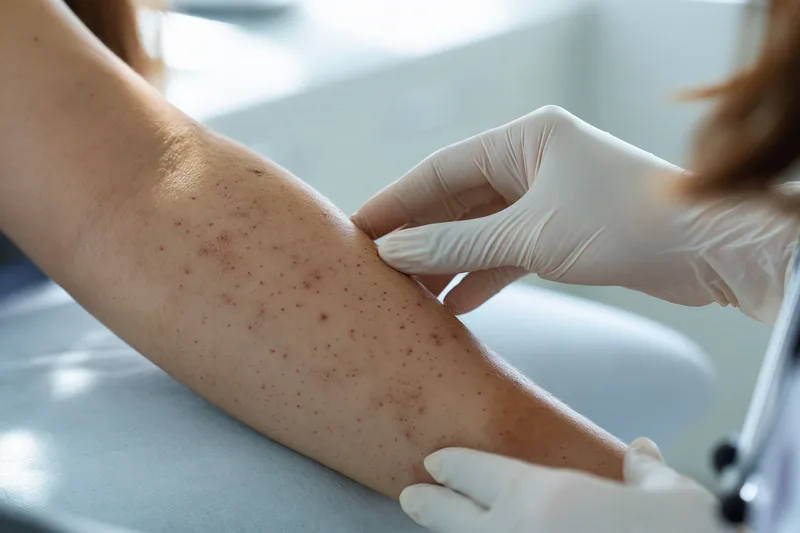
Petechiae are tiny, flat spots caused by small amounts of blood leaking from capillaries beneath the skin. They may appear red or purple on lighter skin and purple, brown or darker than the surrounding area on deeper skin tones. Petechiae generally do not become pale or disappear when pressed because the colour comes from blood beneath the skin rather than increased blood flow at the surface. [1,3]
Purpura are larger areas of bleeding beneath the skin. They may appear as red, purple, brown or dark patches and can develop when several smaller areas of bleeding occur close together. Petechiae and purpura are sometimes described as a rash, but they are clinically different from allergic, infectious or inflammatory rashes because they represent bleeding rather than surface inflammation. [1,3]
Bruises, also called ecchymoses, form when more blood collects beneath the skin. A bruise associated with leukemia may look identical to an ordinary bruise caused by an impact. There is no unique colour, shape or location that identifies leukemia bruises by visual examination alone. The pattern becomes more concerning when bruises repeatedly appear without remembered injury, develop after very minor contact or occur together with petechiae, gum bleeding, nosebleeds or prolonged bleeding from small cuts. [2,5,6,9]
Leukemia can interfere with the bone marrow’s production of normal platelets. Platelets are blood-cell fragments that help seal damaged vessels and initiate clot formation. When platelet numbers become low, or when platelet function is impaired, small vessels may leak blood after minimal pressure or without any obvious injury. This can produce petechiae, purpura and frequent bruising, but the severity of the skin changes does not always correspond directly to a single platelet result. [1,2,5,6]
Low platelets are not exclusive to leukemia. Similar skin findings may occur with viral or bacterial infections, immune thrombocytopenia, liver disease, blood-vessel inflammation, clotting disorders and certain medicines. Aspirin, anticoagulants, antiplatelet medicines, corticosteroids and some antibiotics may also increase bruising or bleeding. Forceful coughing, vomiting, straining or pressure can occasionally produce localized petechiae without an underlying blood cancer. [3,9]
Infection- and Treatment-Related Rashes
A person who has already been diagnosed with leukemia may develop a true inflammatory or infectious rash that is unrelated to bleeding beneath the skin. Reduced immune defence can increase susceptibility to bacterial, viral and fungal infections, while chemotherapy, targeted therapy, immunotherapy, antibiotics and other supportive medicines can cause drug-related eruptions. [7]
These rashes may be red, raised, itchy, painful, scaly, blistering or widespread. Unlike petechiae, some inflammatory rashes may temporarily fade when pressed. However, blanching, itching or redness cannot reliably identify the cause, and several different skin conditions may occur at the same time in a person receiving leukemia treatment. [7]
Most new rashes reported by people with leukemia are caused by infection or treatment-related reactions rather than direct invasion of the skin by leukemia cells. This distinction matters because an infectious rash may require antimicrobial treatment, while a medicine reaction may require assessment of the suspected drug and the severity of the reaction. A new rash should therefore not automatically be interpreted as leukemia progression or as evidence that treatment has failed. [7]
Patients receiving leukemia treatment should not stop prescribed medicines or begin steroid creams, herbal products or over-the-counter remedies solely on the basis of a photograph or online comparison. Suppressing redness or itching without identifying the cause could make an infection or serious drug reaction more difficult to recognize. The treating hematology or oncology team should review new, persistent or rapidly changing skin findings. [7]
Leukemia Cutis
Leukemia cutis is a comparatively uncommon condition in which leukemia cells infiltrate the skin. It is different from petechiae, purpura and ordinary bruising, which usually result from abnormalities in platelets, clotting or small blood vessels rather than direct accumulation of leukemia cells in the skin. [8]
Leukemia cutis may appear as firm papules, nodules, plaques, swellings, discoloured patches or, less commonly, ulcers. The lesions may be skin-coloured, red, brown, purple or blue and may occur as a single lesion, grouped lesions or a widespread eruption. They can develop on many parts of the body, and their appearance varies according to the underlying leukemia subtype and the depth of skin involvement. [8]
The lesions are often painless, although itching, tenderness or discomfort can occur. Their colour and shape may resemble infections, inflammatory disorders, medicine reactions, lymphoma and other skin cancers. Petechiae-like presentations have also been reported, but they are uncommon and cannot be distinguished reliably from other causes by visual inspection. [8]
A photograph cannot confirm leukemia cutis. Diagnosis generally requires a skin biopsy so that the tissue can be examined under a microscope and tested for characteristic cellular markers. The biopsy findings must then be interpreted together with blood counts, peripheral blood findings, bone marrow results and the patient’s established or suspected leukemia subtype. [8]
How Leukemia-Related Skin Changes Can Appear on Different Skin Tones
Medical photographs have historically shown petechiae and bruising mainly on light skin, which can make recognition more difficult in patients with brown or Black skin. On lighter skin, petechiae may look bright red, burgundy or purple. On deeper skin tones, they may appear dark purple, brown, grey-brown or simply darker than the surrounding skin. [1]
Bruises on deeper skin may be less noticeable during their early stages. They may become easier to identify as a darker patch develops or through associated tenderness, swelling or a difference in texture. Comparing both sides of the body and examining areas such as the palms, soles, inside the mouth and lower eyelids may help a clinician identify changes that are difficult to see elsewhere. [1]
Colour alone should not determine whether a lesion is considered serious. Lighting, skin pigmentation, camera settings, recent pressure and the stage of healing can all alter its appearance. A skin change that looks dramatic may have a benign explanation, while a subtle cluster of petechiae may be clinically important when it is new, spreading or associated with bleeding and systemic symptoms. [1,3]
Why Skin Appearance Cannot Diagnose Leukemia
No rash, bruise or pinpoint spot is specific enough to diagnose leukemia without further evaluation. Common skin disorders, infections, medicines, platelet disorders, liver disease, trauma and nutritional problems can produce overlapping appearances. Conversely, some people with leukemia have no visible rash or unusual bruising at the time of diagnosis. [1–3,5–7,9]
The clinically useful question is therefore not whether a mark “looks like leukemia,” but whether its pattern, timing and accompanying symptoms justify blood testing and medical examination. The distinction between bleeding beneath the skin, an inflammatory eruption, infection and leukemia cutis determines which investigations are appropriate and how urgently the patient should be assessed.
Why Leukemia Rash and Bruising Can Develop
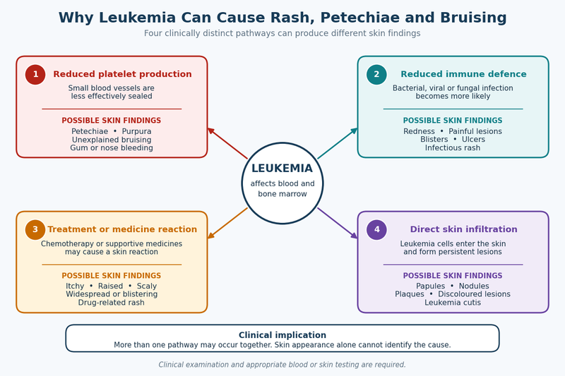
A leukemia rash or unexplained bruising can develop through several different biological pathways. The most familiar mechanism is a reduction in healthy platelets, but infection, treatment reactions, disturbed clotting and direct skin involvement may also contribute. Understanding the mechanism matters because skin changes caused by bleeding require a different assessment from an infectious or medicine-related eruption. [2,5–9]
Reduced Platelet Production in the Bone Marrow
Healthy bone marrow continuously produces red blood cells, white blood cells and platelets. In leukemia, abnormal blood-forming cells can accumulate within the marrow and interfere with the production of normal cells. When platelet production falls, the blood may not seal damaged vessels as efficiently as usual. [2,5,6]
A platelet count below the laboratory reference range is called thrombocytopenia. As the platelet count decreases, minor pressure, friction or an unnoticed injury may allow blood to escape from small vessels beneath the skin. Very small leaks may appear as petechiae, while larger collections of blood may produce purpura or bruises. [2,5,6,9]
This process can also affect areas that are not readily visible. A person may experience recurrent nosebleeds, bleeding from the gums, unusually heavy menstrual bleeding or prolonged bleeding after a small cut. The combination of skin changes and bleeding from another site is generally more concerning than one isolated bruise with a clear explanation. [2,5,6,9]
Platelet levels can change as leukemia develops or during treatment. A patient who had an acceptable platelet count at an earlier appointment may therefore require repeat testing when new petechiae, repeated bruising or active bleeding appears. Previous normal results should not be used to dismiss a new or worsening bleeding pattern. [4–6]
Platelet Count Does Not Explain Every Bruise
Although low platelets are an important cause of leukemia bruises, the number reported on a complete blood count does not fully predict what the skin will look like. One patient with a markedly reduced count may have few visible marks, while another may develop extensive bruising because of recent injuries, fragile blood vessels, medicines or an additional clotting abnormality. [9]
The location and size of a bruise also cannot be used to estimate the platelet count. A large bruise does not automatically mean that the count is critically low, and a small number of visible spots does not guarantee that the risk of internal bleeding is low. Treatment decisions are based on the blood count, active bleeding, recent changes, other laboratory results, planned procedures and the patient’s overall condition rather than the appearance of the skin alone. [4,9]
Bleeding may also be influenced by how well platelets function and whether the proteins involved in clot formation are working normally. Medicines, liver dysfunction, infection and other medical conditions can alter these processes. This is why evaluation may extend beyond a platelet count when bruising or bleeding appears disproportionate to the initial blood results. [3,9]
Infection-Related Skin Changes
Leukemia may reduce the body’s ability to produce effective infection-fighting white blood cells. A high white-cell count does not necessarily mean that immune protection is strong because many circulating leukemia cells are abnormal and may not function like healthy mature cells. Some leukemia treatments can further suppress normal immune defence. [5–7]
Bacterial, viral and fungal infections can consequently produce rashes, painful patches, blisters, ulcers, crusted areas or localized redness. These changes may occur independently of the platelet count and may resemble a drug reaction or an ordinary skin disorder. A photograph alone cannot reliably determine whether an eruption is infectious. [7]
Fever accompanying a new rash requires particular attention in a person with diagnosed or suspected leukemia. Infection can progress rapidly when immune function is impaired, and treatment should not be delayed while waiting to see whether the skin changes resolve. Chills, weakness, dizziness, breathlessness or rapid spreading of the eruption further increase the need for urgent assessment. [5–7]
Treatment-Related Rashes
Chemotherapy, targeted therapy, immunotherapy and supportive medicines can cause inflammatory skin reactions. Antibiotics, antiviral medicines and antifungal treatments may also produce rashes, making it difficult to identify the responsible medicine without reviewing when each treatment was started and how the eruption evolved. [7]
A treatment-related rash may be itchy, tender, scaly, raised or widespread. Some reactions are mild and manageable, while others may signal a serious adverse effect. Blistering, skin peeling, facial swelling, difficulty breathing, extensive pain or sores involving the mouth, eyes or genital area require urgent medical review. [7]
The appearance of a rash after treatment begins does not necessarily mean that leukemia is progressing. It also does not prove that the treatment is working or that the body is “detoxifying.” The timing, distribution, associated symptoms and laboratory findings must be assessed before the treatment plan is changed. [7]
Patients should not independently stop an anticancer medicine because of a rash unless emergency clinicians advise them to do so. At the same time, they should not conceal the eruption with steroid creams, antihistamines or home remedies before reporting it, particularly when fever, blistering, mucosal sores or systemic illness is present. Early photographs taken in consistent lighting may help the clinical team understand how quickly the reaction is changing.
Direct Involvement of the Skin
In leukemia cutis, abnormal leukemia cells leave the blood or bone marrow and accumulate within the skin. This mechanism is different from the vessel leakage that produces petechiae and bruising. The resulting lesions are usually more persistent and may feel firm or raised rather than behaving like blood trapped beneath the skin. [8]
The presence of leukemia cutis cannot be determined from lesion colour or location. Similar nodules, plaques and papules can occur with infection, inflammation, medicine reactions and other cancers. A skin biopsy is usually required to identify the cells involved and connect the findings with the patient’s blood, bone marrow and immunophenotyping results. [8]
Why Different Skin Changes Can Occur Together
A patient may have more than one cause of skin change at the same time. For example, low platelets may produce petechiae while an antibiotic causes an itchy inflammatory rash. Scratching the inflamed skin may then create additional bruising because the platelet count is reduced. Infection, treatment reactions and bleeding beneath the skin can therefore overlap and produce a mixed appearance. [7,9]
This overlap is one reason that describing every skin change as a leukemia rash can be misleading. Clinicians may need to assess flat and raised areas separately, identify which marks fade with pressure, review recent medicines, check for fever or bleeding and compare the findings with current blood counts. Determining the mechanism allows urgent problems to be treated without assuming that every new lesion represents leukemia progression.
Leukemia Rash and Bruises Compared With Ordinary Bruising
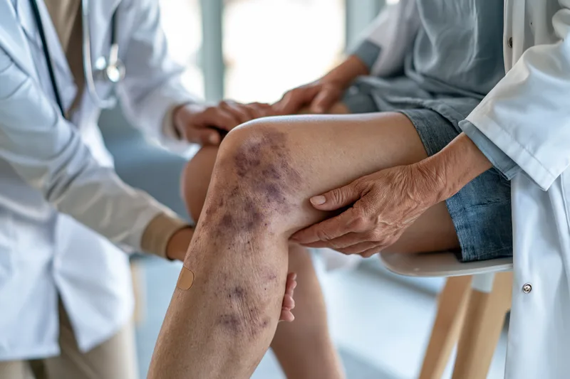
A leukemia rash may include petechiae or purpura, while leukemia-related bruising may look indistinguishable from a bruise caused by everyday trauma. No colour, shape, size or body location can prove that leukemia caused a bruise. The clinically important clues are how the marks developed, how often they recur, whether the degree of bruising matches the injury and whether other bleeding or systemic symptoms are present. [2,5,6,9]
How an Ordinary Bruise Usually Develops
An ordinary bruise commonly appears after a bump, fall, injection, sports injury or sustained pressure. Small blood vessels beneath the skin are damaged, allowing blood to collect in the surrounding tissue. The area may initially feel tender or swollen before gradually changing colour as the trapped blood is broken down and absorbed.
People do not always remember the event that caused a bruise. Minor impacts against furniture, pressure from carrying bags, exercise and everyday physical activity can leave marks that become noticeable only later. One isolated bruise without other symptoms is therefore not usually sufficient reason to suspect leukemia.
Ordinary bruises commonly improve gradually. Their colour may change from red or purple to brown, green or yellow during healing, although the sequence varies according to skin tone, bruise depth, circulation and the amount of blood beneath the skin. Colour cannot reliably determine when a bruise occurred or whether leukemia caused it. [9]
Bruises may be more visible on lighter skin, while early bruising can be subtle on brown or Black skin. On deeper skin tones, tenderness, swelling, texture change or a patch that appears darker than the surrounding skin may be more noticeable than a dramatic colour change. Examining the area in natural light may make subtle differences easier to recognize. [1]
Patterns That Make Leukemia Bruises More Concerning
Leukemia-related bruising becomes more clinically concerning when new bruises repeatedly appear without a clear explanation. A patient may notice several marks developing over a short period, bruising after very minor pressure or a bruise that seems much larger than expected from the reported injury. [2,5,6,9]
The location alone does not identify the cause. Bruising on the shins, knees or forearms may follow ordinary daily contact, especially in physically active people. Marks on the trunk, back, face or other areas that are less commonly exposed to accidental impact may deserve closer review when there is no remembered injury, but even these locations cannot diagnose leukemia.
The overall bleeding pattern is more informative than one skin mark. Bruising accompanied by clusters of petechiae, purple patches, recurrent nosebleeds, bleeding gums, unusually heavy menstrual bleeding or prolonged bleeding from small cuts may indicate a platelet or clotting problem requiring blood tests. [2,5,6,9]
A rapidly increasing number of bruises is also more concerning than a longstanding tendency to bruise easily that has not changed. Comparing the current pattern with the patient’s usual baseline helps determine whether something new is occurring. A person who has always developed occasional bruises after minor contact may have a different level of concern from someone who suddenly develops widespread unexplained marks.
Associated Symptoms That Increase the Need for Evaluation
Unexplained bruising deserves prompt assessment when it occurs with persistent fatigue, pallor, breathlessness, fever, recurrent infections, night sweats, unexplained weight loss, bone discomfort or enlarged lymph nodes. These symptoms remain nonspecific and can occur with many conditions, but their presence makes it less appropriate to assume that the bruising is harmless. [1,2,5,6]
Bleeding from more than one site may indicate a broader problem with platelets or clotting. For example, bruises combined with blood in the urine, black stools, vomiting blood or bleeding that does not stop with pressure require more urgent evaluation than uncomplicated skin bruising. [9]
A severe headache, confusion, sudden weakness, fainting or vision changes occurring with abnormal bruising may indicate internal bleeding or another neurological emergency. These symptoms require immediate medical attention rather than waiting for a routine blood-test appointment.
Fever with new petechiae or purpura is also an urgent combination because serious infections can produce non-fading skin spots and can worsen rapidly. It should not be assumed that the spots are caused by leukemia, an allergy or a harmless viral illness without clinical assessment. [3]
Bruising in Children
Children commonly develop bruises during play, particularly over the knees, shins, elbows and forehead. Frequent bruising in these exposed areas is usually related to activity rather than leukemia. Concern increases when bruises are unusually numerous, appear in protected areas, are accompanied by petechiae or occur together with persistent fever, pallor, reduced activity, bone pain, recurrent infection or abnormal bleeding. [5]
Parents should avoid using online photographs to compare their child’s bruises with images labelled as leukemia. Camera lighting, skin tone, injury stage and image editing can make ordinary and medically significant bruises appear similar. A clinician can assess the distribution, examine the child for other signs and decide whether a complete blood count is appropriate.
Bruising in a child who cannot yet move independently requires careful medical assessment because accidental injury is less likely to explain the finding. The clinician must consider medical causes as well as the possibility of injury, and the evaluation should not be delayed solely because the child otherwise appears well.
Bruising in Older Adults
Older adults may bruise more readily because skin becomes thinner and the tissue supporting small blood vessels becomes more fragile. Bruises may appear after minor contact, especially on the hands and forearms, and may take longer to become less visible. This pattern is common and does not by itself indicate leukemia.
Medicines frequently used by older patients can further increase bruising. Anticoagulants, antiplatelet medicines, aspirin and corticosteroids may make marks more frequent or extensive. However, a known medicine effect should not automatically explain a sudden change in bruising, particularly when petechiae, active bleeding, weakness or other new symptoms are present. [3,9]
A medication review should include prescribed drugs, over-the-counter pain relievers, supplements and herbal products. Patients should not stop an anticoagulant or antiplatelet medicine on their own because abrupt discontinuation may increase the risk of stroke, blood clots or other complications. The prescribing clinician should determine whether testing or treatment adjustment is needed.
Why Bruise Colour and Healing Time Are Unreliable
Patients often search for a particular “leukemia bruise colour,” but leukemia bruises do not have a unique appearance. Both ordinary and medically significant bruises can begin as red, blue, purple, brown or nearly black areas and then change as they heal. Skin pigmentation, bruise depth and lighting substantially affect what is visible.
A bruise that changes colour is not automatically harmless, and one that remains dark is not automatically caused by leukemia. Large or deep bruises may remain visible longer than small superficial marks. Repeated pressure to the same area, impaired circulation and some medicines can also prolong healing.
Similarly, a bruise disappearing does not rule out a blood disorder. The body may absorb one bruise while new bruises continue to develop elsewhere. The pattern over time, associated bleeding and blood-count results are more meaningful than whether one individual mark fades.
Taking dated photographs can help document whether new marks are appearing or existing ones are expanding. Photographs should be taken in similar lighting with a familiar object or ruler nearby for scale, but they should support rather than replace medical examination. A clinician may still need to palpate the area, assess whether a lesion is flat or raised and review current blood results.
When Bruising Should Lead to Blood Testing
A complete blood count with differential and platelet count is commonly used when unexplained bruising raises concern about a blood or bone marrow disorder. The platelet count helps identify thrombocytopenia, while hemoglobin and white-cell findings may reveal additional abnormalities that influence the next step. [4–6]
One abnormal value does not automatically establish leukemia. Infections, immune disorders, medicines, liver disease, nutritional deficiencies and laboratory factors can alter blood counts. Conversely, persistent or progressive symptoms may require further evaluation even when an initial result is not clearly diagnostic.
The decision to test should be based on the complete clinical picture rather than the number or colour of bruises alone. New unexplained bruising, bruising with petechiae or mucosal bleeding, a rapidly changing pattern or bruising accompanied by systemic symptoms provides a stronger reason for medical assessment than an isolated mark following a recognizable injury.
Other Causes That Can Resemble a Leukemia Rash and Bruising
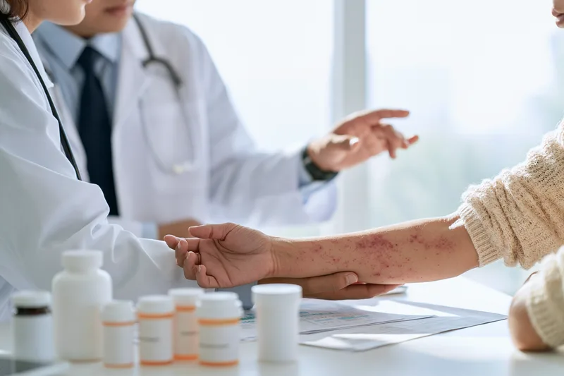
A leukemia rash or pattern of unexplained bruising can be alarming, but several non-leukemic conditions can produce a similar appearance. Considering these alternatives does not mean that new petechiae, purpura or bleeding should be dismissed. It helps the clinician decide whether the immediate concern is a platelet disorder, infection, medicine effect, liver problem, blood-vessel inflammation or another condition requiring a different treatment pathway. [3,7,9]
Medicines That Increase Bruising or Bleeding
Anticoagulants such as warfarin, heparin and direct oral anticoagulants reduce the blood’s ability to form clots. Antiplatelet medicines such as aspirin and clopidogrel reduce platelet activity. Patients taking these medicines may notice that minor injuries produce larger bruises, small cuts bleed for longer or new marks appear after pressure that previously caused no visible change. [9]
A medicine-related explanation should not automatically be assumed when the bruising pattern changes suddenly. New petechiae, repeated nosebleeds, bleeding gums, blood in urine, black stools or rapidly expanding bruises may indicate that the anticoagulant effect is excessive, that the platelet count has fallen or that another medical problem is present. Laboratory testing may be required even when the medicine is a plausible cause. [9]
Corticosteroids can make the skin thinner and small blood vessels more fragile, particularly after prolonged use. Bruising may become more noticeable over the hands, forearms or other exposed areas. The marks may result from minimal contact, but a sudden increase in bruising should still prompt review of the dose, duration, other medicines and current blood results.
Some antibiotics, antiseizure medicines and other drugs can reduce platelet numbers through immune or bone marrow effects. A low platelet count may develop after a medicine has been taken for days or weeks and may not be accompanied by pain or itching. The timing between starting a medicine and developing petechiae or bruising can therefore provide an important diagnostic clue. [3]
Over-the-counter pain relievers can also affect bleeding. Aspirin has a prolonged antiplatelet effect, while nonsteroidal anti-inflammatory medicines may increase bleeding risk, especially when combined with anticoagulants, corticosteroids, kidney disease or gastrointestinal ulcers. Patients should report all non-prescription medicines rather than listing only drugs prescribed by a doctor.
Prescription medicines should not be stopped independently because abrupt discontinuation may create a greater risk than the bruising itself. Stopping an anticoagulant without medical advice can increase the likelihood of stroke or another blood clot. The safer approach is to contact the prescribing clinician promptly, particularly when the skin changes are accompanied by active bleeding.
Supplements and Herbal Products
Nutritional supplements and herbal products should be included in the medication history because they may affect platelet function, clotting, liver metabolism or the activity of prescribed medicines. Taking several products together can make the effect difficult to predict, particularly when the exact ingredients or concentrations are unclear.
Patients receiving individualized Ayurvedic care should provide the treating hematologist with the complete formulation, dose and preparation details. The Ayurvedic physician should also review the current platelet count, liver function, kidney function and all prescribed medicines before making changes. This coordinated approach helps distinguish disease-related bruising from medicine interactions and allows supportive care to remain aligned with objective monitoring.
A product described as natural should not automatically be considered irrelevant to bleeding assessment. Garlic extracts, concentrated ginger preparations, ginkgo and some other supplements may influence platelet activity or interact with anticoagulant therapy. The clinical significance depends on the dose, preparation, combination and the patient’s underlying condition, so isolated ingredient lists should not be used to predict risk without professional review.
Unlabelled or poorly standardized products introduce an additional problem because the patient and clinician may not know the actual dose or all included substances. Bringing the packaging, prescription or ingredient sheet to the appointment can be more useful than trying to recall the product name from memory.
Viral and Bacterial Infections
Infections can cause petechiae, purpura and bruising through several mechanisms. Some infections temporarily reduce platelet production, increase platelet destruction or trigger inflammation within small blood vessels. Others cause widespread disturbances in clotting during severe systemic illness. [3]
A viral infection may produce a conventional rash, temporary thrombocytopenia or both. The patient may have fever, fatigue, body aches, sore throat or swollen lymph nodes, creating symptoms that overlap with leukemia. Blood tests and the clinical course are often necessary to distinguish a self-limited infection from a bone marrow disorder.
Serious bacterial infections can produce rapidly spreading petechiae or purpura and may be accompanied by fever, severe weakness, confusion, breathing difficulty, cold extremities or low blood pressure. This pattern requires emergency medical care. A non-fading rash with fever should not be monitored at home while waiting to see whether it behaves like an allergy or viral eruption. [3]
Infection is also important in patients already diagnosed with leukemia. Reduced immune defence may make an otherwise localized skin infection progress more quickly, while redness and swelling can be less obvious when white-cell responses are impaired. Pain, warmth, drainage, blistering or rapid change around a lesion should be reported even when fever is absent. [7]
Immune Thrombocytopenia and Other Platelet Disorders
Immune thrombocytopenia occurs when the immune system destroys platelets more rapidly than the body can replace them. It can produce petechiae, purpura, frequent bruising, nosebleeds, gum bleeding or heavy menstrual bleeding, closely resembling the skin findings that may occur with leukemia.
The complete blood count may show an isolated reduction in platelets while hemoglobin and white-cell findings remain relatively preserved. This pattern can help guide further investigation, but immune thrombocytopenia remains a diagnosis that requires clinical assessment and exclusion of other causes. One blood count should not be interpreted without reviewing the patient’s symptoms, medication history and peripheral smear.
Other immune disorders can affect blood vessels or clotting proteins rather than platelets alone. These conditions may cause painful purpura, joint symptoms, abdominal discomfort, kidney abnormalities or lesions concentrated on the lower legs. The rash pattern may suggest vascular inflammation, but laboratory testing and sometimes skin biopsy are needed to confirm the mechanism.
Liver Disease and Clotting Abnormalities
The liver produces many of the proteins required for normal coagulation. Advanced liver dysfunction may reduce these proteins, alter platelet production and contribute to an enlarged spleen that traps circulating platelets. Bruising, nosebleeds or prolonged bleeding may therefore occur even when leukemia is not present.
Additional clues may include jaundice, abdominal swelling, itching, dark urine, pale stools, reduced appetite or a history of significant alcohol use, viral hepatitis or chronic liver disease. However, liver disease can be present before obvious symptoms develop, so liver-function and coagulation tests may be needed when the bruising pattern cannot be explained by the initial blood count.
Clotting disorders may be inherited or acquired. Some patients have a longstanding history of excessive bleeding after dental procedures, surgery or childbirth, while others develop new abnormalities because of liver disease, infection, medicines or autoimmune conditions. The absence of petechiae does not exclude a coagulation problem because some clotting-factor disorders produce deeper bruises or bleeding into muscles and joints rather than pinpoint spots.
Blood-Vessel Inflammation and Fragility
Vasculitis refers to inflammation of blood-vessel walls. When small vessels in the skin become inflamed, blood may leak into the surrounding tissue and create purpura that does not fade with pressure. The lesions may be raised or tender and can be associated with joint pain, fever, abdominal symptoms, numbness or kidney involvement.
This pattern differs biologically from bruising caused only by a low platelet count, although the skin findings can overlap. A clinician may order blood tests, urine tests and, in selected cases, a skin biopsy to determine whether vascular inflammation is present.
Blood vessels may also become more fragile with ageing, prolonged corticosteroid use, sun damage or connective-tissue disorders. Older adults frequently develop dark patches on the backs of the hands and forearms after minimal contact. A stable, longstanding pattern may be benign, but a sudden increase in number, size or distribution still requires reassessment.
Nutritional Deficiencies
Severe vitamin C deficiency can weaken the tissues supporting small blood vessels and may cause bruising, petechiae, gum changes and impaired wound healing. It is uncommon in people consuming a varied diet but may occur with severe dietary restriction, malabsorption, alcohol dependence or prolonged illness.
Vitamin K is required for the production of several clotting factors. Deficiency may arise from malabsorption, liver or biliary disease, prolonged antibiotic use or inadequate intake in vulnerable patients. It is more likely to cause prolonged bleeding than a distinctive rash, and the diagnosis requires clinical and laboratory evaluation rather than dietary assumptions.
Vitamin B12 or folate deficiency can sometimes produce low platelets alongside anemia and other blood-count abnormalities. These deficiencies may also cause fatigue, pallor, tongue changes or neurological symptoms. Because the laboratory picture can overlap with bone marrow disease, treatment should be based on confirmed test results rather than beginning multiple supplements before evaluation.
Pressure, Straining and Minor Trauma
Localized petechiae can develop after forceful coughing, vomiting, crying, weightlifting or straining because temporary pressure increases within small blood vessels. These spots often occur around the face, eyes, neck or upper chest and may have an obvious relationship to the triggering event. [3]
Pressure from tight clothing, straps, medical equipment or prolonged positioning can also produce localized spots or bruising. When the distribution matches the area of pressure and no additional bleeding or systemic symptoms are present, the explanation may be mechanical. Persistent, spreading or recurrent lesions still deserve assessment.
Minor trauma remains the most common explanation for an isolated bruise. The absence of a remembered injury does not prove that the mark developed spontaneously, because small impacts are easily forgotten. Concern increases when the number of unexplained bruises changes from the patient’s usual pattern or when additional bleeding appears.
Common Skin Conditions Mistaken for Leukemia Rash
Eczema, contact dermatitis, heat rash, insect bites, fungal infections and allergic reactions can all produce red or discoloured spots. These conditions are more likely to cause itching, scaling, raised borders, fluid-filled lesions or surface irritation than bleeding beneath the skin, but visual overlap is possible.
Folliculitis may create clusters of red or tender bumps around hair follicles, while insect bites can form grouped itchy lesions. Fungal infections may develop a scaly edge or expand gradually. These characteristics can help guide examination, but they should not be treated as absolute diagnostic rules.
Applying a steroid cream before the cause is clear may temporarily reduce redness while worsening or masking a fungal or bacterial infection. Patients with leukemia, low white-cell counts or active cancer treatment should be especially cautious about self-treating a new eruption because infection can progress despite limited visible inflammation. [7]
Why Another Possible Cause Does Not Remove the Need for Assessment
Finding a plausible alternative explanation does not always eliminate the need for blood testing. A patient may be taking aspirin and also develop thrombocytopenia, or may have a viral infection that reveals an unrelated blood abnormality. Several causes can coexist and intensify the same bruising pattern.
Medical assessment becomes more important when skin changes are new, progressive, widespread or accompanied by bleeding from another site. Persistent fatigue, pallor, recurrent fever, unusual infections, weight loss, night sweats or enlarged lymph nodes also strengthen the reason to investigate rather than attributing the findings to sensitive skin or minor trauma. [1,2,5,6]
The safest distinction is not between “leukemia” and “harmless.” Some non-leukemic causes, including serious infection, severe immune thrombocytopenia, liver failure and excessive anticoagulation, can also require urgent treatment. The purpose of evaluation is to identify the actual mechanism promptly rather than relying on the skin’s appearance to estimate the level of risk.
When a Leukemia Rash or Bruising Needs Urgent Medical Attention

A leukemia rash or new pattern of unexplained bruising becomes urgent when it is accompanied by active bleeding, fever, neurological symptoms, breathing difficulty or rapid physical deterioration. The colour or number of skin marks alone cannot determine the level of danger. Urgency depends on whether bleeding is continuing, whether infection may be present and whether the patient has known leukemia, low platelets or current cancer treatment. [3,7,9]
Table 2: When Leukemia Rash or Bruising Needs Medical Care
| Level of care | Skin changes and associated symptoms | Recommended action |
|---|---|---|
| Emergency assessment now | Fever with a rapidly spreading non-fading rash, uncontrolled bleeding, black stools, blood in urine or vomit, severe headache, confusion, fainting, breathlessness, blistering or skin peeling | Attend emergency care immediately. Do not wait for a routine appointment or attempt home treatment. [3,7,9] |
| Same-day oncology or medical review | New petechiae during leukemia treatment, rapidly increasing bruises, repeated nosebleeds, bleeding gums, unusually heavy menstrual bleeding or a new rash after starting treatment | Contact the hematology, oncology or treating medical team on the same day. [2,5–7,9] |
| Prompt clinical appointment | Recurrent unexplained bruising, persistent petechiae, progressive skin changes or bruising with fatigue, pallor, fever, recurrent infections or weight loss | Arrange examination and a complete blood count with differential and platelet count. [1–6] |
| Observe with appropriate follow-up | One isolated bruise after a recognizable injury that gradually improves without additional bleeding or systemic symptoms | Monitor for new bruises, petechiae or bleeding. Seek assessment if the pattern changes or the bruise continues expanding. [9] |
Seek Emergency Care for Uncontrolled or Internal Bleeding
Bleeding that does not stop after continuous firm pressure requires emergency medical care. This includes persistent bleeding from a cut, the nose, gums, an injection site or another visible area. Releasing the pressure repeatedly to check the wound can disturb clot formation, so uninterrupted pressure should be maintained while urgent help is arranged. [9]
Blood in vomit, urine or stool may indicate internal bleeding. Black, tar-like stools can also represent bleeding higher in the digestive tract, even when bright-red blood is not visible. Coughing or vomiting blood, passing a substantial amount of blood or developing abdominal pain with weakness or dizziness should not be managed at home. [9]
A sudden severe headache, confusion, unusual drowsiness, slurred speech, seizure, loss of balance, repeated vomiting, vision changes or new weakness in an arm or leg may indicate bleeding in or around the brain or another neurological emergency. These symptoms require immediate assessment, particularly in someone with low platelets, leukemia or anticoagulant use. [9]
A fall or blow to the head should also be taken seriously when platelet levels are very low or the patient is taking an anticoagulant. Significant bleeding can occur without a large external bruise or visible wound. The absence of immediate symptoms does not always exclude internal injury, so the oncology team or emergency service should advise whether observation, imaging or hospital assessment is required. [9]
Rapidly enlarging bruises, extensive spontaneous bruising or painful swelling beneath the skin may indicate continuing bleeding into the tissues. Emergency assessment is particularly important when these changes are accompanied by faintness, rapid heartbeat, breathlessness, marked weakness or pallor.
Fever With Petechiae or a Rapidly Changing Rash
Fever with newly developing petechiae, purpura or a rapidly spreading rash requires urgent medical assessment. Serious infections can cause non-fading skin spots and may progress quickly, especially in patients whose immune defence has been reduced by leukemia or its treatment. The rash should not automatically be assumed to result from low platelets or the leukemia itself. [3,7]
Chills, shaking, confusion, difficulty breathing, severe weakness, cold or clammy skin, reduced alertness or a feeling of becoming rapidly more unwell increases the urgency. Emergency care should not be delayed while attempting to determine whether the spots fade with pressure or while waiting for a scheduled blood test.
Patients undergoing leukemia treatment should follow the fever instructions provided by their oncology team. The temperature threshold and recommended destination may differ according to the treatment, neutrophil count and local protocol. When no individualized instruction is available, any fever during intensive leukemia treatment should be reported immediately rather than treated with fever-reducing medicine and observed at home.
Taking paracetamol or another fever-reducing medicine before contacting the treatment team may temporarily lower the temperature without addressing the cause. Patients should inform the clinician about the highest recorded temperature, when it began and whether antibiotics, corticosteroids or fever medicines have already been taken.
A new rash with blistering, skin peeling, facial swelling, breathing difficulty or painful sores affecting the mouth, eyes or genital area may represent a severe medicine reaction. This pattern requires emergency assessment even when petechiae or bruising are not present.
Contact the Hematology or Oncology Team the Same Day
A person already diagnosed with leukemia should contact the treating team on the same day when new petechiae, unexplained bruises or mucosal bleeding develops. Platelet and white-cell counts can change between appointments, so a previously acceptable complete blood count should not be used to dismiss a new symptom pattern.
Repeated nosebleeds, bleeding gums, heavier-than-usual menstrual bleeding, prolonged bleeding from minor cuts or a sudden increase in bruising may indicate a clinically important change even when the patient otherwise feels well. The treating team may recommend an urgent blood count, examination, medicine review or assessment for infection and clotting abnormalities. [2,5,6,9]
A new rash during chemotherapy, targeted therapy, immunotherapy or antimicrobial treatment should also be reported. Infection and medicine reactions are more frequent explanations for new rashes in treated leukemia patients than direct infiltration of the skin by leukemia cells. The clinical team may need photographs, treatment dates, recent blood results and a description of whether the rash is painful, itchy, raised, blistering or spreading. [7]
Patients should not independently stop anticancer treatment, antibiotics, antifungal medicines, anticoagulants or corticosteroids because a rash or bruise has appeared. Abruptly stopping treatment may create additional risks. The prescribing team should determine whether the medicine should continue, be paused or be replaced.
Topical steroid creams, antihistamines and over-the-counter skin treatments should not be used to conceal a new eruption before it has been reported, particularly when the patient has fever or reduced immunity. These products may alter the appearance of the lesions and can sometimes worsen or obscure an infection.
Arrange Prompt Assessment When Symptoms Are Stable
New unexplained petechiae appearing across several areas of the body should be medically assessed even when there is no fever or active bleeding. The same applies when the cause cannot be identified, the spots continue to increase or they recur after initially fading. Some causes are minor, but others require a blood count and clinical examination. [3]
Repeated unexplained bruising should be assessed when it represents a clear change from the person’s usual pattern. Bruising accompanied by persistent fatigue, pallor, recurrent infections, night sweats, reduced appetite, unexplained weight loss, bone discomfort or enlarged lymph nodes provides a stronger reason for prompt investigation. These symptoms remain nonspecific and do not prove leukemia, but they should not be attributed to sensitive skin without evaluation. [1,2,5,6]
An isolated bruise after a recognizable injury is generally less concerning when it improves normally and no other symptoms are present. Medical review becomes more appropriate when the mark continues expanding, remains unusually painful, is accompanied by a firm swelling or appears alongside new bruises elsewhere.
Patients taking anticoagulants, antiplatelet medicines or long-term corticosteroids should have a lower threshold for reporting a sudden change in bruising. The medicine may contribute, but it should not automatically be assumed to explain every new bleeding pattern. Dose changes should be made only by the prescribing clinician.
What to Do While Seeking Medical Care
Visible external bleeding should be covered with clean material and held under continuous firm pressure. A patient who is dizzy, confused, faint or actively bleeding should not drive to hospital. Emergency transport is safer when there is a possibility of significant blood loss, neurological impairment or rapid deterioration.
Patients should bring or provide an accurate list of prescribed medicines, non-prescription medicines, supplements and Ayurvedic formulations. Recent complete blood counts, platelet results, discharge summaries and oncology contact information can help the emergency team understand the level of risk and avoid unnecessary delays.
Dated photographs may document how quickly a rash or bruise is changing, but taking photographs must not postpone urgent assessment. Images should be taken in consistent lighting and should include both a close view and a wider view showing the distribution.
A worsening rash, petechiae, bruising, fever or bleeding should never be interpreted as detoxification, disease elimination or a temporary healing response. Ayurvedic treatment should remain coordinated with hematology assessment, and no formulation should be started, stopped or intensified as a substitute for urgent blood testing or emergency management.
How a Leukemia Rash and Bruising Are Assessed
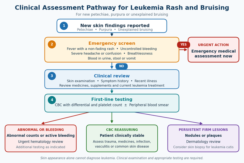
A leukemia rash cannot be diagnosed from a photograph, a pressure test or the colour of the skin changes. Assessment begins by determining whether the marks represent bleeding beneath the skin, an inflammatory rash, infection, a medicine reaction or a persistent lesion that may require biopsy. The clinician then considers the distribution, accompanying symptoms, current medicines and blood-test results before deciding whether further hematology or dermatology investigation is necessary. [3,4,7–9]
Medical History and Symptom Review
The clinician will usually ask when the skin changes first appeared, whether they developed suddenly or gradually and whether new areas are continuing to form. Petechiae that appeared after forceful coughing, vomiting or pressure may have a different explanation from spots that are spreading across several body regions without an obvious trigger. [3]
Bruising is assessed in relation to recent injuries, physical activity, injections, blood draws, falls and sustained pressure from clothing or equipment. Patients may not remember every minor impact, but a rapid increase in unexplained bruises or bruising that seems disproportionate to ordinary contact may justify blood testing. [9]
The presence of bleeding elsewhere is particularly important. The clinician may ask about recurrent nosebleeds, bleeding gums, prolonged bleeding from cuts, unusually heavy menstrual bleeding, blood in urine, black stools or blood in vomit. Bruising combined with bleeding from another site raises greater concern for thrombocytopenia or a clotting abnormality than an isolated bruise after recognizable trauma. [2,5,6,9]
Associated symptoms help determine whether a blood or bone marrow disorder should be investigated. Persistent fatigue, pallor, breathlessness, fever, recurrent infections, night sweats, unintended weight loss, bone discomfort or enlarged lymph nodes do not confirm leukemia, but their presence can increase the need for a complete blood count and clinical examination. [1,2,5,6]
Patients should disclose all recently started or discontinued medicines. Anticoagulants, antiplatelet medicines, aspirin, corticosteroids, some antibiotics and several other treatments may contribute to bruising, platelet reduction or inflammatory rashes. The clinician also needs to know about non-prescription pain relievers, nutritional supplements and Ayurvedic formulations because interactions or combined effects may alter bleeding risk.
For patients already receiving leukemia treatment, the timing of the eruption in relation to chemotherapy, targeted therapy, immunotherapy, antibiotics, antiviral medicines or antifungal treatment can provide an important clue. A rash beginning shortly after a new medicine may suggest a treatment reaction, while pain, warmth, drainage or fever may increase concern for infection. [7]
Examination of the Skin and Bleeding Pattern
The skin examination evaluates whether the lesions are flat or raised, whether they fade with pressure, whether they are tender or itchy and whether there is scaling, blistering, ulceration or drainage. Petechiae and purpura are generally flat and non-blanching because the colour is produced by blood beneath the skin. Raised, itchy or scaly lesions are more suggestive of an inflammatory, allergic or infectious process, although appearances can overlap. [3,7]
Distribution also provides useful information. Localized petechiae around the face after vomiting may be pressure-related, while widespread petechiae involving the limbs and trunk may prompt investigation for a platelet, infectious or vascular cause. Bruising over exposed areas may reflect ordinary daily contact, whereas repeated bruises in several unexpected locations without remembered injury may require closer evaluation.
The clinician may inspect the mouth for gum bleeding, petechiae, ulcers or infection and examine the eyes for pallor or bleeding. Lymph nodes, the abdomen and other areas may be assessed for findings such as enlarged lymph nodes, liver enlargement or spleen enlargement that could influence the diagnostic pathway. [4–6]
The examination should account for skin tone. Petechiae may appear bright red or purple on lighter skin but brown, grey-purple or darker than the surrounding area on deeper skin tones. Subtle changes may be easier to detect inside the mouth, on the palms and soles or in areas with less variation in pigmentation. [1]
Pressing the skin can help the clinician describe whether a lesion blanches, but a home pressure or glass test cannot identify the underlying disease. A non-blanching spot may be caused by platelet reduction, infection, vasculitis, trauma or another condition. A blanching rash can still require urgent assessment when it is accompanied by fever, blistering, breathing difficulty or rapid deterioration.
Complete Blood Count With Differential and Platelet Count
A complete blood count with differential is commonly the first laboratory investigation when unexplained petechiae, purpura or repeated bruising raises concern about a blood disorder. It measures the main circulating blood-cell groups and provides information about hemoglobin, white blood cells and platelets. [4–6]
The platelet count helps determine whether thrombocytopenia could be contributing to bleeding beneath the skin. A reduced count can occur in leukemia, but it can also result from infection, immune platelet destruction, medicines, liver disease and other bone marrow conditions. A low platelet result therefore identifies a bleeding-related abnormality rather than confirming its cause. [3,9]
Hemoglobin and red-cell measurements can identify anemia, which may help explain fatigue, pallor, dizziness or breathlessness. Anemia has many causes and does not prove leukemia, but its presence alongside thrombocytopenia or abnormal white-cell findings may influence how urgently the patient is referred for further investigation. [4–6]
The white-cell count may be high, low or within the laboratory reference range in different leukemia presentations. The differential shows the proportions and absolute numbers of different white-cell types. An abnormal total count is not diagnostic by itself because infections, inflammation, corticosteroids and other conditions can alter white-cell results. [4–6]
The complete blood count should be interpreted as a pattern rather than as isolated numbers. A platelet count slightly outside the reference range has a different significance from a rapidly falling count accompanied by anemia, abnormal white cells and active bleeding. Previous results are valuable because they show whether the abnormalities are new, stable or progressively changing.
Laboratory reference ranges may vary between facilities, and automated reports may flag minor variations that are not clinically significant. Patients should not attempt to diagnose leukemia from one highlighted result without considering the blood film, symptoms, examination and previous counts.
Peripheral Blood Smear
A peripheral blood smear allows laboratory specialists to examine the size, shape, maturity and appearance of blood cells under a microscope. It can help confirm whether an automated platelet count is accurate and may identify platelet clumping, abnormal white cells, immature cells or other features that require further hematology assessment. [4–6]
Platelet clumping can occasionally produce an artificially low automated platelet count. Reviewing the smear or repeating the sample in a different collection tube may help determine whether the thrombocytopenia is genuine. This prevents decisions from being based on a laboratory artefact.
The presence of immature or abnormal cells may increase suspicion for leukemia, but the smear alone may not establish the exact subtype. Additional testing is usually required to characterize the cells and determine whether they represent acute leukemia, a chronic blood disorder, a reactive response to infection or another hematological condition. [4–6]
A smear that does not show obvious leukemia cells is reassuring but does not answer every diagnostic question. Some blood and bone marrow disorders may not release large numbers of abnormal cells into the circulation. The decision to investigate further depends on the entire blood-count pattern, symptoms, examination and persistence of the abnormalities.
Coagulation, Organ-Function and Infection Testing
When the bruising or bleeding seems greater than expected from the platelet count, the clinician may request coagulation studies. These tests assess parts of the clotting process and can help identify abnormalities related to clotting factors, liver dysfunction, anticoagulant medicines or severe systemic illness. [9]
Liver-function tests may be appropriate because the liver produces several proteins needed for normal coagulation. Kidney-function testing can also influence the assessment because impaired kidney function may affect platelet performance and alter the handling of medicines that increase bleeding risk.
When nutritional deficiency is clinically plausible, targeted testing may include vitamin B12, folate or other measurements. Testing should be directed by the history and blood-count pattern rather than assuming that unexplained bruising can be corrected by taking multiple vitamins. Beginning supplements before testing may also complicate interpretation of later results.
Fever, chills or signs of localized infection may lead to blood cultures, viral testing, swabs or other microbiological investigations. Infection is especially important in patients receiving leukemia treatment because immune suppression can allow skin or systemic infections to progress rapidly. [7]
The tests selected depend on the presentation. A stable patient with a few unexplained bruises may need only an initial blood count and medicine review, while someone with fever, widespread petechiae, low blood pressure or active bleeding may require immediate hospital-based investigation and treatment.
Flow Cytometry and Bone Marrow Examination
When blood results or the peripheral smear raise concern for leukemia, flow cytometry may be used to analyze proteins expressed on the surface or inside abnormal cells. This process helps determine the cell lineage and supports classification of the suspected leukemia. It is not routinely required for every patient with bruising or petechiae.
A bone marrow aspiration and biopsy may be recommended when blood findings suggest leukemia or another marrow disorder. The procedure collects liquid marrow and a small core of marrow tissue, usually from the back of the pelvic bone, so that specialists can examine blood-cell production and look for abnormal cells. [4–6]
Bone marrow testing can help confirm the diagnosis, establish the leukemia subtype and provide material for immunophenotyping, chromosome analysis and molecular testing. These results may affect prognosis and treatment selection, so the investigation provides substantially more information than simply confirming that abnormal cells are present. [4–6]
Not every patient with a leukemia rash concern needs a bone marrow biopsy. Most people with ordinary bruises, common rashes or normal clinical findings will not proceed directly to an invasive test. Bone marrow examination is considered when the history, examination, blood counts, blood smear or specialist assessment provides a sufficient reason.
Skin Swabs, Dermatology Review and Skin Biopsy
A rash that is painful, crusted, blistering, draining or associated with fever may require testing for bacterial, viral or fungal infection. Depending on the appearance, the clinician may collect fluid, material from the lesion or a skin scraping. Immunocompromised patients may require earlier dermatology or infectious-disease review because infection can appear less inflamed than expected. [7]
A persistent raised lesion, nodule or plaque may require dermatological examination when the diagnosis remains uncertain. Dermoscopy or clinical photography may assist documentation, but neither can confirm leukemia cutis.
A skin biopsy removes a small sample of the lesion for examination under a microscope. When leukemia cutis is suspected, additional staining and immunophenotypic testing may be required to determine whether the skin contains leukemia cells and whether those cells correspond with the patient’s blood or bone marrow disease. [8]
Biopsy is not generally needed for typical petechiae or ordinary bruises when the cause can be established through blood tests and clinical assessment. It becomes more relevant when lesions are firm, persistent, progressive, unusual in appearance or not explained by platelet and coagulation results.
What a Normal Complete Blood Count Means
A normal complete blood count is reassuring when the concern is petechiae or bruising caused by severe thrombocytopenia or major disruption of normal marrow production. Normal platelets make low-platelet bleeding less likely at the time the sample was taken, while normal hemoglobin and white-cell findings reduce concern for several common blood-count patterns associated with leukemia.
A normal result does not automatically identify the skin condition. The patient may still have a medicine reaction, infection, vascular inflammation, a clotting-factor disorder, fragile blood vessels or a common dermatological condition. Further evaluation should therefore be guided by the lesion type and associated symptoms rather than ending solely because the CBC is within range.
Persistent or progressive skin findings may justify repeat assessment when the initial blood test was performed very early, when symptoms have changed or when the clinician remains concerned about the examination. Repeating a CBC should be a clinical decision rather than a routine response to every isolated bruise.
Patients should not assume that repeated normal blood counts indicate a hidden leukemia that standard tests are continually missing. When the CBC, examination and clinical history remain reassuring, more common causes of rash or bruising become increasingly likely. A hematology referral or additional testing is reserved for cases with persistent abnormalities, concerning systemic symptoms or specific examination findings.
Interpreting Results After a Leukemia Diagnosis
For a patient with confirmed leukemia, blood results are interpreted against the established subtype, treatment phase and previous trends. A falling platelet count after chemotherapy may have a different explanation from thrombocytopenia before treatment or a sudden decline accompanied by infection and abnormal coagulation tests.
The oncology team may assess whether the skin changes are related to the leukemia, expected treatment-related marrow suppression, infection, a medicine reaction or another complication. The same appearance can require different management depending on whether the patient is newly diagnosed, receiving intensive chemotherapy, taking targeted treatment or recovering after a stem-cell transplant. [7,9]
Visible improvement in petechiae or bruising does not prove that leukemia is in remission. Similarly, temporary worsening of the skin should not automatically be interpreted as progression. Disease response is assessed through the appropriate combination of blood counts, bone marrow findings, molecular or cytogenetic tests and the monitoring plan for the specific leukemia subtype.
Any Ayurvedic treatment plan should be reviewed alongside these objective findings. Dosha assessment cannot determine the platelet count, identify infection or distinguish leukemia cutis from a medicine reaction. Decisions about supportive Ayurvedic care should therefore follow diagnostic clarification and remain coordinated with hematology monitoring, especially when the patient has active bleeding, fever, changing blood counts or current anticancer treatment.
New Leukemia Rash or Bruising After Diagnosis

A new leukemia rash, cluster of petechiae or increase in unexplained bruising after diagnosis should be reported to the hematology or oncology team rather than interpreted from appearance alone. During treatment, skin changes may result from falling platelets, infection, medicine reactions, pressure or trauma, disturbed clotting or, less commonly, direct leukemia involvement of the skin. The most likely explanation depends on the treatment phase, recent blood-count trends and accompanying symptoms. [7–9]
Why the Timing of Skin Changes Matters
A rash that begins before treatment may have a different cause from one that develops soon after chemotherapy, targeted therapy, immunotherapy, antibiotics or antifungal treatment. Recording the date of onset and any recent medicine changes can help clinicians distinguish a treatment reaction from infection, bleeding beneath the skin or an unrelated dermatological condition. [7]
Skin changes arising several days after chemotherapy may coincide with expected reductions in platelets or infection-fighting white cells. Petechiae or bruising during this period may therefore prompt an urgent complete blood count, even when the previous result was acceptable. Blood-cell levels can change substantially between scheduled appointments, particularly during intensive treatment. [4–7,9]
A rash that appears shortly after starting a new medicine may suggest a drug reaction, but timing alone cannot confirm the responsible treatment. Patients with leukemia often receive several medicines simultaneously, including anticancer drugs, antibiotics, antiviral agents, antifungal treatments, anti-nausea medicines and pain relief. Infection and medicine reactions can also coexist, making clinical assessment more reliable than stopping the most recently added drug without guidance. [7]
New skin findings during remission monitoring should also be assessed according to their character rather than assumed to represent relapse. An isolated itchy rash or ordinary bruise is usually not sufficient evidence that leukemia has returned. Relapse assessment depends on blood counts, examination, bone marrow findings and disease-specific molecular or cytogenetic monitoring where appropriate. [4–8]
Petechiae and Bruising During Treatment
Chemotherapy and some other leukemia treatments temporarily suppress normal bone marrow activity. When platelet production falls, petechiae, purpura, gum bleeding, nosebleeds or larger bruises may become more visible. These findings can occur even when treatment is working as intended, but they still require review because the degree of bleeding risk cannot be estimated accurately from the skin alone. [5,6,9]
Patients should contact their treatment team when new petechiae appear, bruises increase rapidly or bleeding develops from the mouth, nose, urine, stool or another site. The team may recommend an immediate platelet count, repeat complete blood count, coagulation studies or examination for another contributing cause. [4–6,9]
A bruise near an intravenous line, injection or blood-draw site may result from local vessel injury, especially when platelets are reduced. However, continued enlargement, increasing pain, firm swelling or bleeding through the dressing should be reported. These features may indicate ongoing bleeding, infection or tissue injury that requires direct examination.
Treatment decisions are not based on a universal platelet number alone. The significance of thrombocytopenia depends on whether the patient is actively bleeding, how quickly the count is falling, whether fever or infection is present and whether a procedure is planned. The oncology team may also consider recent transfusions, anticoagulant use and the expected duration of marrow suppression. [4–6,9]
Patients should avoid starting aspirin, nonsteroidal anti-inflammatory pain relievers or over-the-counter products that may affect platelet function unless their oncology team has confirmed that they are appropriate. Prescribed anticoagulants or antiplatelet medicines should not be stopped independently, because discontinuation may expose the patient to serious clotting complications.
Skin Changes That May Indicate Infection
Leukemia and its treatment can weaken immune defence even when the total white-cell count appears normal or elevated. When functional neutrophils are reduced, a skin infection may spread with less redness, swelling or pus than would usually be expected. Pain, warmth, tenderness, drainage, ulceration or rapid progression may therefore be clinically important even when the lesion initially looks minor. [5–7]
Viral infections can produce grouped blisters, painful ulcers or widespread eruptions. Bacterial infections may appear as an expanding red or dark area, a tender swelling, crusting or drainage. Fungal infections may cause persistent plaques, nodules, ulcers or areas of skin breakdown. These descriptions overlap substantially, so the cause should not be determined from online photographs. [7]
Fever with any new rash during leukemia treatment requires immediate contact with the oncology team. Patients should follow the temperature threshold and emergency instructions provided by their treatment centre because the required response may depend on the neutrophil count and current treatment protocol. Waiting for the rash to spread or for additional symptoms to appear can delay treatment of a serious infection. [7]
A painful lesion around a central venous catheter, port, injection site or surgical wound should be reported promptly. Redness, discharge, swelling, tenderness or a new fever may indicate a device-related or wound infection. Patients should not apply herbal pastes, oils, powders or unprescribed topical preparations to catheter sites because contamination can increase infection risk.
Medicine-Related Rashes
Medicine-related eruptions can range from mild, localized redness to widespread inflammatory reactions. They may be itchy, raised, scaly, painful or accompanied by facial swelling and mucosal symptoms. Because treatment-related rashes can resemble infection, the oncology team may need to examine the patient, review recent medicines and assess whether other organs are involved. [7]
A mild rash does not always require permanent discontinuation of the anticancer treatment. Depending on the cause and severity, the team may continue treatment with supportive skin care, adjust the dose, temporarily pause therapy or substitute another medicine. Independent interruption may reduce treatment effectiveness or create additional medical risks.
Blistering, peeling, marked skin pain, facial swelling, breathing difficulty or sores affecting the mouth, eyes or genital area require urgent assessment. These findings may represent a severe medicine reaction rather than an ordinary treatment rash. Fever, dizziness, low blood pressure or rapidly worsening weakness further increases the urgency. [7]
Patients should photograph a new eruption before applying creams when doing so does not delay medical care. Images taken in similar lighting can document whether the rash is spreading, becoming darker or developing blisters. The treating team should also be told about any antihistamines, corticosteroid creams or other remedies already used because these may alter the appearance.
Persistent Nodules, Plaques or Firm Lesions
Firm papules, nodules or plaques that persist or progressively enlarge require a different assessment from temporary bruises or flat petechiae. Infection, inflammatory skin disease, medicine reactions and other cancers can produce similar lesions, but leukemia cutis may also be considered in a patient with an established leukemia diagnosis. [8]
The appearance of a firm lesion does not prove that leukemia cells have entered the skin. Dermatological examination and skin biopsy may be required when the lesion is unexplained, progressive or clinically suspicious. Histopathology and immunophenotypic testing can then determine whether the cells in the skin correspond with the patient’s leukemia. [8]
A biopsy result must be interpreted alongside blood counts, bone marrow findings and the established leukemia subtype. Skin involvement may occasionally appear when blood findings are not dramatically abnormal, which is why persistent lesions should not be dismissed solely because a recent complete blood count was stable. [8]
Conversely, most new rashes in patients with leukemia are not leukemia cutis. Infection and treatment reactions are more frequent explanations. Presenting every persistent rash as evidence of skin infiltration can create unnecessary fear and may distract from treatable causes. [7,8]
Skin Changes After Stem-Cell Transplantation
Patients who have undergone stem-cell transplantation should report new widespread redness, itching, scaling, blistering or skin tightening directly to the transplant team. Post-transplant skin changes may arise from infection, medicines, immune complications or ordinary dermatological conditions, and the correct assessment depends on the transplant type, timing and associated symptoms.
A rash accompanied by diarrhea, jaundice, mouth symptoms, fever or a decline in general condition deserves particularly prompt review. Patients should not begin corticosteroid treatment on their own because steroids may suppress visible inflammation while increasing susceptibility to infection.
The transplant team may request photographs, blood tests, microbiological testing or dermatological assessment. In selected cases, a skin biopsy may be needed to distinguish immune-mediated injury from infection, drug toxicity or direct malignant involvement.
Reporting the Skin Change Clearly
A useful description should include when the change began, where it first appeared and whether it is spreading. Patients should note whether the lesions are flat or raised, painful or itchy, and whether there is blistering, scaling, ulceration, warmth or drainage. Associated fever, bleeding, breathlessness, dizziness, mouth sores, diarrhea or facial swelling should be reported at the same time.
The oncology team should receive a complete list of anticancer medicines, antibiotics, antifungal agents, anticoagulants, pain relievers, supplements and Ayurvedic formulations. The date each product was started, stopped or changed can be diagnostically important.
Dated photographs can supplement the description, particularly when the eruption changes before the appointment. A close image should show lesion detail, while a wider image should show distribution. Photographs should not replace urgent evaluation when fever, active bleeding, blistering, severe pain or systemic illness is present.
Patients should also report whether the marks followed an injection, blood draw, adhesive dressing, pressure from clothing or a minor injury. This information may identify a local trigger, but it should not be used to dismiss widespread petechiae or rapidly increasing bruising.
Coordinating Ayurvedic Care During Leukemia Treatment
Ayurvedic care should remain coordinated with the hematology treatment plan when a new rash or bruising develops. The complete formulation, dose and preparation details should be reviewed because herbs, mineral preparations and supplements may interact with anticoagulants, affect liver metabolism or complicate the interpretation of a medicine reaction.
A new skin eruption should not be described as Ama elimination, detoxification or a necessary healing response without medical evaluation. Ama is an Ayurvedic concept describing improperly processed metabolic material; it cannot determine whether a rash is infectious, allergic, drug-related or caused by bleeding beneath the skin.
Ayurvedic formulations should not be started, stopped or intensified solely to improve the appearance of petechiae or bruises. Objective evaluation of platelet count, hemoglobin, white-cell differential, liver function and coagulation may be required before the supportive plan is adjusted.
When infection, active bleeding or a severe medicine reaction is suspected, emergency or oncology-directed treatment takes priority. After the cause has been identified and the patient is clinically stable, individualized Ayurvedic supportive care can be reconsidered in coordination with the treating team and the current blood-count trend.
After a Leukemia Rash and Bruising Are Evaluated: What Happens Next

After a leukemia rash, petechiae or unexplained bruising has been assessed, the next step depends on the clinical examination, complete blood count, platelet level and any evidence of infection, abnormal clotting or bone marrow disease. The skin finding itself does not determine treatment. Management should target the confirmed cause rather than the colour, size or number of visible marks. [3,4,7–9]
When the Blood Count Is Reassuring
A normal platelet count makes bruising caused by significant thrombocytopenia less likely at the time of testing. Normal hemoglobin and white-cell findings also reduce concern for several common blood-count patterns associated with leukemia, although the clinician must still interpret the results alongside the examination and symptom history. [4–6]
When the complete blood count is reassuring, attention may shift toward more common explanations such as minor trauma, medicine effects, fragile blood vessels, allergic or inflammatory skin disease, localized infection or pressure-related petechiae. A dermatologist, primary-care physician or another relevant specialist may be involved when the skin changes persist despite normal hematological findings.
A normal blood count does not mean that every skin change should be ignored. A spreading rash, persistent firm lesion, blistering eruption, fever or continuing bleeding can require further evaluation even when platelets are within range. The appropriate next investigation depends on the suspected mechanism and may include coagulation studies, infection testing, medicine review or skin biopsy.
Repeated blood testing is not automatically necessary for every isolated bruise. It becomes more reasonable when the symptoms progress, the clinical pattern changes or the original sample was taken before the full illness developed. The decision should be guided by a clinician rather than by repeatedly checking counts solely for reassurance.
When Platelets Are Low
A reduced platelet count requires assessment of both severity and cause. Leukemia is one possible explanation, but infection, immune thrombocytopenia, medicines, liver disease, recent chemotherapy and other bone marrow disorders can also lower platelet numbers. The surrounding blood-count pattern and peripheral smear help determine which possibilities require further investigation. [3–6,9]
The urgency of management depends on more than the platelet number. Active bleeding, a rapidly falling count, fever, neurological symptoms, anticoagulant use and planned procedures can substantially change the level of risk. A stable patient without bleeding may follow a different pathway from someone with the same count who has black stools, persistent nosebleeds or a recent head injury. [9]
Treatment may include addressing an infection, reviewing a suspected medicine, managing an immune platelet disorder or treating the underlying leukemia. Platelet transfusion may be considered in selected clinical situations, but transfusion decisions depend on the patient’s bleeding status, platelet trend, treatment phase and local hematology protocol rather than the appearance of bruising alone.
Patients should not attempt to raise platelets by starting several supplements, herbal products or restrictive diets before the cause is identified. A temporary change in the platelet count after any intervention does not establish that the intervention treated the underlying disease. Objective monitoring remains necessary until the count stabilizes and the responsible mechanism is understood.
When Blood Findings Raise Concern for Leukemia
Abnormal blood counts may prompt review of the peripheral smear, flow cytometry and bone marrow examination. These investigations help determine whether abnormal cells are present, which blood-cell lineage is involved and whether the findings represent leukemia or another hematological condition. [4–6]
A leukemia diagnosis cannot be made solely because petechiae and bruising occur alongside a low platelet count. The exact diagnosis depends on cellular morphology, immunophenotyping and, where appropriate, cytogenetic and molecular testing. These results distinguish leukemia subtypes that may require substantially different treatment approaches. [4–6]
Once leukemia is confirmed, clinicians evaluate the urgency of treatment, infection risk, bleeding risk, organ function and any complications requiring immediate stabilization. Skin changes may improve as platelet production recovers or the underlying disease is brought under control, but visible improvement is not sufficient to determine whether remission has been achieved.
Disease response is assessed through the monitoring methods appropriate to the leukemia subtype. These may include serial blood counts, bone marrow examinations, flow cytometry and molecular testing for measurable residual disease. A bruise fading or petechiae disappearing can indicate that bleeding beneath the skin is resolving, but it cannot replace these objective assessments.
When the Rash Is Caused by Infection or Treatment
When infection is identified, treatment is directed toward the responsible organism and the patient’s immune status. A localized infection in an otherwise stable patient may be managed differently from fever with neutropenia or rapidly progressive skin involvement, which may require hospital-based antimicrobial treatment. [7]
A suspected medicine reaction is assessed according to its severity and the likelihood that a particular treatment caused it. Mild eruptions may sometimes be managed without permanently stopping anticancer treatment, while blistering, skin peeling, mucosal involvement or systemic illness may require urgent withdrawal of the suspected medicine and specialist care. [7]
Patients should not restart a medicine that has been stopped for a serious skin reaction unless the treating team specifically advises it. The name of the suspected medicine and the nature of the reaction should be recorded clearly because future exposure may cause a more severe response.
When the cause remains uncertain, dermatology assessment, microbial sampling or skin biopsy may be needed. This is particularly important for persistent nodules, plaques, ulcers or lesions that do not behave like ordinary bruises or inflammatory rashes.
When Leukemia Cutis Is Confirmed
Leukemia cutis is confirmed by examining a skin-biopsy sample and demonstrating that the infiltrating cells correspond with the patient’s leukemia. The result must be integrated with the blood, bone marrow and molecular findings rather than treated as an isolated dermatological diagnosis. [8]
Management is generally directed toward the underlying leukemia because visible skin lesions represent involvement by the same malignant cell population. Local treatment may be considered in selected circumstances, but it does not replace systemic assessment and treatment planning. [8]
The disappearance of skin lesions can be clinically encouraging, but it does not by itself establish complete disease control. Blood, marrow and disease-specific response testing remain necessary because leukemia may persist even when the skin appears normal.
Conversely, a persistent discoloured patch after treatment does not always indicate active leukemia cutis. Post-inflammatory pigmentation, scarring and tissue changes can remain after the active lesion has resolved. Repeat examination or biopsy may be considered when the lesion continues to enlarge, becomes firm again or develops other concerning features.
Monitoring Skin Changes During Follow-Up
Follow-up should focus on whether new lesions are appearing, whether existing areas are expanding and whether bleeding or systemic symptoms have developed. Patients may document changes with dated photographs, but clinical decisions should continue to rely on examination and laboratory monitoring.
A new bruise during follow-up does not automatically indicate treatment failure or relapse. Ordinary trauma, medicines and age-related vessel fragility remain possible after a leukemia diagnosis. Concern increases when bruising becomes progressively more frequent, is accompanied by petechiae or mucosal bleeding, or occurs alongside deteriorating blood counts.
Patients should report fever, active bleeding, rapidly changing lesions and new neurological symptoms immediately rather than waiting for the next scheduled appointment. Stable, non-urgent skin changes can be discussed during routine follow-up, particularly when photographs and medication details are available.
The treating team should also review any new prescription medicine, over-the-counter product, supplement or herbal formulation. Changes in supportive treatment can affect bleeding risk, liver function, drug metabolism or the interpretation of a new rash.
Exploring Ayurvedic Care After a Confirmed Leukemia Diagnosis
Patients who have completed diagnostic evaluation and received a confirmed leukemia diagnosis may wish to explore individualized Ayurvedic care alongside their hematology plan. The appropriate starting point is a review of the exact leukemia subtype, bone marrow findings, current blood counts, molecular or cytogenetic results, infection history, organ function and ongoing treatment. These factors are more clinically useful than attempting to classify the disease from skin appearance.
Ayurvedic assessment may consider the patient’s constitution, digestion, strength, treatment tolerance, sleep, appetite and recovery needs, but these observations should not be used to replace leukemia classification or laboratory monitoring. Petechiae and bruising must continue to be interpreted through platelet counts, coagulation findings and clinical assessment.
The purpose of coordinated care is to ensure that supportive Ayurvedic decisions do not increase bleeding risk, delay treatment of infection or interfere with anticancer medicines. The hematologist should be informed about every formulation, while the Ayurvedic physician should review current prescriptions, blood-count trends and treatment-related complications before advising changes.
A formulation should not be selected merely because it is traditionally associated with blood, skin colour or bleeding. Ingredients, dose, preparation method, liver and kidney function, platelet status and possible medicine interactions must be considered individually. Mineral-containing preparations require particular attention to sourcing, manufacturing quality, dose and clinical supervision.
Patients can continue to the detailed guide Ayurvedic Treatment for Leukemia after diagnosis to understand individualized assessment, treatment coordination, laboratory monitoring and the role of supportive care during different phases of leukemia management. This information is most useful after urgent causes of rash and bruising have been investigated and the leukemia subtype has been objectively established.
Frequently Asked Questions
A leukemia rash may include petechiae, purpura, unexplained bruising, treatment-related eruptions or, rarely, leukemia cutis. Because many infections, medicines and non-cancerous conditions can produce similar skin changes, diagnosis requires clinical examination and appropriate blood testing.
What Does a Leukemia Rash Look Like?
A leukemia rash has no single appearance. It may involve tiny red, purple or brown petechiae, larger purpura patches, unexplained bruises or persistent raised lesions. The colour can vary with skin tone, and appearance alone cannot distinguish leukemia from infection, medicine reactions or other platelet disorders.
Is a Leukemia Rash Itchy?
Petechiae, purpura and bruises caused by bleeding beneath the skin are usually not itchy. Itching is more commonly associated with allergy, eczema, infection or a medicine reaction. However, symptoms can overlap, so whether a rash itches cannot confirm or rule out leukemia.
Does a Leukemia Rash Disappear When Pressed?
Petechiae and purpura generally do not fade when pressed because blood has leaked beneath the skin. However, many non-leukemic conditions can also cause non-blanching spots. A pressure or glass test may describe the lesion but cannot determine its cause or replace medical assessment.
Where Do Leukemia Bruises Usually Appear?
Leukemia bruises can occur anywhere and do not follow a diagnostic body pattern. Concern increases when multiple bruises repeatedly appear without remembered injury, seem disproportionate to minor contact or occur with petechiae, nosebleeds, bleeding gums or prolonged bleeding from small cuts.
Can Leukemia Rash Be the Only Symptom?
A skin change may occasionally be the first reason a person seeks medical assessment, but most rashes are not caused by leukemia. Evaluation becomes more important when petechiae or unexplained bruising occurs with fatigue, pallor, fever, recurrent infections, weight loss, night sweats or abnormal bleeding.
Does Leukemia Rash Mean the Disease Is Advanced?
Petechiae and bruising do not reveal how advanced leukemia is. They may reflect low platelets, treatment-related bone marrow suppression, infection, medicines or another bleeding disorder. Disease status must be assessed through blood counts, bone marrow findings and tests appropriate to the confirmed leukemia subtype.
Can Leukemia Bruises Heal Normally?
Leukemia-associated bruises may gradually change colour and fade as the trapped blood is absorbed, especially when platelet levels recover. Healing of one bruise does not exclude leukemia or another platelet disorder because new marks may continue to develop. The overall pattern and blood-count trend are more informative.
Can a Normal Platelet Count Rule Out Leukemia?
A normal platelet count makes bleeding caused by significant thrombocytopenia less likely at that time, but it cannot independently exclude every type of leukemia. Hemoglobin, white-cell findings, the peripheral blood smear, symptoms and examination results must also be considered when deciding whether further testing is needed.
What Blood Test Checks Unexplained Petechiae and Bruising?
Assessment commonly begins with a complete blood count with differential and platelet count. A peripheral blood smear may then examine platelet clumping, immature cells and other abnormalities. Coagulation tests, liver-function tests, flow cytometry or bone marrow examination are added only when the clinical findings justify them.
Can Stress Cause Petechiae or Leukemia Bruises?
Emotional stress alone is not a typical cause of petechiae or spontaneous bruising. Physical strain from forceful coughing, vomiting, crying or heavy lifting may produce localized petechiae by increasing pressure in small vessels. Widespread, recurrent or unexplained spots should still be medically assessed.
Can Medicines Cause a Rash That Looks Like Leukemia?
Chemotherapy, targeted therapy, immunotherapy, antibiotics and other medicines can cause rashes that resemble infection or inflammatory skin disease. Anticoagulants, antiplatelet medicines and corticosteroids may increase bruising. Patients should report new skin changes promptly but should not stop prescribed treatment without medical advice.
When Is Leukemia Rash or Bruising an Emergency?
Seek emergency care when skin changes occur with fever, uncontrolled bleeding, black stools, blood in urine or vomit, severe headache, confusion, fainting, breathlessness, rapidly spreading lesions, blistering or skin peeling. Patients receiving leukemia treatment should follow their oncology team’s emergency instructions immediately.
Can Ayurvedic Treatment Begin Before Blood Testing?
Unexplained petechiae, purpura or repeated bruising should be investigated before treatment is selected. Ayurvedic assessment cannot determine platelet levels, diagnose infection or confirm leukemia cutis. After the cause and leukemia subtype are established, individualized Ayurvedic care may be coordinated with hematology treatment and objective monitoring.
References
- Blood Cancer UK. (n.d.). Blood cancer symptoms and signs. https://bloodcancer.org.uk/understanding-blood-cancer/about-blood-cancer/blood-cancer-signs-symptoms/
Used for: Petechiae, purpura, unexplained bruising, associated bleeding and the appearance of skin changes across different skin tones. Blood Cancer UK - Blood Cancer United. (n.d.). Acute lymphoblastic leukemia signs and symptoms. https://bloodcancerunited.org/blood-cancer/leukemia/acute-lymphoblastic-leukemia-all/signs-and-symptoms
Used for: Easy bruising, prolonged bleeding, petechiae, nosebleeds and other symptoms associated with reduced healthy blood-cell production. Blood Cancer United - Mayo Clinic Staff. (2023, May 26). Petechiae: Causes and definition. Mayo Clinic. https://www.mayoclinic.org/symptoms/petechiae/basics/causes/sym-20050724
Used for: Petechiae definition, non-blanching appearance, capillary bleeding, medicine-related causes, infections, physical straining, thrombocytopenia, vasculitis and vitamin C deficiency. Mayo Clinic - Mayo Clinic Staff. (2024, December 20). Acute myelogenous leukemia: Diagnosis and treatment. Mayo Clinic. https://www.mayoclinic.org/diseases-conditions/acute-myelogenous-leukemia/diagnosis-treatment/drc-20369115
Used for: Complete blood count, peripheral blood evaluation, bone marrow examination, leukemia diagnosis and treatment planning. Mayo Clinic - National Cancer Institute. (2025, April 15). Childhood acute lymphoblastic leukemia (PDQ®)–Patient version. https://www.cancer.gov/types/leukemia/patient/child-all-treatment-pdq
Used for: Childhood leukemia symptoms, easy bruising, bleeding, petechiae, pallor, infections and diagnostic evaluation. Cancer.gov - National Cancer Institute. (2025, May 12). Acute lymphoblastic leukemia treatment (PDQ®)–Patient version. https://www.cancer.gov/types/leukemia/patient/adult-all-treatment-pdq
Used for: Adult leukemia symptoms, petechiae, bruising, complete blood count, differential count and bone marrow investigation. Cancer.gov - Pemmaraju, N. (2021, July 23). Leukemia rash: What you need to know. The University of Texas MD Anderson Cancer Center. https://www.mdanderson.org/cancerwise/skin-rashes-and-leukemia–what-you-need-to-know.h00-159462423.html
Used for: Infection-related eruptions, chemotherapy and targeted-therapy reactions, immunotherapy-related skin changes, petechiae and leukemia cutis. UT MD Anderson - Robak, E., Braun, M., & Robak, T. (2023). Leukemia cutis—The current view on pathogenesis, diagnosis, and treatment. Cancers, 15(22), 5393. https://pubmed.ncbi.nlm.nih.gov/38001655/
Used for: Leukemia cutis morphology, papules, nodules, plaques, ulcers, tissue infiltration, skin biopsy, immunophenotyping and treatment considerations. PubMed - The University of Texas MD Anderson Cancer Center. (n.d.). Bleeding and bruising in cancer patients. https://www.mdanderson.org/patients-family/diagnosis-treatment/emotional-physical-effects/bleeding-bruising-cancer.html
Used for: Thrombocytopenia, petechiae, hematomas, excessive bruising, internal bleeding warning signs, anticoagulant effects and coagulation abnormalities. UT MD Anderson