


- Can Ayurvedic Treatment for Oral Cancer Achieve Cure?
- Ayurvedic Treatment for Oral Cancer Case Studies
- Early Ayurvedic Treatment for Oral Cancer
- Ayurvedic Treatment for Oral Cancer by Site and Type
- Early Signs Before Ayurvedic Treatment
- Precancerous Conditions Before Ayurvedic Treatment
- Ayurvedic Treatment for Oral Cancer and Risk Factors
- Tests Before Ayurvedic Treatment for Oral Cancer
- Ayurvedic Treatment for Oral Cancer by Stage
- Ayurvedic Treatment for Oral Cancer With Oncology
- Ayurvedic Treatment for Oral Cancer in Classical Texts
- Curative Ayurvedic Treatment for Oral Cancer
- Heerak Bhasma in Ayurvedic Treatment for Oral Cancer
- Mukharbuda Kanchanaradi Heeraka Rasayana Avaleha (Medicine)
- Do Not Buy or Prepare Oral Cancer Avaleha Without Medical Supervision
- First 30 Days of Ayurvedic Treatment for Oral Cancer
- Measuring Response to Ayurvedic Treatment for Oral Cancer
- Local Care in Ayurvedic Treatment for Oral Cancer
- Diet During Ayurvedic Treatment for Oral Cancer
- Safety of Ayurvedic Treatment for Oral Cancer
- Preventing Recurrence After Ayurvedic Treatment for Oral Cancer
- Urgent Hospital Care During Ayurvedic Treatment for Oral Cancer
- Questions Before Ayurvedic Treatment for Oral Cancer
- Starting Ayurvedic Treatment for Oral Cancer
- Personalising Ayurvedic Treatment for Oral Cancer
- FAQs
- References
Can Ayurvedic Treatment for Oral Cancer Achieve Cure?
Ayurvedic treatment for oral cancer can be planned with curative intent when the diagnosis, tumour stage, depth of invasion, lymph node status, previous treatment and overall health indicate that complete disease control remains achievable. The treatment objective must extend beyond temporary relief from pain, ulceration, bleeding, poor appetite or weakness. It should pursue measurable reduction of the primary tumour, control of regional disease, complete response where clinically possible and sustained remission confirmed through medical follow-up.
Our personalised Ayurvedic treatment may include Heerak Bhasma, a classically processed diamond-derived medicine, together with selected herbs and other properly prepared mineral medicines. These medicines are not prescribed as a fixed formula for every patient. Their selection, quantity, combination and duration depend on the site of the cancer, biopsy findings, stage, tissue involvement, nutritional condition, digestive tolerance, liver and kidney function, previous treatment and current medicines.
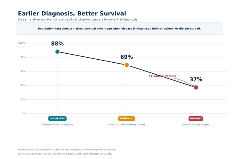
Curative Intent in Oral Cancer Care
Curative intent means that treatment is directed toward eliminating active cancer rather than only controlling symptoms. It does not mean that the same outcome can be predicted for every patient before treatment begins. Oral cancer includes tumours arising from different anatomical sites, and each site may behave differently. A small lip lesion confined to the surface cannot be treated as equivalent to a deeply invasive tongue tumour involving cervical lymph nodes.
Early cancers of the lip and oral cavity may have a substantial possibility of cure when they are diagnosed promptly and treated appropriately. Surgery or radiation may be required to remove or destroy the primary tumour. More advanced disease may require neck dissection, radiation, chemotherapy, immunotherapy or a combination of treatments. The treatment decision depends on tumour size, depth of invasion, bone involvement, lymph node spread, surgical accessibility and the patient’s ability to tolerate treatment.[1]
Ayurveda should be incorporated without allowing a potentially curable cancer to progress during an avoidable delay. When surgery provides the strongest opportunity for complete tumour removal, Ayurvedic treatment should be coordinated before and after the procedure rather than used to postpone it. When radiation or systemic therapy is medically indicated, the Ayurvedic prescription should be selected after reviewing the proposed treatment schedule, blood counts, liver function, kidney function, nutritional status and possible medicine interactions.
Some patients may seek Ayurveda after surgery, radiation or chemotherapy because residual disease, recurrence, treatment toxicity or reduced physical strength remains a concern. Others may seek it immediately after diagnosis. In either situation, the treatment plan should begin with the biopsy report and complete staging rather than assumptions based only on the appearance of the oral lesion.
The aim is to preserve every realistic opportunity for cure. Ayurveda can be used as part of a comprehensive strategy that addresses the tumour, cervical lymphatic risk, ulcerated oral tissues, appetite, body weight, swallowing, sleep, bowel function and overall treatment tolerance. Symptom improvement is clinically valuable, but the curative objective must remain measurable control of the cancer itself.
Role of Heerak Bhasma and Other Herbo Mineral Medicines
Heerak Bhasma is used in selected patients as part of a personalised curative-intent protocol. It is not prescribed as a standalone medicine, and its inclusion does not make biopsy, staging, imaging or oncology review unnecessary. It is combined with disease-directed herbs and other mineral preparations according to the patient’s clinical requirements.
Preclinical research has examined a Heerak Bhasma preparation as a nanodiamond-based formulation. In an experimental lymphoma model, the formulation was associated with reduced tumour growth, reduced distant spread, prolonged survival and changes in dendritic cell and T-cell responses.[5] These findings support further investigation into its immunological and antitumour potential, but they do not establish that Heerak Bhasma alone cures oral cancer in humans.
Published clinical literature also includes individual reports describing Ayurvedic treatment in patients with oral or tongue squamous cell carcinoma. One reported patient with recurrent tongue cancer received personalised Ayurvedic herbo-mineral medicines and Panchakarma following previous radiation, chemotherapy and repeated surgery. The publication described prolonged progression-free survival and improved quality of life during long-term follow-up.[3] Because the patient had received conventional treatment before Ayurveda, the outcome must be interpreted as an integrative clinical result rather than evidence that one medicine independently produced the remission.
Another published oral cancer case described a protocol containing Heerak Bhasma, Swarna Bhasma, Vaikranta Bhasma, selected pishti preparations, iron-based medicine, Nimba, Kanchanara, Manjistha, Gandhaka and local oral treatments. The authors reported improvement in the ulcerative growth, pain, swelling and discharge.[4] This observation supports the clinical investigation of personalised herbo-mineral treatment, but one case cannot determine the expected response for every patient.
The practical value of these reports is that they demonstrate how Ayurveda may be applied as a structured, disease-directed system rather than as general wellness care. They also show why the complete treatment protocol matters. Heerak Bhasma is used alongside other medicines selected for abnormal tissue growth, inflammation, ulceration, discharge, weakness, reduced appetite and impaired recovery.
The quality of mineral medicine is equally important. Raw diamond powder is not equivalent to properly prepared Heerak Bhasma. Classical pharmaceutical processing, authenticated raw materials, controlled preparation, appropriate dosing and laboratory monitoring are necessary. Unverified market products should not be substituted for a physician-supervised formulation.
How Cure Must Be Confirmed
Pain reduction, improved mouth opening, reduced discharge, better appetite, weight stabilisation and increased energy may indicate that treatment is helping the patient, but these changes cannot independently confirm that the cancer has disappeared. Oral lesions can appear smaller or cleaner while deeper tissue or lymph node disease remains active.
Before treatment begins, the lesion should be clinically examined, photographed and measured. The biopsy report should confirm the tumour type and grade where available. Imaging should assess the depth of invasion, surrounding tissue involvement, jaw involvement and cervical lymph nodes. The patient’s weight, eating ability, swallowing, speech, mouth opening, pain level and functional status should also be documented.
Treatment response should be assessed using the same clinical measurements and appropriate repeat imaging. A partial response means that the measurable cancer has decreased but remains detectable. A complete clinical response means that no visible or palpable tumour is found during examination. A complete radiological response means that imaging no longer demonstrates active disease. When suspicious tissue remains, repeat biopsy may be required to determine whether viable cancer cells are still present.
No evidence of disease should be recorded only when clinical examination, imaging and pathology where indicated do not identify active cancer. Durable remission requires this disease-free state to continue during structured follow-up. Cure should be considered only after sustained observation because oral cancer may recur at the original site, appear in cervical lymph nodes or be followed by another primary tumour within the upper aerodigestive tract.
A curative Ayurvedic programme must therefore include predefined reassessment. Continued enlargement of the lesion, increasing firmness, new bleeding, worsening swallowing, increasing neck swelling, weight loss or new disease on imaging requires immediate review of the treatment strategy. Continuing an unchanged protocol despite documented progression may allow the curative window to narrow.
Early Diagnosis Protects Survival and Function
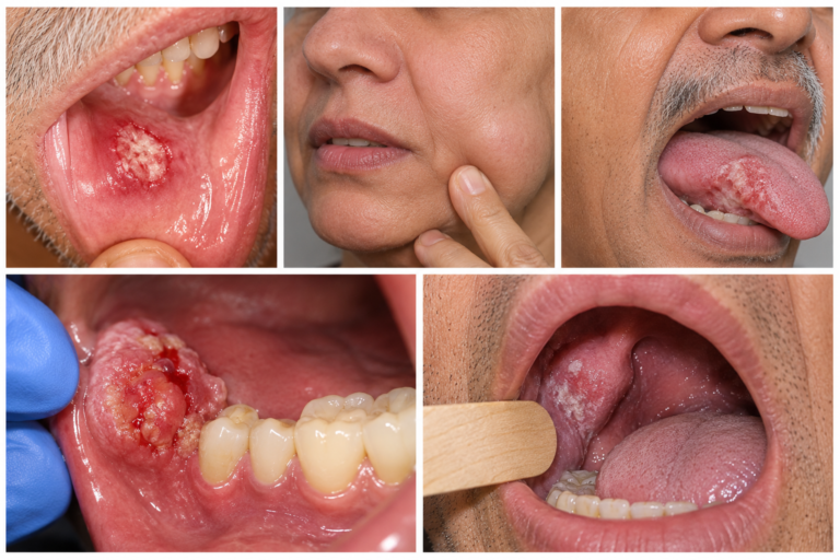
A mouth ulcer that does not heal, a persistent red or white patch, unexplained oral bleeding, a firm growth, reduced tongue movement, numbness, difficulty swallowing, reduced mouth opening, an unexplained loose tooth or a neck lump requires timely evaluation. A suspicious lesion should undergo biopsy or specialist referral because photographs, salivary tests, light-based screening devices and temporary symptom improvement cannot reliably exclude invasive cancer.[2]
Delayed diagnosis can allow a limited lesion to invade the tongue muscles, floor of the mouth, jaw or skin. It may also permit spread to cervical lymph nodes. This progression can change a smaller operation into major oral resection, neck dissection and reconstruction, with greater effects on speech, swallowing, appearance and nutritional independence.[1]
Patients can pursue Ayurveda without rejecting accurate diagnosis or necessary oncology treatment. The safest curative approach begins with biopsy confirmation, complete staging and a documented treatment objective. A personalised Ayurvedic protocol may then use Heerak Bhasma, selected herbs, other properly prepared mineral medicines, local oral care and restorative treatment to pursue measurable tumour control and durable remission.
The claim of cure must arise from the patient’s verified outcome. It should be supported by disappearance of active disease on examination, appropriate imaging, pathology where necessary and continued disease-free follow-up.
Ayurvedic Treatment for Oral Cancer Case Studies

Ayurvedic treatment for oral cancer has been documented in patients with recurrent tongue cancer and ulcerative buccal squamous cell carcinoma involving the mandible. The following published cases include the patient profile, diagnostic findings, previous treatment, Ayurvedic medicines, clinical response and available follow-up data.[3][4]
Recurrent Stage III Tongue Cancer
A 49-year-old woman was diagnosed with stage III moderately differentiated squamous cell carcinoma of the left lateral border of the tongue in August 2005. She had no history of tobacco or alcohol use. Her father had reportedly developed oropharyngeal cancer. She was later diagnosed with type 2 diabetes and had a history of multiple dental extractions for dental caries.[3]
| Clinical field | Published patient data |
|---|---|
| Age at diagnosis | 49 years |
| Age at Ayurvedic registration | 54 years |
| Sex | Female |
| Primary cancer site | Left lateral border of the tongue |
| Histopathology | Moderately differentiated squamous cell carcinoma |
| Initial stage | Stage III |
| Tobacco history | None reported |
| Alcohol history | None reported |
| Family history | Father with reported oropharyngeal cancer |
| Important comorbidity | Type 2 diabetes diagnosed in 2011 |
| Initial diagnosis date | August 2005 |
| Ayurvedic treatment start date | 28 June 2011 |
| Main functional problems | Dysphagia, trismus, loss of appetite, constipation, pain and impaired speech |
| Baseline performance status | Karnofsky Performance Score of 80 |
| Baseline recorded weight | Approximately 65 kg before intensive treatment-related weight loss |
Her first cancer treatment consisted of external-beam radiation delivered in 35 fractions to a total dose of 70 Gy, together with six cycles of low-dose cisplatin. This treatment was completed on 21 October 2005. Residual induration remained in the tongue after chemoradiation.[3]
A knife biopsy performed in April 2007 again showed squamous cell carcinoma infiltrating the lingual muscles. In May 2007, she underwent wide-excision glossectomy with left supra-omohyoid neck dissection. Further recurrent or residual disease later required total glossectomy, hemimandibulectomy and another left supra-omohyoid neck dissection in August 2011.[3]
| Cancer treatment phase | Treatment and clinical findings |
|---|---|
| Initial treatment in 2005 | Radiation therapy of 70 Gy in 35 fractions with six cycles of low-dose cisplatin |
| Residual disease assessment | Persistent induration remained after chemoradiation |
| Repeat biopsy in April 2007 | Squamous cell carcinoma infiltrating the lingual muscles |
| Surgery in May 2007 | Wide-excision glossectomy with left supra-omohyoid neck dissection |
| Major surgery in August 2011 | Total glossectomy, hemimandibulectomy and repeat left supra-omohyoid neck dissection |
| Long-term consequences | Persistent speech impairment, altered swallowing and facial disfigurement |
Ayurvedic treatment began on 28 June 2011. The initial prescription included Yashtimadhu Ghrita, Shatavari Vati and Ananta Vati. Yashtimadhu Ghrita was administered at 5 g with warm water after breakfast and after the evening meal. Shatavari Vati and Ananta Vati were prescribed in divided daily doses according to the published protocol.[3]
Suvarna Bhasmadi Yoga was subsequently added. The formulation contained Suvarna Bhasma, Mukta Bhasma and Guduchi Satva and was administered at 395 mg with ghee after breakfast. The prescription was modified during long-term follow-up according to oral symptoms, appetite, digestion, body weight, treatment tolerance and general strength.[3]
| Ayurvedic treatment phase | Principal medicines recorded in the case |
|---|---|
| Initial phase | Yashtimadhu Ghrita, Shatavari Vati and Ananta Vati |
| Subsequent Rasayana phase | Suvarna Bhasmadi Yoga containing Suvarna Bhasma, Mukta Bhasma and Guduchi Satva |
| Later personalised treatment | Suvarna Malini Vasant, Suvarna Sutashekhara, Arogyavardhini Vati, Mahamanjisthadi Ghana Vati, Praval Pishti Vati, Mouktika Yukta Kamdudha Vati and Sukshma Triphala |
| Appetite and digestion management | Pippalyasava, Shatavari Kalpa and individually selected digestive medicines |
| Local oral treatment | Yashtimadhu Ghrita application, medicated gargling and herbal oral-cleansing preparations |
| Treatment duration | Serial personalised Ayurvedic treatment continued for more than a decade |
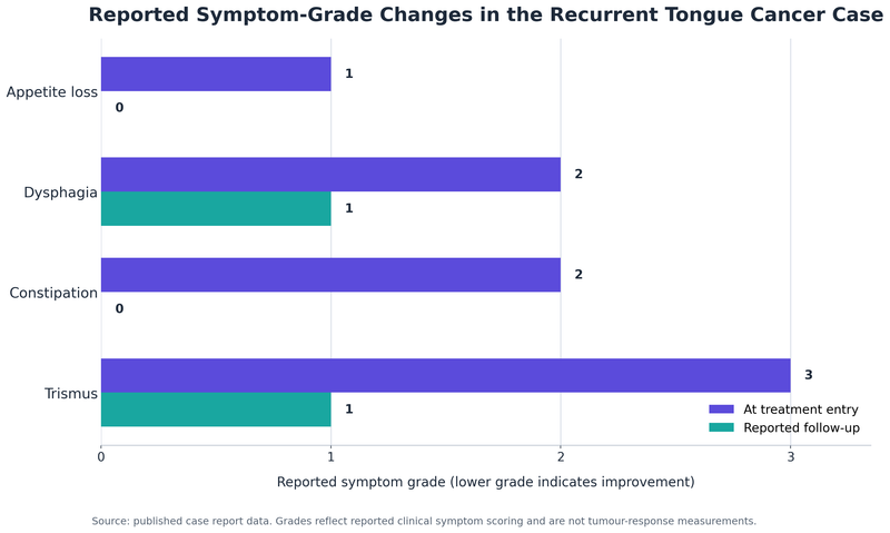
Loss of appetite improved from grade 1 to grade 0. Dysphagia improved from grade 2 to grade 1. Constipation improved from grade 2 to grade 0, while trismus improved from grade 3 to grade 1. The recorded pain score decreased from 6 at the beginning of treatment to 2 and subsequently to 0.[3]
The graph presents the symptom grades recorded in the published case. Lower grades represent improvement in the reported symptom burden and do not independently measure tumour disappearance.[3]
| Recorded outcome | Baseline status | Reported follow-up status |
|---|---|---|
| Loss of appetite | Grade 1 | Grade 0 |
| Dysphagia | Grade 2 | Grade 1 |
| Constipation | Grade 2 | Grade 0 |
| Trismus | Grade 3 | Grade 1 |
| Pain score | 6 | 0 during long-term follow-up |
| Karnofsky Performance Score | 80 | Reached 100 during later assessments |
| Weight | Fell to approximately 55 kg during intensive treatment | Recovered to approximately 62 to 65 kg during long-term follow-up |
| Peripheral neuropathy | Present after cancer treatment | Reported as resolved by December 2012 |
| Appetite and bowel function | Impaired | Reported as improved |
| Speech | Severely impaired after total glossectomy | Persistent anatomical speech limitation |
Radiological follow-up in 2016 showed postoperative reconstruction without reported recurrent tumour. A PET-CT performed in April 2023 identified calcified scalp nodules but did not report metabolically active recurrent or distant oral cancer. Serial haematological, liver and kidney investigations remained within acceptable ranges during the recorded treatment period.[3]
| Long-term assessment | Published finding |
|---|---|
| Local recurrence on later imaging | Not reported |
| Distant metastatic disease on the 2023 PET-CT | Not reported |
| Laboratory tolerance | Serial blood, liver and kidney parameters remained acceptable |
| Quality of life | Improvement or stability in several measured domains |
| Functional limitations | Persistent speech and swallowing impairment related to extensive surgery |
| Progression-free survival | Approximately 15 years reported by the authors |
| Latest clinical status | Asymptomatic during the latest reported follow-up while continuing personalised Ayurvedic medicines |
The patient’s long-term outcome followed radiation, chemotherapy, repeated surgery and personalised Ayurvedic treatment. The sustained follow-up, stable laboratory findings and absence of reported recurrent disease distinguish this case from short-term reports based only on pain reduction or visible ulcer healing.[3]
Buccal Cancer With Mandibular Erosion
A 74-year-old man presented with an ulcerative growth over the right mandibular and buccal region. He had experienced pain, pus discharge, inflammatory swelling, constipation and generalised weakness for approximately two years. He also had hypertension and had been receiving antihypertensive medication for about ten years.[4]
| Clinical field | Published patient data |
|---|---|
| Age | 74 years |
| Sex | Male |
| Primary site | Right buccal mucosa and right lower gingivobuccal sulcus |
| Main presentation | Ulcerative growth over the right mandibular region |
| Symptom duration | Approximately two years |
| Associated symptoms | Pain, pus discharge, swelling, constipation and weakness |
| Important comorbidity | Hypertension for approximately ten years |
| Cytology date | 24 September 2023 |
| Cytological finding | Keratinising squamous cell carcinoma |
| PET scan date | 9 October 2023 |
| Maximum lesion thickness | 2.5 cm |
| Maximum reported SUV | 6.4 |
| Bone involvement | Lytic erosion of the right hemimandible |
| Cervical nodes | FDG-avid nodes at level IA and right levels IB, II and III |
| Formal TNM stage | Not provided in the case report |
The PET scan showed FDG-avid ill-defined soft-tissue thickening with an exophytic component involving the right buccal mucosa and right lower gingivobuccal sulcus. Lytic erosion of the right hemimandible confirmed that the disease had extended beyond the superficial oral mucosa into the jaw bone.[4]
The report stated that conventional oncology treatment had been considered, but advanced age and general debility affected treatment eligibility. The patient then received a standalone Ayurvedic programme containing internal herbo-mineral medicines, an Avaleha and local oral treatment.[4]
| Medicine or formulation | Published dose and method |
|---|---|
| Kanchanara Chhala, Nimba Chhala and Brihat Manjishtadi Kashaya Churna decoction | 50 ml twice daily on an empty stomach, prepared with water and Gomutra as reported |
| Rasamanikya, Shuddha Gandhaka and Trivanga Bhasma | Combined dose of 1 g twice daily on an empty stomach with lukewarm water |
| Heerak Bhasma combination | Heerak Bhasma with Swarna Bhasma, Vaikranta Bhasma, Jaharmohra Pishti, Kaharva Pishti and Tapyadi Lauha |
| Combined herbo-mineral dose | 445 mg twice daily |
| Anupana | Administered with Drakshavaleha before milk |
| Drakshavaleha | 5 g twice daily |
| Local wound cleansing | Panchavalkala Kwatha |
| Local medicated application | Jatyadi Ghrita and Yashtimadhu Ghrita |
| Oral gargling | Yashtimadhu Churna Phanta |
Heerak Bhasma was therefore used as part of a combined herbo-mineral prescription rather than as a single medicine. Swarna Bhasma, Vaikranta Bhasma, Jaharmohra Pishti, Kaharva Pishti and Tapyadi Lauha were administered within the same disease-directed protocol.[4]
| Clinical outcome | Reported change |
|---|---|
| Visible ulcerative growth | Decreased during treatment |
| Buccal pain | Reduced |
| Inflammatory swelling | Reduced |
| Pus discharge | Reduced |
| External wound appearance | Improved in published clinical photographs |
| Repeat PET scan | Not reported |
| Repeat biopsy or cytology | Not reported |
| Mandibular bone reassessment | Not reported |
| Pathological complete response | Not documented |
| Long-term disease-free survival | Not reported |
The reported outcome represents clinical regression of the visible ulcerative lesion and improvement in pain, swelling and discharge. The case report did not include repeat PET measurements, repeat biopsy, pathological confirmation of complete response or long-term survival data.[4]
Early Ayurvedic Treatment for Oral Cancer
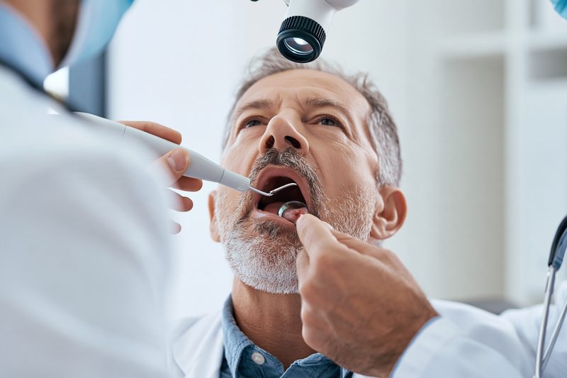
Early Ayurvedic treatment for oral cancer is most valuable when it begins after biopsy confirmation and complete staging, while the patient still has adequate strength, nutrition, swallowing ability and organ function to pursue complete disease control. Beginning Ayurveda early does not mean delaying surgery, radiation or systemic treatment when these provide the strongest curative opportunity. It means introducing a personalised disease-directed protocol before extensive local invasion, cervical lymph-node spread, severe weight loss or treatment-related decline narrows the available options.
The Curative Window Can Narrow Quickly
The oral cavity contains closely connected muscles, nerves, blood vessels, lymphatic channels, salivary structures and jaw bones. A lesion that initially appears limited to the tongue, cheek, gum or floor of the mouth may penetrate deeper tissues and spread to cervical lymph nodes without producing proportionate symptoms. Increasing depth of invasion, bone involvement, multiple lymph nodes and extranodal extension can substantially change the treatment plan and prognosis.[1]
A small, localised tumour may sometimes be treated with a limited surgical procedure or carefully planned radiation. A larger tumour involving the tongue muscles, mandible, skin or floor of the mouth may require wider resection, neck dissection, reconstruction and additional radiation or chemoradiation. The delay may therefore affect survival as well as speech, swallowing, facial appearance, jaw movement and nutritional independence.
Cervical lymph-node disease may be present even when the neck does not appear enlarged. Randomised clinical evidence in early node-negative oral squamous cell carcinoma found improved overall and disease-free survival when elective neck dissection was performed during primary surgery rather than waiting for clinically apparent nodal relapse.[6] This demonstrates why apparently silent regional disease must be considered before relying only on the visible size of the oral lesion.
A persistent ulcer, firm growth, red patch, white patch, unexplained bleeding, numbness, reduced tongue movement, difficulty swallowing, reduced mouth opening or neck swelling requires prompt examination. Repeated courses of antibiotics, mouth gels or home remedies should not replace biopsy when a lesion remains clinically suspicious.[2]
Ayurveda Before Severe Physical Decline
Patients frequently approach Ayurveda only after the cancer has become extensive, eating has become painful and body weight has fallen substantially. At this stage, the patient may also have anaemia, dehydration, infection, disturbed sleep, constipation, weakness or impaired liver and kidney function. These changes can reduce tolerance to surgery, radiation, chemotherapy and even intensive Ayurvedic herbo-mineral treatment.
Beginning personalised Ayurveda earlier allows time to address the patient’s digestive capacity, nutritional intake, bowel regularity, sleep, oral comfort and functional strength while tumour-directed treatment is being planned. In Ayurveda, Agni refers to digestive and metabolic capacity, Bala refers to functional strength, and Ojas represents the body’s overall resilience during illness. Preserving these factors is clinically relevant because a patient who can eat, maintain weight and remain physically active is better positioned to complete disease-directed treatment.
The early Ayurvedic programme may include selected herbs, a personalised Avaleha, local oral care and properly prepared mineral medicines. Heerak Bhasma may be included in carefully selected patients after reviewing the tumour stage, treatment history, laboratory values and concurrent medicines. It should not be used as an isolated remedy or prescribed merely because a biopsy has confirmed cancer.
The treatment objective should remain measurable. Improvement in appetite, pain, discharge, mouth opening or energy may help the patient continue treatment, but these changes do not establish tumour clearance. The oral lesion and cervical lymph nodes must continue to be assessed through examination, standardised measurements, imaging and repeat pathology when clinically indicated.
Early Integration With Oncology Treatment
A patient with an operable oral tumour should have the Ayurvedic plan coordinated around the proposed surgery. Treatment before surgery may focus on nutrition, bowel function, sleep, oral hygiene and general strength without increasing bleeding risk or interfering with anaesthesia. After surgery, the prescription can be modified according to wound status, margin findings, lymph-node pathology, swallowing ability and the need for radiation or chemoradiation.
Patients receiving radiation may develop mucositis, dryness, altered taste, painful swallowing, trismus and weight loss. Early Ayurvedic assessment allows the prescription and diet to be adapted before these complications become severe. Local applications and gargles must be selected carefully because an ulcerated tumour, postoperative wound and radiation-damaged mucosa do not require the same treatment.
Patients receiving chemotherapy or immunotherapy require additional monitoring. Blood counts, liver function, kidney function, bleeding tendency and current medicines should be reviewed before introducing potent herbs or mineral preparations. The treatment should be modified promptly if laboratory abnormalities, infection, dehydration or serious toxicity develops.
Early integration also provides a clear baseline against which the Ayurvedic response can be measured. Clinical photographs, lesion dimensions, mouth-opening measurements, body weight, swallowing ability, pain scores, laboratory findings and imaging should be documented before treatment. Without baseline data, temporary symptom relief may be mistaken for cancer regression.
Waiting Until the Last Stage Reduces Options
Ayurveda should not be reserved only for patients who have exhausted every other treatment and become too weak to eat or tolerate intervention. The opportunity to pursue complete remission is generally greater before extensive tissue destruction, distant metastasis, severe malnutrition or organ dysfunction develops.
A patient who seeks Ayurveda early can receive a coordinated plan that protects nutrition, supports recovery, addresses treatment-related complications and introduces selected disease-directed medicines without abandoning established curative options. A patient presenting very late may still receive meaningful care, but the realistic goal may shift from complete eradication to disease control, preservation of function or relief of severe symptoms.
Difficulty breathing, inability to swallow liquids, uncontrolled oral bleeding, rapidly increasing facial or neck swelling, high fever, severe dehydration, aspiration or sudden functional deterioration requires immediate hospital assessment. Ayurvedic medicines should not delay emergency airway management, bleeding control, infection treatment or nutritional support.
The decision to begin Ayurveda early should be based on the intention to preserve life, oral function and every realistic path to remission. Biopsy, staging and oncology review identify the available curative options, while personalised Ayurveda can be introduced within that plan to address the tumour, oral tissues, nutrition, treatment tolerance and long-term recovery.
Ayurvedic Treatment for Oral Cancer by Site and Type
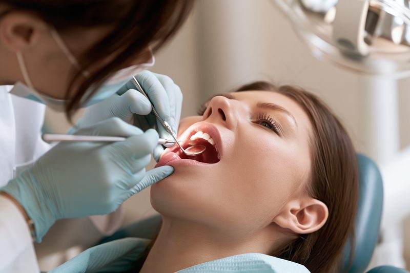
Ayurvedic treatment for oral cancer must be personalised according to the exact anatomical site, histological type, depth of invasion and pattern of spread. A cancer arising from the lip, tongue, cheek, gum or floor of the mouth cannot be treated as the same disease simply because each lesion appears inside or near the mouth. The affected tissue determines the risks to speech, swallowing, jaw movement, facial structure, cervical lymph nodes and nutrition.
Oral Cavity Sites
The oral cavity begins at the lips and extends backward to the junction between the hard and soft palate above and the circumvallate papillae of the tongue below. It includes the mucosal surface of the lips, the front two-thirds of the tongue, the inner lining of the cheeks, the upper and lower gums, the floor of the mouth beneath the tongue, the retromolar trigone behind the last lower molar and the hard palate.[1]
Cancer of the external skin surface of the lip may behave differently from cancer arising from the inner mucosal lip. Lip cancers are often noticed earlier because the lesion is visible, but a persistent crust, ulcer, thickened area or bleeding lesion may still extend into the muscle or regional lymph nodes if treatment is delayed.
The anterior tongue contains muscles responsible for speech, chewing and movement of food. Tongue cancers may appear as a painful ulcer, firm induration or exophytic growth. Even a relatively small surface lesion may extend deeply into the tongue muscles. Depth of invasion is clinically important because deeper tumours have a greater risk of cervical lymph-node spread and may require more extensive surgery or additional treatment.[1]
The floor of the mouth contains thin mucosa overlying salivary structures, nerves and muscles. Tumours in this area may spread into the tongue, mandible or nearby soft tissues. Patients may notice pain, reduced tongue mobility, difficulty swallowing, excessive salivation or a firm area beneath the tongue.
Buccal mucosa cancer develops in the inner lining of the cheek. It may present as a non-healing ulcer, thickened white or red patch, painful growth or reduced mouth opening. Advanced lesions can extend into the cheek muscles, skin, gingivobuccal sulcus or mandible. Persistent trismus may indicate involvement of deeper tissues and can interfere with eating, oral examination and treatment access.
Gingival cancer arises from the upper or lower gum. It can resemble dental infection, periodontal disease or a non-healing extraction socket. When the underlying jaw is involved, the patient may develop pain, loose teeth, numbness, swelling or radiological bone destruction. A tooth extraction performed without recognising the underlying tumour may delay diagnosis.
Retromolar trigone cancer occurs in the small area behind the last lower molar. The site is close to the mandible, chewing muscles and structures of the oropharynx. Tumours in this region may cause pain radiating toward the ear, difficulty opening the mouth and swallowing discomfort.
Hard-palate cancer may arise from the surface epithelium or from minor salivary glands beneath the mucosa. Persistent swelling, ulceration, bleeding, pain or an ill-fitting upper denture may be the first sign. The histological type is particularly important because not every hard-palate tumour is squamous cell carcinoma.
The base of the tongue, tonsils and soft palate belong to the oropharynx rather than the oral cavity. Oropharyngeal cancers differ in their relationship with human papillomavirus, lymph-node behaviour, staging and treatment. A tumour described by a patient as tongue cancer must therefore be located precisely before an Ayurvedic or oncology treatment plan is prepared.[1]
Squamous Cell Carcinoma
Squamous cell carcinoma is the most common malignant tumour of the oral cavity. It develops from the squamous cells lining the oral mucosa. Histopathology may describe the tumour as well differentiated, moderately differentiated or poorly differentiated according to how closely the malignant cells resemble normal squamous tissue.
Differentiation contributes to the pathological assessment, but it does not determine prognosis by itself. Tumour size, depth of invasion, lymphovascular invasion, perineural invasion, surgical margins, cervical lymph nodes and extranodal extension may have greater influence on treatment selection and recurrence risk.
Oral squamous cell carcinoma may appear ulcerative, infiltrative, exophytic or verrucous. An ulcerative cancer produces tissue breakdown and may bleed or become secondarily infected. An infiltrative tumour can extend beneath relatively limited surface changes and produce deep firmness. An exophytic tumour grows outward into the mouth, while a verrucous tumour may form a broad, warty surface.
Verrucous carcinoma is a distinct, usually well-differentiated variant that often grows locally and may invade surrounding tissues while showing a lower tendency for distant spread. Its clinical appearance can resemble benign or precancerous lesions, making adequate biopsy essential.
Carcinoma in situ is confined to the epithelial layer and has not invaded the underlying tissue. Invasive squamous cell carcinoma has crossed the basement membrane and entered deeper structures. This distinction affects the urgency, extent and type of treatment.
Less Common Oral Cancers
Minor salivary-gland cancers may develop in the hard palate, lips, cheeks or other areas containing salivary tissue. These include mucoepidermoid carcinoma, adenoid cystic carcinoma and other histological types. Some salivary cancers may grow slowly but spread along nerves, producing pain or numbness before a large mass becomes visible.
Oral melanoma is an uncommon but aggressive cancer that may appear as a dark brown, black, grey or occasionally non-pigmented lesion. Pigmentation alone cannot distinguish melanoma from benign oral pigmentation, and suspicious changes require biopsy.
Sarcomas arise from connective tissues such as muscle, bone, blood vessels or fibrous tissue. Lymphoma may also involve oral tissues or present as a rapidly enlarging mass. Metastatic cancer from another organ can occasionally appear in the jaw or oral soft tissues. Each of these diseases requires a different treatment pathway from squamous cell carcinoma.
The biopsy report must therefore provide the exact histological diagnosis before a disease-specific Ayurvedic formulation is selected. A formulation designed for ulcerative squamous cell carcinoma may not be appropriate for salivary-gland cancer, melanoma, lymphoma or sarcoma.
Site Specific Ayurvedic Planning
The anatomical site influences the selection of oral applications, decoctions, Avaleha consistency and internal herbo-mineral medicines. A painful tongue ulcer with impaired swallowing requires a different approach from a hard gingival lesion with mandibular erosion or a salivary-gland tumour of the palate.
Tongue and floor-of-mouth cancers require close attention to swallowing, aspiration risk, tongue mobility, speech and cervical lymph nodes. Buccal and retromolar lesions require assessment of mouth opening, chewing muscles and mandibular involvement. Gingival and palatal cancers require careful evaluation of bone and dental structures.
Heerak Bhasma may be considered within a personalised curative-intent protocol, but the remaining herbs and minerals must reflect the tumour site, tissue involvement, ulceration, discharge, bleeding, pain, digestive capacity and general strength. Local oral treatment must also be adapted to the condition of the mucosa. A dry, radiation-injured mouth, an infected ulcerative tumour and a fresh postoperative wound require different preparations.
Ayurvedic assessment can further evaluate the pattern of tissue involvement, inflammation, pain, dryness, discharge and loss of strength. These observations guide individualisation but do not replace histopathology, imaging or modern staging. The exact cancer type and anatomical extent remain essential for deciding whether the patient requires surgery, radiation, systemic treatment, personalised Ayurveda or a coordinated combination.
Early Signs Before Ayurvedic Treatment
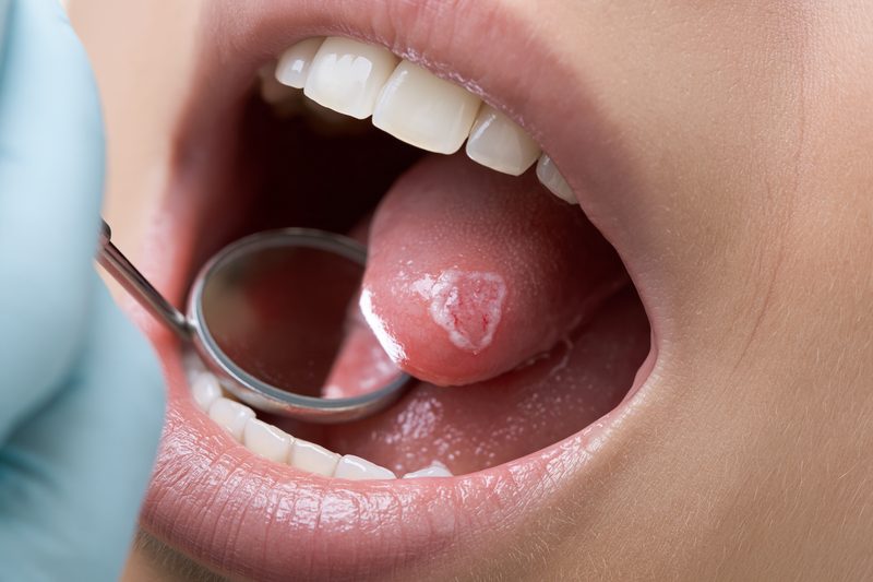
Ayurvedic treatment for oral cancer should never begin on the assumption that a persistent mouth lesion is harmless. A non-healing ulcer, red or white patch, unexplained swelling or change in oral function may represent infection, trauma, a precancerous disorder or invasive cancer. Clinical examination and biopsy are required when a lesion remains suspicious, because appearance and symptom severity cannot reliably determine whether malignant cells are present.[1][2]
A Mouth Ulcer That Does Not Heal
An oral ulcer caused by accidental biting, a sharp tooth, dental treatment or hot food should usually show progressive healing after the source of injury is removed. An ulcer that persists for approximately two weeks, repeatedly reopens, becomes firmer or increases in size requires professional assessment.[2]
Cancerous ulcers may have raised or irregular margins, a hard base, surrounding redness, spontaneous bleeding or mixed red-and-white areas. Some remain painful, while others cause little discomfort during the early stage. The absence of severe pain should therefore not reassure a patient that the lesion is benign.
An ulcer beneath the tongue, along the lateral border of the tongue, inside the cheek, behind the last molar or on the gum can be difficult to inspect without adequate lighting and palpation. A dentist or head-and-neck specialist should examine the entire oral cavity and neck rather than assessing only the visible centre of the lesion.
Persistent Red, White or Mixed Patches
A persistent white patch may represent frictional keratosis, fungal infection, leukoplakia or another mucosal disorder. A red patch may represent inflammation, erythroplakia or invasive disease. Mixed red-and-white lesions can contain areas of epithelial dysplasia or carcinoma and require particular attention.
A patch that cannot be wiped away, develops surface irregularity, becomes thickened or produces a firm area beneath the mucosa should be investigated. Treatment with antifungal medicine or mouthwash should not be repeatedly continued without reassessment when the lesion fails to resolve.
The colour of a lesion cannot establish its diagnosis. Some oral cancers appear predominantly white, red, ulcerated, nodular or nearly normal in colour. Biopsy is required when the clinical appearance or persistence raises concern.[2]
A Lump, Thickening or Hard Area
Oral cancer may present as a lump, nodular growth or area of deep firmness rather than an open ulcer. Induration means abnormal hardness within or beneath the tissue and may indicate infiltration into deeper structures.
A thickened area in the tongue may interfere with movement before it becomes visibly large. A cheek lesion may feel fixed to deeper muscle, while gingival cancer may resemble dental swelling. A hard-palate tumour may first be noticed because a denture becomes uncomfortable or no longer fits correctly.
A growth that becomes progressively larger, bleeds with minor contact or remains fixed to surrounding tissues requires prompt assessment. Temporary reduction in swelling after antibiotics does not exclude an underlying tumour.
Pain, Numbness and Altered Sensation
Oral cancer can produce persistent pain, burning, tenderness or discomfort during chewing and swallowing. Pain may radiate toward the ear, particularly when lesions involve the tongue, floor of the mouth, retromolar area or nearby nerves.
Numbness of the lip, chin, tongue or cheek may indicate nerve involvement. Unexplained tingling, reduced sensation or a persistent feeling that part of the mouth is “asleep” should not be attributed automatically to dental disease.
Pain intensity does not reliably indicate stage. A small lesion near a nerve can be painful, while a larger tumour may remain relatively painless until deeper tissues are involved.
Difficulty Moving the Tongue or Opening the Mouth
Reduced tongue movement may occur when a tumour infiltrates the tongue muscles or floor of the mouth. Patients may notice difficulty moving food, unclear speech, drooling or reduced ability to clean food from the mouth.
Progressive difficulty opening the mouth, known as trismus, may result from oral submucous fibrosis, inflammation, treatment-related fibrosis or tumour involvement of the chewing muscles. In a patient with an oral lesion, worsening trismus requires investigation because it can indicate deeper extension.
Reduced mouth opening may also make eating, dental care, biopsy and treatment more difficult. Early assessment allows the cause to be identified before severe functional restriction develops.
Difficulty Chewing or Swallowing
Painful chewing, food sticking in the mouth, coughing during meals or difficulty swallowing liquids can indicate impaired oral function. These symptoms may arise from the tumour itself, nerve involvement, reduced tongue movement, infection or previous treatment.
Patients may begin avoiding solid food and gradually lose weight without recognising the seriousness of the change. Unintentional weight loss, dehydration or prolonged mealtimes should be documented during the initial assessment.
Swallowing difficulty also increases the risk of aspiration and inadequate nutrition. A patient who coughs repeatedly while drinking, develops chest infections or cannot maintain fluid intake requires urgent evaluation.
Unexplained Bleeding or Discharge
An oral tumour may bleed spontaneously or after minor contact with food, a toothbrush or denture. Persistent blood-stained saliva, repeated bleeding from the same site or bleeding associated with an ulcer requires examination.
Ulcerated tumours may develop secondary infection, foul odour or pus-like discharge. Although antimicrobial treatment may reduce infection and odour, it does not treat the malignant tissue beneath the ulcer.
Heavy or continuous bleeding requires emergency assessment. Local Ayurvedic applications should not be placed on an actively bleeding lesion without identifying the source and severity.
Loose Teeth and Non-Healing Extraction Sites
An unexplained loose tooth, persistent gum swelling or extraction socket that does not heal may indicate cancer involving the gingiva or jaw. Pain may initially be attributed to periodontal disease or dental infection.
Tooth extraction can temporarily remove the most obvious symptom while the underlying lesion continues to progress. Tissue from an abnormal extraction site should be submitted for histopathological examination when malignancy is suspected.
Numbness of the lower lip or chin, mandibular pain and radiological bone destruction require careful evaluation for jaw involvement.
A Neck Lump or Enlarged Lymph Node
Oral cancer can spread to cervical lymph nodes before the patient notices major changes in the primary lesion. A firm, enlarging or persistent lump beneath the jaw or along the side of the neck requires assessment.
A lymph node may be painless and remain mobile during the early stage. More advanced nodal disease may become fixed, involve several levels of the neck or extend beyond the lymph-node capsule. The absence of a palpable neck lump does not exclude microscopic nodal disease.[1][6]
Neck examination and appropriate imaging should therefore form part of the initial staging process even when the oral lesion appears small.
Changes in Speech, Voice or Denture Fit
A tongue or floor-of-mouth tumour may alter articulation and make speech less clear. Patients may also notice changes in saliva control, tongue position or the ability to pronounce particular sounds.
A persistent voice change is more commonly associated with the throat or larynx than the oral cavity, but it may accompany extensive disease or a second lesion. Examination should include adjacent head-and-neck structures when symptoms extend beyond the mouth.
A denture that becomes painful, unstable or unusually tight may indicate underlying swelling or tissue change. Repeated denture adjustment should not replace examination of the mucosa beneath it.
Symptoms Requiring Immediate Assessment
Difficulty breathing, inability to swallow liquids, uncontrolled oral bleeding, rapidly increasing tongue or neck swelling, severe dehydration, high fever or repeated choking requires immediate hospital assessment. These symptoms may indicate airway compromise, major bleeding, severe infection or aspiration.
Ayurvedic treatment for oral cancer can be planned only after the patient is medically stable, the lesion has been diagnosed and the full extent of disease has been assessed. Early recognition of suspicious symptoms protects the opportunity for less extensive treatment, preservation of oral function and complete disease control.
Precancerous Conditions Before Ayurvedic Treatment
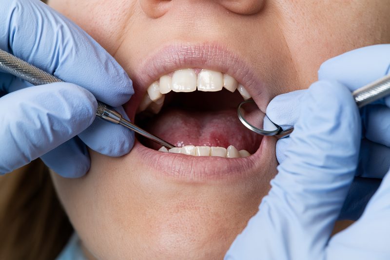
Ayurvedic treatment for oral cancer should begin only after persistent oral patches, ulcers, thickening or fibrosis have been evaluated for epithelial dysplasia and invasive disease. Some oral lesions are classified as oral potentially malignant disorders because they carry a greater risk of developing cancer than normal mucosa. Their appearance alone cannot determine whether malignant change has already occurred, making biopsy and histopathological assessment essential when clinical features are suspicious.[2]
Leukoplakia
Leukoplakia is a persistent white oral patch that cannot be explained by another recognised disease after clinical assessment. It may appear smooth, thickened, fissured, nodular or mixed with red areas. Common sites include the inner cheek, tongue, gum, floor of the mouth and areas exposed to tobacco, areca nut or repeated irritation.
A uniform white patch may carry less immediate concern than a non-homogeneous lesion containing red areas, nodules, ulceration or surface irregularity. However, colour and texture cannot reliably establish whether dysplasia is present. A clinically mild-looking leukoplakia may contain abnormal cells, while another extensive white patch may show no dysplasia on biopsy.
The level of concern increases when the lesion occurs on the lateral tongue or floor of the mouth, persists after removal of a local irritant, enlarges over time, develops firmness or appears in a person with previous oral cancer. A lesion that does not resolve after correcting trauma, denture friction or infection should be reassessed rather than repeatedly treated with mouthwash or topical medication.
Ayurvedic treatment may address associated burning, dryness, mucosal inflammation, tobacco dependence, poor oral hygiene and reduced tissue recovery. It should not replace biopsy when the lesion is persistent, irregular or clinically suspicious. Treatment response must be judged by documented change in lesion size, colour, texture and histopathological findings rather than symptom relief alone.
Erythroplakia
Erythroplakia appears as a persistent red patch that cannot be explained by trauma, infection or another recognised condition. It is less common than leukoplakia but is more frequently associated with severe epithelial dysplasia, carcinoma in situ or invasive squamous cell carcinoma.
The lesion may appear velvety, smooth, slightly depressed or mixed with white areas. Because it may remain painless, patients can underestimate its significance. Persistent red mucosal change on the tongue, floor of the mouth, soft tissues behind the molars or inner cheek requires prompt specialist assessment.
Empirical treatment should not delay biopsy. Temporary reduction in redness after anti-inflammatory or antifungal treatment does not confirm that the lesion is benign. A representative tissue sample is required to determine whether abnormal cells remain within the surface layer or have invaded deeper tissue.[2]
Ayurvedic medicines may be considered after diagnosis for mucosal inflammation, burning, tissue vulnerability and general health, but a red lesion with significant dysplasia requires a clearly documented treatment and surveillance plan. Any increase in size, ulceration, bleeding or underlying hardness requires immediate reassessment.
Mixed Red and White Lesions
A mixed red-and-white lesion may be described clinically as erythroleukoplakia or speckled leukoplakia. The red component may represent thinning or loss of surface keratin, inflammation, dysplasia or early invasion. These lesions generally require greater diagnostic urgency than a uniform white patch.
Biopsy should target the most suspicious area rather than only the thickest white region. Red, ulcerated, nodular or firm areas may contain the highest-grade abnormality. Large or uneven lesions may require samples from more than one site because dysplasia can vary across the same patch.
A benign biopsy from one small area does not permanently exclude future change elsewhere in a widespread lesion. Continued clinical examination is required, especially when the lesion persists, returns after removal or changes in colour and surface pattern.
Ayurvedic treatment should be coordinated with histopathological surveillance. Reduced burning or improved mucosal appearance cannot establish that dysplastic cells have disappeared. Repeat biopsy may be required when the clinical pattern changes or when a lesion fails to respond as expected.
Oral Submucous Fibrosis
Oral submucous fibrosis is a chronic progressive disorder strongly associated with areca nut and betel quid use. It produces inflammation followed by fibrosis beneath the oral mucosa. Patients may initially experience burning after spicy foods, recurrent ulceration or intolerance to hot foods. Progressive fibrosis can then cause stiffness of the cheeks, reduced tongue mobility and increasing difficulty opening the mouth.
The mucosa may appear pale, blanched or marble-like, with palpable fibrous bands in the cheeks, lips or soft palate. Advanced disease can interfere with eating, speech, oral hygiene, dental care and complete examination of the mouth.
Oral submucous fibrosis carries a risk of malignant transformation and requires long-term surveillance. A new ulcer, red patch, thickened area, focal induration, unexplained bleeding or sudden worsening of pain within an already fibrotic mouth requires biopsy rather than attribution to fibrosis alone.
Stopping areca nut, betel quid and tobacco is essential. Continuing exposure can sustain inflammation, fibrosis and malignant risk despite treatment. Ayurvedic care may address burning, dryness, reduced mouth opening, digestion and nutritional status, but it must be combined with clinical examination, physiotherapy where appropriate and biopsy of suspicious areas.
Forceful mouth-opening practices can injure fragile tissues and worsen pain. Mouth-opening exercises should be progressive and selected according to the degree of fibrosis, dental condition and presence of active ulceration or cancer.
Proliferative Verrucous Leukoplakia
Proliferative verrucous leukoplakia is a persistent, multifocal white disorder that can involve several oral sites and expand gradually over years. It may begin as a flat white patch and later become thickened, nodular or wart-like. It can recur after removal and may develop new lesions at different sites.
This condition is clinically important because repeated biopsies may show changing grades of abnormality over time. An early sample may reveal only hyperkeratosis or mild dysplasia, while a later area may develop severe dysplasia, verrucous carcinoma or conventional squamous cell carcinoma.
Patients require mapping of all affected sites, standardised photographs and repeated examination. Biopsy should be repeated when a lesion becomes thicker, develops redness or ulceration, begins bleeding or changes rapidly.
Ayurvedic treatment may be used to improve oral tissue condition, inflammation and general resilience, but it should not be presented as proof that long-term malignant risk has been removed. Persistent multifocal disease requires lifelong surveillance even when individual patches appear to improve.
Actinic Cheilitis
Actinic cheilitis is a sun-related change affecting the lip, most often the lower lip. It may produce persistent dryness, scaling, loss of the normal border between the lip and surrounding skin, pale or mottled areas, crusting, fissures or recurrent ulceration.
Chronic ultraviolet exposure can produce epithelial dysplasia and may progress to lip squamous cell carcinoma. A firm area, persistent ulcer, bleeding site or rapidly enlarging crust requires biopsy.
Sun protection is a central part of management. Patients should reduce prolonged direct exposure and use appropriate lip protection. Ayurvedic topical preparations should not be applied over an undiagnosed crusted or bleeding lesion in place of examination.
Once dysplasia has been identified, treatment decisions depend on its severity, distribution and response to prior therapy. Continued surveillance remains necessary because visible improvement does not always reflect complete removal of abnormal tissue.
Oral Lichen Planus and Lichenoid Disease
Oral lichen planus can produce white lace-like markings, plaques, redness, erosions or painful ulceration. It may involve the cheeks, tongue, gums or several sites simultaneously. Lichenoid reactions can resemble oral lichen planus but may be associated with medication, dental materials, contact sensitivity or other triggers.
Classic symmetrical white striations may be recognisable clinically, but biopsy is often required when the pattern is erosive, ulcerative, unilateral, thickened or otherwise atypical. A lesion that changes its established pattern should be reassessed.
Persistent erosive disease may interfere with eating, oral hygiene and quality of life. It also requires long-term monitoring because a small proportion of affected patients may develop malignant change. New firmness, focal growth, persistent ulceration or unexplained bleeding should not be assumed to be part of the original inflammatory disorder.
Ayurvedic treatment may be directed toward burning, recurrent erosion, mucosal dryness, digestive disturbance and tissue recovery. It should be coordinated with review of possible medication triggers, dental factors and histopathology. Repeated unsupervised use of irritating local agents can worsen erosive mucosa.
Dysplasia and Carcinoma in Situ
Epithelial dysplasia means that abnormal cells and disordered maturation are present within the oral epithelium. Reports may describe mild, moderate or severe dysplasia according to the extent and severity of cellular abnormality.
Mild dysplasia does not always progress to cancer, but it requires risk-factor control and surveillance. Moderate or severe dysplasia usually demands more active management because the probability of progression is greater. The treatment decision may include complete removal, close observation, repeated biopsy or another procedure depending on lesion size, site, patient health and pathological findings.
Carcinoma in situ means that malignant-appearing cells occupy the full thickness of the epithelium but have not yet invaded the underlying connective tissue. Once tumour cells cross the basement membrane, the disease becomes invasive squamous cell carcinoma.[1]
A pathology report should be interpreted with the clinical findings. A small biopsy may not represent the most advanced area of a large or uneven lesion. When the appearance is more concerning than the initial pathology, further sampling or complete excision may be required.
Ayurvedic treatment should not be used to downgrade the significance of moderate dysplasia, severe dysplasia or carcinoma in situ. These findings identify a limited period in which treatment may prevent progression to invasive cancer. A personalised Ayurvedic protocol may accompany definitive management and long-term mucosal care, but histological reassessment remains necessary when the lesion persists.
Monitoring Precancerous Oral Lesions
Each lesion should be documented by site, dimensions, colour, surface pattern, texture and degree of firmness. Standardised photographs are useful when they are taken with similar lighting, distance and mouth position. Cervical lymph nodes should also be examined when invasive disease is suspected.
The interval between reviews depends on the pathology, anatomical site, exposure to tobacco or areca nut, previous oral cancer and clinical behaviour of the lesion. High-risk lesions require closer follow-up than stable low-risk changes.
Complete avoidance of tobacco, areca nut, betel quid and excessive alcohol reduces continued mucosal injury. Sharp teeth, unstable dentures and chronic trauma should be corrected, but removal of irritation does not eliminate the need for biopsy when a lesion remains abnormal.
Ayurvedic medicines, local oral preparations and dietary measures may improve comfort and tissue condition. The decisive clinical question remains whether dysplasia is regressing, persisting or progressing. That determination requires continued examination and repeat histopathology when indicated.
Ayurvedic Treatment for Oral Cancer and Risk Factors
Ayurvedic treatment for oral cancer must include removal of the exposures that initiated or continue to aggravate oral tissue damage. Tobacco, smokeless tobacco, alcohol and areca nut are the most important preventable risk factors, but oral cancer can also occur in people who have never used them. Identifying risk factors helps guide cessation, examination of the entire mouth, surveillance for additional lesions and prevention of a second primary cancer, but it does not determine the diagnosis or stage of an existing tumour.[11][12]
Smoking and Tobacco Exposure
Cigarettes, cigars, pipes, bidis and other smoked tobacco products expose the oral mucosa to carcinogens capable of damaging cellular DNA. Risk generally increases with the duration and intensity of exposure. Smoke reaches the lips, tongue, floor of the mouth, cheeks, gums and throat, so the absence of a lesion at the point where a cigarette is held does not exclude tobacco-related disease.[11]
Smokeless tobacco includes chewing tobacco, snuff, moist snuff, snus and regional products containing tobacco. Holding these products against the cheek or gum creates prolonged contact between carcinogens and the oral mucosa. Patients may develop localised thickening, white patches, gum recession, ulceration or an invasive tumour at or near the habitual placement site.
Switching from smoking to smokeless tobacco does not remove oral cancer risk. Changing brands, using filtered cigarettes or reducing the number of cigarettes may lower exposure but does not provide the protection achieved through complete cessation. Secondhand tobacco smoke also contributes carcinogenic exposure and should be reduced within the home, workplace and vehicle.[11]
Stopping tobacco remains clinically valuable after oral cancer has been diagnosed. Continued exposure can impair oral tissue recovery, sustain inflammation and increase the risk of another cancer within the mouth, throat, larynx, oesophagus or lungs. Cessation should begin as early as possible rather than waiting until surgery, radiation or Ayurvedic treatment has been completed.[12]
Nicotine dependence may require behavioural counselling and evidence-based cessation treatment. Withdrawal symptoms such as irritability, poor concentration, sleep disturbance and cravings should be managed actively because untreated dependence increases the likelihood of relapse. Ayurvedic treatment may be used to address appetite, sleep, bowel disturbance and stress during cessation, but it should accompany a structured plan for complete tobacco abstinence.
Alcohol
Alcohol increases the risk of oral cavity cancer, particularly when consumption is frequent or substantial. Alcohol can increase mucosal permeability, facilitate contact with tobacco carcinogens and contribute to nutritional and metabolic disturbances. The combination of alcohol and tobacco creates a substantially greater risk than either exposure alone.[11]
Beer, wine and spirits all contribute ethanol exposure. The relevant factor is not whether the drink is considered premium, organic or socially acceptable, but the total amount and pattern of consumption. Regular heavy drinking and repeated binge drinking are particularly concerning.
A patient should not assume that changing from spirits to wine or beer removes cancer risk. Reducing consumption may decrease exposure, while stopping alcohol provides the clearest approach during active oral cancer treatment. Cessation can also improve hydration, sleep, appetite, liver function, medication safety and treatment adherence.
Alcohol withdrawal can be medically serious in a person with physiological dependence. Tremor, sweating, agitation, hallucinations, seizures or confusion after stopping alcohol requires urgent medical care. Such patients need supervised withdrawal management rather than abrupt unsupervised cessation.
Alcohol-containing mouthwash is not equivalent to drinking alcoholic beverages, and evidence has not established ordinary mouthwash use as a major independent cause of oral cancer. A patient with painful, ulcerated or radiation-sensitive mucosa may nevertheless tolerate an alcohol-free mouthwash more comfortably.
Areca Nut and Betel Quid
Areca nut is carcinogenic to humans and can increase oral cancer risk even when it is consumed without tobacco. It may be used alone or within betel quid, paan, gutka, pan masala, supari and other packaged or culturally prepared products. Adding tobacco further increases carcinogenic exposure.[12][13]
Areca nut chewing can produce repeated chemical and mechanical injury to the oral mucosa. It is strongly associated with oral submucous fibrosis, in which the cheeks and other oral tissues become progressively stiff. Patients may experience burning with spicy food, pale mucosa, fibrous bands, reduced tongue mobility and decreasing mouth opening.
Removing tobacco from a betel preparation does not make the remaining areca nut safe. Products labelled as herbal, tobacco-free, mouth freshener or natural may still contain areca nut. Patients should review the actual ingredients rather than relying on the front label or cultural familiarity of the product.
Complete cessation is required after oral submucous fibrosis, dysplasia or oral cancer has been diagnosed. Continuing even small amounts can maintain mucosal exposure and make evaluation of new lesions more difficult. Family members should also avoid offering these products during social, religious or celebratory occasions.
Reduced mouth opening should be measured during follow-up. A sudden increase in pain, a new ulcer, focal hardness, bleeding or a change within an area of fibrosis requires biopsy because malignant transformation can occur within an already restricted mouth.
Tobacco and Alcohol Used Together
Tobacco and alcohol interact in a way that produces greater risk than simple exposure to either factor alone. Alcohol can make the mucosal barrier more permeable, while tobacco supplies multiple carcinogens that remain in direct contact with the oral tissues.[11]
A patient who stops only one exposure may reduce risk, but continued use of the other still matters. Complete cessation of both provides the most clinically meaningful risk reduction and reduces the possibility that persistent symptoms will be repeatedly attributed to irritation rather than investigated.
Treatment planning should document the type of tobacco, daily amount, duration, placement site for smokeless products, alcohol frequency and date of cessation. This history helps identify high-risk oral sites and guides examination of the lungs, throat and oesophagus when additional symptoms are present.
Sun Exposure and Lip Cancer
Chronic ultraviolet exposure is an important risk factor for cancer of the external lip, particularly the lower lip. Outdoor workers and people with prolonged recreational sun exposure may develop persistent dryness, scaling, loss of the normal lip border, recurrent crusting or actinic cheilitis before invasive cancer appears.[11]
A lip lesion that repeatedly crusts, bleeds, ulcerates or becomes firm requires examination and possible biopsy. Lip balm alone should not be used to manage a persistent focal lesion without establishing the diagnosis.
Protection includes limiting intense ultraviolet exposure, using a broad-spectrum lip product with adequate sun protection and wearing a wide-brimmed hat during prolonged outdoor activity. These measures remain necessary after treatment because the surrounding lip may retain accumulated sun damage.
Cancer involving the wet inner lip is biologically part of the oral cavity, while cancer of the external sun-exposed lip may behave more like a cutaneous malignancy. Precise localisation is therefore necessary before selecting surgery, radiation, surveillance and Ayurvedic local care.
Human Papillomavirus
High-risk human papillomavirus, particularly HPV type 16, is a major cause of cancers arising from the tonsils and base of the tongue within the oropharynx. Its association with cancers of the true oral cavity is weaker than its association with oropharyngeal cancer.[11][14]
Patients often use the term tongue cancer for tumours arising from different regions. Cancer of the anterior two-thirds of the tongue belongs to the oral cavity, while cancer of the base of the tongue belongs to the oropharynx. This distinction affects staging, HPV testing, expected behaviour and treatment.
HPV status should not be assumed from age, sexual history or the absence of tobacco exposure. Testing is performed on tumour tissue when clinically indicated. A positive oral HPV test without a visible lesion does not independently diagnose cancer, and a negative test does not exclude a non-HPV-related tumour.
HPV vaccination can reduce infection with cancer-associated HPV types and is an important preventive measure for eligible individuals. It does not treat an established oral or oropharyngeal tumour and does not replace examination of persistent symptoms.
Poor Oral Health and Chronic Irritation
Broken teeth, sharp dental edges, unstable dentures and poorly fitting appliances can repeatedly injure the same area of oral mucosa. These problems should be corrected because they can cause persistent ulceration, pain and inflammation. A lesion that remains after the source of trauma has been removed requires reassessment and possible biopsy.[2]
Current evidence does not establish chronic mechanical irritation as a strong independent cause of oral squamous cell carcinoma in the same way as tobacco, alcohol or areca nut. It may act as a cofactor in susceptible or already altered tissue, but the available studies are inconsistent and affected by confounding factors.[15]
Persistent trauma should therefore not be used as a reassuring explanation for a non-healing ulcer. Cancer can be mistakenly attributed to a sharp tooth or denture when the lesion itself has changed the local anatomy. Removing the irritant is appropriate, but the lesion must still be reviewed until complete healing is documented.
Poor oral hygiene, periodontal disease and missing or diseased teeth have been associated with oral cancer in observational research. These findings may partly reflect tobacco use, alcohol use, access to dental care and socioeconomic factors. Good oral hygiene is clinically valuable, but brushing and mouthwash cannot reverse dysplasia or eliminate an established tumour.
Before surgery or radiation, dental infection, unstable teeth and denture problems should be assessed because they can complicate healing and oral intake. Cleaning must be gentle around an ulcerated tumour, and abrasive powders or caustic applications should be avoided.
Previous Precancer or Oral Cancer
A history of leukoplakia, erythroplakia, oral submucous fibrosis, severe epithelial dysplasia, carcinoma in situ or previous oral squamous cell carcinoma increases the need for long-term surveillance. Treatment of one lesion does not remove risk from the remaining oral mucosa.
Patients previously treated for oral cancer can develop recurrence at the original site, regional lymph-node disease or a separate new primary tumour. Any new ulcer, patch, growth, numbness, swallowing change or neck swelling should be evaluated promptly rather than attributed automatically to scar tissue or treatment effects.
Surgical reconstruction and radiation can make examination more difficult. Fibrosis, altered anatomy, chronic dryness and pigmentation may obscure early changes. Follow-up should therefore compare current findings with previous photographs, operative records, imaging and pathology.
Ayurvedic treatment after remission may focus on nutrition, mucosal health, treatment recovery and Rasayana, a restorative approach intended to preserve strength and tissue function. It must remain connected to scheduled cancer surveillance because feeling well does not exclude an early recurrence.
Age, Immunity and General Health
Oral cancer becomes more common with increasing age because cellular damage accumulates over time and exposure to carcinogens may continue for decades. Younger adults can also develop the disease, including individuals without a history of tobacco or alcohol.
Immune suppression may increase vulnerability to several cancers and can alter infection risk, wound healing and treatment tolerance. Patients taking immunosuppressive medicines or living with an immune disorder require coordinated care because abrupt alteration of necessary medication can be dangerous.
Nutritional deficiency, low body weight and chronic illness may weaken recovery but should not be presented as single proven causes of oral cancer. Their clinical importance lies in treatment readiness, wound healing, infection risk and the ability to maintain adequate intake during therapy.
Family history may indicate shared genetic susceptibility or shared exposure, but most patients with oral cancer do not have a clearly inherited cancer syndrome. A family history should prompt careful documentation rather than the assumption that disease is unavoidable.
Oral Cancer Without Known Risk Factors
Some patients develop oral cancer despite never smoking, drinking alcohol or chewing areca nut. The absence of these exposures must not delay biopsy or reduce the seriousness given to a persistent lesion.
Cancers in non-users may arise through a combination of age-related cellular changes, genetic alterations, immune factors, environmental exposures and biological mechanisms that remain incompletely defined. A search for a single personal mistake rarely changes the immediate need for staging and treatment.
Patients without traditional risk factors may be younger or may present with lesions at sites such as the tongue. Their cancer is not automatically less aggressive, and treatment should be based on histopathology, depth of invasion, lymph-node status and molecular findings where relevant.
A patient should not be blamed for developing oral cancer. Risk factors alter probability but do not determine individual outcome. Treatment decisions must remain focused on the present disease and the factors that can still be changed.
Risk Factor Removal in Ayurveda
Ayurveda describes removal of causative and aggravating factors as Nidana Parivarjana. In oral cancer care, this includes complete avoidance of tobacco, areca nut and betel quid, cessation of alcohol, correction of traumatic dental factors, protection of the lips from ultraviolet exposure and maintenance of safe oral hygiene.
Risk-factor removal is necessary but cannot eradicate an established invasive tumour by itself. It must accompany biopsy-based diagnosis, staging, disease-directed treatment and objective follow-up.
A personalised Ayurvedic protocol may support tobacco or alcohol cessation by addressing sleep disturbance, appetite, bowel irregularity, anxiety and reduced strength. The prescription must account for withdrawal, current cancer treatment, liver and kidney function and all concurrent medicines.
Continued exposure can undermine any curative-intent plan. Patients using Heerak Bhasma, herbal medicines, an Avaleha or local oral preparations must still eliminate carcinogenic habits. The intended Ayurvedic treatment response should be assessed alongside sustained abstinence, nutritional recovery, lesion measurements, imaging and pathology where indicated.
Tests Before Ayurvedic Treatment for Oral Cancer
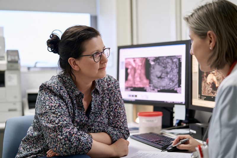
Tests before Ayurvedic treatment for oral cancer must confirm the diagnosis, determine the complete extent of disease and establish whether surgery, radiation or systemic treatment offers an immediate curative opportunity. The same investigations also provide a measurable baseline for deciding whether a personalised protocol containing Heerak Bhasma, herbs and other mineral medicines is producing tumour regression, stable disease or progression. Symptoms and visual examination alone cannot provide this information.
Clinical Oral and Neck Examination
The first assessment should document the exact anatomical origin of the lesion. The external lip, inner lip, anterior tongue, floor of the mouth, buccal mucosa, upper and lower gingiva, retromolar trigone and hard palate have different patterns of local invasion and lymphatic spread. A lesion at the base of the tongue or tonsil belongs to the oropharynx and requires a different staging approach.
The entire oral cavity should be examined under adequate light rather than focusing only on the most painful or visible area. The physician should record the lesion’s length, width, surface appearance, ulceration, bleeding, discharge, firmness, fixation and relationship to nearby teeth, tongue muscles, jaw and skin. Palpation is important because an infiltrative tumour may extend deeper than its visible margins.
Tongue mobility, speech, mouth opening, chewing, swallowing and sensation of the lip, chin and tongue should be recorded. Reduced tongue movement may indicate muscular invasion, while numbness can suggest nerve involvement. Progressive trismus may indicate extension into deeper cheek tissues or the muscles used for chewing.
The neck should be examined for lymph nodes on both sides. The number, size, level, mobility and firmness of palpable nodes should be recorded. A clinically normal neck does not exclude microscopic lymph-node disease, and nodal assessment remains necessary even when the primary lesion appears small.[1][6]
Clear baseline photographs can document the visible lesion, but photographs cannot determine depth, bone invasion or lymph-node involvement. Images should be taken from consistent angles with comparable lighting and a measurement scale so that later changes are not judged by memory alone.
Biopsy and Histopathology
A biopsy is required to confirm oral cancer. A persistent or suspicious lesion should undergo a scalpel, punch or appropriately selected surgical biopsy followed by histopathological examination. Brush cytology, salivary testing, vital staining and light-based devices cannot replace an indicated tissue biopsy.[2]
An incisional biopsy removes a representative part of a larger lesion. The sample should include viable tissue from the edge of the lesion and sufficient depth to assess invasion. Tissue taken only from the necrotic centre may fail to provide adequate diagnostic information. A small lesion may sometimes be removed completely through an excisional biopsy when this is oncologically appropriate.
The pathology report should identify the exact tumour type. Most oral cavity cancers are squamous cell carcinomas, but salivary-gland cancer, melanoma, lymphoma, sarcoma and metastatic disease require different treatment pathways. The report should also record the degree of differentiation when applicable and describe whether invasive malignant cells are present.
For a surgically removed squamous cell carcinoma, the final pathology report should include tumour dimensions, depth of invasion, margin status, lymphovascular invasion, perineural invasion, bone involvement and the number of lymph nodes examined and involved. Extranodal extension should be reported when cancer has grown beyond the lymph-node capsule. These findings affect recurrence risk and the need for postoperative radiation or chemoradiation.[1]
A pathology review may be appropriate when the biopsy is small, the diagnosis is unusual, the clinical appearance does not match the report or treatment decisions carry major functional consequences. The original tissue blocks and slides should be retained because further review or additional testing may be required.
A previously benign or dysplastic biopsy does not permanently exclude cancer if the lesion enlarges, ulcerates, develops firmness or changes colour. Repeat biopsy should target the most suspicious area when clinical progression occurs despite an earlier non-malignant result.[2]
CT and MRI of the Primary Tumour
Contrast-enhanced CT or MRI helps determine how far the tumour extends beneath the visible surface. Imaging may show invasion into the tongue muscles, floor of the mouth, cheek, mandible, maxilla, skin, salivary structures or adjacent spaces that cannot be assessed completely through oral examination.[1]
MRI provides detailed assessment of soft tissues and is particularly useful for tongue, floor-of-mouth, deep cheek and possible nerve involvement. It may define the relationship between the tumour and muscles more clearly than clinical examination alone. MRI can also help evaluate perineural spread when pain, numbness or other neurological symptoms are present.
CT is useful for assessing the primary tumour, cervical lymph nodes and jaw involvement. Bone-window images can demonstrate erosion or destruction of the mandible or maxilla. Dental artefacts may reduce image clarity, making the choice between CT and MRI dependent on the tumour site and the information required.
Contrast imaging requires review of kidney function and previous contrast reactions. A patient with renal impairment, dehydration or a previous severe contrast allergy may require modification of the imaging plan rather than proceeding without assessment.
Imaging performed before treatment should be retained for comparison. A report stating only that a lesion is present is insufficient for response monitoring when the tumour dimensions, involved structures and lymph-node findings are not documented.
Cervical Lymph Node Assessment
Cervical lymph-node status is one of the most important factors in oral cancer treatment planning. Oral cancers can spread through lymphatic channels to nodes beneath the chin, beneath the jaw and along the sides of the neck. This spread may occur before a lump becomes visible or palpable.
Contrast-enhanced CT, MRI or ultrasound can identify suspicious nodes according to their size, shape, internal structure, margins and distribution. A small node may still contain cancer, while an enlarged node may be inflammatory. Imaging findings must therefore be interpreted with the primary tumour, examination and pathology.
Ultrasound-guided fine-needle aspiration or core biopsy may be required when a cervical node is suspicious. This can determine whether the node contains metastatic squamous cell carcinoma or another disease. Cytology from a neck node does not replace biopsy of the primary oral lesion when that lesion remains undiagnosed.
Patients with an apparently node-negative neck may still require elective neck treatment according to the primary site, tumour stage and depth of invasion. Clinical evidence has shown that waiting for a cervical relapse can reduce the opportunity for successful disease control in selected early oral cancers.[6]
An Ayurvedic prescription should not be evaluated only by changes in the visible mouth lesion. Stable or enlarging cervical nodes may indicate persistent regional disease even when pain and ulceration improve.
PET CT and Distant Disease Evaluation
PET-CT is not required for every small, newly diagnosed oral lesion. It is more commonly considered when the cancer is advanced, cervical lymph nodes are involved, distant spread is suspected, the primary site is uncertain or recurrence must be distinguished from post-treatment changes.[1]
PET-CT identifies areas with increased metabolic activity, but inflammation, infection and healing tissues can also appear active. A metabolically active area may therefore require targeted imaging, examination or biopsy before it is labelled as cancer.
Chest imaging is important because the lungs are a potential site of distant spread and a separate smoking-related primary cancer may coexist. Depending on the stage and risk profile, assessment may involve a chest radiograph, CT of the chest or PET-CT.
Bone, liver or brain imaging is selected according to symptoms, stage and existing scan findings rather than performed routinely in every patient. Persistent focal bone pain, neurological symptoms, unexplained liver abnormalities or advanced nodal disease may justify additional investigation.
When distant metastasis is present, the treatment objective may differ from that of a localised oral tumour. Complete remission may remain possible in selected circumstances, but widespread metastatic disease generally requires systemic treatment planning and careful definition of realistic goals.
Dental and Jaw Assessment
A dental examination is necessary before major oral surgery or radiation. Active dental infection, unstable teeth, periodontal disease, sharp dental edges and poorly fitting dentures can complicate wound healing, oral intake and radiation-related tissue injury.
A panoramic dental radiograph or other jaw imaging may be required when the tumour is close to the mandible or maxilla, teeth are loose without a clear dental cause or the patient has persistent jaw pain or numbness. Bone invasion can change both the cancer stage and the extent of surgery.
Teeth with a poor long-term prognosis may require treatment or extraction before radiation. The timing must allow adequate healing whenever clinically possible. Unplanned dental extraction after high-dose radiation to the jaw may carry a greater risk of delayed healing and osteoradionecrosis.
A non-healing extraction socket should not be repeatedly treated as a routine dental infection without histopathological assessment. Gingival and jaw cancers may initially resemble periodontal disease, dental abscess or traumatic ulceration.
Local Ayurvedic oils, powders or gargles should be selected only after evaluating the teeth, gums, tumour surface and surrounding mucosa. Caustic or abrasive preparations can worsen ulceration, bleeding and pain.
Blood and Urine Tests
A complete blood count provides baseline haemoglobin, white blood cell and platelet values. Anaemia may contribute to fatigue, breathlessness and reduced treatment tolerance. A low neutrophil count increases infection risk, while a low platelet count can increase bleeding risk during biopsy, surgery or treatment of an ulcerated lesion.
Liver-function tests should include bilirubin, transaminases, alkaline phosphatase, albumin and other clinically relevant measurements. Liver impairment may affect the metabolism and safety of conventional medicines, herbs and mineral preparations. Low albumin may also reflect poor nutrition, inflammation or advanced disease.
Kidney assessment should include serum creatinine, estimated glomerular filtration rate, urea and electrolytes. Renal function influences contrast imaging, chemotherapy selection, hydration planning and the safe use of several medicines.
Blood glucose and glycated haemoglobin are important in patients with diabetes or suspected impaired glucose control. Poorly controlled diabetes can increase infection risk, delay wound healing and complicate the use of an Avaleha containing a sweet base.
Coagulation testing may be required when the patient has significant bleeding, liver disease, anticoagulant use or an invasive procedure is planned. Tests should be selected according to the medical history rather than ordered as a substitute for clinical assessment.
Urine examination can provide additional information about hydration, glucose, protein, infection and kidney health. Abnormal findings require evaluation before intensive herbo-mineral treatment is started.
Safety Testing for Heerak Bhasma and Mineral Medicines
Baseline blood counts, liver function and kidney function are required before a prolonged protocol containing Heerak Bhasma, Swarna Bhasma or other potent mineral preparations. These investigations provide a reference for distinguishing pre-existing abnormalities from changes that develop during treatment.
Laboratory tests on the patient cannot establish the purity of a medicine. The formulation itself requires authenticated raw materials, documented classical processing, batch identification and appropriate analytical quality testing. Pharmacopoeial identity and purity standards are necessary for Ayurvedic ingredients and finished preparations.[10]
Blood lead, mercury or arsenic testing may be considered when the patient has previously consumed unverified metallic products, has occupational or environmental exposure, or develops symptoms compatible with toxicity. A normal baseline result does not make an untested product acceptable for use.
The complete prescription should be reviewed alongside anticoagulants, antiplatelet medicines, antihypertensives, diabetes medicines, chemotherapy, immunotherapy and other supplements. A medicine that is suitable after surgery may require interruption or modification before another invasive procedure or during a period of severe treatment toxicity.
Repeat monitoring depends on the formulation, dose, duration, concurrent oncology treatment and the patient’s baseline health. New jaundice, reduced urination, persistent vomiting, confusion, unexplained anaemia, abdominal pain or marked weakness requires prompt laboratory reassessment.
Nutrition and Swallowing Assessment
Body weight, recent weight loss, usual food intake and ability to chew and swallow should be documented before treatment. A patient may appear to be eating while consuming too little protein, energy or fluid to maintain weight.
The assessment should identify pain during eating, coughing with liquids, food remaining in the mouth, prolonged mealtimes, drooling and recurrent chest infection. These features may indicate impaired oral control or aspiration risk and may require evaluation by a speech and swallowing specialist.
Dietary restriction should not produce further weight loss. A patient entering surgery, radiation or combined treatment with severe malnutrition has less reserve for wound healing, infection control and treatment completion.
Mouth opening should be measured in millimetres. This provides an objective baseline for trismus and allows later changes to be tracked. Tongue movement, speech clarity and the ability to manage saliva should also be recorded.
An Avaleha may be easier for some patients to take, but its texture and method of administration must match the patient’s swallowing capacity. A patient who aspirates liquids or semi-solid food requires swallowing assessment before oral medicines are continued in the usual form.
Performance and Organ Function
Performance status records how independently the patient can manage daily activities. A patient who remains active and self-caring may tolerate a more intensive curative programme than a patient who is confined to bed for much of the day.
Heart and lung assessment may be required before major surgery, anaesthesia or certain systemic treatments. Electrocardiography, echocardiography, pulmonary testing or specialist clearance should be obtained when indicated by age, symptoms, medical history or the proposed treatment.
The treatment plan must account for hypertension, diabetes, coronary disease, previous stroke, kidney disease, liver disease, immune suppression and other serious conditions. These illnesses do not automatically exclude Ayurvedic treatment, but they influence medicine selection, dose, diet and monitoring frequency.
Frailty should be assessed separately from age. An older patient with preserved strength and organ function may tolerate active treatment, while a younger patient with severe malnutrition or advanced comorbidity may require stabilisation before an intensive protocol.
Biomarker Testing
Routine biomarker testing is not identical for every oral cancer. True oral cavity squamous cell carcinoma should be distinguished from cancer of the tonsil or base of the tongue, where p16 or HPV-related testing may influence classification and prognosis.
In recurrent or metastatic squamous cell carcinoma, programmed death ligand testing may be required when pembrolizumab or another immune-based treatment is being considered. The result must be interpreted with the disease burden, symptoms, previous treatment and the need for a rapid response.[1]
Unusual histological types may require additional immunohistochemistry or molecular testing. Salivary-gland cancers, melanoma, lymphoma and sarcoma have different biomarker pathways from conventional oral squamous cell carcinoma.
Molecular findings should be used to identify treatment opportunities rather than to replace pathology and staging. A biomarker result does not show the complete anatomical extent of the cancer.
Baseline for Measuring Ayurvedic Response
Before Ayurvedic treatment begins, the clinical record should contain the biopsy report, staging information, imaging reports and images, lesion measurements, neck findings, body weight, mouth opening, swallowing status, performance status and relevant laboratory results.
The treatment objective should be stated as complete response, partial response, disease stabilisation or control of advanced disease according to the patient’s stage and available options. This prevents temporary symptom improvement from being confused with confirmed tumour eradication.
Follow-up should compare the same clinical and radiological parameters used at baseline. Reduced pain, better appetite and improved energy demonstrate patient benefit, while tumour response requires documented change in the lesion, lymph nodes and any distant disease.
A protocol containing Heerak Bhasma, herbs, other mineral medicines and Avaleha can be adjusted according to these findings. Progression on examination or imaging requires immediate reassessment of the treatment pathway rather than continuation based only on improved wellbeing.
Ayurvedic Treatment for Oral Cancer by Stage
Ayurvedic treatment for oral cancer must be planned according to the tumour stage, depth of invasion, cervical lymph-node involvement and presence or absence of distant spread. Stage determines whether the realistic objective is complete eradication, reduction before definitive treatment, prevention of recurrence or prolonged control of advanced disease. The Ayurvedic prescription, including the possible use of Heerak Bhasma, other mineral medicines, herbs and Avaleha, must therefore follow the documented extent of cancer rather than the visible size of the mouth lesion alone.
How Oral Cancer Stage Is Determined
Oral cancer is staged through the TNM system. The T category describes the size and local depth of the primary tumour. The N category records whether cancer has spread to cervical lymph nodes, how many nodes are involved, their size and location, and whether the cancer has extended beyond the lymph-node capsule. The M category identifies distant metastasis.[1]
For oral cavity squamous cell carcinoma, depth of invasion is incorporated into tumour staging because a small surface lesion may extend deeply into the tongue or surrounding tissues. A lesion that appears limited in width may therefore carry a higher stage when it penetrates more deeply.
Clinical staging is based on examination, biopsy and imaging before definitive treatment. Pathological staging is determined after surgery by examining the removed tumour, surgical margins and lymph nodes. Pathological staging may reveal deeper invasion or microscopic nodal disease that was not evident clinically.
The treatment plan should use the most complete available stage. A prescription based only on a photograph or biopsy diagnosis without imaging and neck assessment may underestimate the disease and delay a necessary curative procedure.
Stage I and Stage II Oral Cancer
Stage I and stage II generally describe cancers confined to the primary oral site without clinically detected cervical lymph-node or distant spread. The distinction depends on tumour size and depth of invasion.[1]
These stages usually provide the strongest opportunity for complete disease eradication. Depending on the anatomical site and expected functional outcome, treatment may involve surgery, radiation or a carefully selected combination. The neck may still require treatment because microscopic lymph-node spread can exist even when examination and imaging appear normal.
Ayurvedic treatment at an early stage should protect rather than replace the curative opportunity. A personalised protocol may begin around definitive treatment to support nutritional strength, oral tissue recovery, bowel function, sleep and treatment tolerance. Disease-directed herbs and selected herbo-mineral medicines may be introduced after reviewing the planned surgery or radiation and the patient’s laboratory findings.
Heerak Bhasma may be considered in selected patients as part of a wider curative-intent protocol. Its inclusion should not postpone removal or destruction of a localised tumour when definitive treatment can provide a strong possibility of cure.
The oral lesion, mouth opening, body weight and cervical lymph nodes should be documented before treatment. After definitive therapy, the Ayurvedic plan may continue with the objective of restoring tissue function, improving strength and reducing modifiable recurrence risks while scheduled surveillance continues.
Stage III Oral Cancer
Stage III may include a larger or more deeply invasive primary tumour without lymph-node spread, or a tumour with limited spread to a single regional lymph node.[1]
At this stage, treatment commonly requires more than local symptom management. Surgery may involve removal of part of the tongue, cheek, floor of the mouth, gum or jaw, together with treatment of the neck. Radiation or chemoradiation may be recommended according to margin status, lymph-node findings and other high-risk pathological features.
A patient may still have a realistic curative opportunity, but the treatment window is narrower than in a small localised tumour. Delaying definitive treatment while observing whether an oral preparation reduces pain or ulceration may allow further invasion or nodal progression.
Personalised Ayurveda may be coordinated before surgery to improve nutritional status, bowel regularity, sleep and general strength. After surgery, the prescription should be revised according to wound healing, final pathology, swallowing ability and the need for radiation or chemotherapy.
When Heerak Bhasma, Swarna Bhasma or other mineral medicines are used, the dose and combination should reflect blood counts, liver and kidney function, concurrent oncology medicines and the patient’s ability to swallow. An Avaleha may require adjustment in texture and quantity when tongue movement, mouth opening or swallowing is impaired.
Response assessment must include the neck as well as the visible oral site. Improvement in the primary ulcer does not establish control of cervical lymph-node disease.
Stage IVA Oral Cancer
Stage IVA generally describes moderately advanced local disease, more extensive regional lymph-node involvement or a combination of both, without distant metastasis.[1]
The tumour may invade structures such as the jaw bone, facial skin, deep tongue tissues or adjacent anatomical spaces. Lymph-node disease may involve multiple nodes, nodes on the opposite side of the neck or larger regional nodes.
Treatment is often intensive and may include major surgery, neck dissection, reconstruction and postoperative radiation or chemoradiation. Some tumours are treated primarily with combined radiation and systemic therapy when surgery would not provide an acceptable or complete removal.
Curative treatment may still be possible in selected stage IVA patients. The decision depends on resectability, nodal burden, general health, nutritional status and whether the patient can complete the required multimodal treatment.
Ayurvedic treatment should be organised around this curative pathway. Before definitive treatment, the immediate priorities may include correcting dehydration, improving protein and energy intake, controlling constipation, reducing secondary oral infection and stabilising body weight. During recovery, the prescription can be adapted for mucosal injury, dryness, painful swallowing, trismus, fatigue and reduced strength.
A personalised herbo-mineral protocol may include Heerak Bhasma and other selected medicines, but the treatment response must be measured through examination and imaging. Reduced pain, discharge or swelling is clinically useful but cannot independently confirm control of advanced local or nodal disease.
Stage IVB Oral Cancer
Stage IVB describes very advanced local disease or extensive cervical lymph-node disease without documented distant metastasis.[1]
The primary tumour may invade the masticator space, pterygoid plates, skull base or encase the internal carotid artery. Nodal disease may be large, involve several regions of the neck or show extranodal extension.
Some stage IVB tumours are not safely removable through surgery. Treatment may involve chemoradiation, systemic therapy, immunotherapy or another multidisciplinary approach selected according to tumour extent and patient fitness.
The possibility of complete remission depends on the individual pattern of disease and response to treatment. Curative intent may remain appropriate in selected patients, while in others the objective may be prolonged disease control, preservation of swallowing and airway function, prevention of bleeding and maintenance of nutrition.
Ayurvedic treatment must be adapted to the patient’s capacity. Severe weight loss, infection, dehydration, impaired kidney function, low blood counts or inability to swallow may require stabilisation before potent herbo-mineral medicines are introduced.
Heerak Bhasma should not be presented as a substitute for airway assessment, feeding support, radiation or systemic treatment in a patient with extensive disease. Its role, when selected, is within a monitored programme that follows measurable tumour and lymph-node response.
Stage IVC Oral Cancer
Stage IVC means that oral cancer has spread to a distant organ or distant lymphatic site.[1]
The lungs are among the more common sites of distant spread. Metastatic disease may also involve bone, liver or other organs. Symptoms can include persistent cough, breathlessness, focal bone pain, weakness, unexplained weight loss or organ-specific abnormalities, although some metastases are detected on imaging before symptoms develop.
The treatment objective in widespread metastatic disease is usually different from that of localised oral cancer. Systemic therapy, immunotherapy, radiation to selected sites and nutritional or airway support may be required. A small number of patients with limited metastatic disease may receive more aggressive treatment, but this depends on tumour biology, disease distribution and overall health.
Personalised Ayurveda may be used to pursue tumour control, preserve appetite and strength, support treatment tolerance and improve quality of life. The possibility of complete remission should be assessed individually and should not be promised solely because the oral lesion improves.
Heerak Bhasma and other mineral medicines may be considered only after reviewing organ function, metastatic sites, current systemic treatment and laboratory values. The treatment must be modified if liver, kidney, bone marrow or swallowing function deteriorates.
Depth of Invasion
Depth of invasion measures how far the tumour extends below the level of the surrounding normal mucosa. It is different from tumour thickness because it measures invasion from the expected original mucosal surface rather than simply measuring the thickest part of the mass.
A relatively small tumour with substantial depth of invasion may have a greater risk of cervical lymph-node spread than a broader superficial lesion. Depth therefore influences tumour staging, neck treatment and recurrence assessment.[1]
The biopsy may suggest invasion, but complete depth is often determined more accurately from the surgical specimen. Imaging can provide additional information when the tumour extends into the tongue, floor of the mouth or cheek.
Ayurvedic treatment planning should recognise that visible surface improvement may not represent regression of deeper infiltrative disease. Palpation, imaging and pathology remain necessary when the tumour has significant depth.
Cervical Lymph Node Spread
Oral cancer commonly spreads first to cervical lymph nodes. The likely nodal regions depend on the primary site, but nodes beneath the chin, beneath the jaw and along the internal jugular chain are frequently assessed.
Lymph-node involvement increases recurrence risk and may change a limited oral procedure into a combined oral and neck treatment. The number of involved nodes, their size, side of the neck and relationship to surrounding structures influence stage and treatment intensity.
Microscopic nodal disease may exist in a neck that appears normal on examination and imaging. Evidence from early oral cancer has shown that planned neck treatment in selected patients can improve disease control and survival compared with waiting for nodal relapse.[6]
An Ayurvedic response should not be judged solely from the mouth lesion. Cervical nodes must be examined and imaged at appropriate intervals. A shrinking oral ulcer with an enlarging neck node indicates incomplete disease control.
Extranodal Extension
Extranodal extension means that cancer has grown through the capsule of a lymph node into surrounding tissue. It is a high-risk pathological finding associated with a greater likelihood of regional recurrence and distant spread.
Extranodal extension may be suspected on imaging but is confirmed most accurately through pathological examination after neck dissection. It can influence the recommendation for postoperative chemoradiation rather than radiation alone.[1]
Patients with extranodal extension require close coordination between surgical, radiation, medical oncology and Ayurvedic care. The Ayurvedic prescription should be selected with awareness of the intensified treatment schedule, blood-count changes, nutritional decline and increased mucosal toxicity that may occur.
Symptom improvement cannot downgrade the pathological significance of extranodal extension. Follow-up must include the treated neck, surgical bed and distant disease surveillance.
Bone and Deep Tissue Involvement
Cancer involving the mandible or maxilla may cause pain, loose teeth, numbness, non-healing extraction sockets or radiological bone destruction. Superficial contact with bone is different from true cortical or medullary invasion, and imaging or surgical pathology is required to define the extent.
Tumours involving the cheek muscles, deep tongue, floor of the mouth or skin may require wider surgery and reconstruction. Involvement of the chewing muscles can produce progressive trismus and restrict eating, examination and treatment access.
Local Ayurvedic applications may reduce pain, discharge or secondary inflammation, but they cannot establish whether bone or deep tissue invasion has resolved. Repeat imaging and surgical assessment remain necessary.
A personalised Avaleha may be useful when ordinary tablets are difficult to swallow, but the formulation must match the patient’s mouth opening, aspiration risk and nutritional needs.
Stage Based Ayurvedic Monitoring
Early-stage disease requires confirmation that the primary tumour and at-risk neck have been adequately treated. More advanced disease requires repeated assessment of the oral site, cervical lymph nodes, reconstructed tissues, lungs and any other involved organ.
The baseline stage should remain recorded throughout treatment. A patient may improve symptomatically without changing stage, while another may achieve a measurable partial or complete response after treatment.
Clinical photographs, lesion measurements, neck examination, body weight, swallowing ability, imaging and laboratory values should be compared at defined intervals. A suspicious residual area may require repeat biopsy even when other findings have improved.
The Ayurvedic protocol should change when the disease pattern changes. Localised residual disease, progressive lymph nodes, distant metastasis and treatment-related weakness require different combinations of herbs, mineral medicines, local care, nutrition and medical intervention.
Ayurvedic Treatment for Oral Cancer With Oncology
Ayurvedic treatment for oral cancer must be coordinated with surgery, neck treatment, radiation and systemic therapy according to the tumour site, stage, pathological risk and previous treatment. The immediate priority is to preserve the strongest available opportunity for complete disease removal while maintaining speech, swallowing, nutrition, facial structure and quality of life. Heerak Bhasma, other carefully selected mineral medicines, herbs and Avaleha can be incorporated into a personalised plan without delaying a treatment that is necessary for local, regional or distant disease control.
Surgery
Surgery is the principal curative treatment for many oral cavity cancers. The operation is planned to remove the visible tumour together with an adequate margin of surrounding tissue that may contain microscopic disease. The extent of surgery depends on the tumour’s anatomical site, dimensions, depth of invasion and involvement of muscle, bone, skin, nerves or adjacent oral structures.[1]
A small lesion of the lip, tongue, cheek or gum may sometimes be removed through a limited procedure. A deeply infiltrative tongue cancer may require partial glossectomy, while more extensive disease may require removal of a larger portion of the tongue. Cancer involving the mandible may require marginal removal of the upper edge of the bone or segmental removal of an involved section. Tumours of the upper gum or hard palate may require partial maxillary resection.
The surgical objective is complete removal with histologically negative margins. A negative margin means that no cancer is identified at the cut edge of the removed tissue. A close margin or positive margin increases the risk of local recurrence and may lead to further surgery, radiation or chemoradiation. The final pathology report therefore has greater treatment value than the visible appearance of the wound after surgery.
The surgical specimen also provides accurate information about depth of invasion, lymphovascular invasion, perineural invasion, bone involvement and tumour differentiation. These findings can reveal risks that were not apparent on examination or imaging and may change the postoperative treatment plan.
Ayurvedic treatment before surgery may be directed toward maintaining food intake, correcting constipation, improving sleep, stabilising body weight and addressing weakness. Medicines must be reviewed before the operation because preparations that influence bleeding, blood glucose, blood pressure, liver function or sedation may require temporary modification.
After surgery, the Ayurvedic prescription must reflect the actual wound, final pathology and oral function. A patient with a limited excision requires a different treatment from a patient who has undergone total glossectomy, mandibulectomy or flap reconstruction. Oral applications should not be placed over a fresh surgical wound unless they have been approved within the postoperative plan.
Heerak Bhasma and other potent herbo-mineral medicines may be introduced or continued according to blood counts, liver and kidney function, wound healing, swallowing capacity and the requirement for postoperative radiation or chemotherapy. Symptom improvement after surgery does not remove the need to act on positive margins, involved lymph nodes or extranodal extension.
Neck Dissection
Oral cancer can spread from the primary lesion to cervical lymph nodes. Neck dissection removes selected groups of lymph nodes and surrounding tissues according to the known or estimated risk of regional disease. It may be performed at the same time as removal of the oral tumour or as a separate procedure.
A clinically normal neck does not guarantee that the lymph nodes are free of cancer. Microscopic nodal deposits may be too small to detect through palpation or imaging. Tumour site, depth of invasion, pathological features and estimated nodal risk help determine whether elective treatment of the neck is required.
A randomised trial involving patients with early node-negative oral squamous cell carcinoma found that elective neck dissection performed with primary surgery produced better overall and disease-free survival than waiting to perform neck dissection after nodal relapse. Three-year overall survival was 80.0% with elective neck dissection and 67.5% with therapeutic dissection after nodal recurrence.[6]
When lymph nodes are clinically or radiologically suspicious, fine-needle aspiration, core biopsy or surgical pathological assessment may confirm metastatic involvement. The number of positive nodes, their size, side of the neck and presence of extranodal extension influence the need for postoperative radiation or chemoradiation.
Neck dissection may produce shoulder weakness, altered neck sensation, stiffness, swelling or lymphatic discomfort. Rehabilitation and physiotherapy can preserve movement and reduce long-term functional restriction. A personalised Ayurvedic plan may address appetite, sleep, bowel function, tissue recovery and general strength while the neck is monitored for postoperative complications.
A reduction in the oral lesion after Ayurvedic treatment cannot be used to assume that cervical lymph-node disease has also resolved. Regional response must be documented through neck examination, imaging and pathology when tissue has been removed.
Radiation Therapy
Radiation therapy uses precisely planned ionising radiation to destroy cancer cells within the primary tumour, surgical bed and selected lymph-node regions. It may be used as definitive treatment for selected early cancers, after surgery when recurrence risk is elevated or with chemotherapy for more advanced disease.[1]
The decision between surgery and radiation for an early lesion depends on the exact site, expected tumour control and likely functional outcome. Radiation may preserve anatomy in selected lip or tongue lesions, while surgery may provide better control for some gingival, jaw-involving or deeply infiltrative tumours.
Postoperative radiation may be recommended when pathology identifies an advanced primary tumour, close margins, multiple involved lymph nodes, perineural invasion, lymphovascular invasion or other features associated with recurrence. Positive margins and extranodal extension may require concurrent chemotherapy when the patient can tolerate it.
Radiation planning begins with immobilisation and imaging so that the treatment team can define the tumour bed, regional lymph nodes and nearby organs. The total dose is divided into multiple treatment sessions to deliver an effective cancer dose while limiting damage to normal tissues.
Common oral effects include mucositis, painful swallowing, altered taste, dry mouth, thick saliva, skin irritation, fatigue, reduced appetite and weight loss. Later effects can include persistent dryness, dental decay, trismus, fibrosis, swallowing impairment and jaw complications. The severity depends on the radiation dose, treated area, concurrent chemotherapy, oral health and nutritional condition.
Dental assessment before radiation is important because infected or unstable teeth within the treatment field may require management before therapy begins. Fluoride use, gentle oral hygiene and long-term dental surveillance can reduce preventable complications. Dental extraction after high-dose jaw radiation requires careful specialist planning because healing may be impaired.
Ayurvedic treatment during radiation must be adapted to the condition of the mucosa. Strong, abrasive, caustic or irritating local preparations can worsen an already inflamed mouth. Gentle mucosal protection, nutritional planning, bowel regulation and maintenance of hydration may improve the patient’s ability to complete the prescribed course.
An Avaleha may be used only when its texture, sweetness and method of administration are suitable for the patient’s swallowing ability, glucose control and oral sensitivity. A formulation that is comfortable before radiation may require modification when mucositis develops.
Heerak Bhasma and other mineral medicines require continued laboratory monitoring during radiation, particularly when chemotherapy is given concurrently. New dehydration, vomiting, reduced food intake or renal impairment may require dose adjustment or temporary interruption.
Chemoradiation
Chemoradiation combines systemic anticancer medicine with radiation to increase the effect of radiation on tumour cells. It may be used for unresectable locally advanced disease or after surgery when pathology shows high-risk features such as positive margins or extranodal extension.[1]
Cisplatin is commonly used in eligible patients, but the regimen depends on kidney function, hearing, neurological health, performance status and previous treatment. Patients unable to tolerate the preferred regimen may require another systemic approach selected by the oncology team.
Chemoradiation can produce greater toxicity than radiation alone. Possible effects include severe mucositis, nausea, vomiting, dehydration, loss of taste, difficulty swallowing, low blood counts, infection, kidney impairment, hearing changes, neuropathy and substantial weight loss. Early nutritional and laboratory monitoring is required because treatment interruption can compromise the intended cancer control.
A patient who cannot maintain adequate oral intake may need high-calorie liquid nutrition, intravenous hydration or feeding-tube support. Nutritional support does not indicate treatment failure. It can preserve strength and allow completion of a potentially curative programme.
Ayurvedic medicines used during chemoradiation must be reviewed against the patient’s complete oncology regimen. The prescription should be simplified when swallowing is difficult or laboratory values become unstable. Medicines that aggravate nausea, diarrhoea, bleeding, liver stress or kidney stress should not be continued without reassessment.
Heerak Bhasma should not be added automatically during chemoradiation. Its use depends on baseline organ function, the complete herbo-mineral combination, the severity of treatment toxicity and the ability to perform regular monitoring. Blood counts, creatinine, electrolytes and liver parameters provide essential information for treatment decisions.
Improved energy or reduced oral pain during chemoradiation is beneficial, but the cancer response must be evaluated after an appropriate interval through clinical examination and imaging. Inflammation immediately after treatment can make early scans difficult to interpret.
Immunotherapy
Immunotherapy helps the immune system recognise or attack cancer cells and is used mainly for selected recurrent, unresectable or metastatic head-and-neck squamous cell cancers. Pembrolizumab and nivolumab are among the immune checkpoint inhibitors used in this setting.[1]
The decision to use pembrolizumab alone or with chemotherapy may depend on tumour burden, symptoms, previous treatment, general health and programmed death ligand expression. A rapidly progressive or symptomatic cancer may require a combination capable of producing a faster response, while another patient may be suitable for immunotherapy alone.
Immunotherapy can produce prolonged disease control in a proportion of patients, but not every tumour responds. Some cancers remain stable, some shrink and others continue to progress. Response must be assessed through repeated examination and imaging rather than changes in appetite or pain alone.
Immune-related adverse effects occur when activated immunity affects normal organs. These effects can involve the thyroid, lungs, liver, bowel, skin, kidneys, adrenal glands, nervous system or other tissues. Persistent diarrhoea, breathlessness, jaundice, severe rash, unusual weakness, confusion or new endocrine symptoms require prompt medical assessment.
Ayurvedic medicines used during immunotherapy require careful review because both treatments may affect immune activity or organ function. New laboratory abnormalities should not be attributed automatically to cancer, Ayurveda or immunotherapy without investigation.
Heerak Bhasma has preclinical immunological findings, but human evidence is not sufficient to predict how it interacts with immune checkpoint therapy.[5] Its use during immunotherapy must therefore be individualised, documented and monitored rather than assumed to enhance the response.
Chemotherapy and Other Systemic Treatment
Chemotherapy may be combined with immunotherapy or used in other regimens for recurrent, unresectable or metastatic oral cancer. It can reduce tumour burden, control symptoms and slow progression, but the expected benefit depends on previous treatment, disease distribution and patient fitness.[1]
Systemic treatment may be required when cancer cannot be controlled by local surgery or radiation alone. It may also be used when the disease has spread to the lungs, bones, liver or distant lymph nodes.
The treatment choice is influenced by performance status, kidney and liver function, blood counts, neuropathy, hearing, nutritional condition and the speed at which the cancer is progressing. A patient with major organ dysfunction or severe frailty may require a modified regimen.
Ayurvedic treatment may accompany systemic therapy to preserve food intake, bowel regularity, sleep, oral comfort and functional strength. The prescription must be reviewed whenever the systemic regimen changes because drug interactions and organ toxicity can alter what is safe.
Repeated blood tests are essential when herbs and mineral medicines are combined with systemic treatment. A declining haemoglobin level, low white blood cell count, low platelets, increasing creatinine or abnormal liver enzymes requires assessment before the next dose of any treatment.
Recurrent Oral Cancer
Recurrent oral cancer may develop at the original site, within cervical lymph nodes or at a distant location. Treatment depends on where the cancer has returned, which treatments were used previously and whether complete removal or re-irradiation remains possible.[1]
A suspicious recurrence should be confirmed through examination, imaging and biopsy when feasible. Scar tissue, radiation changes and infection can resemble recurrent disease, while a small recurrent tumour may be hidden within reconstructed or fibrotic tissue.
Salvage surgery may provide another curative opportunity when the recurrence is localised and removable. Previous radiation can make surgery and wound healing more complex, while previous surgery may affect the feasibility of another resection.
Re-irradiation may be considered in selected patients, but the previous dose, treated structures and risk of severe tissue injury must be reviewed. Immunotherapy, chemotherapy or another systemic treatment may be required when local salvage is not possible.
The Ayurvedic plan for recurrence must be rebuilt from the updated biopsy and scans. Repeating the original prescription without reassessing the new disease pattern may miss changes in tumour location, lymph-node involvement, organ function or treatment sensitivity.
Reconstruction and Rehabilitation
Reconstruction may be required after removal of part of the tongue, cheek, jaw, lip or palate. Tissue can be transferred from another part of the body to restore separation between the mouth and neck, cover exposed structures and improve appearance and function.
Reconstruction cannot always restore normal speech or swallowing, but it can provide meaningful functional improvement. The outcome depends on the amount and site of tissue removed, nerve preservation, radiation exposure and rehabilitation.
Speech and swallowing therapy may begin before treatment to document baseline function and prepare the patient for expected changes. After treatment, therapy can improve articulation, swallowing safety, tongue compensation, jaw movement and adaptation to altered anatomy.
Patients with palatal or jaw defects may benefit from dental prostheses or obturators. Dental rehabilitation can improve chewing, speech, facial support and social confidence.
Nutrition must be adjusted to the reconstructed anatomy and swallowing ability. Texture-modified foods, high-protein meals, oral supplements or feeding support may be required during recovery. Ayurvedic dietary planning should preserve adequate energy and protein rather than impose restrictions that accelerate weight loss.
Treatment That Cannot Be Delayed
Rapidly increasing tongue or neck swelling can threaten the airway and requires urgent hospital evaluation. Difficulty breathing, noisy breathing, inability to handle saliva or inability to lie flat may indicate impending airway obstruction.
Persistent or heavy oral bleeding requires immediate assessment. A tumour can erode fragile blood vessels, while infection, low platelets or anticoagulant medicines may increase bleeding. Local powders or home applications must not delay haemorrhage control.
Inability to swallow liquids, repeated choking, dehydration, severe weakness or confusion may indicate aspiration, infection, electrolyte disturbance or inadequate nutrition. Intravenous fluids, antibiotics, airway management or feeding support may be required before oral medicines can be administered safely.
A rapidly enlarging cancer, new neurological deficit, severe jaw pain, progressive numbness or a growing cervical node requires prompt reassessment. These changes may indicate advancing local, nerve, bone or regional disease.
Ayurvedic treatment can continue within an urgent oncology plan when the patient is medically stable and the medicines remain appropriate. The treatment sequence must preserve the opportunity for complete tumour control while preventing avoidable loss of airway, swallowing, nutrition or organ function.
Ayurvedic Treatment for Oral Cancer in Classical Texts

Ayurvedic treatment for oral cancer draws from classical descriptions of Mukha Roga, diseases of the oral cavity, and Arbuda, a deep-seated abnormal growth involving affected tissues. These descriptions help guide personalised assessment of the tumour, ulceration, bleeding, pain, discharge, tissue destruction and loss of strength. They do not replace the modern diagnosis of oral cancer, which depends on biopsy, histopathology, imaging and staging.
Mukha Arbuda and Granthi
Sushruta describes Granthi as a localised nodular swelling and Arbuda as a larger, deeper and more persistent abnormal tissue growth. Arbuda is characterised by progressive enlargement, firmness, deep attachment and limited spontaneous resolution. The descriptions appear in Sushruta Samhita, Nidana Sthana, Chapter 11, together with the diagnostic principles for Granthi, Apachi, Arbuda and Galaganda.[8]
The term Mukha Arbuda may be applied conceptually when an abnormal growth develops within the lips, tongue, cheek, gum, palate or floor of the mouth. It should not be used to assume that every oral swelling is malignant. Dental infection, salivary disease, cysts, benign tumours, precancerous lesions and invasive cancers can appear similar during early examination.
A firm oral lesion that continues to enlarge, develops ulceration, bleeds easily or becomes fixed to deeper tissues reflects features that require urgent investigation. Classical assessment may describe the tissue pattern, but biopsy determines whether malignant cells are present and identifies the exact cancer type.
Mamsa and Rakta Involvement
Mamsa Dhatu refers to the structural tissue framework that includes muscle and fleshy tissue. Oral cancers commonly arise from the surface mucosa and may progressively involve the tongue muscles, cheek tissues, floor of the mouth or surrounding structures. Increasing firmness, restricted movement and deep fixation indicate that the disease may extend beyond the visible surface.
Rakta Dhatu represents the blood-related tissue and vascular component. Ulceration, redness, recurrent bleeding, burning and inflamed margins may indicate prominent Rakta and Pitta disturbance within the Ayurvedic assessment. These findings also require modern evaluation because tumour-related bleeding, infection and fragile blood vessels can produce similar symptoms.
Treatment selection must account for both the visible ulcer and the deeper tissue involvement. Local improvement in redness or discharge cannot establish that infiltrative disease within muscle, bone or lymph nodes has resolved.
Kapha and Meda in Tumour Growth
Kapha contributes stability, cohesion and tissue structure. When pathologically increased within an Arbuda pattern, it may be associated with slow enlargement, heaviness, firmness, thickness and reduced mobility of the growth.
Meda Dhatu refers to fatty and supportive tissue. Its involvement may contribute to bulky or deeply seated growth, particularly when the lesion extends into the cheek, subcutaneous tissue or surrounding anatomical spaces.
A predominantly firm and slowly enlarging lesion may appear less urgent because it produces little pain. This pattern can still contain invasive disease. Lack of pain should not delay biopsy or imaging when progressive thickening or fixation is present.
Kaphaja features may influence the selection of herbs directed toward abnormal tissue accumulation and lymphatic congestion. They do not indicate that the cancer can be treated by Kapha-reducing measures alone.
Pitta and Rakta in Ulceration
Pitta is associated with heat, transformation and inflammatory activity. Prominent Pitta and Rakta features may include burning, redness, rapid ulceration, bleeding, foul discharge, tenderness and local tissue breakdown.
These features may be especially important in an ulcerated tongue, cheek or gingival tumour. Secondary infection can increase pain, odour, swelling and discharge, while tumour necrosis can produce a visibly unhealthy wound.
Ayurvedic treatment may include medicines selected to reduce excessive heat, protect the mucosa and support wound recovery. The treatment must still distinguish between improvement in the ulcer surface and reduction of the underlying malignant tissue.
Persistent bleeding, rapidly increasing ulceration or blackened tissue requires immediate reassessment. These changes may indicate progression, infection, vascular involvement or severe tissue necrosis.
Vata in Pain and Functional Loss
Vata disturbance may become prominent when the disease produces pain, dryness, numbness, speech difficulty, restricted tongue movement, trismus or impaired swallowing. Tissue destruction, nerve involvement, surgery and radiation can further aggravate these features.
Sharp or radiating pain may suggest nerve irritation or perineural involvement. Numbness of the lip, chin or tongue requires careful assessment because it may indicate tumour extension along a nerve or into the jaw.
Restricted mouth opening can result from tumour invasion, fibrosis, oral submucous fibrosis or previous radiation. Treatment should therefore address the cause rather than treating every case of trismus as the same Vata disorder.
Vata-directed measures may be selected to improve comfort, lubrication, bowel function, sleep and functional recovery. They cannot substitute for imaging or oncological assessment when neurological or mechanical symptoms are progressing.
Tridosha and Tissue Destruction
Advanced oral cancer commonly presents with combined features rather than a single isolated dosha pattern. Firm tumour mass and lymphatic involvement may reflect Kapha predominance, ulceration and bleeding may reflect Pitta and Rakta involvement, while pain, dryness, wasting and loss of function may indicate Vata aggravation.
This combined pattern is often described as Tridoshaja, involving all three doshas. The relative dominance may change during the course of disease. A firm painless lesion may later become ulcerated, infected and painful. Surgery or radiation may reduce the tumour while producing dryness, fibrosis, weakness and swallowing difficulty.
The prescription must therefore change as the clinical pattern changes. A formulation selected before surgery may not remain appropriate during severe radiation mucositis or after major reconstructive surgery.
Dhatu Kshaya and Loss of Strength
Progressive oral cancer can reduce food intake, body weight, muscle mass and functional capacity. Pain, difficulty chewing, altered taste, swallowing impairment and treatment-related mucositis may lead to nutritional depletion.
Mamsa Kshaya refers to loss of muscle and structural tissue. It may become visible as reduced body mass, weakness, poor wound recovery and declining physical activity. Severe loss of strength can limit the patient’s ability to complete surgery, radiation or systemic treatment.
Ayurvedic treatment must therefore combine tumour-directed medicines with nutritional restoration and carefully selected Brimhana measures. Brimhana refers to nourishment intended to rebuild depleted tissues and body strength. It must be adapted to swallowing ability, digestion, blood glucose and the presence of active ulceration.
Heavy nourishing treatment is not suitable for every patient at every stage. A patient with poor digestion, nausea, severe mucositis or aspiration risk may require smaller, easier-to-swallow and more digestible preparations.
Classical Treatment Principles
Sushruta Samhita, Chikitsa Sthana, Chapter 18 describes treatment principles for Granthi, Apachi, Arbuda and related abnormal growths.[9] The approach includes assessment of the dosha pattern, tissue involvement, depth, location, maturity and operability of the lesion.
Classical treatment is not limited to internal medicine. Surgical removal and local treatment form important parts of the Ayurvedic surgical tradition. This remains clinically relevant because a removable localised tumour may require complete excision rather than prolonged observation with internal medicines alone.
When a tumour is operable, the Ayurvedic plan can be coordinated around surgery to preserve nutrition, improve recovery and address residual risk. When disease is unresectable, recurrent or metastatic, the formulation must be selected according to the current tumour burden, previous treatment and organ function.
The classical framework also places importance on the patient’s strength, digestive capacity and stage of disease. Two patients with the same histopathological diagnosis may require different treatment because one has a small localised lesion and preserved function, while the other has deep invasion, lymph-node disease, weight loss and impaired swallowing.
Personalised Ayurvedic Assessment
The Ayurvedic examination should document whether the lesion is firm, ulcerative, infiltrative, bleeding, painful, dry, infected or associated with discharge. It should also assess tongue movement, mouth opening, swallowing, appetite, bowel function, sleep, weight and general strength.
The dominant dosha and tissue pattern helps determine the supporting herbs, local preparations and restorative measures. The biopsy, stage and imaging determine the cancer-directed urgency and whether surgery, radiation or systemic treatment must be incorporated.
Heerak Bhasma and other mineral medicines may be considered within this personalised framework. Their role cannot be decided from the label of oral cancer alone. Selection requires assessment of disease stage, tissue destruction, nutritional status, laboratory values, concurrent treatment and the patient’s capacity to tolerate a potent herbo-mineral protocol.
Ayurvedic treatment remains curative in intent when it pursues complete disease control through a coordinated strategy and verifies the response objectively. Classical assessment guides individualisation, while examination, imaging and pathology determine whether the tumour is regressing, persisting or progressing.
Curative Ayurvedic Treatment for Oral Cancer

Ayurvedic treatment for oral cancer should be organised around a clearly defined curative objective whenever complete disease control remains medically achievable. The treatment plan must address the primary tumour, regional lymph nodes, surrounding oral tissues, nutrition, organ function and recurrence risk together. Relief from pain, bleeding, discharge or difficulty eating remains important, but these improvements must accompany measurable control of the cancer itself.
Targeting the Primary Tumour
The primary oral lesion should be documented by exact site, dimensions, depth, surface appearance, firmness, fixation and involvement of nearby structures. A superficial lip lesion, deeply infiltrative tongue tumour, ulcerated cheek cancer and gingival tumour eroding the mandible require different treatment priorities.
The Ayurvedic prescription is selected according to the tumour pattern, histopathology, stage, tissue involvement and previous treatment. A firm infiltrative lesion may require stronger emphasis on medicines traditionally selected for abnormal tissue growth, while an ulcerative and bleeding lesion also requires attention to inflammation, mucosal injury and local wound condition.
Heerak Bhasma may be included in selected patients as one component of a broader herbo-mineral protocol. Its use should be combined with other medicines selected for the patient’s tumour site, tissue pattern, strength, digestion and concurrent oncology treatment. It should not be treated as a single universal medicine for every oral cancer.
Published clinical reports have described improvement in oral cancer patients receiving personalised multi-medicine Ayurvedic protocols, including formulations containing Heerak Bhasma, Swarna Bhasma, selected herbs and local oral treatment.[3][4] These observations support disease-directed treatment planning, while tumour response still requires confirmation through examination, imaging and pathology where indicated.
Controlling Cervical Lymph Node Disease
Oral cancer can spread to cervical lymph nodes even when the primary lesion appears small or the neck feels normal. Treatment aimed only at the visible mouth lesion may therefore leave regional disease untreated.
The curative plan must include documented examination and imaging of both sides of the neck. Suspicious nodes may require ultrasound-guided sampling, neck dissection, radiation or systemic treatment according to stage and resectability.[1]
Ayurvedic medicines may be selected to address the patient’s overall disease pattern and lymphatic involvement, but node response must be assessed independently. Reduction in pain or ulcer size cannot be used to assume that cervical metastases have resolved.
When lymph-node disease is present, the Ayurvedic protocol should be coordinated with the planned neck treatment. Evidence from early oral cancer has shown that timely management of occult cervical disease can improve survival compared with waiting for nodal relapse.[6]
Managing Ulceration, Bleeding and Discharge
Ulcerated oral cancer can produce severe pain, bleeding, foul odour, discharge, secondary infection and difficulty eating. These complications can rapidly reduce sleep, food intake, body weight and treatment tolerance.
Internal medicines and carefully selected local preparations may be used to reduce inflammation, protect mucosal tissue, control secondary infection and improve oral comfort. Local treatment must be matched to the lesion. A friable bleeding tumour, radiation-injured mucosa, infected ulcer and postoperative wound cannot receive the same application.
Panchavalkala Kwatha, Yashtimadhu-based preparations, selected medicated ghrita and other local medicines may be considered after examining the tissue condition. Strong alkaline, corrosive or abrasive substances should not be applied to an ulcerated tumour because they can worsen tissue injury, bleeding and pain.
Improvement in discharge, odour or surface ulceration benefits the patient but does not confirm eradication of deeper malignant tissue. Palpation, lesion measurements, imaging and repeat biopsy remain necessary when residual disease is suspected.
Preserving Nutrition and Swallowing
Curative treatment becomes more difficult when the patient loses substantial body weight, muscle mass and swallowing ability. Painful oral lesions, restricted tongue movement, trismus, altered taste and fear of eating can cause progressive nutritional depletion.
The Ayurvedic programme should preserve appetite, digestion and bowel function while ensuring adequate protein, energy and hydration. Diet should be soft enough to reduce pain but nutritionally dense enough to prevent further weakness.
A personalised Avaleha may be selected for patients who can safely swallow a semi-solid preparation. Its texture, sweetness, dose and accompanying liquid must be adapted to mouth opening, swallowing function, diabetes risk and aspiration potential.
Patients who cough while drinking, repeatedly choke, develop chest infections or cannot maintain hydration require formal swallowing assessment. Feeding support may be necessary to preserve strength during surgery, radiation or chemoradiation.
Nutritional improvement should be measured through body weight, food intake, muscle strength, serum albumin where relevant and functional capacity. Appetite alone is not sufficient if the patient continues to lose weight.
Restoring Treatment Capacity
Oral cancer and its treatment can produce fatigue, disturbed sleep, constipation, reduced mobility, anxiety and loss of physical strength. These changes may prevent a patient from completing a curative treatment schedule.
The Ayurvedic prescription may include Rasayana and Brimhana measures selected to restore functional strength, preserve body tissues and improve recovery. Restorative treatment must remain appropriate for the patient’s digestive capacity, tumour burden and swallowing ability.
A patient with severe mucositis, nausea, infection or poor digestion may require a lighter and simpler prescription before stronger nourishment is introduced. A patient recovering after surgery may require gradual rebuilding according to wound healing and oral function.
Improved energy and performance status can increase the patient’s ability to tolerate rehabilitation, radiation or systemic therapy. These gains should be assessed alongside tumour response rather than presented as evidence that the cancer has disappeared.
Coordinating Heerak Bhasma and Other Mineral Medicines
Heerak Bhasma is considered within a personalised protocol when the patient’s stage, laboratory values, previous treatment and general condition permit its use. Other mineral medicines may include Swarna Bhasma, Abhraka Bhasma, Rajata Bhasma, Mukta preparations, Vaikranta Bhasma or selected Lauha preparations according to the clinical requirement.
The presence of oral cancer alone is not sufficient to decide the combination or dose. A patient receiving cisplatin, immunotherapy or anticoagulants requires a different safety assessment from a patient who has completed surgery and is not receiving systemic treatment.
Preclinical research has reported antitumour and immune-related effects from a Heerak Bhasma preparation in an experimental lymphoma model.[5] This evidence supports further clinical investigation but does not establish a fixed oral cancer dose or confirm that Heerak Bhasma alone can produce cure.
The quality of preparation is essential. Authenticated raw materials, documented pharmaceutical processing, batch identification, appropriate analytical testing and physician-supervised dosing are required. Blood counts, liver function and kidney function should be monitored during prolonged treatment.
Measuring Curative Response
A curative Ayurvedic protocol must begin with objective baseline documentation. The record should include lesion dimensions, clinical photographs, palpation findings, neck examination, biopsy, imaging, body weight, mouth opening, swallowing function and laboratory values.
Early improvement may include reduced pain, improved appetite, reduced discharge, better sleep and increased ability to eat. These changes demonstrate patient benefit but do not define tumour response.
Partial response requires measurable reduction in the cancer while residual disease remains. Complete clinical response requires disappearance of visible and palpable disease. Complete radiological response requires the absence of detectable active disease on appropriate imaging. Residual suspicious tissue may still require biopsy.
The frequency of reassessment depends on the stage, treatment pathway and speed of progression. A rapidly enlarging tumour or increasing neck node requires earlier review than a stable postoperative patient under surveillance.
Treatment should be modified when objective findings show inadequate response, unacceptable toxicity or progression. The purpose of personalised treatment is not to continue the same formula indefinitely, but to adjust the plan according to the changing disease and the patient’s capacity.
Preventing Recurrence
After complete response or definitive treatment, the Ayurvedic plan shifts toward maintaining nutritional recovery, mucosal health, strength and long-term surveillance. The risk of recurrence depends on the original stage, margins, lymph-node findings, extranodal extension, continued carcinogenic exposure and tumour biology.
Tobacco, areca nut, betel quid and alcohol must remain completely discontinued. Dental infection, chronic trauma and poor oral hygiene should be corrected. New ulcers, patches, growths, numbness, swallowing changes or neck swellings require prompt examination.
Rasayana may be continued according to the patient’s condition, but it cannot replace scheduled clinical and radiological follow-up. Recurrence may develop before major symptoms appear.
A curative Ayurvedic treatment model therefore combines disease-directed herbo-mineral medicines, local oral care, nutrition, functional restoration and objective monitoring. Cure is established only when active disease is no longer detectable and the patient remains disease-free during continued follow-up.
Heerak Bhasma in Ayurvedic Treatment for Oral Cancer
Heerak Bhasma may be incorporated into Ayurvedic treatment for oral cancer when the biopsy, stage, tissue involvement, nutritional status, organ function and concurrent oncology treatment support the use of a potent herbo-mineral protocol. It is not prescribed as a single universal remedy. Its role is determined within a personalised strategy intended to pursue measurable tumour regression, complete response where achievable, preservation of oral function and durable disease control.
Classical Basis of Heerak Bhasma
Heeraka, also known as Vajra, is described under Ratna Varga in classical Rasashastra literature. Rasa Ratna Samuccaya, Chapter 4, discusses precious mineral substances, their identification, purification and pharmaceutical processing. Classical use requires Shodhana, a purification and processing procedure, followed by Marana, a controlled method of converting the material into a fine bhasma suitable for physician-directed administration.
Raw diamond powder is not equivalent to Heerak Bhasma. The therapeutic preparation depends on authenticated raw material, the processing medium, repeated heating cycles, particle transformation and quality assessment. A product labelled as diamond ash without documented preparation cannot be assumed to possess the characteristics of a properly manufactured classical medicine.
Heerak Bhasma is traditionally placed within the Rasayana group of medicines. Rasayana refers to treatment intended to preserve strength, tissue function, recovery capacity and resistance during chronic or severe disease. In oral cancer care, this restorative role must be combined with direct tumour assessment rather than used only as a general immunity measure.
Selection of Patients
Heerak Bhasma may be considered in newly diagnosed, recurrent or previously treated oral cancer after the complete disease pattern has been assessed. The decision depends on the exact tumour site, histological type, stage, depth of invasion, cervical lymph-node status, previous surgery, radiation exposure, systemic treatment and current rate of progression.
A patient with a small operable tongue lesion has different immediate requirements from a patient with mandibular erosion, multiple cervical lymph nodes or metastatic disease. Heerak Bhasma should not delay surgery, neck dissection, radiation or systemic treatment when those interventions provide the strongest available curative opportunity.
The patient’s ability to eat, swallow and maintain body weight also influences treatment. Severe dehydration, uncontrolled infection, major bleeding, profound weakness or inability to swallow may require medical stabilisation before an intensive herbo-mineral programme is introduced.
Baseline blood counts, liver function, kidney function, blood glucose and current medicines should be reviewed. Previous exposure to unverified metallic products, occupational heavy-metal exposure and unexplained neurological, gastrointestinal or renal symptoms may require additional investigation before treatment begins.
Role Within a Curative Protocol
Heerak Bhasma is used as one component of a multi-targeted treatment plan. The remaining medicines are selected according to whether the oral tumour is firm, infiltrative, ulcerative, bleeding, painful, infected or associated with lymph-node disease and tissue destruction.
A personalised combination may include herbs directed toward abnormal tissue growth, inflammation, mucosal injury, appetite, digestion and recovery. Other properly prepared mineral medicines may be added when indicated by the patient’s strength, disease stage and laboratory findings.
The treatment may also include a personalised Avaleha, local oral care, nutritional planning and restoration of Bala and Ojas. Bala refers to functional strength, while Ojas represents the patient’s overall resilience during prolonged illness and treatment.
Heerak Bhasma does not replace local control of the tumour. Surgery may still be required for a removable primary lesion. Radiation may be required for high-risk margins or regional disease. Systemic treatment may be necessary when the disease is unresectable, recurrent or metastatic. The Ayurvedic protocol should be coordinated with these interventions rather than developed without knowledge of them.
Modern Research Findings
A preclinical study characterised a Heerak Bhasma preparation as a nanodiamond-based formulation and evaluated it in an experimental lymphoma model. The treated animals showed reduced tumour growth, less distant spread, prolonged survival and changes involving dendritic cells and T-cell immune responses.[5]
The findings suggest that the preparation may influence both tumour behaviour and immune regulation. They provide a mechanistic basis for further research into Heerak Bhasma as part of cancer treatment, but the experiment involved animals with lymphoma rather than patients with oral squamous cell carcinoma.
The study does not establish a human oral cancer dose, predict response in an individual patient or prove that Heerak Bhasma alone can produce complete remission. Clinical use must therefore remain personalised and linked to objective monitoring.
Human Oral Cancer Evidence
A published case of keratinising squamous cell carcinoma involving the right buccal mucosa and lower gingivobuccal sulcus described the use of Heerak Bhasma within a combined Ayurvedic protocol. The patient had an ulcerative mandibular lesion with pain, swelling, discharge and radiological erosion of the right hemimandible.[4]
Heerak Bhasma was administered with Swarna Bhasma, Vaikranta Bhasma, Jaharmohra Pishti, Kaharva Pishti and Tapyadi Lauha. The wider treatment also included Kanchanara, Nimba, Manjistha, Gandhaka, an Avaleha and local oral care. The authors reported reduction in the visible ulcerative growth, pain, inflammatory swelling and discharge.[4]
The response occurred during a multi-medicine programme, so it cannot be attributed to Heerak Bhasma alone. The published report also did not provide repeat biopsy, post-treatment PET measurements or long-term disease-free survival. The documented outcome therefore represents clinical regression and symptom improvement rather than confirmed pathological cure.
This case remains clinically relevant because it demonstrates the use of Heerak Bhasma in a disease-directed oral cancer protocol rather than as a general restorative medicine. It also shows why treatment response must be assessed beyond the visible wound.
Combining Heerak Bhasma With Herbs and Minerals
The accompanying medicines are selected according to the biological and Ayurvedic features of the disease. Kanchanara and Varuna may be considered when firm growth and regional tissue involvement are prominent. Nimba, Haridra, Daruharidra and Manjistha may be selected when ulceration, inflammation, discharge and Rakta involvement require attention.
Yashtimadhu and selected medicated ghrita preparations may be used for mucosal protection when the tissue condition permits. Guduchi, Amalaki and Ashwagandha may be considered within a restorative plan when appetite, strength and recovery capacity are reduced.
Swarna Bhasma may be selected in specific Rasayana protocols. Abhraka Bhasma, Mukta preparations, Rajata Bhasma, Vaikranta Bhasma or selected Lauha medicines may be considered according to the patient’s clinical pattern and laboratory findings. Their inclusion should not be automatic simply because the diagnosis is cancer.
The complete combination must account for bleeding tendency, constipation, diabetes, hypertension, kidney function, liver function and concurrent oncology medicines. A formulation that is suitable after recovery from surgery may require modification during chemoradiation or severe oral mucositis.
Dose and Administration
The dose of Heerak Bhasma cannot be standardised publicly for every oral cancer patient. It depends on the authenticity and pharmaceutical characteristics of the preparation, accompanying medicines, age, digestive capacity, disease burden, organ function and duration of treatment.
The Anupana, or vehicle used to administer the medicine, may include ghee, honey, an Avaleha or another clinically selected medium. The choice affects palatability, digestion and administration and must be modified for diabetes, swallowing impairment, mucositis or aspiration risk.
A patient with severe trismus or partial glossectomy may require a different dosage form from a patient with preserved oral function. A person who coughs while swallowing semi-solid medicines requires swallowing assessment before an Avaleha or ghee-based preparation is continued.
Dose escalation should not be based only on disease severity. Increasing the quantity without reviewing tolerance, laboratory findings and the complete prescription can increase risk without demonstrating greater anticancer benefit.
Quality and Safety
Heerak Bhasma should be dispensed only when the raw material source, processing record, batch identity and analytical quality are documented. Pharmacopoeial standards are used to assess the identity, purity and strength of Ayurvedic ingredients and finished preparations.[10]
The medicine should be examined according to appropriate classical and modern quality parameters. Traditional bhasma tests provide pharmaceutical information, while analytical testing can evaluate elemental composition, contamination, particle characteristics and batch consistency.
A patient’s normal liver and kidney tests do not prove that an untested formulation is safe. Medicine quality and patient monitoring address different risks and both are required.
Heerak Bhasma should not be purchased from an unknown online seller or substituted with an unverified product bearing a similar name. The prescribed dose is linked to the specific preparation used by the treating physician and should not be transferred to another brand or batch without review.
Monitoring During Treatment
Clinical monitoring should record the oral lesion’s dimensions, firmness, bleeding, discharge, surrounding tissue and relationship to deeper structures. Standardised photographs can document visible changes, while palpation and imaging assess disease that is not apparent on the surface.
The cervical lymph nodes must be monitored independently. A smaller oral ulcer with an enlarging neck node represents incomplete disease control and requires immediate reassessment.
Complete blood count, liver function and kidney function should be repeated according to the dose, treatment duration, baseline health and concurrent cancer therapy. More frequent monitoring may be required during chemotherapy, immunotherapy, dehydration, infection or rapid weight loss.
New jaundice, reduced urine output, persistent vomiting, confusion, severe abdominal symptoms, unexplained anaemia or marked weakness requires prompt evaluation. The formulation should be reviewed rather than continued unchanged until the cause is known.
Assessing the Response
Early benefits may include reduced pain, improved appetite, less discharge, better sleep and increased ability to eat. These improvements can preserve strength and treatment capacity but do not confirm that the cancer has disappeared.
Partial response requires measurable reduction of the tumour or involved lymph nodes while residual disease remains. Complete clinical response requires disappearance of visible and palpable disease. Complete radiological response requires the absence of detectable active disease on appropriate imaging.
Residual firmness, persistent ulceration or an abnormal mucosal area may require repeat biopsy even when symptoms have improved. Pathological examination is necessary when viable tumour must be distinguished from scar tissue, inflammation or treatment-related change.
Heerak Bhasma remains part of a curative-intent Ayurvedic protocol only when its use is connected to documented tumour response, safety monitoring and timely modification of treatment. Cure is established through the sustained absence of active disease rather than through the inclusion of any single medicine.
Mukharbuda Kanchanaradi Heeraka Rasayana Avaleha (Medicine)
Ayurvedic treatment for oral cancer may include Mukharbuda Kanchanaradi Heeraka Rasayana Avaleha, a physician-designed herbo-mineral formulation intended for curative-intent care alongside biopsy-based diagnosis, staging and objective response monitoring. No classical text records this exact formula under this name. It is an individually designed Avaleha inspired by classical principles for Arbuda, Granthi, oral tissue disease, Rasayana treatment and herbo-mineral pharmaceutics.
The formula combines a concentrated Kanchanara-based decoction, evidence-supported herbs for oral squamous cell carcinoma research, mucosal-protective medicines and carefully measured Heerak Bhasma, Swarna Bhasma, Abhraka Bhasma, Mukta Pishti and Gandhaka Rasayana. The finished quantity is 900 g, providing 15 g twice daily for 30 days.
Classical Inspiration
The disease framework is inspired by Sushruta Samhita, Nidana Sthana, Chapter 11, which describes Arbuda as a deep, firm and progressively enlarging tissue growth involving Mamsa and Rakta. The treatment principles are drawn from Sushruta Samhita, Chikitsa Sthana, Chapter 18, which discusses the management of Granthi, Apachi, Arbuda and related abnormal growths.[8][9]
The herbal structure is also inspired by Kanchanara Guggulu described in Sharangadhara Samhita, Madhyama Khanda, Chapter 7, verses 95–98. Its classical composition brings together Kanchanara, Triphala, Trikatu, Varuna, aromatic herbs and purified Guggulu for Granthi, Apachi and related firm tissue disorders.[18] PCIMH Portal
The semi-solid pharmaceutical method follows Sharangadhara Samhita, Madhyama Khanda, Chapter 8, Avaleha Kalpana. This chapter describes the preparation of a concentrated decoction base, proper Paka, addition of fine powders and suitable administration media.[16] Vedic Reserve
Heeraka, or Vajra, is described under Ratna Varga in Rasa Ratna Samuccaya, Chapter 4, including its identification, Shodhana and Marana. Only properly processed Heerak Bhasma with documented identity and batch quality is suitable for physician-supervised internal administration.[17] Lotus Gemology
Decoction Ingredients
The following 400 g of coarse herbal material is used to prepare the concentrated decoction. The raw herbs are filtered out after extraction and are not retained as coarse particles in the finished Avaleha.
| Ingredient | Botanical name | Part used | Thirty-day quantity | Principal formulation role |
|---|---|---|---|---|
| Kanchanara | Bauhinia variegata | Stem bark | 60 g | Classical lead herb for Granthi and firm abnormal tissue growth |
| Varuna | Crataeva nurvala | Stem bark | 40 g | Supports the Kanchanara-based Granthi and Arbuda framework |
| Nimba | Azadirachta indica | Leaves | 40 g | Ulcerative tissue, inflammatory signalling and tumour-directed preclinical activity |
| Guduchi | Tinospora cordifolia | Mature stem | 40 g | Tumour-directed preclinical activity, treatment tolerance and Rasayana |
| Khadira | Acacia catechu | Heartwood | 30 g | Oral tissue protection and apoptosis-related preclinical evidence |
| Ashwagandha | Withania somnifera | Root | 30 g | Tissue depletion, functional strength and anti-invasive preclinical activity |
| Yashtimadhu | Glycyrrhiza glabra | Root | 20 g | Mucosal protection, burning, painful swallowing and oral-cancer cell research |
| Amalaki | Phyllanthus emblica | Fruit pericarp | 30 g | Rasayana, antioxidant defence and oral carcinogenesis research |
| Haritaki | Terminalia chebula | Fruit pericarp | 25 g | Bowel regulation and preclinical oral squamous cell carcinoma activity |
| Bibhitaki | Terminalia bellirica | Fruit pericarp | 25 g | Triphala component and multi-targeted preclinical activity |
| Tulsi | Ocimum tenuiflorum | Leaves | 20 g | Oral-cancer cell antiproliferative and apoptosis-related activity |
| Kalmegha | Andrographis paniculata | Whole plant | 20 g | NF-κB-directed oral carcinogenesis research |
| Draksha | Vitis vinifera | Dried fruit | 20 g | Palatability, nourishment and support for depleted patients |
| Total decoction herbs | 400 g |
Final Thirty-Day Composition
The following weights produce a 900 g finished batch after the decoction has been filtered and concentrated.
| Ingredient | Pharmaceutical form | Thirty-day quantity | Amount delivered in each 15 g dose |
|---|---|---|---|
| Concentrated filtered herbal decoction | Reduced extract prepared from the 400 g decoction group | 300 g | 5 g |
| Jaggery | Purified Avaleha base | 250 g | Approximately 4.17 g |
| Draksha pulp | Fine seedless pulp | 40 g | Approximately 667 mg |
| Cow ghee | Pharmaceutical-grade | 30 g | 500 mg |
| Honey | Quality-tested and added after cooling | 72 g | 1.2 g |
| Haridra | Curcuma longa rhizome fine powder | 40 g | Approximately 667 mg |
| Daruharidra | Berberis aristata stem fine powder | 20 g | Approximately 333 mg |
| Pippali | Piper longum fruit fine powder | 12 g | 200 mg |
| Shunthi | Zingiber officinale dried rhizome powder | 12 g | 200 mg |
| Maricha | Piper nigrum fruit fine powder | 6 g | 100 mg |
| Tulsi | Ocimum tenuiflorum leaf fine powder | 15 g | 250 mg |
| Yashtimadhu | Glycyrrhiza glabra root fine powder | 25 g | Approximately 417 mg |
| Amalaki | Phyllanthus emblica fruit fine powder | 20 g | Approximately 333 mg |
| Kalmegha | Andrographis paniculata fine powder | 15 g | 250 mg |
| Shuddha Guggulu | Purified Commiphora wightii exudate | 15 g | 250 mg |
| Heerak Bhasma | Batch-tested diamond-derived bhasma | 0.30 g | 5 mg |
| Swarna Bhasma | Batch-tested gold bhasma | 1.20 g | 20 mg |
| Abhraka Bhasma | Properly prepared and batch-tested | 7.50 g | 125 mg |
| Mukta Pishti | Pharmaceutical-grade pearl preparation | 4 g | Approximately 67 mg |
| Gandhaka Rasayana | Quality-tested classical preparation | 15 g | 250 mg |
| Finished Avaleha | 900 g | 60 doses of 15 g |
The mineral combination is a proposed clinician-compounded microdose model rather than a universal oral-cancer standard. Abhraka Bhasma, Mukta Pishti and Gandhaka Rasayana quantities remain within recognised Ayurvedic therapeutic ranges, but patient-specific suitability still depends on laboratory findings, concurrent treatment and verified product quality.[10] Namayush
Rasamanikya, Hartala, Manahshila, Naga Bhasma, Trivanga Bhasma, Tamra Bhasma and mercurial Rasa preparations are not included in this universal batch. Their routine combination would introduce arsenic, lead, copper or mercury-related risks without validated human oral-cancer dosing. Any such medicine requires a separate indication, individual risk assessment and independent laboratory monitoring.
Preparation Method
The Kanchanara, Varuna, Nimba, Guduchi, Khadira, Ashwagandha, Yashtimadhu, Amalaki, Haritaki, Bibhitaki, Tulsi, Kalmegha and Draksha are cleaned, dried and coarsely crushed. Fine powder should not be used at this stage because it can pass through the filter and make the finished Avaleha gritty.
The 400 g decoction group is soaked overnight in approximately 6.4 litres of purified water. The mixture is simmered gently in a stainless-steel vessel until approximately one-quarter of the liquid remains. It is then filtered through a fine pharmaceutical filter while warm.
The filtered decoction is reduced further on low heat until 300 g of concentrated extract remains. Direct high heat should be avoided because it can burn the herbal solids and produce an uneven final preparation.
The jaggery is dissolved separately, filtered to remove impurities and added to the 300 g concentrated decoction. Draksha pulp is incorporated, and the mixture is cooked slowly until it reaches Avaleha consistency. A cooled drop should retain its shape, show a soft thread between the fingers and not spread immediately when placed in water.
The vessel is removed from direct heat, and the ghee is blended evenly. Haridra, Daruharidra, Pippali, Shunthi, Maricha, Tulsi, Yashtimadhu, Amalaki, Kalmegha and Shuddha Guggulu are passed through a fine sieve and added gradually while the mixture remains warm but is no longer boiling.
Heerak Bhasma must first be mixed by geometric dilution. The 300 mg quantity is triturated with a small measured portion of Amalaki or Yashtimadhu powder. Successively larger portions of the remaining powder mixture are added until an even premix is obtained. Swarna Bhasma, Abhraka Bhasma, Mukta Pishti and Gandhaka Rasayana are incorporated into the same premix using a calibrated laboratory balance.
The mineral premix is added only after the Avaleha has cooled sufficiently and direct heating has stopped. Hand-stirring rare bhasmas directly into a large jar is unsuitable because uneven distribution could expose different doses to markedly different mineral quantities. Validated blending or preparation as 60 individual 15 g unit doses provides better dose uniformity.
Honey is added after the formulation cools below approximately 40°C. It should not be boiled or exposed to prolonged direct heating. The preparation is blended until the colour, texture and consistency are uniform.
The finished batch is weighed and standardised to 900 g. Any minor process loss should be corrected before adding the honey and bhasma premix, using a small reserved quantity of concentrated filtered decoction rather than plain water. The Avaleha is transferred into two 450 g containers or 60 unit-dose packs, labelled with the batch number, preparation date, dose and storage instructions.
Dosage
The prescribed dosage is 15 g twice daily for 30 days, providing a total finished quantity of 900 g.
The morning dose is taken after breakfast and the evening dose after the evening meal. It may be followed by 30 to 50 ml of lukewarm water. The Avaleha should not be rubbed over an ulcerated tumour or deliberately retained against a bleeding lesion.
The dose should be measured with a calibrated weighing spoon or supplied as individual 15 g units. A missed dose should not be replaced with a double dose.
Patients with dysphagia, repeated coughing while drinking, aspiration risk, severe trismus or major tongue resection require an assessment of whether a semi-solid preparation can be swallowed safely. Dilution or an alternative dosage form may be required.
Research Evidence for the Herbal Components
The studies below evaluated individual extracts, isolated phytochemicals or different formulations. Their experimental doses and preparations are not equivalent to this Avaleha. They provide biological and clinical rationale for ingredient selection but do not prove that the combined formulation independently cures human oral cancer.
| Ingredient | Research data relevant to oral cancer | Evidence level and clinical meaning |
|---|---|---|
| Haridra | A randomised double-blind trial involving 223 patients with oral leukoplakia evaluated curcumin at 3.6 g daily for six months. Clinical response occurred in 67.5% of the curcumin group and 55.3% of the placebo group. The difference in histological response alone was not significant, although combined clinical and histological response favoured curcumin.[19] PubMed | Human evidence in a potentially malignant oral disorder, not proof of treatment for invasive oral cancer |
| Guduchi | An aqueous Tinospora cordifolia extract produced concentration-dependent apoptosis in AW13516 oral cancer cells and reduced expression of genes associated with epithelial-to-mesenchymal transition.[20] PubMed | Direct oral-cancer cell-line evidence; no confirmatory human trial |
| Amalaki, Haritaki and Bibhitaki | Triphala extracts inhibited OSCC-cell metabolic activity, proliferation, migration and invasion, promoted apoptosis and reduced tumour growth and metastatic spread in a zebrafish xenograft model through PI3K/Akt pathway modulation.[21] PubMed | Cell and animal evidence; the tested extract is not identical to this Avaleha |
| Haritaki | A hydroalcoholic Terminalia chebula fraction reduced viability, proliferation and migration in SCC9 oral cancer cells, induced apoptosis and showed preclinical synergy with cisplatin.[30] PubMed | Preclinical evidence only; interaction with chemotherapy remains unconfirmed in humans |
| Amalaki | Phyllanthus emblica fruit extract reduced oral carcinogenesis and altered oxidative-stress markers in a chemically induced hamster buccal-pouch model.[29] PubMed | Animal chemoprevention evidence rather than treatment evidence in established human cancer |
| Nimba | Neem leaf extract inhibited OSCC-cell proliferation and migration, reduced tumour volume in xenograft models and modulated NF-κB, COX-2, STAT3, AKT and inflammatory cytokines. Nimbolide has also shown activity in OSCC cellular, animal and patient-derived xenograft models.[31] PubMed | Strong preclinical evidence, but no established human oral-cancer dose |
| Ashwagandha | Withaferin A reduced oral squamous cell carcinoma motility and metastatic nodule formation in preclinical models through effects involving claudin-1. Ashwagandha root fractions have also produced cytotoxic and apoptosis-related effects across gingival and tongue OSCC cell lines.[22] PubMed | Direct OSCC cell and animal evidence; no definitive human treatment trial |
| Pippali | Piperlongumine inhibited TrxR1, increased reactive oxygen species, suppressed WNT-associated epithelial-to-mesenchymal transition and activated mitochondrial apoptosis in OSCC models.[23] PubMed | Mechanistic preclinical evidence; crude Pippali powder is not equivalent to isolated piperlongumine |
| Maricha | Piperine induced apoptosis and autophagy in HSC-3 oral cancer cells through PI3K/Akt/mTOR regulation and reduced tumour growth in an animal model.[34] PubMed | Preclinical evidence; piperine may alter drug transport and exposure |
| Daruharidra | Berberine reduced oral squamous cell carcinoma proliferation and migration and promoted apoptosis through inhibition of RAGE/PI3K/AKT/mTOR signalling.[24] PubMed | Direct OSCC cell evidence; human efficacy remains unproven |
| Shunthi | Six-gingerol inhibited oral cancer cell growth, migration and invasion, induced apoptosis and altered AMPK and AKT/mTOR signalling.[33] PubMed | Preclinical evidence; culinary ginger exposure is not equivalent to experimental concentrations |
| Shuddha Guggulu | Guggulsterone inhibited STAT3 and HIF-1α signalling, promoted apoptosis and enhanced cetuximab and cisplatin activity in head-and-neck squamous cell carcinoma cell and xenograft models.[25] PubMed | Preclinical evidence with potential thyroid, anticoagulant and drug-interaction concerns |
| Khadira | Acacia catechu bark extract produced cytotoxicity and apoptosis-related changes in SCC-25 human oral squamous carcinoma cells.[26] PubMed | Direct cell-line evidence; no human oral-cancer trial |
| Tulsi | Aqueous Tulsi leaf extract reduced oral cancer cell viability and produced apoptosis-related changes in experimental studies.[27] PubMed | Direct oral-cancer cell-line evidence; clinical dose and efficacy remain undefined |
| Yashtimadhu | Glycyrrhiza glabra extract showed concentration-dependent cytotoxicity in oral cancer cells, while mechanistic research has identified PI3K/Akt-related pathways as potential targets.[28] PubMed | Cell and computational evidence; prolonged use requires blood-pressure and electrolyte monitoring |
| Kalmegha | Andrographolide suppressed chemically induced oral squamous carcinogenesis, reduced NF-κB activation and tumour-cell proliferation and promoted apoptosis in experimental models.[32] PubMed | Oral-cancer cell and animal evidence; human clinical efficacy remains unconfirmed |
| Kanchanara | Bauhinia variegata extracts have shown antioxidant and cytotoxic activity across several cancer-cell lines, supporting further investigation of its classical Granthi use.[35] PubMed | Classical lead herb with indirect anticancer evidence; direct human oral-cancer evidence is absent |
| Varuna | Compounds isolated from Crataeva nurvala stem bark inhibited proliferation, migration and colony formation and promoted apoptosis in non-oral cancer-cell models.[36] PubMed | Indirect preclinical evidence rather than direct oral-cancer evidence |
| Heerak Bhasma | A nanodiamond-based Heerak Bhasma preparation reduced tumour proliferation and distant spread, increased apoptosis and altered dendritic-cell and T-cell responses in a mouse lymphoma model.[5] PubMed | Animal lymphoma evidence; not proof of efficacy or a validated dose in human oral cancer |
| Swarna Bhasma | A prospective study in 43 patients with mixed solid cancers reported responses in 17 patients during one year of Swarna Bhasma treatment, but the study was small, uncontrolled and not specific to oral cancer.[37] PubMed | Limited human evidence with substantial methodological restrictions |
| Abhraka Bhasma | Shataputi Abhraka Bhasma demonstrated preparation-dependent in-vitro activity in lung, leukaemia and prostate cancer cell lines.[7] PubMed | Non-oral preclinical evidence; no validated human oral-cancer efficacy |
| Mukta Pishti | Mukta Pishti is included for burning, mucosal sensitivity and Pitta-dominant symptoms rather than as a proven tumour-killing medicine. Current experimental evidence is mainly gastroprotective and does not establish anticancer activity. PubMed | Supportive formulation component without direct oral-cancer evidence |
| Gandhaka Rasayana | Gandhaka Rasayana is included for the ulcerative, inflammatory and discharge-associated tissue pattern. Official Ayurvedic sources list a therapeutic range of 250 to 500 mg and identify hypersensitivity and loose stools as relevant precautions.[10] Namayush | Classical supportive medicine without direct evidence of human oral-cancer regression |
Clinical Safety and Monitoring
This 30-day formulation requires pretreatment complete blood count, liver-function tests, kidney-function tests, blood glucose, electrolytes and review of all prescription medicines, supplements and oncology treatment. Blood tests should be repeated during treatment when the patient is receiving chemotherapy, immunotherapy or radiation, or when dehydration, vomiting, poor intake, infection or rapid weight loss develops.
The sweet Avaleha base may require replacement with a separately validated low-sugar dosage form in uncontrolled diabetes, severe oral candidiasis or major dental disease. Reducing the jaggery without redesigning the pharmaceutical process can lower preservation and alter the stability of the finished preparation.
Haridra, Pippali, Maricha, Guggulu, Ashwagandha and Yashtimadhu can interact with anticoagulants, antiplatelet medicines, thyroid treatment, antihypertensive medicines, diabetes medicines or systemic anticancer drugs. The formula must be reviewed before surgery and whenever chemotherapy, immunotherapy or another prescription regimen changes.
The formulation is not used as a universal prescription during pregnancy, breastfeeding, severe hepatic impairment, severe renal impairment, active major oral bleeding, uncontrolled infection or an unassessed swallowing disorder.
Every batch requires authenticated botanicals, documented Shodhana and Marana records for the bhasmas, microbial testing, aflatoxin and pesticide testing, elemental characterisation and dose-uniformity testing. Improvement in pain, appetite, discharge, mouth opening or energy does not confirm tumour eradication. Response must be assessed through lesion measurements, neck examination, appropriate imaging and repeat biopsy when residual abnormal tissue remains.
Do Not Buy or Prepare Oral Cancer Avaleha Without Medical Supervision

Ayurvedic treatment for oral cancer requires a patient-specific prescription, controlled preparation and objective medical monitoring. A patient must not purchase an oral cancer Avaleha from the open market, copy an online ingredient list or ask an untrained person to prepare it. A commercially available product cannot be assumed to contain the required herbs, properly processed bhasmas, correct dose, suitable Avaleha base or disease-specific proportions. It should not be expected to produce the same result as a formulation prepared individually after reviewing the biopsy, stage, imaging, laboratory findings, previous treatment and current health of the patient.
Mukharbuda Kanchanaradi Heeraka Rasayana Avaleha is not a standard over-the-counter formulation. The same ingredient combination and dose cannot be used for every patient with lip, tongue, cheek, gingival, floor-of-mouth or palatal cancer. Published Ayurvedic oral cancer cases have used different combinations according to the patient’s disease pattern, previous oncology treatment, symptoms and general strength.[3][4]
Cancer Site, Type and Stage Change the Prescription
The exact anatomical site changes the required treatment. A superficial lip lesion differs from a deeply infiltrative tongue tumour, a buccal lesion extending into the chewing muscles, a gingival tumour eroding the jaw or a floor-of-mouth cancer affecting swallowing and cervical lymph nodes.
Histopathology also matters. Squamous cell carcinoma, verrucous carcinoma, salivary-gland cancer, melanoma, lymphoma and sarcoma are different diseases. A market product cannot distinguish between them or adapt its composition to their biological behaviour.
Tumour stage strongly affects the realistic treatment objective. A localised stage I lesion may still have a strong opportunity for complete removal, while stage III or stage IV disease may involve deep tissues, jaw bone, several cervical lymph nodes or distant organs. The quantities of Heerak Bhasma, restorative medicines, ulcer-directed herbs and local oral preparations cannot remain identical across these stages.[1]
Depth of invasion, lymphovascular invasion, perineural invasion, surgical margins, cervical lymph-node involvement and extranodal extension influence recurrence risk and treatment selection. A market Avaleha is sold without examining these findings and may create false reassurance while deeper or regional disease continues to progress.
The duration of the disease also changes treatment response. A recently diagnosed localised lesion with preserved oral function is different from a tumour that has been present for months or years and has produced fibrosis, infection, bone erosion, severe trismus, weight loss or recurrent bleeding. A ready-made formula cannot account for the accumulated tissue damage and loss of strength.
Age and General Health Affect Whether the Avaleha Can Work
Age alone does not decide treatment eligibility, but it affects digestion, nutritional reserve, organ function, swallowing, medicine tolerance and recovery. A younger patient with preserved body weight may tolerate a different formulation from an older patient with frailty, reduced kidney function, poor appetite or several prescription medicines.
Diabetes can impair wound healing, increase infection risk and make a jaggery-based or honey-containing Avaleha unsuitable without modification. Blood glucose must be considered when selecting the sweet base, dose and administration schedule.
Hypertension, coronary artery disease, heart failure and previous stroke can influence the use of Yashtimadhu, ghee, mineral medicines and medicines that affect fluid balance or blood pressure. A market product does not review these risks before sale.
Kidney disease changes the safety of many medicines and affects contrast imaging, hydration and concurrent chemotherapy. Liver disease may alter the metabolism and tolerance of herbs, mineral preparations and oncology medicines. Baseline and repeat kidney and liver tests are therefore required during prolonged herbo-mineral treatment.
Anaemia, low white blood cells or low platelets can affect fatigue, infection risk and bleeding. A patient with low platelets and an ulcerated oral tumour requires a different immediate plan from a patient with normal blood counts.
Thyroid disease, autoimmune disorders, immune suppression, chronic lung disease, gastrointestinal disease, neurological illness and previous tuberculosis may alter the treatment plan. Guggulu, Ashwagandha, Pippali, mineral medicines and immune-active herbs may not be suitable in the same quantity for every patient with these conditions.
The formulation must also account for constipation, diarrhoea, poor digestion, nausea, vomiting, oral candidiasis and food intolerance. An Avaleha that is too heavy, sweet, pungent or oily may worsen symptoms and reduce adherence in a patient whose digestive capacity is already impaired.
Previous Cancer Treatment Changes the Formula
A newly diagnosed patient requires a different prescription from someone recovering from glossectomy, mandibulectomy, neck dissection, flap reconstruction, radiation, chemotherapy or immunotherapy.
After oral surgery, the wound, margins, lymph-node pathology and swallowing ability must be reviewed before internal or local medicines are selected. A market formulation cannot determine whether the patient has a fresh wound, fistula, exposed bone, infected reconstruction or aspiration risk.
Radiation can cause severe mucositis, dryness, altered taste, fibrosis, dental damage and reduced mouth opening. Pungent herbs, concentrated powders or inappropriate local applications may aggravate radiation-sensitive mucosa. The formulation used before radiation may therefore become unsuitable during or immediately after treatment.
Chemotherapy can lower blood counts, affect kidney and liver function and produce nausea, dehydration, neuropathy and oral infection. Immunotherapy can cause inflammatory injury to the thyroid, lungs, liver, bowel, kidneys and other organs. A market-bought Avaleha cannot modify its ingredients when laboratory values or oncology treatment change.
Patients taking anticoagulants, antiplatelet medicines, diabetes medicines, antihypertensives, thyroid medicines, steroids, chemotherapy or immunotherapy require interaction review. Haridra, Pippali, Maricha, Guggulu, Ashwagandha, Yashtimadhu and other ingredients may alter bleeding risk, blood glucose, blood pressure, thyroid function or medicine exposure in susceptible patients.
Nutrition, Swallowing and Functional Status Change Administration
Oral cancer frequently affects chewing, tongue movement, mouth opening and swallowing. A thick and sticky Avaleha may be difficult or unsafe for a patient with severe trismus, total or partial glossectomy, repeated choking or aspiration.
A patient who coughs while swallowing liquids or semi-solid foods requires swallowing assessment before the medicine is administered orally. Diluting the Avaleha at home without medical guidance can alter the dose and may not make swallowing safe.
Severe weight loss, low muscle mass, dehydration and reduced performance status may limit tolerance to potent herbo-mineral treatment. The prescription may first need to prioritise hydration, infection control, nutritional support and easier dosage forms.
A patient with active oral bleeding, airway difficulty, uncontrolled infection or inability to swallow liquids requires hospital treatment. Taking a market Avaleha in this situation can delay bleeding control, airway protection, intravenous fluids, antibiotics or feeding support.
Raw Material and Bhasma Quality Determine Safety
The name Heerak Bhasma on a label does not establish its identity, purity, pharmaceutical processing or dose. Raw diamond powder, industrial material and properly prepared Heerak Bhasma are not equivalent.
Heerak Bhasma requires authenticated raw material, documented Shodhana and Marana, batch identification, appropriate analytical testing and accurate microdose measurement. Swarna Bhasma, Abhraka Bhasma, Mukta Pishti, Gandhaka Rasayana and other mineral medicines require the same level of pharmaceutical control.[10]
An unknown market product may contain a different bhasma concentration from the prescribed formulation. It may also contain undeclared mineral preparations, poor-quality substitutes, adulterants or contamination. A patient cannot determine these risks from colour, taste, price or promotional claims.
Herbs can also vary according to species, plant part, cultivation, storage, age and processing. Kanchanara stem bark cannot be replaced casually with another part of the plant. Nimba leaf, Varuna bark, Guduchi stem and Yashtimadhu root each require correct botanical identification.
Herbal ingredients may contain pesticides, microbial contamination, aflatoxins, excessive moisture or adulterants. Finished Avaleha requires testing for microbial quality, stability and contaminants. A product prepared in an uncontrolled kitchen or purchased without reliable documentation may not meet these requirements.
Storage also affects quality. Exposure to moisture, heat, repeated contamination by a wet spoon or prolonged use after opening may promote spoilage. A market jar may be stored or transported under unsuitable conditions before reaching the patient.
Incorrect Preparation Can Produce Uneven or Unsafe Doses
The decoction herbs, fine powders, sweet base, ghee, honey and bhasmas must be added at specific stages. Incorrect heating can burn the herbal extract, alter consistency and damage heat-sensitive ingredients.
Honey must not be boiled with the Avaleha. Heerak Bhasma, Swarna Bhasma and other fine mineral preparations should be incorporated only after direct heat has stopped and the mixture has cooled appropriately.
Rare bhasmas cannot be stirred casually into a large batch. Heerak Bhasma requires accurate weighing and geometric dilution so that the small quantity is distributed evenly throughout all sixty doses. Inadequate mixing can leave some doses with too little medicine and others with excessive concentration.
The final weight must be standardised to 900 g for a thirty-day course of 15 g twice daily. Process loss, evaporation and inconsistent addition of the base can change the concentration of every ingredient. Adding water at the end without pharmaceutical control can also reduce preservation and stability.
A kitchen spoon cannot reliably measure 15 g. Dose variation becomes clinically important when the Avaleha contains potent mineral medicines. Calibrated measurement or individually packed doses are required.
Fixed Market Formulas Ignore the Patient’s Current Disease Pattern
An oral tumour can change during treatment. A firm lesion may become ulcerated and infected. A bleeding lesion may develop necrosis. A patient may move from preserved swallowing to severe mucositis or aspiration risk. A formulation that was suitable at the beginning may require modification after surgery, radiation, infection or laboratory changes.
Prominent bleeding may require reduction or removal of medicines that increase irritation or affect coagulation. Severe burning and mucositis may require a less pungent formula. Constipation may require additional bowel-directed treatment, while diarrhoea requires withdrawal of medicines that further loosen the stool.
A patient with marked weakness and tissue loss may require greater Brimhana and Rasayana support. A patient with poor digestion, nausea or heavy coating may not tolerate the same quantity of ghee, jaggery or nourishing herbs.
A market formula remains unchanged regardless of whether the tumour is shrinking, stable or progressing. Personalised treatment requires reassessment and reformulation according to clinical findings.
Treatment May Fail Because of Disease and Patient Factors
Treatment response may be reduced by advanced stage, extensive tumour burden, deep muscle invasion, jaw-bone involvement, multiple cervical lymph nodes, extranodal extension or distant metastasis. These features can make complete disease control more difficult even when the formulation is correctly prepared.[1]
A long disease duration may allow greater tissue destruction, fibrosis, nerve involvement and nutritional decline. Recurrent cancer after surgery or radiation can behave differently from a newly diagnosed tumour.
Older age, severe frailty, poor performance status and major weight loss may reduce the patient’s capacity to tolerate prolonged treatment. Uncontrolled diabetes, kidney disease, liver disease, heart disease, anaemia, infection and immune suppression can further affect response and safety.
Persistent tobacco, areca nut, betel quid or alcohol use can continue to expose the oral tissues to carcinogens and increase the risk of recurrence or another primary tumour. Complete cessation remains necessary even when the patient is taking a potent Ayurvedic formulation.
Irregular dosing, missed follow-up, incorrect storage, use of an ordinary spoon, prolonged treatment gaps and substitution with another product can alter the delivered dose. Taking several unreported supplements or market remedies at the same time can also produce interactions and make it difficult to identify the cause of toxicity or progression.
Severe dietary restriction may cause protein deficiency and weight loss. The medicine cannot produce the intended result when the patient becomes progressively malnourished, dehydrated or unable to swallow. Nutrition must be adapted to the oral condition rather than reduced through fear of feeding the cancer.
Tumour biology also affects response. Two patients with the same stage may respond differently because of variations in pathological risk, genetic changes, immune environment and previous treatment sensitivity. No fixed Avaleha can remove these biological differences.
Delayed surgery, radiation or systemic treatment can allow progression during a period when the cancer was potentially curable. Ayurvedic treatment must preserve the curative window rather than replace an urgently indicated procedure.
Medical Supervision Is Mandatory
This Avaleha must never be prepared or used without supervision by a qualified Ayurvedic physician experienced in oncology and herbo-mineral medicines. The physician must review the biopsy, imaging, stage, previous treatment, blood tests, organ function, swallowing ability and complete medicine list before finalising the formula.
Preparation should be performed in a controlled setting with authenticated ingredients, calibrated balances, documented processing, batch records, dose-uniformity procedures and appropriate laboratory testing. The patient should receive only the batch prepared for the prescribed clinical plan.
Complete blood count, liver function, kidney function and blood glucose should be monitored according to the ingredients and concurrent treatment. The oral lesion, neck nodes, body weight, mouth opening, swallowing and imaging findings must be reviewed at defined intervals.
Reduced pain, cleaner ulceration, improved appetite or increased energy does not prove that the cancer has disappeared. Treatment is continued, modified or stopped according to measurable tumour response and safety findings.
A patient must not buy this Avaleha from the market, prepare it at home or ask another patient to share the medicine. The intended result depends on accurate diagnosis, correct stage, patient-specific ingredients, verified bhasmas, controlled preparation, precise dosing, concurrent treatment planning and continuous medical monitoring.
First 30 Days of Ayurvedic Treatment for Oral Cancer
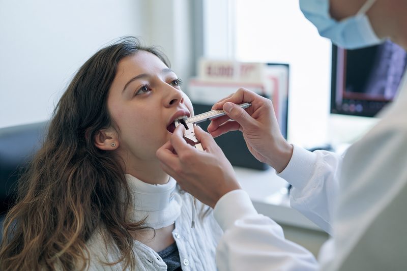
The first 30 days of Ayurvedic treatment for oral cancer are used to assess medicine tolerance, stabilise nutrition, reduce the immediate symptom burden and establish whether the disease is responding, remaining stable or progressing. The treatment may include a personalised Avaleha, Heerak Bhasma, other selected herbo-mineral medicines and local oral care, but the early objective is not limited to making the patient feel better. Clinical changes must be compared with the pretreatment lesion measurements, neck findings, photographs, body weight, swallowing capacity, laboratory values and imaging.
Pain and Burning
Pain may arise from ulceration, inflammation, nerve involvement, secondary infection, exposed tissue, previous surgery or radiation injury. A reduction in pain can allow the patient to speak, sleep and eat more comfortably.
The pain score should be recorded before treatment and reassessed using the same scale. A lower pain score is clinically valuable, but pain reduction does not establish that the tumour has regressed. Analgesic use, ulcer condition, infection and swallowing function should be reviewed at the same time.
In the published recurrent tongue cancer case, the recorded pain score decreased during treatment and later reached zero during long-term follow-up.[3] The result occurred within a broader course that included previous chemotherapy, radiation, repeated surgery and prolonged personalised Ayurvedic care.
Ulceration, Discharge and Odour
An ulcerated oral tumour may produce discharge, foul odour, bleeding and secondary infection. Carefully selected internal medicines and local oral care may help reduce inflammatory swelling, improve cleanliness of the lesion and make eating and oral hygiene easier.
The amount and character of discharge should be documented. Increasing pus, spreading facial swelling, fever, rapidly worsening odour or increasing pain may indicate infection or tumour progression and requires prompt reassessment.
The published buccal cancer case reported reduction in visible ulcerative growth, pain, swelling and pus discharge during a combined Ayurvedic protocol containing Heerak Bhasma, other mineral medicines, herbs, Avaleha and local oral treatment.[4] Repeat imaging and biopsy were not reported, so the documented result represents clinical improvement rather than confirmed complete tumour eradication.
Appetite and Food Intake
Improved appetite is an important early goal because oral cancer frequently reduces food intake through pain, altered taste, restricted tongue movement, trismus and swallowing difficulty. Better appetite must result in adequate consumption rather than remaining only a subjective feeling.
Food consistency should be adapted to the location of the tumour and the patient’s swallowing ability. Soft, moist and energy-dense meals may be easier to tolerate than dry or abrasive foods. Protein intake, hydration and total daily energy remain important during surgery, radiation, chemotherapy and recovery.
The patient’s actual food intake and weekly body weight should be recorded. Continued weight loss despite improved appetite indicates that nutritional needs are not being met or that swallowing, absorption, infection or tumour burden requires further assessment.
Swallowing and Mouth Opening
Reduced pain and inflammation may improve swallowing and mouth opening during the first month. These changes can make oral hygiene, examination, eating and medicine administration easier.
Mouth opening should be measured in millimetres using the same method at each review. Swallowing assessment should record difficulty with solids, semi-solid foods and liquids, as well as coughing, choking, drooling or food remaining in the mouth.
In the published recurrent tongue cancer case, dysphagia and trismus improved from their recorded baseline grades during Ayurvedic treatment.[3] Persistent structural impairment remained because the patient had undergone extensive tongue and mandibular surgery.
A patient who coughs repeatedly while drinking, develops a wet voice after swallowing or experiences recurrent chest infection may be aspirating. Oral medicines should not be forced when swallowing is unsafe.
Sleep, Bowel Function and Energy
Pain, anxiety, reduced food intake, opioids, dehydration and physical inactivity can disturb sleep and bowel function. Constipation can further reduce appetite, increase abdominal discomfort and make the patient less able to tolerate treatment.
The personalised prescription may be adjusted to improve bowel regularity without producing diarrhoea or dehydration. Sleep improvement may follow better pain control, easier breathing, reduced anxiety and correction of nighttime oral discomfort.
Increased energy should be assessed through daily activity rather than reported feeling alone. The patient’s ability to walk, manage personal care, communicate, eat independently and attend treatment provides a more useful measure of functional improvement.
The recurrent tongue cancer case recorded improvement in appetite, constipation and performance status during the reported treatment course.[3] These changes helped document recovery of the patient’s general condition but did not independently establish the absence of cancer.
Body Weight and Strength
Weight stabilisation is a meaningful first-month goal, particularly in patients who have been losing weight before treatment. A stable weight may indicate that pain control, swallowing support and nutritional intake are becoming adequate.
Rapid weight gain caused by fluid retention should not be confused with nutritional recovery. Swelling of the legs, breathlessness, reduced urination or sudden increase in weight requires evaluation of heart, kidney, liver and protein status.
Muscle strength, mobility and performance status should be assessed alongside body weight. A patient may maintain weight while continuing to lose muscle if intake is inadequate or physical activity remains very low.
Brimhana measures may be introduced to rebuild depleted tissues, but the formulation must remain digestible and safe for the patient’s glucose control, swallowing function and tumour-related symptoms.
Laboratory Tolerance
Complete blood count, liver function, kidney function, blood glucose and other relevant tests may require reassessment during the first 30 days, particularly when Heerak Bhasma, Swarna Bhasma or multiple herbo-mineral medicines are prescribed.
More frequent testing may be required when the patient is receiving chemotherapy, immunotherapy or radiation, or when vomiting, dehydration, infection, reduced food intake or rapid weight loss develops.
A fall in haemoglobin, white blood cells or platelets can affect treatment tolerance and bleeding or infection risk. Rising creatinine, bilirubin or liver enzymes requires review of all oncology medicines, Ayurvedic medicines, supplements, hydration and disease-related causes.
The formulation should be modified rather than continued unchanged when laboratory safety deteriorates.
Early Tumour Response
Visible reduction in the ulcer or growth may occur during the first month, but surface change must be interpreted carefully. A lesion can appear cleaner while deep firmness, muscle invasion, jaw involvement or cervical lymph-node disease remains active.
The same clinical measurements and photographic technique used before treatment should be repeated. The lesion should be assessed for dimensions, depth, induration, fixation, bleeding, discharge and relationship to nearby structures.
Cervical lymph nodes must be examined independently. Reduction in the primary lesion with enlargement of a neck node indicates incomplete control and requires imaging or tissue assessment.
Imaging is repeated according to the stage, rate of progression and complete treatment plan rather than automatically after a fixed number of days. A rapidly progressive tumour may require earlier imaging, while post-radiation inflammation may make very early scans difficult to interpret.[1]
Signs That Treatment Is Not Working
Increasing lesion size, deeper firmness, new bleeding, worsening pain, progressive trismus, declining swallowing, continued weight loss or enlarging neck nodes may indicate inadequate disease control.
New numbness of the lip, chin or tongue may reflect nerve involvement. Increasing jaw pain, loose teeth or a non-healing extraction site may indicate bone extension. Breathlessness, persistent cough, focal bone pain or severe unexplained weakness may require evaluation for distant disease.
Feeling more energetic does not justify continuing the same treatment when examination or imaging shows progression. The Ayurvedic formulation, oncology plan and treatment objective must be reassessed immediately.
Thirty-Day Clinical Review
At the end of 30 days, the review should compare the current lesion with the documented baseline. Pain, discharge, bleeding, mouth opening, swallowing, appetite, bowel function, sleep, body weight, performance status and laboratory safety should be recorded using the same methods.
The patient’s adherence should also be assessed. Missed doses, incorrect measurement, treatment gaps, continued tobacco or areca nut use, inadequate nutrition and unreported supplements can affect the result.
The next prescription should be based on the observed response. A responding patient may continue with adjusted tumour-directed and restorative treatment. A patient with severe mucositis or poor digestion may require a gentler formulation. A patient with stable symptoms but progressive tumour measurements requires a change in disease-directed treatment rather than simple continuation.
The first 30 days can demonstrate improved comfort, nutrition, strength and early clinical response. Complete remission requires disappearance of detectable disease on appropriate examination, imaging and pathology where necessary, followed by continued disease-free monitoring.
Measuring Response to Ayurvedic Treatment for Oral Cancer
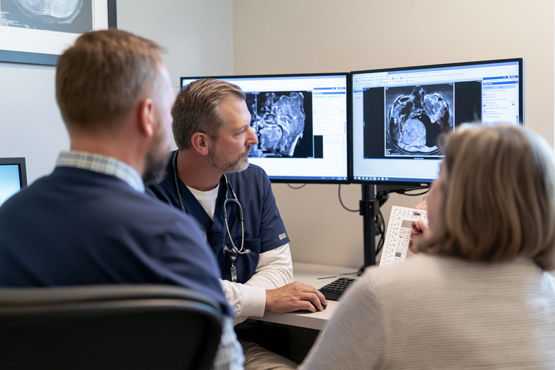
Response to Ayurvedic treatment for oral cancer must be determined through repeatable clinical measurements, imaging and pathology rather than symptom improvement alone. Reduced pain, cleaner ulceration, better appetite and increased strength show that the patient is benefiting, but they do not establish that malignant tissue has disappeared from the primary site, cervical lymph nodes or distant organs.
Baseline Disease Documentation
Assessment begins before the first dose of the personalised formulation. The record should contain the biopsy diagnosis, histological type, tumour site, clinical and pathological stage where available, depth of invasion, cervical lymph-node findings and any evidence of bone, nerve or distant-organ involvement.
The visible lesion should be measured in three dimensions whenever possible. Its surface pattern, ulceration, bleeding, discharge, firmness, fixation and relationship to the tongue, cheek, gum, floor of the mouth, jaw and skin should be documented. Standardised photographs should use similar lighting, distance, mouth position and measurement scale at every review.
The neck should be examined separately because improvement in the oral lesion does not confirm control of regional disease. The size, side, level, firmness and mobility of palpable lymph nodes should be recorded. Baseline CT, MRI, ultrasound or PET-CT findings should be retained for direct comparison with later studies.[1]
Body weight, mouth opening, swallowing ability, speech, food intake, pain score, performance status and laboratory values should also be recorded. These measurements evaluate the patient’s functional recovery and treatment tolerance but must remain distinct from tumour-response assessment.
Clinical Tumour Response
Clinical response is assessed by comparing the oral lesion with its documented baseline. A responding lesion may show a measurable reduction in dimensions, less deep firmness, improved mobility, reduced bleeding and healing of ulcerated margins.
Surface healing without reduction in underlying induration may indicate that inflammation has improved while invasive tissue remains. Palpation is therefore essential, particularly for tongue, floor-of-mouth and buccal tumours that can extend beneath a relatively small visible lesion.
A partial clinical response means that the tumour has clearly decreased but remains detectable. Stable disease means that there is no meaningful reduction or progression. Progressive disease includes enlargement of the primary lesion, increasing depth or fixation, new bone or skin involvement, worsening trismus or development of new disease sites.
The cervical nodes must be assessed at the same time. A smaller mouth lesion accompanied by a new or enlarging neck node represents regional progression rather than complete response.
Symptom Response
Pain reduction, less discharge, improved mouth opening, easier swallowing, better sleep and increased appetite are important patient outcomes. They may improve nutrition, mobility and the ability to complete surgery, radiation or systemic treatment.
These outcomes should be measured consistently. Pain should be recorded with the same scale, mouth opening should be measured in millimetres, body weight should be checked under similar conditions, and swallowing should be evaluated separately for liquids, semi-solid foods and solids.
Symptom improvement can occur because inflammation, infection or tissue irritation has decreased even when viable tumour remains. Analgesic use can also reduce pain without changing the cancer. Symptoms must therefore be interpreted alongside examination and imaging.
Continued weight loss, declining swallowing, new numbness, increasing bleeding or worsening fatigue despite reduced oral pain requires broader reassessment. Such changes may indicate nutritional failure, treatment toxicity, infection or progression outside the visible lesion.
Radiological Response
CT and MRI assess deeper tumour extension, muscle involvement, jaw invasion and cervical lymph nodes that cannot be measured reliably through visual examination. The follow-up scan should be compared directly with the pretreatment study rather than interpreted as an isolated report.[1]
The radiologist should evaluate changes in tumour dimensions, tissue infiltration, bone destruction, lymph-node size and internal characteristics. Reduced swelling or metabolic activity may indicate response, but post-surgical inflammation, radiation changes, infection and fibrosis can resemble residual or recurrent cancer.
PET-CT may help assess metabolic activity and distant disease in selected patients, but increased uptake is not specific to cancer. Inflammation and healing tissues may also appear metabolically active. Suspicious findings may require repeat imaging, targeted examination or biopsy before a final response is assigned.
The timing of imaging depends on the stage, treatment received and speed of clinical change. Rapid progression on examination may require earlier imaging, while scans performed too soon after radiation or surgery may be difficult to interpret because of treatment-related inflammation.
A complete radiological response means that appropriate imaging no longer identifies active disease. It does not always prove that microscopic cancer cells are absent, particularly when residual scar tissue or mucosal abnormality remains.
Pathological Response
Pathology provides the strongest confirmation when suspicious tissue can be sampled safely. A repeat biopsy may be required when an ulcer, firm area, red or white patch, nodular change or unexplained tissue abnormality persists after treatment.
A biopsy showing no viable malignant cells supports pathological response in the sampled tissue. The result must still be interpreted with the quality and location of the sample because a small biopsy may miss residual disease elsewhere within a large or irregular treatment bed.
Pathological complete response means that no viable tumour is found in the examined tissue after treatment. When surgery is performed, the resected primary tumour and lymph nodes provide more extensive information than a small surface biopsy.
Negative pathology from the oral site does not establish that the cervical nodes or distant organs are disease-free. Response must be assessed across every previously involved site.
Complete Response and No Evidence of Disease
Complete clinical response requires disappearance of visible and palpable tumour during examination. Complete radiological response requires no detectable active disease on the appropriate scans. Pathological complete response requires the absence of viable tumour in examined tissue.
No evidence of disease is recorded when clinical examination, imaging and pathology where indicated do not identify active cancer. This status is stronger than symptom improvement but still requires continued surveillance because recurrence can develop after an apparently complete response.[1]
Residual speech difficulty, altered swallowing, numbness, dry mouth or facial changes may remain after successful tumour control because of surgery, radiation or permanent tissue loss. Persistent functional impairment does not necessarily indicate active cancer, but new or worsening symptoms require investigation.
A patient may also feel completely well while harbouring early recurrent disease. Absence of pain or normal appetite cannot replace scheduled oral and neck examination.
Durable Remission and Cure
Remission describes a period during which cancer has reduced substantially or is no longer detectable. Complete remission requires the absence of detectable active disease, while partial remission means that measurable disease remains but has decreased.
Durable remission means that complete disease control continues over prolonged follow-up without local, regional or distant recurrence. The strength of this conclusion increases as the disease-free interval becomes longer and surveillance remains consistently negative.
Cure cannot be confirmed from one negative scan, temporary ulcer healing or a short period without symptoms. It requires sustained absence of disease through repeated oral and neck examination, appropriate imaging and pathology when suspicious tissue appears.
The original stage remains relevant after remission. Patients with deep invasion, involved lymph nodes, extranodal extension, positive or close margins and recurrent disease require particularly careful surveillance because their risk pattern differs from that of a small localised tumour.[1]
Follow-Up After Complete Response
Follow-up should examine the treated oral site, the rest of the oral mucosa, both sides of the neck and any reconstructed or irradiated tissue. New ulcers, patches, growths, firmness, bleeding, numbness, swallowing changes or neck swelling require prompt assessment.
Standardised photographs and measurements allow small changes to be identified earlier than memory alone. Dental health, mouth opening, nutrition, body weight, thyroid function after neck irradiation and treatment-related late effects should also be reviewed.
Patients who previously used tobacco, areca nut, betel quid or alcohol must remain abstinent. Continued exposure increases the risk of recurrence and a second primary cancer within the mouth, throat, oesophagus or lungs.
The personalised Avaleha, Heerak Bhasma and other medicines should be modified according to objective findings. A responding patient may continue a disease-directed protocol before transitioning to longer-term Rasayana care. Stable but persistent disease may require reformulation or additional oncology treatment. Documented progression requires immediate revision of the complete treatment strategy.
Ayurvedic treatment for oral cancer has achieved its curative objective only when the patient has no detectable active disease and this outcome remains stable through continued clinical, radiological and pathological surveillance.
Local Care in Ayurvedic Treatment for Oral Cancer
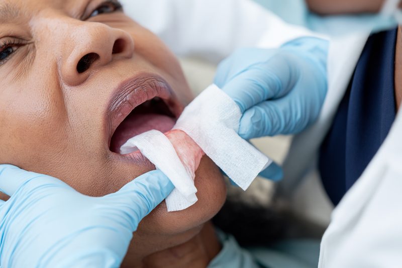
Local care in Ayurvedic treatment for oral cancer is selected according to the condition of the tumour surface, surrounding mucosa, previous surgery, radiation exposure, bleeding tendency, infection risk and swallowing ability. The purpose is to reduce pain, protect damaged tissues, improve oral hygiene, control secondary infection and help the patient eat more comfortably while the primary tumour and cervical lymph nodes continue to receive disease-directed treatment.
Painful and Ulcerated Lesions
An ulcerated oral tumour may expose sensitive tissue and produce constant pain, burning or discomfort during chewing and swallowing. The lesion should be examined for depth, necrotic tissue, bleeding, discharge, surrounding inflammation and contact with teeth or dentures before any local preparation is selected.
Yashtimadhu-based rinses or carefully prepared mucosal applications may be considered when dryness, burning and painful irritation predominate. A mild infusion should be filtered thoroughly so that coarse particles do not remain against the ulcer. The temperature should be lukewarm rather than hot because heat may increase pain and bleeding.
Jatyadi Ghrita or another medicated ghrita may be considered when the surrounding tissue is dry, inflamed or slow to recover. It should be applied only in a thin, measured layer and should not be packed deeply into a malignant cavity. A thick oily coating may interfere with examination, trap debris or increase aspiration risk in a patient with impaired swallowing.
Local relief should be assessed by changes in pain during eating, sleep disturbance, analgesic requirement and ability to maintain oral hygiene. Persistent deep pain, radiating ear pain, new numbness or increasing firmness requires reassessment for nerve, muscle or bone involvement rather than repeated topical treatment.
Bleeding From the Oral Lesion
Minor contact bleeding may occur when an ulcerated tumour is touched by food, a toothbrush, denture or examination instrument. The frequency, duration and estimated volume of bleeding should be documented because increasing bleeding can indicate tumour progression, infection, fragile tissue or vessel involvement.
Gentle pressure with clean gauze may control minor superficial bleeding. Vigorous rubbing, scraping or repeated manipulation of the lesion should be avoided. Strong powders, concentrated alkalis, acidic preparations and irritating herbal pastes can worsen tissue injury and provoke further bleeding.
The patient’s platelet count, coagulation status, liver function and use of anticoagulant or antiplatelet medicines should be reviewed when bleeding becomes frequent. Haridra, Guggulu, Pippali, Maricha and other ingredients within the complete prescription may also require review when the patient is taking medicines that affect coagulation.
Continuous bleeding, repeated passage of large clots, dizziness, weakness, breathlessness or rapidly filling blood in the mouth requires urgent hospital assessment. Oral applications and internal Ayurvedic medicines should not delay vascular control, transfusion, airway protection or other emergency treatment.
Discharge, Odour and Secondary Infection
Ulcerated tumours can accumulate food debris, necrotic tissue and saliva, producing foul odour and discharge. Secondary bacterial or fungal infection may increase pain, swelling, fever and difficulty eating.
Panchavalkala Kwatha may be considered as a gently filtered oral rinse when the tissue condition, swallowing safety and treating physician’s assessment permit its use. The rinse should not be swallowed deliberately, and it should not be used at a temperature or concentration that causes burning or dryness.
Nimba-based or Triphala-based oral rinses may be selected in some patients, but concentration and frequency require individual adjustment. Strong decoctions can irritate exposed tissue, while inadequately filtered preparations can leave particles within the ulcer.
Fever, increasing facial swelling, spreading redness, worsening pus discharge, severe tenderness or difficulty opening the mouth may indicate a significant infection. Such findings require clinical examination and may require antimicrobial treatment, drainage or hospital care. Improvement in odour after cleansing does not establish control of the underlying cancer.
Oral candidiasis can develop during radiation, chemotherapy, antibiotic treatment, diabetes or severe dry mouth. It may cause white removable plaques, burning, altered taste and painful swallowing. Antifungal treatment may be required, and a sweet Avaleha base may need modification when candidiasis repeatedly returns.
Daily Oral Hygiene
Oral hygiene reduces debris, infection risk, dental decay and discomfort. The teeth, tongue and unaffected mucosa should be cleaned gently with a soft toothbrush or another method suited to the patient’s mouth opening and pain level.
The tumour should not be scrubbed directly. Aggressive brushing can produce bleeding, while complete avoidance of oral cleaning allows debris and microbial growth to increase. The cleaning method should therefore preserve hygiene without repeatedly injuring the lesion.
A mild, non-alcoholic mouth rinse may be more comfortable than strongly flavoured commercial products in patients with ulceration, radiation mucositis or dry mouth. Products that cause burning should be stopped rather than tolerated in the belief that stronger irritation provides better cleansing.
Dentures should be removed and cleaned regularly. A denture that rubs against the tumour, postoperative wound or irradiated mucosa should not be worn until it has been reviewed and adjusted. Repeated trauma can increase pain and obscure changes in the underlying lesion.
The patient should use a clean, dry measuring spoon for the Avaleha and should rinse the mouth gently after the dose when the formulation contains jaggery, honey or another sweet base. This reduces prolonged contact with the teeth and damaged mucosa.
Dry Mouth and Thick Saliva
Dry mouth may develop from dehydration, medication, salivary-gland involvement or radiation. It can make speaking, chewing and swallowing difficult and can increase dental decay, oral infection and sleep disturbance.
Frequent small sips of water may be useful when swallowing is safe. The patient may also benefit from moist foods, sauces and texture modification rather than dry or crumbly meals. Caffeinated drinks, alcohol and tobacco can worsen dryness and should be avoided.
A small amount of physician-selected medicated ghrita may provide temporary lubrication in suitable patients. It should not be used when the patient has uncontrolled nausea, poor fat tolerance, repeated aspiration or inability to clear oral secretions.
Thick or rope-like saliva may be more difficult to manage than complete dryness. Gentle hydration, humidification and regular oral cleansing can reduce discomfort. New difficulty handling saliva, repeated choking or drooling requires swallowing and airway assessment.
Radiation Mucositis
Radiation mucositis can produce diffuse redness, ulceration, severe pain, altered taste and difficulty swallowing. Its appearance and treatment needs differ from those of an untreated malignant ulcer.
Local preparations should be simplified during severe mucositis. Pungent, concentrated, salty, acidic, very hot or abrasive substances can increase pain and reduce food intake. Even an herbal preparation that was previously tolerated may become unsuitable when the mucosa is acutely inflamed.
Yashtimadhu-based rinses, bland mucosal protection and carefully selected ghrita preparations may be considered according to the condition of the tissue and the oncology treatment plan. Each preparation should be introduced separately so that irritation or intolerance can be identified.
Pain control must be adequate enough to permit hydration and nutrition. Severe mucositis may require prescription analgesia, topical anaesthetic treatment, intravenous fluids or feeding support. Ayurvedic local care may accompany these measures but should not replace them when oral intake is failing.
Mucositis generally reflects treatment-related injury rather than tumour growth, but a persistent focal ulcer, increasing induration or new mass after the expected healing period requires reassessment for residual or recurrent disease.
Mouth Opening and Trismus
Trismus can result from oral submucous fibrosis, tumour invasion, surgery, radiation fibrosis, pain or inflammation. It can interfere with eating, dental care, speech, examination and delivery of treatment.
Mouth opening should be measured in millimetres at baseline and during follow-up. A subjective impression that the mouth opens more easily is less reliable than repeated measurement using the same method.
Gentle jaw exercises may be introduced when the tumour, surgical wound and bone condition permit. Excessive force can cause tissue injury, bleeding, joint pain or fracture in a weakened jaw. Exercise intensity should be determined by the treating team rather than increased independently by the patient.
A sudden reduction in mouth opening, new severe pain or increasing cheek firmness requires reassessment for infection, fibrosis or deeper tumour extension. Local oils or massage should not be applied forcefully over a tumour, enlarged lymph node or recently operated area.
Dental and Jaw Care
Dental infection, loose teeth, sharp edges and unstable restorations can worsen pain and oral injury. Dental examination is required before radiation and whenever a tumour lies close to the gum, tooth socket, mandible or maxilla.
A sharp tooth or denture edge should be corrected, but persistent ulceration after removal of the irritant still requires clinical review. Cancer should not be excluded simply because a local traumatic factor was found.
A non-healing extraction socket, unexplained tooth mobility, lower-lip numbness or persistent jaw pain may indicate bone involvement. Panoramic radiography, CT, MRI or biopsy may be required according to the findings.
After high-dose radiation to the jaw, dental extraction and invasive procedures require specialist planning because healing may be impaired. The patient should not undergo unplanned extraction solely to relieve pain without review of the radiation field and bone condition.
Ayurvedic tooth powders containing coarse, salty, alkaline or pungent substances should not be rubbed over irradiated mucosa, an ulcerated tumour or exposed bone. Gentle cleaning is preferable to aggressive stimulation.
Local Care After Surgery
Postoperative local treatment depends on wound closure, flap reconstruction, sutures, drains, infection risk and the surgeon’s instructions. The mouth may contain healing tissue that should not be exposed to unapproved oils, powders or decoctions.
During the early postoperative period, prescribed oral rinses and wound-care instructions should be followed. Ayurvedic local preparations may be introduced only after the wound has been examined and the risk of aspiration, infection and interference with healing has been considered.
A reconstructed tongue, cheek, palate or jaw may have altered sensation and reduced movement. The patient may not feel trauma, excessive heat or retained food normally. Regular visual inspection and professional review are therefore important.
Increasing swelling, wound separation, foul discharge, fever, exposed plate or bone, sudden bleeding or a new fistula requires surgical assessment. These findings should not be managed by adding stronger local herbal applications.
When Local Applications Should Be Avoided
Local applications should be withheld when the diagnosis is uncertain, active major bleeding is present, airway compromise is developing, swallowing is unsafe or the patient has a fresh unreviewed surgical wound.
They should also be avoided when the preparation produces burning, increased bleeding, swelling, rash, nausea, coughing or difficulty handling saliva. A product that is considered natural can still irritate damaged mucosa or cause an allergic reaction.
Caustic alkalis, corrosive substances, concentrated essential oils and unverified powders should never be used to burn or dissolve an oral tumour. Such injury can increase pain, infection, bleeding and tissue destruction while delaying effective treatment.
The local treatment plan should be reviewed whenever the tumour changes, radiation begins, surgery is performed or swallowing deteriorates. The preparation suitable for one phase of treatment may become unsafe during another.
Local oral care is most effective when it improves comfort, hygiene, food intake and tissue protection without obscuring the lesion or delaying tumour-directed treatment. Clinical examination, imaging and pathology remain necessary to determine whether the cancer itself is responding.
Diet During Ayurvedic Treatment for Oral Cancer
Diet during Ayurvedic treatment for oral cancer must preserve body weight, muscle strength, hydration and swallowing safety while reducing pain and irritation within the mouth. Food should be selected according to the tumour site, surgical changes, radiation effects, mouth opening, digestive capacity, diabetes status and ability to swallow. Severe restriction is unsafe when it causes further weight loss or prevents the patient from completing curative treatment.
Maintaining Weight and Muscle
Oral cancer can reduce food intake through pain, ulceration, altered taste, reduced tongue movement, trismus, dry mouth and fear of bleeding. Surgery, radiation and systemic treatment can further increase nutritional requirements while making eating more difficult. Unintentional weight loss may therefore occur rapidly even when the patient continues to eat small meals.
Body weight should be recorded before treatment and monitored at least weekly during periods of active weight loss or intensive therapy. The trend is more useful than a single measurement. A patient who loses more weight each week requires immediate review of food intake, swallowing, infection, pain control and treatment toxicity.
Maintaining muscle is as important as maintaining total body weight. A patient may lose muscle while appearing to maintain weight because of fluid retention or reduced physical activity. Strength, walking ability, independence in daily activities and performance status should therefore be reviewed alongside the weighing scale.
Adequate energy intake helps preserve the patient’s ability to undergo surgery, heal wounds, complete radiation and tolerate systemic treatment. The aim is not simply to increase calories through sugar. Meals should provide a balance of protein, digestible carbohydrates, suitable fats, fluids and micronutrients.
Protein for Healing and Recovery
Protein is required for wound healing, immune function and preservation of muscle. The amount should be individualised according to body weight, kidney function, liver health, current treatment and nutritional status.
Soft protein sources may include well-cooked lentils, smooth dal, yoghurt where tolerated, paneer, tofu, eggs, fish, poultry or other culturally suitable foods. The choice depends on the patient’s dietary pattern and ability to chew and swallow. Foods should be blended, mashed or moistened when ordinary texture causes pain.
A patient following a vegetarian or vegan diet may need several smaller protein servings through the day because large portions can be difficult to consume. Smooth lentil soups, tofu preparations, nut or seed pastes and medically appropriate nutrition supplements may be used when chewing is limited.
Very dry protein foods can become difficult to swallow in a patient with reduced saliva. Meat, bread, paneer, tofu or legumes may need broth, sauce, ghee or another suitable moistening medium. Food should not require forceful chewing when the tumour, jaw or postoperative tissues are painful.
Protein powders should be selected carefully. Products containing excessive sugar, strong flavouring, acidic additives or unverified herbs may irritate the mouth or interact with treatment. Kidney impairment requires medical review before high-protein supplementation is introduced.
Food Texture and Mouth Comfort
Food texture should match the patient’s mouth opening, tongue movement, dentition and swallowing safety. Soft, moist foods are often better tolerated than dry, rough, crisp or sticky foods.
A patient with a painful tongue or cheek lesion may manage smooth soups, porridge, soft rice, mashed vegetables, yoghurt, soft eggs or blended meals more comfortably. A patient with reduced tongue movement may require a texture that forms a cohesive bolus and does not scatter throughout the mouth.
Dry biscuits, toast, hard crusts, chips, nuts and coarse raw vegetables can scrape an ulcerated tumour or radiation-injured mucosa. Their nutritional value can often be provided through softer alternatives until the tissue has recovered.
Very sticky foods can remain around the tumour, teeth or reconstructed tissues. Thick nut butter, dense sweets and an excessively sticky Avaleha may be difficult to clear when saliva is reduced or tongue movement is impaired.
Food temperature also matters. Very hot foods can increase pain and bleeding, while very cold foods may be uncomfortable for patients with dental sensitivity. Lukewarm or cool meals are often better tolerated during active mucositis.
Swallowing Safety
Difficulty swallowing can result from the tumour, surgery, radiation fibrosis, reduced tongue control, pain or nerve dysfunction. Coughing during meals, choking, a wet voice after swallowing, food remaining in the mouth or repeated chest infections may indicate aspiration.
Swallowing should be assessed separately for liquids, semi-solid foods and solids. A patient may tolerate one texture safely while aspirating another. Thin liquids are not always easier to swallow because they move rapidly and may enter the airway before the patient can control them.
A speech and swallowing specialist may recommend changes in posture, pacing, spoon size, liquid thickness or food texture. These instructions should be followed consistently because unsafe swallowing can lead to dehydration, malnutrition and aspiration pneumonia.
The patient should eat while sitting upright and remain upright after the meal. Small mouthfuls, slow pacing and adequate time between swallows may reduce choking. Food should not be forced when the patient is fatigued, breathless or unable to handle saliva.
An Avaleha should be given only when the patient can swallow a semi-solid preparation safely. Diluting it at home does not guarantee safety and may alter dose accuracy. Repeated coughing after the medicine requires immediate reassessment of the dosage form.
Diet During Oral Mucositis
Radiation and chemoradiation can cause painful inflammation and ulceration throughout the mouth. During severe mucositis, foods that were previously tolerated may produce intense burning.
Soft, bland and moist foods are usually more comfortable. Strong chilli, pepper, concentrated ginger, very salty food, vinegar, citrus juice and acidic sauces may aggravate exposed mucosa. The patient’s response should guide temporary modification rather than applying the same restriction after the mucosa has healed.
Dry or coarse spices should not be rubbed over an oral lesion. Herbal ingredients included within a prescribed Avaleha are processed and dosed differently from raw powders applied directly to damaged tissue.
Pain control should be adequate enough to allow food and fluid intake. A patient who cannot eat because of pain may require prescription analgesia, topical anaesthetic treatment, intravenous hydration or feeding support. Continuing to lose weight while attempting to tolerate pain is not an acceptable nutritional plan.
Oral candidiasis may cause burning, altered taste and removable white plaques. It requires clinical assessment and may need antifungal treatment. A highly sweet diet or frequent sticky sweets may worsen oral hygiene and should be reduced when candidiasis or uncontrolled diabetes is present.
Dry Mouth and Altered Taste
Reduced saliva makes chewing and swallowing difficult and increases the risk of dental decay. Frequent small sips of water may help when swallowing is safe. Meals can be softened with broth, gravy, yoghurt, ghee or another tolerated moistening medium.
Dry foods should be modified rather than eliminated permanently. Bread can be softened in soup, rice can be served with dal or sauce, and protein foods can be minced or blended with a moist base.
Altered taste may make food seem metallic, bitter or flavourless. Using mild herbs, changing serving temperature and rotating protein sources may improve intake. Strong spices should not be added when they irritate the mucosa.
Metallic taste can sometimes be reduced by using non-metal utensils, but taste changes may also arise from medication, infection, dry mouth or nutritional deficiency. Persistent taste disturbance should be evaluated rather than managed only by adding stronger flavours.
Saliva substitutes or other prescribed measures may be required after radiation. Sweet lozenges and frequent sugary drinks should not become the main strategy because they increase dental and glucose-related risks.
Hydration
Dehydration can worsen weakness, constipation, kidney stress, dizziness and treatment toxicity. Patients with painful swallowing may gradually reduce fluids without recognising the extent of dehydration.
Fluid intake should be spread throughout the day rather than attempted in large amounts at once. Water, broths, suitable oral rehydration fluids and other non-irritating beverages may be used according to swallowing safety and medical condition.
Alcohol, tobacco and areca nut should be avoided completely. Alcohol can irritate damaged mucosa, worsen dehydration and increase oral cancer risk. Smoking and smokeless tobacco continue to expose the mouth to carcinogens and can impair recovery.[11][12]
Excessive caffeine may worsen dryness or sleep disturbance in susceptible patients. Carbonated or acidic drinks may produce burning when mucositis or ulceration is present.
Reduced urine output, dark urine, dizziness, confusion, dry tongue or inability to retain fluids requires prompt assessment. Intravenous hydration may be necessary during severe mucositis, vomiting, diarrhoea or kidney dysfunction.
Diabetes and the Avaleha Base
A traditional Avaleha often contains jaggery, sugar or honey to create the required consistency and preservation. This may be unsuitable for uncontrolled diabetes, recurrent oral candidiasis or significant dental disease.
Blood glucose should be checked before and during treatment in patients with diabetes or steroid-related hyperglycaemia. The sweet base, total daily dose and meal plan may require modification.
Reducing jaggery or honey without redesigning the formulation can alter its consistency, stability, concentration and dose uniformity. A low-sugar version should therefore be prepared as a separate pharmaceutical formula rather than improvised by the patient.
The Avaleha should be measured accurately and taken according to the prescribed schedule. It should not be consumed repeatedly throughout the day like a food supplement because this increases sugar exposure and disrupts the intended dosing.
The mouth should be rinsed gently after the dose when swallowing is safe. Teeth and dentures should be cleaned regularly to reduce prolonged contact with the sweet base.
Pathya and Apathya
Pathya refers to foods and daily practices suited to the patient’s disease, digestion, treatment and recovery. Apathya refers to foods or habits likely to aggravate symptoms, interfere with treatment or reduce the patient’s strength.
Pathya in oral cancer commonly includes soft, moist, freshly prepared and nutritionally dense meals that the patient can swallow without trauma. The exact diet changes according to mucositis, surgery, diabetes, constipation, diarrhoea, kidney function and treatment phase.
Apathya includes tobacco, areca nut, betel quid, alcohol and any food that repeatedly causes bleeding, severe burning, choking or worsening pain. Extremely hot foods, harsh spices and rough textures are generally avoided when the mucosa is actively ulcerated.
Pathya should not become an excessively restrictive cancer diet. Eliminating major food groups without a medical reason can worsen protein deficiency, weight loss and treatment tolerance. The patient’s ability to maintain nutrition remains more important than following a rigid list.
Foods considered wholesome in general Ayurveda may still be inappropriate for a specific patient. Ghee may be useful for dryness but unsuitable during severe nausea or fat intolerance. Milk may provide nutrition for one patient and worsen discomfort in another. The prescription should follow the individual response.
Feeding Tube Support
A feeding tube may be required when the patient cannot swallow safely or cannot consume enough food to maintain weight. It may be placed temporarily during intensive treatment or used for longer periods when major oral surgery has altered swallowing.
Tube feeding does not mean that the curative plan has failed. It can preserve nutrition, hydration and medicine tolerance during surgery, radiation or chemoradiation.
The feeding formula should provide adequate protein, energy, fluids and micronutrients. Diabetes, kidney disease, diarrhoea, constipation and fluid restriction may require a specialised formula.
Ayurvedic medicines should not be placed through a feeding tube without confirming compatibility, particle size, dose accuracy and risk of blockage. Coarse herbal powders, thick Avaleha and oily preparations may obstruct the tube or deliver an unreliable dose.
A patient who receives most nutrition through a tube may still require oral hygiene, swallowing rehabilitation and regular assessment of whether oral intake can be resumed safely.
Supplements and Herbal Foods
Vitamin and mineral supplements should be used when a deficiency is documented or a clinician identifies a clear nutritional need. Very high doses should not be added independently during radiation, chemotherapy or immunotherapy.
Commercial cancer supplements may contain multiple herbs, antioxidants, concentrated extracts or minerals not listed clearly on the front label. Taking these alongside a personalised herbo-mineral prescription can increase interaction risk and make adverse effects difficult to identify.
Protein supplements, meal replacements and electrolyte products should be reviewed for sugar content, acidity, potassium, sodium and swallowing suitability. A product suitable for one patient may be inappropriate for another with diabetes, kidney disease or severe mucositis.
The personalised Avaleha, Heerak Bhasma and other prescribed medicines should remain the only Ayurvedic cancer formulation unless the treating physician has reviewed and approved additional products.
Monitoring Nutritional Response
Nutritional progress should be assessed through body weight, food intake, muscle strength, hydration, bowel function and functional capacity. Serum albumin and other laboratory measurements may provide additional information but should not be interpreted in isolation because inflammation and disease burden can influence them.
A stable or increasing weight accompanied by better strength and food intake suggests nutritional recovery. Weight gain caused by swelling, reduced urination or fluid accumulation requires medical assessment.
Continued weight loss despite a carefully planned diet may indicate uncontrolled pain, unsafe swallowing, infection, treatment toxicity, malabsorption or progressive cancer. The treatment plan should be reassessed rather than relying only on larger portions or more supplements.
Diet supports curative treatment by preserving the patient’s ability to heal, tolerate therapy and maintain oral function. It does not replace tumour-directed treatment or confirm cancer regression. Disease response remains dependent on examination, imaging and pathology where indicated.[1]
Safety of Ayurvedic Treatment for Oral Cancer

Ayurvedic treatment for oral cancer containing Heerak Bhasma, Swarna Bhasma, Abhraka Bhasma, Mukta preparations, Gandhaka Rasayana or other potent herbo-mineral medicines requires verified ingredients, precise dosing, patient-specific selection and regular medical monitoring. Safety cannot be judged from the traditional name of a formulation, its price, colour or the claim that it is natural. The complete prescription must be evaluated in relation to the patient’s age, cancer stage, nutritional condition, liver and kidney function, swallowing ability, previous treatment and current medicines.
Properly Prepared Bhasma and Raw Mineral Are Not Equivalent
A bhasma is produced through defined Ayurvedic pharmaceutical procedures that include identification of the raw material, Shodhana, controlled processing, Marana and assessment of the finished preparation. Raw diamond, gold, mica, sulphur or another mineral should never be powdered and consumed as a substitute for the corresponding bhasma.
The safety and pharmaceutical properties of Heerak Bhasma depend on the authenticity of the starting material, the processing media, the number and control of heating cycles, particle transformation, removal of contamination and uniformity of the final product. The same principle applies to Swarna Bhasma, Abhraka Bhasma and other mineral medicines.
A label stating “Heerak Bhasma” does not prove that these requirements have been met. The batch should have traceable manufacturing records, authenticated ingredients, documented processing and appropriate analytical quality testing. Official pharmacopoeial standards are used to establish identity, purity and strength for Ayurvedic ingredients and formulations.[10]
Heerak Bhasma used in research cannot automatically be considered equivalent to every commercial product carrying the same name. The biological findings reported for a characterised preparation apply to the tested batch and do not validate untested formulations from unrelated manufacturers.[5]
Batch Testing and Pharmaceutical Quality
Each batch used in an oral cancer protocol should be identifiable from raw-material receipt to final dispensing. The manufacturing record should document the botanical or mineral identity, quantities used, processing stages, preparation date, final yield and number of patient doses.
Herbal ingredients should be examined for correct species and plant part. Kanchanara bark, Nimba leaf, Guduchi stem, Yashtimadhu root and Amalaki fruit have different pharmaceutical characteristics and should not be replaced casually with another plant part or a visually similar herb.
The herbs and finished Avaleha should be assessed for microbial contamination, excessive moisture, pesticides, aflatoxins and adulteration. A semi-solid preparation containing jaggery, honey, herbal extract and ghee can spoil when its moisture content, preservation, handling or storage is inadequate.
Mineral ingredients require assessment of identity, elemental composition, contamination and batch consistency. Testing should be conducted on the actual batch supplied to the patient rather than relying only on a certificate from a different production lot.
Dose uniformity is particularly important when Heerak Bhasma or Swarna Bhasma is used in a small quantity within a large Avaleha batch. Inadequate mixing can produce substantial variation between doses. Geometric dilution, calibrated weighing and validated blending are required before the mineral premix is incorporated into the finished formulation.
Patient Selection Before Treatment
Not every patient with oral cancer is suitable for the same herbo-mineral prescription. A patient with a small localised lip tumour and normal organ function differs from a patient with advanced tongue cancer, cervical lymph-node disease, severe weight loss and renal impairment.
Age should be considered together with physiological reserve rather than used alone. Older patients may have reduced kidney filtration, lower muscle mass, frailty, swallowing impairment or several prescription medicines. Younger patients may also require dose reduction when malnutrition, organ dysfunction or intensive oncology treatment is present.
The prescription must be reconsidered in patients with uncontrolled diabetes, chronic kidney disease, liver disease, heart failure, severe hypertension, active gastrointestinal disease, neurological illness, immune suppression or significant anaemia. These conditions may alter the suitability of the Avaleha base, accompanying herbs, bhasma dose and monitoring frequency.
Active major oral bleeding, severe dehydration, uncontrolled infection, inability to swallow liquids, repeated aspiration or airway compromise requires immediate medical stabilisation. Potent internal medicines should not be started merely because the patient has reached the clinic while an urgent complication remains untreated.
Pregnancy and breastfeeding require a separate assessment. A complex herbo-mineral oncology formulation should not be prescribed routinely without specialist evaluation of maternal disease, potential fetal or infant exposure and available treatment alternatives.
Baseline Laboratory Assessment
A complete blood count should be obtained before prolonged herbo-mineral treatment. Haemoglobin provides information about anaemia and oxygen-carrying capacity. White blood cells help assess infection and bone-marrow reserve, while platelets influence the risk of bleeding from an ulcerated tumour or an invasive procedure.
Liver assessment should include bilirubin, transaminases, alkaline phosphatase, albumin and other measurements selected according to the patient’s medical history. Abnormal liver function may result from existing disease, malnutrition, infection, metastatic cancer, chemotherapy, other medicines or previous supplement use.
Kidney assessment should include serum creatinine, estimated glomerular filtration rate, urea and electrolytes. Reduced kidney function can alter medicine tolerance, hydration requirements, contrast-imaging decisions and the safety of concurrent chemotherapy.
Blood glucose and glycated haemoglobin are particularly important when the formulation contains jaggery or honey. A patient with uncontrolled diabetes may require a redesigned low-sugar dosage form rather than a simple reduction of the Avaleha base at home.
Coagulation studies may be required when the patient has recurrent bleeding, liver disease, low platelets or uses anticoagulant treatment. Urine examination may provide additional information about hydration, infection, glucose, protein and kidney health.
Testing for blood lead, mercury, arsenic or other elements may be appropriate when the patient has previously consumed unverified metallic preparations, used several market products simultaneously or has symptoms compatible with toxic exposure. These tests do not replace testing of the formulation itself.
Monitoring During the First Month
The first month should assess both treatment response and safety. Blood counts, liver function, kidney function, electrolytes and blood glucose should be repeated according to the potency of the formulation, baseline abnormalities and concurrent oncology treatment.
Monitoring may need to be more frequent during chemotherapy, immunotherapy, radiation, infection, vomiting, dehydration or rapid weight loss. A patient with stable organ function after surgery may require a different schedule from a patient receiving cisplatin or another medicine capable of affecting the kidneys or bone marrow.
The treating physician should record appetite, bowel function, urine output, body weight, swelling, skin changes, neurological symptoms, sleep and ability to perform normal activities. These findings may identify intolerance before severe laboratory abnormalities develop.
A normal test at the beginning of treatment does not guarantee continued safety. Organ function can change because of dehydration, infection, cancer progression, oncology treatment or the combined effect of several medicines.
The next batch should not be prepared automatically without reviewing the first-month clinical and laboratory findings. The quantities of Heerak Bhasma, Swarna Bhasma, Abhraka Bhasma, Gandhaka Rasayana, Guggulu, Pippali and the Avaleha base may require modification.
Safety During Surgery and Invasive Procedures
The complete Ayurvedic prescription should be reviewed before biopsy, tumour resection, neck dissection, dental extraction or another invasive procedure. Some herbs may affect blood glucose, blood pressure, platelet activity, gastrointestinal function or the metabolism of prescription medicines.
The surgical and anaesthesia teams should know every medicine and supplement the patient is taking. The decision to continue or temporarily withhold a formulation depends on its ingredients, the type of operation, bleeding risk, fasting period and postoperative swallowing ability.
An Avaleha should not be administered when the patient is fasting for anaesthesia or cannot swallow safely after surgery. A fresh oral wound, flap reconstruction or exposed bone should not receive unapproved local oils, powders or herbal pastes.
After surgery, treatment should resume only when the patient is medically stable and the dosage form is compatible with swallowing and wound care. The prescription must be revised according to the final pathology, surgical margins, lymph-node findings and need for radiation or chemoradiation.
Safety During Radiation
Radiation can produce severe mucositis, dry mouth, altered taste, reduced intake, dehydration and tissue sensitivity. A formulation tolerated before radiation may become too pungent, sweet, sticky or difficult to swallow during treatment.
Pippali, Maricha, Shunthi and other heating herbs may require reduction when the mucosa becomes intensely inflamed. The local use of concentrated decoctions, powders or essential oils can increase burning and reduce food intake.
Dehydration during radiation may affect kidney function and increase the risk associated with concurrent medicines. Laboratory monitoring and fluid intake should therefore be reviewed when oral intake declines.
A sweet Avaleha may remain around the teeth and damaged mucosa when saliva is reduced. Gentle oral cleansing after each dose is important when swallowing is safe, and the formulation may require temporary modification during severe candidiasis or uncontrolled blood glucose.
Safety During Chemotherapy and Immunotherapy
Chemotherapy can affect blood counts, kidneys, liver, nerves, gastrointestinal function and oral tissues. A patient receiving chemotherapy should not continue a potent herbo-mineral prescription unchanged when neutrophils, platelets, creatinine or liver enzymes become abnormal.
Nausea and vomiting may prevent reliable absorption of the Avaleha and increase dehydration. Repeated dosing after vomiting can also result in uncertain exposure. The treating physician should decide whether the dose should be repeated, reduced or temporarily withheld.
Immunotherapy may cause inflammatory injury to the thyroid, lungs, liver, bowel, kidneys, skin, nervous system or other organs. New diarrhoea, breathlessness, jaundice, rash, unusual weakness or confusion requires immediate assessment rather than automatic treatment with additional herbs.
Preclinical research has reported immune-related effects from a characterised Heerak Bhasma preparation, but this does not establish how it interacts with immune checkpoint medicines in humans.[5] Concurrent use requires individual review and close monitoring.
Swarna Bhasma has limited human research in mixed cancer populations, while Abhraka Bhasma has mainly preparation-dependent laboratory evidence rather than established oral cancer safety and efficacy data.[7][37] These findings do not justify using higher doses or reducing laboratory surveillance.
Medicine and Herb Interactions
The complete prescription must be compared with anticoagulants, antiplatelet medicines, antihypertensives, diabetes medicines, thyroid treatment, steroids, seizure medicines, antibiotics, chemotherapy, immunotherapy and pain medicines.
Haridra, Pippali, Maricha, Guggulu, Ashwagandha and Yashtimadhu may require particular review when the patient uses medicines affecting coagulation, glucose, blood pressure, thyroid function or drug metabolism. The clinical importance depends on the dose, extract, treatment duration and individual susceptibility.
Yashtimadhu may be unsuitable in prolonged high exposure for patients with uncontrolled hypertension, fluid retention, low potassium or certain heart and kidney conditions. Its inclusion in an oral cancer Avaleha should therefore be reviewed rather than assumed to be harmless.
Guggulu may require caution in thyroid disorders, gastrointestinal sensitivity and patients taking several prescription medicines. Pippali and Maricha can affect the absorption or exposure of some medicines and should not be used solely to increase potency without reviewing the complete regimen.
The patient should not add separate turmeric capsules, concentrated herbal extracts, metallic products or commercial immunity supplements while taking a personalised formulation. Multiple overlapping products can increase exposure and make it difficult to identify the cause of an adverse effect.
Symptoms Requiring Immediate Review
Yellowing of the eyes or skin, dark urine, persistent vomiting, severe abdominal pain or marked loss of appetite may indicate liver or gastrointestinal injury and requires prompt investigation.
Reduced urine output, new swelling, severe weakness, confusion or sudden weight gain may indicate dehydration, kidney dysfunction, fluid retention or another medical complication.
New tremor, numbness, burning sensations, weakness, confusion or altered behaviour requires neurological assessment. These symptoms may arise from previous chemotherapy, metabolic disturbance, cancer progression, nutritional deficiency, unverified mineral exposure or another illness.
Persistent diarrhoea can cause dehydration and electrolyte loss, while prolonged constipation may worsen nausea, appetite and discomfort. The prescription should be modified according to the cause rather than continued unchanged.
A new rash, facial swelling, wheezing or difficulty breathing may indicate an allergic reaction and requires urgent assessment. Heavy oral bleeding, airway difficulty, inability to swallow liquids or rapidly enlarging neck swelling requires emergency hospital care.
The formulation should be withheld when a serious adverse effect is suspected until the patient has been examined and the cause has been investigated. Restarting the same dose without reassessment can expose the patient to repeated harm.
Accurate Dosing and Storage
The prescribed 15 g dose should be measured with a calibrated spoon or supplied in individual unit packs. Household spoons vary substantially and should not be used for a formulation containing microdosed bhasmas.
The container should remain tightly closed and should be stored according to the preparation instructions, away from excessive heat, sunlight and moisture. A clean, dry spoon should be used for every dose.
Water should not be added directly to the storage jar because increased moisture can alter stability and encourage microbial growth. A required dose may be mixed separately immediately before administration when dilution has been approved.
Changes in smell, colour, texture, gas formation, fungal growth or separation beyond the expected consistency require the batch to be withheld and examined. Spoiled Avaleha should not be reheated and reused.
The medicine should not be shared with another patient, even when both have oral cancer. Differences in stage, organ function, treatment history, diabetes, swallowing and concurrent medicines can make the same batch ineffective or unsafe.
Continuing Treatment Safely
Safety is established through the combined review of medicine quality, patient symptoms, laboratory findings and tumour response. A formulation that produces no laboratory toxicity but allows the cancer to progress is not providing safe curative care.
The prescription should be continued only when the patient tolerates it, organ function remains acceptable and objective assessment shows response or clinically justified disease control. Increasing the bhasma dose in response to progression without revisiting the diagnosis, stage and oncology options can increase risk while losing valuable treatment time.
A personalised herbo-mineral protocol remains appropriate only when every batch is traceable, every dose is controlled and every clinical decision is based on the patient’s changing disease and health status.
Preventing Recurrence After Ayurvedic Treatment for Oral Cancer
Ayurvedic treatment for oral cancer should continue after complete response with the objective of preserving disease-free survival, restoring damaged oral tissues, maintaining nutrition and identifying recurrence at the earliest treatable stage. Heerak Bhasma, a personalised Rasayana programme, local oral care and dietary correction may form part of long-term management, but no medicine can guarantee that oral cancer will never return. Prevention depends on complete initial disease control, removal of continuing carcinogenic exposures, correction of modifiable risks and regular surveillance of the mouth, neck and previously involved tissues.[1]
Why Oral Cancer Can Return
Oral cancer can recur when microscopic malignant cells remain after treatment or when biologically aggressive cells survive surgery, radiation, chemotherapy or another disease-directed intervention. These cells may remain undetectable for a period before producing a visible lesion, an enlarging cervical lymph node or distant disease.
Local recurrence develops at or close to the original oral tumour site. Regional recurrence develops within cervical lymph nodes. Distant recurrence occurs when oral cancer spreads to organs such as the lungs, bones or liver. A patient may also develop a second primary cancer that is biologically separate from the original tumour.[1]
Recurrence risk varies according to the original site, tumour dimensions, depth of invasion, surgical margins, bone involvement, perineural invasion, lymphovascular invasion, cervical lymph-node disease and extranodal extension. Advanced stage, multiple involved nodes and recurrent disease before complete remission generally require more intensive surveillance.
A patient may feel well while early recurrent disease remains clinically silent. Normal appetite, absence of pain and improved strength cannot replace scheduled oral and neck examinations.
Surgical Margins and Residual Disease
A negative surgical margin means that malignant cells were not identified at the cut edge of the removed specimen. A positive margin means that cancer cells reached the edge and may remain in the surgical bed. A close margin can also increase concern depending on the site, pathological interpretation and other risk factors.
Positive or close margins may lead to further surgery, radiation or chemoradiation when these treatments remain feasible. Ayurvedic medicines should not be used to disregard a margin finding that requires immediate oncological action.
The postoperative Ayurvedic plan should be prepared after reviewing the final histopathology rather than relying only on the preoperative biopsy. The final specimen may reveal greater depth, bone involvement, nerve invasion or microscopic lymph-node disease that was not evident before surgery.
Heerak Bhasma and other tumour-directed medicines may be incorporated into a personalised post-treatment protocol, but residual disease must be assessed through examination, imaging and pathology. Improvement in wound appearance does not confirm that microscopic malignant cells have been eliminated.
Cervical Lymph Node Surveillance
Cervical lymph nodes remain an important site of recurrence after treatment of oral cancer. Regional disease may develop beneath the chin, beneath the jaw or along either side of the neck.
The neck should be examined during every cancer surveillance visit. A new firm lump, persistent fullness, asymmetry, pain or reduced neck mobility requires investigation. Small metastatic nodes may not be palpable, making imaging necessary when the original tumour carried a significant nodal risk.
Previous neck dissection does not remove the need for surveillance. Recurrence may develop in remaining nodal regions, the surgical bed or the opposite side of the neck. Radiation-related fibrosis and postoperative scarring can make physical examination more difficult.
Ayurvedic treatment response should never be judged only from the oral cavity. A healed mouth lesion with an enlarging cervical node represents regional disease until investigation proves otherwise.
Follow-Up After Complete Response
Follow-up should be most intensive during the earlier years after treatment, when the risk of recurrence is generally greater. The exact schedule depends on the original stage, pathological risk, treatment received, current symptoms and general health.[1]
Each review should examine the original tumour site, the remaining oral mucosa, reconstructed tissues, tongue movement, mouth opening and both sides of the neck. New lesions should be compared with previous photographs, operative records, pathology and imaging.
Imaging is selected according to the original disease and current findings. CT, MRI, ultrasound or PET-CT may be used to assess deep oral tissues, jaw bone, reconstructed areas, cervical lymph nodes or distant organs. Imaging should be performed earlier when new clinical findings suggest recurrence.
A persistent abnormal area may require biopsy even when imaging is reassuring. Conversely, post-treatment inflammation, fibrosis and healing can produce abnormal imaging findings without viable cancer. Clinical findings, radiology and pathology must therefore be interpreted together.
The patient should retain copies of the biopsy report, final surgical pathology, radiation summary, systemic-treatment records and baseline scans. These records allow a new lesion to be compared accurately with the original disease.
Warning Signs of Local Recurrence
A new ulcer at or near the treated site requires prompt examination when it persists, enlarges or becomes firm. Recurrent cancer may initially resemble trauma, infection, radiation injury or a denture-related sore.
New redness, white patches, mixed red-and-white change, nodular tissue, unexplained bleeding or progressive thickening requires assessment. A lesion should not be repeatedly treated with mouthwash or topical medication without establishing why it persists.
Increasing tongue restriction, worsening speech, new swallowing difficulty or progressive trismus may indicate deeper local disease. These changes can also result from fibrosis or surgery, but a new or worsening pattern requires investigation.
Numbness of the lip, chin, tongue or cheek may indicate nerve involvement. Persistent jaw pain, unexplained tooth mobility or a non-healing extraction socket can indicate recurrent disease involving the gingiva or bone.
Pain that returns after a period of stability should be assessed according to its location and character. Deep pain, radiating ear pain or pain associated with increasing firmness requires more than symptomatic treatment.
Distinguishing Recurrence From Treatment Effects
Surgery and radiation can produce permanent changes in the mouth and neck. Scar tissue may feel firm, flap reconstruction may look different from normal mucosa, and radiation can cause dryness, pigmentation, ulceration and fibrosis.
Stable changes that remain unchanged over repeated examinations are less concerning than a progressive alteration. Increasing firmness, new ulceration, rapid colour change, unexplained bleeding or a growing mass requires investigation even when the area has previously been attributed to scar tissue.
Radiation-related bone injury can cause pain, exposed bone and poor healing. These features may resemble recurrent cancer and may also occur together with it. Imaging and biopsy may be required to distinguish osteoradionecrosis from malignant involvement.
Oral candidiasis can cause white patches and burning, while bacterial infection can produce swelling and discharge. Appropriate antimicrobial treatment may improve these conditions, but failure to resolve requires renewed assessment.
Ayurvedic local care may reduce dryness, inflammation and oral discomfort. It should not conceal a progressive lesion or delay biopsy of abnormal tissue.
Preventing a Second Primary Cancer
Patients treated for oral cancer remain at increased risk of developing another cancer within the mouth or other parts of the head and neck. A second primary cancer is not the same as recurrence because it develops independently from a new group of malignant cells.[1]
The entire oral cavity should therefore be examined rather than only the original tumour site. New lesions can develop on the opposite side of the tongue, cheek, gum, palate, lip or floor of the mouth.
Persistent throat discomfort, voice change, ear pain, swallowing difficulty or a new neck lump may indicate disease outside the original oral location. Respiratory symptoms may require chest assessment according to the patient’s exposure history and previous disease.
Long-term surveillance remains necessary even after several disease-free years. A prolonged remission lowers immediate concern but does not eliminate the possibility of a later recurrence or second primary cancer.
Complete Tobacco and Areca Nut Cessation
Smoking, smokeless tobacco, areca nut, betel quid, gutka, paan and pan masala must remain completely discontinued after oral cancer treatment. Continued exposure can damage remaining oral tissue and increase the risk of recurrence or a second primary cancer.[11][12]
Occasional use is not safe for a person who has already developed oral cancer. A patient should not restart tobacco or areca nut after the ulcer heals, after surgery or after completing radiation.
Products labelled tobacco-free may still contain areca nut. Ingredient labels should be reviewed carefully, and culturally familiar mouth fresheners should not be assumed to be harmless.
Nicotine dependence and areca nut dependence may require behavioural treatment, family involvement and structured relapse prevention. Ayurvedic care may address sleep disturbance, bowel changes, appetite and anxiety during cessation, but continued carcinogenic exposure will weaken any long-term curative strategy.
Alcohol Abstinence
Alcohol should remain discontinued after treatment because it can irritate damaged mucosa, worsen nutritional and liver-related problems and increase the risk associated with tobacco exposure.[11]
A patient who previously drank heavily may require medically supervised cessation. Tremor, sweating, agitation, confusion, hallucinations or seizures after stopping alcohol requires urgent medical care.
Alcohol-free social and dietary alternatives can support long-term adherence. Alcohol should not be restarted because the patient has achieved complete remission or because Ayurvedic medicines are being taken.
Oral Hygiene and Dental Surveillance
Regular dental care helps prevent infection, tooth loss and chronic mucosal injury after oral cancer treatment. Teeth, gums, dentures and reconstructed tissues should be reviewed according to the patient’s surgery and radiation history.
Dry mouth increases the risk of dental decay and oral infection. Gentle oral cleansing, prescribed fluoride measures, hydration and saliva-supportive care may be required after radiation.
Sharp teeth, unstable dentures and poorly fitting prostheses should be corrected. Persistent tissue change should still be examined after the irritant is removed because trauma and recurrent disease can coexist.
Dental extraction after high-dose radiation to the jaw requires specialist planning. Poor healing, exposed bone or prolonged pain should not be managed only with local remedies.
A sweet Avaleha should be followed by gentle oral cleansing when swallowing is safe. The medicine should be taken at the prescribed times rather than repeatedly throughout the day, reducing prolonged sugar contact with the teeth and mucosa.
Nutrition and Weight Maintenance
Stable nutrition supports tissue repair, immune function, physical activity and tolerance of any additional treatment. Persistent weight loss after remission requires evaluation of swallowing, dental function, dry mouth, depression, gastrointestinal disease and possible recurrence.
Meals should provide adequate protein, energy and hydration in a texture suited to the patient’s oral function. Severe dietary restriction can produce muscle loss and reduce the ability to recover from surgery or radiation.
Weight gain alone does not confirm recovery when it results from fluid retention. Body strength, mobility, food intake and muscle preservation should be assessed together.
Diabetes, kidney disease, liver disease and cardiovascular illness require continued management because uncontrolled chronic disease can interfere with healing, nutrition and the safe use of long-term herbo-mineral medicines.
Long-Term Rasayana Care
After complete response, the Ayurvedic prescription may shift from intensive tumour-directed treatment toward a personalised Rasayana programme. The objective is to maintain Bala, tissue recovery, digestive capacity, sleep, nutrition and functional strength while surveillance continues.
Heerak Bhasma may remain part of selected long-term protocols when the physician determines that continued use is appropriate. The dose and accompanying medicines should be reviewed according to remission status, age, laboratory values and previous treatment rather than continued automatically.
Swarna Bhasma, Guduchi, Amalaki, Ashwagandha, Yashtimadhu or other restorative medicines may be selected according to the patient’s individual needs. A patient with radiation-related dryness, a patient with severe muscle depletion and a patient with diabetes require different formulations.
The Avaleha may also require reformulation after remission. A tumour-directed batch containing several potent herbs and minerals should not be repeated indefinitely without reassessing whether the patient now requires restorative, mucosal, nutritional or metabolic treatment.
Complete blood count, liver function, kidney function and blood glucose should continue to be monitored when long-term herbo-mineral treatment is prescribed. Each new batch should reflect the current clinical condition rather than the historical diagnosis alone.
When Recurrence Is Suspected
A suspected recurrence requires updated examination, imaging and biopsy where feasible. The disease should not be labelled recurrent solely from symptoms, and a progressive lesion should not be treated empirically for months without confirmation.
The assessment should determine whether the recurrence is local, regional or distant and whether it can be completely removed. Previous surgery, radiation dose, systemic treatment, organ function and current performance status influence the available options.[1]
Localised recurrence may still offer another curative opportunity through salvage surgery or another selected intervention. Delay can allow the tumour to become unresectable or spread beyond the original region.
The Ayurvedic prescription must be rebuilt according to the new disease pattern. Repeating the remission-phase Rasayana without adjusting for active tumour burden may provide inadequate disease-directed treatment.
Heerak Bhasma, selected herbs, other mineral medicines, Avaleha and local oral care may be incorporated into the revised plan when clinically suitable. Their continued use must be linked to measurable tumour response, treatment tolerance and preservation of any remaining curative opportunity.
Urgent Hospital Care During Ayurvedic Treatment for Oral Cancer
Ayurvedic treatment for oral cancer must not delay emergency hospital care when breathing, swallowing, bleeding, infection, hydration or organ function becomes unstable. Heerak Bhasma, Avaleha, local oral preparations and other herbo-mineral medicines cannot correct an obstructed airway, major haemorrhage, severe aspiration, sepsis or acute treatment toxicity. These complications require immediate medical stabilisation before the personalised Ayurvedic protocol is continued.
Breathing Difficulty and Airway Obstruction
A tumour involving the tongue, floor of the mouth, throat or cervical lymph nodes can narrow the airway directly or produce swelling that interferes with breathing. Infection, bleeding and treatment-related inflammation may further reduce the available airway space.
Noisy breathing, a high-pitched sound during inhalation, inability to lie flat, rapidly increasing tongue or neck swelling, drooling, difficulty handling saliva and visible effort during breathing require emergency assessment. Increasing anxiety, confusion, bluish lips or extreme drowsiness may indicate inadequate oxygen delivery.
A patient with impending airway obstruction may require oxygen, intravenous medicines, endoscopic assessment, intubation or an emergency surgical airway. Oral medicines should not be forced when breathing or swallowing is compromised.
Temporary reduction in swelling after an herbal medicine does not establish that the airway is secure. Airway deterioration can occur quickly, particularly when the tumour lies near the tongue base, floor of the mouth or deep neck spaces.[1]
Uncontrolled Oral Bleeding
Oral tumours can bleed because of ulceration, fragile vessels, infection, trauma or invasion of larger blood vessels. Radiation, low platelets, liver dysfunction and anticoagulant medicines may increase the severity of bleeding.
Continuous bleeding, repeated large clots, rapid filling of the mouth with blood, vomiting swallowed blood, dizziness, faintness, breathlessness or increasing weakness requires immediate hospital care. Bleeding that initially appears minor can threaten both circulation and the airway if blood accumulates in the mouth or is aspirated.
Clean gauze and gentle direct pressure may be used temporarily for accessible superficial bleeding while emergency care is arranged. The lesion should not be scraped, cauterised at home or covered with concentrated herbal powders, alkaline preparations or unverified styptic products.
Hospital treatment may include airway protection, blood tests, transfusion, endoscopic or surgical control, interventional radiology or correction of coagulation abnormalities. The Ayurvedic prescription must be reviewed before it is resumed, particularly when the patient is taking herbs or medicines that may influence platelets, blood pressure or coagulation.
Inability to Swallow and Severe Dehydration
Oral cancer, surgery, radiation mucositis, pain, trismus and neurological impairment can make swallowing unsafe or impossible. A patient who cannot swallow water, repeatedly chokes, coughs after every sip or is unable to handle saliva requires urgent assessment.
Reduced urine output, dark urine, dizziness on standing, dry mouth, rapid heartbeat, confusion and profound weakness may indicate clinically significant dehydration. Vomiting, diarrhoea, fever and poor intake can worsen fluid and electrolyte loss.
An Avaleha should not be forced into a patient who cannot swallow semi-solid food safely. Diluting it with water does not eliminate aspiration risk and may produce an inaccurate dose.
Hospital care may include intravenous fluids, electrolyte correction, pain control, treatment of infection and temporary feeding support. A nasogastric tube, gastrostomy or another nutrition route may be required when oral intake cannot meet the patient’s needs safely.
Aspiration and Chest Infection
Reduced tongue control, altered anatomy after surgery, impaired sensation and swallowing weakness can allow food, liquid, saliva or medicine to enter the airway. Aspiration may occur silently without a dramatic choking episode.
Repeated coughing during meals, a wet or gurgling voice after swallowing, breathlessness, wheezing, fever, chest discomfort or recurrent chest infections requires clinical assessment. Sudden breathing difficulty after food or medicine intake may indicate acute aspiration.
Aspiration pneumonia can progress rapidly in a weakened patient. Fever, low oxygen levels, confusion and reduced blood pressure require urgent hospital care.
Oral Ayurvedic medicines should be paused when swallowing safety is uncertain. A formal swallowing assessment can determine whether the patient requires thickened fluids, modified food texture, posture changes or temporary non-oral feeding.
Severe Infection and Sepsis
An ulcerated oral tumour can become secondarily infected, particularly when immunity is reduced by chemotherapy, malnutrition, diabetes or advanced disease. Infection may spread into the cheek, jaw, floor of the mouth, deep neck spaces or bloodstream.
High fever, chills, rapidly increasing facial or neck swelling, worsening pus discharge, severe tenderness, foul odour with systemic weakness, confusion or low blood pressure requires urgent medical treatment. Infection beneath the tongue or within the deep neck can also threaten the airway.
A patient receiving chemotherapy may develop serious infection with only a mild fever because the white blood cell count is low. Fever during neutropenia requires urgent assessment even when the oral lesion does not appear significantly worse.
Hospital treatment may include blood cultures, intravenous antibiotics, drainage, airway monitoring, glucose control and correction of dehydration. Local rinses and internal Ayurvedic medicines cannot replace this care when invasive infection or sepsis is suspected.
Rapid Tumour Progression
Rapid enlargement of the oral lesion, increasing neck swelling, new skin involvement, progressive jaw fixation or sudden reduction in mouth opening may indicate advancing disease. New bleeding, numbness of the lip or chin, loss of tongue movement or severe radiating pain may suggest deeper tissue, nerve or bone involvement.
A neck mass that grows quickly can compress the airway, blood vessels or nerves. Swelling accompanied by facial congestion, severe headache, voice change or breathing difficulty requires urgent investigation.
New weakness of the face, difficulty speaking beyond the expected oral limitation, altered consciousness, severe headache, limb weakness or seizure requires emergency neurological assessment. These symptoms should not be attributed automatically to weakness, stress or medicine intolerance.
Updated imaging and biopsy may be needed to determine whether the disease has progressed locally, regionally or to distant organs. The Ayurvedic formulation must not be continued unchanged when objective progression is suspected.
Severe Radiation or Chemotherapy Toxicity
Radiation and chemoradiation can produce intense mucositis, painful swallowing, dehydration, infection and rapid nutritional decline. A patient who cannot maintain fluids, has uncontrolled pain or develops extensive ulceration may require hospital-based symptom control and feeding support.
Chemotherapy can cause low white blood cells, low platelets, anaemia, kidney injury, electrolyte disturbance, vomiting and diarrhoea. Fever, unusual bruising, uncontrolled bleeding, severe weakness or markedly reduced urine output requires urgent laboratory assessment.
Cisplatin and other systemic medicines may affect kidney function, hearing and nerves. Persistent vomiting, dehydration and reduced urine output increase the risk of treatment-related kidney injury.
The personalised Avaleha and herbo-mineral medicines should be reviewed whenever chemotherapy toxicity develops. Continuing the full prescription during acute organ dysfunction can make treatment exposure and adverse effects more difficult to assess.
Immune Therapy Complications
Immune checkpoint treatment can produce inflammation in normal organs. Persistent diarrhoea, severe abdominal pain, breathlessness, new cough, jaundice, profound weakness, confusion, severe rash or rapidly changing thyroid-related symptoms may indicate an immune-mediated adverse effect.
These reactions can occur during treatment or after a treatment cycle has ended. They require prompt oncology assessment because corticosteroids or other immune-suppressing treatment may be necessary.
Additional immune-active herbs or mineral medicines should not be introduced to manage these symptoms without confirming the cause. Delay in treating immune-related lung, liver, bowel, adrenal or neurological inflammation can result in serious organ injury.
Liver and Kidney Warning Signs
Yellowing of the skin or eyes, dark urine, pale stools, persistent nausea, abdominal swelling or severe loss of appetite may indicate liver dysfunction. Causes can include infection, cancer involvement, systemic treatment, other medicines or combined product exposure.
Markedly reduced urination, swelling of the legs or face, confusion, persistent vomiting and sudden weight gain may indicate kidney dysfunction or fluid retention. Dehydration can also reduce kidney function before obvious swelling appears.
Heerak Bhasma, Swarna Bhasma and other mineral medicines should not be continued automatically during unexplained organ dysfunction. Complete blood count, liver tests, kidney tests, electrolytes and review of all medicines are required before deciding whether the prescription can be restarted or modified.
Severe Neurological or Metabolic Symptoms
New confusion, extreme drowsiness, agitation, seizures, muscle weakness or altered behaviour may arise from infection, dehydration, electrolyte imbalance, low oxygen, medicine toxicity, high calcium or cancer progression.
These symptoms require immediate medical evaluation rather than treatment through additional Rasayana or sedating preparations. The patient may be unable to report symptoms accurately or swallow medicine safely.
Sudden neurological deterioration should be treated as an emergency even when the patient has no known brain involvement. Blood tests, oxygen assessment, imaging and review of medicines may be required to identify the cause.
Pausing Oral Ayurvedic Medicines
Oral Avaleha and other medicines should be temporarily withheld when the patient cannot swallow safely, is repeatedly vomiting, has active major bleeding, develops severe confusion or requires emergency surgery. The prescribing physician should review the treatment after the patient has been medically stabilised.
The medicine should also be reconsidered when liver or kidney tests deteriorate, blood counts fall substantially, severe allergy is suspected or a new systemic anticancer regimen begins.
A temporary interruption made for medical safety does not represent abandonment of Ayurvedic treatment. Treatment can be resumed in a revised form when swallowing, organ function, laboratory findings and the oncology plan permit safe administration.
Difficulty breathing, uncontrolled bleeding, inability to swallow fluids, rapidly increasing swelling, severe infection, aspiration, confusion or acute organ dysfunction requires immediate hospital care rather than waiting for the next scheduled Ayurvedic consultation.
Questions Before Ayurvedic Treatment for Oral Cancer

Has the Diagnosis Been Confirmed by Biopsy?
A suspicious oral ulcer, growth, red patch, white patch or area of deep firmness should not be treated as cancer solely from appearance. A tissue biopsy should identify whether the lesion is squamous cell carcinoma, verrucous carcinoma, salivary-gland cancer, melanoma, lymphoma, sarcoma or another condition because each diagnosis requires a different treatment plan.[2]
The pathology report should be available before a tumour-directed Avaleha or herbo-mineral protocol is finalised. When the clinical appearance is more concerning than the initial biopsy, pathology review, deeper sampling or repeat biopsy may be required.
Has the Cancer Been Completely Staged?
The treatment plan should not be based only on the visible oral lesion. Staging must assess tumour dimensions, depth of invasion, involvement of the tongue muscles, cheek, floor of the mouth, jaw or skin, cervical lymph nodes and possible distant spread.[1]
The patient should ask whether CT, MRI, ultrasound, PET-CT or chest imaging is required for the particular stage and tumour site. A formulation designed for a localised lesion may be inadequate when deep invasion, mandibular erosion, multiple lymph nodes or distant metastasis is present.
Is the Treatment Goal Cure or Disease Control?
The treatment objective should be stated clearly before therapy begins. Curative intent means attempting to eliminate all detectable disease and achieve durable remission. Disease control means reducing or stabilising cancer when complete eradication may not be immediately achievable.
Pain relief, improved appetite and reduced discharge are important benefits, but they do not by themselves define curative treatment. The patient should know whether the plan is intended to achieve complete response, reduce tumour burden before another intervention, prevent recurrence after definitive treatment or control advanced disease.
Why Is Heerak Bhasma Being Prescribed?
The physician should explain why Heerak Bhasma is suitable for the patient’s particular tumour, stage, general strength and treatment history. It should not be included merely because the diagnosis contains the word cancer.
Heerak Bhasma is used as one component of a personalised herbo-mineral protocol. Preclinical research has reported immune-related and antitumour effects from a characterised preparation, but the available evidence does not establish a universal human oral-cancer dose or prove that it can independently cure every tumour.[5]
The patient should be told which other herbs and mineral medicines are being combined with it, the intended role of each component, the dose delivered in every serving and the planned duration of treatment.
Are the Bhasmas Properly Prepared and Tested?
The patient should ask for information about the source, batch identity, pharmaceutical processing and analytical testing of Heerak Bhasma, Swarna Bhasma, Abhraka Bhasma and any other mineral medicine.
Properly prepared bhasma is not equivalent to raw mineral powder or an unverified product with a similar label. The formulation should use authenticated raw materials, documented Shodhana and Marana, calibrated weighing, controlled blending and batch-specific quality assessment.[10]
The finished Avaleha should also be evaluated for dose uniformity, microbial quality, contamination and storage stability. A certificate from a different batch does not establish the quality of the medicine actually supplied to the patient.
How Will the Response Be Measured?
The physician should define the baseline against which treatment will be assessed. This may include lesion dimensions, standardised photographs, deep firmness, mouth opening, swallowing, cervical lymph nodes, body weight, blood tests and imaging findings.
The patient should know when the first clinical review will occur and when imaging or repeat biopsy may be required. Improvement should be classified as symptomatic benefit, partial response, stable disease, complete clinical response or complete radiological response according to measurable findings.
A cleaner ulcer or reduction in pain should not be presented as complete remission while residual firmness, cervical lymph nodes or abnormal imaging remains.
How Will Safety Be Monitored?
Baseline complete blood count, liver-function tests, kidney-function tests and blood glucose should be reviewed before prolonged treatment with a potent herbo-mineral Avaleha. Additional tests may be required according to age, chronic disease, previous supplement exposure and concurrent oncology treatment.
The patient should know when these tests will be repeated and which symptoms require immediate reporting. Jaundice, reduced urination, persistent vomiting, severe abdominal pain, confusion, unusual bleeding, marked weakness or a new neurological symptom requires prompt reassessment.
The next batch should be prescribed only after reviewing tolerance, laboratory findings and tumour response. Repeating the same formulation automatically can overlook changing organ function or disease progression.
Can Ayurveda Be Coordinated With Surgery or Radiation?
The patient should ask whether the Ayurvedic physician has reviewed the proposed surgery, final pathology, radiation field, chemotherapy schedule or immunotherapy plan. A potentially curative oncology procedure should not be delayed while waiting to see whether a market medicine reduces symptoms.[1]
Ayurvedic medicines may require modification before surgery because of fasting, bleeding risk, anaesthesia and postoperative swallowing changes. During radiation, pungent herbs or sticky preparations may become difficult to tolerate when severe mucositis develops. During chemotherapy or immunotherapy, blood counts, kidney function, liver function and medicine interactions require closer monitoring.
Coordination allows the prescription to change with each treatment phase rather than continuing one fixed formula regardless of surgery, radiation toxicity or laboratory abnormalities.
What Evidence Supports the Proposed Treatment?
The physician should distinguish between classical references, laboratory research, animal studies, published case reports and controlled human clinical trials. Each type of evidence answers a different question.
Laboratory evidence may show that an extract affects oral cancer cells, but it does not determine the effective dose in a human patient. Animal evidence may identify a biological mechanism but cannot establish clinical cure. A case report can document an individual outcome but cannot predict how every patient will respond.
The patient should also ask whether the published case involved Ayurveda alone or followed surgery, chemotherapy or radiation. Integrative outcomes should not be described as proof that one herb or bhasma produced the entire response.
What Happens if the Cancer Progresses?
A responsible treatment plan must define progression before it occurs. Increasing lesion size, deeper firmness, new bleeding, worsening swallowing, progressive trismus, continued weight loss, enlarging cervical nodes or new disease on imaging should trigger immediate reassessment.
The physician should explain whether the next step would involve reformulating the Ayurvedic prescription, obtaining repeat biopsy, arranging updated imaging, referring for surgery, beginning radiation or changing systemic treatment.
Continuing the same Avaleha despite documented progression can allow a previously treatable tumour to invade deeper tissues or become unresectable. Treatment flexibility is essential because the disease pattern and patient’s capacity can change rapidly.
Is the Avaleha Prepared Specifically for This Patient?
The patient should confirm that the formulation has been designed from the biopsy, stage, tumour site, symptoms, laboratory findings, chronic illnesses and current medicines. A fixed commercial product cannot account for all these factors.
The concentration of Heerak Bhasma and other mineral medicines should be recorded accurately. The sweet base should be modified when diabetes, candidiasis or severe dental disease is present. The consistency should match mouth opening and swallowing safety.
The patient should not purchase a similarly named Avaleha from the market, copy an online recipe or use another patient’s medicine. The intended result depends on the exact ingredients, verified batch quality, dose uniformity, clinical selection and continued supervision.
What Follow-Up Is Required After Complete Response?
The patient should understand that treatment does not end when the ulcer disappears. Continued oral examination, neck assessment, imaging and repeat pathology where indicated are required to confirm complete response and detect recurrence early.[1]
The follow-up plan should include surveillance of the original site, reconstructed or irradiated tissues, remaining oral mucosa and cervical lymph nodes. Tobacco, areca nut, betel quid and alcohol must remain discontinued.
Long-term Rasayana and Heerak Bhasma may be considered in selected patients, but they should not replace surveillance. Cure is established through sustained absence of active disease rather than through completion of a fixed number of medicine jars.
Starting Ayurvedic Treatment for Oral Cancer
Ayurvedic treatment for oral cancer should begin only after the diagnosis has been confirmed by biopsy and the full extent of disease has been established through clinical examination and appropriate imaging. The treatment plan must define whether the immediate objective is complete remission, reduction of tumour burden before definitive treatment, control of recurrent disease or restoration after surgery, radiation or systemic therapy. A personalised protocol may include Heerak Bhasma, selected herbs, other properly prepared mineral medicines, Avaleha, local oral care and nutritional treatment, but the prescription must follow the documented cancer pattern rather than the appearance of the oral ulcer alone.
Who May Be Suitable for Curative Intent Treatment
Curative-intent Ayurvedic treatment may be considered when complete disease control remains clinically possible and the patient can undergo structured assessment and follow-up. Suitability depends on the anatomical site, histological diagnosis, tumour stage, depth of invasion, cervical lymph-node status, distant spread, previous treatment and general health.
Patients with localised disease may have a strong opportunity for complete tumour removal through surgery or destruction through radiation. Ayurveda can be introduced within this curative pathway to preserve nutrition, maintain body strength, protect oral tissues, support recovery and address residual or recurrence risk. An operable tumour should not be allowed to progress while the patient takes unmonitored oral medicines.[1]
Patients with stage III or locally advanced disease may also be treated with curative intent when the tumour and neck disease remain resectable or responsive to combined treatment. Their Ayurvedic protocol must account for deeper tissue involvement, lymph-node risk, nutritional decline and the greater likelihood that surgery, radiation or chemoradiation will be required.
Patients with recurrent disease need a new biopsy or updated imaging when clinically indicated. The earlier prescription should not be repeated automatically because the recurrent tumour may differ in location, extent, lymph-node involvement and treatment sensitivity. Salvage surgery or another oncology intervention may still offer a curative opportunity in selected cases.[1]
Patients with distant metastatic disease require an individual definition of the treatment goal. Complete remission may remain possible in limited circumstances, but widespread disease more often requires systemic tumour control, preservation of swallowing, maintenance of strength and prevention of serious complications. The treatment objective must reflect the documented disease burden rather than an unverified promise.
Age does not independently determine suitability. A physically active older patient with preserved organ function may tolerate treatment better than a younger patient with severe malnutrition, infection, renal impairment or advanced functional decline. Frailty, swallowing safety, body weight, laboratory findings and performance status are more informative than age alone.
Planning Heerak Bhasma and Avaleha
Heerak Bhasma may be selected when the treating Ayurvedic physician considers it appropriate for the patient’s disease burden, strength, organ function and complete treatment plan. It is prescribed as one component of a multi-targeted protocol rather than as an isolated cancer medicine.
The accompanying medicines are selected according to whether the tumour is firm, infiltrative, ulcerative, bleeding, painful, infected or associated with cervical lymph-node disease. The formulation must also address appetite, digestion, bowel function, sleep, weight loss, treatment-related tissue injury and the patient’s ability to swallow.
A personalised Avaleha may be prepared when a semi-solid dosage form is suitable. Its herbal composition, bhasma concentration, sweet base, texture and daily dose must be adjusted for diabetes, oral candidiasis, mouth opening, tongue movement and aspiration risk.
A patient with an ulcerative buccal tumour and mandibular erosion requires a different formulation from a patient recovering after partial glossectomy or receiving radiation for a tongue lesion. The same jar should not be prescribed solely because both patients have squamous cell carcinoma.
Heerak Bhasma, Swarna Bhasma, Abhraka Bhasma and other mineral medicines require authenticated raw materials, documented pharmaceutical processing, calibrated measurement and batch-specific quality assessment.[10] The medicine should not be purchased from the market, copied from an online formula or prepared at home without supervision from a qualified Ayurvedic physician experienced in herbo-mineral treatment.
The prescription should also be reviewed against anticoagulants, antiplatelet medicines, diabetes medicines, thyroid treatment, antihypertensives, chemotherapy, immunotherapy and other supplements. The quantities may require modification before surgery or during periods of radiation mucositis, dehydration, low blood counts or altered liver and kidney function.
What Happens During the First Consultation
The first consultation should establish the exact diagnosis, disease extent and available curative options. The biopsy report is reviewed to confirm the tumour type, differentiation and other available pathological findings. Imaging is reviewed for tumour dimensions, depth, bone involvement, cervical lymph nodes and distant disease.
The complete treatment history should include previous surgery, final surgical pathology, radiation dose and treatment field, chemotherapy, immunotherapy and any earlier Ayurvedic or herbal medicines. The response and adverse effects from previous treatment influence the next prescription.
The oral assessment should document the lesion’s location, measurements, ulceration, bleeding, discharge, firmness and fixation. Tongue movement, mouth opening, chewing, speech and swallowing should also be recorded. Both sides of the neck require examination because lymph-node disease may remain present even when the mouth lesion appears limited.[1]
The consultation should record body weight, recent weight loss, appetite, food intake, bowel function, sleep, pain and ability to complete normal daily activities. These findings determine whether the patient can begin an intensive protocol or first requires hydration, nutritional correction, infection treatment or hospital stabilisation.
The physician should explain how response will be measured. Baseline photographs, lesion dimensions, neck findings, imaging and laboratory results allow later improvement or progression to be identified objectively. Reduced pain and improved appetite are beneficial, but complete response requires absence of detectable active disease on appropriate examination and investigations.
The initial prescription should cover a defined review period. It should not be continued indefinitely without reassessing tumour size, cervical nodes, body weight, swallowing, organ function and medicine tolerance. Treatment must be modified promptly when the cancer progresses or the patient’s condition changes.
Reports Required Before Consultation
The biopsy and complete histopathology report should be submitted before a tumour-directed formulation is finalised. When surgery has already been performed, the final surgical pathology should include tumour size, depth of invasion, surgical margins, bone involvement, perineural invasion, lymphovascular invasion, lymph-node findings and extranodal extension where available.
Recent CT, MRI, ultrasound or PET-CT reports should be provided with the original images whenever possible. Images allow the tumour site, deeper invasion, jaw involvement and cervical lymph nodes to be assessed more accurately than a written summary alone.
Clear photographs of the oral lesion should be taken under adequate lighting from several angles. A measurement scale can improve comparison, but photographs cannot replace clinical examination, imaging or biopsy.
Previous operative notes, discharge summaries, radiation-treatment records, chemotherapy schedules and immunotherapy details should be included. The current treatment team must know the total radiation dose, the area treated and any significant toxicity that developed.
Recent laboratory reports should include complete blood count, liver-function tests, kidney-function tests, electrolytes and blood glucose. Glycated haemoglobin is important in patients with diabetes or when a sweet Avaleha base is being considered. Additional tests may be required according to the patient’s age, medical history, previous product exposure and planned oncology treatment.
The complete list of prescription medicines, over-the-counter products, vitamins, protein supplements, herbal products and previously used bhasmas should be disclosed. Unreported supplements can affect bleeding risk, blood glucose, liver function, kidney function or systemic treatment exposure.
Current body weight, recent weight change, food consistency, mouth opening and swallowing problems should also be recorded. Repeated coughing during meals, choking, inability to swallow liquids or recurrent chest infections requires swallowing assessment before an Avaleha is prescribed.
Beginning Treatment Safely
Treatment can begin after the diagnosis, stage, organ function and swallowing ability have been reviewed and the immediate oncology requirements have been identified. A localised operable cancer should proceed through the treatment pathway most likely to achieve complete removal. Ayurveda may be coordinated around that pathway rather than used to consume valuable time.
The first month should assess pain, ulceration, discharge, appetite, mouth opening, swallowing, body weight, laboratory tolerance and early tumour change. Any increase in lesion size, cervical-node enlargement, bleeding, numbness, trismus or weight loss requires immediate reassessment.
Difficulty breathing, uncontrolled oral bleeding, inability to swallow liquids, rapidly increasing tongue or neck swelling, severe infection or confusion requires hospital care before oral Ayurvedic medicines are continued.
A personalised Ayurvedic programme remains curative in intent when it protects the available treatment window, uses verified medicines, measures the tumour objectively and changes direction promptly when the disease does not respond.
Personalising Ayurvedic Treatment for Oral Cancer

Ayurvedic treatment for oral cancer must be personalised according to the exact tumour site, histological diagnosis, stage, tissue involvement, previous treatment, nutritional status and organ function. A fixed combination cannot address every patient because a superficial lip cancer, deeply invasive tongue tumour, ulcerative buccal cancer with mandibular erosion and recurrent cancer after radiation present different risks and treatment requirements. The prescription must change as the disease and the patient’s condition change.
Tumour Site and Tissue Involvement
The anatomical site determines which functions are most at risk. Tongue and floor-of-mouth cancers can affect speech, swallowing, tongue mobility and cervical lymph nodes. Buccal and retromolar tumours may restrict mouth opening and invade the chewing muscles. Gingival cancers can extend into the mandible or maxilla, while lip cancers may involve the skin, muscle and regional lymphatic pathways.
A firm, deeply infiltrative tumour requires greater emphasis on medicines selected for abnormal tissue growth and deep attachment. An ulcerative lesion with bleeding, discharge and foul odour requires simultaneous attention to inflammation, secondary infection, fragile mucosa and nutritional decline.
Surface appearance cannot define the complete prescription. A relatively small visible lesion may have substantial depth of invasion or cervical lymph-node spread. Imaging, palpation and biopsy findings must therefore guide the intensity of treatment more strongly than photographs alone.
Local oral medicines must also reflect the tissue condition. A dry and painful tongue after radiation requires a different local approach from an infected buccal ulcer, a fresh surgical wound or exposed mandibular bone. Using the same gargle, ghrita or herbal application for all four conditions may increase pain or delay healing.
Histology and Stage
Most oral cavity cancers are squamous cell carcinomas, but salivary-gland cancers, verrucous carcinoma, melanoma, lymphoma and sarcoma require different treatment pathways. An Ayurvedic formulation developed for squamous cell carcinoma should not be transferred automatically to another histological diagnosis.
Early localised cancer may still have a strong opportunity for complete removal through surgery or destruction through radiation. The Ayurvedic protocol should preserve this opportunity and focus on treatment readiness, nutrition, recovery and long-term disease control.
Locally advanced cancer may require a stronger tumour-directed protocol together with surgery, neck dissection, radiation or systemic treatment. The prescription must account for lymph-node disease, bone involvement, impaired swallowing, severe pain and a greater risk of weight loss.
Recurrent cancer requires updated imaging and pathology whenever feasible. The medicines used during the original illness should not be repeated automatically because the recurrent tumour may involve different tissues, show a different growth rate or appear in a patient whose liver, kidneys, nutrition and treatment tolerance have changed.
Metastatic disease requires a separate assessment of the realistic treatment objective. The formulation may still pursue measurable regression and prolonged disease control, but the patient may also require systemic oncology treatment, airway protection, feeding support or radiation to a painful or threatening site.
Selecting Heerak Bhasma
Heerak Bhasma may be included when the physician considers it appropriate for the tumour burden, disease stage, previous treatment and patient strength. It should not be added solely because cancer has been diagnosed.
Preclinical research has reported antitumour and immune-related effects from a characterised Heerak Bhasma preparation, including reduced tumour growth and changes involving dendritic cells and T-cell responses.[5] These findings support its inclusion within selected research-informed protocols, but they do not establish a universal dose or prove that it can independently cure oral cancer.
The amount must be determined from the verified batch, complete formulation, body weight, organ function and intended treatment duration. A patient recovering from surgery may require a different dose and accompanying medicines from a patient receiving chemoradiation or living with renal impairment.
Heerak Bhasma should be combined through accurate geometric dilution so that every dose contains a consistent quantity. Uneven hand-mixing can produce some doses with inadequate medicine and others with excessive concentration.
Its continued use should depend on tumour response, laboratory tolerance and the patient’s overall condition. Increasing the dose because the cancer is progressing is not appropriate without reassessing the stage, diagnosis, medicine quality and available oncology options.
Selecting Other Mineral Medicines
Swarna Bhasma may be considered when prolonged illness, major tissue loss or reduced recovery capacity requires an additional Rasayana component. Limited human research in mixed cancer populations has reported clinical responses, but it does not establish a standard oral-cancer indication or dose.[37]
Abhraka Bhasma may be considered in selected patients with prolonged weakness, reduced stamina and tissue depletion. Laboratory research has demonstrated preparation-dependent biological activity, showing that the pharmaceutical method can influence the final product.[7]
Mukta Pishti or Mukta Bhasma may be selected when burning, mucosal sensitivity and Pitta-predominant oral discomfort are prominent. Their inclusion is directed more toward symptom pattern and tissue protection than proven direct tumour regression.
Gandhaka Rasayana may be considered when ulceration, discharge, recurrent infection or poor tissue recovery is present. The dose must account for bowel tolerance and the other sulphur-containing or mineral medicines in the prescription.
Lauha preparations may be appropriate when documented anaemia or related weakness is present, but anaemia in oral cancer can arise from bleeding, inflammation, nutritional deficiency, bone-marrow suppression or chronic disease. Iron-containing medicines should not be added without identifying the likely cause.
Rajata Bhasma, Vaikranta Bhasma and other mineral preparations may be used only when a specific clinical indication exists. Adding more mineral medicines does not automatically make a formulation stronger. A powerful prescription is one that accurately matches the patient’s disease and can be taken safely for the required duration.
Selecting Tumour Directed Herbs
Kanchanara may form the principal herbal framework when the tumour is firm, infiltrative or associated with glandular or lymphatic thickening. Its classical use in Granthi-related formulations makes it relevant to abnormal tissue growth, although direct human oral-cancer evidence remains limited.
Varuna may be selected when the tumour lies close to deeper oral structures, glandular tissue or the jaw. It is commonly combined with Kanchanara rather than relied upon as an isolated anticancer herb.
Nimba may be useful when ulceration, inflammation, discharge and microbial contamination are prominent. Preclinical research has reported effects on oral squamous cell carcinoma proliferation, migration and tumour-associated signalling pathways.[31]
Guduchi may be selected for prolonged treatment when disease-directed action must be combined with restoration of treatment tolerance. Experimental oral-cancer research has reported apoptosis-related activity and effects on epithelial-to-mesenchymal transition pathways.[20]
Haridra may be included when inflammation, bleeding tendency and tissue irritation are prominent. Human research in oral leukoplakia has shown clinical activity, although evidence in potentially malignant disease cannot be treated as proof of cure for invasive cancer.[19]
Daruharidra may be selected for inflammatory and ulcerative patterns. Berberine-related research has reported reduced oral squamous cell carcinoma proliferation and migration through signalling pathways associated with tumour survival.[24]
Manjistha may be considered when Rakta-associated findings such as redness, inflammation, bleeding and discoloured ulcer margins are present. Its role is decided from the complete clinical pattern rather than from the cancer diagnosis alone.
Khadira may be selected for oral tissue changes, excessive discharge and mucosal vulnerability. Laboratory research has reported cytotoxic and apoptosis-related effects in oral squamous carcinoma cells.[26]
Tulsi and Kalmegha may be considered when the formulation requires additional tumour-directed and inflammatory-pathway activity. Both have preclinical oral-cancer evidence, but the experimental extracts and concentrations are not equivalent to ordinary herbal powder in an Avaleha.[27][32]
Selecting Restorative Herbs
Ashwagandha may be considered when the patient has loss of muscle, disturbed sleep, reduced strength and prolonged recovery. Preclinical oral squamous cell carcinoma studies have reported anti-invasive and apoptosis-related activity.[22]
Its use should be modified when the patient has poor digestion, marked congestion, thyroid disease or interaction concerns. A patient with severe mucositis may also be unable to tolerate a heavy or strongly flavoured preparation.
Amalaki may support Rasayana, nutritional recovery and oxidative balance. Experimental oral carcinogenesis research has reported protective activity, although this does not establish treatment efficacy in established human cancer.[29]
Yashtimadhu may be selected for dryness, burning, painful swallowing and fragile mucosa. Prolonged use requires caution in patients with hypertension, fluid retention, kidney disease or low potassium.
Shatavari may be considered when dryness, weight loss and nutritional depletion are prominent. It should not be used automatically when digestion is weak or the patient cannot tolerate a heavier formulation.
Draksha may improve palatability and provide a gentler nourishing component for patients with reduced appetite. Its quantity must be reviewed in diabetes and when a sweet Avaleha base is already present.
Selecting Digestive and Absorption Support
Pippali, Maricha and Shunthi may be included in small quantities when digestion is weak and the patient requires improved tolerance of a complex formulation. Their quantities should be reduced or omitted when the oral mucosa is intensely inflamed, bleeding or highly sensitive.
Pippali and Maricha can also influence the absorption and exposure of other medicines. They require review when the patient is receiving chemotherapy, anticoagulants, antiplatelet medicines or drugs with a narrow therapeutic range.
Triphala may support bowel regularity and contribute to the tumour-directed herbal framework. Preclinical research has reported reduced oral squamous cell carcinoma proliferation, migration and invasion.[21] Its quantity should be reduced when diarrhoea, dehydration or severe weight loss is present.
Constipation caused by opioids, dehydration or reduced food intake may require separate management rather than simply increasing Triphala within the cancer formulation. The cause of the bowel disturbance should guide treatment.
Chronic Diseases and Medicine Interactions
Diabetes affects the Avaleha base, wound healing, infection risk and blood-glucose monitoring. A jaggery or honey-based formulation may require complete redesign rather than minor home adjustment.
Hypertension, heart disease and fluid retention influence the suitability of Yashtimadhu, ghee and several supportive medicines. Blood pressure should be monitored when treatment continues for several months.
Kidney disease affects medicine selection, hydration and the frequency of laboratory testing. A patient receiving cisplatin or another kidney-stressing medicine requires particularly careful coordination.
Liver disease may alter tolerance to herbs, mineral preparations and systemic cancer medicines. Increasing bilirubin or liver enzymes requires review of every medicine and supplement rather than attributing the change automatically to one treatment.
Thyroid disease may influence the use of Guggulu and Ashwagandha. Anticoagulant and antiplatelet treatment requires review of Haridra, Pippali, Maricha and other ingredients that may alter bleeding risk or medicine exposure.
The complete prescription should be shared with all treating clinicians. Concealing Ayurvedic medicines from the oncology team or withholding oncology medicines from the Ayurvedic physician can create preventable interaction and safety risks.
Reformulating During Treatment
The first formulation should not be repeated automatically for several months. The prescription must be reviewed after changes in tumour dimensions, cervical lymph nodes, oral bleeding, swallowing, body weight, laboratory values and oncology treatment.
A patient whose tumour is regressing but who develops severe mucositis may need the tumour-directed medicines continued in a gentler dosage form. A patient whose oral symptoms improve while imaging shows progression requires a major change in treatment rather than additional restorative herbs.
After surgery, the formulation should be rebuilt according to final pathology, wound healing and the need for postoperative radiation. During chemoradiation, the prescription may require simplification because blood counts, kidney function and oral tolerance can change rapidly.
After complete response, the formulation may transition from an intensive tumour-directed protocol toward long-term Rasayana, mucosal recovery and recurrence surveillance. Continuing the original high-intensity Avaleha indefinitely may expose the patient to unnecessary medicines after the clinical objective has changed.
Personalisation remains effective only when the formulation is connected to objective findings. Every change in the cancer, treatment phase or general health should lead to a corresponding review of the herbs, bhasmas, Avaleha base, dose and monitoring plan.
FAQs
Ayurvedic treatment for oral cancer may combine personalised herbo-mineral medicines, Heerak Bhasma, Avaleha, local oral care, nutrition and objective monitoring. Treatment should begin only after biopsy confirmation and staging, with the goal defined as complete response, disease control or recovery support according to the patient’s condition.
Can Ayurveda Cure Oral Cancer?
Ayurveda may be used with curative intent in carefully selected patients. The possibility of cure depends on the cancer site, stage, depth, lymph-node involvement, previous treatment and general health. Cure must be confirmed through examination, imaging, pathology when required and continued disease-free follow-up.
Can Ayurvedic Treatment Shrink an Oral Cancer Tumour?
A personalised Ayurvedic protocol may pursue measurable tumour regression using selected herbs, properly prepared mineral medicines and local care. Tumour shrinkage must be confirmed through repeat measurements, palpation, photographs, neck assessment and imaging rather than pain relief or surface healing alone.
Is Heerak Bhasma Used for Oral Cancer?
Heerak Bhasma may be included as one component of a personalised oral cancer protocol. It is not a universal standalone medicine. Its dose and combination depend on the tumour stage, organ function, body strength, previous treatment and the other medicines prescribed.
Is Heerak Bhasma Safe?
Heerak Bhasma should be used only when its raw material, classical processing, batch identity, quality testing and dose are verified. Safety also depends on the patient’s liver function, kidney function, blood counts, current medicines and treatment duration.
Can Oral Cancer Be Treated Without Surgery?
Some patients may receive radiation, systemic treatment or personalised Ayurveda without surgery, depending on tumour location, stage and operability. Surgery should not be delayed when complete removal offers the strongest opportunity for cure or prevention of deeper invasion and lymph-node spread.
Can Ayurveda Be Taken Before Oral Cancer Surgery?
Ayurveda may be used before surgery to maintain nutrition, bowel function, sleep and general strength. All medicines must be reviewed before the operation because some herbs may affect bleeding, blood glucose, blood pressure, anaesthesia or postoperative recovery.
Can Ayurveda Be Taken After Oral Cancer Surgery?
Ayurveda may be introduced or modified after surgery according to wound healing, final pathology, surgical margins, lymph-node findings and swallowing ability. The postoperative prescription should support recovery without delaying radiation or chemoradiation when these are medically indicated.
Can Ayurveda Be Taken During Radiation?
Ayurvedic treatment may be coordinated with radiation after reviewing blood tests, hydration, swallowing and oral tissue condition. The prescription may require modification when mucositis, dry mouth, candidiasis, painful swallowing or reduced kidney function develops.
Can Ayurveda Be Taken With Chemotherapy?
Ayurvedic medicines may be combined with chemotherapy only under coordinated supervision. Blood counts, kidney function, liver function, hydration and medicine interactions must be monitored. The prescription may need reduction or temporary interruption during infection, vomiting, low platelets or organ dysfunction.
Can Ayurveda Be Taken With Immunotherapy?
Ayurvedic treatment may be considered during immunotherapy, but new diarrhoea, breathlessness, jaundice, rash, thyroid symptoms or severe weakness requires urgent assessment. Immune-active medicines should not be added or increased without evaluating possible immunotherapy-related organ inflammation.
What Is an Oral Cancer Avaleha?
An oral cancer Avaleha is a personalised semi-solid Ayurvedic formulation containing concentrated herbal extracts, fine herbal powders and selected bhasmas. Its ingredients, mineral concentration, texture and sweet base must be adjusted according to the cancer stage, swallowing ability, diabetes status and concurrent treatment.
Can a Patient Buy Oral Cancer Avaleha From the Market?
A patient should not buy a similarly named Avaleha from the market. A commercial product cannot account for tumour site, histology, stage, lymph nodes, age, chronic disease, laboratory results, swallowing ability, previous treatment or current medicines.
Can Oral Cancer Avaleha Be Prepared at Home?
A herbo-mineral oral cancer Avaleha should never be prepared at home without direct supervision from a qualified Ayurvedic physician. Accurate weighing, pharmaceutical heating, geometric dilution, dose uniformity, authenticated bhasmas and batch testing are required for safe preparation.
Why May the Same Avaleha Not Work for Every Patient?
Treatment response varies with age, disease duration, tumour stage, lymph-node spread, bone involvement, previous treatment, nutrition, organ function, chronic illness, continued tobacco exposure and tumour biology. The formulation must therefore be changed according to the individual disease pattern and response.
What Tests Are Required Before Ayurvedic Treatment?
The patient usually requires biopsy, histopathology, staging imaging, cervical lymph-node assessment, complete blood count, liver function, kidney function and blood glucose. Nutritional status, body weight, swallowing ability, mouth opening and all current medicines should also be documented.
How Long Does Ayurvedic Treatment for Oral Cancer Take?
Treatment duration depends on tumour burden, stage, previous treatment, response and patient strength. The first treatment period assesses tolerance, nutrition, symptoms and early tumour changes. Continued treatment requires objective evidence of response and acceptable laboratory safety.
What Improvement May Occur During the First Month?
The patient may experience less pain, reduced discharge, improved appetite, better mouth opening, improved sleep, easier bowel movements and stabilised weight. These benefits improve daily function but do not confirm that the cancer has disappeared.
Does Reduced Pain Mean Oral Cancer Is Cured?
Reduced pain shows symptomatic improvement but does not prove tumour clearance. Pain may improve because inflammation, infection or surface ulceration has reduced while malignant tissue remains within deeper structures, bone or cervical lymph nodes.
Does a Healed Mouth Ulcer Mean Complete Remission?
A healed surface ulcer may still conceal deep induration, muscle invasion, bone involvement or lymph-node disease. Complete remission requires no detectable active cancer on clinical examination, appropriate imaging and pathology when suspicious tissue remains.
How Is Ayurvedic Treatment Response Measured?
Response is measured by comparing lesion dimensions, deep firmness, oral photographs, cervical lymph nodes, mouth opening, swallowing, body weight, imaging and laboratory results with the pretreatment baseline. Symptom relief and tumour regression must be recorded separately.
What Happens if the Tumour Continues to Grow?
Increasing lesion size, worsening firmness, new bleeding, reduced tongue movement, progressive trismus, enlarging neck nodes or continued weight loss requires immediate reassessment. The Ayurvedic prescription and oncology plan should be revised rather than continued unchanged.
Can Ayurveda Prevent Oral Cancer Recurrence?
Ayurveda may support long-term nutrition, mucosal recovery, strength and Rasayana care after complete response. Recurrence prevention also requires complete avoidance of tobacco, areca nut and alcohol, regular dental care and scheduled oral, neck and imaging surveillance.
What Foods Should Be Taken During Treatment?
Food should be soft, moist, protein-rich and easy to swallow. Meals must provide adequate energy and hydration without causing burning, bleeding or choking. Severe dietary restriction should be avoided because weight and muscle loss can reduce treatment tolerance.
When Is Oral Cancer an Emergency?
Difficulty breathing, uncontrolled bleeding, inability to swallow liquids, repeated choking, rapidly increasing tongue or neck swelling, severe dehydration, high fever, confusion or markedly reduced urination requires immediate hospital care. Oral medicines should not be forced during these emergencies.
How Is Complete Remission Confirmed?
Complete clinical remission requires no visible or palpable active tumour. Complete radiological remission requires no detectable disease on appropriate imaging. Persistent or suspicious tissue may require biopsy before the patient is classified as having no evidence of disease.
When Can Oral Cancer Be Called Cured?
Oral cancer can be called cured only after active disease has been eliminated and the patient remains free from local, lymph-node and distant recurrence during continued follow-up. One negative scan, disappearance of pain or completion of treatment is not sufficient.
References
[1] National Cancer Institute. (2025). Lip and oral cavity cancer treatment (PDQ®): Health professional version. https://www.cancer.gov/types/head-and-neck/hp/adult/lip-mouth-treatment-pdq
Brief: Covers oral-cavity anatomy, histopathology, TNM staging, depth of invasion, extranodal extension, treatment options, recurrence and follow-up.
[2] American Dental Association. (2026). Oral cancer guideline: 2026 update. https://www.ada.org/resources/research/science/evidence-based-dental-research/oral-cancer-guideline/
Brief: Addresses assessment of suspicious oral lesions, referral and biopsy, and the limits of adjunctive tests such as cytology, vital staining and light-based tools.
[3] Sardeshmukh, S., Deshmukh, V., Godse, V., Datar, S., Kulkarni, S., Dalvi, S., Bhuvad, S., Gujar, S., & Chavan, S. (2025). Multimodal Ayurvedic intervention provided 15 years of progression-free survival to a female patient with recurrent squamous cell carcinoma of tongue: A case report. Journal of Ayurveda and Integrative Medicine, 16(3), 100984. https://pmc.ncbi.nlm.nih.gov/articles/PMC12148575/
Brief: Reports a recurrent stage III tongue-cancer case, prior conventional treatment, personalised Ayurvedic care, symptoms, quality of life and follow-up. The article describes a 15-year clinical timeline and approximately 12 years of progression-free survival after the Ayurvedic intervention. A single case cannot establish general treatment efficacy.
[4] Abhijitha, K. S., Sharma, A., & Porte, S. M. (2025). Ayurvedic management of oral squamous cell carcinoma with ulcerative growth on mandibular region: A case study. Journal of Ayurveda and Integrated Medical Sciences, 10(11), 454–458. https://jaims.in/jaims/article/view/5053
Brief: Describes a buccal and mandibular oral-cancer case involving mineral preparations, herbs, Avaleha and local oral care. The report describes changes in visible growth, pain, swelling and discharge; it does not document repeat PET imaging, repeat biopsy or pathological complete remission.
[5] Paladhi, A., Rej, A., Sarkar, D., Singh, R., Bhattacharyya, S., Sarkar, P. K., Kar, P. K., Manna, P. P., & Hira, S. K. (2022). Nanoscale diamond-based formulation as an immunomodulator and potential therapeutic for lymphoma. Frontiers in Pharmacology, 13, 852065. https://pmc.ncbi.nlm.nih.gov/articles/PMC9014173/
Brief: Examines a Heerak Bhasma preparation and reports antitumour and immune effects in a mouse lymphoma model. This is preclinical lymphoma research, not evidence of human oral-cancer cure.
[6] D’Cruz, A. K., Vaish, R., Kapre, N., Dandekar, M., Gupta, S., Hawaldar, R., Agarwal, J. P., Pantvaidya, G., Chaukar, D., Deshmukh, A., Kane, S., Arya, S., Ghosh-Laskar, S., Chaturvedi, P., Pai, P., Nair, S., Nair, D., & Badwe, R. (2015). Elective versus therapeutic neck dissection in node-negative oral cancer. The New England Journal of Medicine, 373(6), 521–529. https://pubmed.ncbi.nlm.nih.gov/26027881/
Brief: Reports survival and disease-control outcomes for timely elective neck dissection in selected patients with early oral squamous cell carcinoma and no clinically evident lymph-node involvement.
[7] Tamhankar, Y. L., & Gharote, A. P. (2020). Effect of Puta on in vitro anticancer activity of Shataputi Abhrak Bhasma on lung, leukemia and prostate cancer cell lines. Journal of Ayurveda and Integrative Medicine, 11(2), 118–123. https://pmc.ncbi.nlm.nih.gov/articles/PMC7329716/
Brief: Investigates how preparation methods relate to Abhraka Bhasma activity in laboratory cancer-cell models. It did not test oral cancer or establish clinical efficacy in patients.
[8] Suśruta. (1911). Diagnosis of Granthi, Apachi, Arbuda and Galaganda. In K. K. L. Bhishagratna (Trans.), An English translation of the Sushruta Samhita (Vol. 2, Nidāna Sthāna, Chapter 11). https://www.wisdomlib.org/hinduism/book/sushruta-samhita-volume-2-nidanasthana/d/doc142869.html
Brief: Provides classical descriptions of Granthi and Arbuda, including growth and tissue involvement. Arbuda should not be assumed to correspond directly to every modern malignant tumour.
[9] Suśruta. (1911). The medical treatment of Granthi, Apachi, Arbuda and Galaganda. In K. K. L. Bhishagratna (Trans.), An English translation of the Sushruta Samhita (Vol. 4, Chikitsā Sthāna, Chapter 18). https://www.wisdomlib.org/hinduism/book/sushruta-samhita-volume-4-cikitsasthana/d/doc142922.html
Brief: Describes classical treatment principles for Granthi and Arbuda, including assessment and traditional local and surgical measures.
[10] Pharmacopoeia Commission for Indian Medicine & Homoeopathy. (n.d.). Official pharmacopoeial and formulary standards for Indian systems of medicine. https://www.portal.pcimh.gov.in/content/faqs
Brief: Provides official information on standards for identity, purity, strength, botanical authentication and analytical evaluation of Ayurvedic ingredients and formulations.
[11] National Cancer Institute. (2025). Oral cavity, oropharyngeal, hypopharyngeal, and laryngeal cancers prevention (PDQ®): Health professional version. https://www.cancer.gov/types/head-and-neck/hp/oral-prevention-pdq
Brief: Reviews tobacco, alcohol, betel quid, areca nut and HPV as risk factors, along with cessation and prevention considerations.
[12] IARC Working Group on the Evaluation of Cancer-Preventive Interventions. (2023). Oral cancer prevention (IARC Handbooks of Cancer Prevention, Vol. 19). International Agency for Research on Cancer. https://publications.iarc.who.int/617
Brief: Summarises evidence on oral-cancer prevention, including tobacco and areca-nut cessation, risk-factor reduction and clinical oral examination.
[13] IARC Working Group on the Evaluation of Carcinogenic Risks to Humans. (2004). Betel-quid and areca-nut chewing and some areca-nut-derived nitrosamines (IARC Monographs on the Evaluation of Carcinogenic Risks to Humans, Vol. 85). International Agency for Research on Cancer. https://publications.iarc.who.int/103
Brief: Evaluates the carcinogenicity of areca nut and betel quid, including preparations used without tobacco.
[14] Lingen, M. W., Xiao, W., Schmitt, A., Jiang, B., Pickard, R., Kreinbrink, P., Perez-Ordonez, B., Jordan, R. C., & Gillison, M. L. (2013). Low etiologic fraction for high-risk human papillomavirus in oral cavity squamous cell carcinomas. Oral Oncology, 49(1), 1–8. https://pubmed.ncbi.nlm.nih.gov/22841678/
Brief: Helps distinguish oral-cavity squamous cell carcinoma from HPV-associated oropharyngeal cancer and reports that high-risk HPV accounts for a relatively small proportion of oral-cavity tumours.
[15] Pentenero, M., Azzi, L., Lodi, G., Manfredi, M., & Varoni, E. M. (2022). Chronic mechanical trauma/irritation and oral carcinoma: A systematic review showing low evidence to support an association. Oral Diseases, 28(8), 2110–2118. https://pmc.ncbi.nlm.nih.gov/articles/PMC9787889/
Brief: Finds limited evidence that chronic mechanical irritation independently causes oral cancer. A persistent lesion should not be attributed solely to a sharp tooth or denture without appropriate assessment.
[16] Śārṅgadhara. (n.d.). Śārṅgadhara Saṃhitā, Madhyama Khaṇḍa, Chapter 8: Avaleha Kalpanā. https://nighantu.ageayurveda.com/text/sharangadhara-samhita/
Brief: Describes classical pharmaceutical principles for Avaleha preparation, including decoction concentration, Pāka, addition of fine powders and preparation of a semi-solid dosage form.
[17] Joshi, D. (Trans.). (1989). Supplement: Rasa Ratna Samuccaya continued: Fourth chapter. Indian Journal of History of Science, 24(1), 131–181. https://www.lotusgemology.com/resources/4-treasures-reference-database/3279
Brief: Covers Ratna Varga and the classical description of Vajra or Heeraka, including the stated need for Śodhana and Māraṇa before medicinal use.
[18] Pharmacopoeia Commission for Indian Medicine & Homoeopathy. (2003). Kāñcanāra Guggulu (Ayurvedic Formulary of India, Part I). https://www.portal.pcimh.gov.in/product_details/9b8f2fcd-442f-43a4-af14-929a67901999
Brief: Provides official formulary recognition of Kāñcanāra Guggulu and identifies its classical source in Śārṅgadhara Saṃhitā, Madhyama Khaṇḍa, Chapter 7, verses 95–98.
[19] Kuriakose, M. A., Ramdas, K., Dey, B., Iyer, S., Rajan, G., Elango, K. K., Suresh, A., Ravindran, D., Kumar, R. R., Prathiba, R., Ramachandran, S., Kumar, N. A., Thomas, G., Somanathan, T., Ravindran, H. K., Ranganathan, K., Katakam, S. B., Parashuram, S., Jayaprakash, V., & Pillai, M. R. (2016). A randomized double-blind placebo-controlled phase IIB trial of curcumin in oral leukoplakia. Cancer Prevention Research, 9(8), 683–691. https://pubmed.ncbi.nlm.nih.gov/27267893/
Brief: Provides human clinical evidence on curcumin for oral leukoplakia, a potentially malignant disorder. It does not establish efficacy for invasive oral cancer.
[20] Patil, S., Ashi, H., Hosmani, J., Almalki, A. Y., Alhazmi, Y. A., Mushtaq, S., Parveen, S., Baeshen, H. A., Varadarajan, S., Raj, A. T., Patil, V. R., & Vyas, N. (2021). Tinospora cordifolia inhibits oral cancer cells in a dose-dependent manner by inducing apoptosis and attenuating epithelial-mesenchymal transition. Saudi Journal of Biological Sciences, 28(8), 4553–4559. https://pmc.ncbi.nlm.nih.gov/articles/PMC8324948/
Brief: Reports laboratory findings on Guduchi extract, apoptosis and epithelial-to-mesenchymal transition in oral-cancer cells. These results do not establish benefit in patients.
[21] Hu, S., Li, S., Xu, Y., Huang, X., Mai, Z., Chen, Y., Xiao, H., Ning, W., Gaus, S., Savkovic, V., Lethaus, B., Zimmerer, R., Acharya, A., Ziebolz, D., Schmalz, G., Huang, S., Zhao, J., & Hu, X. (2024). The antitumor effects of herbal medicine Triphala on oral cancer by inactivating the PI3K/Akt signalling pathway: Network pharmacology, molecular docking, in vitro and in vivo experimental validation. Phytomedicine, 128, 155488. https://pubmed.ncbi.nlm.nih.gov/38493718/
Brief: Reports preclinical Triphala findings involving oral-cancer cell growth, migration, invasion, apoptosis and PI3K/Akt signalling.
[22] Shin, J.-A., Kim, L.-H., Ryu, M. H., Choi, S.-Y., Jin, B., Lee, W., Jung, Y. C., Ahn, C.-H., Ahn, M.-H., Hong, K.-O., Swarup, N., Chawla, K., Kang, S. C., Hong, S. D., & Cho, S.-D. (2022). Withaferin A mitigates metastatic traits in human oral squamous cell carcinoma caused by aberrant claudin-1 expression. Cell Biology and Toxicology, 38(1), 147–165. https://pubmed.ncbi.nlm.nih.gov/33665778/
Brief: Reports preclinical findings on withaferin A and metastatic traits in oral squamous cell carcinoma. Isolated withaferin A is not equivalent to ordinary Ashwagandha root powder.
[23] Tian, M., Deng, C., Tao, D., Wang, Y., Chen, L., & Shi, L. (2026). Piperlongumine inhibits oral squamous cell carcinoma epithelial-mesenchymal transition and promotes tumour-cell apoptosis by targeting TrxR1 to activate reactive oxygen species and related pathways. Journal of Craniofacial Surgery, 37(5), 960–968. https://pubmed.ncbi.nlm.nih.gov/41296501/
Brief: Reports preclinical mechanistic findings on piperlongumine, oxidative signalling, epithelial-to-mesenchymal transition and apoptosis in oral squamous cell carcinoma. Piperlongumine is an isolated compound, not equivalent to Pippali powder.
[24] Liu, D., Li, L., Zhang, J., Qin, H., Zhang, M., Sun, X., Han, Y., Wang, F., Wang, Z., & Cai, Z. (2025). Berberine promotes apoptosis and inhibits the migration of oral squamous carcinoma cells through inhibition of the RAGE/PI3K/AKT/mTOR pathway. Biomedicine & Pharmacotherapy, 187, 118147. https://pubmed.ncbi.nlm.nih.gov/40339228/
Brief: Reports in vitro findings on berberine, apoptosis, cell migration and signalling pathways in oral squamous cell carcinoma. Berberine is not equivalent to whole Daruharidra powder.
[25] Leeman-Neill, R. J., Wheeler, S. E., Singh, S. V., Thomas, S. M., Seethala, R. R., Neill, D. B., Panahandeh, M. C., Hahm, E.-R., Joyce, S. C., Sen, M., Cai, Q., Freilino, M. L., Li, C., Johnson, D. E., & Grandis, J. R. (2009). Guggulsterone enhances head-and-neck cancer therapies via inhibition of signal transducer and activator of transcription-3. Carcinogenesis, 30(11), 1848–1856. https://pmc.ncbi.nlm.nih.gov/articles/PMC2783003/
Brief: Reports preclinical findings on guggulsterone, STAT3 signalling, apoptosis and combination activity with cancer treatments. The isolated constituent is not equivalent to Shuddha Guggulu.
[26] Lakshmi, T., Ezhilarasan, D., Nagaich, U., & Vijayaragavan, R. (2017). Acacia catechu ethanolic seed extract triggers apoptosis of SCC-25 cells. Pharmacognosy Magazine, 13(Suppl. 3), S405–S411. https://pmc.ncbi.nlm.nih.gov/articles/PMC5669074/
Brief: Reports in vitro cytotoxicity and apoptosis-related findings for Acacia catechu extract in SCC-25 oral squamous carcinoma cells.
[27] Luke, A. M., Patnaik, R., Kuriadom, S. T., Jaber, M., & Mathew, S. (2021). An in vitro study of Ocimum sanctum as a chemotherapeutic agent on an oral cancer cell line. Saudi Journal of Biological Sciences, 28(1), 887–890. https://pmc.ncbi.nlm.nih.gov/articles/PMC7783796/
Brief: Reports laboratory antiproliferative findings involving Tulsi extracts and an oral squamous cell carcinoma cell line.
[28] Leo Caroline, M., Sathish Muthukumar, R., Harini Priya, A. H., & Nachiammai, N. (2023). Anticancer effect of Plectranthus amboinicus and Glycyrrhiza glabra on an oral cancer cell line: An in vitro experimental study. Asian Pacific Journal of Cancer Prevention, 24(3), 881–887. https://pmc.ncbi.nlm.nih.gov/articles/PMC10334075/
Brief: Reports in vitro cytotoxicity of Yashtimadhu root extract against an oral-cancer cell line. It does not establish clinical efficacy or an effective dose in people.
[29] Krishnaveni, M., & Mirunalini, S. (2012). Chemopreventive efficacy of Phyllanthus emblica fruit extract on 7,12-dimethylbenz[a]anthracene-induced oral carcinogenesis: A dose-response study. Environmental Toxicology and Pharmacology, 34(3), 801–810. https://pubmed.ncbi.nlm.nih.gov/23058484/
Brief: Reports animal-model chemoprevention findings involving Amalaki in hamster oral carcinogenesis. It is not treatment evidence for established human oral cancer.
[30] Devi, S. T., Kshetrimayum, V., Heisnam, R., Akula, S. J., Radhakrishnanand, P., Mukherjee, P. K., Singh, K. B., & Sharma, N. (2025). Investigating the impact of Terminalia chebula, an underutilised functional fruit, on oral squamous cell carcinoma: Exploring cell-death mechanisms. Journal of Ethnopharmacology, 344, 119482. https://pubmed.ncbi.nlm.nih.gov/39938761/
Brief: Reports laboratory findings on Haritaki extract, cell death and migration in oral squamous cell carcinoma cells, including a reported interaction with cisplatin.
[31] Morris, J., Gonzales, C. B., De La Chapa, J. J., Cabang, A. B., Fountzilas, C., Patel, M., Orozco, S., & Wargovich, M. J. (2019). The highly pure neem leaf extract SCNE inhibits tumorigenesis in oral squamous cell carcinoma through disruption of protumour inflammatory cytokines and cell signalling. Frontiers in Oncology, 9, 890. https://pmc.ncbi.nlm.nih.gov/articles/PMC6753233/
Brief: Reports preclinical findings on neem leaf extract, inflammatory signalling and oral squamous cell carcinoma models.
[32] Wang, L.-J., Zhou, X., Wang, W., Tang, F., Qi, C.-L., Yang, X., Wu, S., Lin, Y.-Q., Wang, J.-T., & Geng, J.-G. (2011). Andrographolide inhibits oral squamous cell carcinogenesis through NF-κB inactivation. Journal of Dental Research, 90(10), 1246–1252. https://pmc.ncbi.nlm.nih.gov/articles/PMC6761731/
Brief: Reports oral-cancer cell and animal findings involving andrographolide, NF-κB signalling, proliferation, apoptosis and angiogenesis. Andrographolide is an isolated compound, not equivalent to whole Kalmegha powder.
[33] Zhang, H., Kim, E., Yi, J., Hai, H., Kim, H., Park, S., Lim, S.-G., Kim, S.-Y., Jang, S., Kim, K., Kim, E.-K., Lee, Y., Ryoo, Z., & Kim, M. (2021). [6]-Gingerol suppresses oral cancer cell growth by inducing AMPK activation and suppressing the AKT/mTOR signalling pathway. In Vivo, 35(6), 3193–3201. https://pmc.ncbi.nlm.nih.gov/articles/PMC8627739/
Brief: Reports preclinical findings on 6-gingerol, apoptosis, migration, invasion and AMPK/AKT/mTOR signalling in oral-cancer models. Isolated 6-gingerol is not equivalent to ordinary Shunthi powder.
[34] Han, E.-J., Choi, E.-Y., Jeon, S.-J., Lee, S.-W., Moon, J.-M., Jung, S.-H., & Jung, J.-Y. (2023). Piperine induces apoptosis and autophagy in HSC-3 human oral cancer cells by regulating the PI3K signalling pathway. International Journal of Molecular Sciences, 24(18), 13949. https://pmc.ncbi.nlm.nih.gov/articles/PMC10530752/
Brief: Reports preclinical findings on piperine, apoptosis, autophagy and PI3K/Akt/mTOR signalling in oral-cancer cells. Piperine is not equivalent to whole Maricha powder.
[35] Mishra, A., Sharma, A. K., Kumar, S., Saxena, A. K., & Pandey, A. K. (2013). Bauhinia variegata leaf extracts exhibit considerable antibacterial, antioxidant and anticancer activities. BioMed Research International, 2013, 915436. https://pmc.ncbi.nlm.nih.gov/articles/PMC3777169/
Brief: Reports general laboratory anticancer findings involving Kanchanara extracts. The tested cell lines were not oral-cancer models, so the evidence is indirect.
[36] Sinha, S., Mishra, P., Amin, H., Rah, B., Nayak, D., Goswami, A., Kumar, N., Vishwakarma, R., & Ghosal, S. (2013). A new cytotoxic quinolone alkaloid and a pentacyclic steroidal glycoside from the stem bark of Crataeva nurvala: Study of antiproliferative and apoptosis-inducing properties. European Journal of Medicinal Chemistry, 60, 490–496. https://pubmed.ncbi.nlm.nih.gov/23354071/
Brief: Reports preclinical findings on isolated compounds from Varuna stem bark in cervical, prostate and breast cancer-cell models. It did not study oral cancer.
[37] Das, S., Das, M. C., & Paul, R. (2012). Swarna Bhasma in cancer: A prospective clinical study. Ayu, 33(3), 365–367. https://pmc.ncbi.nlm.nih.gov/articles/PMC3665104/
Brief: Describes a small, uncontrolled observational study of Swarna Bhasma in patients with different solid cancers. It was not specific to oral cancer and cannot establish a cure rate, universal dose or independent treatment effect.