

- Why Prostate Cancer PSA Levels Are Measured
- Do High Prostate Cancer PSA Levels Confirm Cancer?
- Prostate Cancer PSA Levels by Age
- What Prostate Cancer PSA Levels Mean
- Prostate Cancer PSA Levels Before Testing
- How Prostate Cancer PSA Levels Are Interpreted
- Next Steps After High Prostate Cancer PSA Levels
- Prostate Cancer PSA Levels by Country
- Prostate Cancer PSA Levels, Grade and Stage
- Prostate Cancer PSA Levels During Follow-Up
- Do Lower Prostate Cancer PSA Levels Mean Cure?
- PSA Monitoring Within Complete Prostate Cancer Treatment
- Prostate Cancer PSA Levels in Complete Care
- Prostate Cancer PSA Levels in an Ayurvedic Case
- Prostate Cancer PSA Levels and Ayurvedic Formulation
- When Prostate Cancer PSA Levels Need Urgent Review
- Questions About Prostate Cancer PSA Levels
- FAQs
- References
Why Prostate Cancer PSA Levels Are Measured
Prostate cancer PSA levels are measured for three different clinical purposes: screening before symptoms appear, investigating possible prostate disease and monitoring a known cancer over time. The same laboratory value can have a different meaning in each setting, so interpretation must begin with the reason the test was ordered.[1,6]
PSA Screening
Screening is performed in men who do not have symptoms or a previous prostate cancer diagnosis. Its purpose is to identify an increased likelihood of clinically significant cancer at a stage when further assessment may still allow curative treatment. Screening does not diagnose cancer and may also detect slow-growing tumours that would never have caused illness during the patient’s lifetime.[2,6]
The decision to undergo screening should consider age, life expectancy, family history, Black ancestry, inherited pathogenic variants such as BRCA2 and the patient’s preferences regarding possible MRI, biopsy and treatment. Men at increased inherited or familial risk may be offered discussion and testing at a younger age than men at average risk.[2]
A baseline result can also help determine the interval before the next test. A relatively low PSA may support a longer testing interval, while a higher value that remains below the local investigation threshold may justify earlier reassessment. Screening schedules should follow the patient’s national guidance and individual risk rather than an identical annual test for every man.[2,6]
PSA Investigation
PSA becomes part of a diagnostic investigation when a man has persistent urinary symptoms, visible blood in the urine, an abnormal prostate examination, unexplained pelvic symptoms or another clinical concern. It may also be ordered when a strong family history or genetic result raises suspicion even in the absence of symptoms.[1,6]
Urinary frequency, urgency, hesitancy, weak flow and incomplete emptying are commonly caused by benign prostate enlargement and do not establish cancer. The PSA result helps determine whether additional assessment is warranted, but it must be interpreted with examination findings, urine testing, prostate volume and any evidence of inflammation or infection.[1,6]
When an elevated result persists, further evaluation may include percent-free PSA, PSA density, multiparametric magnetic resonance imaging and prostate biopsy. MRI can identify suspicious regions and guide targeted sampling, while biopsy determines whether malignant cells are present and provides the Gleason score and Grade Group required for treatment planning.[1,6]
PSA Monitoring
After prostate cancer has been diagnosed, PSA is used to assess disease activity during active surveillance, treatment and long-term follow-up. Serial measurements can show whether the marker is stable, decreasing or rising, but the result must remain connected to imaging, pathology, symptoms and the treatment already received.[1,6]
During active surveillance, PSA helps identify changes that may require earlier MRI or repeat biopsy. A rising value does not automatically mean that the cancer has become aggressive because benign prostate growth, inflammation and measurement variation can still affect the result while the prostate remains present.[1,6]
After radical prostatectomy, PSA is expected to fall to a very low or undetectable level because almost all PSA-producing prostate tissue has been removed. A persistent or subsequently rising value may indicate residual or recurrent disease and requires assessment according to post-prostatectomy criteria rather than screening thresholds.[6]
After radiotherapy, PSA usually declines more gradually because benign prostate tissue remains. Temporary fluctuations can occur, and biochemical recurrence is assessed through a treatment-specific rise above the lowest post-treatment value rather than by applying the conventional screening threshold of 4 ng/mL.[6]
During androgen-deprivation therapy, a decline in PSA may indicate suppression of androgen-sensitive tumour activity. The result should be reviewed with serum testosterone, imaging and the duration of response because a low PSA during hormonal suppression does not independently demonstrate complete or permanent elimination of cancer.[6]
Do High Prostate Cancer PSA Levels Confirm Cancer?
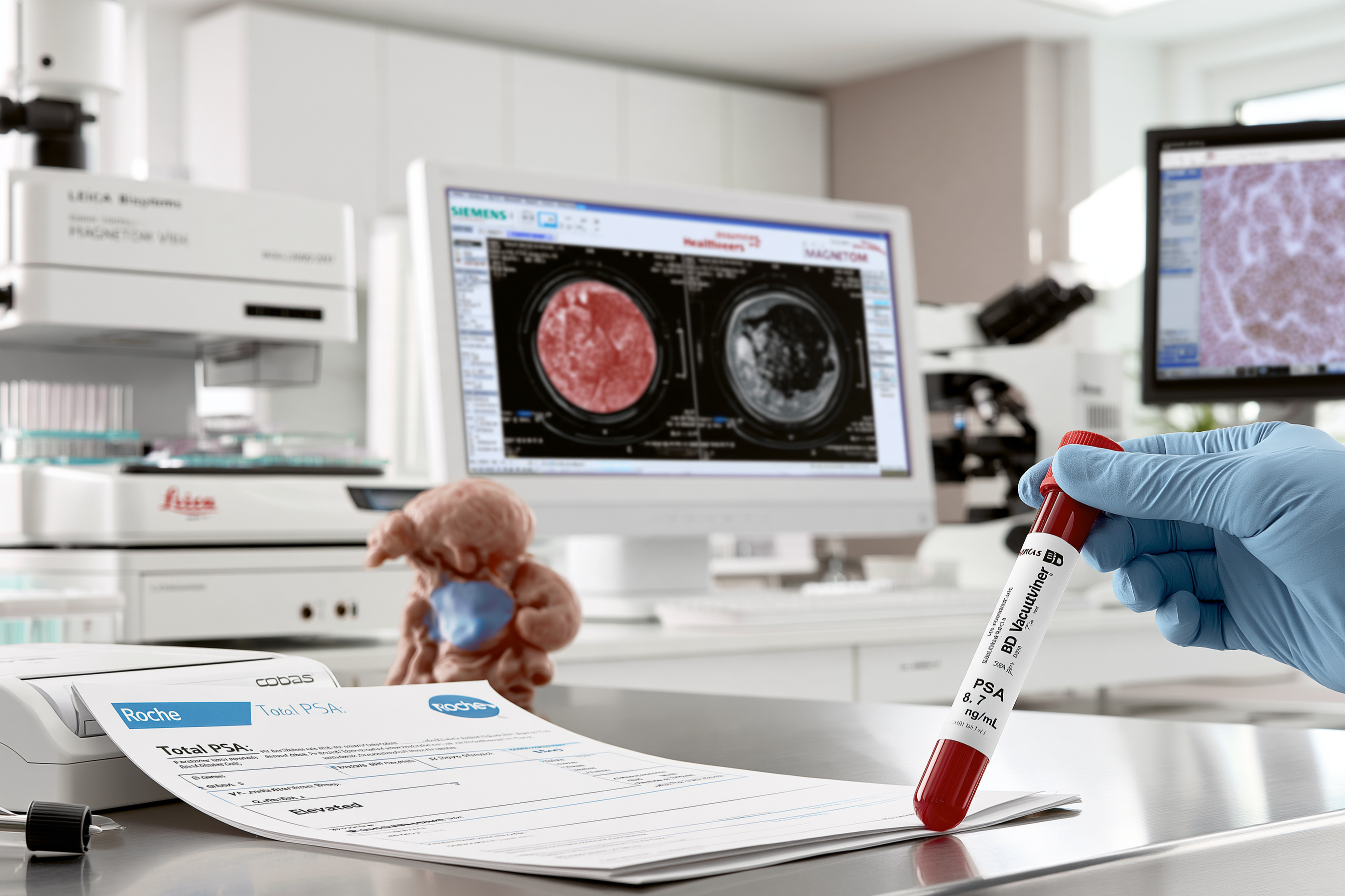
High prostate cancer PSA levels do not confirm that cancer is present. PSA can rise whenever prostate tissue becomes enlarged, inflamed, irritated or mechanically disturbed, so the result identifies a need for clinical assessment rather than providing a diagnosis by itself. Cancer confirmation requires tissue examination when biopsy is clinically indicated.[1,6]
| Cause or influence | Expected effect on PSA | Clinical interpretation |
|---|---|---|
| Benign prostatic hyperplasia | May cause a persistent increase because a larger prostate produces more PSA | Assess prostate volume, urinary symptoms and PSA density because benign enlargement and cancer can coexist. |
| Prostatitis | May cause a moderate or marked temporary increase | Review pelvic pain, urinary symptoms and inflammatory findings; repeat PSA after the acute condition resolves. |
| Urinary tract infection | May temporarily increase PSA | Perform urine assessment when clinically indicated and avoid interpreting PSA as a stable cancer-risk marker during active infection. |
| Acute urinary retention | Can produce a temporary increase | Treat the obstruction first and repeat PSA at an appropriate interval. |
| Ejaculation | May cause a short-term increase | Avoid ejaculation for approximately 48 hours before testing when advised. |
| Vigorous cycling or perineal pressure | May produce a temporary increase in some men | Avoid strenuous cycling before a planned repeat test when practical. |
| Prostate biopsy or instrumentation | May substantially increase PSA for a period | Record the procedure date and delay routine baseline testing until the procedure-related effect has resolved. |
| Catheterisation or cystoscopy | May alter PSA, particularly when associated with retention or inflammation | Interpret the result according to the procedure, clinical circumstances and timing. |
| Finasteride or dutasteride | Usually lowers the measured PSA | The clinician must interpret the result according to treatment duration and previous PSA values. |
| Androgen-deprivation therapy | Can markedly lower PSA by suppressing androgen-sensitive cancer activity | Interpret PSA with serum testosterone, imaging and treatment response rather than untreated screening ranges. |
| Testosterone therapy | May alter PSA behaviour | Record the dose, duration and previous PSA before interpreting a change. |
| Laboratory and biological variation | May cause small fluctuations between tests | Use the same laboratory where practical and confirm a meaningful change with serial measurements. |
Common Non-Cancer Causes of High PSA
Benign prostatic hyperplasia can raise PSA because a larger prostate contains more PSA-producing tissue. This is especially common with increasing age and may be accompanied by weak urinary flow, hesitancy, urgency, nocturia or incomplete bladder emptying. A raised PSA in this setting still requires appropriate assessment because benign enlargement and prostate cancer can occur together.[1]
Prostatitis and urinary tract infection may produce a temporary or substantial PSA increase. Urinary pain, fever, pelvic discomfort, cloudy urine or a sudden change in urinary symptoms may support an inflammatory or infectious cause, although prostatitis can also occur without prominent symptoms. PSA is usually reassessed after the acute condition has resolved rather than interpreted as a stable cancer-risk marker during active inflammation.[1,6]
Acute urinary retention can elevate PSA because pressure and inflammation affect prostate tissue. Recent catheterisation, cystoscopy, prostate biopsy or another procedure involving the urinary tract may also disturb the gland and alter the result. The test date should therefore be reviewed in relation to any recent procedure or episode of retention.[1,6]
Ejaculation and prolonged pressure on the perineum, including vigorous cycling, may cause a temporary rise in some men. These effects are generally modest, but they can influence interpretation when the baseline value is close to an investigation threshold. Consistent preparation before repeat testing helps determine whether the elevation persists.[6]
Finasteride and dutasteride can lower the measured PSA despite continued prostate disease. A result that appears low may therefore require adjustment by the treating clinician according to treatment duration and previous values. Testosterone therapy may also change PSA behaviour and should always be disclosed before interpretation.[1,6]
When a High PSA Is More Concerning
Concern increases when PSA remains elevated on repeat testing, rises progressively over time, is disproportionately high for prostate size or is accompanied by an abnormal prostate examination. A lower percent-free PSA, higher PSA density, suspicious multiparametric magnetic resonance imaging findings, strong family history, Black ancestry or a pathogenic inherited variant can further increase the estimated probability of clinically significant cancer.[1,2,6]
The absolute value also influences urgency. PSA between 4 and 10 ng/mL commonly requires risk refinement because many men in this range do not have cancer. PSA above 10 ng/mL carries a substantially higher probability of malignancy, while values above 20 ng/mL become an important adverse risk feature if cancer is confirmed. Even markedly elevated results cannot independently determine tumour grade, local extension or metastatic spread.[1,6]
A rapid rise may be clinically important, but PSA velocity should not be used as the sole reason for biopsy. Short-term fluctuations can reflect infection, laboratory variation or benign prostate activity. The trend becomes more meaningful when several results are obtained under comparable conditions and interpreted with prostate volume, imaging and other risk factors.[1,6]
How Prostate Cancer Is Confirmed
Multiparametric magnetic resonance imaging can identify suspicious areas, estimate local tumour extent and guide targeted biopsy. A suspicious MRI increases concern but does not independently prove cancer, while a non-suspicious MRI reduces risk without completely excluding clinically significant disease.[6]
Prostate biopsy provides the tissue required to confirm malignancy. The pathology report identifies whether cancer is present and records the Gleason pattern, Grade Group, number of involved cores and amount of tumour in each sample. These findings are then combined with PSA, imaging and clinical stage to determine whether the disease is low, intermediate or high risk.[1,6]
A high PSA should therefore lead to a structured investigation rather than an immediate conclusion. Repeating the test when appropriate, excluding temporary causes, reviewing medicines, assessing prostate size and obtaining MRI or biopsy when indicated provide a more reliable estimate of cancer risk than the PSA value alone.[1,6]
Prostate Cancer PSA Levels by Age

Prostate cancer PSA levels generally rise with age because the prostate often becomes larger and produces more PSA over time. Age can therefore influence interpretation, but it does not create a universal “normal PSA” range. A result that appears modest in an older man may be clinically important in a younger man, particularly when there is a strong family history, Black ancestry, an inherited cancer-risk variant, an abnormal examination or a rising PSA trend.[1,2,5,6]
PSA Levels Before Age 40
Routine PSA screening is not generally recommended for average-risk men younger than 40 because prostate cancer is uncommon in this age group and the likelihood of screening-related harm may exceed the expected benefit. Testing may nevertheless be considered when there are concerning urinary symptoms, an abnormal prostate examination, a known pathogenic variant or an unusually strong family history of early prostate cancer.[2,5]
An elevated PSA in a younger man should not be dismissed solely because cancer is uncommon at that age. Infection, prostatitis and other benign causes remain frequent explanations, but persistent elevation requires appropriate assessment because cancers arising in younger patients may have a stronger hereditary component.[2,6]
PSA Levels From Age 40 to 49
Men at increased risk may begin informed PSA discussions between 40 and 45 years of age. Higher-risk groups include men with Black ancestry, a first-degree relative diagnosed with prostate cancer at a young age, several affected relatives or inherited variants such as BRCA2.[2]
A baseline PSA during this period can help estimate future risk and guide the interval before the next test. Even when the value is below a traditional threshold of 4 ng/mL, a result that is unexpectedly high for age, rising on repeated tests or accompanied by other risk factors may justify earlier reassessment.[1,2]
PSA Levels From Age 50 to 59
The risk of prostate cancer increases during the 50s, and this age group is included in the principal screening population in several international guidelines. PSA should be interpreted through shared decision-making that considers life expectancy, personal values, family history, ancestry and willingness to undergo MRI or biopsy if the result is abnormal.[2,5,8,9]
A PSA between 3 and 4 ng/mL may prompt further evaluation in some national pathways even though it remains below the historical 4 ng/mL threshold. The decision should not depend on the laboratory reference range alone because clinically significant cancer can occur at lower values, while benign enlargement or inflammation can cause higher results.[1,6]
PSA Levels From Age 60 to 69
Men in their 60s have a higher prevalence of both benign prostate enlargement and prostate cancer. This makes interpretation more complex because the same PSA elevation may arise from increased benign tissue, malignant activity or both conditions occurring together.[1,6]
A stable PSA should still be reviewed in relation to prostate volume and previous measurements. A progressively rising value, higher PSA density, lower percent-free PSA or suspicious MRI finding may be more informative than an isolated age-adjusted number.[1,6]
PSA Levels From Age 70 Onward
PSA testing after age 70 should be individualised according to overall health, estimated life expectancy, previous PSA history and whether a diagnosis would alter treatment. Routine screening may provide limited benefit for men with substantial competing illnesses, while healthy older men with a long life expectancy may still benefit from assessment when PSA is persistently elevated or symptoms are concerning.[2,4,5]
A higher PSA should not automatically be attributed to age. New urinary retention, visible blood in the urine, an abnormal examination, unexplained weight loss or persistent bone pain requires clinical evaluation regardless of age. Conversely, detecting a small, low-grade tumour in a frail older patient may lead to observation rather than immediate treatment when the cancer is unlikely to affect survival or quality of life.[1,4,6]
Why Age-Specific PSA Cut-Offs Differ
Age-related PSA thresholds vary because national health systems weigh early detection, unnecessary biopsy, overdiagnosis and treatment-related harm differently. Some pathways use fixed thresholds, while others rely on age, repeat PSA, prostate volume, MRI and validated risk calculators.[2,5,7,8]
A laboratory value should therefore be interpreted according to the patient’s country, clinical setting and individual risk profile. Screening thresholds for an asymptomatic man should not be applied to a patient with symptoms, and neither should be used to interpret PSA after prostate cancer treatment.[2,6,7,8]
What Prostate Cancer PSA Levels Mean
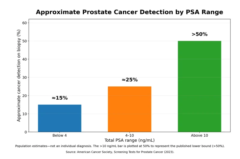
Prostate cancer PSA levels are best interpreted as ranges of probability and clinical urgency rather than fixed values that determine diagnosis or stage. The likelihood of detecting cancer generally increases as PSA rises, but the same result can have different meanings depending on age, prostate volume, inflammation, medication use, previous results, examination findings and magnetic resonance imaging.[1,6]
| PSA level | General cancer-risk meaning | Patient-focused next step |
|---|---|---|
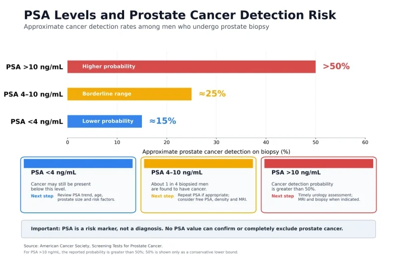
| Below 4 ng/mL | Relatively reassuring, but not cancer-free. Among men selected for biopsy, about 15% are found to have prostate cancer. | Review the PSA trend, age, prostate size, examination findings, family history and inherited risk rather than relying on one result. |
| 4–10 ng/mL | Commonly called the borderline range. Approximately one in four biopsied men is found to have prostate cancer. | Exclude temporary causes, repeat PSA when appropriate and consider percent-free PSA, PSA density and multiparametric MRI. |
| Above 10 ng/mL | Associated with a greater than 50% probability of detecting prostate cancer on biopsy. | Arrange timely urological assessment, MRI and biopsy when clinically indicated. |
| 10–20 ng/mL | Can contribute to intermediate-risk classification after cancer is confirmed. | Interpret with Grade Group, biopsy tumour volume, MRI findings and clinical stage. |
| Above 20 ng/mL | An important high-risk feature when prostate cancer is confirmed. | Complete staging is required; the value does not independently prove metastatic disease. |
| Above 50 or 100 ng/mL | Strongly concerning for substantial prostate disease and may occur with advanced cancer. | Prompt specialist assessment, biopsy and appropriate staging imaging are required; spread cannot be diagnosed from PSA alone. |
PSA Below 4 ng/mL
A PSA below 4 ng/mL is often considered relatively low in an untreated man, but it does not exclude prostate cancer. Approximately 15% of men with a PSA below 4 ng/mL who undergo biopsy are found to have cancer, although this estimate includes cancers with different grades and clinical importance.[1]
Further assessment may still be appropriate when a result below 4 ng/mL is rising consistently, unexpectedly high for the patient’s age or prostate size, associated with a low percent-free PSA or accompanied by an abnormal prostate examination. A strong family history, Black ancestry or an inherited pathogenic variant can also increase concern at a lower PSA level.[1,2]
A result below 4 ng/mL should not be considered permanently protective. The direction of change over several tests may reveal increasing prostate activity before the value crosses a conventional investigation threshold. Comparisons are most reliable when testing is performed under similar conditions and, where practical, through the same laboratory.
PSA Between 4 and 10 ng/mL
PSA between 4 and 10 ng/mL is commonly described as the borderline range. Approximately one in four men biopsied within this range is found to have prostate cancer, which also means that most men in this range do not have cancer.[1]
The clinical objective is to distinguish men who are more likely to have clinically significant disease from those whose elevation is caused by benign prostate enlargement, inflammation or temporary biological variation. Reviewing earlier PSA results can show whether the value is stable, fluctuating or progressively increasing. Urine testing and symptom assessment may identify infection, while prostate volume allows the clinician to determine whether the PSA is proportionate to the size of the gland.
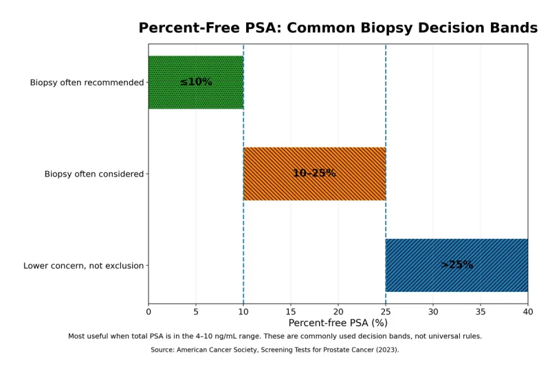
Percent-free PSA can add useful information within this range. A lower percentage of free PSA is associated with a higher likelihood of prostate cancer. Many clinicians recommend discussing biopsy when percent-free PSA is 10% or lower and considering biopsy when it is between 10% and 25%, although these thresholds must be combined with the patient’s complete risk profile.[1]
Multiparametric magnetic resonance imaging may identify a suspicious lesion, estimate its location and help determine whether targeted biopsy is required. A non-suspicious MRI can reduce the estimated probability of clinically significant cancer, but it cannot completely exclude a tumour. Persistent PSA elevation may therefore require continued monitoring or biopsy even when imaging is reassuring.
PSA Between 10 and 20 ng/mL
A PSA above 10 ng/mL is associated with a greater than 50% overall probability of detecting prostate cancer on biopsy.[1] This estimate does not mean that every patient within the range has cancer, but it usually warrants prompt urological assessment rather than prolonged observation without investigation.
Benign enlargement, prostatitis, urinary retention and recent prostate procedures can still produce values within this range. The possibility of a temporary cause should be assessed, but treatment for presumed infection should not be given solely to lower PSA when there is no clinical evidence of infection.
When cancer is confirmed, PSA between 10 and 20 ng/mL contributes to risk assessment alongside Grade Group, biopsy tumour burden, MRI findings and clinical stage. A PSA of 12 ng/mL with a small, low-grade lesion does not carry the same implications as the same value with extensive high-grade cancer. Treatment decisions must therefore be based on the combined disease profile rather than PSA alone.
PSA Above 20 ng/mL
A PSA above 20 ng/mL substantially increases concern for prostate cancer and requires timely investigation. If malignancy is confirmed, this level is an adverse feature that may influence staging investigations and treatment planning. It does not independently establish that the tumour has extended beyond the prostate or reached distant organs.[1,6]
Clinical assessment may include multiparametric MRI, prostate biopsy and imaging selected according to the biopsy grade, symptoms and estimated probability of disease outside the prostate. The presence of persistent pelvic or bone pain, unexplained weight loss, urinary obstruction or neurological symptoms can increase the need for expedited staging.
A marked fall on repeat testing may indicate that inflammation, infection, retention or another temporary factor contributed to the original result. It does not eliminate the need to investigate an initially substantial elevation, particularly when examination or imaging remains abnormal.
PSA Above 50 or 100 ng/mL
PSA above 50 or 100 ng/mL is strongly concerning for extensive prostate cancer activity and should lead to urgent specialist assessment. Very high values increase suspicion for locally advanced or metastatic disease, but the anatomical extent of cancer must be established through imaging rather than inferred from PSA alone.[6]
Severe prostatitis, acute urinary retention and recent instrumentation can occasionally cause marked PSA elevation. The clinical history, urine findings, examination and timing of the blood test help determine whether these factors may have contributed. When cancer is suspected, evaluation should not be delayed solely to observe whether the value falls naturally.
Biopsy remains necessary when tissue confirmation is clinically appropriate, while staging imaging determines whether disease is confined to the prostate, involves regional structures or has spread to distant sites. PSA should then be followed as a treatment-response marker, with the trend interpreted alongside symptoms, imaging and the therapy received.[6]
Prostate Cancer PSA Levels Before Testing
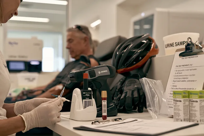
Prostate cancer PSA levels can be temporarily altered by inflammation, infection, ejaculation, strenuous cycling and recent procedures involving the prostate or urinary tract. Preparing consistently before each blood test reduces avoidable variation and helps the clinician determine whether a change represents persistent prostate activity or a short-term physiological effect.[1,6]
Avoid Temporary PSA Changes
Ejaculation can produce a temporary PSA increase in some men. Avoiding ejaculation for approximately 48 hours before the blood test may improve comparability, particularly when a previous result was close to the investigation threshold or when small changes are being monitored over time.[1,6]
Vigorous cycling and activities that place sustained pressure on the perineum may also influence PSA temporarily. Avoiding strenuous cycling for approximately 48 hours before testing can reduce the possibility that mechanical pressure on the prostate contributes to an unexpected rise.[1,6]
Ordinary walking, normal daily activity and routine non-strenuous exercise do not usually require restriction. The main objective is to avoid an unusual activity immediately before testing that could make the result less comparable with earlier measurements.
The blood sample does not usually require fasting unless other tests ordered at the same appointment have separate dietary requirements. Patients should follow the laboratory instructions provided for the complete blood panel rather than fasting solely because PSA is being measured.
Report Symptoms and Recent Procedures
Urinary burning, fever, pelvic discomfort, cloudy urine, sudden urgency or a recent worsening of urinary symptoms may indicate infection or inflammation capable of increasing PSA. The treating clinician may perform urine testing and manage the underlying condition before using PSA to estimate prostate cancer risk.[1,6]
A recent prostate biopsy can keep PSA elevated for several weeks. Cystoscopy, urinary catheterisation, acute urinary retention and other procedures affecting the urinary tract may also influence interpretation. The clinician should be told what procedure occurred and when it was performed so that the test can be scheduled at an appropriate interval.[6]
A digital rectal examination may cause a small PSA change in some circumstances, although its effect is generally less important than infection, biopsy or retention. When both examinations are planned, the blood sample may be collected before prostate manipulation to maintain consistent testing conditions.
Patients should not conceal urinary symptoms or proceed with routine PSA testing during an acute infection merely to avoid rescheduling. Testing after the temporary condition has resolved usually provides a more clinically meaningful baseline.[6]
Disclose Medicines and Hormonal Treatment
Finasteride and dutasteride reduce PSA by suppressing benign prostate activity and shrinking prostate tissue. A measured value may therefore appear lower than expected even when clinically important disease is present. The clinician must know the medicine, dose and duration of use before interpreting the result.[1,6]
Testosterone replacement therapy can affect PSA behaviour and requires structured monitoring. Patients should report prescribed testosterone, anabolic hormones and any recent change in hormonal treatment rather than stopping therapy independently before the blood test.
Androgen-deprivation therapy used for prostate cancer can produce a substantial PSA reduction by suppressing androgen-sensitive tumour activity. PSA measured during this treatment cannot be interpreted using untreated screening thresholds and should be reviewed with the testosterone level, treatment duration and previous cancer findings.[6]
Herbal medicines, nutritional supplements and over-the-counter products should also be disclosed. A patient should not discontinue prescribed medicines or begin an unverified product for the purpose of lowering PSA before testing, because an artificially altered marker can delay appropriate investigation.
Keep Repeat Tests Comparable
Repeat testing is most informative when the samples are collected under similar conditions. Using the same laboratory where practical can reduce variation caused by different testing platforms, calibration methods and reference ranges.
The date, PSA value, laboratory unit and relevant circumstances should be recorded after every test. PSA is commonly reported in nanograms per millilitre, while some countries report micrograms per litre; these units are numerically equivalent. A result of 4 ng/mL is therefore the same numerical concentration as 4 µg/L.
The clinician should know whether the patient had infection, retention, ejaculation, vigorous cycling, a recent procedure or a medication change before each measurement. Without this context, a temporary rise may be mistaken for disease progression or a treatment-related fall may be interpreted too confidently.
A small difference between two tests may reflect normal biological or laboratory variation rather than a clinically meaningful change. The trend becomes more reliable when several values are obtained over an appropriate interval under comparable conditions and assessed with prostate volume, symptoms, examination findings and imaging.[1,6]
When PSA Testing Should Be Delayed
Routine PSA testing may be postponed when the patient has active prostatitis, a urinary tract infection, acute urinary retention or a recent prostate biopsy. The appropriate delay depends on the cause, severity, treatment response and procedure performed, so the timing should be determined by the treating clinician.[6]
Testing should not be delayed when there are urgent warning signs such as inability to pass urine, fever with urinary obstruction, visible blood in the urine, new neurological symptoms or severe pelvic or bone pain. These findings require prompt clinical assessment, and the doctor may order PSA as part of a broader urgent investigation.
A markedly elevated previous PSA, an abnormal prostate examination or a persistent upward trend should not be managed through repeated postponement. Temporary influences should be addressed, but appropriate urological assessment, magnetic resonance imaging or biopsy should proceed when the overall cancer risk remains concerning.[1,6]
How Prostate Cancer PSA Levels Are Interpreted
Prostate cancer PSA levels are interpreted more accurately when the total PSA result is combined with previous measurements, prostate volume, percent-free PSA, imaging findings and the patient’s individual risk factors. These additional measures help distinguish a temporary or benign PSA elevation from a pattern that is more concerning for clinically significant prostate cancer.[1,2,6]
| PSA measure | How it is determined | When it is useful | Clinical meaning |
|---|---|---|---|
| Total PSA | Measures free and protein-bound PSA together | Initial screening, diagnostic investigation and follow-up | A higher result increases concern but cannot confirm cancer, grade or stage. |
| Percent-free PSA | Free PSA divided by total PSA and multiplied by 100 | Most useful when total PSA is approximately 4–10 ng/mL | A lower percentage is associated with greater cancer probability. |
| Percent-free PSA ≤10% | Lower proportion of unbound PSA | When deciding whether biopsy should be discussed | Commonly considered a stronger reason to recommend or discuss biopsy. |
| Percent-free PSA 10–25% | Intermediate free-PSA proportion | Borderline biopsy decisions | Biopsy may be considered according to age, MRI, prostate size, examination and inherited risk. |
| PSA density | Total PSA divided by prostate volume | Elevated PSA with benign enlargement or uncertain MRI findings | A PSA disproportionately high for prostate size increases concern for clinically significant cancer. |
| PSA velocity | Change in PSA per year | Evaluating a longitudinal pattern | A persistent rise adds context but should not be the sole reason for biopsy. |
| PSA doubling time | Estimated time required for PSA to double | Active surveillance and assessment of recurrence after treatment | A shorter doubling time is generally more concerning than a slow increase. |
| Prostate Health Index | Combines total PSA, free PSA and a PSA precursor | Refining biopsy decisions in selected patients | Estimates the probability of clinically significant cancer but does not diagnose it. |
| 4Kscore | Combines four kallikrein measurements with clinical factors | Selected patients with uncertain biopsy need | Estimates the likelihood of detecting higher-grade cancer on biopsy. |
| Risk calculators | Combine PSA with age, examination, family history, prostate volume and previous biopsy | Shared decision-making before MRI or biopsy | Provide a probability estimate rather than an individual diagnosis. |
Repeat PSA Testing
A newly elevated PSA is often repeated before magnetic resonance imaging or biopsy when the patient is clinically stable and there are no urgent warning signs. PSA can vary because of infection, inflammation, urinary retention, ejaculation, recent prostate procedures, laboratory variation and changes in medication. Repeating the test after these influences have been addressed can show whether the elevation is persistent.[1,6]
The repeat result should be compared with all earlier PSA measurements rather than only the most recent value. A value that returns toward the patient’s previous baseline may support continued monitoring, whereas a persistently elevated or progressively rising result may justify additional risk assessment. A substantial fall does not completely exclude cancer, particularly when examination findings, family history or imaging remain concerning.
The interval before repeat testing depends on the initial PSA, symptoms, possible temporary causes and the patient’s overall risk. A mildly elevated result without concerning symptoms may be reassessed after an appropriate interval, while a markedly elevated PSA, abnormal prostate examination or suspected advanced disease requires faster specialist evaluation rather than repeated testing alone.[2,6]
Percent-Free PSA
PSA circulates in the blood in two principal forms: PSA attached to blood proteins and PSA that remains unbound, known as free PSA. Percent-free PSA is calculated by dividing free PSA by total PSA and multiplying the result by 100. Men with prostate cancer tend, on average, to have a lower percentage of free PSA than men whose elevation is caused by benign prostate enlargement.[1]
Percent-free PSA is most useful when total PSA lies within a borderline range and the decision about biopsy remains uncertain. A percent-free PSA of 10% or lower is generally associated with a higher likelihood of cancer and often leads to a biopsy discussion. Values between 10% and 25% may also support biopsy depending on age, prostate size, family history, examination findings and MRI results.[1]
A higher percent-free PSA can reduce the estimated probability of cancer but cannot exclude it. The test should not be interpreted as an independent diagnosis because laboratory methods, prostate volume and individual biological variation can affect the result. It is most useful when incorporated into a broader risk calculation.
PSA Density
PSA density adjusts the total PSA for the size of the prostate. It is calculated by dividing the PSA value by prostate volume, usually measured with magnetic resonance imaging or transrectal ultrasound. A PSA of 6 ng/mL arising from a very large benign prostate may carry a different level of concern from the same PSA produced by a much smaller gland.[1]
A higher PSA density indicates that the amount of PSA is disproportionately elevated relative to prostate volume and can increase suspicion for clinically significant cancer. A lower PSA density may support benign enlargement as a contributor, although it cannot rule out malignancy.[1,2]
Prostate volume measurement is not perfectly identical across imaging methods or readers, so PSA density should not be treated as an absolute dividing line. Its clinical value is greatest when assessed with MRI findings, previous biopsy results, Grade Group risk and the complete PSA trend.
PSA density can be particularly helpful when MRI shows no suspicious lesion but PSA remains elevated. In this setting, a low density may support surveillance in a carefully selected patient, while a higher density may strengthen the reason to consider biopsy despite reassuring imaging.[2]
PSA Velocity
PSA velocity describes how quickly PSA changes over time and is usually expressed as the increase in ng/mL per year. A sustained rise may increase concern, especially when it occurs across several comparable tests rather than between only two measurements.
A rapid change can also result from prostatitis, urinary infection, retention, recent instrumentation or inconsistent testing conditions. PSA velocity should therefore not be used as the sole reason to order a biomarker test, MRI or biopsy. Current early-detection guidance recommends integrating the rate of change with absolute PSA, age, prostate volume, examination findings, inherited risk and imaging.[2]
A gradual rise caused by benign prostate growth may appear less concerning than a persistent increase that is disproportionate to prostate size. However, no single rate of increase reliably separates benign disease from cancer in every patient. The clinical pattern is more informative than an isolated calculation.
PSA Doubling Time
PSA doubling time estimates how long it would take for the PSA concentration to double if the observed pattern continued. It is calculated from several measurements collected over time and is used more often after a prostate cancer diagnosis than during initial screening.[6]
During active surveillance, a shortening doubling time may prompt closer assessment with MRI or repeat biopsy, but it does not independently prove that the tumour has become more aggressive. Prostatitis, benign prostate growth and differences in testing conditions can still influence the calculation while the prostate remains present.
After prostatectomy or radiotherapy, PSA doubling time can help estimate the behaviour of biochemical recurrence. A shorter doubling time is generally more concerning than a slow increase because it may indicate more rapidly progressing disease. Interpretation must also include the original Grade Group, pathological stage, time since treatment, current PSA level and imaging findings.
The calculation becomes unreliable when PSA values are extremely low, fluctuate irregularly or are measured at inconsistent intervals. Treatment decisions should not be made from a doubling-time estimate without confirming that the underlying PSA trend is genuine.
Additional Blood and Urine Tests
When the decision about MRI or biopsy remains uncertain, additional biomarker tests may refine the probability of clinically significant prostate cancer. Blood-based options can include the Prostate Health Index, 4Kscore and IsoPSA, while selected urine tests assess prostate cancer-associated genes or proteins. Availability and clinical use differ across the USA, UK, Canada, Singapore and Australia.[1,2]
The Prostate Health Index combines total PSA, free PSA and a PSA precursor into a single risk score. The 4Kscore incorporates four kallikrein measurements with clinical factors to estimate the likelihood of finding higher-grade cancer on biopsy. These tests are intended to improve risk selection and may help some patients avoid an unnecessary biopsy.[1]
Biomarker results do not confirm cancer and should not replace biopsy when clinical suspicion is high. They are also not required for every patient with an elevated PSA. Their value is greatest when the result is likely to change the decision between continued monitoring, MRI and tissue sampling.[1,2]
Risk calculators can combine age, PSA, digital rectal examination, family history, previous biopsy findings, prostate volume and other variables. They provide an estimated probability rather than an individual certainty, and their accuracy may differ between populations. The result should be discussed in the context of the patient’s country, ancestry, access to MRI and willingness to proceed with biopsy.[2]
Next Steps After High Prostate Cancer PSA Levels
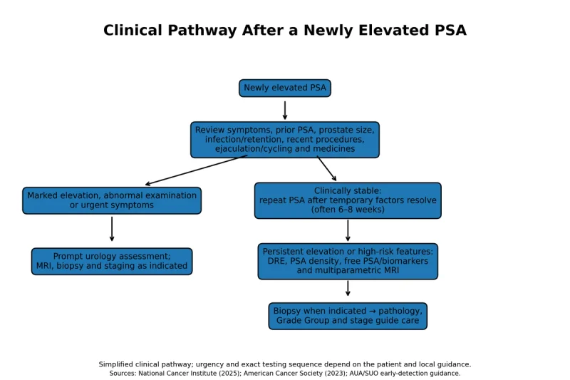
High prostate cancer PSA levels usually lead to a staged clinical assessment rather than an immediate diagnosis or treatment decision. The speed and extent of investigation depend on the absolute PSA value, previous results, symptoms, prostate examination, age, life expectancy, family history, inherited risk, prostate volume and whether temporary causes of elevation are present.[1,2,6]
| Clinical situation | Usual next step | Purpose |
|---|---|---|
| First mildly elevated PSA with no urgent findings | Review infection, retention, ejaculation, cycling, recent procedures and medicines; repeat PSA after temporary factors resolve | Confirm whether the elevation is persistent rather than temporary. |
| Abnormal screening PSA in a clinically stable patient | Repeat PSA in approximately six to eight weeks or according to the applicable national pathway | Reduce unnecessary imaging or biopsy caused by a transient elevation. |
| Persistent borderline elevation | Assess prostate examination, percent-free PSA, PSA density, prostate volume and validated risk calculators | Estimate the probability of clinically significant cancer. |
| Persistent elevation with meaningful cancer risk | Obtain multiparametric prostate MRI | Identify suspicious lesions, calculate prostate volume and guide biopsy planning. |
| Suspicious MRI finding | Consider MRI-targeted biopsy with systematic sampling | Confirm whether cancer is present and determine Grade Group and tumour burden. |
| Non-suspicious MRI but high PSA density or strong risk factors | Consider biopsy, additional biomarkers or structured close monitoring | MRI reduces risk but does not completely exclude clinically significant cancer. |
| Markedly elevated PSA or abnormal prostate examination | Prompt urological review, MRI and biopsy when appropriate | Avoid delaying diagnosis of clinically important disease. |
| Urinary retention, fever, haematuria, bone pain or neurological symptoms | Urgent clinical assessment rather than routine repeat testing alone | Identify obstruction, severe infection, advanced disease or neurological compression. |
| Biopsy-confirmed prostate cancer | Combine PSA with Gleason score, Grade Group, clinical stage, MRI and staging imaging | Determine whether active surveillance, local treatment or systemic treatment is appropriate. |
Clinical Risk Review
The initial review should compare every available PSA result with its date and testing circumstances. A persistent upward pattern carries different implications from a single unexpected elevation following urinary infection, retention, catheterisation or another recent prostate procedure. The clinician should also record urinary symptoms, visible blood in the urine, pelvic discomfort, unexplained weight loss, bone pain and any new neurological symptoms.[1,6]
Medication review is essential because finasteride and dutasteride can lower the measured PSA, while testosterone therapy and prostate cancer hormonal treatment can alter its behaviour. Family history, Black ancestry and inherited pathogenic variants such as BRCA2 may increase concern at a PSA value that would otherwise appear borderline.[1,2]
A digital rectal examination can detect prostate asymmetry, a hard nodule, irregularity or fixation. An abnormal finding strengthens the reason for specialist assessment, but a normal examination does not exclude cancer because many tumours are too small or are located beyond the area that can be felt.[1,6]
Urine testing may be appropriate when urinary infection or inflammation is suspected. Fever, painful urination, pelvic discomfort, cloudy urine or a sudden deterioration in urinary flow can indicate a temporary inflammatory cause, but infection and prostate cancer can coexist. Persistent PSA elevation should therefore be reassessed after the acute condition has resolved.[1,6]
Confirming the Elevated Result
A newly elevated PSA is commonly repeated before proceeding to secondary biomarkers, magnetic resonance imaging or biopsy when the patient is clinically stable. Repeat testing can confirm whether the elevation persists after temporary biological, laboratory or procedural influences have been addressed.[2,6]
The interval before retesting should reflect the suspected cause and the degree of elevation. A borderline result without concerning symptoms may be repeated after an appropriate waiting period, whereas a markedly elevated PSA, abnormal prostate examination or symptoms suggesting advanced disease should prompt faster urological assessment.[2,6]
A lower repeat result may reduce immediate concern but does not automatically end the investigation. Continued assessment may still be necessary when PSA remains high for age or prostate size, has shown a sustained upward trend, or occurs alongside strong hereditary risk or abnormal clinical findings.[1,2]
Repeated blood testing should not be used to postpone appropriate investigation indefinitely. Persistent elevation requires a decision about further risk assessment, imaging or biopsy based on the probability of clinically significant disease rather than the objective of waiting for the PSA to return to a laboratory reference range.[2,6]
Multiparametric MRI
Multiparametric magnetic resonance imaging provides detailed information about prostate anatomy, gland volume and suspicious lesions. It can help identify where a tumour may be located, estimate whether it extends beyond the prostate capsule and guide tissue sampling toward the most concerning area.[2,7]
MRI findings are commonly reported using a five-category suspicion score. Lower categories indicate that clinically significant cancer is less likely, while higher categories indicate progressively greater suspicion and a stronger reason to consider targeted biopsy. The MRI score must still be interpreted with PSA density, family history, examination findings and previous biopsy results.[2,7]
A suspicious MRI does not confirm cancer because benign inflammation and other prostate changes can resemble a tumour. Tissue examination remains necessary when pathological confirmation is required. MRI primarily improves localisation and helps select the regions most likely to contain clinically significant disease.[2,7]
A non-suspicious MRI lowers the probability of clinically significant cancer but cannot exclude it completely. Biopsy may still be appropriate when PSA density is high, PSA continues to rise, the prostate examination is abnormal or the patient has substantial familial or genetic risk.[2,7]
MRI also provides the prostate volume required to calculate PSA density. This can clarify whether the total PSA is proportionate to a substantially enlarged gland or remains unusually high relative to the amount of prostate tissue present.[1,2]
Prostate Biopsy
Prostate biopsy provides tissue for confirming whether malignant cells are present when the overall risk justifies sampling. It may be performed through the perineal skin or through the rectum, depending on local practice, patient factors, infection risk, available equipment and specialist expertise.[2,6]
MRI-targeted biopsy samples lesions identified on imaging, while systematic biopsy collects tissue from mapped areas throughout the prostate. A combined approach may be used when MRI reveals a suspicious lesion because targeted cores improve sampling of that lesion and systematic cores can detect clinically important cancer elsewhere in the gland.[2,6]
The pathology report identifies the cancer type, primary and secondary Gleason patterns, Grade Group, number of positive cores and proportion of tissue involved. These findings show how abnormal the tumour appears microscopically and contribute more directly to treatment planning than the PSA value alone.[1,6]
Biopsy can cause temporary blood in the urine, semen or stool, as well as discomfort, urinary retention or infection. The route of biopsy, preventive measures and warning symptoms requiring urgent care should be discussed before consent.[6]
A negative biopsy means cancer was not detected in the tissue sampled; it does not guarantee that no tumour exists elsewhere in the prostate. Persistent clinical suspicion may require continued PSA monitoring, repeat MRI, additional biomarker assessment or another biopsy. PSA alone should not be the only factor used to determine whether repeat biopsy is necessary.[2,6]
Staging After Cancer Is Confirmed
Once biopsy confirms prostate cancer, PSA is combined with Grade Group, clinical examination, MRI findings and the amount of cancer in the biopsy cores to classify the disease. These findings help distinguish lower-risk cancer that may be suitable for active surveillance from disease requiring definitive or systemic treatment.[1,6]
Local staging assesses whether the tumour appears confined to the prostate, has extended through the capsule or involves nearby structures such as the seminal vesicles. MRI contributes to this assessment, although microscopic extension may not always be visible before treatment.[6,7]
Patients with higher PSA, higher Grade Group, locally advanced findings or symptoms suggesting spread may require additional staging. Depending on risk and local availability, this may include prostate-specific membrane antigen positron emission tomography combined with computed tomography, conventional computed tomography, bone imaging or other directed investigations.[6]
Distant staging is not automatically required for every patient with a raised PSA or newly diagnosed low-risk tumour. Its use depends on the combined probability of lymph-node, bone or organ involvement. The final clinical risk category should therefore be based on PSA, pathology and anatomical staging together rather than on any one result in isolation.[1,6]
Prostate Cancer PSA Levels by Country

Prostate cancer PSA levels are not interpreted through one universal international screening threshold. The USA, UK, Canada, Singapore and Australia use different starting ages, testing intervals, referral values and risk-based pathways. A result considered sufficient for repeat testing or specialist referral in one country may be managed differently elsewhere, particularly when age, family history, ancestry or inherited cancer-risk variants are considered.
USA
The 2026 American Urological Association and Society of Urologic Oncology guideline recommends offering regular prostate cancer screening every two to four years to people aged 50 to 69 after an informed discussion about potential benefits and harms. The testing interval may be adjusted according to the baseline PSA, previous results, health status, life expectancy and personal preferences.[2]
Men at increased risk should be offered screening from approximately 40 to 45 years of age. Higher-risk factors include Black ancestry, a strong family history of prostate cancer and inherited pathogenic variants associated with prostate cancer susceptibility. Several affected relatives, prostate cancer diagnosed at a young age or a family history of metastatic or fatal disease may justify earlier and more individualised surveillance.[2]
A newly elevated PSA is generally confirmed with a repeat measurement before secondary biomarkers, magnetic resonance imaging or biopsy when the patient is clinically stable. This approach does not apply when the PSA is markedly elevated, the prostate examination is abnormal or symptoms raise concern for advanced disease, because these findings may require expedited urological assessment.[2]
Screening recommendations apply to people without symptoms. Urinary retention, visible blood in the urine, an abnormal prostate examination, persistent pelvic pain or suspected metastatic symptoms require diagnostic assessment regardless of whether the patient falls within the routine screening age range.
UK
The UK does not recommend population-wide PSA screening for all men. In 2026, the UK National Screening Committee recommended targeted PSA testing every two years from age 45 to 61 for men who have both a pathogenic BRCA2 variant and a relevant family history of breast, ovarian, pancreatic or prostate cancer.[10]
This targeted recommendation should not be interpreted as evidence that other men are ineligible for PSA testing. Men without symptoms may discuss the advantages and disadvantages of testing with their general practitioner, while those with urinary symptoms, visible haematuria, an abnormal prostate examination or other concerning findings should be assessed through a diagnostic pathway rather than a screening programme.
For people with possible symptoms of prostate cancer, NICE uses age-specific PSA values to guide referral consideration. The thresholds are more than 2.5 ng/mL at ages 40 to 49, more than 3.5 ng/mL at ages 50 to 59, more than 4.5 ng/mL at ages 60 to 69 and more than 6.5 ng/mL at ages 70 to 79.[7]
These values are referral thresholds for symptomatic assessment, not boundaries below which cancer is impossible. A result below the relevant threshold may still require specialist review when the prostate feels malignant, PSA is rising persistently, genetic risk is substantial or symptoms remain unexplained. A value above the threshold raises the need for further investigation but does not independently confirm cancer.[7]
Canada
Canadian patients may encounter different recommendations because urological and preventive-health authorities have taken different positions on population PSA screening. The Canadian Urological Association recommends that men who choose screening generally begin at age 50, while men at increased risk may begin at age 45. Subsequent testing intervals should be individualised according to previous PSA values and the patient’s overall risk.[5]
Increased risk may arise from a strong family history, prostate cancer affecting relatives at younger ages or inherited variants such as BRCA1, BRCA2 or HOXB13. Men with a known pathogenic variant require an individualised strategy developed with clinicians experienced in hereditary cancer risk rather than reliance on an average-risk screening schedule.[5]
The 2014 Canadian Task Force recommendation advised against routine PSA screening across age groups. Its recommendation against screening was weak for men aged 55 to 69 because a small possible reduction in prostate cancer mortality had to be balanced against false-positive results, unnecessary biopsy, overdiagnosis and treatment-related harm. The recommendation was stronger for men younger than 55 and those aged 70 or older.[4]
These differing positions make informed decision-making particularly important. A Canadian patient should discuss his age, life expectancy, family history, ancestry, previous PSA values and willingness to undergo MRI or biopsy before choosing screening. Provincial referral procedures and access to prostate MRI may also affect how an elevated result is investigated.
The recommendations against routine screening do not apply in the same way to men with symptoms or abnormal clinical findings. Persistent urinary obstruction, visible haematuria, a suspicious prostate examination or unexplained bone pain requires diagnostic evaluation even when routine screening would not otherwise have been offered.
Singapore
The Singapore Cancer Society identifies men aged 50 to 70 and men with a strong family history of prostate cancer as groups in whom PSA testing should be discussed with a doctor. A strong family history includes one or more first-degree relatives, such as a father or brother, diagnosed before age 65.[9]
PSA testing in Singapore should be based on individual risk and preferences rather than treated as an automatic annual investigation for every man. The discussion should include the possibility of a false-positive result, detection of an indolent tumour, MRI, biopsy and treatment-related consequences, as well as the potential benefit of identifying clinically significant cancer before symptoms or metastatic spread develop.[9]
Men with a previous borderline result should bring all dated PSA reports to the consultation because the longitudinal pattern may influence the need for repeat testing or referral. A persistent rise, an abnormal prostate examination, a high PSA density or a strong hereditary risk may justify further assessment even when the absolute value is not markedly elevated.
Men presenting with urinary symptoms should not assume that PSA testing alone will determine the cause. Benign prostate enlargement is common, but urine testing, clinical examination, assessment of residual urine, prostate-volume measurement and urological review may be necessary to distinguish benign obstruction, infection and suspected malignancy.
Australia
Australia’s 2026 national guideline recommends PSA testing every two years for men aged 50 to 69 who choose testing after an informed discussion with their general practitioner. For average-risk men in this age group, a PSA below 3.0 µg/L generally leads to continued two-yearly testing. A result of 3.0 µg/L or higher should usually be repeated within one to three months, and persistent elevation should lead to an offer of specialist referral.[8]
Routine testing is not recommended for average-risk men aged 45 to 49, although an interested patient may be offered an initial PSA measurement. A result below 1.0 µg/L generally permits further testing to be deferred until age 50. A result of 1.0 µg/L or higher should be repeated within one to three months, with specialist referral considered when the elevation is confirmed.[8]
Men at higher risk are advised to begin two-yearly testing from age 45. Higher-risk features include a brother with prostate cancer, a father diagnosed before age 65, two or more second-degree relatives who died from prostate cancer, Black sub-Saharan ancestry or a confirmed BRCA2 pathogenic variant.[8]
The recommended action level for higher-risk men is 1.0 µg/L or higher at ages 45 to 49 and 2.0 µg/L or higher at ages 50 to 69. These lower thresholds recognise that an apparently modest PSA may have greater clinical significance when the probability of developing consequential prostate cancer is substantially above average.[8]
For men aged 70 or older, testing is based on clinical assessment, competing health conditions, personal preferences and whether life expectancy is sufficient for early detection to provide meaningful benefit. Testing is generally considered when life expectancy exceeds seven years. A PSA of 5.5 µg/L or higher should usually be repeated within one to three months, followed by consideration of specialist referral if the result remains elevated.[8]
When PSA remains at or above the applicable Australian action level, multiparametric magnetic resonance imaging is recommended before biopsy. MRI can refine the probability of clinically significant cancer, identify suspicious regions for targeted sampling and reduce unnecessary biopsy, but persistent high-risk findings may still require tissue assessment when MRI is not suspicious.[8]
Prostate Cancer PSA Levels, Grade and Stage

Prostate cancer PSA levels help estimate disease risk, but they do not reveal how abnormal the cancer cells are or how far the tumour has spread. Treatment planning therefore combines PSA with biopsy findings, Grade Group, clinical stage, imaging, tumour volume, symptoms and the patient’s general health.[1,6]
PSA and Gleason Score
The Gleason score is determined by examining prostate biopsy tissue under a microscope. The pathologist identifies the two most common growth patterns, grades each pattern from 3 to 5 in contemporary reporting and adds them together. A result written as Gleason 3+4=7 means that pattern 3 is predominant and pattern 4 is the second most common pattern.[1,6]
The order of the two numbers matters. Gleason 3+4=7 generally has a more favourable biological profile than Gleason 4+3=7 because the more aggressive pattern 4 component predominates in the second result. PSA alone cannot distinguish between these tumours, even when both patients have the same blood level.[1,6]
A low PSA does not guarantee a low Gleason score. Some high-grade tumours produce relatively little PSA, while a large benign prostate may produce a higher PSA without aggressive cancer. Biopsy remains necessary when tissue confirmation and tumour grading are clinically indicated.[1,6]
PSA and Grade Group
Grade Group converts the Gleason score into five clinically useful categories. Grade Group 1 corresponds to Gleason 3+3=6, Grade Group 2 to Gleason 3+4=7, Grade Group 3 to Gleason 4+3=7, Grade Group 4 to Gleason 8 and Grade Group 5 to Gleason 9 or 10.[1,6]
Grade Group 1 usually represents the least aggressive form detected on biopsy and may be suitable for active surveillance when PSA, MRI, tumour volume and clinical stage are also favourable. Grade Groups 4 and 5 indicate substantially more aggressive disease and usually require more extensive staging and treatment assessment.[1,6]
The amount of cancer identified in each biopsy core also matters. A small focus of Grade Group 1 disease in one core has different implications from Grade Group 1 cancer occupying many cores or involving a large proportion of sampled tissue. PSA density and MRI findings can help determine whether the biopsy result may underestimate the true tumour burden.
A high PSA combined with a low Grade Group may reflect a large benign prostate, inflammation, greater tumour volume or cancer not fully represented by the sampled tissue. Conversely, a high Grade Group with only a modest PSA may still indicate clinically important disease. Discordant findings usually require careful pathology review, imaging correlation and specialist assessment.[1,6]
PSA and Clinical Stage
Clinical stage describes where the cancer appears to be located before definitive treatment. It is based on prostate examination, imaging, biopsy findings and other investigations rather than PSA alone.[6]
T1 disease cannot be felt during prostate examination and is often detected because of an elevated PSA or an incidental tissue finding. T2 disease appears confined to the prostate but may involve part of one lobe, more than half of one lobe or both lobes. T3 disease extends beyond the prostate capsule or into the seminal vesicles, while T4 disease involves adjacent structures.[6]
A PSA below 10 ng/mL can occur in organ-confined disease, but it does not guarantee that the tumour is confined to the prostate. PSA between 10 and 20 ng/mL increases clinical concern, while PSA above 20 ng/mL is an adverse risk feature. Final staging still depends on examination, MRI, biopsy grade and, when appropriate, additional imaging.[1,6]
Clinical stage and pathological stage are not identical. Clinical stage is estimated before surgery, whereas pathological stage is determined after the prostate and surrounding tissues have been examined following radical prostatectomy. Microscopic extension beyond the prostate may occasionally be discovered even when pre-treatment imaging appeared reassuring.[6]
PSA and Cancer Spread
A rising or markedly elevated PSA can increase suspicion that cancer extends beyond the prostate, but no PSA value can independently prove lymph-node, bone or organ metastasis. Disease spread must be evaluated with imaging selected according to the patient’s PSA, Grade Group, clinical stage, symptoms and previous treatment.[6]
Men with higher-risk findings may undergo prostate-specific membrane antigen positron emission tomography combined with computed tomography, conventional computed tomography, bone imaging or other directed investigations. The choice depends on local availability, national guidance and whether the result is likely to change treatment.[6]
Bone pain, unexplained weight loss, increasing fatigue, leg weakness, numbness or loss of bladder or bowel control can indicate advanced disease and require prompt medical assessment. These symptoms are not specific to prostate cancer, but they should not be managed by repeating PSA alone.
A very high PSA may occur with metastatic disease, yet some advanced tumours produce less PSA than expected. This is one reason symptoms, imaging and tumour grade remain important even when the blood result appears only moderately elevated.[6]
Combined Prostate Cancer Risk
Prostate cancer is commonly classified into lower, intermediate or higher clinical risk by combining PSA, Grade Group and stage. PSA below 10 ng/mL may support a lower-risk classification when the tumour is Grade Group 1 and confined to the prostate. PSA between 10 and 20 ng/mL, Grade Groups 2 or 3, or more extensive local disease can move a patient into an intermediate-risk category.[1,6]
PSA above 20 ng/mL, Grade Groups 4 or 5, or tumour extension beyond the prostate can indicate high-risk disease. A patient may enter a higher category because of any one major adverse feature, even when the remaining findings appear more favourable.[1,6]
Risk categories guide decisions about active surveillance, surgery, radiotherapy, hormonal treatment, systemic therapy and the need for staging investigations. They estimate probability across patient groups but do not predict an individual outcome with certainty.
A complete treatment decision should also consider age, life expectancy, urinary function, sexual function, other medical conditions, genetic risk, patient preferences and the expected benefits and harms of each option. PSA contributes important information, but Grade Group and anatomical stage determine whether the disease appears biologically indolent, locally aggressive or potentially metastatic.[1,6]
Prostate Cancer PSA Levels During Follow-Up
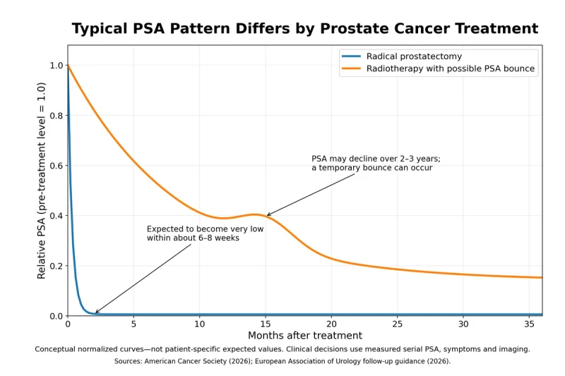
Prostate cancer PSA levels are interpreted according to whether the patient is undergoing active surveillance, has received surgery or radiotherapy, is taking androgen-deprivation therapy or has undergone focal treatment. The expected PSA pattern differs in each setting, so screening thresholds such as 4 ng/mL should not be used to assess treatment response or recurrence.[6,11]
During Active Surveillance
Active surveillance is used for selected patients whose cancer is considered unlikely to cause immediate harm. PSA is monitored alongside magnetic resonance imaging, clinical assessment and repeat biopsy to detect evidence that the tumour is becoming more extensive or biologically aggressive.[11]
PSA is commonly measured at least every six months, although the interval may be individualised according to Grade Group, PSA density, MRI findings, age, health status and the surveillance protocol used in the patient’s country. A digital rectal examination may be performed periodically, while repeat MRI and biopsy are scheduled according to baseline risk and subsequent changes.[11]
A rising PSA should trigger further assessment rather than automatic treatment. PSA can increase because of benign prostate growth, inflammation or biological variation while the tumour remains unchanged. A shortening PSA doubling time may justify earlier MRI and biopsy, but a change from surveillance to active treatment should usually be based on confirmed pathological progression, increasing tumour volume or other adverse findings rather than PSA alone.[11]
A stable PSA is reassuring but does not eliminate the need for scheduled imaging or biopsy. Some tumours can undergo Grade Group progression without a marked PSA increase, particularly when the cancer produces relatively little antigen. Continued surveillance therefore depends on agreement between PSA, MRI, pathology and clinical findings.[11]
After Radical Prostatectomy
Radical prostatectomy removes the prostate and seminal vesicles, leaving very little normal tissue capable of producing PSA. The level is therefore expected to fall to an undetectable or extremely low concentration, generally within approximately two months after surgery.[11]
A PSA that never becomes undetectable is described as persistent PSA and may indicate residual benign tissue, residual local cancer or disease outside the surgical field. The probability of clinically important residual disease depends on the PSA value, postoperative trend, surgical-margin status, pathological stage, lymph-node findings and Grade Group.[11,12]
A PSA that initially becomes undetectable and later begins to rise may indicate biochemical recurrence. A commonly used definition after prostatectomy is PSA of at least 0.2 ng/mL followed by a second confirmatory value at or above that level. Lower detectable values measured with an ultrasensitive assay may justify closer follow-up, particularly in patients with adverse pathological findings, but they should be confirmed before recurrence is declared.[12]
Biochemical recurrence does not necessarily mean that visible metastatic disease is present. PSA often rises before recurrent cancer can be identified on conventional imaging. The time from surgery to recurrence, PSA doubling time, Grade Group, pathological stage and surgical margins help estimate whether the likely source is confined to the prostate bed or represents disease elsewhere.[11,12]
Salvage radiotherapy is generally more effective when delivered while PSA remains low. A confirmed postoperative rise should therefore prompt timely specialist review rather than waiting for PSA to reach the screening threshold used in an untreated prostate. Imaging may assist treatment planning, but a negative scan at a low PSA does not exclude microscopic recurrent disease.[12]
After Radiotherapy
Radiotherapy leaves the prostate in place, so benign prostate cells continue to produce PSA. The level is not expected to become immediately undetectable and may decline gradually over many months or several years before reaching its lowest value, known as the PSA nadir.[6,11]
The lowest PSA achieved and the subsequent trend are more informative than whether the result falls below 4 ng/mL. A gradual decline is generally favourable, while a sustained upward pattern may raise concern for persistent or recurrent cancer. Interpretation should consider whether androgen-deprivation therapy was given with radiotherapy because hormonal suppression can produce an additional temporary reduction.[6,11]
Some patients experience a temporary PSA bounce after external-beam radiotherapy or brachytherapy. PSA rises for a limited period and then falls again without additional cancer treatment. A bounce can be difficult to distinguish from early recurrence from one measurement, making repeat testing and evaluation of the overall trajectory essential.[11]
Biochemical recurrence after radiotherapy is commonly defined as a PSA rise of 2 ng/mL or more above the post-treatment nadir. This definition differs from the postoperative threshold because the irradiated prostate remains capable of producing PSA.[11]
A patient may still require assessment before the formal biochemical-recurrence threshold is reached when PSA rises consistently, doubling time is short, symptoms develop or the original cancer had high-risk features. Magnetic resonance imaging, prostate-specific membrane antigen imaging and biopsy may be considered when the findings are likely to influence salvage treatment.[11]
During Androgen-Deprivation Therapy
Androgen-deprivation therapy lowers testosterone, which can suppress androgen-sensitive prostate cancer and produce a substantial PSA decline. PSA should be assessed together with serum testosterone because a low PSA has a different meaning when tumour activity is being hormonally suppressed.[11]
The depth and duration of the PSA response can provide prognostic information, but a falling PSA does not independently establish that all cancer has been eliminated. Residual tumour cells may remain controlled while treatment continues and can later become less responsive to hormonal suppression.[6,11]
A rising PSA during treatment requires confirmation and assessment of whether testosterone has reached the intended suppressed range. Inadequate testosterone suppression may result from missed doses, delayed injections, absorption problems or treatment-related factors that require correction.[11]
A confirmed PSA rise despite adequately suppressed testosterone may indicate progression toward castration-resistant prostate cancer. The next assessment may include symptom review, blood tests and imaging, but treatment should not be changed from PSA progression alone when imaging and clinical findings remain stable. The complete pattern of biochemical, radiological and symptomatic change guides management.[11]
Patients receiving androgen-deprivation therapy also require monitoring for treatment-related effects, including bone loss, metabolic changes, cardiovascular risk, anaemia, hot flushes, fatigue, sexual dysfunction and loss of muscle mass. PSA follow-up should therefore form part of a broader review rather than being the only measure of treatment success.[11]
After Focal Therapy
Focal therapies such as high-intensity focused ultrasound, cryotherapy and irreversible electroporation treat selected regions of the prostate while leaving other prostate tissue intact. PSA usually falls after treatment but remains detectable because untreated benign tissue continues to produce the antigen.[11]
There is no single universally accepted PSA threshold that confirms recurrence after focal treatment. Interpretation considers the percentage decline from baseline, the lowest post-treatment value, subsequent PSA trend, prostate volume and the amount of tissue treated.[11]
A favourable PSA response cannot confirm complete ablation of the targeted tumour or exclude cancer in untreated areas. Follow-up commonly includes multiparametric magnetic resonance imaging and targeted or systematic biopsy according to the treatment protocol and individual risk.[11]
A rising PSA, new suspicious MRI lesion or adverse biopsy finding may indicate residual cancer in the treated region, recurrence at the treatment margin or a separate tumour elsewhere in the gland. Treatment decisions should therefore be based on anatomical and pathological confirmation rather than PSA alone.[11]
Biochemical Recurrence
Biochemical recurrence means that PSA has risen according to treatment-specific criteria after an initial attempt at local disease control. It does not automatically mean that the patient has symptoms, visible tumour growth or metastatic cancer.[11,12]
Risk is greater when recurrence occurs soon after treatment, PSA doubles rapidly, the original tumour had a high Grade Group or pathological stage, or surgical margins and lymph nodes showed adverse findings. A slowly rising PSA many years after treatment may follow a less aggressive course, although continued assessment remains necessary.[11,12]
Prostate-specific membrane antigen positron emission tomography can identify recurrent disease at lower PSA values than many conventional imaging methods. Detection becomes more likely as PSA rises, but a negative scan cannot exclude microscopic cancer. Imaging should be performed when the result is likely to alter salvage radiotherapy, systemic treatment or another management decision.[11,12]
New bone pain, unexplained weight loss, urinary obstruction, haematuria, leg weakness or neurological symptoms require clinical evaluation regardless of the PSA value. Some aggressive prostate cancers produce relatively little PSA, so symptoms and imaging remain important even when the biochemical change appears modest.[6,11]
Do Lower Prostate Cancer PSA Levels Mean Cure?
Lower prostate cancer PSA levels are usually an encouraging biochemical finding, but they do not independently prove that every malignant cell has been eliminated. PSA can decrease because prostate inflammation has resolved, benign tissue has become less active, medication has altered antigen production or cancer treatment has temporarily suppressed tumour activity. The result must therefore be interpreted with the treatment received, the lowest PSA achieved, the duration of response, imaging, pathology and continued follow-up.[1,6]
Why PSA Can Fall Without Tumour Elimination
PSA may decline after prostatitis, urinary infection or acute urinary retention has resolved. In this setting, the reduction may reflect recovery from inflammation or pressure on prostate tissue rather than any change in an undiagnosed cancer. A substantial fall after treatment of infection is reassuring, but persistent elevation, an abnormal prostate examination or suspicious imaging may still require urological investigation.[1,6]
Normal biological and laboratory variation can also produce a modest change between tests. Differences in testing platform, recent ejaculation, cycling, prostate manipulation and sample timing can make one value lower than another. A small reduction should therefore be confirmed through serial measurements obtained under comparable conditions before it is interpreted as a durable clinical response.[6]
Finasteride and dutasteride can reduce PSA by shrinking benign prostate tissue and suppressing PSA production. The lower result does not necessarily mean that the patient’s cancer risk has disappeared. Clinicians must interpret the measured value according to the medicine used, treatment duration, baseline PSA and subsequent trend.[1,6]
A reduction in benign prostate volume can also lower PSA even when a separate cancer focus remains present. This is particularly important in men with both benign prostatic hyperplasia and prostate cancer because the blood result reflects antigen released from the entire gland rather than from malignant tissue alone.
PSA Response During Cancer Treatment
After radical prostatectomy, PSA is expected to fall to an undetectable or extremely low concentration because almost all PSA-producing prostate tissue has been removed. A sustained undetectable result is favourable, but continued testing remains necessary because microscopic residual disease may not produce a measurable PSA immediately after surgery.[6]
After radiotherapy, PSA usually falls gradually because benign prostate tissue remains in the body. The lowest value reached, known as the nadir, and the subsequent trend are more clinically useful than expecting PSA to become completely undetectable. A favourable nadir indicates treatment response but does not independently establish permanent tumour eradication.[6]
Androgen-deprivation therapy can cause a rapid and substantial PSA decline by suppressing testosterone-dependent tumour activity. Cancer cells may remain viable while their growth and PSA production are controlled. A very low PSA during hormonal treatment therefore represents biochemical suppression and should be interpreted with serum testosterone, imaging, symptoms and the duration of response.[6]
Some prostate cancers can become less dependent on androgen signalling and progress despite a low or only moderately rising PSA. This is uncommon but clinically important, particularly in aggressive or previously treated disease. New pain, weight loss, urinary obstruction or neurological symptoms require assessment even when the blood marker appears relatively favourable.
Focal therapies leave untreated prostate tissue in place, so PSA usually declines without becoming undetectable. The magnitude of reduction cannot establish whether the treated lesion has been completely ablated or whether cancer remains in another part of the gland. Magnetic resonance imaging and protocol-directed biopsy may still be required to assess local control.
Evidence Needed to Confirm Disease Control
A meaningful PSA response should persist across several measurements rather than appear on one isolated report. The lowest value achieved, the time required to reach it, the duration of stability and any later rise help distinguish sustained disease control from temporary suppression or ordinary biological fluctuation.[6]
The interpretation must agree with the patient’s original diagnosis. A falling PSA is more reassuring when imaging shows regression or stability, symptoms improve appropriately and repeat pathology does not identify residual or progressive malignancy. A favourable blood result is less reliable when MRI remains suspicious, biopsy shows higher-grade disease or new symptoms suggest progression.
After surgery, durable control is assessed through sustained very low or undetectable PSA and the absence of clinical or radiological recurrence. After radiotherapy, assessment relies on the PSA nadir, treatment-specific recurrence criteria and long-term follow-up. During active surveillance, PSA stability does not mean that the existing cancer has been cured because the tumour remains under observation and scheduled MRI or biopsy is still required.
A negative prostate biopsy provides stronger anatomical evidence than PSA alone but is not an absolute guarantee that no cancer remains. Biopsy samples selected areas of the gland and may miss a small residual focus. Confidence increases when repeated pathology, imaging, PSA behaviour and long-term clinical follow-up remain concordant.
Cure should therefore be described through objective and durable evidence appropriate to the treatment setting. This may include sustained biochemical control, no clinically significant malignancy on indicated pathology, no suspicious or progressive disease on imaging, no metastatic findings and sufficiently long follow-up without recurrence. The required evidence differs between a patient treated with surgery, radiotherapy, focal therapy, systemic treatment or a personalised non-surgical protocol.
PSA Reduction During Ayurvedic Care
When PSA declines during personalised Ayurvedic treatment, the change should be documented as a biochemical response rather than presented immediately as proof of cure. The result becomes more clinically meaningful when it is reproduced on serial testing and accompanied by stable or improving MRI findings, favourable pathology where repeat biopsy is indicated, reduced disease burden and continued absence of progression.
A physician-directed Ayurvedic plan may also improve urinary inflammation, bladder function, bowel regularity, sleep, appetite or treatment tolerance. Improvement in these outcomes can be valuable, but symptom relief does not confirm that prostate adenocarcinoma has regressed. Benign urinary symptoms and malignant disease can change independently.
PSA testing should continue through the same urologic or oncologic pathway while Ayurvedic care is being administered. Prescribed surgery, radiotherapy, hormonal treatment or active-surveillance investigations should not be stopped solely because one PSA result has fallen. Any proposed treatment change requires review of the full disease profile rather than reliance on the blood marker alone.
An unexpected or unusually rapid reduction should be verified using the same laboratory where practical. The clinician should review recent infection treatment, urinary retention, finasteride or dutasteride use, hormonal medicines, laboratory units and any change in testing conditions. Confirming these factors prevents a treatment effect from being overstated or an important residual risk from being overlooked.[1,6]
PSA Monitoring Within Complete Prostate Cancer Treatment
Prostate cancer PSA levels are an essential monitoring tool, but they should never determine treatment in isolation. A complete treatment plan should integrate serial PSA measurements with prostate MRI, biopsy findings, Gleason score, Grade Group, clinical stage, PSA density, urinary symptoms, prostate volume, previous treatment and the patient’s overall health.
A falling PSA may indicate favourable biochemical activity, while a stable PSA can support continued monitoring in selected patients. However, neither result independently proves that prostate cancer has disappeared. Treatment response becomes more meaningful when PSA findings agree with imaging, pathology where indicated, clinical examination and long-term absence of disease progression.
For patients considering a personalised Ayurvedic approach, treatment should remain disease-directed and objectively monitored. The complete framework, including Ayurvedic assessment, personalised herbal and herbo-mineral formulation, published clinical evidence, safety considerations, treatment goals and long-term monitoring, is explained in our Ayurvedic treatment for prostate cancer guide.
The objective is not merely to reduce the PSA number. Treatment should pursue measurable disease control through appropriate PSA trends, stable or improving imaging, favourable pathology when repeat biopsy is clinically indicated, improvement in urinary function and absence of regional or distant progression.
Prostate Cancer PSA Levels in Complete Care
Prostate cancer PSA levels are most useful when they are interpreted as one part of a complete disease assessment. PSA can show biochemical change, but treatment planning requires agreement between the blood trend, magnetic resonance imaging, biopsy findings, Grade Group, clinical stage, symptoms, prostate volume, previous treatment and the patient’s overall health.[1,2,6]
Building the Complete Disease Profile
The starting point is the original diagnostic evidence. Patients should retain every dated PSA report, free and total PSA result, magnetic resonance imaging report, biopsy report, Gleason score, Grade Group, clinical stage and staging scan. Reviewing these records together helps distinguish a small, lower-grade tumour from biologically aggressive or more extensive disease, even when the PSA values appear similar.[1,6]
The direction of PSA change is more clinically informative than one isolated result. A stable value may support continued monitoring in a carefully selected patient, while a persistent rise may justify repeat imaging, biopsy or treatment review. The significance of that rise depends on prostate size, infection, medication use, testing conditions and whether the prostate remains present after treatment.[1,2,6]
Magnetic resonance imaging contributes anatomical information that PSA cannot provide. It can identify suspicious regions, estimate prostate volume, support PSA-density calculation and assess possible extension beyond the gland. Biopsy provides the pathological information required to confirm malignancy and determine Grade Group, while staging investigations evaluate lymph-node, bone or organ involvement when the overall risk warrants them.[1,2,6]
Symptoms should be recorded separately from tumour markers. Urinary frequency, weak flow and incomplete emptying may improve because inflammation or benign enlargement has reduced even when cancer remains present. Conversely, a patient may feel well despite a rising PSA or pathological progression. Objective monitoring is therefore required even when urinary function, appetite, sleep and general strength improve.
Selecting Treatment From the Combined Risk
Treatment choice should reflect the combined disease profile rather than the PSA result alone. Selected patients with low-volume, lower-grade cancer may be suitable for active surveillance, while others may require surgery, radiotherapy, focal treatment, hormonal treatment or systemic therapy. Age, life expectancy, urinary function, sexual function, cardiovascular risk, bone health and patient preferences also influence the decision.[1,2,6]
A patient with PSA below 10 ng/mL may still require definitive treatment when biopsy shows a high Grade Group or imaging suggests extension beyond the prostate. Another patient with PSA above 10 ng/mL may have a large benign prostate and a small lower-grade tumour. These differences explain why treatment cannot be selected from a PSA threshold without reviewing pathology and imaging.
Active surveillance is not the absence of treatment planning. It is a structured monitoring strategy that includes serial PSA, clinical review, magnetic resonance imaging and repeat biopsy according to the patient’s risk. A change in PSA may trigger reassessment, but confirmed pathological or radiological progression usually carries greater weight when deciding whether to begin active treatment.[2]
After definitive treatment, the expected PSA pattern depends on the procedure received. Following radical prostatectomy, PSA should fall to a very low or undetectable concentration. After radiotherapy or focal therapy, benign prostate tissue remains and PSA may continue to be measurable. Treatment response and recurrence must therefore be assessed through treatment-specific criteria rather than the screening thresholds used before diagnosis.[6]
Coordinating Ayurvedic and Oncology Care
Personalised Ayurvedic treatment should be planned from the same diagnostic evidence used in urological and oncological care. The physician should review PSA history, Grade Group, clinical stage, imaging, biopsy burden, urinary symptoms, current cancer treatment, liver and kidney function, blood counts, comorbidities and prescribed medicines before selecting a formulation.
The therapeutic objectives may include improving urinary function, reducing inflammation, supporting appetite and strength, maintaining bowel regularity, improving treatment tolerance and pursuing measurable disease control. Disease-directed claims should be assessed through serial PSA, repeat imaging, pathology when clinically indicated and sufficiently long follow-up rather than symptom improvement alone.
Ayurvedic treatment should not obscure the PSA pattern through unsupervised use of products intended only to lower the laboratory value. A falling PSA is useful when it reflects genuine and durable disease control, but it becomes misleading when medication effects, infection treatment, hormonal suppression or inconsistent laboratory conditions are not documented.
Conventional treatment should not be stopped solely because PSA declines during Ayurvedic care. Surgery, radiotherapy, androgen-deprivation therapy, systemic treatment or active-surveillance investigations should be reviewed with the treating specialist using the complete disease profile. Coordinated care reduces the risk of delaying treatment for aggressive cancer while allowing supportive and disease-directed Ayurvedic management to be evaluated objectively.
The companion pillar article, Ayurvedic Treatment for Prostate Cancer, explains the broader treatment framework, formulation principles, published clinical evidence, safety requirements and long-term monitoring used in personalised care.
Measuring Objective Treatment Response
A credible treatment response requires repeated and concordant findings. PSA should decline or remain appropriately stable across serial measurements, imaging should show stability or regression, and repeat pathology should be favourable when biopsy is clinically indicated. Absence of new lymph-node, bone or organ disease strengthens the evidence for sustained control.
The lowest PSA achieved should be documented together with the time required to reach it. A rapid fall can be encouraging, but its meaning depends on whether the patient received surgery, radiotherapy, hormonal treatment, infection treatment or another intervention capable of changing PSA production. The duration of stability is often more clinically valuable than the magnitude of one early reduction.
A rising PSA should prompt verification before treatment failure is declared. Testing conditions, laboratory method, infection, urinary retention, recent procedures and medication changes should be reviewed. When the rise is confirmed, the next step may include magnetic resonance imaging, prostate-specific membrane antigen imaging, repeat biopsy or specialist treatment review according to the original diagnosis and treatment received.[2,6]
Cure should be assessed through objective evidence appropriate to the clinical setting. This may include durable biochemical control, no clinically significant malignancy on indicated pathology, no progressive disease on imaging, absence of metastatic findings and long-term follow-up without recurrence. PSA is central to this assessment, but it cannot establish cure independently.
Records Needed for Personalised Assessment
A patient seeking a complete treatment review should provide all PSA reports in chronological order rather than only the latest result. Free PSA, PSA density, prostate volume and testosterone levels should be included when available because they may substantially change interpretation.
The prostate magnetic resonance imaging report should include the date, gland volume, lesion location, PI-RADS score and any comment on capsular, seminal-vesicle or lymph-node involvement. The pathology report should include the cancer type, Gleason pattern, Grade Group, number of positive cores, percentage involvement and any adverse pathological features.
Previous treatment records should document surgery, radiotherapy, focal therapy, hormonal injections, anti-androgen medicines, chemotherapy, targeted therapy and their dates. Current prescriptions, supplements and Ayurvedic medicines should also be listed because some treatments can alter PSA, liver function, kidney function, blood counts or bleeding risk.
Baseline and follow-up blood tests should include complete blood count, liver function, kidney function and other investigations selected according to the treatment being considered. This complete documentation allows PSA changes to be interpreted accurately and enables conventional and Ayurvedic treatment decisions to be measured against the same objective clinical outcomes.
Prostate Cancer PSA Levels in an Ayurvedic Case
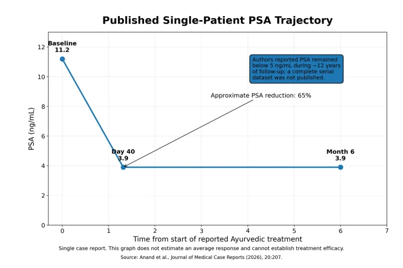
Prostate cancer PSA levels declined from 11.2 ng/mL to 3.9 ng/mL in a published case involving a 45-year-old man of Indian origin living in the United States. The biochemical response was accompanied by negative follow-up biopsies, improvement in urinary and quality-of-life outcomes, and an approximately 12-year period without reported relapse. These findings provide clinically relevant evidence from one patient but cannot establish the expected response for other men with prostate cancer.[3]
| Treatment or monitoring setting | Expected PSA pattern | Finding requiring further assessment |
|---|---|---|
| Active surveillance | PSA may remain detectable and relatively stable because the prostate and diagnosed tumour remain present | Persistent rise, shortening doubling time, increasing PSA density, suspicious MRI or pathological progression on repeat biopsy |
| Radical prostatectomy | PSA should fall to a very low or undetectable concentration because most PSA-producing tissue has been removed | AUA defines biochemical recurrence as PSA of at least 0.2 ng/mL on two separate determinations |
| Radiotherapy | PSA declines gradually and may take two to three years or longer to reach its lowest level | Biochemical recurrence is commonly defined as PSA rising at least 2 ng/mL above the post-treatment nadir |
| PSA bounce after radiotherapy | A temporary increase may occur before PSA falls again | Consecutive rises, short doubling time or continued increase require assessment rather than assuming a benign bounce |
| Androgen-deprivation therapy | PSA commonly falls as testosterone-sensitive tumour activity is suppressed | Rising PSA despite adequately suppressed testosterone may indicate treatment-resistant disease |
| Focal therapy | PSA usually falls but remains detectable because untreated prostate tissue remains | No universal PSA recurrence threshold; serial PSA, MRI and protocol-directed biopsy are required |
| Biochemical recurrence without visible disease | PSA may rise before recurrent cancer becomes visible on imaging | Assess doubling time, original Grade Group, stage, treatment history and whether imaging will alter salvage treatment |
| Ayurvedic or integrative treatment | A favourable PSA decline may indicate biochemical response | PSA reduction must be confirmed through serial tests and interpreted with imaging, pathology and long-term absence of progression |
Baseline Diagnosis
The patient had experienced urinary frequency, urgency, incomplete bladder emptying, nocturia, difficulty initiating urination and difficulty maintaining a steady urinary stream for approximately six months. He also reported recurrent urinary infections, abdominal distension and long-standing constipation. Rectal examination identified an enlarged prostate with firm, discrete nodules, while the baseline PSA was approximately 11.2 ng/mL.[3]
Transrectal ultrasonography showed multiple hypoechoic areas but did not identify invasion into surrounding structures. Biopsy mapping confirmed adenocarcinoma in the right mid-region of the prostate. The reported cancer focus measured 0.06 cm, involved approximately 2% of the sampled specimen and was identified in one of the two cores obtained from that region.[3]
Histopathology showed Gleason 3+3=6 prostate adenocarcinoma, corresponding to Grade Group 1. The reported clinical stage was T1, indicating that the cancer was not clinically apparent as a clearly palpable or visible tumour confined to a defined anatomical area. The available assessment did not identify distant metastasis.[3]
The small tumour focus and Gleason 3+3 pattern represented favourable diagnostic features, but the PSA of 11.2 ng/mL was higher than ordinarily expected for a very small low-grade lesion. This difference made serial PSA, repeat pathology and long-term follow-up particularly important because PSA could have reflected a combination of malignant activity, benign enlargement and inflammatory prostate changes.
The patient was advised to undergo radical prostatectomy but expressed concern about removal of the prostate and elected to pursue Ayurvedic treatment after discussion of the potential benefits and risks. The reported outcome therefore reflects an individual treatment decision in early-stage, low-volume disease and should not be extrapolated to men with high-grade, locally advanced or metastatic prostate cancer.[3]
Personalised Ayurvedic Treatment
The treatment approach was based on the patient’s urinary obstruction, dysuria, constipation, abdominal distension, appetite and broader metabolic presentation. The authors used the Ayurvedic frameworks of Mūtrakṛcchra, meaning difficult or painful urination; Udāvarta, a disturbance associated with constipation and abnormal upward movement of physiological functions; and Mūtragranthi, a classical description of glandular swelling or obstruction affecting the urinary pathway.[3]
These Ayurvedic concepts were used to guide individualised treatment and were not presented as exact equivalents of prostate adenocarcinoma. Modern diagnosis continued to depend on PSA, ultrasonography and biopsy-confirmed histopathology.[3]
The initial oral regimen included Chirivilwadi Kashayam, Laghu Lokeswara Rasa, Vasthyamayanthaka Kashaya Churnam and Karpoora Silajithu Bhasma. Treatment was subsequently modified according to appetite, urinary control, bowel function and clinical response. Chukkuchundadi Kashaya, Vasthyamayanthaka Ghrita and Chandraprabha Gutika were introduced during later stages of management.[3]
The patient received seven formulations across the treatment period rather than one fixed medicine. Some were polyherbal preparations, while others were herbo-mineral formulations requiring specialised pharmaceutical preparation and professional supervision. The response therefore cannot be attributed reliably to one herb, one mineral ingredient or one formulation.[3]
The reported prescriptions were selected for this patient’s constitution, digestion, urinary symptoms, bowel pattern and disease findings. They should not be copied for self-treatment because herbo-mineral medicines can differ in composition, purification, manufacturing quality and dose, and may interact with prescribed medicines or be unsuitable in impaired liver or kidney function.
PSA Response
After approximately 40 days of Ayurvedic treatment, PSA decreased from 11.2 ng/mL to 3.9 ng/mL. This represented an absolute reduction of 7.3 ng/mL and an approximate 65% decline from baseline.[3]
The percentage reduction describes the change in the blood marker and does not mean that tumour size decreased by 65%. PSA can arise from malignant tissue, benign prostate enlargement and inflammation, so the biochemical change required confirmation through pathology and continued monitoring.
At the six-month follow-up, PSA reportedly remained stable at 3.9 ng/mL. The patient was asymptomatic, had regained normal daily activity and had gained approximately 3 kg. Stability across repeated measurements provided stronger evidence of a sustained biochemical response than a single low value would have provided.[3]
The authors later reported that PSA remained below 5 ng/mL during approximately 12 years of follow-up. Because the prostate remained in place, a detectable PSA was expected. Long-term stability, rather than complete PSA disappearance, was therefore the clinically relevant pattern.[3]
Follow-Up Biopsy
A subsequent 12-core prostate biopsy reportedly showed benign prostatic hyperplasia in every sampled core, with no malignant cells detected. Because one negative biopsy can miss a small residual tumour, the patient was advised to undergo another biopsy, and the authors reported that the second biopsy also did not detect cancer.[3]
The negative biopsies added objective pathological evidence beyond the PSA reduction. However, prostate biopsy samples selected areas rather than the entire gland, so a negative result means that cancer was not identified in the tissue obtained; it cannot provide the same whole-gland assessment as examination of a surgically removed prostate.[1,6]
Confidence in disease regression was strengthened by the combination of two reported negative biopsies, sustained PSA control and prolonged follow-up without documented relapse. Each finding supports the others, whereas any one of them would be less conclusive when considered alone.[3]
Urinary and Quality-of-Life Outcomes
The patient’s urinary symptoms improved during treatment. The published Expanded Prostate Cancer Index Composite for Clinical Practice assessment recorded better urinary control, reduced pain and frequency, and improvement from substantial urinary leakage to total urinary control.[3]
Bowel symptoms also improved, with the reported urgency and frequent bowel movements resolving after treatment. These changes were clinically relevant because chronic constipation and abdominal distension formed part of the patient’s initial presentation and influenced the personalised Ayurvedic strategy.[3]
Sexual-function scores improved from very poor or absent orgasmic ability to a good level and from erections insufficient for sexual activity to erections described as firm enough for intercourse. These patient-reported outcomes suggest meaningful improvement in quality of life, although retrospective reporting and the absence of a comparison group limit the certainty with which the changes can be attributed to treatment.[3]
No adverse drug reactions were reported during or after the documented treatment period. This observation applies only to the treated patient and does not establish that the formulations are universally safe, particularly when herbo-mineral preparations are manufactured, prescribed or monitored under different conditions.[3]
Clinical Interpretation
The outcome was supported by three distinct categories of evidence: a large and sustained PSA reduction, repeated biopsies without detected malignancy and long-term follow-up without reported relapse. Improvement in urinary, bowel and sexual function provided additional patient-centred evidence of clinical recovery.[3]
The baseline tumour was small, unifocal and Gleason 3+3=6. Lower-grade prostate cancers can follow an indolent course, and small lesions can be missed during repeat sampling. The case therefore cannot determine how the same treatment would perform in patients with Grade Group 2 to 5 disease, extracapsular extension, lymph-node involvement or distant metastasis.
The report involved one patient and did not include a control group, randomisation or comparison with active surveillance or conventional treatment. Seven Ayurvedic medicines were prescribed at different stages, making it impossible to identify the independent effect of any one formulation. The authors also acknowledged that these specific combinations have not been validated as prostate cancer treatments in randomised controlled trials.[3]
The PSA decline occurred early, while pathological confirmation required subsequent biopsies and extended observation. This sequence demonstrates why PSA reduction should be treated as an initial response marker rather than immediate proof of cure. Stronger evidence developed only after the biochemical response remained stable and the later pathological assessments did not detect malignancy.
For an individual patient, comparable monitoring would require dated serial PSA measurements, prostate imaging, repeat pathology when clinically indicated, assessment for regional or distant disease, and sufficiently long follow-up. Symptom relief alone would be inadequate because urinary function can improve even when malignant tissue remains.
The case supports further clinical investigation of carefully standardised and monitored Ayurvedic treatment for selected prostate cancer patients. It does not provide a universal replacement for active surveillance, surgery, radiotherapy, hormonal treatment or systemic therapy, and its findings should be applied only after reviewing tumour grade, stage, PSA behaviour, imaging and the patient’s overall risk.[3]
Prostate Cancer PSA Levels and Ayurvedic Formulation
Prostate cancer PSA levels can be monitored during personalised Ayurvedic treatment, but the prescription must be based on the complete disease profile rather than the PSA value alone. Varuna–Kanchanara Rasayana Avaleha is a physician-designed formulation that may be considered after reviewing biopsy findings, Grade Group, clinical stage, magnetic resonance imaging, prostate volume, urinary symptoms, current oncology treatment, liver and kidney function, blood counts, digestion and concurrent medicines. It is not a ready-made classical formula or a fixed prescription for every patient.
Classical and Pharmaceutical Basis
Avaleha is a semi-solid Ayurvedic dosage form prepared by concentrating a medicinal decoction and incorporating prescribed powders and suitable excipients. Its pharmaceutical principles are described in Sharangadhara Samhita, Madhyama Khanda, Chapter 8, Avaleha Kalpana, which explains preparation, consistency, finishing characteristics, dose considerations and suitable accompanying liquids.[14]
Rasayana refers to a therapeutic approach intended to support tissue nourishment, strength, physiological resilience and recovery. Its foundational principles are described in Charaka Samhita, Chikitsa Sthana, Chapter 1, Rasayana Adhyaya. In prostate cancer care, Rasayana should not be reduced to a general tonic concept; it must be selected according to disease burden, digestive capacity, treatment-related weakness and measurable clinical objectives.[13]
The formulation design may also consider the classical descriptions of Granthi and Arbuda in Sushruta Samhita, Nidana Sthana, Chapter 11. Granthi refers broadly to a localised glandular or nodular swelling, while Arbuda describes a larger and more persistent growth pattern. These classical categories help organise Ayurvedic assessment but should not be presented as direct equivalents of biopsy-confirmed prostate adenocarcinoma, Gleason grade or modern tumour stage.[15]
Varuna–Kanchanara Rasayana Avaleha is therefore a contemporary individualised formulation developed from classical therapeutic and pharmaceutical principles. Its indication, composition and dose must arise from the patient’s modern diagnosis and Ayurvedic assessment rather than from the disease name alone.
Herbal Core
Varuna, botanically identified as Crataeva nurvala, and Kanchanara, identified as Bauhinia variegata, may form the principal gland-directed herbal base in selected patients. Their inclusion is considered when prostate enlargement, urinary obstruction, nodularity or a Granthi-like clinical pattern accompanies the confirmed diagnosis. They should not be described as independently proven cures for prostate cancer.
Punarnava, or Boerhavia diffusa, may be selected when urinary difficulty, fluid imbalance, swelling or impaired elimination is present. Gokshura, or Tribulus terrestris, may be added for urinary comfort and pelvic support, while Pashanabheda, or Bergenia ligulata, may be considered when the urinary presentation requires additional tract-directed treatment. Their proportions should change according to urine flow, residual urine, kidney function, infection history and the presence of obstruction.
Guduchi, or Tinospora cordifolia, and Amalaki, or Phyllanthus emblica, may be incorporated for Rasayana-oriented recovery when appetite, strength, treatment tolerance and tissue resilience require support. Guduchi is not appropriate for every patient, particularly when immune-mediated illness, altered liver function or interacting medication creates a safety concern.
Haritaki, Amalaki and Bibhitaki may be used together as Triphala when constipation, irregular bowel evacuation, abdominal distension or impaired digestive function contributes to pelvic discomfort. Bowel regulation can improve quality of life and reduce straining, but it should not be interpreted as evidence that the prostate tumour has regressed.
Purified Guggulu from Commiphora wightii, Shallaki from Boswellia serrata and Haridra from Curcuma longa may be considered when inflammatory, metabolic or tissue-remodelling objectives are clinically relevant. Their use requires review of anticoagulants, antiplatelet medicines, gastrointestinal sensitivity, liver function and planned surgery because herbal medicines can alter tolerability or interact with conventional treatment.
Pippali, Shunthi and Maricha may be used in carefully controlled quantities to support Agni, the Ayurvedic concept of digestive and metabolic capacity, and improve tolerance of a complex formulation. They may require reduction or exclusion in patients with reflux, gastritis, mucosal irritation, bleeding risk or marked Pitta-predominant symptoms.
The objective of this herbal design is not merely to force a lower laboratory value. It may be directed toward sustained PSA control, reduction of urinary obstruction, improvement in bladder emptying, maintenance of strength during treatment and measurable stability or regression on imaging and pathology where clinically reassessed.
Bhasma and Pishti Components
Bhasma refers to a specialised Ayurvedic preparation produced through prescribed purification, processing and controlled incineration of a metal, mineral or other substance. Pishti refers to a finely triturated preparation produced with specified liquid media, generally without the same incineration process used for bhasma. These preparations require substantially greater pharmaceutical control than ordinary herbal powders.
Selected patients may be assessed for microquantities of Swarna Bhasma, Heeraka Bhasma, Abhraka Bhasma, Yashada Bhasma, Vanga Bhasma, Swarna Makshika Bhasma, Rajata Bhasma or Lauha Bhasma. Praval Pishti and Mukta or Moti Pishti may be considered when mineral support, digestive tolerance or treatment-related debility forms part of the personalised indication.
These ingredients are not mandatory components of every prostate cancer prescription. Their selection depends on tumour risk, age, strength, anaemia, nutritional status, liver and kidney function, cardiovascular history, concurrent treatment and the physician’s assessment of whether the expected therapeutic value justifies their inclusion.
Traditional processing terminology does not by itself establish identity, purity, bioavailability or safety. Every botanical, bhasma and pishti ingredient should be traceable to an authenticated source and the completed batch should undergo appropriate identity, physicochemical, microbial, pesticide and contaminant testing. Products containing metal or mineral preparations require particular attention to elemental composition, dose consistency and toxicological quality control.[16,17]
Loose bhasma, unlabelled powders and formulations copied from another patient should not be used. Inadequately prepared or contaminated Ayurvedic products have been associated with clinically significant metal exposure, making batch documentation, qualified supervision and laboratory monitoring essential.[17]
Preparation and Personalisation
The herbal decoction component is prepared from prescribed coarse ingredients and concentrated under controlled conditions. The selected Avaleha base is incorporated at the appropriate stage, followed by ghee where clinically suitable. Fine herbal powders are added after the preparation reaches the required consistency, while heat-sensitive and physician-selected mineral components are incorporated according to the applicable pharmaceutical method.[14]
The finished medicine should have consistent texture, aroma, stability and dose uniformity. It should not be prepared at home because variations in temperature, concentration, ingredient identity, particle size and finishing stage can substantially change the final product.
A traditional sweetening base may be unsuitable for patients with uncontrolled diabetes, severe insulin resistance or a medically restricted carbohydrate intake. The dosage form, excipient quantity and total daily dose must therefore be adapted to the metabolic condition rather than assuming that every patient can tolerate a conventional Avaleha base.
Dose selection depends on body weight, digestive capacity, bowel function, treatment tolerance, stage of disease and concurrent medication. A patient receiving radiotherapy, androgen-deprivation therapy, anticoagulation or systemic cancer treatment may require a different composition and dose from a patient undergoing active surveillance.
The formulation should be reassessed when PSA changes unexpectedly, urinary retention develops, liver or kidney tests worsen, blood counts fall or a new conventional treatment begins. Continuing an unchanged prescription despite a changing disease profile can reduce effectiveness and increase avoidable risk.
Objective Treatment Monitoring
Baseline documentation should include dated PSA values, free PSA when available, prostate volume, PSA density, magnetic resonance imaging, biopsy findings, Grade Group, clinical stage, urinary function and current treatment. Complete blood count, liver function and kidney function provide additional safety data before a complex herbal or herbo-mineral formulation is prescribed.
Follow-up PSA should be obtained under comparable testing conditions and interpreted according to whether the prostate remains intact and whether surgery, radiotherapy, hormonal treatment or focal therapy has been used. A decline during Ayurvedic care should be recorded as a biochemical response until it is supported by sustained follow-up and concordant imaging or pathology.
A durable response may include an appropriate PSA reduction or stabilisation, improved urinary function, stable or regressing lesions on imaging, favourable pathology when repeat biopsy is clinically indicated and absence of new regional or distant disease. Symptom improvement without objective disease assessment is insufficient for determining tumour control.
A rising PSA requires verification of infection, retention, medication changes, laboratory variation and treatment adherence. When the increase persists, the patient may require repeat magnetic resonance imaging, prostate-specific membrane antigen imaging, biopsy or oncology reassessment rather than simply increasing the Ayurvedic dose.
Surgery, radiotherapy, androgen-deprivation therapy, systemic treatment or active-surveillance investigations should not be discontinued solely because PSA falls after beginning the formulation. Any change in the treatment plan should be based on the combined PSA trend, Grade Group, stage, imaging, pathology and specialist assessment.
When Prostate Cancer PSA Levels Need Urgent Review
Prostate cancer PSA levels do not usually represent an emergency by themselves, but a markedly elevated or rapidly rising result can require prompt specialist assessment when it is accompanied by abnormal examination findings, urinary obstruction, systemic illness, neurological symptoms or a previous prostate cancer diagnosis. Waiting for repeated routine testing may be unsafe when the overall clinical picture suggests significant obstruction, severe infection or advancing disease.[1,6]
Markedly Elevated or Rapidly Rising PSA
An unexpectedly high PSA should be reviewed promptly to confirm the result, identify temporary causes and determine whether imaging or biopsy is required. Values above 20 ng/mL carry greater concern for clinically significant cancer, while PSA above 50 or 100 ng/mL may indicate substantial tumour activity, particularly when the result is persistent and cannot be explained by acute infection, urinary retention or recent instrumentation.[1,6]
A rapid increase across several comparable tests can also justify earlier assessment, especially when prostate volume does not explain the change. The rate of rise should not be used alone to diagnose cancer, but it becomes more concerning when accompanied by a hard or irregular prostate, suspicious magnetic resonance imaging, low percent-free PSA, high PSA density or strong hereditary risk.[1,2]
A substantially elevated result should not be managed through repeated blood tests alone. Once temporary influences have been considered, persistent abnormality may require urological examination, multiparametric magnetic resonance imaging, biopsy and staging investigations selected according to the patient’s complete risk profile.[1,2,6]
Acute Urinary Obstruction
Inability to pass urine requires urgent medical assessment because prolonged retention can damage the bladder and kidneys. Lower abdominal pain, a visibly distended bladder, severe difficulty beginning urination or passage of only small amounts of urine can indicate significant obstruction.
Acute retention may raise PSA and can occur because of benign prostate enlargement, inflammation, medication effects or locally advanced cancer. Relief of the obstruction takes priority over interpreting the PSA result, and the marker may need to be repeated after bladder drainage and recovery.[1,6]
Reduced urine output accompanied by swelling, vomiting, confusion or worsening kidney function requires urgent investigation. These findings may indicate obstruction affecting the upper urinary tract and should not be managed solely with medicines intended to improve urinary flow.
Fever and Urinary Symptoms
Fever, chills, painful urination, pelvic pain and difficulty passing urine may indicate acute bacterial prostatitis or a complicated urinary infection. These conditions can cause a pronounced PSA increase and may progress to bloodstream infection if assessment and treatment are delayed.[1,6]
A patient with fever, weakness, rapid breathing, confusion, low blood pressure or inability to retain fluids requires emergency medical care. PSA interpretation should be deferred until the acute infection has been treated and the patient has recovered sufficiently for a clinically meaningful repeat measurement.
Antibiotics should not be prescribed merely to reduce an unexplained PSA when there are no symptoms, urine findings or clinical evidence of infection. Unnecessary antibiotic use can delay cancer investigation, cause adverse effects and contribute to antimicrobial resistance.
Visible Blood in Urine
Visible blood in the urine requires medical evaluation because it may arise from infection, stones, benign prostate enlargement, bladder disease, prostate cancer or another urinary tract condition. The presence of clots, difficulty passing urine or increasing lower abdominal pain can indicate blockage and requires urgent assessment.
Haematuria should not be attributed automatically to prostate cancer or to a raised PSA. Urine testing, imaging, cystoscopic assessment and prostate evaluation may be required to identify the bleeding source, particularly when the symptom persists or recurs.
Bone and Pelvic Symptoms
Persistent pain in the spine, hips, ribs or pelvis can require expedited assessment when prostate cancer is known or strongly suspected. Bone pain is not specific to metastatic disease, but new, progressive or night-predominant pain should not be managed by monitoring PSA alone.[6,11]
Unexplained weight loss, increasing fatigue, anaemia or declining physical function can add to concern when they occur with a rising PSA or high-risk biopsy findings. Clinical examination, blood tests and appropriate staging imaging may be necessary to determine whether the symptoms are related to cancer or another condition.
A low or moderately elevated PSA does not completely exclude advanced disease because some aggressive prostate cancers produce relatively little antigen. Symptoms and imaging therefore remain important even when the PSA value appears lower than expected for the patient’s clinical condition.[6,11]
Neurological Warning Signs
New leg weakness, numbness, difficulty walking, loss of sensation around the buttocks or inner thighs, or loss of bladder or bowel control requires emergency assessment. In a patient with known or suspected prostate cancer, these symptoms can indicate compression of the spinal cord or cauda equina by vertebral metastatic disease.[6,11]
Treatment is time-sensitive because delayed decompression can result in permanent neurological disability. The patient should not wait for a routine PSA appointment, repeat blood test or scheduled oncology review when these symptoms develop.
Rising PSA After Treatment
A confirmed PSA rise after radical prostatectomy, radiotherapy or other prostate cancer treatment requires timely review using treatment-specific recurrence criteria. The result may represent biochemical recurrence before symptoms or visible disease develop, and earlier assessment can preserve access to potentially effective salvage treatment.[11,12]
After prostatectomy, a detectable and rising PSA should not be allowed to approach the untreated screening threshold of 4 ng/mL before action is considered. Salvage radiotherapy is generally more effective when initiated at a low PSA in appropriately selected patients.[12]
After radiotherapy, a consistent upward trend, short PSA doubling time or rise of 2 ng/mL or more above the treatment nadir requires assessment for recurrence. Imaging and biopsy may be needed when the findings will influence local salvage treatment, hormonal therapy or systemic management.[11]
During androgen-deprivation therapy, rising PSA despite adequately suppressed testosterone may indicate treatment-resistant disease. Confirmation of testosterone suppression, review of treatment adherence, symptom assessment and updated imaging are required before the management plan is changed.[11]
When Repeat Testing Is Not Enough
Routine repeat testing is appropriate for some clinically stable patients with a mildly elevated PSA and a plausible temporary cause. It is not sufficient when the patient has urinary retention, systemic infection, visible haematuria, neurological symptoms, severe progressive pain, a suspicious prostate examination or a substantial post-treatment PSA rise.[1,6,11]
Prompt assessment does not mean that cancer or metastatic spread has already been confirmed. It allows urgent complications to be treated, clinically important disease to be identified earlier and unnecessary delay to be avoided while the cause of the PSA abnormality is established.
Questions About Prostate Cancer PSA Levels
Prostate cancer PSA levels should be discussed in relation to previous results, prostate size, symptoms, medicines, family history, imaging and biopsy findings. Asking focused questions helps patients understand whether the result requires repeat testing, magnetic resonance imaging, biopsy, staging or continued monitoring.[1,2,6]
Should My PSA Be Repeated?
A newly elevated PSA may be repeated when the patient is clinically stable and there is no urgent indication for immediate imaging or biopsy. Repeat testing can confirm whether the elevation persists after temporary influences such as urinary infection, prostatitis, retention, ejaculation, cycling or a recent prostate procedure have resolved.[1,2,6]
The appropriate interval depends on the PSA value, symptoms, examination findings and suspected cause of elevation. A markedly abnormal result, suspicious prostate examination or concerning symptoms may require prompt urological assessment rather than waiting for several repeat tests.[2,6]
Could Infection or Inflammation Have Raised My PSA?
Prostatitis and urinary tract infection can produce a temporary or substantial PSA increase. Fever, painful urination, pelvic discomfort, cloudy urine, urinary urgency or a sudden deterioration in flow may support an infectious or inflammatory cause, although inflammation can sometimes occur with few symptoms.[1,6]
Urine testing and clinical assessment may be required before the PSA is interpreted as a cancer-risk marker. A lower result after recovery is reassuring, but persistent elevation, an abnormal prostate examination or suspicious imaging may still require further investigation because infection and cancer can coexist.[1,6]
Is My PSA High for My Prostate Size?
A larger benign prostate can produce more PSA because it contains more PSA-secreting tissue. PSA density adjusts the blood result for prostate volume and can help determine whether the PSA is proportionate to benign enlargement or unexpectedly high for the size of the gland.[1,2]
Prostate volume is commonly measured through magnetic resonance imaging or transrectal ultrasound. A higher PSA density can increase concern for clinically significant cancer, while a lower density may support benign enlargement as a contributing factor without completely excluding malignancy.[1,2]
What Does My Percent-Free PSA Mean?
Percent-free PSA compares the amount of unbound PSA with total PSA. A lower percentage is associated with a greater likelihood of prostate cancer and can be especially useful when total PSA lies within the borderline range.[1]
A value of 10% or lower commonly strengthens the reason to discuss biopsy, while a value between 10% and 25% may also justify biopsy depending on age, prostate volume, family history, examination findings and magnetic resonance imaging. A higher percentage reduces estimated risk but cannot rule out clinically significant cancer.[1]
How Has My PSA Changed Over Time?
The pattern across several dated results is usually more informative than one isolated measurement. A stable PSA may support continued monitoring, while a persistent rise may justify additional assessment even when the absolute value remains below a traditional threshold.[1,2,6]
The clinician should determine whether the change represents a consistent trend or ordinary biological and laboratory variation. Infection, retention, medication changes, different laboratories and inconsistent testing conditions can create apparent rises that do not reflect cancer progression.[1,6]
Do I Need a Multiparametric MRI?
Multiparametric magnetic resonance imaging can identify suspicious areas, calculate prostate volume, estimate local tumour extent and guide targeted biopsy. It is commonly considered when PSA remains elevated after repeat testing or when the complete clinical risk profile raises concern for clinically significant cancer.[2,6]
A suspicious MRI increases the reason for biopsy but does not confirm malignancy. A non-suspicious scan lowers the estimated risk without eliminating it, particularly when PSA density is high, PSA continues to rise, examination findings are abnormal or hereditary risk is substantial.[2,6]
Do I Need a Prostate Biopsy?
Biopsy is considered when PSA, magnetic resonance imaging, examination findings and other risk factors indicate a meaningful probability of clinically significant cancer. It provides the tissue required to confirm malignancy and determine the Gleason pattern, Grade Group and amount of cancer present.[1,2,6]
MRI-targeted biopsy samples visible suspicious lesions, while systematic biopsy samples mapped areas throughout the gland. A combined approach may detect clinically important disease both within and outside the MRI-visible region.[2,6]
A negative biopsy means that cancer was not detected in the sampled tissue, but it does not guarantee that no tumour exists elsewhere in the prostate. Persistent clinical suspicion may require continued PSA monitoring, repeat imaging, additional biomarkers or another biopsy.[2,6]
What Are My Gleason Score and Grade Group?
The Gleason score describes the microscopic growth patterns seen in biopsy tissue, while Grade Group places the tumour into one of five categories of increasing biological aggressiveness. These findings are more directly related to tumour behaviour than PSA alone.[1,6]
Patients should ask whether the biopsy showed Grade Group 1 disease or a higher-grade cancer, how many cores were positive and how much tumour was present in each core. A small Grade Group 1 focus has different implications from extensive Grade Group 4 or 5 disease, even when both patients have similar PSA values.[1,6]
Has the Cancer Spread Beyond the Prostate?
PSA can increase suspicion of disease outside the prostate but cannot determine anatomical spread. Clinical stage is established through examination, magnetic resonance imaging, biopsy findings and additional staging investigations when the combined risk justifies them.[6]
Higher PSA, higher Grade Group, locally advanced findings or symptoms such as persistent bone pain may lead to prostate-specific membrane antigen imaging, computed tomography, bone imaging or another directed assessment. A high PSA does not independently prove metastatic disease, while a lower PSA does not completely exclude aggressive cancer.[6]
Could My Medicines Be Changing the PSA Result?
Finasteride and dutasteride can substantially lower measured PSA, making an apparently modest result more clinically important than it would be in an untreated patient. The clinician should know the medicine, dose, duration and PSA value before treatment began.[1,6]
Testosterone replacement, androgen-deprivation therapy and other hormonal treatments can also alter PSA behaviour. Herbal medicines and nutritional supplements should be disclosed because they may affect symptoms, laboratory findings, bleeding risk, liver function or interactions with prescribed treatment.
What Follow-Up Schedule Do I Need?
The follow-up interval depends on whether the patient is being screened, investigated for possible cancer, monitored through active surveillance or followed after treatment. The same schedule is not appropriate for every patient or every clinical setting.[2,6,11]
Patients undergoing active surveillance usually require serial PSA, periodic clinical review, magnetic resonance imaging and repeat biopsy according to their risk. After surgery, radiotherapy, hormonal treatment or focal therapy, follow-up should use treatment-specific expectations and recurrence criteria rather than untreated screening thresholds.[6,11,12]
Which Results Should Be Reviewed Before Ayurvedic Treatment?
Personalised Ayurvedic treatment should be planned only after reviewing all dated PSA values, free PSA, prostate volume, PSA density, magnetic resonance imaging, biopsy findings, Gleason score, Grade Group, clinical stage and previous treatment. Complete blood count, liver function, kidney function and the current medication list are also required for safety and treatment selection.
A PSA decline during Ayurvedic care should be evaluated through serial testing and supported by imaging, pathology when indicated and long-term follow-up. Surgery, radiotherapy, hormonal treatment or active-surveillance investigations should not be stopped solely because one PSA result has improved.
FAQs
Prostate cancer PSA levels estimate the probability of prostate disease but cannot confirm cancer, determine its stage or prove cure without supporting clinical evidence. Interpretation depends on age, previous PSA results, prostate volume, medicines, magnetic resonance imaging, biopsy findings and the treatment already received.
What PSA Level Usually Indicates Prostate Cancer?
No single PSA level confirms prostate cancer. The likelihood of detecting cancer generally increases as PSA rises, but biopsy is required when tissue confirmation is clinically indicated.
Is a PSA Level of 4 High?
A PSA of 4 ng/mL is above the investigation threshold used in some clinical pathways, but it is not a universal boundary between normal and cancerous results. Age, prostate size, previous values, symptoms and individual risk factors determine whether repeat testing or further investigation is needed.
What Is the Cancer Risk When PSA Is Between 4 and 10?
PSA between 4 and 10 ng/mL is commonly called the borderline range. Approximately one in four men biopsied within this range is found to have prostate cancer, while most have a benign or temporary cause of elevation.
Can Prostate Cancer Occur When PSA Is Below 4?
Prostate cancer can occur with PSA below 4 ng/mL. Further assessment may still be appropriate when PSA is rising, the prostate examination is abnormal, magnetic resonance imaging is suspicious or the patient has substantial familial, ancestral or genetic risk.
What Does a PSA Above 10 Mean?
PSA above 10 ng/mL is associated with a substantially higher probability of prostate cancer and normally requires timely urological assessment. Infection, benign enlargement, urinary retention and recent prostate procedures must still be considered before the result is attributed to malignancy.
Does PSA Above 20 Mean Advanced Prostate Cancer?
PSA above 20 ng/mL is an adverse risk feature if cancer is confirmed, but it does not independently prove locally advanced or metastatic disease. Biopsy, Grade Group, magnetic resonance imaging and staging investigations are required to establish the extent of cancer.
Does a PSA of 50 or 100 Mean the Cancer Has Spread?
A PSA of 50 or 100 ng/mL creates strong concern for substantial malignant activity, but metastatic spread cannot be diagnosed from PSA alone. Appropriate imaging is needed to determine whether disease is confined to the prostate, involves nearby structures or has reached distant sites.
Can Benign Prostate Enlargement Cause High PSA?
Benign prostatic hyperplasia can raise PSA because a larger prostate contains more PSA-producing tissue. Prostate cancer can coexist with benign enlargement, so persistent elevation should still be assessed according to the patient’s complete risk profile.
Can Prostatitis or Urinary Infection Raise PSA?
Prostatitis and urinary tract infection can cause temporary and sometimes marked PSA elevation. PSA is usually reassessed after the acute condition has resolved, although persistent elevation or abnormal clinical findings may still require imaging or biopsy.
What Percent-Free PSA Is Concerning?
A percent-free PSA of 10% or lower is generally associated with a higher likelihood of prostate cancer. Values between 10% and 25% may also support biopsy depending on age, prostate size, magnetic resonance imaging and other risk factors.
Is PSA Density More Accurate Than Total PSA?
PSA density can improve risk assessment by comparing total PSA with prostate volume. A higher density suggests that PSA is disproportionately elevated for the size of the gland, but it cannot diagnose or exclude cancer by itself.
Should an Elevated PSA Be Repeated Before Biopsy?
A newly elevated PSA is often repeated when the patient is clinically stable and there is no urgent concern. Markedly elevated PSA, an abnormal prostate examination, progressive symptoms or suspected advanced disease may require faster imaging and specialist assessment rather than routine retesting alone.
Can PSA Fall While Prostate Cancer Is Still Present?
PSA can fall even when malignant cells remain. Hormonal treatment, finasteride, dutasteride, reduced inflammation, radiotherapy and changes in benign prostate tissue can all lower PSA without proving complete tumour elimination.
Does a Lower PSA Mean Prostate Cancer Is Cured?
A lower PSA is an encouraging biochemical response but does not independently establish cure. Durable disease control requires treatment-specific PSA follow-up together with appropriate imaging, pathology, clinical assessment and absence of progression over time.
Can PSA Rise After Radiotherapy Without Cancer Recurrence?
A temporary PSA bounce can occur after radiotherapy or brachytherapy and may later resolve without additional cancer treatment. Recurrence is assessed from the sustained trend and the rise above the post-treatment nadir rather than from one elevated result.
What PSA Is Expected After Prostate Surgery?
After radical prostatectomy, PSA is expected to fall to an undetectable or extremely low level because most PSA-producing prostate tissue has been removed. A persistent or confirmed rising value requires assessment for residual or recurrent disease.
Can a Normal MRI Exclude Prostate Cancer?
A non-suspicious multiparametric MRI reduces the probability of clinically significant prostate cancer but does not eliminate it. Biopsy may still be considered when PSA density is high, PSA continues to rise, examination findings are abnormal or inherited risk is substantial.
Can a Negative Biopsy Exclude Prostate Cancer?
A negative biopsy means cancer was not found in the tissue sampled. It cannot guarantee that no tumour exists elsewhere in the prostate, so persistent clinical suspicion may require continued PSA monitoring, repeat imaging or another biopsy.
Do PSA Recommendations Differ Between Countries?
PSA screening ages, testing intervals and referral thresholds differ across the USA, UK, Canada, Singapore and Australia. The correct pathway depends on the patient’s country, symptoms, age, life expectancy and individual prostate cancer risk.
References
[1] American Cancer Society. (2023, November 22). Screening tests for prostate cancer.
https://www.cancer.org/cancer/types/prostate-cancer/detection-diagnosis-staging/tests.html
Brief: Explains PSA testing, factors that influence PSA levels, and additional tests used to investigate abnormal results.
[2] American Urological Association, & Society of Urologic Oncology. (2026). Early detection of prostate cancer: AUA/SUO guideline (Published 2023; amended 2026).
https://www.auanet.org/x18449.xml
Brief: Provides recommendations on shared decision-making, PSA screening, risk assessment, and evaluation before prostate biopsy.
[3] Anand, S., Anjana, C. S., Mythri, H. S., & Galib, R. (2026). Complete tumor regression with exclusive Ayurvedic treatment in adenocarcinoma of prostate: A case report. Journal of Medical Case Reports, 20, Article 207.
https://pmc.ncbi.nlm.nih.gov/articles/PMC13091260
Brief: Documents reported prostate tumour regression in one patient receiving Ayurvedic treatment. A single case cannot establish general effectiveness or a reproducible cure rate.
[4] Bell, N., Connor Gorber, S., Shane, A., Joffres, M., Singh, H., Dickinson, J., Shaw, E., Dunfield, L., Tonelli, M., & Canadian Task Force on Preventive Health Care. (2014). Recommendations on screening for prostate cancer with the prostate-specific antigen test. CMAJ, 186(16), 1225–1234.
https://pmc.ncbi.nlm.nih.gov/articles/PMC4216256
Brief: Evaluates PSA screening benefits and harms, including overdiagnosis, unnecessary biopsies, and treatment-related complications.
[5] Mason, R. J., Marzouk, K., Finelli, A., Saad, F., So, A. I., Violette, P. D., Breau, R. H., & Rendon, R. A. (2022). Update—2022 Canadian Urological Association recommendations on prostate cancer screening and early diagnosis: Endorsement of the 2021 Cancer Care Ontario guidelines on prostate multiparametric magnetic resonance imaging. Canadian Urological Association Journal, 16(4), E184–E196.
https://cuaj.ca/index.php/journal/article/view/7851
Brief: Updates Canadian screening and early diagnosis recommendations, including the role of multiparametric MRI in deciding whether a biopsy is needed.
[6] National Cancer Institute. (2025, January 31). Prostate-specific antigen test.
https://www.cancer.gov/types/prostate/psa-fact-sheet
Brief: Explains PSA testing for screening and monitoring, including why benign prostate enlargement and inflammation can also raise PSA.
[7] National Institute for Health and Care Excellence. (2026, April 15). Suspected cancer: Recognition and referral (NICE Guideline NG12).
https://www.nice.org.uk/guidance/ng12
Brief: Provides recommendations for recognising possible cancer and arranging investigations or specialist referral, including suspected prostate cancer.
[8] Prostate Cancer Foundation of Australia. (2026). New guidelines for the early detection of prostate cancer.
https://www.pcfa.org.au/awareness/new-guidelines-for-the-early-detection-of-prostate-cancer
Brief: Outlines Australian recommendations on individual risk assessment, PSA testing intervals, action levels, and further investigation.
[9] Singapore Cancer Society. (n.d.). Screening for other cancers.
Brief: Discusses PSA screening benefits and limitations, including false-positive results and detection of slow-growing cancers.
[10] UK National Screening Committee. (2026, March). Prostate cancer.
https://view-health-screening-recommendations.service.gov.uk/prostate-cancer
Brief: Advises against population-wide screening while recommending targeted screening for a defined group with a pathogenic BRCA2 variant and relevant family history.
[11] European Association of Urology. (2026). EAU guidelines on prostate cancer. EAU Guidelines Office.
https://uroweb.org/guidelines/prostate-cancer
Brief: Covers prostate cancer diagnosis, staging, treatment selection, follow-up, and quality-of-life outcomes.
[12] American Urological Association, American Society for Radiation Oncology, & Society of Urologic Oncology. (2024). Salvage therapy for prostate cancer: AUA/ASTRO/SUO guideline.
https://www.auanet.org/guidelines-and-quality/guidelines/salvage-therapy-for-prostate-cancer
Brief: Addresses evaluation and treatment of persistent or recurrent prostate cancer after initial therapy, including a rising PSA.
[13] National Institute of Indian Medical Heritage. (n.d.). Caraka Saṃhitā: Cikitsā Sthāna, Chapter 1, Rasāyana Adhyāya [e-Saṃhitā]. Central Council for Research in Ayurvedic Sciences.
https://niimh.nic.in/ebooks/ecaraka
Brief: Describes Rasāyana, the classical Ayurvedic approach to rejuvenation and maintenance of health. It provides traditional context rather than clinical evidence of prostate cancer efficacy.
[14] Śārṅgadhara. (2012). Śārṅgadhara Saṃhitā (K. R. Srikantha Murthy, Trans.). Chaukhambha Orientalia.
https://chaukhambabooks.co.in/product/Sarngadhara-Samhita-BIoH7
Brief: Madhyama Khaṇḍa, Chapter 8, Avaleha Kalpanā, describes preparation principles for semisolid Ayurvedic formulations; it does not establish disease-specific effectiveness.
[15] National Institute of Indian Medical Heritage. (n.d.). Suśruta Saṃhitā: Nidāna Sthāna, Chapter 11, Granthi, Apacī, Arbuda and Galagaṇḍa Nidāna [e-Saṃhitā]. Central Council for Research in Ayurvedic Sciences.
https://niimh.nic.in/ebooks/esushruta
Brief: Describes classical categories of swellings and masses, including Granthi and Arbuda. These categories are not directly interchangeable with modern cancer diagnoses.
[16] World Health Organization. (2011). Quality control methods for herbal materials.
https://www.who.int/publications/i/item/9789241500739
Brief: Provides methods for assessing herbal material identity, purity, and quality, supporting laboratory testing and manufacturing quality assurance.
[17] Saper, R. B., Phillips, R. S., Sehgal, A., Khouri, N., Davis, R. B., Paquin, J., Thuppil, V., & Kales, S. N. (2008). Lead, mercury, and arsenic in US- and Indian-manufactured Ayurvedic medicines sold via the Internet. JAMA, 300(8), 915–923.
https://pmc.ncbi.nlm.nih.gov/articles/PMC2755247
Brief: Examines toxic metals in Ayurvedic medicines purchased online, demonstrating the need for independent product testing and safety assessment.