



- Thalassemia Major vs Minor vs Intermedia at a Glance
- Major, Minor and Intermedia in Alpha Thalassemia
- Why Thalassemia Severity Differs
- Thalassemia Major vs Minor vs Intermedia Symptoms
- Age at Diagnosis in Thalassemia Major vs Minor vs Intermedia
- Blood Test Reports in Thalassemia Major vs Minor vs Intermedia
- HbA, HbA2 and HbF in Thalassemia Major vs Minor vs Intermedia
- Thalassemia Minor vs Iron Deficiency Anaemia
- Thalassemia Major vs Minor vs Intermedia: Can the Type Change?
- Risks in Thalassemia Major vs Minor vs Intermedia
- Inheritance Risk in Thalassemia Major vs Minor vs Intermedia
- Treatment in Thalassemia Major vs Minor vs Intermedia
- Transfusion and Chelation in Thalassemia Major vs Minor vs Intermedia
- Advanced Treatment in Thalassemia Major vs Minor vs Intermedia
- Can Thalassemia Major, Intermedia or Minor Be Cured?
- Ayurvedic Assessment in Thalassemia Major vs Minor vs Intermedia
- Case Evidence in Thalassemia Major vs Minor vs Intermedia
- Thalassemia Recovery and Cure Options
- Measuring Progress in Thalassemia Major vs Minor vs Intermedia
- FAQs
- References
Thalassemia Major vs Minor vs Intermedia at a Glance
The difference between thalassemia major vs minor vs intermedia is determined by the severity of impaired haemoglobin production, the degree of anaemia, the patient’s transfusion requirement and the risk of long-term complications. These terms are used most often for beta-thalassemia. Thalassemia major generally causes severe anaemia beginning in infancy and usually requires regular red-cell transfusions. Thalassemia intermedia produces a variable clinical picture that lies between severe transfusion-dependent disease and the carrier state. Thalassemia minor is usually a carrier condition associated with small red blood cells and, in some people, mild anaemia rather than progressive organ-damaging disease [4,5,6].
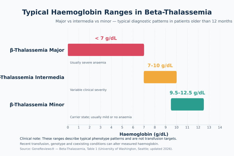
Modern haematology increasingly classifies clinically significant beta-thalassemia as transfusion-dependent thalassemia, abbreviated TDT, or non-transfusion-dependent thalassemia, abbreviated NTDT. This approach is more useful than relying on the words major or intermedia alone because it reflects the patient’s actual treatment needs. Most people traditionally diagnosed with beta-thalassemia major have TDT, whereas many diagnosed with beta-thalassemia intermedia initially have NTDT. However, an NTDT patient may still need transfusions during infection, surgery, pregnancy, rapid growth or worsening disease and may eventually require a regular transfusion programme [6,8,9].
These categories are not stages through which every patient progresses. Thalassemia minor does not gradually develop into intermedia or major. The inherited genetic pattern remains present throughout life. A person previously labelled as having minor disease may occasionally be reclassified after detailed haemoglobin analysis or genetic testing reveals a more complex genotype, but this reflects correction of the original diagnosis rather than progression of the carrier state [4,5].
| Feature | Thalassemia Major | Thalassemia Intermedia | Thalassemia Minor |
|---|---|---|---|
| Usual severity | Severe | Mild to moderately severe; highly variable | Usually mild carrier state |
| Typical presentation | Infancy, often 6–24 months | Childhood to adulthood | Often found incidentally |
| Haemoglobin pattern | Usually markedly reduced without transfusion | Moderately reduced | Normal or mildly reduced |
| Transfusion need | Usually regular | None, occasional or regular depending on severity | Usually none |
| Iron overload risk | High, especially from transfusions | Significant even without regular transfusions | Usually low unless another cause exists |
| Spleen enlargement | Common if inadequately controlled | Common in clinically significant disease | Not typical |
| Growth problems | Possible without adequate treatment | Possible in more severe disease | Not typical |
| Long-term organ complications | Significant without effective monitoring | Can occur despite no regular transfusion | Generally not expected |
| Curative-treatment assessment | Often relevant | Relevant in selected severe cases | Not normally indicated |
| Main management priority | Transfusion, chelation, complication prevention and curative options | Control anaemia, iron burden and complications | Correct diagnosis, avoid unnecessary iron and assess reproductive risk |
Thalassemia Major
Beta-thalassemia major is the most severe of the three traditional categories. Affected infants are usually well at birth because fetal haemoglobin temporarily compensates for inadequate beta-globin production. Symptoms commonly emerge between six and 24 months of age as fetal haemoglobin declines. Parents may notice increasing pallor, poor feeding, irritability, reduced weight gain, delayed growth, abdominal enlargement from an enlarged liver or spleen, jaundice, recurrent illness or reduced activity [4].
Without an effective transfusion programme, severe anaemia stimulates excessive but ineffective red-cell production. This can expand the bone marrow, alter facial and skeletal development, enlarge the spleen and interfere with normal growth. Regular red-cell transfusions suppress this ineffective blood formation and maintain a haemoglobin level that supports growth, activity and organ function. Because repeated transfusions introduce additional iron, most patients also require iron-chelation therapy and long-term monitoring of the heart, liver, endocrine glands, bones and reproductive health [8].
A diagnosis of thalassemia major should not be based on haemoglobin concentration alone. The haematologist considers the child’s age at presentation, symptoms, transfusion history, haemoglobin electrophoresis or high-performance liquid chromatography findings, genetic variants, spleen size, growth pattern and evidence of ineffective erythropoiesis. Recent transfusions can alter haemoglobin analysis by adding donor haemoglobin, so the date and volume of the last transfusion must accompany any report submitted for interpretation [4,8].
Thalassemia Intermedia
Thalassemia intermedia describes a broad and clinically variable group. Some patients have only moderate anaemia and remain free from regular transfusions for many years, while others develop substantial complications or eventually become transfusion dependent. The modern term NTDT is often more informative because it indicates that the patient does not require a continuing transfusion schedule for survival at the time of assessment. It does not mean that the condition is mild or that transfusions will never be needed [6,9].
Symptoms may first appear during later childhood, adolescence or adulthood. They can include persistent fatigue, reduced exercise capacity, pallor, jaundice, poor growth, delayed puberty, gallstones, bone changes or progressive enlargement of the spleen. Pregnancy, serious infection, surgery or rapid childhood growth may expose a previously compensated anaemia and create a temporary or continuing need for transfusion [6,9].
Patients with thalassemia intermedia can develop iron overload even when they have received few transfusions. Ineffective red-cell production suppresses the body’s normal control of intestinal iron absorption, allowing excessive dietary iron to accumulate gradually in the liver and other organs. Serum ferritin can help identify trends, but it may underestimate iron burden in NTDT. Liver iron measurement by validated magnetic resonance imaging may therefore be necessary when iron accumulation is suspected [6,9].
Other important risks include leg ulcers, reduced bone density, fractures, gallstones, extramedullary haematopoiesis, pulmonary hypertension and blood clots, particularly after splenectomy. Consequently, the absence of regular transfusions should never be interpreted as absence of disease activity. Monitoring should be based on haemoglobin trends, symptoms, growth, spleen size, iron measurements, organ function and emerging complications rather than the diagnostic label alone [6,9].
Thalassemia Minor
Beta-thalassemia minor, also called beta-thalassemia trait or the beta-thalassemia carrier state, usually results from one altered beta-globin gene and one functioning beta-globin gene. Most carriers remain healthy and do not require transfusions, chelation or disease-directed treatment. Their complete blood count commonly shows a low mean corpuscular volume and low mean corpuscular haemoglobin because the red blood cells are smaller and contain less haemoglobin than usual [4,5].
Some carriers have mild anaemia, but significant fatigue, breathlessness or a substantial fall in haemoglobin should not automatically be attributed to thalassemia minor. Iron deficiency, vitamin B12 or folate deficiency, menstrual blood loss, pregnancy, thyroid disease, chronic inflammation, kidney disease, sleep disorders or another haemoglobin variant may coexist. Ferritin and appropriate iron studies should be checked before prescribing iron because microcytosis alone does not prove iron deficiency [4,5].
The principal long-term implication of thalassemia minor is reproductive rather than progressive organ damage. Partner testing becomes important before conception or during early pregnancy because two carriers can have a child with clinically significant thalassemia. Testing may include a complete blood count, haemoglobin analysis and molecular genetic testing, particularly when results are borderline, the partners have different haemoglobin variants or ancestry is associated with alpha-thalassemia, beta-thalassemia, haemoglobin E or sickle-cell variants [4,5].
A laboratory report containing the word “minor” should therefore not be interpreted as a mild form that may later become major. It identifies a carrier pattern that usually requires accurate documentation, avoidance of unnecessary treatment and informed reproductive counselling. In contrast, major and intermedia require continued clinical assessment because transfusion needs, iron burden and organ risks may change over time [5,6,8,9].
Major, Minor and Intermedia in Alpha Thalassemia

The comparison of thalassemia major vs minor vs intermedia applies most directly to beta-thalassemia. Alpha-thalassemia follows a different genetic pattern because most people inherit four alpha-globin genes, two from each parent. Clinical severity usually depends on how many of these genes are absent or unable to function, whether the changes occur on the same chromosome or opposite chromosomes, and whether a non-deletional variant produces a more severe phenotype [1,11].
Silent Carrier and Alpha-Thalassemia Trait
A silent alpha-thalassemia carrier usually has one affected alpha-globin gene. The complete blood count may be normal or may show mildly reduced mean corpuscular volume and mean corpuscular haemoglobin. Haemoglobin electrophoresis or high-performance liquid chromatography can also appear normal after infancy, so molecular genetic testing may be required when family history, ancestry or reproductive risk remains clinically important [1,11].
Alpha-thalassemia trait usually involves two affected alpha-globin genes and is the closest equivalent to a minor or carrier phenotype. Patients commonly have lifelong microcytosis with no anaemia or only mild anaemia. This pattern may be mistaken for iron deficiency, particularly during pregnancy or routine health screening. Ferritin and transferrin saturation should therefore be assessed before prescribing iron, because iron supplementation will not correct inherited alpha-globin deficiency unless genuine iron deficiency is also present [1,11].
The arrangement of the two affected genes has important reproductive consequences. In the cis form, both affected genes are located on the same chromosome, while the other chromosome carries two functioning genes. In the trans form, one affected gene is present on each chromosome. The person may have similar blood-count findings in either arrangement, but the risk to a future child can differ substantially. Cis deletions are particularly important in families with ancestry from Southeast Asia and parts of southern China, while trans deletions are more frequent in people with African ancestry [11].
Routine haemoglobin analysis cannot always determine whether alpha-thalassemia trait is cis or trans. Genetic testing of the patient and partner may be needed before pregnancy, especially when both have unexplained microcytosis, alpha-thalassemia trait or ancestry associated with clinically significant alpha-globin variants. This is relevant for multicultural families receiving care in India, Singapore, the United Kingdom, the United States, Canada and Australia [11].
Haemoglobin H Disease
Haemoglobin H disease usually develops when three alpha-globin genes are affected. It represents the principal intermediate clinical form of alpha-thalassemia and is sometimes described as alpha-thalassemia intermedia. The remaining alpha-globin production is insufficient to maintain normal haemoglobin formation, leading to chronic haemolytic anaemia and the formation of haemoglobin H, which consists of four beta-globin chains [1,11].
Clinical severity varies considerably. Some patients maintain a relatively stable haemoglobin level and require only periodic monitoring, while others experience persistent fatigue, pallor, jaundice, enlarged spleen, gallstones, reduced exercise tolerance or delayed growth. Haemoglobin can fall sharply during fever, infection, pregnancy or exposure to oxidant medicines, creating a temporary need for blood transfusion [11].
Non-deletional forms of haemoglobin H disease are often more severe than deletional forms. Patients may develop lower baseline haemoglobin, greater splenic enlargement, more frequent haemolytic episodes and a higher likelihood of transfusion. The exact genotype therefore has practical value when estimating expected severity, planning pregnancy care and determining the intensity of long-term monitoring [11].
Iron overload can occur in haemoglobin H disease even when transfusions have been infrequent. Chronic ineffective erythropoiesis can increase intestinal iron absorption, allowing iron to accumulate gradually in the liver and other tissues. Ferritin trends, transfusion history, liver function and magnetic resonance imaging of liver iron should be considered according to the patient’s age and clinical risk rather than assuming that iron overload occurs only after regular transfusion [11].
Haemoglobin Bart Hydrops Fetalis Syndrome
When all four alpha-globin genes are absent or non-functional, the fetus cannot produce the alpha chains required for normal fetal and adult haemoglobin. Excess gamma chains form haemoglobin Bart, which binds oxygen very tightly and releases it poorly to fetal tissues. This can cause profound fetal anaemia, heart failure, generalized fluid accumulation and hydrops fetalis. The condition is also called alpha-thalassemia major or haemoglobin Bart hydrops fetalis syndrome [1,11].
Without specialised fetal treatment, the condition commonly results in stillbirth or death around the time of birth. In selected pregnancies, intrauterine transfusions followed by intensive neonatal and lifelong haematology care may permit survival. Survivors can remain transfusion dependent and may require assessment for stem-cell transplantation or other advanced treatment at a specialist centre [11].
The pregnancy can also create serious maternal risks, including pre-eclampsia, severe swelling, excessive amniotic fluid, haemorrhage and mirror syndrome, in which the mother develops symptoms resembling the hydropic fetus. Early identification is therefore important not only for fetal prognosis but also for maternal safety [11].
Couples in whom both partners carry alpha-zero thalassemia in the cis arrangement may have a pregnancy at risk of haemoglobin Bart syndrome. Preconception genetic counselling, parental molecular testing and discussion of reproductive options can establish the precise risk before pregnancy. During pregnancy, chorionic villus sampling, amniocentesis or specialised fetal assessment may be considered according to gestational age, local practice and the parents’ informed preferences [11].
The terms major, intermedia and minor may therefore appear in alpha-thalassemia reports, but the more precise diagnostic labels are silent carrier, alpha-thalassemia trait, haemoglobin H disease and haemoglobin Bart hydrops fetalis syndrome. Patients should not infer their alpha-thalassemia category from a beta-thalassemia comparison chart because the gene structure, haemoglobin pattern, pregnancy risk and expected clinical course are different [1,11].
Why Thalassemia Severity Differs

The clinical variation seen in thalassemia major vs minor vs intermedia is not determined by one haemoglobin result or mutation name alone. Severity reflects how much functional beta-globin the body can produce, the degree of imbalance between alpha- and non-alpha-globin chains, the amount of fetal haemoglobin that persists after infancy, the presence of other inherited haemoglobin changes and the complications that develop over time. Two patients with apparently similar beta-globin variants may therefore have different haemoglobin levels, transfusion requirements, spleen sizes and long-term risks [4,8,9].
Beta-Zero and Beta-Plus Variants
A beta-zero variant prevents the affected HBB gene from producing functional beta-globin. A beta-plus variant allows some beta-globin production, although the residual amount differs among variants. When two clinically significant variants are inherited, their combined effect helps determine whether the patient develops transfusion-dependent thalassemia or a milder non-transfusion-dependent phenotype [4,8].
A patient who inherits two beta-zero variants often has little or no adult haemoglobin production and is more likely to develop severe disease during infancy. A person with one or two beta-plus variants may retain some adult haemoglobin and present later or require fewer transfusions. These associations are clinically useful but are not absolute. Some beta-plus combinations cause severe transfusion-dependent disease, while some patients with two beta-zero variants have a less severe course because other genetic factors increase fetal haemoglobin or reduce globin-chain imbalance [4,8].
Beta-thalassemia minor generally results when one HBB gene carries a clinically significant variant and the other gene produces sufficient beta-globin to prevent severe anaemia. However, a patient with unexpectedly low haemoglobin, substantial splenic enlargement or transfusion dependence should not be assumed to have uncomplicated minor disease solely because one beta-thalassemia variant was identified. Further testing may reveal a second HBB variant, an unstable haemoglobin, an additional alpha-globin gene or another cause of anaemia [4,8].
Alpha-Globin Gene Balance
In beta-thalassemia, reduced beta-globin production leaves excess alpha-globin chains without enough beta chains with which to form stable adult haemoglobin. These unmatched alpha chains damage developing red blood cells in the bone marrow and shorten the survival of red cells that reach the circulation. The degree of this chain imbalance is a major determinant of ineffective erythropoiesis, haemolysis and clinical severity [8].
Coinherited alpha-thalassemia can sometimes reduce the excess production of alpha chains and partially restore the balance between alpha and non-alpha chains. A patient with clinically important beta-thalassemia who also inherits reduced alpha-globin production may consequently have a higher baseline haemoglobin, later onset of symptoms or a lower transfusion requirement than expected from the HBB variants alone [4,8,9].
The opposite effect can occur when a patient inherits an additional functional alpha-globin gene through alpha-gene triplication or quadruplication. Increased alpha-chain production can worsen the imbalance created by a single beta-thalassemia variant. A person initially expected to have beta-thalassemia minor may then develop a thalassemia intermedia phenotype with more pronounced anaemia, splenic enlargement or occasional transfusion requirements [4,8].
Alpha-globin testing becomes particularly valuable when the clinical condition does not match the beta-globin report. Molecular assessment of both HBB and the alpha-globin genes can clarify why members of the same family have different disease severity and can improve reproductive counselling for couples carrying more than one haemoglobin-related variant [4,8].
Fetal Haemoglobin Modifiers
Fetal haemoglobin contains gamma-globin chains instead of beta-globin chains. Gamma chains can pair with alpha chains and reduce the number of damaging unpaired alpha chains. Patients who continue to produce higher amounts of fetal haemoglobin after infancy may therefore have less severe anaemia, a later need for regular transfusion or an intermedia phenotype despite inheriting otherwise severe beta-thalassemia variants [4,8].
Fetal haemoglobin production is influenced by several genetic regions, including HBG2, BCL11A and the HBS1L-MYB region. The combination of these modifiers can partly explain why siblings with the same principal HBB variants may not have identical transfusion requirements or complication patterns. Their effects are variable, and no single modifier can reliably predict the complete clinical course [4,8].
An elevated fetal haemoglobin percentage should also be interpreted alongside the total haemoglobin, patient age and transfusion history. A high percentage does not necessarily mean that the absolute amount is sufficient to prevent anaemia. Recent donor transfusions can alter the measured proportions of adult and fetal haemoglobin, while severe reduction of adult haemoglobin may make the fetal fraction appear proportionally high [4,8].
Haemoglobin E Beta Thalassemia
Haemoglobin E beta-thalassemia occurs when a person inherits a haemoglobin E variant from one parent and a beta-thalassemia variant from the other. Haemoglobin E is both a structural haemoglobin variant and a change that reduces normal beta-globin production. This combination is particularly relevant to families with ancestry from India, Bangladesh and Southeast Asia, including patients receiving care in Singapore, Australia, the United Kingdom, Canada and the United States [4,9].
Its clinical expression ranges from mild anaemia requiring observation to severe transfusion-dependent disease resembling beta-thalassemia major. Some children remain clinically stable during early life but develop worsening anaemia, poor growth or splenic enlargement as they age. Others require regular transfusions from childhood. The diagnosis of haemoglobin E beta-thalassemia therefore does not by itself predict whether the patient will follow a minor, intermedia or major clinical course [4,9].
Assessment should consider baseline haemoglobin, growth, exercise tolerance, spleen size, haemolysis, transfusion history, fetal haemoglobin production, iron burden and the exact beta-thalassemia variant. On some laboratory platforms, haemoglobin E may appear within the same measurement window as haemoglobin A2. Molecular confirmation may be required when haemoglobin analysis is unclear or when the result will influence reproductive counselling or treatment planning [4,9].
Age, Illness, Pregnancy and Spleen Function
The inherited variants remain unchanged throughout life, but their clinical expression can change. Infants with severe beta-thalassemia may appear healthy at birth because fetal haemoglobin remains dominant. Anaemia becomes evident as fetal haemoglobin declines and adult haemoglobin production becomes necessary. In contrast, a patient with intermedia may remain stable through childhood and develop clinically important complications only during adolescence or adulthood [4,6,8,9].
Infection, surgery, rapid growth and pregnancy can increase oxygen requirements or accelerate red-cell destruction. A patient who usually manages without transfusion may temporarily need transfused red cells during these periods. Repeated or continuing transfusion may become appropriate when anaemia causes impaired growth, reduced daily function, skeletal changes, progressive splenic enlargement, pulmonary hypertension or other complications [6,9].
An enlarged spleen can trap and destroy red cells, platelets and white blood cells, contributing to worsening anaemia, low blood counts, abdominal discomfort and increasing transfusion requirements. Splenectomy may reduce red-cell consumption in selected patients, but it also creates lifelong risks such as severe infection and thrombosis. The decision requires specialist assessment rather than reliance on spleen size alone [6,8,9].
Iron overload, nutritional deficiencies, endocrine dysfunction, liver disease, cardiac impairment and reduced adherence to chelation can further influence how well a patient tolerates chronic anaemia. These acquired factors do not change the genetic form of thalassemia, but they can substantially increase fatigue, delay growth, reduce exercise capacity and worsen long-term outcomes [6,8,9].
For patients transferring care between countries, the medical record should include the confirmed genotype, haemoglobin analysis, age at first transfusion, current transfusion interval, annual red-cell requirement, antibody history, chelation exposure, ferritin trend and magnetic resonance measurements of liver and cardiac iron. Terms such as major, intermedia, TDT and NTDT may be applied differently across treatment centres, whereas these objective findings provide a more reliable description of present disease severity [4,8,9].
Thalassemia Major vs Minor vs Intermedia Symptoms

The symptoms of thalassemia major vs minor vs intermedia differ mainly in their age of onset, intensity, effect on growth and activity, and relationship to chronic anaemia and ineffective red-cell production. Symptoms alone cannot confirm the category because iron deficiency, infection, pregnancy, thyroid disease, nutritional deficiencies and other haemoglobin disorders may produce overlapping complaints. The pattern must be interpreted with the complete blood count, haemoglobin analysis, transfusion history, physical examination and genetic findings [1,4].
Thalassemia Major Symptoms
Children with beta-thalassemia major usually appear healthy at birth because fetal haemoglobin remains the dominant form of haemoglobin during early infancy. Symptoms commonly emerge between six and 24 months of age as fetal haemoglobin declines and the body becomes increasingly dependent on beta-globin production. The earliest signs may include progressive pallor, reduced feeding, irritability, unusual sleepiness, limited interest in play and poor weight gain [4].
Parents may first recognise that the child tires more quickly than other children, breathes faster during feeding or physical activity, or fails to follow the expected growth curve. Persistent severe anaemia can increase the heart rate and respiratory rate because the body is attempting to deliver more oxygen to its tissues. Older children may report headaches, dizziness, breathlessness, palpitations, reduced concentration and marked exercise intolerance [4,8].
Jaundice may develop when fragile red blood cells are destroyed more rapidly than normal. The urine may become darker during periods of increased haemolysis, although dark urine also requires assessment for other causes. The liver and spleen may enlarge as they participate in the removal of abnormal red cells and in blood-cell production outside the bone marrow. Abdominal swelling or a firm mass beneath the left ribs may therefore be an important presenting feature [4,8].
When severe anaemia remains insufficiently treated, the bone marrow expands in an attempt to produce more red blood cells. This can contribute to widening of facial bones, prominence of the forehead or upper jaw, thinning of the outer bone structure, bone pain and increased fracture risk. These skeletal changes are less likely to develop when an effective transfusion programme is started early and maintained consistently [4,8].
Poor growth is not explained by anaemia alone. Inadequate transfusion, iron-related endocrine injury, delayed puberty, chronic liver disease, nutritional problems and reduced bone health may all contribute. A child who crosses downward through height or weight percentiles requires a broader assessment rather than simply receiving dietary advice or a higher-calorie diet [8].
Symptoms can also appear shortly before the next scheduled transfusion. Increasing fatigue, pallor, breathlessness, headaches, poor school participation or reduced physical activity may indicate that the pretransfusion haemoglobin is falling below the level required for adequate daily function. Repeated symptoms at the end of every transfusion cycle should be documented because they may help the haematology team adjust the transfusion interval or target [8].
Regular transfusion can control many symptoms of severe anaemia, but it does not eliminate the need to monitor new complaints. Reduced exercise tolerance, ankle swelling, an irregular heartbeat, persistent abdominal discomfort, bone pain, delayed puberty or excessive thirst may indicate cardiac, liver, endocrine, skeletal or iron-related complications. These symptoms require objective investigation rather than being accepted as an unavoidable part of thalassemia [8].
Thalassemia Intermedia Symptoms
Thalassemia intermedia may remain unnoticed during infancy and become apparent later in childhood, adolescence or adulthood. The presentation depends on how effectively the patient compensates for chronic anaemia and whether complications have already developed. Some people experience only reduced stamina, while others develop growth impairment, marked splenic enlargement, skeletal changes or organ complications despite never having received regular transfusions [6,9].
Common symptoms include chronic fatigue, pallor, breathlessness during exertion, headaches, dizziness, palpitations and reduced exercise tolerance. A patient may be able to perform routine activities but struggle with running, climbing stairs, physically demanding employment or sustained concentration. Functional limitation should be assessed in relation to the patient’s usual activities rather than judged only from whether the haemoglobin value is considered moderately low [6].
Persistent haemolysis may cause jaundice, dark urine and pigment gallstones. Gallstones can produce pain in the right upper abdomen, nausea after meals, fever or worsening jaundice. Recurrent abdominal pain in a patient with intermedia should not automatically be attributed to an enlarged spleen because gallbladder, liver and other abdominal conditions may coexist [6,9].
Progressive splenic enlargement can create abdominal fullness, early satiety and discomfort beneath the left ribs. The patient may eat less because the enlarged spleen presses against the stomach. Hypersplenism can further lower red-cell, white-cell and platelet counts, contributing to worsening anaemia, susceptibility to infection or easy bruising. Changes in spleen size and blood counts are therefore more clinically useful than the presence of splenomegaly alone [6,9].
Expansion of the bone marrow may cause facial bone changes, bone pain, reduced bone density and fractures. Extramedullary haematopoiesis may produce masses of blood-forming tissue outside the bone marrow, commonly near the spine or within the chest. Back pain accompanied by leg weakness, altered sensation or changes in bladder or bowel control requires urgent assessment because spinal cord compression can occur [6,9].
Leg ulcers may develop around the ankles and can be painful, slow to heal and prone to recurrence. Their presence may reflect chronic tissue hypoxia, abnormal red-cell circulation and vascular changes rather than a simple skin infection. Persistent wounds require coordinated assessment of anaemia, circulation, infection, nutrition and local wound care [6,9].
Patients may also develop chest discomfort, breathlessness that is disproportionate to the measured haemoglobin, fainting or declining exercise capacity because of pulmonary hypertension or cardiac strain. These symptoms should not be dismissed as ordinary consequences of chronic anaemia. Echocardiography and other cardiopulmonary investigations may be required when symptoms are persistent or progressive [6,9].
A previously stable patient may become more symptomatic during infection, pregnancy, surgery or rapid growth. Increasing fatigue, worsening jaundice, falling haemoglobin, rapid splenic enlargement or reduced daily function may create a temporary or continuing need for transfusion. The decision should be based on the complete clinical picture rather than one haemoglobin value [6,9].
Thalassemia Minor Symptoms
Most people with beta-thalassemia minor have no symptoms and discover the carrier state during a routine blood count, pregnancy screening, premarital testing or investigation of small red blood cells. Some have mild pallor or tiredness associated with a slightly reduced haemoglobin level, but they generally maintain normal growth, physical activity and life expectancy without transfusion or chelation [1,5].
Symptoms that are severe, persistent or newly developing should not automatically be assigned to thalassemia minor. Marked exhaustion, substantial breathlessness, fainting, chest pain, progressive weakness, weight loss, fever, jaundice or splenic enlargement is not typical of an uncomplicated carrier state and warrants further investigation [1,5].
Iron deficiency is an important coexisting cause of symptoms, particularly in menstruating patients, pregnant women, frequent blood donors and people with restricted diets or gastrointestinal blood loss. Ferritin and transferrin saturation should be assessed before prescribing iron because low mean corpuscular volume and low mean corpuscular haemoglobin are expected features of the carrier state and do not independently establish iron deficiency [1,5].
Vitamin B12 deficiency, folate deficiency, thyroid dysfunction, kidney disease, inflammatory disorders, sleep disturbance and heavy menstrual bleeding may also explain symptoms that appear disproportionate to the mild anaemia. Treating the correct coexisting condition can improve energy and concentration even though the inherited microcytosis remains present [1].
Pregnancy can make mild anaemia more noticeable because plasma volume expands and nutritional requirements increase. A pregnant carrier requires assessment for iron and folate status according to obstetric guidance, but the haemoglobin should not be expected to normalise completely if microcytosis is caused by beta-thalassemia trait. Partner testing remains essential because the mother’s mild symptoms do not predict the genetic risk to the fetus [5].
A patient recorded as having thalassemia minor who develops a persistently low haemoglobin level, jaundice, an enlarged spleen, bone changes or a transfusion requirement needs haematology reassessment. The original diagnosis may be incomplete, or the patient may have an additional alpha-globin abnormality, another beta-globin variant, haemoglobin E, an unstable haemoglobin or a separate acquired cause of anaemia [1,4].
Age at Diagnosis in Thalassemia Major vs Minor vs Intermedia

The age at diagnosis in thalassemia major vs minor vs intermedia depends on the amount of functional globin produced, the rate at which fetal haemoglobin declines, the severity of anaemia and whether screening occurs before symptoms develop. Age at diagnosis is not always the same as age of disease onset. A child may have inherited thalassemia from conception but receive a confirmed diagnosis only after symptoms, an abnormal blood count, newborn screening or family testing prompts further investigation [4,8].
Thalassemia Major Diagnosis in Infancy
Beta-thalassemia major commonly becomes clinically apparent between six and 24 months of age. Newborns may initially appear healthy because fetal haemoglobin remains the principal oxygen-carrying haemoglobin during early infancy. As fetal haemoglobin declines, inadequate beta-globin production becomes more evident and severe anaemia may develop [4,8].
Early signs leading to investigation can include progressive pallor, poor feeding, reduced activity, inadequate weight gain, abdominal enlargement, jaundice or delayed development. A complete blood count may show severe microcytic, hypochromic anaemia, while the blood film may demonstrate marked variation in red-cell size and shape, target cells and nucleated red blood cells. Haemoglobin analysis commonly shows substantially increased fetal haemoglobin with reduced or absent adult haemoglobin, depending on the genotype and whether transfusions have already begun [4,8].
Newborn screening can identify an abnormal haemoglobin pattern before symptoms develop, but the initial result may not fully distinguish beta-thalassemia major from another severe beta-globin disorder. Because HbA2 is not reliably established during the neonatal period and fetal haemoglobin remains dominant, repeat haemoglobin analysis and molecular genetic testing may be required to confirm the diagnosis [4].
A child with suspected severe thalassemia should be referred promptly to paediatric haematology rather than waiting for repeated episodes of symptomatic anaemia. Early confirmation allows timely transfusion planning, extended red-cell antigen typing, family testing, genetic counselling and baseline assessment of growth, liver and spleen size, infection status and future iron-overload risk [8].
When a child has already received a transfusion, donor red cells can produce measurable adult haemoglobin and temporarily alter the haemoglobin pattern. The laboratory and haematologist should be informed of the date, volume and location of every recent transfusion. Molecular genetic testing may provide a clearer diagnosis when haemoglobin analysis has been affected by donor blood [4,8].
Thalassemia Intermedia Diagnosis Later in Life
Thalassemia intermedia may be diagnosed in later childhood, adolescence or adulthood because the patient produces enough functional haemoglobin to avoid severe symptoms during infancy. The first indication may be persistent anaemia, poor exercise tolerance, jaundice, gallstones, delayed growth, splenic enlargement, bone changes or an unexpectedly low haemoglobin result during routine testing [6,9].
Some patients are diagnosed during an infection, surgery or period of rapid growth when their usual compensation becomes inadequate. Others are identified during pregnancy because routine antenatal blood testing reveals pronounced microcytosis or anaemia. Pregnancy may also intensify symptoms and expose a previously unrecognised non-transfusion-dependent phenotype [6,9].
A person may initially be labelled as having thalassemia minor because regular transfusions are not required. Persistent haemoglobin levels lower than expected for a carrier, progressive splenomegaly, jaundice, skeletal changes, iron overload or occasional transfusion requirements should prompt reassessment. Comprehensive testing may identify compound beta-thalassemia, haemoglobin E beta-thalassemia, alpha-gene triplication or another genetic combination producing an intermedia phenotype [4,6].
Diagnosis should integrate clinical history with the complete blood count, reticulocyte count, peripheral blood film, bilirubin, lactate dehydrogenase, haemoglobin analysis and molecular findings. Transfusion history, growth, spleen size and evidence of iron accumulation help determine whether the patient is clinically non-transfusion dependent or is moving toward a regular transfusion requirement [6,9].
Adults diagnosed after migration or transfer between healthcare systems may have incomplete childhood records or terminology that differs between countries. Reassessment should document the exact genotype, age at first transfusion, annual transfusion burden, previous splenectomy, antibody history, ferritin trend and magnetic resonance measurements of liver and cardiac iron. These findings describe current disease severity more accurately than the word intermedia alone [6,8,9].
Thalassemia Minor Diagnosis During Screening
Thalassemia minor is often diagnosed incidentally during a routine complete blood count. The report may show a low mean corpuscular volume and mean corpuscular haemoglobin despite a normal or only mildly reduced haemoglobin level. A relatively preserved or increased red-cell count can raise suspicion of a carrier state, but these findings cannot confirm the diagnosis without haemoglobin analysis or genetic testing [1,4].
Many carriers are identified during antenatal screening, premarital testing, fertility assessment, blood donation, preoperative testing or investigation of suspected iron deficiency. Others are tested after a child or relative receives a thalassemia diagnosis. Because carriers are usually well, the diagnosis may not occur until adulthood even though the genetic variant has been present throughout life [4,5].
Beta-thalassemia trait is commonly supported by an increased HbA2 level, although iron deficiency, delta-globin variants, recent transfusion and laboratory methodology can complicate interpretation. Borderline or discordant findings may require repeat testing after correction of iron deficiency and, where clinically appropriate, molecular analysis of the HBB gene [1,4].
Alpha-thalassemia trait may be more difficult to confirm because haemoglobin electrophoresis can be normal outside the newborn period. Persistent microcytosis with normal iron studies and no diagnostic beta-thalassemia pattern may justify alpha-globin genetic testing, particularly when both partners have microcytosis or when reproductive risk includes haemoglobin H disease or haemoglobin Bart hydrops fetalis syndrome [1,11].
A carrier diagnosis should be recorded clearly in the medical record so that future clinicians do not repeatedly treat inherited microcytosis as iron deficiency. Iron should be prescribed only when deficiency is demonstrated. Partner testing and genetic counselling are more clinically important than repeated attempts to normalise the mean corpuscular volume [1,5].
Delayed or Missed Diagnosis
Thalassemia may be diagnosed late when microcytosis is repeatedly attributed to iron deficiency without ferritin or haemoglobin analysis. Failure of the blood count to improve as expected after confirmed iron deficiency has been corrected should prompt evaluation for an inherited haemoglobin disorder. Continued iron treatment without evidence of deficiency can expose the patient to unnecessary supplementation while delaying the correct diagnosis [1,5].
Recent transfusion, mixed iron deficiency, pregnancy and coexisting haemoglobin variants can obscure the expected laboratory pattern. Diagnostic uncertainty should be resolved through review of earlier blood counts, family studies, repeat haemoglobin analysis at an appropriate interval and targeted genetic testing rather than assigning a type from a single report [4,8].
Siblings can receive diagnoses at different ages even when they inherit the same principal variants because fetal haemoglobin modifiers, alpha-globin changes, illness and access to screening influence clinical presentation. Once one family member is diagnosed, appropriate testing of parents and siblings can identify affected children or carriers before complications or reproductive decisions arise [4,8].
Blood Test Reports in Thalassemia Major vs Minor vs Intermedia

Blood test reports in thalassemia major vs minor vs intermedia differ in the severity of anaemia, red-cell size, haemoglobin fractions, evidence of haemolysis, iron burden and the effect of previous transfusions. No single result can reliably determine the category. Interpretation requires the complete blood count, peripheral blood smear, haemoglobin analysis, iron studies, transfusion history, clinical findings and, when necessary, molecular genetic testing [1,4,8,9].
| Blood Report Finding | Major | Intermedia | Minor |
|---|---|---|---|
| Haemoglobin | Usually severely reduced without treatment | Moderately reduced and variable | Normal or mildly reduced |
| MCV | Low | Low | Often markedly low despite mild anaemia |
| MCH | Low | Low | Low |
| RBC count | Variable | Variable | Often normal or relatively high |
| Reticulocytes | Often increased | Often increased | Usually normal or mildly increased |
| HbA | Reduced or absent depending on genotype and transfusion | Variable | Usually predominant |
| HbA2 | Variable | Variable | Commonly elevated in beta-thalassemia trait |
| HbF | Usually markedly increased | Often increased | Normal or mildly increased |
| Ferritin | Often elevated with transfusion exposure | May rise even without transfusion | Usually normal unless another condition exists |
| Genetic testing | Confirms causative variants | Helps explain phenotype | Confirms carrier status when needed |
Laboratories in the United States commonly report haemoglobin in grams per decilitre, while some laboratories in the United Kingdom, Canada, Australia and other countries use grams per litre. A haemoglobin value of 9 g/dL is equivalent to 90 g/L. Ferritin expressed as nanograms per millilitre has the same numerical value as micrograms per litre. Confirming the units prevents serious interpretation errors when reports are reviewed across different healthcare systems.
Complete Blood Count
The complete blood count provides the first indication that haemoglobin production is abnormal. The most useful measurements include haemoglobin, red blood cell count, mean corpuscular volume, mean corpuscular haemoglobin, red cell distribution width, reticulocyte count, white blood cell count and platelet count. These values should be interpreted together rather than using one abnormal measurement to assign the diagnosis [1,4].
Untreated or inadequately transfused thalassemia major generally produces severe anaemia with markedly reduced mean corpuscular volume and mean corpuscular haemoglobin. The red cells are small and contain less haemoglobin, but the overall pattern may change after regular transfusion because circulating donor red cells are larger and better haemoglobinised than the patient’s own cells. A blood count taken shortly after transfusion may therefore underestimate the degree of underlying microcytosis [4,8].
The clinically most useful haemoglobin value in a regularly transfused patient is often the level measured immediately before the next scheduled transfusion. Repeated pretransfusion results help determine whether the transfusion programme is maintaining adequate tissue oxygenation and suppressing excessive bone-marrow activity. A value taken one or two days after transfusion cannot be compared directly with a pretransfusion value because it answers a different clinical question [8].
Thalassemia intermedia commonly produces persistent anaemia that is more pronounced than the mild anaemia associated with a carrier state but less immediately severe than untreated major disease. The exact haemoglobin level varies with genotype, age, splenic activity, infection, pregnancy, nutritional status and previous transfusion. Some patients function with a chronically low haemoglobin level, while others develop poor growth, bone changes, splenic enlargement or reduced exercise capacity at a similar value [6,9].
The red blood cell count can help distinguish thalassemia minor from iron deficiency. In beta-thalassemia minor, the mean corpuscular volume may be markedly reduced even when the haemoglobin is normal or only mildly low, and the red blood cell count is often preserved or relatively increased. In iron deficiency, the marrow may be unable to maintain the same red-cell number as iron depletion progresses. This pattern is useful for screening but is not sufficiently specific to replace ferritin, haemoglobin analysis or genetic testing [1,4].
Red cell distribution width measures the variation in red-cell size. It is often substantially increased in iron deficiency because newly produced cells become progressively smaller as iron stores decline. It may be normal or only mildly increased in uncomplicated thalassemia minor because the inherited microcytosis affects many cells more uniformly. However, red cell distribution width can also rise in thalassemia when iron deficiency, haemolysis, recent transfusion or another nutritional deficiency is present [1].
The reticulocyte count reflects the number of young red blood cells entering the circulation. It may be elevated in major and intermedia because of ongoing haemolysis and increased marrow stimulation, but the increase can be less than expected for the severity of anaemia because much of the developing red-cell population is destroyed within the marrow before entering the bloodstream. The absolute reticulocyte count is generally more informative than the percentage alone when haemoglobin is substantially reduced [4,8,9].
A sudden fall in haemoglobin accompanied by a low or unexpectedly normal reticulocyte count may indicate temporary suppression of red-cell production, including parvovirus B19 infection, severe illness or a medication effect. A rapid haemoglobin decline with an increased reticulocyte count may be more consistent with accelerated haemolysis or blood loss. The result must still be interpreted alongside symptoms, bilirubin, lactate dehydrogenase, transfusion history and examination findings.
White blood cell and platelet counts may fall when an enlarged spleen traps circulating blood cells. This pattern, called hypersplenism, can occur in inadequately controlled major disease and in intermedia with substantial splenomegaly. Conversely, platelet and white-cell counts may remain persistently increased after splenectomy. These changes should not be interpreted as isolated laboratory abnormalities because they can influence infection, bleeding and thrombotic risk [6,8,9].
Peripheral Blood Smear
A peripheral blood smear allows direct examination of red-cell size, colour, shape and maturity. Thalassemia commonly produces microcytosis, hypochromia and target cells. The smear may also show anisocytosis, which means variation in size, and poikilocytosis, which means variation in shape. These changes are usually more extensive in thalassemia major and intermedia than in uncomplicated thalassemia minor [1,4].
Target cells contain a central area of haemoglobin surrounded by a pale ring and an outer haemoglobinised rim. They are common in thalassemia but are not specific to it because they may also occur in liver disease, after splenectomy and in other haemoglobin disorders. Basophilic stippling may be present but should not be treated as diagnostic because it can occur in several inherited and acquired conditions [1].
Nucleated red blood cells and pronounced polychromasia may appear when the marrow is under substantial stress or when blood formation is occurring outside the marrow. Their presence is more consistent with severe or inadequately controlled disease than with uncomplicated carrier status. The number may also increase after splenectomy because the spleen is no longer removing these immature cells from circulation [4,8].
A regularly transfused patient may show two distinct red-cell populations on the smear. Donor cells may appear closer to normal size and colour, while the patient’s own cells remain microcytic and hypochromic. This mixed population can increase red cell distribution width and complicate interpretation of the mean corpuscular volume. The laboratory should therefore be informed of the most recent transfusion date.
The smear cannot independently distinguish major from intermedia or prove that microcytosis represents thalassemia rather than iron deficiency. Its value lies in supporting the broader pattern, identifying unexpected features and guiding further testing when the automated blood count does not fully explain the severity of anaemia.
Haemoglobin Electrophoresis and HPLC
Haemoglobin electrophoresis and high-performance liquid chromatography measure the proportions of different haemoglobin fractions. The principal fractions considered in beta-thalassemia are adult haemoglobin, fetal haemoglobin and haemoglobin A2. The pattern varies with age, genotype, iron status, recent transfusion and the presence of another haemoglobin variant [1,4].
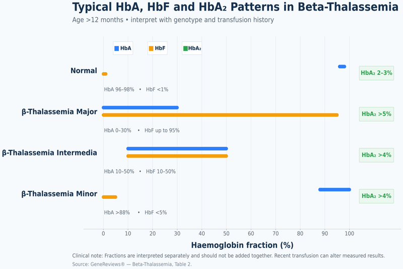
Beta-thalassemia minor commonly shows an increased haemoglobin A2 proportion, sometimes with a mild increase in fetal haemoglobin. Most of the circulating haemoglobin remains adult haemoglobin because the unaffected HBB gene continues to produce beta-globin. A normal or borderline haemoglobin A2 value does not always exclude a carrier state, particularly when iron deficiency, a delta-globin variant, recent transfusion or a mild beta-globin variant affects the result [1,4].
In untreated beta-thalassemia major, fetal haemoglobin usually forms most of the measurable haemoglobin. Adult haemoglobin may be absent when both inherited variants prevent beta-globin production, while a variable amount may remain when one or both variants permit residual beta-globin synthesis. Regular transfusions introduce donor adult haemoglobin, so the measured adult fraction may predominantly reflect transfused cells rather than the patient’s own production [4,8].
Thalassemia intermedia produces a more variable haemoglobin pattern. Fetal haemoglobin may be substantially increased, but the amount of adult haemoglobin depends on the underlying beta-globin variants and modifying genes. Haemoglobin fractions cannot be used in isolation to decide whether a patient is transfusion dependent because treatment requirement is determined by clinical severity, growth, complications and longitudinal transfusion needs [6,9].
Haemoglobin E beta-thalassemia requires careful laboratory interpretation. On some high-performance liquid chromatography systems, haemoglobin E appears in the same analytical region as haemoglobin A2. A report showing an unusually large haemoglobin A2 or A2/E fraction may therefore require confirmation by another method or molecular genetic testing. The clinical phenotype can range from mild non-transfusion-dependent anaemia to severe transfusion-dependent disease [4,9].
Age is particularly important when interpreting infant reports. Fetal haemoglobin naturally predominates at birth, while haemoglobin A2 is not yet present at the proportion expected later in childhood. An abnormal newborn screening pattern may identify the risk of a severe beta-globin disorder, but repeat testing and molecular confirmation may be needed before assigning the final subtype [4].
Recent transfusion can obscure the patient’s natural haemoglobin pattern for several months. The report request should include the date of the last transfusion, the number of units received and any previous stem-cell transplantation. When an immediate diagnosis is essential, genetic testing may be more informative than repeating haemoglobin analysis while donor cells remain in circulation [4,8].
Iron Studies
Ferritin, transferrin saturation, serum iron and total iron-binding capacity help distinguish iron deficiency from iron accumulation. Ferritin reflects stored iron but also rises during inflammation, infection and liver injury. A single high ferritin result therefore does not quantify total body iron, and a single apparently acceptable value does not exclude clinically important iron accumulation [8,9].
Thalassemia minor does not itself prove either iron deficiency or iron overload. A carrier may develop iron deficiency from menstrual blood loss, pregnancy, gastrointestinal bleeding, reduced dietary intake or another cause. Iron treatment is appropriate when deficiency has been demonstrated, but persistent microcytosis may remain after iron stores are corrected because the inherited reduction in globin production has not changed [1,5].
Regular transfusion is the principal cause of iron loading in thalassemia major. The human body has no effective mechanism for excreting the large quantity of iron delivered by repeated red-cell transfusions. Ferritin trends can help monitor exposure and response to chelation, but magnetic resonance measurement of liver iron concentration and cardiac T2* provides more direct information about iron deposition in major organs [8].
Patients with thalassemia intermedia may develop substantial iron overload despite receiving few or no transfusions. Ineffective erythropoiesis suppresses hepcidin, a hormone that normally limits intestinal iron absorption. The digestive tract consequently absorbs more iron than the body needs, and the excess accumulates gradually, particularly in the liver. Ferritin may underestimate tissue iron in non-transfusion-dependent thalassemia, making magnetic resonance assessment important when the cumulative risk is significant [6,9].
Transferrin saturation provides additional information about circulating iron availability. Markedly increased saturation may indicate that iron-binding capacity is being exceeded, while low saturation with low ferritin supports iron deficiency. These measurements should be interpreted with inflammatory markers, liver function, transfusion history and current chelation because acute illness can alter several iron parameters simultaneously.
Haemolysis and Organ Function Tests
Indirect bilirubin and lactate dehydrogenase may increase when red cells are being destroyed more rapidly than normal. Haptoglobin may fall because it binds haemoglobin released into the circulation, although it is influenced by liver function and inflammation. These tests support the presence of haemolysis but do not independently distinguish major from intermedia [4,6].
Persistent haemolysis can contribute to jaundice and pigment gallstones. A rising bilirubin level accompanied by abdominal pain, fever or worsening jaundice requires assessment for gallbladder obstruction, infection or liver disease rather than being attributed automatically to thalassemia. The direct and indirect bilirubin fractions help determine whether the increase is predominantly related to haemolysis or impaired bile flow.
Liver function testing commonly includes alanine aminotransferase, aspartate aminotransferase, alkaline phosphatase, bilirubin, albumin and coagulation measurements. Abnormal results may reflect iron deposition, viral hepatitis, gallstones, medication toxicity, fatty liver disease or another condition. Ferritin alone cannot identify the cause of liver injury [8,9].
Kidney monitoring may include creatinine, estimated glomerular filtration rate, electrolytes, urine protein and markers of tubular function. Some abnormalities arise from chronic anaemia or iron overload, while others may be related to chelation therapy. Results should be compared with the specific chelator, dosage, hydration status and previous values rather than interpreted from a single measurement [8].
Patients with major and clinically significant intermedia may also require fasting glucose, thyroid function, calcium, phosphate, vitamin D, parathyroid hormone and reproductive hormone testing. Iron-related endocrine injury can present as poor growth, delayed puberty, diabetes, thyroid dysfunction, reduced fertility or bone loss before severe symptoms develop. Regular surveillance allows abnormalities to be identified while intervention remains possible [8,9].
Genetic Testing
Molecular genetic testing confirms the variants responsible for thalassemia when blood-count and haemoglobin-analysis findings are incomplete, conflicting or affected by transfusion. Beta-thalassemia assessment commonly begins with analysis of the HBB gene. Depending on the laboratory method and ancestry, deletion or duplication testing may also be required when routine sequencing identifies only one or no explanatory variant [4].
Alpha-globin testing may be necessary when the measured severity does not match the beta-globin findings. Coinherited alpha-thalassemia can reduce beta-thalassemia severity, while an additional alpha-globin gene can worsen globin-chain imbalance. Testing may also clarify persistent microcytosis when iron studies are normal and beta-thalassemia has not been demonstrated [4].
A genetic report should describe the variant on each chromosome whenever possible, whether the variants are confirmed to be on opposite parental copies, and how they have been classified. Parental testing can establish whether two variants were inherited separately and can improve counselling about risks to siblings and future children.
Genotype contributes to prognosis but does not replace clinical assessment. Patients carrying the same principal HBB variants may differ in fetal haemoglobin production, alpha-globin gene number, transfusion requirement, spleen size and complication burden. Treatment decisions should therefore combine molecular findings with longitudinal haemoglobin levels, transfusion records, growth, iron measurements and organ surveillance [4,8,9].
HbA, HbA2 and HbF in Thalassemia Major vs Minor vs Intermedia
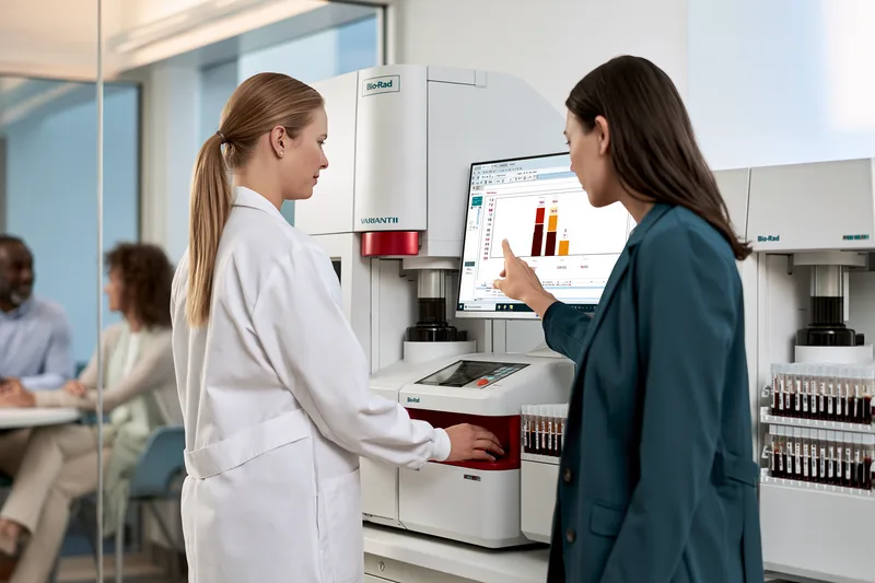
The proportions of HbA, HbA2 and HbF help distinguish thalassemia major vs minor vs intermedia, but they must be interpreted with the patient’s age, complete blood count, symptoms, genotype and transfusion history. A haemoglobin fraction that appears diagnostic in an untreated adult may be misleading in a newborn, a recently transfused patient, a person with iron deficiency or someone carrying an additional haemoglobin variant [1,4].
HbA
HbA is the predominant adult haemoglobin and contains two alpha-globin and two beta-globin chains. Its proportion reflects how much functional beta-globin the patient can produce, but donor red cells, age and coexisting variants can substantially alter the reported percentage [4].
In beta-thalassemia minor, HbA usually remains the dominant fraction because one HBB gene continues to produce beta-globin. The HbA percentage may be slightly lower than expected because HbA2, and sometimes HbF, occupies a larger proportion of the total haemoglobin. A dominant HbA fraction therefore does not exclude beta-thalassemia trait [1,4].
In untreated beta-thalassemia major, HbA may be absent when both inherited variants completely prevent beta-globin production. If one or both variants permit some residual beta-globin synthesis, a measurable HbA fraction may remain. The presence of HbA should therefore not automatically exclude severe disease, particularly when the patient has profound anaemia, early symptoms or an established transfusion requirement [4,8].
HbA in thalassemia intermedia is more variable. Some patients retain a meaningful amount of endogenous HbA because their beta-globin variants permit partial production, while others produce very little HbA but remain less severely affected because fetal haemoglobin is high or other genetic modifiers reduce globin-chain imbalance. Clinical severity cannot be assigned from the HbA percentage alone [4,6,9].
Regular transfusions introduce donor red cells containing predominantly HbA. A patient with beta-thalassemia major may consequently have a report showing a large HbA fraction even when the patient produces little or no HbA independently. The result must be interpreted alongside the date of the last transfusion, the quantity transfused and the usual transfusion interval [4,8].
After stem-cell transplantation, measurable HbA may originate from donor-derived blood formation rather than transfused red cells. Interpretation then requires information about donor chimerism, transfusion independence, haemoglobin stability and the timing of the test after transplantation. HbA alone cannot establish complete correction of the underlying haematological disorder.
HbA2
HbA2 is a minor adult haemoglobin composed of two alpha-globin and two delta-globin chains. An HbA2 proportion above the laboratory reference interval is a characteristic finding in many people with beta-thalassemia minor because reduced beta-chain production favours a relative increase in delta-chain-containing haemoglobin [1,4].
The HbA2 value should be interpreted with the complete blood count. A persistently low mean corpuscular volume and mean corpuscular haemoglobin, a relatively preserved red-cell count and elevated HbA2 together strongly support beta-thalassemia trait. An isolated mild increase in HbA2 without compatible red-cell indices may require repeat assessment or genetic confirmation [1,4].
A normal or borderline HbA2 result does not completely exclude beta-thalassemia minor. Iron deficiency can lower HbA2 in some patients and may shift a carrier result toward the reference range. Severe iron deficiency should therefore be corrected before repeating haemoglobin analysis when the blood count remains suggestive of beta-thalassemia [1,4].
Delta-globin variants or delta-thalassemia can also reduce HbA2 and mask the expected beta-thalassemia trait pattern. Conversely, some non-thalassemia conditions and treatment-related changes may produce a modest HbA2 elevation. Molecular testing becomes important when the haemoglobin fractions and red-cell indices do not provide a consistent explanation [4].
HbA2 is less useful for separating major from intermedia because both conditions are primarily assessed through HbF, residual HbA, genotype, clinical severity and transfusion requirement. In severe beta-thalassemia, HbA2 may form only a small part of the total haemoglobin because HbF predominates [4,8,9].
Haemoglobin E can create a major interpretation problem because some high-performance liquid chromatography systems measure HbE within the same analytical window as HbA2. A report showing an unusually large HbA2 or combined A2/E fraction should prompt review of the laboratory method and, when required, confirmatory electrophoresis or molecular testing. This is particularly important in families with ancestry from India, Bangladesh or Southeast Asia [4,9].
HbF
HbF is fetal haemoglobin and contains alpha-globin and gamma-globin chains. It is the dominant haemoglobin before birth and during early infancy. Its proportion normally declines as beta-globin production increases, so an elevated HbF percentage must always be interpreted according to age [4].
In beta-thalassemia major, HbF commonly constitutes most of the patient’s naturally produced haemoglobin because normal or near-normal HbA formation is severely impaired. A very high HbF proportion in an untransfused infant or child with severe microcytic anaemia supports a serious beta-globin disorder, but genotype and clinical findings are needed to distinguish beta-thalassemia major from other conditions associated with persistent HbF [4,8].
Patients with thalassemia intermedia may also have substantially elevated HbF. In some individuals, inherited modifiers increase gamma-globin production and reduce the damaging excess of unmatched alpha chains. Higher HbF can lessen anaemia and delay or reduce the need for regular transfusion, although the protective effect depends on the absolute amount and distribution of HbF within circulating red cells [4,6,9].
Beta-thalassemia minor may produce a normal or mildly elevated HbF result. A pronounced HbF increase is not typical of every carrier and may suggest hereditary persistence of fetal haemoglobin, delta-beta-thalassemia, treatment effects or a more complex genotype. Family testing and molecular analysis may be needed when HbF is much higher than expected for an uncomplicated carrier state [1,4].
The HbF percentage can appear high simply because HbA is severely reduced. A result reported as 80% HbF does not mean that the patient has 80% of a normal total haemoglobin concentration. A patient with a total haemoglobin of 6 g/dL and 80% HbF has a much lower absolute amount of HbF than a person with a total haemoglobin of 12 g/dL and the same percentage. Clinical oxygen-carrying capacity therefore depends on total haemoglobin as well as the reported fraction.
Hydroxyurea and other treatments that increase fetal haemoglobin can alter HbF over time. A rising HbF percentage may support a biological response, but treatment benefit should also be assessed through total haemoglobin, transfusion burden, symptoms, spleen size and complications. A higher HbF percentage without meaningful clinical improvement should not be considered sufficient evidence of disease control [6,9].
Factors That Can Distort the Report
Age is one of the most important sources of misinterpretation. HbF naturally predominates in newborns, while HbA2 has not yet reached the proportion expected in older children and adults. Newborn screening may identify an abnormal pattern, but definitive classification can require repeat haemoglobin analysis and genetic testing after the normal developmental transition in haemoglobin production [4].
Recent transfusion can alter HbA, HbA2 and HbF simultaneously because the laboratory measures a mixture of donor and patient red cells. The report should include the transfusion date, number of units and whether transfusions are occasional or regular. When an urgent diagnosis is required, molecular testing may provide more reliable information than repeating fraction analysis while donor cells remain present [4,8].
Iron deficiency can make beta-thalassemia trait harder to recognise by further lowering the mean corpuscular volume and sometimes reducing HbA2. Treating confirmed iron deficiency may improve haemoglobin and clarify the HbA2 pattern, but inherited microcytosis often persists after iron stores have normalised [1,4].
Alpha-thalassemia trait may produce microcytosis with normal HbA, HbA2 and HbF proportions after infancy. A normal haemoglobin electrophoresis or high-performance liquid chromatography result therefore does not exclude alpha-thalassemia. Persistent microcytosis with normal iron studies and no diagnostic beta-thalassemia pattern may require alpha-globin genetic testing [1,11].
Pregnancy can change plasma volume, haemoglobin concentration and iron requirements without changing the inherited globin variants. Haemoglobin fractions should be interpreted with ferritin, red-cell indices and partner screening rather than assuming that pregnancy-related anaemia represents progression of thalassemia.
The final interpretation should identify whether the pattern supports beta-thalassemia trait, a clinically significant beta-thalassemia syndrome, haemoglobin E beta-thalassemia, another haemoglobin variant or an inconclusive result requiring molecular confirmation. The percentages of HbA, HbA2 and HbF provide diagnostic evidence, but the patient’s transfusion need, growth, symptoms, spleen size, iron burden and organ health determine the practical severity of the disease [4,8,9].
Thalassemia Minor vs Iron Deficiency Anaemia
The distinction between thalassemia minor vs iron deficiency anaemia is important because both can produce a low mean corpuscular volume and mean corpuscular haemoglobin, yet their causes, treatment and reproductive implications are different. Thalassemia minor is an inherited reduction in globin-chain production, whereas iron deficiency develops when available iron is insufficient because of blood loss, increased requirements, reduced intake or impaired absorption. A patient may also have both conditions at the same time [1,4].
Microcytosis alone cannot establish iron deficiency. Prescribing iron solely because the red blood cells are small may leave thalassemia trait unrecognised and expose a patient with adequate iron stores to unnecessary treatment. The diagnosis should combine the complete blood count with ferritin, transferrin saturation, haemoglobin analysis and relevant clinical history [1,5].
Symptoms and Clinical History
Both conditions can cause tiredness, pallor, headaches, reduced concentration and breathlessness during exertion. Symptoms are often absent or mild in uncomplicated thalassemia minor, while progressive iron deficiency may produce worsening fatigue, reduced exercise tolerance, dizziness, palpitations, restless legs, pica or cravings for non-food substances. Symptoms overlap substantially and cannot distinguish the two conditions without laboratory testing [1,5].
A lifelong pattern of microcytosis, similar blood-count findings in relatives or a family history of haemoglobin disorders supports thalassemia trait. Iron deficiency is more likely when anaemia develops after heavy menstrual bleeding, pregnancy, repeated blood donation, gastrointestinal blood loss, a restricted diet or a condition that interferes with iron absorption. A previously normal mean corpuscular volume that becomes progressively lower also favours an acquired cause rather than an isolated inherited trait.
The two histories are not mutually exclusive. A person with beta-thalassemia minor may develop iron deficiency during pregnancy, after prolonged menstrual loss or because of gastrointestinal disease. Symptoms that are more severe than expected for the carrier state should therefore prompt investigation rather than being attributed automatically to thalassemia [1,4].
Complete Blood Count Differences
Thalassemia minor commonly produces a disproportionately low mean corpuscular volume in relation to the degree of anaemia. The haemoglobin may be normal or mildly reduced, while the red blood cell count is often preserved or relatively increased because the marrow continues to produce a large number of small cells. This pattern is particularly characteristic of beta-thalassemia trait but is not present in every carrier [1,4].
Iron deficiency usually reduces haemoglobin, mean corpuscular volume and mean corpuscular haemoglobin as the deficiency progresses. The red blood cell count may eventually fall because the marrow lacks enough iron to sustain normal red-cell production. Early iron deficiency can still produce an apparently preserved red blood cell count, so the count cannot independently confirm or exclude either diagnosis [1].
Red cell distribution width measures how much the red cells vary in size. It is frequently increased in iron deficiency because newly formed cells become progressively smaller as iron availability declines. It may remain normal or rise only mildly in uncomplicated thalassemia minor because many of the cells are uniformly microcytic. However, substantial overlap occurs, particularly when thalassemia and iron deficiency coexist, and red cell distribution width should not be treated as a definitive discriminator [1].
Haemoglobin concentration alone is also insufficient. A carrier with another cause of anaemia may have a haemoglobin level much lower than expected, while a person with early iron deficiency may still have haemoglobin within the laboratory reference interval. Reviewing previous blood counts can reveal whether microcytosis has been lifelong and stable or has developed progressively.
Mentzer Index
The Mentzer index is calculated by dividing the mean corpuscular volume by the red blood cell count. A lower result may suggest thalassemia trait, whereas a higher result may suggest iron deficiency. It can be useful as an initial screening calculation when a microcytic blood count is first reviewed [1].
The index cannot diagnose either condition. Its accuracy varies with age, population, severity of iron deficiency, pregnancy, recent transfusion and the presence of both disorders. Similar limitations apply to other mathematical indices based on mean corpuscular volume, haemoglobin, red blood cell count and red cell distribution width. These calculations can identify patients who need further testing but cannot replace ferritin, haemoglobin analysis or molecular confirmation [1,4].
A borderline Mentzer index should not be resolved by assuming that one diagnosis is more likely based only on ancestry. Thalassemia variants occur across many populations, and iron deficiency can affect patients from every ethnic background. Laboratory confirmation is required before long-term treatment or reproductive counselling.
Ferritin and Iron Studies
Serum ferritin is generally the most useful initial test for assessing iron stores. A low ferritin level strongly supports iron deficiency in a patient with microcytosis. Thalassemia minor by itself does not cause depleted iron stores, so a low ferritin result in a carrier indicates that iron deficiency is also present and requires appropriate evaluation and treatment [1].
A normal or elevated ferritin value does not always exclude deficiency. Ferritin can rise during infection, chronic inflammation, liver disease and other inflammatory states. When the clinical suspicion remains high, interpretation may require transferrin saturation, serum iron, total iron-binding capacity and an inflammatory marker such as C-reactive protein [1].
Low transferrin saturation supports inadequate circulating iron availability, particularly when ferritin is low or difficult to interpret. In uncomplicated thalassemia minor without iron deficiency, ferritin and transferrin saturation are generally not reduced solely because of the carrier state. An isolated serum iron result is less reliable because it can fluctuate with meals, time of day, illness and recent supplementation.
Ferritin should not be used to determine whether a person has beta-thalassemia trait. A normal ferritin makes iron deficiency less likely in the absence of inflammation, but the inherited diagnosis still requires compatible red-cell indices, haemoglobin analysis or genetic testing. Conversely, low ferritin does not exclude thalassemia because both conditions may coexist [1,4].
Peripheral Blood Smear
Both disorders can produce small, pale red blood cells on the peripheral smear. Target cells may support thalassemia, while pronounced variation in red-cell size, elongated cells and pencil-shaped cells may be more noticeable in iron deficiency. None of these appearances is specific enough to establish the diagnosis independently [1].
Thalassemia minor often produces relatively uniform microcytosis with target cells and mild variation in shape. More severe anisocytosis and poikilocytosis may occur when iron deficiency is also present. The smear should therefore be interpreted with the complete blood count and iron studies rather than used as a stand-alone test.
Nucleated red blood cells, marked haemolysis or extensive red-cell abnormalities are not expected in uncomplicated thalassemia minor. Their presence should prompt assessment for a clinically significant thalassemia syndrome, another haemoglobin disorder, severe haemolysis or a separate haematological condition [4].
HbA2 and Haemoglobin Analysis
An increased HbA2 proportion commonly supports beta-thalassemia minor. The finding is most meaningful when it accompanies persistent microcytosis, a normal or relatively high red blood cell count and no evidence that recent transfusion has altered the haemoglobin fractions [1,4].
Iron deficiency may reduce HbA2 in some beta-thalassemia carriers and shift the result toward the normal or borderline range. A patient with convincing microcytosis, low ferritin and a non-diagnostic HbA2 result may require iron replacement followed by repeat haemoglobin analysis after iron stores have recovered. Molecular testing may be appropriate when the result will influence pregnancy planning or family counselling [1,4].
Alpha-thalassemia trait commonly produces microcytosis with normal HbA, HbA2 and HbF proportions after infancy. A normal electrophoresis or high-performance liquid chromatography report therefore does not exclude alpha-thalassemia. Persistent microcytosis with normal iron studies and no beta-thalassemia pattern may require alpha-globin genetic testing [1,11].
Recent transfusion can make haemoglobin analysis difficult by introducing donor HbA and changing the measured proportions of HbA2 and HbF. The laboratory should be informed of the transfusion date and quantity. Genetic testing may be more informative when carrier assessment cannot be postponed until donor cells have cleared from the circulation [4].
When Both Conditions Occur Together
A patient with thalassemia minor can become iron deficient for the same reasons as anyone else. The combined condition may lower haemoglobin more than the carrier state alone, increase red cell distribution width and intensify fatigue, breathlessness or reduced exercise tolerance. The blood-count pattern may then no longer resemble the typical high-red-cell-count pattern associated with uncomplicated trait [1,4].
Confirmed iron deficiency should be treated even when thalassemia minor is present. Correcting the deficiency can improve haemoglobin, energy and functional capacity, although the inherited microcytosis may remain. Failure of the mean corpuscular volume to return to the laboratory reference range does not necessarily mean that iron treatment has failed [1,5].
Follow-up should assess haemoglobin response and restoration of iron stores rather than continuing supplementation until the mean corpuscular volume becomes normal. In a carrier, lifelong small red blood cells may persist despite adequate ferritin and transferrin saturation. Continued iron after deficiency has been corrected provides no treatment for the underlying thalassemia trait [1,5].
When haemoglobin remains substantially lower than expected after iron deficiency has been corrected, other causes should be considered. These include folate or vitamin B12 deficiency, chronic inflammation, kidney disease, thyroid dysfunction, ongoing blood loss, haemoglobin E, alpha-gene triplication or a more clinically significant thalassemia genotype [1,4].
Iron Treatment and Follow-Up
Iron should be prescribed when laboratory findings demonstrate deficiency or when a clinician has established a strong indication based on the complete clinical assessment. Thalassemia minor without iron deficiency does not improve with routine iron supplementation because the microcytosis results from inherited globin production rather than inadequate iron availability [1,5].
The dose, formulation and duration of iron treatment should reflect the patient’s age, pregnancy status, severity of deficiency, tolerance, ongoing blood loss and ability to absorb oral iron. Repeat blood counts and iron studies are needed to confirm that haemoglobin and iron stores are recovering. Failure to respond should prompt assessment of adherence, continued bleeding, malabsorption, inflammation or an alternative diagnosis.
In pregnancy, a carrier should not be assumed to be iron deficient merely because haemoglobin and mean corpuscular volume are low. Ferritin and obstetric assessment are needed because unnecessary iron does not correct thalassemia-related microcytosis, while untreated genuine iron deficiency can affect maternal wellbeing and fetal development. Partner carrier testing remains a separate priority and should not be delayed while the mother’s iron status is being corrected [4,5].
Children with persistent microcytosis should also be assessed before repeated courses of iron are given. A documented response to treatment supports iron deficiency, but temporary haemoglobin improvement does not exclude an underlying carrier state when microcytosis persists or family history is suggestive. Haemoglobin analysis and parental testing may be required [1,4].
The most reliable distinction is obtained by combining the blood-count pattern with ferritin and other iron studies, haemoglobin analysis, previous results and family history. Thalassemia minor explains inherited microcytosis, iron deficiency identifies a treatable shortage of available iron, and coexistence requires both accurate replacement therapy and continued recognition of the carrier state [1,4,5].
Thalassemia Major vs Minor vs Intermedia: Can the Type Change?

In thalassemia major vs minor vs intermedia, the inherited genetic pattern does not progressively change from one category to another. Thalassemia minor does not become intermedia or major with age, pregnancy, infection, stress, diet or lack of treatment. A carrier may develop worsening anaemia from another cause, but this does not transform the carrier state into a severe form of thalassemia [4,5].
The clinical classification of intermedia can change because transfusion needs and complications may increase over time. A patient who previously managed without regular transfusions may later require scheduled transfusion support. This represents a change from a non-transfusion-dependent to a transfusion-dependent clinical phenotype, not a new mutation or progression from minor to major [6,9].
Why Thalassemia Minor Does Not Become Major
The beta-globin variants responsible for thalassemia are inherited at conception and generally remain unchanged throughout life. A person with uncomplicated beta-thalassemia minor has one clinically significant HBB variant and sufficient beta-globin production from the other gene to avoid the severe ineffective erythropoiesis associated with thalassemia major [4].
Ageing does not cause the functioning HBB gene to become a second thalassemia gene. Menopause, pregnancy, emotional stress, infection, dietary changes or physical exertion also cannot convert a carrier into a person with beta-thalassemia major. The persistent small red-cell pattern may remain visible throughout life, but it does not represent continuing deterioration of the genetic disorder [4,5].
A carrier may nevertheless experience a fall in haemoglobin. Iron deficiency, folate or vitamin B12 deficiency, heavy menstrual bleeding, gastrointestinal blood loss, pregnancy, chronic inflammation, thyroid dysfunction, kidney disease or another acquired condition can worsen anaemia. Treating the additional cause may improve haemoglobin and symptoms even though the inherited microcytosis remains [1,4].
A haemoglobin value that becomes lower than expected should therefore trigger investigation rather than an assumption that minor has progressed. Ferritin, transferrin saturation, reticulocyte count, vitamin B12, folate, kidney function, thyroid function and evidence of bleeding may be required according to the patient’s history and symptoms.
Pregnancy can make anaemia more noticeable because blood plasma volume expands and iron and folate requirements increase. A pregnant carrier may consequently have a lower haemoglobin concentration than before pregnancy, particularly when iron deficiency is also present. This physiological and nutritional change does not convert thalassemia minor into intermedia or major [4,5].
Iron should not be started solely because the haemoglobin or mean corpuscular volume is low. A carrier who develops confirmed iron deficiency requires appropriate replacement, but the treatment goal is restoration of iron stores and improvement of iron-deficiency anaemia. The mean corpuscular volume may remain below the reference interval because the inherited carrier pattern persists [1,5].
Why an Earlier Minor Diagnosis May Be Revised
A person previously told that they have thalassemia minor may later receive an intermedia diagnosis when the original evaluation was incomplete. This is diagnostic reclassification rather than biological progression. The more severe phenotype was already determined by the inherited genetic combination but may not have been recognised during the first assessment [4].
One explanation is the presence of a second HBB variant that was not detected by the initial test. Basic haemoglobin analysis may suggest beta-thalassemia trait, while comprehensive molecular testing later identifies compound heterozygous beta-thalassemia or another clinically significant beta-globin combination. This is particularly relevant when haemoglobin is substantially lower than expected for a carrier [4].
An additional alpha-globin gene can also worsen beta-globin chain imbalance. A patient carrying beta-thalassemia together with alpha-gene triplication or quadruplication may develop greater anaemia, jaundice, splenic enlargement or an occasional transfusion requirement. The alpha-globin change was inherited from birth but may remain undetected until targeted genetic testing is performed [4].
Haemoglobin E beta-thalassemia can be misclassified when the haemoglobin fractions are not interpreted with the correct laboratory method. Haemoglobin E may overlap analytically with HbA2 on some high-performance liquid chromatography systems. Molecular confirmation may reveal a clinically significant combined disorder rather than uncomplicated beta-thalassemia trait [4,9].
Recent transfusion can obscure the natural haemoglobin pattern because donor red cells introduce HbA and alter the apparent proportions of HbA2 and HbF. A patient tested after transfusion may initially receive an incomplete or uncertain classification. Reviewing pretransfusion reports or performing molecular genetic testing can clarify the underlying diagnosis [4,8].
A diagnosis of uncomplicated minor should be reconsidered when the patient has persistent jaundice, marked splenic enlargement, bone changes, poor growth, gallstones, haemoglobin substantially below the expected carrier range, iron overload without an alternative explanation or any requirement for transfusion. These findings suggest either a more complex thalassemia genotype or an additional haematological condition [4,6].
Why Thalassemia Intermedia Can Become Transfusion Dependent
Thalassemia intermedia is clinically variable, and the distinction between non-transfusion-dependent and transfusion-dependent disease may change over time. Some patients remain free from regular transfusions for decades, while others gradually require more frequent support because chronic anaemia is no longer adequately tolerated or complications begin to progress [6,9].
A temporary transfusion during severe infection, surgery, pregnancy or acute haemoglobin decline does not necessarily mean that the patient has become permanently transfusion dependent. The haematologist evaluates whether haemoglobin and daily function return to the previous baseline after the precipitating event has resolved [6,9].
Regular transfusions may become appropriate when chronic anaemia impairs growth, school participation, employment, exercise capacity or normal physical development. Increasing skeletal changes, progressive splenic enlargement, recurrent haemolytic crises, extramedullary haematopoiesis or declining cardiopulmonary function may also support a continuing transfusion programme [6,9].
The decision should not be based on one haemoglobin threshold alone. Some patients tolerate a chronically low haemoglobin without major functional impairment, while others develop substantial symptoms or organ complications at a similar value. Longitudinal changes in growth, activity, spleen size, bone health, haemolysis and organ function provide more useful evidence than a single blood count [6,9].
An enlarging spleen can increase red-cell destruction and shorten the interval between transfusions. This may create a cycle of worsening anaemia, abdominal discomfort, early satiety and increasing transfusion burden. The effect of hypersplenism should be assessed through serial spleen measurements, blood counts and transfusion records rather than spleen size alone [6,9].
Pregnancy may expose limited haematological reserve in a patient with intermedia. Maternal blood-volume expansion and increased oxygen requirements can worsen anaemia, while fetal growth places additional physiological demands on the mother. Some patients need transfusions only during pregnancy, whereas others continue to require regular support afterwards. The long-term classification should be reassessed after recovery from pregnancy and delivery [6,9].
Age-related complications can also change treatment needs. Pulmonary hypertension, extramedullary haematopoiesis, leg ulcers, thrombosis, bone disease or progressive iron-related organ injury may develop despite years without regular transfusion. The appearance of these complications does not indicate that the genetic disorder has transformed, but it may require a more intensive treatment strategy [6,9].
What a Change in Classification Means
A change from NTDT to TDT describes the patient’s current dependence on regular transfusion support. It does not mean that thalassemia intermedia has genetically mutated into thalassemia major. The terminology is intended to guide treatment, iron monitoring and complication surveillance rather than describe a fixed sequence of disease stages [6,8,9].
The reverse may also occur after an effective disease-modifying intervention. A patient who becomes transfusion independent after successful stem-cell transplantation or gene-based treatment no longer has the same transfusion requirement. However, this outcome should be established through sustained haemoglobin production, absence of transfusions, treatment-specific follow-up and assessment of residual organ damage rather than a short interruption in transfusion [8].
Patients transferring between hospitals or countries may receive different labels because treatment centres do not always use major, intermedia, TDT and NTDT in exactly the same way. A reliable clinical record should therefore include the genotype, age at first transfusion, reason transfusions were started, current interval, annual red-cell requirement, pretransfusion haemoglobin, spleen status, antibody history, iron burden and major complications [8,9].
For a patient recorded as having thalassemia minor, new or disproportionate symptoms require investigation for an additional condition or incomplete original diagnosis. For a patient with intermedia, increasing transfusion requirements indicate a change in clinical severity that requires reassessment of iron monitoring, chelation, organ surveillance and eligibility for disease-modifying or potentially curative treatment [4,6,8,9].
Risks in Thalassemia Major vs Minor vs Intermedia

The long-term risks in thalassemia major vs minor vs intermedia differ in their causes and required surveillance. Thalassemia major is associated mainly with severe anaemia, lifelong transfusion exposure, iron accumulation and treatment-related complications. Thalassemia intermedia can cause progressive organ damage through chronic anaemia, ineffective erythropoiesis, haemolysis and increased intestinal iron absorption even without regular transfusions. Thalassemia minor usually does not cause progressive organ injury, but inaccurate diagnosis, unnecessary iron treatment and unrecognised reproductive risk can have important consequences [4,5,6,8,9].
A patient’s risk cannot be estimated from the diagnostic label or haemoglobin value alone. Age, transfusion burden, chelation adherence, spleen status, genotype, pregnancy, previous complications and magnetic resonance measurements of organ iron influence the probability of future disease. Regularly updated clinical records are particularly important when patients transfer between treatment centres or healthcare systems [6,8,9].
Thalassemia Major Risks
Iron overload is one of the principal long-term risks in thalassemia major. Each red-cell transfusion introduces iron that the body cannot actively excrete in sufficient quantities. Without effective chelation, excess iron gradually accumulates in the liver, heart, endocrine glands and other tissues, where it can cause progressive organ dysfunction [8].
Serum ferritin is useful for following general trends but can rise during infection, inflammation or liver injury and may not accurately represent iron within individual organs. Liver iron concentration measured by magnetic resonance imaging and cardiac T2* magnetic resonance imaging provide more direct assessments of iron deposition. A patient can have improving ferritin while clinically important cardiac or hepatic iron remains present, so chelation decisions should not depend on one ferritin result [8].
Cardiac iron accumulation can impair contraction of the heart muscle and interfere with its electrical rhythm. Early myocardial iron may cause no symptoms, while advanced involvement can produce palpitations, breathlessness, reduced exercise tolerance, ankle swelling, fainting, arrhythmia or heart failure. Cardiac magnetic resonance imaging can identify iron before conventional symptoms or echocardiographic abnormalities become evident [8].
Chest pain, fainting, a new irregular heartbeat, breathlessness at rest or rapidly declining exercise tolerance requires urgent assessment. These symptoms should not be assumed to result only from anaemia because cardiac iron, pulmonary hypertension, infection, arrhythmia and other cardiopulmonary disorders may require immediate treatment [8].
The liver receives a substantial proportion of transfusional iron and is often the earliest organ in which excess iron can be measured. Progressive accumulation may cause inflammation, fibrosis, cirrhosis and an increased risk of liver failure. Viral hepatitis, metabolic fatty liver disease, alcohol exposure and medication toxicity can coexist and accelerate injury, making liver enzymes, liver iron concentration and fibrosis assessment clinically important [8].
A normal alanine aminotransferase or aspartate aminotransferase result does not exclude significant liver iron. Conversely, raised liver enzymes do not prove that iron is the only cause. Evaluation may require hepatitis testing, imaging, metabolic assessment and review of chelation exposure in addition to iron measurements [8].
Iron deposition and chronic disease can affect the pituitary gland, thyroid, pancreas, parathyroid glands and reproductive organs. Consequences may include poor growth, delayed or absent puberty, reduced sex hormone production, hypothyroidism, diabetes, impaired calcium regulation and infertility. These abnormalities can develop gradually and may be detectable through scheduled laboratory testing before the patient recognises symptoms [8].
A child whose height or weight begins to fall across growth percentiles requires assessment of pretransfusion haemoglobin, nutritional intake, thyroid function, glucose regulation, puberty, liver health and overall treatment adequacy. Increasing calories alone will not correct growth failure caused by inadequate transfusion, iron-related endocrine injury or delayed puberty [8].
Excessive thirst, frequent urination, unexplained weight loss or recurrent infections may indicate impaired glucose control or diabetes. Persistent cold intolerance, constipation, dry skin, reduced concentration or unexplained weight gain may indicate thyroid dysfunction. Delayed puberty, menstrual irregularity, reduced libido or fertility difficulty warrants endocrine and reproductive assessment rather than being accepted as an unavoidable consequence of thalassemia [8].
Reduced bone density and fractures can result from several interacting factors, including marrow expansion, delayed puberty, hormonal dysfunction, low vitamin D, reduced physical activity, nutritional insufficiency and iron-related organ injury. Bone pain or reduced height should not be attributed automatically to anaemia. Assessment may include calcium and vitamin D status, endocrine testing and bone-density measurement according to age and clinical risk [8].
Inadequate transfusion can allow continued bone-marrow expansion, contributing to skeletal deformity, thinning of cortical bone and fracture risk. Effective transfusion suppresses excessive marrow activity, but it does not eliminate bone risks arising from endocrine dysfunction or established skeletal injury. Bone surveillance remains necessary even in patients whose transfusion programme is stable [8].
Repeated transfusions can cause alloimmunisation, in which the immune system develops antibodies against donor red-cell antigens. These antibodies can make compatible blood more difficult to locate and increase the risk of delayed haemolytic transfusion reactions. Extended antigen matching, accurate recording of all previous antibodies and continued communication between transfusion centres reduce avoidable risk [8].
An antibody may become undetectable in routine testing but still cause a reaction if the patient later receives blood carrying the corresponding antigen. Patients should retain a permanent record of identified antibodies and provide it whenever transfusions are administered in another hospital or country. A negative current antibody screen does not erase a documented historical antibody [8].
Acute transfusion reactions may present with fever, chills, rash, breathing difficulty, chest or back pain, dark urine, low blood pressure or sudden discomfort during or shortly after transfusion. Transfusion should be stopped and assessed according to the treating centre’s protocol when a reaction is suspected. Delayed reactions may appear days later with increasing fatigue, jaundice, falling haemoglobin or dark urine [8].
Modern blood screening and donor testing have substantially reduced transfusion-transmitted infections in well-regulated systems, but surveillance remains relevant because exposure is lifelong and screening practices differ between regions. Previous transfusions received in different countries, earlier treatment periods and incomplete vaccination records should be documented when infection risk is assessed [8].
Splenic enlargement may persist or progress when abnormal red cells are removed rapidly or bone-marrow activity is inadequately suppressed. Hypersplenism can reduce haemoglobin, white blood cells and platelets and may increase transfusion requirements. Abdominal fullness, early satiety, pain beneath the left ribs or a measurable increase in spleen size should be evaluated with blood-count trends and transfusion records [8].
Splenectomy can reduce red-cell consumption in selected patients but creates lifelong susceptibility to severe bacterial infection and increases thrombotic risk. Fever in a patient without a functioning spleen requires urgent medical assessment because serious infection can progress rapidly. Vaccination, infection-prevention measures and an individual emergency plan remain necessary throughout life [8].
Chelation therapy protects organs from iron accumulation but can produce adverse effects that differ among chelating medicines. Kidney dysfunction, liver abnormalities, gastrointestinal symptoms, reduced blood-cell counts, hearing changes or visual effects may occur depending on the agent and patient. Monitoring should follow the specific chelator rather than using one generic laboratory schedule for every patient [8].
A rising iron burden does not always mean that the prescribed chelator is ineffective. Missed doses, gastrointestinal intolerance, unsuitable timing, inadequate dosage, drug interactions or changing transfusion exposure may reduce net iron removal. Identifying the reason for inadequate control is safer than increasing the dose without reviewing adherence, organ function and treatment tolerance [8].
Pregnancy can place additional strain on the heart and increase transfusion requirements. Preconception assessment should examine cardiac function, liver iron, endocrine health, diabetes control, fertility, antibody history and current medication safety. A patient who feels well before pregnancy may still have clinically important organ iron requiring optimisation before conception [8].
The continuing burden of transfusions, chelation, monitoring and treatment complications can affect education, employment, relationships, sleep and emotional health. Anxiety, treatment fatigue and depression can reduce adherence and thereby increase physical risk. Psychological support and practical treatment planning are components of comprehensive thalassemia care rather than optional additions [8].
Thalassemia Intermedia Risks
Thalassemia intermedia can produce serious complications despite the absence of a regular transfusion schedule. Chronic ineffective erythropoiesis, haemolysis, tissue hypoxia and increased intestinal iron absorption continue over many years. Complications may therefore become more apparent with age even when the haemoglobin level appears stable [6,9].
Iron overload is a major risk in intermedia because ineffective erythropoiesis suppresses hepcidin, allowing the intestine to absorb more iron than the body requires. The excess accumulates primarily in the liver and can eventually contribute to fibrosis, endocrine dysfunction and other organ injury. This process can occur in patients who have never received regular transfusions [6,9].
Ferritin may underestimate liver iron in non-transfusion-dependent thalassemia. A value that appears only moderately elevated should therefore be interpreted with age, transfusion history, liver function and previous trends. Magnetic resonance measurement of liver iron is particularly valuable when cumulative exposure is suspected or chelation is being considered [6,9].
Intermittent transfusions received during infection, surgery, pregnancy or haemolytic episodes add further iron. A patient who is described as non-transfusion dependent may still accumulate a substantial lifetime transfusion burden. Every transfusion should be recorded so that iron risk is estimated from the complete history rather than the current treatment schedule alone [9].
Chronic haemolysis can produce jaundice, dark urine and pigment gallstones. Gallstones may remain asymptomatic or cause right upper abdominal pain, nausea, fever, bile-duct obstruction or worsening jaundice. A sudden change in abdominal symptoms should not be attributed automatically to chronic haemolysis because acute gallbladder or bile-duct complications may require urgent treatment [6,9].
Progressive splenic enlargement can worsen anaemia by trapping and destroying red cells. It may also reduce white blood cells and platelets, cause abdominal discomfort and produce early satiety that interferes with nutrition. Serial spleen measurements, complete blood counts and functional symptoms provide more useful information than one ultrasound measurement [6,9].
Splenectomy has historically been used to reduce anaemia or transfusion burden, but patients with intermedia have an increased risk of thrombosis and pulmonary hypertension after the procedure. Removal of the spleen also creates lifelong susceptibility to severe infection. The decision requires individual assessment of anaemia, spleen-related symptoms, transfusion requirements and alternative treatment options [6,9].
Thrombotic complications may include deep-vein thrombosis, pulmonary embolism, portal-vein thrombosis, stroke or thrombosis in other vascular sites. Risk is influenced by chronic haemolysis, abnormal red-cell membranes, platelet activation, age, splenectomy, pregnancy, immobility, previous thrombosis and additional inherited or acquired factors [6,9].
Not every patient with intermedia requires routine anticoagulation. Thrombotic prevention should be individualised, particularly during surgery, prolonged immobility, pregnancy, the postpartum period or hospital admission. Unilateral leg swelling, sudden chest pain, unexplained breathlessness, coughing blood or new neurological symptoms requires urgent assessment [6,9].
Pulmonary hypertension can develop through interactions among chronic haemolysis, anaemia, thrombosis, vascular dysfunction, iron burden and splenectomy. Early symptoms may include gradually worsening breathlessness, reduced exercise tolerance, chest pressure, palpitations, dizziness or fainting. These symptoms may resemble ordinary anaemia and require cardiopulmonary evaluation when they are new or progressive [6,9].
Extramedullary haematopoiesis develops when blood-forming tissue expands outside the bone marrow in response to chronic ineffective erythropoiesis. Masses may form near the spine, within the chest or in other sites. Some remain asymptomatic, while others cause pain, breathing difficulty or pressure on nearby structures [6,9].
Back pain accompanied by leg weakness, numbness, altered walking, loss of bladder control or loss of bowel control may indicate spinal cord compression from paraspinal extramedullary haematopoiesis. This requires urgent imaging and specialist treatment because delayed intervention can lead to permanent neurological injury [6,9].
Bone-marrow expansion can alter facial and skeletal structure and contribute to cortical thinning, bone pain and fractures. Reduced bone density may be compounded by endocrine dysfunction, low vitamin D, inadequate physical activity and nutritional factors. Patients with persistent bone pain, height loss or low-trauma fractures require investigation beyond routine haemoglobin testing [6,9].
Leg ulcers commonly develop near the ankles and may be painful, slow to heal and recurrent. Chronic tissue hypoxia, anaemia, vascular dysfunction and local trauma can contribute. Management requires assessment of infection, circulation, haemoglobin adequacy, nutrition and wound care rather than repeated topical treatment alone [6,9].
Endocrine and reproductive complications can occur when iron overload, chronic anaemia or tissue hypoxia affects hormonal function. Delayed puberty, reduced fertility, impaired glucose regulation, thyroid abnormalities and bone disease may develop, although the pattern can differ from regularly transfused disease. Symptoms and laboratory surveillance should guide investigation rather than assuming that endocrine injury occurs only in thalassemia major [9].
Pregnancy may worsen anaemia and increase the need for transfusion. It can also increase thrombotic and cardiopulmonary stress, particularly in patients with splenectomy, pulmonary hypertension or significant iron overload. Preconception evaluation should assess haemoglobin stability, cardiac status, liver iron, endocrine health, antibodies and previous thrombotic events [9].
Occasional transfusion does not eliminate the risk of alloimmunisation. Patients exposed to donor blood intermittently may develop antibodies that complicate future transfusion during pregnancy, surgery or acute illness. Red-cell antigen typing and accurate preservation of antibody history remain important even when transfusions are years apart [9].
Progressively reduced activity, impaired growth, increasing spleen size, skeletal changes, recurrent extramedullary haematopoiesis or cardiopulmonary complications may indicate that the previous non-transfusion-dependent approach is no longer adequate. Reassessment of transfusion strategy and disease-modifying treatment can reduce complications arising from prolonged ineffective erythropoiesis [6,9].
Thalassemia Minor Risks
Uncomplicated thalassemia minor generally does not cause the progressive cardiac, hepatic, endocrine or skeletal injury associated with major and intermedia. Most carriers have normal life expectancy and do not require transfusion, chelation or disease-directed medication. The principal risks arise from misinterpretation of the blood count and failure to recognise the genetic implications of carrier status [1,4,5].
Persistent microcytosis may be repeatedly diagnosed as iron deficiency without confirmation from ferritin or transferrin saturation. Unnecessary long-term iron treatment does not correct the inherited reduction in globin production and may cause gastrointestinal adverse effects or excessive iron exposure. Iron should be used when deficiency has been demonstrated rather than prescribed solely because the mean corpuscular volume is low [1,5].
The opposite error can also occur when genuine iron deficiency is dismissed as part of the carrier state. Menstrual blood loss, pregnancy, gastrointestinal bleeding, dietary insufficiency and impaired absorption can reduce iron stores in a person with thalassemia minor. A new fall in haemoglobin, increasing fatigue or a change from the patient’s established blood-count pattern requires investigation [1,4].
Symptoms that are disproportionate to mild carrier anaemia may result from vitamin B12 deficiency, folate deficiency, thyroid dysfunction, kidney disease, inflammation, sleep disturbance, heavy menstrual bleeding or another medical condition. Attributing all fatigue to thalassemia minor can delay diagnosis of a treatable coexisting disorder [1,4].
Pregnancy may lower haemoglobin through plasma-volume expansion and increased nutritional requirements, but it does not convert thalassemia minor into major or intermedia. Ferritin and obstetric assessment help distinguish expected carrier microcytosis from superimposed iron deficiency. Iron supplementation should reflect documented maternal need rather than the carrier label alone [4,5].
The most consequential risk is having a child with clinically significant haemoglobin disease when the reproductive partner also carries beta-thalassemia or another relevant haemoglobin variant. A partner may carry beta-thalassemia, haemoglobin E, sickle haemoglobin, delta-beta-thalassemia or an alpha-globin abnormality without having symptoms. Partner testing should therefore be based on laboratory evaluation rather than physical wellbeing or assumptions about ancestry [4,5].
A carrier result should be documented permanently and communicated during preconception or antenatal care. Complete blood count and haemoglobin analysis may be sufficient for initial partner screening, while molecular genetic testing may be required when the results are borderline, recent transfusion obscures the pattern or the parental combination could produce severe disease [4,5].
An original diagnosis of minor should be reassessed when the patient has persistent jaundice, significant splenic enlargement, gallstones, bone changes, haemoglobin substantially below the expected carrier range, unexplained iron overload or any transfusion requirement. These findings are not typical of uncomplicated thalassemia minor and may indicate an additional globin variant, alpha-gene triplication, haemoglobin E beta-thalassemia or another haematological disorder [4].
Repeated medical testing is not required merely to prove that the carrier state still exists. Follow-up should be directed by symptoms, pregnancy, confirmed nutritional deficiency or another clinical indication. Accurate diagnosis prevents both undertreatment of genuine health problems and unnecessary treatment of an inherited blood-count pattern that is usually clinically stable [1,4,5].
Inheritance Risk in Thalassemia Major vs Minor vs Intermedia

The inheritance risk in thalassemia major vs minor vs intermedia depends on the exact globin-gene variants carried by both biological parents, not on how anaemic either parent appears. A healthy person with thalassemia minor can pass a clinically significant variant to a child, while a patient with major or intermedia can also transmit one of their affected genes. Genetic testing and partner screening are therefore more reliable than symptoms, haemoglobin level or family appearance when estimating risk to future children [4,5,11].
| Parent Combination | Possible Child Outcome |
|---|---|
| One beta-thalassemia carrier + unaffected partner | About 50% chance of carrier status and 50% chance of not inheriting that variant |
| Both parents carry beta-thalassemia | Each pregnancy may have 25% affected, 50% carrier and 25% unaffected |
| Major/intermedia patient + unaffected partner | Children generally inherit at least one affected beta-globin variant and are usually carriers |
| Major/intermedia patient + beta-thalassemia carrier | Approximately 50% risk of an affected child and 50% carrier risk, depending on variants |
| Beta-thalassemia carrier + HbE carrier | Risk of HbE/beta-thalassemia |
| Beta-thalassemia carrier + sickle-cell carrier | Risk of sickle beta-thalassemia |
| Both parents with alpha-zero thalassemia in cis | Each pregnancy may have a 25% risk of Hb Bart hydrops fetalis syndrome |
Beta-thalassemia is usually inherited in an autosomal recessive pattern. A child receives one HBB gene from each parent. A person with beta-thalassemia minor generally has one clinically significant HBB variant and one functioning copy, while a person with beta-thalassemia major or intermedia usually has clinically important variants affecting both inherited copies. The clinical severity in an affected child then depends on the specific combination inherited and on modifying factors such as fetal haemoglobin production and alpha-globin gene number [4].
Each pregnancy represents a new and independent genetic event. The outcome of one pregnancy does not alter the probability in the next. Having one unaffected child does not mean that future children are protected, and having one affected child does not mean that every subsequent child will have the same condition [4,5].
When One Parent Has Thalassemia Minor
When one parent carries beta-thalassemia and the other parent has no clinically significant beta-globin or interacting haemoglobin variant, each pregnancy generally has a 50% probability of producing a carrier child and a 50% probability of producing a child who did not inherit that parental variant. The children would not ordinarily be expected to have beta-thalassemia major from this parental combination alone [4,5].
The non-carrier partner should still undergo appropriate testing rather than being assumed unaffected because the complete blood count is normal or there is no family history. Some mild beta-globin variants, alpha-thalassemia changes and structural haemoglobin variants may cause few or no symptoms. Recent transfusion, iron deficiency or pregnancy can also make screening results more difficult to interpret [4].
A normal mean corpuscular volume does not exclude every clinically relevant haemoglobin variant. Initial partner assessment commonly includes a complete blood count and haemoglobin analysis, followed by molecular testing when results are borderline, family history is suggestive or one partner carries a variant that could interact with haemoglobin E, sickle haemoglobin or another beta-globin abnormality [4].
When Both Parents Have Beta-Thalassemia Minor
When both parents carry clinically significant beta-thalassemia variants, each pregnancy generally has a 25% probability of an affected child, a 50% probability of a carrier child and a 25% probability of a child who inherits neither parental variant. These probabilities apply separately to every conception [4,5].
The affected child’s eventual phenotype cannot always be predicted simply as major or intermedia from the word carrier on the parental reports. Two beta-zero variants are more often associated with severe transfusion-dependent disease, while combinations that retain partial beta-globin production may produce a milder phenotype. Fetal haemoglobin modifiers, alpha-globin changes and other genetic factors can substantially influence the clinical course [4].
Parents may therefore receive a fetal or newborn genetic diagnosis before the exact future transfusion requirement can be predicted. The molecular result identifies the inherited variants, but longitudinal assessment of haemoglobin, growth, symptoms, spleen size and transfusion need determines whether the child develops a transfusion-dependent or non-transfusion-dependent phenotype [4,8,9].
When One Parent Has Major or Intermedia
A person with beta-thalassemia major or intermedia usually carries clinically significant variants in both HBB genes and will pass one of those variants to every biological child. If the other parent has no relevant beta-globin variant, the children will generally inherit one affected HBB gene and are expected to be carriers rather than to have major disease [4].
When a person with major or intermedia has a reproductive partner who carries beta-thalassemia, each pregnancy may have an approximately 50% probability of an affected child and a 50% probability of a carrier child. The exact phenotype in an affected child depends on which parental variants are inherited and on additional modifying genes [4].
When both biological parents have clinically significant beta-thalassemia affecting both HBB genes, all children will inherit an HBB variant from each parent and are expected to have an affected genotype. The clinical severity may still differ between siblings because different variant combinations or modifying factors can be inherited [4].
Fertility and pregnancy planning may require additional specialist input for patients with major or intermedia. Iron overload, cardiac function, endocrine health, ovarian or testicular function, transfusion antibodies and medication safety can influence conception and pregnancy management, but they do not change the underlying genetic probability transmitted to the child [8,9].
Haemoglobin E and Other Beta-Globin Variants
A beta-thalassemia carrier can have a child with clinically significant disease even when the partner does not carry beta-thalassemia specifically. Haemoglobin E, sickle haemoglobin, haemoglobin C, delta-beta-thalassemia and some unstable haemoglobins can interact with a beta-thalassemia variant and produce a combined disorder [4].
Haemoglobin E beta-thalassemia is particularly relevant to families with ancestry from India, Bangladesh and Southeast Asia. One parent may carry haemoglobin E and the other beta-thalassemia, while both remain well or have only mild blood-count changes. A child who inherits both variants may have a phenotype ranging from mild non-transfusion-dependent anaemia to severe transfusion-dependent disease [4,9].
When one parent carries sickle haemoglobin and the other carries beta-thalassemia, a child may inherit sickle beta-thalassemia. The severity depends partly on whether the beta-thalassemia variant permits residual beta-globin production. Partner screening should therefore evaluate clinically important haemoglobin variants rather than testing only for the same condition already identified in the first parent [4].
Laboratory terminology can vary between India, the United States, the United Kingdom, Canada, Australia and Singapore. A report may use terms such as trait, carrier, heterozygous, variant detected or presumptive haemoglobinopathy. The underlying haemoglobin fractions and molecular findings should be reviewed rather than relying only on the report heading.
Alpha-Thalassemia Inheritance
Alpha-thalassemia reproductive risk depends on the number and arrangement of affected alpha-globin genes. A person with two affected genes may have the changes on the same chromosome, called the cis arrangement, or one affected gene on each chromosome, called the trans arrangement. These configurations can produce similar blood-count findings in the parent but very different risks for the child [11].
When both parents carry alpha-zero thalassemia in the cis arrangement, each pregnancy may have a 25% probability of haemoglobin Bart hydrops fetalis syndrome, a 50% probability of alpha-thalassemia trait and a 25% probability of inheriting no parental alpha-zero deletion. Haemoglobin Bart syndrome causes profound fetal anaemia and can also create serious maternal complications [11].
When one parent carries an alpha-zero deletion and the other carries an alpha-plus change affecting one alpha-globin gene, a child may inherit three affected genes and develop haemoglobin H disease. The severity may be greater when a non-deletional alpha-globin variant is involved [11].
A routine complete blood count or normal adult haemoglobin electrophoresis cannot reliably determine whether two affected alpha genes are in cis or trans. Molecular testing is often required to establish the arrangement and provide an accurate pregnancy-risk estimate [11].
Testing Before Pregnancy
Carrier screening is most informative when completed before conception because it allows time to confirm both partners’ results, obtain genetic counselling and consider reproductive options without the time pressure of an ongoing pregnancy. Testing is particularly important when either partner has known thalassemia, unexplained microcytosis, a family history of haemoglobin disease or ancestry associated with thalassemia or other haemoglobin variants [4,5,11].
Initial testing generally includes a complete blood count with red-cell indices and haemoglobin analysis. Ferritin should be assessed when iron deficiency could affect interpretation. Molecular testing may then be needed to confirm the specific HBB variant, identify alpha-globin deletions or non-deletional variants, determine whether alpha changes are in cis or trans, or clarify an unusual haemoglobin fraction [4,11].
Both partners should ideally be tested through methods capable of detecting the variants relevant to their ancestry and family history. A limited mutation panel may miss rare or population-specific variants. Full sequencing, deletion analysis or additional globin-gene testing may be required when the blood findings remain suspicious despite an initially negative result [4,11].
The final reproductive assessment should state the confirmed variant in each partner, the expected inheritance probabilities, the range of possible clinical severity and the limitations of prediction. Terms such as positive, minor or trait without the molecular details may be insufficient for planning prenatal or preimplantation testing.
Reproductive Options
Couples at increased risk may choose natural conception without fetal testing, natural conception with prenatal diagnosis, in vitro fertilisation with preimplantation genetic testing for a monogenic condition, use of donor sperm or donor eggs, adoption or a decision not to pursue pregnancy. Genetic counselling should provide accurate information about each option while respecting the couple’s values, medical circumstances and legal context [4,5,11].
Preimplantation genetic testing requires identification of the parental variants before an in vitro fertilisation cycle begins. Embryos can then be tested for the familial variants before transfer. The process does not guarantee pregnancy, and confirmatory prenatal testing may still be discussed because no laboratory method is completely free from diagnostic limitations.
Natural conception with prenatal diagnosis can determine whether the fetus inherited the parental variants. Chorionic villus sampling or amniocentesis may be used according to gestational age, local practice and specialist advice. The laboratory must know the exact parental variants before testing so that the fetal result can be interpreted accurately [4,11].
Non-invasive prenatal screening based on maternal blood is widely used for common chromosome conditions, but routine screening does not necessarily provide a definitive diagnosis of fetal thalassemia. Availability and validation of non-invasive testing for specific monogenic haemoglobin disorders vary, and diagnostic decisions should not be based on a commercial screening claim without specialist confirmation.
Testing During Pregnancy
When a pregnant patient is found to have thalassemia minor, partner testing should be arranged promptly. Waiting until late pregnancy can reduce the time available for genetic counselling, confirmatory testing and informed reproductive decision-making [5].
Iron deficiency should be assessed separately because pregnancy-related anaemia can coexist with thalassemia trait. Correcting iron deficiency may improve maternal haemoglobin but does not change the inherited probability for the fetus. Conversely, a normal ferritin result does not remove the need for partner testing when the mother is a confirmed carrier [4,5].
When both parents carry relevant variants, referral to a fetal medicine or clinical genetics service allows discussion of fetal diagnostic options and pregnancy-specific risks. In alpha-thalassemia pregnancies at risk of haemoglobin Bart syndrome, early specialist assessment is important because severe fetal anaemia can affect both fetal survival and maternal health [11].
Testing Children and Other Relatives
When a child is diagnosed with thalassemia major or intermedia, testing the biological parents confirms the inheritance pattern and identifies relatives who may also be carriers. Siblings should be assessed according to their symptoms, blood counts and genetic risk rather than assuming that an unaffected appearance excludes carrier status [4].
Carrier children generally do not require disease-directed treatment, but documenting the diagnosis can prevent repeated misdiagnosis as iron deficiency and provides important information for their future reproductive care. The result should be explained in age-appropriate language as the child matures.
Extended family testing can identify siblings, cousins and other relatives who may benefit from preconception screening. This is particularly valuable in families in which marriages occur within the extended family or where more than one child has been affected. Testing should remain voluntary and confidential, with individual counselling for each person.
The reproductive risk attached to thalassemia minor is therefore determined by the partner’s result and the interaction between both inherited variants. Major and intermedia describe the clinical expression in an affected person, while molecular testing identifies what can be passed to the next generation [4,5,11].
Treatment in Thalassemia Major vs Minor vs Intermedia

Treatment in thalassemia major vs minor vs intermedia differs because the three conditions create different levels of anaemia, ineffective red-cell production, iron accumulation and organ risk. Thalassemia major usually requires a planned lifelong transfusion and iron-chelation programme unless a successful curative treatment removes transfusion dependence. Thalassemia intermedia requires treatment according to symptoms, growth, complications and iron burden rather than automatic regular transfusion. Thalassemia minor usually requires no thalassemia-specific medication, transfusion or chelation [1,2,6,8,9].
| Treatment | Major | Intermedia | Minor |
|---|---|---|---|
| Regular blood transfusion | Usually required | Only when clinically indicated; may become regular | Not normally required |
| Iron chelation | Commonly required | Required when clinically significant iron overload develops | Not routinely required |
| Ferritin monitoring | Essential | Important | Only when clinically indicated |
| Liver iron MRI | Important | Important when iron accumulation is suspected | Usually unnecessary |
| Cardiac T2* MRI | Important according to risk | Selected patients | Usually unnecessary |
| Hydroxyurea or disease-modifying therapy | Selected patients | Selected patients | Not normally indicated |
| Luspatercept or other approved therapy | Eligible patients depending on indication | Selected eligible patients | Not indicated for uncomplicated trait |
| Stem-cell transplantation | Potentially curative option | Selected severe cases | Not indicated |
| Gene-based therapy | Selected eligible patients | Eligibility depends on phenotype and indication | Not indicated |
| Ayurvedic integrative care | Can be coordinated with transfusion and chelation | May be individualised with complication monitoring | Usually directed at coexisting symptoms rather than carrier status |
The treatment category should be based on the patient’s current clinical phenotype rather than the diagnostic label alone. Two patients described as having intermedia may need very different levels of care, while a patient historically labelled as major may become transfusion independent after successful stem-cell transplantation or gene-based treatment. Longitudinal haemoglobin results, transfusion records, growth, spleen size, iron measurements and organ function provide a more reliable basis for treatment planning [4,6,8,9].
Treatment for Thalassemia Major
Regular red-cell transfusion is the foundation of treatment for most patients with thalassemia major or transfusion-dependent thalassemia. The purpose is not only to raise the haemoglobin temporarily but also to maintain adequate oxygen delivery, support normal growth and activity, suppress excessive bone-marrow expansion and reduce progressive enlargement of the liver and spleen [2,8].
Many transfusion programmes aim to maintain the pretransfusion haemoglobin near 9.5 to 10.5 g/dL, equivalent to 95 to 105 g/L, although the individual target may be adjusted according to age, symptoms, cardiac status, growth, spleen size and complications. Some patients require a higher target when there is persistent extramedullary haematopoiesis, significant cardiac disease, pulmonary hypertension or inadequate suppression of bone-marrow activity [8].
The transfusion interval is usually individualised so that haemoglobin does not repeatedly fall below the level needed for daily function. Increasing fatigue, poor school attendance, reduced work capacity, breathlessness or bone pain before every transfusion may indicate that the interval or transfused volume requires reassessment. A post-transfusion haemoglobin result cannot replace serial pretransfusion measurements because the two values reflect different parts of the treatment cycle [8].
Transfused red cells should be compatible with the patient’s ABO and RhD group and selected according to an extended antigen profile whenever possible. Matching for additional red-cell antigens can reduce alloimmunisation, particularly when initiated before the first transfusions. The patient’s complete antibody history must remain available even when a previously detected antibody is no longer measurable on the current screen [8].
Leucocyte-reduced blood products are generally used to reduce febrile reactions and exposure to donor white cells. The transfusion team should also document previous reactions, antibody findings and any difficulty obtaining compatible blood. This information becomes especially important when a patient travels, relocates or receives treatment in a different country [8].
Repeated transfusions introduce iron that the body cannot remove efficiently. Iron-chelation therapy is therefore required once transfusional iron begins to accumulate. Chelation is intended to prevent or reduce iron-related injury to the heart, liver, pancreas, pituitary gland, thyroid, bones and reproductive system [2,8].
Chelation treatment should not be adjusted from one ferritin result alone. Serum ferritin is useful for following trends, but it can rise during inflammation, infection or liver injury and may not accurately represent iron in the heart or liver. Magnetic resonance measurement of liver iron concentration and cardiac T2* provides more direct information about organ iron and helps guide the intensity of chelation [8].
The choice of chelator depends on age, iron distribution, organ function, previous response, treatment adherence, pregnancy plans and local availability. Oral and parenteral chelators have different dosing schedules, adverse-effect profiles and monitoring requirements. Some patients need a change in chelator or combination treatment when iron remains uncontrolled despite an appropriate single-agent regimen [8].
Chelation safety monitoring must be matched to the specific medicine. Kidney function, liver function, blood counts, hearing and vision may require surveillance depending on the chelator used. New abdominal symptoms, reduced urine output, worsening fatigue, fever, mouth ulcers, hearing changes or visual disturbance should be assessed promptly rather than waiting for the next routine appointment [8].
Poor chelation response does not always mean that the medicine lacks biological activity. Missed doses, inconvenient dosing, gastrointestinal intolerance, incorrect timing, insufficient dose, rapid transfusional iron loading or difficulty obtaining the medicine can all reduce effectiveness. Reviewing the practical reasons for inadequate control may improve iron removal more safely than increasing the dose without understanding the cause [8].
Patients with thalassemia major require continuing surveillance beyond haemoglobin and ferritin. Cardiac assessment, liver imaging, endocrine testing, bone-health evaluation, growth monitoring, diabetes screening, thyroid testing, reproductive assessment and infection surveillance are used to identify complications before irreversible organ injury develops [8].
Children should have height, weight, growth velocity and pubertal development documented over time. Growth delay may reflect inadequate transfusion, iron-related endocrine injury, hypothyroidism, poor nutrition, chronic liver disease or delayed puberty. Increasing calorie intake alone will not correct growth impairment caused by inadequate disease control or endocrine dysfunction [8].
Bone health may require assessment of calcium, vitamin D, endocrine function and bone mineral density according to age and risk. Regular physical activity that is appropriate for the patient’s cardiac and skeletal condition can support bone and muscle health. Persistent bone pain, height loss or low-trauma fractures require investigation rather than being attributed automatically to thalassemia [8].
Vaccination, infection prevention and prompt assessment of fever are especially important after splenectomy. Patients without a functioning spleen may develop severe bacterial infection rapidly and should have an individual emergency plan. Splenectomy is not a routine treatment for every patient with an enlarged spleen and should be considered only after evaluating transfusion burden, hypersplenism, infection risk and thrombotic risk [8].
Fertility preservation and reproductive counselling may be relevant before stem-cell transplantation, gene-based treatment or exposure to gonadotoxic conditioning. Patients planning pregnancy require assessment of cardiac function, liver iron, endocrine health, diabetes, transfusion antibodies and medication safety before conception [8].
Allogeneic haematopoietic stem-cell transplantation can provide a potentially curative treatment for selected patients, particularly when a suitable donor and favourable clinical circumstances are present. Gene-based therapies may also provide transfusion independence for eligible patients. These treatments require specialist assessment of age, genotype, organ health, iron-related injury, previous treatment and country-specific access [4,8,10].
Even after successful curative treatment, previous iron overload and established organ injury may not disappear immediately. Continued monitoring of liver iron, cardiac status, endocrine function, fertility and bone health may remain necessary. Transfusion independence is an important outcome, but it does not automatically prove that all earlier complications have resolved [8].
Treatment for Thalassemia Intermedia
Thalassemia intermedia or non-transfusion-dependent thalassemia does not have one standard treatment schedule. Management is based on the severity of chronic anaemia, the patient’s functional capacity, growth, spleen size, iron burden and complications. The absence of regular transfusions does not mean that observation alone is always sufficient [6,9].
A patient with stable haemoglobin, normal growth, good exercise tolerance and no progressive complications may be managed with structured monitoring rather than scheduled transfusion. Follow-up should assess haemoglobin trends, reticulocyte count, bilirubin, spleen size, growth, bone health, iron burden and symptoms that could indicate pulmonary hypertension, thrombosis or extramedullary haematopoiesis [6,9].
Transfusion may be used temporarily during severe infection, surgery, pregnancy, acute haemoglobin decline or a haemolytic episode. A short period of transfusion support does not automatically convert the patient to permanent transfusion-dependent status. Haemoglobin and functional capacity should be reassessed after the temporary trigger has resolved [6,9].
Regular transfusion may become appropriate when chronic anaemia causes poor growth, delayed development, reduced school or work performance, worsening exercise intolerance, skeletal changes, progressive splenic enlargement or clinically important extramedullary haematopoiesis. Recurrent leg ulcers, pulmonary hypertension or other complications of chronic ineffective erythropoiesis may also influence the decision [6,9].
No single haemoglobin value determines when regular transfusion should begin. One patient may remain active and free from complications at a haemoglobin level that produces significant functional impairment in another. Treatment decisions should therefore consider symptoms, age, growth, organ findings and the trajectory of disease rather than applying one threshold to every patient [6,9].
When transfusions become regular, the treatment plan should include red-cell antigen matching, antibody surveillance and systematic iron monitoring. An intermedia patient who receives transfusions intermittently over many years can still develop a substantial cumulative iron burden and alloimmunisation [6,9].
Iron overload in thalassemia intermedia may develop even without frequent transfusion because ineffective erythropoiesis increases intestinal iron absorption. Ferritin can underestimate tissue iron in non-transfusion-dependent disease, so magnetic resonance measurement of liver iron may be required when cumulative risk is significant [6,9].
Chelation may be prescribed when liver iron concentration, ferritin trends and the overall clinical assessment demonstrate clinically important iron accumulation. The decision should consider age, previous transfusions, liver function, reproductive plans and the risks of the selected chelator. A patient should not be denied iron assessment merely because regular transfusions have never been required [6,9].
Routine iron supplementation is inappropriate unless iron deficiency has been demonstrated. Chronic anaemia in intermedia does not mean that the body lacks iron. Many patients already have increased iron stores, and empirical supplementation can worsen iron loading [6,9].
Folic acid may be prescribed when erythropoietic demand is high, dietary intake is inadequate or pregnancy increases requirements. It should not be used as a substitute for investigating iron status, vitamin B12 deficiency or another cause of worsening anaemia. The dose and duration should reflect the patient’s age, diet and clinical circumstances [6,9].
Hydroxyurea may increase fetal haemoglobin and improve total haemoglobin in selected patients, particularly when the genotype and clinical pattern suggest a reasonable likelihood of response. Benefit should be judged through haemoglobin, symptoms, transfusion requirement, spleen size and complications rather than the HbF percentage alone. Blood-count and toxicity monitoring are required during treatment [6,9].
Other disease-modifying medicines that improve red-cell maturation or reduce ineffective erythropoiesis may be considered for selected eligible patients. Indications, age restrictions, contraindications, reimbursement and availability differ between India, the United States, the United Kingdom, Canada, Australia and Singapore. Treatment selection should therefore be made through a specialist centre familiar with the patient’s genotype and local regulatory pathway [8,9].
Splenectomy may improve haemoglobin or reduce transfusion need in selected patients with severe hypersplenism, but it increases the risks of overwhelming infection, thrombosis and pulmonary hypertension. It should not be used solely because the spleen is enlarged. The decision requires assessment of symptoms, blood-cell sequestration, transfusion burden and less invasive treatment options [6,9].
Patients who have undergone splenectomy require lifelong infection precautions, appropriate vaccination and urgent assessment of fever. Thrombotic risk should be reviewed during surgery, pregnancy, hospital admission, prolonged immobility and other high-risk periods. Routine anticoagulation is not appropriate for every patient and requires individual assessment [6,9].
Extramedullary haematopoiesis may respond to transfusion-based suppression of ineffective erythropoiesis, medication, radiotherapy or surgery depending on its location and severity. Back pain with weakness, numbness, walking difficulty or loss of bladder or bowel control requires urgent evaluation for spinal cord compression [6,9].
Pulmonary hypertension, recurrent thrombosis, leg ulcers, gallstones and bone disease require complication-specific treatment in addition to management of anaemia. Increasing the haemoglobin alone may not reverse established vascular, skeletal or hepatobiliary disease. Early recognition is therefore important even when the patient remains non-transfusion dependent [6,9].
Pregnancy in thalassemia intermedia may require closer haemoglobin, cardiac and thrombotic monitoring. Some patients need temporary transfusion support, while others require continuation of transfusions after delivery. Preconception assessment should review iron burden, spleen status, antibodies, previous thrombosis and the safety of current medicines [9].
Patients with significant intermedia may also be assessed for stem-cell transplantation or gene-based therapy. Eligibility depends on clinical severity, transfusion exposure, genotype, organ health, donor availability and treatment access. A patient does not need to carry the word major in the diagnosis to have disease serious enough to justify evaluation for an advanced treatment [4,9,10].
Treatment for Thalassemia Minor
Thalassemia minor usually requires no transfusion, iron-chelation therapy, hydroxyurea or disease-modifying treatment. Most carriers maintain normal daily activity and life expectancy without thalassemia-specific medication. The central clinical priorities are accurate diagnosis, avoidance of unnecessary treatment and recognition of reproductive risk [1,4,5].
Persistent microcytosis does not need to be corrected when iron stores are adequate. The mean corpuscular volume and mean corpuscular haemoglobin may remain below the laboratory reference interval throughout life because the inherited globin pattern has not changed. Repeated attempts to normalise these values with iron provide no benefit when deficiency is absent [1,5].
Iron should be prescribed when ferritin and other findings confirm iron deficiency or when the treating clinician establishes a clear indication. A carrier can develop iron deficiency from menstrual blood loss, pregnancy, gastrointestinal bleeding, dietary insufficiency or impaired absorption. Treating confirmed deficiency may improve haemoglobin and fatigue, although inherited microcytosis commonly persists [1,4].
The response to iron should be assessed through haemoglobin recovery and restoration of iron stores rather than complete normalisation of the mean corpuscular volume. Continuing supplementation solely because the red cells remain small can lead to unnecessary long-term treatment [1,5].
Folate supplementation is not automatically required for every carrier. It may be advised during pregnancy, documented dietary deficiency or another period of increased requirement according to standard clinical guidance. Marked anaemia should not be treated with folate without assessing iron, vitamin B12 and other possible causes [1,4].
Blood transfusion is not a routine treatment for thalassemia minor. A carrier who requires transfusion because of severe bleeding, surgery or another acute illness is being treated for that separate clinical event rather than for uncomplicated carrier status. Recurrent transfusion need is not typical and should prompt reassessment of the diagnosis [4].
Chelation and repeated magnetic resonance measurements of organ iron are also unnecessary in an uncomplicated carrier who has not developed iron overload from another cause. Elevated ferritin should be investigated for inflammation, liver disease, metabolic conditions, repeated transfusion, excessive supplementation or a more significant thalassemia phenotype rather than attributed automatically to minor [1,4].
Persistent fatigue, breathlessness or weakness should be evaluated for iron deficiency, vitamin B12 or folate deficiency, thyroid disease, kidney disease, sleep disorders, chronic inflammation, heavy menstrual bleeding or another medical condition. Thalassemia minor should not become a default explanation for symptoms that are disproportionate to the mild blood-count abnormality [1,4].
Pregnant carriers require assessment of haemoglobin and iron status, but pregnancy does not convert minor into major or intermedia. Iron should be prescribed according to documented maternal need. Partner testing should occur promptly because the mother’s mild clinical phenotype does not indicate whether the fetus is at risk of a severe haemoglobin disorder [4,5].
Genetic counselling is the most important thalassemia-specific intervention for many carriers. When the reproductive partner also carries beta-thalassemia, haemoglobin E, sickle haemoglobin or another clinically significant globin variant, the pregnancy may be at risk of an affected child. Molecular confirmation may be required when routine screening is inconclusive [4,5].
A diagnosis recorded as minor should be reconsidered when the patient has substantial anaemia, persistent jaundice, splenic enlargement, gallstones, bone changes, iron overload without another explanation or any continuing transfusion requirement. These findings may indicate a second globin variant, alpha-gene triplication, haemoglobin E beta-thalassemia or another haematological disorder [4].
How Treatment Changes Over Time
Treatment intensity can change even though the inherited variants remain unchanged. A child with thalassemia major may require adjustment of transfusion volume as body weight increases. Chelation may need modification as iron distribution, adherence, organ function or transfusion exposure changes. An adult with stable disease may require additional cardiac, endocrine, fertility or bone-health treatment as age-related risks emerge [8].
A patient with intermedia may move from observation to occasional transfusion and later to a regular transfusion programme if growth, function or complications worsen. This represents a change in clinical need rather than genetic conversion into a new form of thalassemia. Iron surveillance and chelation planning should be revised whenever transfusion exposure increases [6,9].
A carrier with thalassemia minor usually remains clinically stable, but pregnancy, blood loss or another acquired illness may lower haemoglobin temporarily. Treatment should target the additional condition rather than escalating to therapies used for major or intermedia [1,4,5].
Treatment success should be measured through sustained functional and objective outcomes. These include haemoglobin stability, transfusion requirement, growth, activity, spleen size, ferritin trend, liver and cardiac iron, organ function, treatment tolerance and quality of life. A temporary rise in haemoglobin or improvement in energy does not by itself establish disease control or cure [6,8,9].
Transfusion and Chelation in Thalassemia Major vs Minor vs Intermedia

Blood transfusion and iron chelation have different roles in thalassemia major vs minor vs intermedia. Thalassemia major usually requires regular transfusions to maintain haemoglobin and suppress harmful bone-marrow expansion, followed by chelation to control transfusional iron. Thalassemia intermedia may require no transfusion, occasional transfusion or a continuing transfusion programme depending on symptoms and complications, while iron overload can develop even without regular transfusion. Thalassemia minor ordinarily requires neither treatment [2,4,6,8,9].
Transfusion in Thalassemia Major
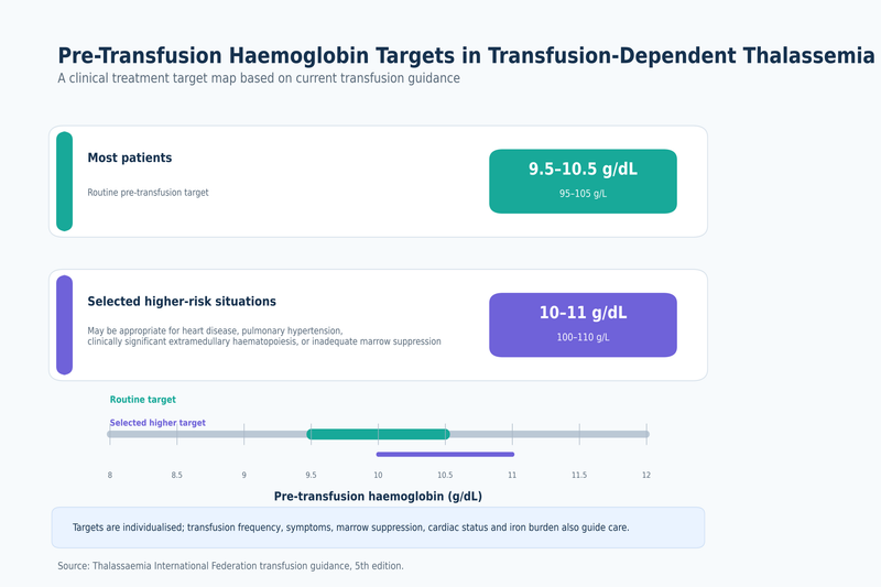
A regular transfusion programme should begin after the diagnosis and clinical phenotype have been confirmed. The decision should not be based solely on genotype, one haemoglobin result or a temporary decline during infection. The haematologist considers persistent anaemia, growth, activity, feeding, skeletal changes, spleen enlargement and evidence of ineffective erythropoiesis before establishing long-term transfusion dependence [8].
Patients with transfusion-dependent beta-thalassemia commonly receive packed red cells every two to five weeks. Current international guidance generally targets a pretransfusion haemoglobin of approximately 9.5 to 10.5 g/dL, equivalent to 95 to 105 g/L. A higher target of approximately 10 to 11 g/dL may be appropriate when cardiac disease, pulmonary hypertension, clinically significant extramedullary haematopoiesis or inadequate suppression of bone-marrow activity is present [8].
The target is individualised rather than applied as a fixed number to every patient. A child with impaired growth, progressive splenic enlargement or skeletal changes may require adjustment even when the measured haemoglobin appears close to the expected range. An adult with cardiac disease may also need a different target from an otherwise stable patient of the same age [8].
Pretransfusion haemoglobin should be measured immediately before the scheduled transfusion. This result shows how well the previous transfusion supported the patient through the complete interval. A haemoglobin value measured shortly after transfusion cannot be used to determine whether the next interval is adequate because it reflects the immediate rise rather than the lowest point of the cycle [8].
The post-transfusion haemoglobin should also be monitored to avoid excessive elevation and unnecessary blood exposure. International guidance advises maintaining it below approximately 13 to 15 g/dL, with the actual volume calculated from body weight, pretransfusion haemoglobin and the haemoglobin concentration of the blood product [8].
A patient who repeatedly becomes fatigued, pale, breathless or unable to participate normally in school, employment or physical activity before the next transfusion may require reassessment of the interval or transfused volume. Increasing spleen size, poor growth or continued bone-marrow expansion can provide additional evidence that the current programme is not adequately suppressing ineffective erythropoiesis [8].
Before the first transfusion, extended red-cell antigen typing should be performed at least for the major Rh and Kell antigens, with broader phenotyping or genotyping where available. Each transfusion should use ABO-compatible and RhD-compatible blood, with matching for Rh C, c, E, e and Kell antigens whenever possible. This reduces the probability of alloimmunisation, which can make compatible blood difficult to obtain later [8].
Leucocyte-depleted packed red cells are preferred. Screening for new red-cell antibodies should occur before each transfusion, and any previously identified antibody must remain permanently documented. An antibody can become undetectable over time but still cause a haemolytic reaction if blood carrying the corresponding antigen is given again [8].
Patients who receive transfusions in different hospitals or countries should keep a record of their blood group, extended antigen profile, previous antibodies, transfusion reactions and dates of transfusion. Differences in laboratory systems or language should not allow a clinically important historical antibody to be overlooked [8].
Fever, chills, rash, breathing difficulty, chest or back pain, sudden discomfort, low blood pressure or dark urine during a transfusion may indicate an acute reaction. Increasing jaundice, fatigue, dark urine or an unexpected fall in haemoglobin several days later may indicate a delayed haemolytic reaction. These symptoms require prompt assessment rather than waiting for the next scheduled transfusion [8].
Annual blood consumption should be recorded as the total volume of packed red cells received in relation to body weight. A rising requirement may reflect growth, inadequate transfusion targets, increasing spleen activity, red-cell antibodies, haemolysis or another clinical change. It also increases the rate of iron loading and may require modification of the chelation plan [8].
Transfusion in Thalassemia Intermedia
Transfusion decisions in thalassemia intermedia are based on the clinical consequences of anaemia rather than the haemoglobin value alone. Some patients maintain normal growth and useful daily function without transfusion, while others develop skeletal changes, increasing spleen size, poor exercise tolerance or organ complications despite a haemoglobin level that has remained numerically stable [6,9].
A temporary transfusion may be required during severe infection, surgery, pregnancy, acute haemolysis or a sudden decline in haemoglobin. Receiving one or several transfusions during such an event does not necessarily mean that the patient has become permanently transfusion dependent. The clinical baseline should be reassessed after the precipitating condition has resolved [6,9].
Regular transfusions may be considered when chronic anaemia causes impaired growth, delayed development, reduced school or work performance, progressive skeletal changes, worsening splenomegaly or significant functional limitation. Extramedullary haematopoiesis, pulmonary hypertension, recurrent leg ulcers or other complications associated with ineffective erythropoiesis may also influence the decision [6,9].
One low haemoglobin result during infection should not automatically lead to a lifelong programme. Conversely, long-standing tolerance of a low result does not prove that transfusion is unnecessary. Chronic tissue hypoxia and ineffective erythropoiesis can produce cumulative complications even when the patient has adapted subjectively to fatigue [6,9].
The benefit of regular transfusion should be evaluated through growth, activity, symptoms, spleen size, skeletal findings and control of complications. The treatment should not be judged only by the immediate rise in haemoglobin. Transfusion can reduce ineffective erythropoiesis and suppress abnormal blood formation outside the bone marrow, but it also introduces iron and increases exposure to donor antigens [6,9].
Once transfusions become frequent and sustained, iron monitoring and transfusion-safety measures should approach those used for transfusion-dependent disease. Extended antigen matching, antibody screening, annual blood-consumption records and systematic assessment of iron burden become increasingly important [8,9].
Pregnancy can increase transfusion needs because plasma volume, oxygen demand and fetal growth requirements rise. A patient who previously managed without transfusion may need temporary support during pregnancy. The decision should also consider cardiac status, previous antibodies, splenic enlargement and thrombotic risk rather than using haemoglobin alone [9].
After delivery and physiological recovery, the need for continuing transfusion should be reassessed. Some patients return to their previous non-transfusion-dependent baseline, while others remain more symptomatic or continue to require regular support. The classification should reflect the sustained clinical course rather than the pregnancy period alone [9].
Transfusion in Thalassemia Minor
Routine transfusion is not indicated for uncomplicated thalassemia minor. The functioning globin gene generally produces enough haemoglobin to prevent the severe ineffective erythropoiesis seen in major and clinically significant intermedia. Mild microcytosis or mild anaemia does not justify transfusion [1,4,5].
A carrier may require transfusion because of major surgery, severe bleeding, trauma or another unrelated acute condition. In that situation, the transfusion treats the acquired blood loss or illness rather than thalassemia minor itself. The patient does not become transfusion dependent because a transfusion was required during an isolated medical event.
Recurrent or scheduled transfusion is not typical of thalassemia minor. A patient labelled as a carrier who develops continuing transfusion requirements should be reassessed for a second beta-globin variant, haemoglobin E beta-thalassemia, alpha-gene triplication, haemolysis, blood loss, bone-marrow disease or another cause of severe anaemia [4].
Pregnancy alone does not make routine transfusion necessary in a carrier. Maternal symptoms, haemoglobin trajectory, bleeding, iron status and obstetric factors should be assessed individually. Iron deficiency should be corrected when demonstrated, but transfusion should not be used simply to normalise inherited microcytosis [4,5].
Measuring Iron Overload
Iron overload develops differently in major and intermedia. In thalassemia major, regular transfusions are the principal source because each unit of red cells contains iron that the body cannot excrete effectively. In thalassemia intermedia, ineffective erythropoiesis suppresses normal regulation of intestinal iron absorption, allowing excessive dietary iron to accumulate even when transfusion exposure is limited [6,8,9].
A patient with intermedia who also receives occasional or regular transfusions accumulates iron from both mechanisms. The term non-transfusion dependent should therefore never be interpreted as meaning that iron overload is impossible. The complete lifetime transfusion history remains clinically important even when the patient is not currently receiving blood [6,9].
Uncomplicated thalassemia minor does not ordinarily cause the iron-loading pattern associated with major or intermedia. Elevated ferritin in a carrier should prompt assessment for inflammation, liver disease, metabolic dysfunction, repeated transfusion, excessive iron supplementation or another diagnosis rather than being attributed automatically to carrier status [1,4].
Serum ferritin is useful for following iron trends but is not a direct measurement of iron within the heart or liver. Infection, inflammation and liver injury can increase ferritin independently of total iron burden. Interpretation should therefore consider recent illness, liver tests, inflammatory markers, transfusion exposure and previous ferritin results [8,9].
In transfusion-dependent thalassemia, serial ferritin assessment is generally performed at least every three months, with the interval adjusted according to iron burden and changes in chelation. A persistent upward trend may indicate that iron intake is exceeding iron removal, while a falling result may reflect effective treatment, reduced transfusion exposure or resolution of inflammation [8].
Magnetic resonance imaging can quantify liver iron concentration and assess myocardial iron through cardiac T2*. These measurements answer different clinical questions. Liver iron reflects total body iron more closely, while cardiac T2* estimates myocardial iron and related cardiac risk. A satisfactory liver result cannot substitute for cardiac assessment when myocardial iron is a concern [8].
International guidance recommends periodic liver and cardiac magnetic resonance assessment in transfusion-dependent patients, commonly beginning around eight to ten years of age or earlier when technically feasible without sedation. Annual assessment is frequently used, but shorter or longer intervals may be selected according to the degree of overload, previous results and recent chelation changes [8].
Magnetic resonance studies should be performed and interpreted through validated methods because measurements from uncalibrated techniques may not be comparable. Patients transferring between centres should provide previous liver iron concentration and cardiac T2* reports, including the date and technique, rather than only stating that an MRI was normal.
In non-transfusion-dependent thalassemia, liver iron concentration is particularly important because ferritin may underestimate the degree of tissue iron. Current NTDT guidance recommends structured iron assessment from approximately ten years of age, although earlier investigation may be required after substantial transfusion exposure, severe disease or evidence of liver dysfunction [6,9].
In established NTDT guidance, chelation is generally considered when liver iron concentration reaches at least 5 mg of iron per gram of dry liver weight. When validated liver iron measurement is unavailable, a ferritin level of at least 800 ng/mL may support treatment, while values between 300 and 800 ng/mL require interpretation with other clinical and laboratory evidence of iron loading [9].
These thresholds should not be transferred directly to transfusion-dependent disease because the relationship between ferritin, liver iron and organ risk differs between TDT and NTDT. They should also not be used by patients to start or stop treatment without specialist assessment, particularly when inflammation, liver injury or recent transfusion may have altered ferritin [8,9].
Cardiac iron is less common in NTDT than in heavily transfused disease but may occur in older patients, those with severe iron burden or those who have accumulated substantial transfusion exposure. Cardiac T2* may therefore be requested when clinically indicated rather than performed automatically at the same frequency for every NTDT patient [9].
Selecting and Monitoring Chelation
Chelation removes excess iron or prevents further accumulation by increasing iron excretion. It does not correct the globin-gene defect, directly raise haemoglobin or replace transfusion when transfusion is clinically required. Its effectiveness depends on selecting an appropriate medicine, dose and schedule for the patient’s iron-loading pattern [2,8,9].
Chelation in transfusion-dependent thalassemia is intended to balance ongoing transfusional iron intake and reduce previously accumulated iron. Treatment commonly begins after sufficient transfusion exposure and laboratory evidence show that iron is accumulating. The timing depends on age, cumulative transfusion burden, ferritin trends, liver iron measurements and local prescribing guidance rather than one isolated test [8].
The three principal chelators used internationally are deferoxamine, deferasirox and deferiprone. Their availability, licensed indications and formulations differ between India, the United States, the United Kingdom, Canada, Australia and Singapore. Selection also depends on organ iron distribution, kidney and liver function, age, previous adverse effects, adherence and pregnancy plans [8].
Deferoxamine is generally administered by prolonged subcutaneous infusion or intravenously in selected circumstances. Local infusion reactions, hearing changes, visual toxicity, growth effects and infection-related concerns require appropriate monitoring. The practical burden of repeated infusions can influence adherence even when the medicine is biologically effective [8].
Deferasirox is an oral chelator. Kidney function, liver function, gastrointestinal tolerance and urinary findings require monitoring, with dose interruption or modification when clinically indicated. Different formulations are not always interchangeable milligram for milligram, making the prescribed product and dose important when treatment is transferred between countries or pharmacies [8].
Deferiprone is also taken orally and can be effective in removing cardiac iron. It can cause neutropenia or agranulocytosis, making scheduled blood-count monitoring essential. Fever, sore throat, mouth ulcers or other signs of infection require prompt medical review because a serious reduction in neutrophils may initially present through common infection symptoms [8].
Combination chelation may be considered when severe cardiac or hepatic iron persists, when iron continues to rise despite appropriately administered single-agent therapy or when rapid intensification is required. Combination treatment increases complexity and must be supervised through a specialist centre with close toxicity and response monitoring [8].
In non-transfusion-dependent thalassemia, chelation is usually given at lower intensity than in heavily transfused disease because the rate and distribution of iron accumulation differ. Current NTDT guidance uses a liver iron concentration of approximately 3 mg/g dry weight or ferritin near 300 ng/mL as a treatment-interruption target, followed by continued monitoring for reaccumulation [9].
Chelation should not continue unchanged after iron burden has fallen substantially. Excessive treatment can create toxicity when more iron is being removed than the patient is accumulating. Dose reduction, interruption or a change in regimen may be required as ferritin and liver iron decline [8,9].
Monitoring must be matched to the selected chelator. A general statement that kidney and liver tests are normal is insufficient when the prescribed medicine requires additional blood-count, hearing, visual or urinary monitoring. The schedule should also become more frequent after a dose change, new adverse effect or major change in iron burden [8].
Adherence directly affects chelation success because iron continues to enter the body through transfusion or intestinal absorption during missed treatment. When iron remains uncontrolled, the clinical review should examine tolerability, formulation, timing, missed doses, medication access and the patient’s daily routine before concluding that the chelator has failed [8].
A lower transfusion burden does not automatically permit immediate discontinuation of chelation. Previously accumulated iron can remain within organs for years, and myocardial iron is often removed more slowly than liver iron. Changes should be based on ferritin trends, liver iron concentration, cardiac T2* and organ function rather than the number of recent transfusions alone [8].
A successful transfusion and chelation programme should maintain the intended pretransfusion haemoglobin, support growth and normal activity, suppress excessive bone-marrow expansion and prevent progressive organ iron. Its effectiveness is demonstrated through sustained trends in annual blood requirement, ferritin, liver iron concentration, cardiac T2*, liver and endocrine function, treatment tolerance and adherence [8].
The patient’s transferable record should contain the current transfusion interval, pretransfusion haemoglobin range, annual packed-cell requirement, red-cell phenotype or genotype, historical antibodies, chelator name and formulation, dose, adverse reactions, ferritin trend, liver iron concentration and cardiac T2*. These objective data allow a new treatment centre to continue care more safely than the terms major, intermedia or minor alone [8,9].
Advanced Treatment in Thalassemia Major vs Minor vs Intermedia
Advanced treatment in thalassemia major vs minor vs intermedia depends on whether the patient has clinically significant anaemia, continuing transfusion dependence, progressive complications and sufficient organ reserve to tolerate intensive therapy. Allogeneic haematopoietic stem-cell transplantation and approved gene-based therapies can provide sustained transfusion independence in selected patients with severe beta-thalassemia. Disease-modifying medicines may reduce anaemia or transfusion burden without replacing the patient’s blood-forming system. Uncomplicated thalassemia minor does not require transplantation, gene therapy or these disease-modifying medicines [4,8,9].
Allogeneic Stem Cell Transplantation
Allogeneic haematopoietic stem-cell transplantation replaces the patient’s abnormal blood-forming stem cells with healthy stem cells obtained from a donor. When donor cells engraft successfully and continue producing adequate haemoglobin, the patient may achieve durable transfusion independence. It remains an established potentially curative treatment for appropriately selected patients with transfusion-dependent beta-thalassemia [4,8].
The best-established outcomes are generally seen in younger patients who have a well-matched donor and limited iron-related injury to the liver, heart and endocrine organs. Earlier transplantation can reduce the period during which the patient remains exposed to repeated transfusions, alloimmunisation and iron accumulation. Age alone does not determine suitability, but increasing age is often accompanied by greater organ damage and treatment-related risk [8].
A human leukocyte antigen matched sibling donor is commonly preferred when available. Human leukocyte antigens are inherited tissue markers that influence whether donor cells are accepted and whether donor immune cells attack the recipient’s tissues. A close match reduces, but does not eliminate, the risks of graft rejection and graft-versus-host disease [8].
When a matched sibling is unavailable, some patients may be considered for a matched unrelated donor, partially matched family donor or haploidentical donor. A haploidentical donor shares approximately half of the relevant tissue markers and is commonly a parent, child or sibling. Improvements in conditioning, immune suppression and graft manipulation have expanded these options, but the expected benefits and risks differ among transplant approaches and centres [8].
Umbilical-cord blood may provide another source of blood-forming stem cells, although cell dose, engraftment speed and graft-failure risk require careful consideration. Donor selection should assess tissue matching, donor health, infectious screening, blood-group factors, stem-cell source and the transplant centre’s experience with thalassemia [8].
The patient undergoes conditioning treatment before receiving donor stem cells. Conditioning suppresses or removes the patient’s existing marrow cells and immune response so that donor cells can engraft. The intensity and medicines used depend on age, organ health, donor type and the transplant protocol. Conditioning can cause profound temporary suppression of blood-cell production and requires treatment in a specialist transplant unit [8].
During the period before engraftment, patients are vulnerable to bacterial, viral and fungal infections, bleeding, mucositis, nutritional difficulty and organ toxicity. Red-cell and platelet transfusions may still be required until the donor marrow begins producing sufficient blood cells. The patient remains closely monitored after discharge because immune recovery continues for months [8].
Graft failure occurs when donor stem cells do not establish adequate blood formation or are later lost. The patient may remain transfusion dependent and may require additional treatment or another transplantation procedure. Mixed chimerism means that both patient-derived and donor-derived blood-forming cells remain present. Stable mixed chimerism can sometimes maintain adequate haemoglobin, while declining donor chimerism may indicate an increasing risk of rejection [8].
Graft-versus-host disease develops when donor immune cells recognise the recipient’s tissues as foreign. It may affect the skin, gastrointestinal tract, liver, lungs and other organs. Acute disease usually develops earlier after transplantation, while chronic disease may persist or appear later and can substantially affect quality of life. Prevention and treatment require immunosuppressive medicines that can further increase infection risk [8].
Conditioning can impair fertility by damaging ovarian or testicular function. Fertility-preservation options should therefore be discussed before treatment whenever the patient’s age and clinical condition permit. Pubertal status, previous iron-related endocrine injury and the urgency of transplantation influence which preservation methods are feasible [8].
Transplant evaluation includes the confirmed genotype, transfusion history, red-cell antibodies, liver iron concentration, cardiac assessment, liver fibrosis, kidney function, endocrine status, infection screening, spleen status and psychosocial readiness. Poor chelation adherence in the past should not automatically exclude a patient, but existing organ injury and the ability to complete prolonged follow-up must be considered in the risk assessment [8].
Successful engraftment does not immediately remove iron accumulated before transplantation. Patients may remain at risk of liver, cardiac, endocrine and bone complications after transfusions have stopped. Iron reduction through phlebotomy or chelation may be required after stable blood formation has been established, depending on haemoglobin, organ iron and clinical status [8].
The inherited HBB variants remain present in the patient’s reproductive cells after an allogeneic transplant. The new donor-derived marrow can correct blood production, but it does not remove the possibility of passing a beta-thalassemia variant to a biological child. Genetic counselling therefore remains relevant even after sustained transfusion independence.
Gene Addition Therapy
Gene addition therapy uses the patient’s own blood-forming stem cells rather than cells from another person. The collected cells are modified in a laboratory with a lentiviral vector carrying a functional beta-globin gene. After myeloablative conditioning, the modified cells are returned to the patient and can produce a functional form of adult haemoglobin [12].
Betibeglogene autotemcel is approved in the United States for adult and paediatric patients with beta-thalassemia who require regular red-cell transfusions. It is an autologous treatment, meaning the patient receives their own genetically modified cells. A matched donor is not required, and graft-versus-host disease is not expected because the infused cells are not derived from another person [12].
Treatment begins with mobilisation of blood-forming stem cells from the bone marrow into the circulation. The cells are collected through apheresis, a procedure in which blood passes through a machine that removes the required cells and returns the remaining components. The collected cells are transported for individualised manufacturing, and a separate unmodified stem-cell collection may be stored as a rescue product in case engraftment fails [12].
Myeloablative conditioning remains necessary before infusion. Gene addition therapy therefore carries many acute risks associated with intensive conditioning, including severe cytopenias, infection, mucositis, infertility, liver complications and prolonged hospitalisation. Avoiding a donor does not make the treatment non-intensive [12].
Clinical trials supporting approval reported transfusion independence in most treated patients, but the results came from carefully selected participants treated at specialist centres. Transfusion independence is generally assessed through sustained haemoglobin production without regular red-cell transfusions over a defined follow-up period. It should not be inferred from a brief interruption in transfusion or one improved haemoglobin result [12].
Delayed platelet recovery can increase bleeding risk after treatment. Failure of neutrophil engraftment can leave the patient vulnerable to serious infection and may require infusion of rescue cells. Blood counts must be followed closely until stable engraftment has been confirmed [12].
Because a lentiviral vector inserts genetic material into blood-forming cells, long-term monitoring for abnormal clonal blood-cell growth and haematological malignancy is required. Current prescribing information calls for extended surveillance after treatment. A normal early recovery does not remove the need for long-term blood counts and specialist follow-up [12].
The patient’s accumulated iron and established organ damage do not disappear when transfusions stop. Chelation may continue until liver and cardiac iron have reached safer levels. Endocrine dysfunction, bone disease, liver fibrosis and cardiac injury may still require treatment even when gene addition has produced stable haemoglobin [8,12].
Gene Editing Therapy
Gene editing modifies the patient’s own blood-forming stem cells without adding a conventional replacement HBB gene. Exagamglogene autotemcel uses CRISPR-Cas9 technology to edit a regulatory region associated with BCL11A in collected stem cells. This reduces suppression of gamma-globin production and allows the modified cells to produce more fetal haemoglobin, which can compensate for inadequate beta-globin production [10].
The process includes stem-cell mobilisation, apheresis, laboratory editing, product testing, myeloablative conditioning and infusion of the edited cells. Patients require treatment in an authorised centre with expertise in stem-cell transplantation, haemoglobin disorders and management of conditioning-related complications [10].
In the United States, the current indication includes patients aged two years and older with transfusion-dependent beta-thalassemia. Other regulatory jurisdictions may retain different age limits, eligibility requirements or donor-related conditions. Approval in one country does not establish automatic eligibility, funding or access in India, the United Kingdom, Canada, Australia or Singapore [10,15,16].
The principal treatment goal is sustained transfusion independence with adequate haemoglobin. In the evidence supporting the original United States approval, approximately 91% of evaluable participants achieved the defined transfusion-independence endpoint. This result demonstrates substantial potential but does not guarantee that every patient will become permanently transfusion free [10].
Gene editing avoids the need for a donor and the risk of graft-versus-host disease. It does not avoid the risks of stem-cell mobilisation, manufacturing failure, myeloablative conditioning, infection, infertility, delayed blood-count recovery or prolonged hospitalisation. A patient must be medically fit enough to tolerate the complete treatment pathway rather than only the gene-editing infusion [10].
Potential unintended editing outside the intended DNA target remains an important long-term safety consideration. Clinical programmes include extensive product testing and continued surveillance, but the lifelong implications of newer gene-editing technologies cannot be predicted from short follow-up alone. Patients should understand the commitment to long-term monitoring before treatment [10].
Gene editing increases fetal haemoglobin but does not necessarily repair the patient’s original HBB variants directly. The inherited variants remain present in reproductive cells, so the risk of transmitting thalassemia to a biological child continues. Partner testing and genetic counselling remain necessary after successful treatment.
A patient who becomes transfusion independent may still need iron-removal therapy. Liver iron concentration, cardiac T2*, ferritin, endocrine function and bone health should continue to be assessed according to the pre-treatment burden. Stopping transfusion prevents further transfusional iron loading but does not instantly remove iron already deposited in tissues [8,10].
Choosing Between Transplantation and Gene Therapy
Allogeneic transplantation and autologous gene therapy both require intensive conditioning, hospital treatment and prolonged follow-up. The central difference is the source of the blood-forming cells. Allogeneic transplantation uses donor cells and carries risks of rejection and graft-versus-host disease. Gene therapy uses the patient’s own modified cells and avoids those donor-related complications but introduces manufacturing, gene-modification and long-term surveillance considerations [8,10,12].
A well-matched sibling transplant may offer a highly established curative pathway, particularly for a young patient without substantial organ injury. Gene therapy may become especially relevant when no suitable donor is available or when the patient wishes to avoid donor-related complications. The preferred option depends on age, genotype, donor availability, transfusion burden, organ function, fertility priorities, treatment access and the experience of the treating centre [8,10,12].
Gene therapy should not be assumed to be safer merely because it uses the patient’s own cells. Both approaches commonly require myeloablative chemotherapy and can cause infertility, severe infection, liver injury, prolonged low blood counts and treatment-related mortality. The risk comparison must include the entire treatment process rather than only the source of the infused cells [8,10,12].
Previous red-cell antibodies can complicate transfusion support during either treatment. Cardiac iron, liver fibrosis, kidney dysfunction, active infection and significant pulmonary disease can increase conditioning risk. These factors should be optimised before treatment when possible [8].
A patient with intermedia may qualify for advanced treatment when the clinical burden is substantial, even without lifelong scheduled transfusions. Progressive anaemia, repeated transfusions, severe extramedullary haematopoiesis, poor growth, pulmonary hypertension, thrombosis or major quality-of-life impairment may justify specialist evaluation. The word intermedia should not automatically exclude a patient from curative-treatment assessment [6,9].
Thalassemia minor does not justify the risks of transplantation or gene therapy. A carrier has sufficient endogenous blood production and does not face the transfusion-dependent disease burden these intensive treatments are intended to correct. The appropriate interventions are accurate diagnosis, treatment of any separate nutritional deficiency and reproductive counselling [4,5].
Luspatercept
Luspatercept is an erythroid-maturation agent that promotes development of late-stage red-cell precursors. It does not replace the HBB gene or create a new stem-cell population. Its clinical purpose is to improve effective red-cell production and reduce transfusion burden in patients who respond [14].
In the United States, luspatercept is approved for anaemia in adults with beta-thalassemia who require regular red-cell transfusions. Regulatory indications in other jurisdictions may also include selected adults with non-transfusion-dependent beta-thalassemia. The exact indication should be checked according to the patient’s country, age and transfusion pattern [14].
Treatment is administered by subcutaneous injection, generally at three-week intervals. Haemoglobin and transfusion records are reviewed before each dose. A reduction in transfused red-cell units over a sustained interval is more clinically meaningful than one delayed transfusion or a temporary haemoglobin increase [14].
Luspatercept is not a substitute for urgent transfusion when rapid correction of severe anaemia is required. Patients should continue their established transfusion schedule until the haematology team confirms that the interval or volume can be safely adjusted. Abruptly withholding transfusion in anticipation of a medicine response can expose the patient to symptomatic anaemia [14].
Response is variable. Some patients achieve a meaningful reduction in transfusion burden, while others obtain little benefit and should not continue indefinitely without objective evidence of response. The assessment should compare red-cell units, transfusion frequency, pretransfusion haemoglobin, symptoms and adverse effects with the documented baseline [14].
Thromboembolic events are an important risk, particularly in patients with additional risk factors such as previous splenectomy, earlier thrombosis, limited mobility or hormonal therapy. Blood pressure should also be monitored because hypertension can develop during treatment [14].
Extramedullary haematopoietic masses can enlarge or become symptomatic in some patients. New back pain, weakness, altered sensation, difficulty walking or bladder and bowel symptoms require prompt investigation for pressure on the spinal cord or other structures [14].
Luspatercept can cause fetal harm and requires pregnancy-related counselling. It is not used to treat uncomplicated thalassemia minor and does not eliminate the need for iron monitoring or chelation when iron overload is present [14].
Mitapivat
Mitapivat is an oral pyruvate-kinase activator that improves red-cell energy production. In thalassemia, enhanced glycolytic activity can improve red-cell survival and reduce ineffective erythropoiesis. In January 2026, it became the first oral treatment approved in the United States for anaemia in adults with either alpha-thalassemia or beta-thalassemia [13].
For a non-transfusion-dependent adult, the treatment goal may be a sustained increase in haemoglobin accompanied by improved exercise tolerance, reduced fatigue or fewer anaemia-related complications. For a transfusion-dependent adult, the objective may be a measurable reduction in red-cell units or transfusion frequency. Treatment success should be judged against a documented pre-treatment baseline [13].
Mitapivat is not a genetic cure and does not remove existing iron. Patients who remain transfusion dependent must continue appropriate transfusion care, and those with iron overload may still require chelation. A reduced transfusion burden may slow new iron accumulation, but previously deposited iron requires separate monitoring and treatment [13].
Hepatocellular injury has occurred in treated patients with thalassemia. Baseline and repeated liver testing are required, particularly during the first months of treatment. New loss of appetite, nausea, right upper abdominal pain, vomiting, jaundice or dark urine requires prompt assessment because these symptoms may reflect liver injury rather than the underlying thalassemia alone [13].
Mitapivat can interact with medicines that affect drug-metabolising enzymes and can alter the exposure of other prescribed treatments. The medication review should include prescription medicines, non-prescription products and herbal preparations. Existing cirrhosis is an important treatment consideration because current United States prescribing information advises avoiding its use in patients with cirrhosis [13].
Regulatory requirements differ between countries. In the United States, access includes a structured safety programme because of the risk of liver injury. Approval, reimbursement and monitoring arrangements in India, the United Kingdom, Canada, Australia and Singapore may differ or change, so eligibility should be confirmed through a specialist haematology service [13].
Mitapivat has no role in correcting uncomplicated carrier microcytosis. A patient with thalassemia minor and mild stable anaemia should be investigated for iron deficiency, vitamin deficiency, thyroid disease, bleeding or another cause before a disease-modifying drug is considered [1,4].
Advanced Treatment by Thalassemia Type
For thalassemia major, the advanced-treatment discussion commonly includes matched-donor transplantation, alternative-donor transplantation, gene addition, gene editing and medicines intended to reduce transfusion burden. The choice depends on whether the patient seeks a potentially curative intensive intervention or continued disease control with transfusion, chelation and medication [8,10,12,14].
For thalassemia intermedia, advanced treatment is guided by actual disease burden. A patient with stable haemoglobin and no progressive complications may not need transplantation or gene therapy. A patient with increasing transfusions, severe splenomegaly, extramedullary haematopoiesis, pulmonary hypertension, poor growth or major functional impairment may require assessment for disease-modifying or potentially curative treatment [6,9].
For alpha-thalassemia, the role of advanced treatment differs from beta-thalassemia. Transplantation may be considered in highly selected survivors of haemoglobin Bart hydrops fetalis syndrome or severe transfusion-dependent alpha-thalassemia. Current beta-thalassemia gene-addition and gene-editing products should not be assumed to treat alpha-thalassemia, although mitapivat has an approved adult alpha-thalassemia indication in the United States [11,13].
For thalassemia minor, advanced treatment offers no favourable benefit-to-risk balance. Transplantation and gene therapy expose a clinically stable carrier to intensive conditioning, infertility, infection and other serious risks without a corresponding medical need. Disease-modifying drugs should not be used merely to normalise the mean corpuscular volume or remove the carrier label [4,5].
Regulatory approval does not guarantee that a treatment is funded, locally available or suitable for a particular patient. The United States, United Kingdom, Canada, India, Australia and Singapore may differ in authorised ages, treatment-centre availability, insurance or public funding, donor requirements and long-term follow-up systems. International patients should obtain written confirmation of eligibility, total treatment pathway, follow-up obligations and financial coverage before travelling for therapy [10,12,13,15,16].
The most meaningful outcome after transplantation or gene therapy is sustained production of adequate haemoglobin without regular transfusions, accompanied by stable engraftment and acceptable safety. Reduction in transfusion burden is a valuable outcome from disease-modifying medicines, but it is not equivalent to cure. Iron burden, organ function, fertility, endocrine health, bone health and long-term safety remain part of follow-up regardless of how successfully the anaemia responds [8,9,10,12].
Can Thalassemia Major, Intermedia or Minor Be Cured?

Whether thalassemia major vs minor vs intermedia can be cured depends on the exact diagnosis, transfusion requirement, genotype, organ health and meaning assigned to the word cure. Thalassemia major and clinically severe intermedia may be treated with potentially curative stem-cell or gene-based therapies in selected patients. Thalassemia minor is a carrier state that usually does not require curative treatment because it does not ordinarily cause transfusion dependence or progressive thalassemia-related organ damage [4,5,8,9].
What Cure Means in Thalassemia
Cure should mean sustained production of sufficient functional haemoglobin without regular red-cell transfusions, supported by durable laboratory and clinical follow-up. A temporary increase in haemoglobin, a longer interval between transfusions or improved energy does not by itself establish cure. The outcome must persist without recurrent dependence on transfused blood or rescue treatment [8,10,12].
After allogeneic stem-cell transplantation, cure is assessed through stable donor-derived blood formation, adequate haemoglobin, sustained transfusion independence and absence of progressive graft failure. Donor chimerism measures the proportion of blood-forming cells originating from the donor. Complete donor chimerism is not always necessary when stable mixed chimerism continues to produce adequate haemoglobin, but declining donor contribution may indicate a risk of later graft rejection [8].
After gene addition or gene-editing therapy, the principal outcome is sustained transfusion independence produced by the patient’s genetically modified blood-forming stem cells. The exact definition is treatment specific and usually requires maintenance of a minimum haemoglobin level without red-cell transfusion over a defined period. Trial-defined transfusion independence is a stronger outcome than a subjective report of improvement, but continued follow-up is still required to establish durability and long-term safety [10,12].
Transfusion independence should not be confused with complete reversal of every consequence of thalassemia. A patient may no longer need blood transfusions yet continue to have liver iron, cardiac injury, endocrine dysfunction, impaired fertility, reduced bone density or previous splenic complications. These conditions may require continued treatment after the anaemia has been corrected [8].
The inherited globin variants also remain present in the patient’s reproductive cells after stem-cell transplantation or somatic gene therapy. Blood production may be corrected, but the patient can still pass a thalassemia variant to a biological child. Partner screening and genetic counselling therefore remain necessary after successful curative treatment [4,8].
Cure in Thalassemia Major
Allogeneic haematopoietic stem-cell transplantation is an established potentially curative treatment for thalassemia major. Successful transplantation replaces the patient’s ineffective blood-forming system with donor stem cells capable of producing adequate haemoglobin. The likelihood of a favourable outcome is influenced by donor compatibility, age, liver health, cardiac function, iron burden, previous treatment and the experience of the transplant centre [4,8].
A young patient with a well-matched sibling donor and limited organ injury may have a more favourable benefit-to-risk balance than an older patient with advanced iron-related complications. This does not mean that older patients are automatically ineligible, but their evaluation generally requires more detailed assessment of cardiac reserve, liver fibrosis, endocrine function, infection history and conditioning risk [8].
Successful transplantation can eliminate the need for scheduled transfusions and stop further transfusional iron accumulation. It does not immediately remove iron already stored in the liver, heart or endocrine organs. Phlebotomy, continued chelation or another iron-reduction strategy may be required after stable engraftment, depending on haemoglobin, liver iron concentration and cardiac findings [8].
Transplantation also carries significant risks, including graft rejection, graft-versus-host disease, severe infection, infertility, organ toxicity and treatment-related mortality. A treatment should not be called curative merely because transplantation was performed. Cure must be demonstrated through durable engraftment, stable haemoglobin, continuing transfusion independence and long-term follow-up [8].
Gene addition and gene-editing therapies provide another potentially curative pathway for selected patients with transfusion-dependent beta-thalassemia. These treatments use the patient’s own stem cells, avoiding the need for a matched donor and eliminating graft-versus-host disease associated with donor immune cells. They still require stem-cell collection, laboratory manufacturing, intensive conditioning, hospitalisation and prolonged surveillance [10,12].
Gene addition introduces a functional beta-globin gene into collected stem cells, while gene editing can reactivate fetal haemoglobin production by modifying a regulatory pathway. Both approaches aim to produce enough functional haemoglobin to remove the need for regular transfusion. Neither approach guarantees transfusion independence for every eligible patient [10,12].
Failure to achieve the trial-defined transfusion-independence endpoint does not necessarily mean that the treatment produced no benefit. Some patients may achieve a substantial reduction in transfusion burden without becoming completely transfusion independent. This may reduce iron loading and improve daily function, but it should be described as a partial clinical response rather than cure [10,12].
The treatment outcome should be assessed through total haemoglobin, haemoglobin composition, transfusion history, blood-cell recovery, treatment-related complications and long-term stability. Ferritin alone cannot confirm cure because ferritin reflects iron burden and inflammation rather than correction of ineffective haemoglobin production [8,10,12].
Cure in Thalassemia Intermedia
Thalassemia intermedia is too clinically variable for one universal curative-treatment recommendation. Some patients remain stable without regular transfusions and experience limited complications, making the risks of transplantation or gene therapy greater than the expected benefit. Others develop progressive anaemia, repeated transfusion requirements, severe splenomegaly, extramedullary haematopoiesis, pulmonary hypertension, thrombosis, bone disease or major impairment of daily function [6,9].
A patient does not need to carry the label thalassemia major to be considered for potentially curative treatment. Severe non-transfusion-dependent thalassemia may create substantial lifelong morbidity despite the absence of a fixed transfusion schedule. Eligibility should reflect actual clinical burden, accumulated complications, genotype and anticipated future risk [6,9].
Occasional transfusion during infection, surgery or pregnancy does not by itself justify a curative procedure. The haematology team should determine whether anaemia and complications remain manageable with monitoring, temporary transfusion, chelation or disease-modifying treatment. Intensive curative treatment becomes more relevant when these measures do not adequately control disease burden [6,9].
The expected benefit of transplantation or gene-based therapy should be compared with the patient’s current quality of life and future complication risk. A patient with stable haemoglobin, preserved organ function and no progressive complications may reasonably continue structured monitoring. A patient with worsening organ injury or increasing transfusion exposure may gain more from evaluation at a specialist curative-treatment centre [6,8,9].
Transfusion independence after treatment must be interpreted against the patient’s previous pattern. A person who required only one transfusion every several years cannot be assessed by the same endpoint as a patient receiving transfusions every three weeks. Baseline haemoglobin, annual transfusion burden, symptoms and complication history should be documented before treatment so that the response can be measured accurately.
Why Thalassemia Minor Does Not Need Curative Treatment
Thalassemia minor is usually a carrier state rather than an active transfusion-dependent disease. Most carriers have normal daily function, normal life expectancy and no progressive organ injury caused by thalassemia. Stem-cell transplantation, gene therapy and disease-modifying medicines would expose them to serious risks without a corresponding medical need [4,5].
The persistently low mean corpuscular volume seen in thalassemia minor does not represent treatment failure or an incomplete cure. It reflects inherited production of smaller red blood cells and may remain present throughout life. Attempting to normalise the mean corpuscular volume with repeated iron treatment is inappropriate when iron stores are adequate [1,5].
A carrier who develops marked fatigue, falling haemoglobin, jaundice, splenic enlargement or a transfusion requirement requires reassessment. These findings may indicate iron deficiency, blood loss, nutritional deficiency, another medical condition or a more complex haemoglobin genotype. The appropriate goal is to identify and treat the additional problem rather than to seek curative therapy for uncomplicated carrier status [1,4].
For most carriers, the most important intervention is reproductive risk assessment. A clinically healthy partner may carry beta-thalassemia, haemoglobin E, sickle haemoglobin or another interacting variant. Complete blood count, haemoglobin analysis and molecular testing when indicated provide more useful protection for future children than any treatment directed at the carrier’s mild microcytosis [4,5].
How Cure Is Confirmed
Confirmation of cure requires repeated objective assessment rather than a single normal report. The patient’s transfusion dates, pre-treatment transfusion frequency, red-cell units received and haemoglobin levels should be compared with post-treatment findings. A short period without transfusion may occur during illness recovery or treatment adjustment and is insufficient to establish a durable result [8,10,12].
After allogeneic transplantation, follow-up commonly includes complete blood counts, reticulocyte counts, haemoglobin analysis, donor chimerism, immune recovery, infection surveillance and assessment for graft-versus-host disease. A stable haemoglobin without transfusion is necessary, but evidence of sustained donor blood formation and absence of progressive graft loss is also important [8].
After gene-based therapy, follow-up includes complete blood counts, total haemoglobin, therapeutic haemoglobin fractions, transfusion records, blood-cell recovery and surveillance for abnormal clonal blood-cell changes. Long-term monitoring remains essential because newer genetic therapies have a shorter period of real-world follow-up than established transfusion and transplantation programmes [10,12].
Iron burden must be assessed separately. Ferritin trends, liver iron concentration and cardiac T2* determine whether residual iron remains after transfusions stop. A patient can meet criteria for transfusion independence while still requiring chelation or phlebotomy to reduce accumulated iron [8].
Cardiac, liver, endocrine, reproductive and bone assessments remain clinically relevant after cure. Diabetes, hypothyroidism, delayed puberty, infertility, osteoporosis or liver fibrosis may persist even when haemoglobin production has normalised. These conditions require their own objective endpoints and cannot be assumed to have resolved because transfusions are no longer required [8].
Growth and pubertal development should continue to be monitored in children and adolescents. Correcting anaemia may improve growth potential, but previous endocrine injury, bone disease or delayed puberty may need specific treatment. Catch-up growth is not guaranteed when complications were established before curative therapy [8].
A durable curative outcome should therefore include sustained transfusion independence, stable haemoglobin, functioning blood-cell production, acceptable treatment safety and continued control of residual organ complications. Normal energy or appetite may accompany recovery but cannot replace these objective measures [8,10,12].
Ayurvedic Care and Cure Claims
Personalised Ayurvedic care may be integrated to address appetite, digestion, fatigue, sleep, physical strength, treatment tolerance and quality of life. These outcomes can be clinically meaningful, particularly for patients living with chronic transfusion schedules, chelation adverse effects or nutritional difficulty.
Any claim of disease modification should be evaluated through pretransfusion haemoglobin, transfusion interval, annual red-cell requirement, ferritin, liver iron concentration, cardiac T2*, spleen size, growth and organ function. A subjective increase in energy should not be presented as correction of the globin-gene defect.
Current clinical evidence does not establish Ayurvedic treatment alone as a replacement for transfusion, iron chelation, stem-cell transplantation or approved gene-based therapy in thalassemia major. Required transfusions and chelation should not be stopped because symptoms improve or one haemoglobin result rises. Changes should occur only through coordinated haematology assessment and sustained objective evidence.
In thalassemia intermedia, an integrative treatment plan should still monitor iron loading, haemolysis, splenic enlargement, bone health, cardiopulmonary complications and changing transfusion needs. In thalassemia minor, treatment should focus on confirmed coexisting problems rather than attempting to eliminate an inherited carrier pattern.
The term cure is appropriate only when the patient achieves durable correction of clinically significant thalassemia through objectively demonstrated blood production and sustained freedom from required transfusions. Reduced fatigue, improved appetite, a temporary haemoglobin rise or a longer transfusion interval may represent benefit, but each should be reported separately without being labelled as cure [8,9,10,12].
Ayurvedic Assessment in Thalassemia Major vs Minor vs Intermedia

Ayurvedic assessment in thalassemia major vs minor vs intermedia should begin only after the haematological diagnosis, genotype, transfusion status and iron burden have been established. Thalassemia is an inherited globin-gene disorder and is not described as a distinct molecular disease in the classical Ayurvedic texts. Ayurvedic concepts may help assess the patient’s digestion, nourishment, strength, symptoms and treatment tolerance, but they should not be treated as substitutes for the confirmed genetic and haematological diagnosis [4,6,8,9].
Classical Ayurvedic Assessment
Pandu is a classical Ayurvedic disease framework characterised by pallor, weakness, reduced vitality and impaired tissue nourishment. Its detailed treatment discussion appears in the Charaka Samhita, Chikitsa Sthana, Chapter 16, Pandu Roga Chikitsa. Pandu should not be translated directly as thalassemia because the classical category includes several presentations that may arise from nutritional, digestive, haemorrhagic or systemic causes.
Rakta Dhatu is the Ayurvedic tissue concept associated with blood, circulation and nourishment, while Kshaya means depletion or reduction. Rakta Dhatu Kshaya may be considered when the patient has pallor, weakness, dizziness or reduced exercise capacity, but it is not equivalent to a low haemoglobin result or a specific globin-gene variant. Haemoglobin, reticulocyte count, bilirubin, transfusion requirement and haemoglobin analysis remain the objective measures of disease severity [4,8,9].
Dhatu Kshaya means depletion or inadequate nourishment of one or more body tissues. It may be clinically relevant when chronic anaemia is accompanied by low weight, poor muscle development, delayed growth, reduced bone strength or prolonged recovery after illness. These findings still require investigation for inadequate transfusion, endocrine dysfunction, iron toxicity, nutritional deficiency and bone disease rather than being attributed solely to an Ayurvedic imbalance [8,9].
Agni refers to the digestive and metabolic capacity responsible for processing food and supporting tissue nourishment. Assessment includes appetite, post-meal discomfort, bowel regularity, nausea, bloating, food tolerance and the ability to maintain weight. Reduced appetite in thalassemia may also result from splenic enlargement, liver disease, medication adverse effects, infection or pressure on the stomach, so digestive symptoms require both Ayurvedic and medical evaluation [6,8,9].
Bala means functional strength and physical resilience. It is assessed through exercise tolerance, ability to complete daily activities, recovery after exertion, school or work participation and frequency of illness. Bala should not be judged only from how energetic the patient feels on one day because symptoms may fluctuate across the transfusion cycle.
Ojas is the Ayurvedic concept of integrated vitality, stability and resistance to physical stress. It may be considered through sleep, recovery, emotional steadiness, appetite and the ability to tolerate ongoing treatment. Ojas is not a laboratory marker and should not replace evaluation of haemoglobin, organ function, iron burden or infection risk.
Prakriti is the individual’s baseline constitutional pattern, while Vikriti is the current functional disturbance. These concepts may guide food selection, dosage form and tolerability, but they cannot determine whether a patient has major, intermedia or minor disease. The clinical category must remain based on genotype, transfusion dependence, symptoms and objective complications [4,6,8,9].
Rasayana refers to a classical therapeutic approach intended to support nourishment, strength, recovery and healthy ageing. Its principal discussion appears in the Charaka Samhita, Chikitsa Sthana, Chapter 1, Rasayana Adhyaya. In thalassemia, Rasayana care should be adapted to the patient’s age, digestive capacity, iron status, liver and kidney function, transfusion schedule and current medicines.
Brimhana means nourishing and tissue-building care. It may be considered when the patient has poor weight gain, low muscle mass, weakness or delayed recovery, but it should not consist merely of increasing calories. Growth failure can arise from inadequate transfusion, thyroid dysfunction, delayed puberty, diabetes, liver disease or bone complications and requires investigation before treatment is planned [8].
Ayurvedic Assessment in Thalassemia Major
In thalassemia major, the Ayurvedic consultation should begin with the transfusion interval, pretransfusion haemoglobin, annual red-cell requirement, chelator, ferritin trend, liver iron concentration, cardiac T2*, liver function, kidney function, growth and endocrine findings. These measurements determine whether symptoms arise from anaemia, iron overload, organ dysfunction, treatment toxicity or another condition [8].
Fatigue that appears immediately before each transfusion may indicate declining haemoglobin rather than a primary digestive or constitutional imbalance. Persistent fatigue throughout the transfusion cycle may require assessment of cardiac iron, thyroid function, glucose regulation, sleep quality, infection, depression or nutritional deficiency. Ayurvedic treatment should not obscure the need to identify these causes.
Appetite and digestion require careful assessment because splenic enlargement, hepatomegaly, gallbladder disease, chelator intolerance and chronic liver injury can all produce nausea, early satiety or abdominal discomfort. A medicine intended to stimulate appetite should not be prescribed without investigating progressive abdominal swelling, persistent vomiting, right upper abdominal pain or rapidly increasing spleen size [8].
Any Ayurvedic medicine containing iron should be approached with particular caution. Lauha and Mandura are classical iron-containing substances, and low haemoglobin in thalassemia major does not imply iron deficiency. Most regularly transfused patients accumulate excess iron, making ferritin, transferrin saturation and magnetic resonance iron measurements essential before an iron-containing preparation is considered [8].
Herbal or herbo-mineral formulations should be reviewed against the patient’s chelation therapy and organ function. A patient with elevated liver enzymes, impaired kidney function or cardiac disease may require dose reduction, ingredient avoidance or postponement of non-essential treatment. New symptoms after starting a formulation should be investigated rather than interpreted automatically as detoxification or a temporary healing response.
Strong purgation, prolonged fasting, induced vomiting, bloodletting or any procedure likely to cause dehydration or reduced nutritional intake is inappropriate during unstable anaemia, infection, cardiac symptoms or immediately around transfusion. Patients with low platelets, anticoagulant use or hypersplenism also require additional caution because bleeding risk may be increased [8].
The immediate Ayurvedic goals in a stable patient may include improving appetite, bowel regularity, sleep, treatment tolerance, muscle strength and nutritional intake. These outcomes should be measured alongside pretransfusion haemoglobin, transfusion interval, growth, iron burden and organ function rather than being presented as evidence that transfusion-dependent disease has been corrected.
Ayurvedic Assessment in Thalassemia Intermedia
Thalassemia intermedia requires assessment of chronic anaemia, haemolysis, splenic enlargement, gallstones, bone health, iron loading, extramedullary haematopoiesis, pulmonary hypertension and thrombosis risk. A patient may appear clinically stable while progressive complications develop, making objective monitoring essential even when regular transfusions are not required [6,9].
Pallor, fatigue and reduced exercise tolerance may be considered through the Ayurvedic frameworks of Pandu, Rakta Dhatu Kshaya and reduced Bala, but the treatment decision must account for haemoglobin trend, reticulocyte response, bilirubin, lactate dehydrogenase and functional limitation. Persistent symptoms may indicate that the previous non-transfusion-dependent strategy is no longer sufficient [6,9].
An enlarged spleen can cause early satiety, poor appetite and abdominal heaviness. These symptoms should not be treated only as weak Agni because increasing splenic sequestration may lower haemoglobin, platelets and white blood cells. Serial spleen measurements and complete blood counts are necessary when abdominal symptoms worsen [6,9].
Iron-containing Ayurvedic medicines should not be prescribed solely because the patient is anaemic. Non-transfusion-dependent thalassemia can cause substantial iron accumulation through increased intestinal absorption, and serum ferritin may underestimate liver iron. Liver iron concentration should be reviewed when the history or laboratory pattern suggests overload [6,9].
Jaundice and digestive discomfort may arise from haemolysis or pigment gallstones rather than impaired digestion alone. Right upper abdominal pain, fever, vomiting or sudden worsening of jaundice requires assessment for gallbladder or bile-duct complications before an Ayurvedic digestive formulation is introduced.
Back pain requires particular caution because paraspinal extramedullary haematopoiesis can compress the spinal cord. Back pain accompanied by weakness, numbness, altered walking or loss of bladder or bowel control requires urgent imaging and specialist treatment rather than massage, heat therapy or delayed observation [6,9].
A patient with previous splenectomy requires review of infection prevention and thrombotic risk. Fever, sudden chest pain, unilateral leg swelling, unexplained breathlessness or neurological symptoms should not be managed initially as an Ayurvedic imbalance because they may indicate severe infection or thrombosis [6,9].
Ayurvedic care may focus on maintaining appetite, regular bowel function, sleep, muscle conditioning and recovery while the haematology team monitors transfusion need, iron accumulation and organ complications. Any reported reduction in fatigue should be compared with haemoglobin, activity level and cardiopulmonary findings before it is interpreted as disease improvement.
Ayurvedic Assessment in Thalassemia Minor
Thalassemia minor usually does not require disease-directed Ayurvedic treatment. The inherited carrier pattern commonly causes lifelong microcytosis with no symptoms or mild anaemia, and treatment should not be aimed at forcing the mean corpuscular volume or mean corpuscular haemoglobin into the normal range [1,4,5].
Fatigue in a carrier should prompt assessment of iron deficiency, vitamin B12 deficiency, folate deficiency, thyroid dysfunction, menstrual blood loss, pregnancy, kidney disease, chronic inflammation, sleep disturbance and psychological stress. Labelling all weakness as Pandu or Rakta Dhatu Kshaya can delay recognition of a treatable coexisting disorder [1,4].
Iron-containing Ayurvedic preparations should be used only when iron deficiency has been demonstrated. A low mean corpuscular volume is expected in thalassemia minor and is not sufficient evidence of iron deficiency. Treatment response should be judged by restoration of iron stores and improvement in haemoglobin rather than complete correction of inherited microcytosis [1,5].
A carrier with marked jaundice, splenic enlargement, gallstones, substantial anaemia or any continuing transfusion requirement requires haematology reassessment. These findings may indicate haemoglobin E beta-thalassemia, alpha-gene triplication, another globin variant or an unrelated cause of haemolysis rather than uncomplicated minor disease [4].
Pregnant carriers require individual assessment of haemoglobin, ferritin, folate status and obstetric needs. Ayurvedic medicines should be reviewed for pregnancy safety, and partner screening should not be delayed while maternal fatigue or anaemia is being treated. The reproductive risk depends on the partner’s haemoglobin and genetic findings rather than the severity of the mother’s symptoms [4,5].
The principal Ayurvedic role in uncomplicated minor disease may be to support digestion, sleep, general nutrition and management of an independently diagnosed condition. The carrier state itself does not require detoxification, transfusion-directed herbs, chelation substitutes or intensive Rasayana treatment.
Integrating Ayurveda With Haematology Care
An integrative plan should document every prescribed herb, mineral preparation, dose, manufacturer and treatment duration. The haematology team should be informed before treatment begins, particularly when the patient has liver disease, kidney impairment, cardiac disease, pregnancy, anticoagulant use or a planned transplant or gene-based procedure.
Ayurvedic medicines should not replace scheduled transfusion, iron chelation, antibiotics, endocrine therapy or complication-specific treatment. Required conventional treatment should be changed only when sustained clinical and laboratory evidence supports the decision and the treating haematologist agrees [6,8,9].
Symptoms should be reassessed after any new formulation is introduced. Increasing jaundice, dark urine, abdominal pain, vomiting, itching, reduced urine output, swelling, palpitations or an unexpected fall in haemoglobin requires prompt medical evaluation. These findings should not be described as cleansing, toxin release or a normal adjustment reaction.
Preliminary clinical studies have evaluated Dhatri Avaleha and Triphaladi Avaleha as adjunctive interventions in children receiving standard thalassemia care. The studies reported selected symptomatic or transfusion-related findings but were small and did not establish genetic correction, permanent transfusion independence or replacement of transfusion and chelation [3,7].
Ayurvedic benefit should be recorded through defined outcomes such as appetite, weight, sleep, activity, digestive tolerance and treatment adherence, while disease status is tracked through haemoglobin, transfusion requirement, ferritin, magnetic resonance iron measurements, spleen size and organ function. This separation allows supportive improvement to be recognised without overstating its effect on the inherited haemoglobin disorder.
Case Evidence in Thalassemia Major vs Minor vs Intermedia

Case evidence in thalassemia major vs minor vs intermedia is clinically meaningful only when the diagnosis, transfusion schedule, concurrent medicines, laboratory timing and follow-up period are clearly documented. A published case report described an 11-year-old boy with thalassemia major who continued regular blood transfusions and iron-chelation treatment while receiving an Ayurvedic add-on formulation for 12 weeks [18].
Patient Profile and Treatment
The child had severe anaemia, general weakness, reduced appetite, enlargement of the liver and spleen, and raised serum iron and ferritin. Both parents were reported to have thalassemia minor. Before the Ayurvedic intervention, the child was receiving a blood transfusion approximately every 30 days together with conventional medical management and iron chelation [18].
The report identified the condition as thalassemia major but did not provide the patient’s HBB genotype, HbA, HbA2 and HbF fractions, annual red-cell requirement, red-cell antibody history, liver iron concentration or cardiac T2*. These missing details limit assessment of the child’s baseline disease burden and the degree of established iron-related organ risk.
The Ayurvedic intervention was Gandhakadi Yoga with Pippali, given at a reported total dose of 250 mg per day in divided doses with warm water. The preparation contained Shuddha Gandhaka, meaning purified sulfur, Vidanga, Agastya and Pippali, with Bhringaraja used during the sulfur-purification process. The treatment was added to the existing transfusion and chelation programme rather than used as monotherapy [18].
The report related the formulation to a discussion in Ayurveda Prakasha concerning management of adverse effects associated with improperly processed or improperly tolerated iron. It did not provide a sufficiently precise chapter-and-verse citation to establish the preparation as a classical disease-specific treatment for thalassemia. The reported formulation should therefore be understood as a modified clinical protocol rather than a classical cure described specifically for an inherited globin disorder.
Because the preparation contains a processed mineral ingredient, the published dose should not be reproduced using an unverified commercial product. Botanical identity, sulfur purification, manufacturing controls, contamination testing, tablet strength and physician supervision directly affect safety and cannot be assumed from the product name alone.
Before and After Reports
After three months, the report stated that the transfusion interval increased by approximately five to six days. Based on the previously reported 30-day interval, this would represent a temporary interval of approximately 35 to 36 days during the observation period [18].
Haemoglobin was reported to increase from 5.3 to 7.9 g/dL, equivalent to 53 to 79 g/L. The red blood cell count increased from 1.99 to 3.04 million cells per cubic millimetre, while packed cell volume increased from 15.9% to 22.7%. Mean corpuscular volume changed from 77 to 74.7 fL, and mean corpuscular haemoglobin remained at 26 pg [18].
These haemoglobin and red-cell changes cannot be interpreted confidently without knowing whether the before and after samples were collected at the same point in the transfusion cycle. A post-transfusion haemoglobin compared with a pretransfusion haemoglobin could create an apparent improvement unrelated to increased endogenous blood production. The volume and date of the preceding transfusions should therefore accompany every before-and-after report.
Serum ferritin decreased from 2,827 to 1,382 ng/mL, while serum iron decreased from 234 to 156 µg/dL. The reported ferritin reduction was substantial, but the child continued iron-chelation treatment during the same period. A single-patient observation cannot separate the effect of chelation, changes in transfusion exposure, inflammation, liver function and the Ayurvedic formulation [18].
Aspartate aminotransferase decreased from 87 to 46 IU/L, and alanine aminotransferase decreased from 147 to 44 IU/L. These changes suggest improvement in measured liver-enzyme activity during the observation period, but liver enzymes do not measure hepatic iron concentration and cannot establish that iron was removed from the liver [18].
Total bilirubin changed from 1.66 to 1.8 mg/dL rather than decreasing. The white blood cell count declined from 6,100 to 3,730 cells per cubic millimetre. These findings demonstrate why complete reports should be reviewed rather than highlighting only the values that improved. A lower white blood cell count may reflect laboratory variation, splenic sequestration, infection, medicine exposure or another clinical factor and requires interpretation with the differential count and examination findings.
The report also described improvement in general weakness and appetite. These changes may represent genuine patient benefit because better food intake and daily function can improve quality of life. They do not independently demonstrate correction of ineffective erythropoiesis, reversal of the inherited variant or freedom from future transfusion.
Clinical Meaning of the Reported Improvement
An extension of the transfusion interval is beneficial only when the patient maintains an acceptable pretransfusion haemoglobin, normal growth, adequate activity and suppression of excessive bone-marrow expansion. Delaying transfusion while allowing the haemoglobin to fall further can worsen fatigue, splenic enlargement, skeletal changes and tissue hypoxia. Total red-cell units received over a fixed period are therefore more informative than the number of days between two transfusions [8].
A ferritin reduction should be interpreted through repeated measurements taken under comparable clinical conditions. Ferritin can fall when inflammation resolves, liver injury changes, transfusion exposure decreases or chelation adherence improves. Confirmation of reduced body iron requires longer follow-up and, when clinically indicated, magnetic resonance measurement of liver iron concentration and cardiac T2* [8,9].
The case did not report transfusion independence, discontinuation of chelation, normalisation of haemoglobin production or genetic correction. It therefore supports only a possible adjunctive benefit during continued standard treatment. The reported changes should not be described as proof that Gandhakadi Yoga cured thalassemia major or replaced transfusion and chelation [18].
A single case also cannot establish whether the same result would occur in another child. Differences in genotype, fetal haemoglobin production, alpha-globin modifiers, splenic function, transfusion volume, chelation adherence, nutritional status and organ iron can produce different outcomes even when patients receive an apparently similar intervention.
A more convincing replication would require haemoglobin measurements collected immediately before transfusion, documentation of red-cell volume per kilogram, cumulative units over at least three to six months, ferritin trends, liver iron concentration, cardiac T2*, reticulocyte count, bilirubin, liver and kidney function, growth, spleen size and treatment-related adverse effects. The transfusion and chelation regimen should remain sufficiently stable to distinguish a treatment effect from changes in standard care.
Relevance to Intermedia and Minor
This case involved a transfusion-dependent child and should not be transferred directly to thalassemia intermedia. In intermedia, the most relevant outcomes include baseline haemoglobin without recent transfusion, frequency of occasional transfusions, haemolysis, spleen size, exercise capacity, liver iron concentration, extramedullary haematopoiesis and cardiopulmonary complications [6,9].
A patient with intermedia may report improved strength while iron loading, splenic enlargement or pulmonary hypertension continues silently. Symptom improvement must therefore be assessed alongside objective complication monitoring rather than being accepted as evidence that the disease has become mild.
The case is also not applicable to uncomplicated thalassemia minor. A carrier does not need a transfusion-directed or iron-reduction formulation solely because the mean corpuscular volume is low. Before-and-after treatment in a carrier should address a confirmed coexisting problem such as iron deficiency, nutritional deficiency, thyroid dysfunction, menstrual blood loss or poor sleep rather than claiming removal of the inherited carrier state [1,4,5].
A credible patient case should identify what changed, what remained abnormal and which conventional treatments continued during the observation period. In thalassemia, the strongest evidence of meaningful improvement is sustained control of haemoglobin and transfusion burden without worsening growth, spleen size, marrow expansion, iron accumulation or organ function. [8,9,18]
Thalassemia Recovery and Cure Options

The distinction between thalassemia major vs minor vs intermedia determines whether the clinical goal is carrier counselling, long-term disease control, reduced transfusion burden or potentially curative treatment. Thalassemia minor usually requires no curative therapy because it is a carrier state rather than a progressive transfusion-dependent disease. Thalassemia intermedia requires an individual plan based on anaemia, transfusion exposure, iron burden and complications, while thalassemia major generally requires regular transfusions, iron chelation and assessment for advanced treatment [4,8,9].
Blood transfusion and iron chelation can control severe anaemia and reduce iron-related organ injury, but they do not correct the inherited globin-gene abnormality. Selected patients with clinically significant beta-thalassemia may achieve sustained transfusion independence through allogeneic stem-cell transplantation or approved gene-based therapy. Eligibility depends on age, genotype, donor availability, transfusion burden, liver and cardiac health, previous iron injury, fertility considerations and access to an experienced treatment centre [8,10,12].
Recovery must be measured through objective outcomes rather than improvement in symptoms alone. Important indicators include stable pretransfusion haemoglobin, reduced annual red-cell requirement, sustained transfusion independence where achieved, controlled ferritin and organ iron, preserved growth, stable spleen size and improved cardiac, liver, endocrine and bone health. Better appetite or energy can be meaningful, but these changes do not independently confirm correction of thalassemia [8,9].
Patients and families comparing conventional, genetic and integrative approaches can read the complete guide to thalassemia cure options and recovery. It explains transfusion and chelation care, stem-cell transplantation, gene therapy, Ayurvedic clinical perspectives, published case evidence and the objective findings required before remission or cure can be considered.
Treatment availability, approval criteria and financial access differ across countries. International patients should therefore confirm the authorised indication, age requirement, treatment-centre eligibility, insurance or public funding pathway and long-term follow-up requirements before making decisions about transplantation or gene-based therapy [8,10,12].
Measuring Progress in Thalassemia Major vs Minor vs Intermedia
Progress in thalassemia major vs minor vs intermedia should be measured against a documented baseline rather than judged from energy, appetite or one haemoglobin result. The most relevant outcomes differ by type. In thalassemia major, assessment centres on pretransfusion haemoglobin, transfusion burden, iron status, growth and organ function. In intermedia, it includes haemoglobin stability, daily function, haemolysis, spleen size, complications and iron accumulation. In minor, monitoring should focus on any confirmed coexisting cause of anaemia rather than attempting to eliminate the inherited carrier pattern [1,4,5,8,9].
Before any new conventional, Ayurvedic or integrative treatment begins, the record should include the diagnosis, genotype when available, current symptoms, body weight, growth pattern, transfusion dates, red-cell volume received, chelator and other medicines. Recent complete blood count, reticulocyte count, bilirubin, liver and kidney function, ferritin and clinically indicated magnetic resonance iron measurements provide the reference against which subsequent changes can be interpreted [8,9].
Progress at 30 Days
The first 30 days primarily establish tolerability, adherence and early symptom change. Appetite, nausea, abdominal discomfort, bowel pattern, sleep, fatigue, school or work participation and ability to complete ordinary physical activities should be compared with the pretreatment baseline. Body weight can be recorded, but a small short-term change should not be interpreted as recovery without considering hydration, recent illness and normal measurement variation.
In thalassemia major, the pretransfusion haemoglobin should be compared at the same point in the transfusion cycle. A value obtained immediately after transfusion cannot be compared meaningfully with one collected immediately before transfusion. The transfusion interval, number of red-cell units and volume per kilogram should also be documented so that an apparent haemoglobin improvement is not simply the effect of receiving more donor blood [8].
A patient should not deliberately delay the next transfusion to demonstrate that a new treatment is working. An extension of the interval is clinically useful only when pretransfusion haemoglobin remains within the agreed target, daily function is preserved and there is no worsening of pallor, breathlessness, spleen enlargement, bone pain or growth. A longer interval accompanied by a lower haemoglobin represents increased exposure to anaemia rather than improvement [8].
In thalassemia intermedia, 30-day assessment should determine whether fatigue, exercise tolerance and daily function have changed while haemoglobin remains stable. Reticulocyte count, bilirubin and lactate dehydrogenase may help assess whether haemolysis has altered. Increasing jaundice, abdominal pain or rapid splenic enlargement requires medical reassessment rather than continuation of treatment based only on improved appetite [6,9].
In thalassemia minor, a 30-day review is relevant when a coexisting condition such as iron deficiency, vitamin deficiency, heavy menstrual bleeding or thyroid disease is being treated. Early improvement in energy may occur, but inherited microcytosis is expected to remain. Failure of the mean corpuscular volume to normalise does not mean that treatment of confirmed iron deficiency has failed [1,4,5].
Safety testing should be guided by the treatment used and the patient’s existing organ function. New jaundice, dark urine, persistent vomiting, rash, reduced urine output, swelling, fever or an unexpected fall in haemoglobin requires prompt investigation. These changes should not be classified as detoxification, cleansing or a temporary healing reaction.
Progress at 60 Days
At 60 days, repeated measurements begin to show whether an early response is sustained. The comparison should include at least two appropriately timed haemoglobin results, transfusion dates, total red-cell volume, symptom pattern and treatment adherence. One favourable result followed by a return to baseline does not demonstrate continuing disease modification.
For a regularly transfused patient, cumulative blood use is more informative than the interval between two transfusions. The total red-cell volume received over a comparable period should be evaluated together with pretransfusion haemoglobin. A meaningful reduction in transfusion burden requires fewer red-cell units without lower haemoglobin, impaired growth, declining activity or increasing marrow-expansion symptoms [8].
Ferritin can be repeated when clinically appropriate, but a short-term increase or decrease should be interpreted cautiously. Infection, inflammation, liver injury, chelation adherence and transfusion volume can alter ferritin independently of total organ iron. A 60-day ferritin change does not establish removal of cardiac or hepatic iron and should not be used alone to stop or reduce prescribed chelation [8,9].
Weight, appetite and functional capacity should be reassessed using the same method employed at baseline. In children, height and growth velocity require longer observation than two months, but failure to gain weight, declining activity or increasing splenic enlargement can already indicate inadequate disease control. In adults, improved stamina should be distinguished from simple adaptation to chronic anaemia by comparing objective activity and cardiopulmonary symptoms.
Liver and kidney tests are particularly important when a new herbal, herbo-mineral, chelation or disease-modifying treatment has been introduced. Changes should be interpreted against pretreatment values and the known monitoring requirements of every medicine. An improvement in liver enzymes does not prove that hepatic iron has decreased, while normal liver enzymes do not exclude substantial iron accumulation [8,9].
In intermedia, the 60-day assessment should also review jaundice, gallstone symptoms, spleen-related discomfort, leg ulcers, bone pain and any new breathlessness or chest symptoms. Stable haemoglobin does not confirm stable disease when pulmonary hypertension, thrombosis, extramedullary haematopoiesis or liver iron may be progressing independently [6,9].
Progress at 90 Days
At 90 days, the treatment response should be classified from sustained objective and patient-relevant outcomes. The review should include complete blood count, reticulocyte count, bilirubin, liver and kidney function, treatment adherence, adverse effects and the complete transfusion record. Ferritin and other iron measurements should be repeated according to the patient’s iron burden and established monitoring schedule [8,9].
In thalassemia major, a favourable response may include stable pretransfusion haemoglobin, preserved growth and activity, improved appetite or treatment tolerance and no increase in transfusion requirement. A reduction in transfusion burden becomes more credible when fewer units have been required across several consecutive cycles while the haemoglobin target and clinical wellbeing remain maintained [8].
A 90-day period remains too short to confirm permanent transfusion independence, genetic correction or cure. Changes in infection frequency, donor blood availability, body weight, transfusion volume and laboratory timing can influence short-term results. Curative claims require sustained freedom from required transfusions and longer follow-up with stable endogenous haemoglobin production [8,10,12].
In thalassemia intermedia, the three-month review should determine whether the patient remains safely non-transfusion dependent and whether complications are stable. Haemoglobin, exercise tolerance, spleen size, haemolysis, transfusion episodes and daily function should be considered together. A patient who feels better but develops increasing spleen size, worsening jaundice or lower haemoglobin has not achieved complete clinical improvement [6,9].
In thalassemia minor, persistent microcytosis at 90 days is expected and should not trigger indefinite iron treatment when ferritin and transferrin saturation have normalised. The appropriate endpoints are correction of the identified deficiency, improvement in haemoglobin where possible and resolution of related symptoms. The carrier state itself remains present and continues to require reproductive documentation [1,4,5].
Ayurvedic or integrative treatment should be continued only when the intended benefits are measurable, adverse effects are absent and essential haematology care remains uninterrupted. Improved appetite, sleep or wellbeing may justify continuation as supportive care, but these benefits should be reported separately from transfusion burden, iron status and haemoglobin production.
Longer-Term Monitoring
Magnetic resonance measurement of liver iron concentration and cardiac T2* should follow the interval recommended for the patient’s transfusion exposure, age and previous results. These investigations are not usually repeated every 30 or 90 days because meaningful changes in organ iron require longer observation. Earlier reassessment may be needed when iron burden is severe or chelation has been substantially intensified [8,9].
Growth, puberty, fertility, bone density, thyroid function, glucose regulation and cardiac status also require longer-term surveillance. A treatment that improves haemoglobin-related symptoms over three months cannot be considered fully successful if endocrine, skeletal or cardiac complications continue to progress [8].
For international patients, laboratory comparisons should use the same units and clearly identify whether haemoglobin was measured before or after transfusion. Reports should include the collection date, most recent transfusion date, treatment changes and current medicines. This prevents an apparent improvement caused by different laboratory timing or reporting systems from being mistaken for a biological response.
The most reliable evidence of progress is a consistent pattern of stable or improved haemoglobin, appropriate transfusion use, controlled iron burden, preserved organ function, normal growth or functional capacity and acceptable treatment safety. Symptoms and quality of life remain important, but they should strengthen rather than replace the objective evidence used to guide thalassemia care [8,9].
FAQs
What is the difference between thalassemia major, intermedia and minor?
Thalassemia major usually causes severe anaemia and requires regular blood transfusions. Thalassemia intermedia has variable severity and may require occasional or regular transfusions. Thalassemia minor is generally a carrier state with mild anaemia or no symptoms.
Is thalassemia minor the same as thalassemia trait?
Yes. Beta-thalassemia minor, beta-thalassemia trait and beta-thalassemia carrier status usually describe the same inherited condition.
Can thalassemia minor become thalassemia major?
No. Thalassemia minor does not gradually progress into major. Worsening anaemia usually indicates iron deficiency, blood loss, pregnancy, nutritional deficiency, another illness or an incomplete original diagnosis.
Can thalassemia intermedia become transfusion dependent?
Yes. Some patients initially manage without regular transfusions but later require scheduled transfusions because of worsening anaemia, poor growth, splenic enlargement, pregnancy or disease-related complications.
Is thalassemia intermedia the same as non-transfusion-dependent thalassemia?
Many patients with thalassemia intermedia are classified as having non-transfusion-dependent thalassemia. However, they may still need transfusions during infection, surgery, pregnancy or worsening disease.
Which blood test confirms thalassemia?
Diagnosis usually requires a complete blood count, haemoglobin electrophoresis or HPLC and, when needed, genetic testing. No single haemoglobin, MCV or ferritin result confirms every type.
What does low MCV mean in thalassemia?
Low MCV means the red blood cells are smaller than usual. It is common in thalassemia but can also occur in iron deficiency and other conditions.
Does low MCV mean iron is needed?
No. Iron should be given only when iron deficiency is confirmed through ferritin and other appropriate iron studies.
How is thalassemia minor different from iron deficiency?
Thalassemia minor commonly causes persistent microcytosis with a normal or relatively high red blood cell count. Iron deficiency usually causes low ferritin and low transferrin saturation.
Can thalassemia minor and iron deficiency occur together?
Yes. A carrier can develop iron deficiency because of pregnancy, menstrual blood loss, gastrointestinal bleeding, poor intake or impaired absorption.
What does high HbA2 indicate?
Elevated HbA2 commonly supports beta-thalassemia minor when it occurs with compatible red-cell indices. Borderline results may require repeat testing or genetic confirmation.
What does high HbF mean in thalassemia?
High HbF is common in beta-thalassemia major and may also occur in intermedia. The result must be interpreted with age, total haemoglobin, genotype and transfusion history.
Can HbA be present in thalassemia major?
Yes. Some patients produce a small amount of HbA, while transfused donor blood can also create a high HbA percentage on the report.
Can blood transfusion affect HPLC results?
Yes. Donor red cells can alter HbA, HbA2 and HbF percentages for several weeks or months after transfusion.
Does thalassemia minor require treatment?
Most carriers do not require thalassemia-specific treatment. Management focuses on treating confirmed deficiencies, avoiding unnecessary iron and assessing reproductive risk.
Does thalassemia intermedia always need transfusion?
No. Some patients remain stable without regular transfusion, while others require temporary or continuing transfusions based on symptoms and complications.
How often is transfusion needed in thalassemia major?
Many patients receive transfusions every two to five weeks. The exact interval depends on pretransfusion haemoglobin, body weight, growth, symptoms and spleen activity.
Can thalassemia intermedia cause iron overload without transfusion?
Yes. Increased intestinal iron absorption can cause liver iron accumulation even when the patient has received few or no transfusions.
Is ferritin enough to assess iron overload?
No. Ferritin is useful for monitoring trends but may be affected by inflammation, infection and liver disease. Liver iron MRI and cardiac T2* may be required.
Can thalassemia minor cause iron overload?
Uncomplicated thalassemia minor usually does not cause major iron overload. Elevated ferritin should be investigated for another cause.
Can two thalassemia carriers have a child with thalassemia major?
Yes. When both parents carry clinically significant beta-thalassemia variants, each pregnancy may have a risk of an affected child.
Should the partner of a thalassemia carrier be tested?
Yes. Partner testing should include a complete blood count and haemoglobin analysis, followed by genetic testing when required.
Does the severity of the parents’ symptoms predict the child’s risk?
No. A healthy carrier can still pass a clinically significant variant. The child’s risk depends on the genetic findings in both parents.
Can thalassemia major be cured?
Selected patients may achieve sustained transfusion independence through successful stem-cell transplantation or approved gene-based therapy.
Can thalassemia intermedia be cured?
Potentially curative treatment may be considered when intermedia causes significant anaemia, transfusion dependence or progressive complications.
Does thalassemia minor need gene therapy?
No. Thalassemia minor is usually a stable carrier state and does not justify the risks of gene therapy or stem-cell transplantation.
What is the difference between stem-cell transplant and gene therapy?
Stem-cell transplantation uses blood-forming cells from a donor. Gene therapy modifies the patient’s own stem cells before returning them after conditioning treatment.
Does gene therapy remove all previous complications?
No. Existing liver iron, cardiac injury, endocrine dysfunction, infertility or bone disease may persist after transfusions stop.
Can a treated patient still pass thalassemia to a child?
Yes. Current stem-cell and somatic gene therapies correct blood production but do not necessarily remove the inherited variant from reproductive cells.
Can Ayurveda replace transfusion in thalassemia major?
No. Required transfusions should not be stopped or delayed because symptoms improve during Ayurvedic treatment.
Can Ayurveda be used with iron chelation?
It may be integrated under medical supervision after reviewing the formulation, chelator, liver function, kidney function and other medicines.
Should iron-containing Ayurvedic medicines be used for low haemoglobin?
Not automatically. Many patients with major or intermedia already have iron overload. Iron-containing preparations require confirmed iron deficiency and careful supervision.
How should improvement be measured?
Improvement should be assessed through pretransfusion haemoglobin, transfusion burden, ferritin trend, organ iron, spleen size, growth, activity and organ function.
How soon can treatment response be assessed?
Appetite, digestion and treatment tolerance may improve within weeks. Changes in transfusion burden, haemoglobin stability and iron overload require longer objective follow-up.
Does increased energy mean thalassemia is improving?
Not necessarily. Energy may improve after transfusion, better sleep, improved nutrition or treatment of another deficiency. Blood reports and transfusion records must confirm disease-related improvement.
References
[1] Baird, D. C., Batten, S. H., & Sparks, S. K. (2022). Alpha- and beta-thalassemia: Rapid evidence review. American Family Physician, 105(3), 272–280.
This clinical review explains the differences among thalassemia trait, intermedia and major, including CBC patterns, haemoglobin analysis, iron studies, genetic confirmation, complications and treatment.
[2] Centers for Disease Control and Prevention. (2025, January 2). Treatment of thalassemia.
This patient-focused guidance covers regular and occasional blood transfusions, iron overload, chelation therapy and the different treatment requirements of major, intermedia and minor.
[3] Jadhav, S. B., Anand, I. P., & Patel, K. S. (2010). Effect of Triphaladi Avaleha as an adjuvant therapy in the management of thalassemia. AYU, 31(4), 403–409.
This small controlled study evaluated Triphaladi Avaleha alongside continuing transfusion and conventional treatment. It reported improvements in selected symptoms and a between-group ferritin difference but did not establish transfusion independence or genetic correction.
[4] Langer, A. L. (2024). Beta-thalassemia. GeneReviews®. University of Washington, Seattle.
This comprehensive clinical reference covers beta-zero and beta-plus variants, major, intermedia and minor phenotypes, HbE/beta-thalassemia, inheritance, diagnosis, surveillance and curative-treatment options.
[5] National Health Service. (2022). Thalassaemia carriers.
This guidance confirms that thalassemia minor is a carrier state, does not later become thalassemia major and normally does not require treatment unless another cause of anaemia is present.
[6] Saliba, A. N., Musallam, K. M., & Taher, A. T. (2023). How I treat non-transfusion-dependent beta-thalassemia. Blood, 142(11), 949–960.
This clinical review explains diagnosis and management of beta-thalassemia intermedia and NTDT, including iron overload without regular transfusions, splenomegaly, thrombosis, pulmonary hypertension, extramedullary haematopoiesis and changing transfusion needs.
[7] Singh, R., Patel, K. S., & Anand, I. P. (2010). Evaluation of Dhatri Avaleha as adjuvant therapy in thalassemia: Anukta Vyadhi in Ayurveda. AYU, 31(1), 19–23.
This small paediatric study evaluated Dhatri Avaleha for 60 days alongside standard thalassemia management. Selected symptoms and the first measured transfusion interval improved, but significant objective laboratory improvement was not established.
[8] Taher, A. T., Farmakis, D., Porter, J. B., Cappellini, M. D., & Musallam, K. M. (Eds.). (2025). Guidelines for the management of transfusion-dependent beta-thalassaemia (5th ed.). Thalassaemia International Federation.
These international guidelines provide current standards for transfusion targets, antigen matching, chelation, MRI-based iron monitoring, cardiac and endocrine surveillance, transplantation and gene-based therapies.
[9] Taher, A. T., Musallam, K. M., & Cappellini, M. D. (Eds.). (2023). Guidelines for the management of non-transfusion-dependent beta-thalassaemia (3rd ed.). Thalassaemia International Federation.
These guidelines cover iron accumulation through increased intestinal absorption, MRI liver-iron assessment, chelation thresholds, transfusion indications and age-related complications in thalassemia intermedia and NTDT.
[10] U.S. Food and Drug Administration. (2026). CASGEVY: Exagamglogene autotemcel prescribing information.
The prescribing information covers CRISPR-based gene editing, stem-cell collection, myeloablative conditioning, transfusion-independence outcomes, treatment risks and eligibility criteria.
[11] Tamary, H., Greenberg-Kushnir, N., & Dgany, O. (2026). Alpha-thalassemia. GeneReviews®. University of Washington, Seattle.
This reference explains silent carrier status, alpha-thalassemia trait, cis and trans arrangements, haemoglobin H disease, haemoglobin Bart hydrops fetalis syndrome, molecular testing and reproductive risk.
[12] U.S. Food and Drug Administration. (2022). ZYNTEGLO: Betibeglogene autotemcel prescribing information.
This prescribing information covers autologous lentiviral gene-addition therapy for patients with beta-thalassemia requiring regular red-cell transfusions, including conditioning, engraftment, adverse effects and long-term surveillance.
[13] U.S. Food and Drug Administration. (2026, January 5). FDA approves first oral treatment for anemia in thalassemia, an inherited blood disorder.
The FDA approval summary describes mitapivat for adults with alpha- or beta-thalassemia, including haemoglobin response in NTDT, transfusion reduction in TDT and liver-safety monitoring requirements.
[14] U.S. Food and Drug Administration. (2026). REBLOZYL: Luspatercept-aamt prescribing information.
The prescribing information covers luspatercept for anaemia in adults with beta-thalassemia requiring regular transfusions, including dosing, assessment of transfusion reduction, hypertension and thromboembolic risks.
[15] Health Canada. (2024, December 20). Notice: Multiple additions to the Prescription Drug List.
This official Canadian notice records the indication of exagamglogene autotemcel for patients aged 12 years and older with transfusion-dependent beta-thalassemia.
[16] National Institute for Health and Care Excellence. (2024). Exagamglogene autotemcel for treating transfusion-dependent beta-thalassaemia.
This technology appraisal describes managed access to exagamglogene autotemcel in the United Kingdom for eligible people aged 12 years and older with transfusion-dependent beta-thalassemia.
[17] Patalia, A. Y., Kori, V. K., Patel, K. S., & Rajagopala, S. (2014). Efficacy of Triphaladi Avaleha on Beejadushtijanya Pandu (thalassemia). AYU, 35(1), 15–21.
This randomised paediatric study compared Triphaladi Avaleha with deferiprone while required transfusions continued. It reported improvement in selected symptom scores, but laboratory differences were insufficient to prove chelation equivalence or cure.
[18] Meena, U., & Kori, V. K. (2021). Ayurvedic management of thalassemia major—Beeja Dushtijanya Pandu—as an adjuvant therapy: A case study. International Research Journal of Ayurveda & Yoga, 4(9), 126–133.
This single-patient report documented Gandhakadi Yoga with Pippali alongside continuing transfusion and chelation. It reported changes in haemoglobin, ferritin, liver enzymes and transfusion interval but cannot establish causality, durable transfusion independence or general effectiveness.