


- White Patches on Skin: Vitiligo or Fungal Infection?
- White Patches on Skin by Location
- How White Patches on Skin Are Diagnosed
- Other Causes of White Patches on Skin
- Why Antifungal Creams Do Not Treat All White Patches on Skin
- Treatment for White Patches on Skin
- Ayurvedic Treatment for Vitiligo
- White Patches on Skin Consultation
- When White Patches on Skin Need Medical Care
- White Patches on Skin FAQs
- References
White Patches on Skin: Vitiligo or Fungal Infection?
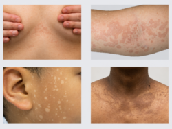
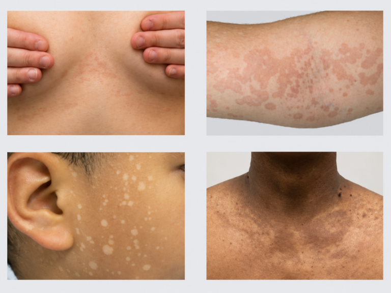
White patches on skin may be caused by vitiligo, a superficial fungal infection such as tinea versicolor, or another disorder that reduces skin pigment. Vitiligo usually produces smooth areas of marked or complete pigment loss, whereas tinea versicolor more often causes lighter, darker, pink, or brown patches with subtle surface scaling. Because early vitiligo and minimally scaly fungal patches can look similar, colour alone is not sufficient for diagnosis.[3,6,7]
The Main Difference Between Vitiligo and Fungal White Patches
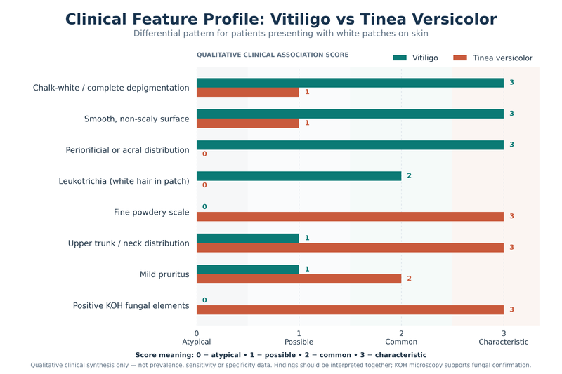
Vitiligo is a pigmentary disorder in which functioning melanocytes, the cells responsible for producing melanin, are lost from affected skin. Established patches are therefore depigmented, meaning that pigment is almost completely or completely absent. The skin surface usually remains smooth because vitiligo primarily changes colour rather than texture.[3]
Tinea versicolor, also called pityriasis versicolor, is caused by an overgrowth of Malassezia yeast that normally lives on the skin. It usually causes hypopigmentation, meaning reduced rather than completely absent pigment, although lesions may also appear tan, brown, pink, or reddish. The affected skin may have a fine, dry, powder-like scale that becomes easier to see when the surface is gently stretched or scraped.[6,7]
| Clinical Feature | Vitiligo | Tinea Versicolor |
|---|---|---|
| Main skin change | Loss of pigment caused by loss or dysfunction of melanocytes | Superficial overgrowth of Malassezia yeast affecting normal pigmentation |
| Typical colour | Milky-white or chalk-white | White, pale, tan, pink, brown or occasionally darker |
| Degree of pigment loss | May become completely depigmented | Usually hypopigmented rather than completely depigmented |
| Surface | Usually smooth | Fine, powdery or bran-like scale may be present |
| Itching | Usually absent, although active lesions may occasionally itch | Mild itching may occur, especially with heat or sweating |
| Borders | Often clearly defined | May be less sharply defined or merge with nearby patches |
| Common locations | Face, around eyes and mouth, hands, fingers, feet, elbows, knees and skin folds | Upper chest, upper back, shoulders, neck and upper arms |
| Hair within patch | May become white in some lesions | Hair colour is generally unaffected |
| Effect of sun exposure | Contrast increases because depigmented skin does not tan normally | Patches may become more noticeable because affected skin tans differently |
| Heat and humidity | Not caused by hot or humid weather | Often becomes more noticeable or recurrent in warm, humid conditions |
| Wood’s lamp | Depigmentation usually becomes more sharply accentuated | May show yellowish, yellow-green or golden fluorescence, but findings are variable |
| KOH skin scraping | No fungal elements | Yeast and short hyphae may be demonstrated |
| Response to antifungal treatment | Does not treat the underlying vitiligo | Active fungal disease usually responds to appropriate antifungal therapy |
| Pigment after treatment | Repigmentation depends on disease activity, site and treatment response | Colour may remain abnormal for weeks or months after fungal clearance |
Neither condition should be judged solely by how white the patch appears. Tinea versicolor may become strikingly pale after sun exposure because the surrounding skin tans more normally, while early vitiligo may initially look only slightly lighter than nearby skin. Previous use of steroid, antifungal, bleaching, or combination creams may further alter the appearance and make visual diagnosis less reliable.
Signs More Suggestive of Vitiligo
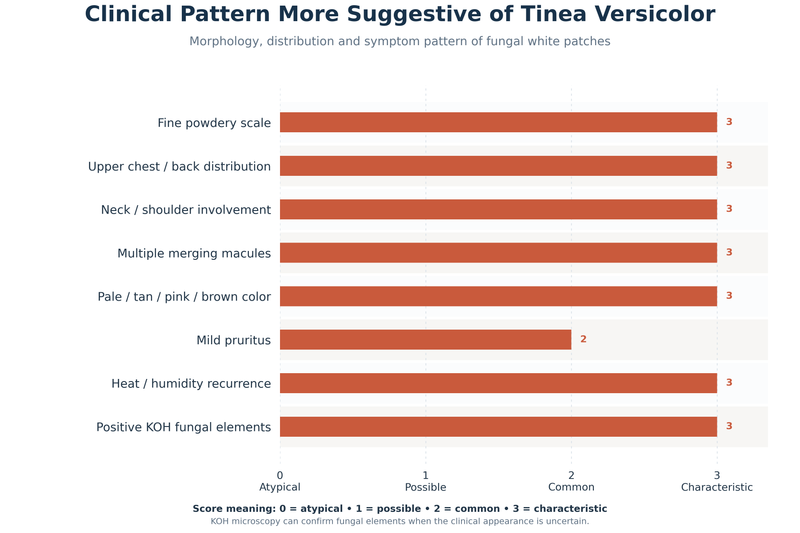
Vitiligo is more likely when the patch is distinctly milky or chalky white, has a clearly defined border, and feels completely smooth without dryness or scale. Commonly affected areas include the face, fingers, hands, feet, elbows, knees, armpits, groin, and the skin around the eyes or mouth. The lips, genital area, and other mucosal surfaces may also be involved.[3]
Nonsegmental vitiligo often develops on both sides of the body, although the distribution is not always symmetrical. New patches may appear after cuts, burns, pressure, scratching, or repeated friction, a response known as the Koebner phenomenon. Hair growing within an established patch may sometimes turn white, which suggests that pigment-producing cells within the hair follicles have also been affected.[3]
Vitiligo usually does not cause persistent itching, peeling, crusting, or pain. Mild itching can occasionally occur around an active or newly developing patch, but prominent scaling or recurrent irritation should prompt consideration of fungal infection, eczema, contact dermatitis, or another inflammatory skin disorder.
Signs More Suggestive of Fungal Infection
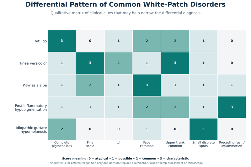
Tinea versicolor is more likely when several pale or discoloured patches appear across the upper chest, back, shoulders, neck, or upper arms. Individual spots may gradually merge into larger irregular areas. Facial involvement can occur, particularly in children and people living in warm climates, but the trunk remains the more typical location.[6,7]
Fine scaling is one of the most useful clinical clues. The scale may be obvious, but it can also be so subtle that the patch initially appears smooth. Mild itching may occur, especially during sweating, hot weather, or periods of increased humidity. Recurrence is common because the yeast responsible for the condition is part of the normal skin flora and can proliferate again under favourable conditions.[6,7]
Tinea versicolor is generally not considered contagious. It does not usually spread from one person to another in the way that ringworm infections can. Warm weather, sweating, oily skin, hormonal changes, and occlusive clothing may make the condition more noticeable or recurrent.
Successful antifungal treatment may eliminate the active yeast before normal skin colour returns. A pale area can therefore remain visible for several weeks or months despite effective treatment. Persistent colour difference alone does not prove that the infection is still active, particularly when scaling and itching have resolved.[7]
Other Conditions That Can Cause White Patches
Pityriasis alba commonly causes pale, poorly defined patches on the cheeks, chin, or upper arms of children and adolescents. The surface may be mildly dry or finely scaly, and the surrounding skin may have a history of eczema or sensitivity. Unlike established vitiligo, pityriasis alba usually causes partial pigment reduction rather than complete depigmentation.[5]
Post-inflammatory hypopigmentation can develop after eczema, allergic reactions, burns, infections, injuries, or irritating skin treatments. The pale area usually follows the shape and location of the earlier inflammation, and pigment may gradually return as the skin recovers.
Small white spots on sun-exposed forearms or lower legs in adults may represent idiopathic guttate hypomelanosis rather than vitiligo or fungus. A stable pale patch present since infancy or early childhood may suggest a congenital pigmentary disorder. Genital white patches accompanied by itching, fissuring, pain, or fragile skin require assessment for inflammatory conditions such as lichen sclerosus.
A pale patch associated with reduced sensation, numbness, weakness, loss of sweating, or a thickened nearby nerve should not be assumed to be vitiligo or fungal infection. These findings require prompt in-person medical evaluation because they may indicate a neurological or infectious condition affecting the skin and peripheral nerves.
How the Diagnosis Is Confirmed
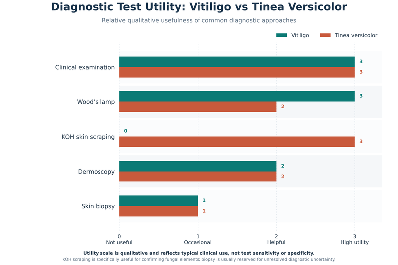
A clinician begins by examining the colour, border, surface texture, distribution, and hair within the patch. The history should establish when the patch appeared, whether it is enlarging, whether new patches are developing, and whether itching, sweating, friction, injury, eczema, or previous treatment has influenced its appearance.
A Wood’s lamp examination may help assess pigment loss under long-wave ultraviolet light in a darkened room. Vitiligo usually becomes more sharply accentuated because of the marked reduction or absence of epidermal melanin. Some tinea versicolor lesions may show yellowish, yellow-green, or coppery fluorescence, although the absence of fluorescence does not exclude fungal infection.[3,6]
When tinea versicolor remains possible, a potassium hydroxide skin scraping can provide stronger confirmation. Superficial scale is collected and examined microscopically for clusters of yeast cells and short fungal hyphae. Dermoscopy may provide additional information about pigment patterns, follicular changes, lesion borders, and subtle scaling.[6,7]
| Diagnostic Method | What the Doctor Looks For | Vitiligo Finding | Tinea Versicolor Finding | When It Is Most Useful |
|---|---|---|---|---|
| Clinical examination | Colour, border, scale, location, symmetry and hair changes | Smooth, clearly depigmented patches may be seen | Fine scale and trunk-predominant patches may be present | First assessment for every patient |
| Wood’s lamp examination | Behaviour of pigmentation under long-wave ultraviolet light | Depigmented areas become more clearly accentuated | Some lesions show yellowish or yellow-green fluorescence | Subtle pigment loss or uncertain lesion borders |
| KOH skin scraping | Microscopic fungal elements from superficial scale | Negative for fungal elements | Yeast cells and short hyphae support the diagnosis | One of the most useful tests when fungal infection is uncertain |
| Dermoscopy | Pigment network, follicles, scale and lesion margins | Loss of pigment network and perifollicular changes may be identified | Fine scale and altered pigmentation may become easier to see | Differentiating clinically similar pigment disorders |
| Sensory examination | Light touch, pain, temperature and sweating | Sensation normally preserved | Sensation normally preserved | Essential when a pale patch is numb or has altered sensation |
| Blood tests | Associated medical conditions rather than the white patch itself | Selected testing may be considered for associated autoimmune disease | Usually not required to diagnose uncomplicated tinea versicolor | When history suggests thyroid or another systemic disorder |
| Skin biopsy | Microscopic tissue architecture | May support melanocyte loss or another pigment disorder | Rarely required | Atypical, persistent or diagnostically unresolved lesions |
Blood tests do not distinguish vitiligo from fungal infection. They may be considered after vitiligo has been diagnosed when symptoms, examination findings, or family history raise concern for an associated autoimmune condition, particularly thyroid disease.[3]
Why Correct Diagnosis Matters Before Treatment
Antifungal medicines can treat tinea versicolor but cannot restore melanocytes lost through vitiligo. Conversely, medicines used to control vitiligo do not eliminate fungal overgrowth. Applying an unsuitable cream may delay the correct diagnosis, cause irritation, suppress visible inflammation, or temporarily disguise scaling without treating the underlying condition.
Medical assessment is especially important when a patch is progressively enlarging, becoming completely white, developing white hair, appearing on the face, hands, lips, or genital area, or failing to improve after an appropriate antifungal course. Rapidly appearing patches, widespread pigment loss, persistent itching, pain, ulceration, bleeding, crusting, or altered sensation also require direct clinical examination.
White Patches on Skin by Location
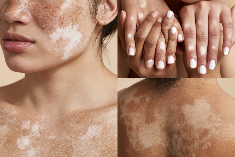
White patches on skin can look similar across different disorders, but their location often provides an important diagnostic clue. Vitiligo commonly affects the face, hands, feet, skin folds and areas exposed to friction, whereas tinea versicolor is concentrated mainly on the upper trunk, neck and upper arms. Location must still be interpreted alongside texture, scaling, symptoms and the degree of pigment loss.[3,6,7]
Face and Around the Mouth
Smooth, sharply defined white patches around the eyes, nostrils or mouth are more suggestive of vitiligo, particularly when the lips or inner lining of the mouth are also affected. Whitening of an eyebrow, eyelash or other hair within the patch further increases clinical suspicion because vitiligo can affect pigment-producing cells in hair follicles.[3]
Pale facial patches in children are frequently caused by pityriasis alba rather than vitiligo. These patches usually occur on the cheeks or chin, have poorly defined borders and may feel slightly dry or show fine scaling. They often become more noticeable after sun exposure because the surrounding skin darkens while the affected area tans less.[5]
Tinea versicolor can affect the face, particularly in children, adolescents and people living in warm or humid climates. Facial fungal patches are more likely to show subtle scale or coexist with similar lesions on the neck, chest, back or upper arms. An isolated smooth facial patch without scale requires evaluation for vitiligo, pityriasis alba and post-inflammatory pigment loss rather than automatic antifungal treatment.[5–7]
Chest, Back and Shoulders
Multiple pale, tan, pink or brown patches across the upper chest, back or shoulders strongly favour tinea versicolor. The lesions may begin as separate spots and gradually merge into larger irregular areas. Fine scale may be visible at the margins or become apparent when the skin is gently stretched or scraped.[6,7]
Mild itching, recurrence during warm weather and increased visibility after tanning also support tinea versicolor. The patches may remain lighter than the surrounding skin after the fungal overgrowth has been controlled because normal pigment production can take weeks or months to recover.[6,7]
Vitiligo can also affect the trunk, but its patches are usually smooth, distinctly depigmented and free from scale. Trunk lesions that appear together with patches on the face, hands, feet or around body openings are more suggestive of vitiligo than an isolated superficial fungal infection.[3]
Neck and Upper Arms
Tinea versicolor commonly extends from the upper chest or back onto the neck and upper arms. Several patches with subtle scaling, variable colour and a history of recurrence during periods of sweating are characteristic clinical clues.[6,7]
Pityriasis alba may also occur on the neck, shoulders and upper arms, especially in children with dry or eczema-prone skin. Its borders are usually less distinct than those of vitiligo, and the surface may feel mildly rough. Examination is particularly useful when facial and upper-arm patches occur together because pityriasis alba, tinea versicolor and early vitiligo can overlap in appearance.[5–7]
Hands, Fingers, Feet and Joints
White patches on the fingers, backs of the hands, wrists, feet, elbows or knees are more characteristic of vitiligo than tinea versicolor. These sites are frequently exposed to friction, pressure and minor trauma, which may contribute to the appearance of new vitiligo lesions in susceptible skin.[3]
Vitiligo affecting the fingertips, toes or areas around the nails may be more difficult to repigment because these regions contain fewer hair follicles that can serve as reservoirs of pigment-producing cells. Early diagnosis is therefore valuable when smooth white patches begin to appear on acral areas such as the hands and feet.
Tinea versicolor is uncommon on the hands and feet. A scaly patch on these sites may instead represent a dermatophyte infection, eczema, contact dermatitis or another inflammatory disorder. The presence of cracking, redness, a raised border or marked itching makes uncomplicated vitiligo less likely.
Armpits, Groin and Other Skin Folds
Vitiligo may develop in the armpits, groin and other areas exposed to repeated friction. The affected skin usually remains smooth and does not produce odour, discharge, moisture, soreness or surface scale.[3]
Fungal or yeast-related rashes within skin folds more often cause itching, redness, maceration, scaling or a clearly active border. These symptoms distinguish an inflammatory fold infection from uncomplicated vitiligo, although pigment alteration may remain after the rash resolves.
White genital patches require direct examination when they are accompanied by itching, pain, fragile skin, fissures, bleeding or anatomical change. These features may indicate an inflammatory genital disorder rather than vitiligo or tinea versicolor and should not be treated repeatedly with over-the-counter antifungal creams without a confirmed diagnosis.
Areas of Previous Rash or Injury
A pale patch that appears exactly where eczema, a burn, an infection, an allergic reaction or another rash previously occurred may represent post-inflammatory hypopigmentation. These areas often retain some pigment, have less sharply defined borders and gradually regain colour as the skin recovers.
Vitiligo may also first appear at sites of cuts, burns, scratches, pressure or repeated rubbing through the Koebner phenomenon. Unlike ordinary post-inflammatory pigment loss, such lesions may become progressively whiter, expand beyond the original injury or appear at additional trauma-prone sites.[3]
How White Patches on Skin Are Diagnosed
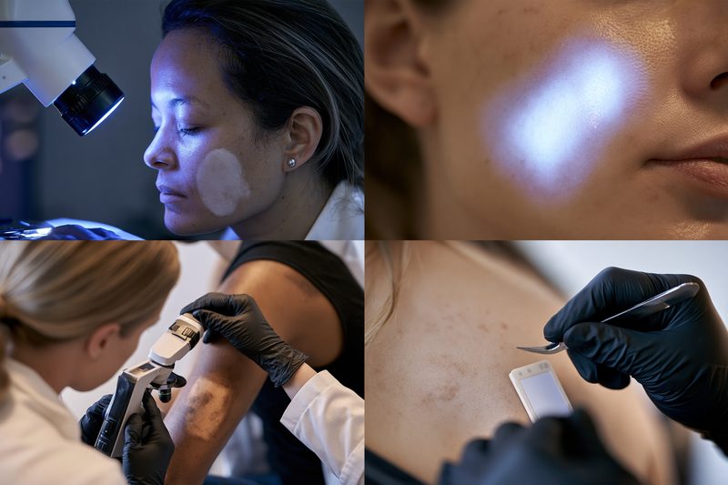
White patches on skin are diagnosed by combining the clinical appearance with the patient’s history and, when needed, targeted tests. No single visual feature can reliably distinguish every case of vitiligo, tinea versicolor, pityriasis alba, post-inflammatory hypopigmentation or another pigmentary disorder.[3,6–8]
Clinical Examination
The clinician assesses the colour, border, texture, scale, distribution and symmetry of each patch. Established vitiligo usually appears smooth and distinctly depigmented, whereas tinea versicolor more often shows partial pigment loss with fine surface scale. Examination also checks whether hair within the patch has turned white and whether similar lesions are present on the face, hands, feet, trunk, skin folds or around body openings.[3,6,7]
The history should establish when the patch first appeared, whether it is enlarging, whether new areas are developing and whether itching, sweating, sun exposure, friction or skin injury affects it. Previous eczema, burns, infections, cosmetic procedures and use of steroid, antifungal or combination creams may explain altered pigmentation or modify the visible signs.
A family history of vitiligo or autoimmune disease can increase clinical suspicion but does not confirm the diagnosis. Seasonal recurrence during warm and humid weather, particularly when lesions affect the chest, back or shoulders, is more suggestive of tinea versicolor.[6–8]
Wood’s Lamp Examination
A Wood’s lamp emits long-wave ultraviolet light and is used in a darkened room to evaluate pigment changes that may be difficult to define under ordinary lighting. Vitiligo generally becomes more sharply accentuated because affected epidermal skin contains markedly reduced or absent melanin.[3,10]
The examination can reveal subtle vitiligo beyond the margins visible in normal light and may help estimate the true extent of involvement. It can also assist in distinguishing depigmentation from milder hypopigmentation, particularly in lighter skin tones where contrast may otherwise be limited.
Some tinea versicolor patches show yellow, yellow-green or coppery fluorescence under a Wood’s lamp. Fluorescence is not present in every case, so a normal Wood’s lamp result does not exclude fungal infection.[6–8]
Skin Scraping and KOH Microscopy
When tinea versicolor is suspected, superficial scale can be collected from the patch and examined after applying potassium hydroxide. Microscopy may reveal clusters of yeast cells and short fungal hyphae, supporting the diagnosis of Malassezia overgrowth.[6–8]
Skin scraping is particularly useful when scaling is minimal, when previous treatment has changed the appearance or when a pale patch has not responded as expected. A negative result may require repeat sampling from a more active or scaly margin if clinical suspicion remains high.
Vitiligo does not produce fungal elements on microscopy. A negative fungal test therefore supports reconsideration of vitiligo, pityriasis alba, post-inflammatory hypopigmentation or another non-fungal cause, but it does not independently establish which alternative diagnosis is present.
Dermoscopy
Dermoscopy allows magnified examination of pigment networks, perifollicular colour, borders, scale and vascular patterns. In vitiligo, it may demonstrate loss of the normal pigment network, perifollicular pigment retention or features associated with activity and repigmentation.[8,10]
In tinea versicolor, dermoscopy can make subtle scale and altered pigment patterns easier to identify. It is most useful as an extension of the clinical examination rather than as a replacement for microscopy when fungal infection remains uncertain.[8]
Sensory Examination
Sensation should be tested when a pale patch is associated with numbness, reduced sweating, weakness or altered temperature perception. Uncomplicated vitiligo and tinea versicolor do not normally cause loss of sensation.
Reduced sensitivity to light touch, pain or temperature requires assessment for disorders affecting cutaneous nerves. This finding changes the diagnostic pathway and should not be managed by repeatedly trying antifungal or pigment-restoring creams.
Blood Tests
Blood tests do not distinguish vitiligo from fungal infection and are not required for every white patch. They may be considered after vitiligo has been diagnosed when symptoms, examination findings or family history suggest an associated autoimmune condition.[3,10]
Thyroid assessment may be appropriate when the patient has fatigue, weight change, cold or heat intolerance, palpitations, neck swelling, menstrual disturbance or a personal or family history of thyroid disease. Testing should be selected according to clinical risk rather than ordered routinely for all patients.
Skin Biopsy
A skin biopsy is rarely necessary when the history, examination, Wood’s lamp findings and fungal testing provide a clear diagnosis. It may be considered when lesions are atypical, progressive, treatment-resistant or difficult to distinguish from inflammatory, infectious or neoplastic skin disease.
Biopsy findings must be interpreted with the clinical pattern because reduced pigment can occur in several conditions. Histopathology can support melanocyte loss in vitiligo or identify inflammation and other tissue changes, but it is usually reserved for unresolved diagnostic uncertainty.[3,10]
Other Causes of White Patches on Skin
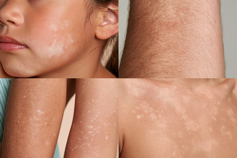
White patches on skin that do not have the typical features of vitiligo or tinea versicolor may result from eczema-related inflammation, previous skin injury, stable developmental pigment differences, chronic sun exposure or another inflammatory disorder. The patient’s age, the appearance of the patch, preceding symptoms, rate of change and body location help narrow the diagnosis.[5,11]
Pityriasis Alba
Pityriasis alba commonly affects children and adolescents, particularly those with dry, sensitive or eczema-prone skin. It usually produces round or irregular pale patches on the cheeks, chin, neck or upper arms. The borders are generally indistinct, and the surface may feel mildly dry or show fine scale.[5,11]
The patches often become more noticeable during summer because the surrounding skin tans while the affected area remains lighter. Unlike established vitiligo, pityriasis alba usually retains some pigment, does not produce sharply defined chalk-white skin and rarely causes whitening of the hair. The colour generally returns gradually, although improvement may take several months.[5,11]
Post-Inflammatory Hypopigmentation
Post-inflammatory hypopigmentation can develop after eczema, psoriasis, allergic dermatitis, infection, burns, abrasions or other inflammatory skin conditions. It may also follow procedures such as cryotherapy, chemical peels, laser treatment or prolonged exposure to irritating topical products.[11]
The pale area usually corresponds to the location and shape of the earlier rash or injury. Residual dryness, redness, scaling or altered texture may remain when the underlying inflammation is still active. Most non-scarring cases gradually regain pigment, whereas deeper inflammation, burns or scarring disorders may cause prolonged or permanent colour loss.[11]
Potent topical corticosteroids and unregulated combination creams can also produce local skin thinning and pigment alteration when used incorrectly. A patch that becomes paler, thinner or more fragile during repeated cream use requires medication review rather than continued unsupervised application.
Idiopathic Guttate Hypomelanosis
Idiopathic guttate hypomelanosis causes multiple small, round or oval white spots, usually on the forearms, shins and other chronically sun-exposed areas. It is more common in middle-aged and older adults, and the number of spots may increase gradually with age.[12]
The spots are usually only a few millimetres wide, remain separate from one another and do not cause itching, scaling or pain. Their small size, sun-exposed distribution and stable behaviour help distinguish them from enlarging vitiligo patches. The condition is benign and does not represent a fungal infection.[12]
Nevus Depigmentosus
Nevus depigmentosus is usually present at birth or becomes noticeable during early childhood. It commonly appears as a single pale patch or a group of patches confined to one region of the body. The affected area grows in proportion to the child but generally does not spread progressively to unrelated sites.[13]
The patch often has an irregular or serrated border and retains more pigment than established vitiligo. A stable lesion with an early onset favours nevus depigmentosus, whereas newly appearing patches, progressive enlargement, whitening of hair or development at distant sites increases concern for vitiligo.[13]
Progressive Macular Hypomelanosis
Progressive macular hypomelanosis most often affects adolescents and young adults. It produces poorly defined, non-scaly pale patches that commonly develop on the lower chest, abdomen, lower back or central trunk. Individual areas may merge and form a larger symmetrical pattern.[11]
The condition usually causes no itching or discomfort and may be more visible in darker skin tones. Negative fungal microscopy, absence of scale and the characteristic trunk distribution help separate it from tinea versicolor. It differs from vitiligo because the pigment reduction is usually incomplete and the borders are less distinct.[11]
Lichen Sclerosus
Lichen sclerosus can produce ivory-white, thin or crinkled patches, most commonly around the vulva, anus or penis. It may cause severe itching, burning, soreness, painful fissures, bleeding or discomfort during sexual activity. Progressive disease can produce scarring and anatomical changes.[14]
Genital white patches with altered texture or persistent symptoms should not be assumed to be vitiligo or treated repeatedly as a fungal infection. Early diagnosis and long-term medical follow-up are important because untreated genital lichen sclerosus may cause functional complications and carries an increased risk of squamous cell carcinoma.[14]
Chemical or Treatment-Related Pigment Loss
Repeated contact with certain hair dyes, adhesives, rubber products, disinfectants, cosmetic chemicals or occupational substances can produce localised pigment loss in susceptible individuals. The pattern may correspond to the points of contact, such as the hairline, hands, wrists or areas beneath protective equipment.
Pigment alteration may also follow injections, radiotherapy, prolonged pressure, skin resurfacing procedures or inappropriate topical treatment. Establishing the timing and distribution of exposure is essential because continued contact can cause additional patches even when the original area is being treated.[11]
Why Antifungal Creams Do Not Treat All White Patches on Skin
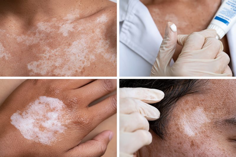
White patches on skin require cause-specific treatment because antifungal medicines act against fungal organisms, while vitiligo results from loss of functioning pigment-producing cells and does not improve simply by eliminating yeast from the skin.[3,6,7]
When Antifungal Treatment Is Appropriate
Antifungal treatment is appropriate when examination supports tinea versicolor or another superficial fungal infection. Features such as fine scaling, mild itching, recurrence in warm or humid weather, involvement of the chest or upper back, and fungal elements on skin scraping strengthen the diagnosis.[6–8]
Topical antifungal creams, lotions or medicated washes can reduce active yeast overgrowth. The selected preparation, treatment area and duration should reflect the extent of disease, previous response and whether the condition is recurrent. Widespread or repeatedly relapsing infection may require clinician-supervised treatment rather than continued self-medication.[6,7]
Why Pale Skin May Remain After the Fungus Clears
Normal skin colour may not return immediately after successful treatment. Tinea versicolor can temporarily interfere with pigment production, so the yeast may be controlled while the affected areas remain lighter or darker than surrounding skin for several weeks or months.[6,7]
Persistent colour difference does not necessarily mean that treatment has failed. Ongoing scale, itching or development of new lesions is more suggestive of continued activity than residual discoloration alone. Repeatedly applying additional antifungal cream to inactive pale skin may cause irritation without accelerating pigment recovery.
Why Vitiligo Does Not Respond to Antifungal Creams
Vitiligo does not result from fungal overgrowth, so antifungal medicines cannot restore melanocytes or stop immune-mediated pigment loss. A smooth, progressively enlarging white patch that remains unchanged after appropriate antifungal treatment should be reassessed rather than treated with repeated courses.[3]
Delayed recognition may allow additional patches to appear before vitiligo-directed treatment begins. This is particularly important when lesions affect the face, hands, feet or areas around the eyes and mouth, or when hair within a patch begins to lose colour.[3]
Risks of Repeated Self-Treatment
Repeated use of unconfirmed treatments can alter the appearance of a lesion and make diagnosis more difficult. Combination creams containing antifungal agents, corticosteroids and antibiotics may temporarily reduce redness or itching while masking infection, thinning the skin or changing pigmentation.
Using potent corticosteroid-containing creams without supervision can cause skin thinning, visible blood vessels, stretch marks and worsening of some fungal infections. Symptoms that recur after each treatment course require diagnostic confirmation rather than repeated use of the same product.
When Treatment Failure Requires Reassessment
Reassessment is needed when scaling and itching persist despite correct antifungal use, when new patches continue to appear, or when the affected area becomes more sharply white and clearly defined. A repeat examination, Wood’s lamp assessment or skin scraping may distinguish persistent fungal disease from vitiligo, pityriasis alba or post-inflammatory pigment change.[3,6–8]
Prompt evaluation is also required when a patch develops pain, ulceration, bleeding, marked inflammation, altered sensation or loss of sweating. These findings are not typical of uncomplicated vitiligo or tinea versicolor and require investigation for another skin or neurological disorder.
Treatment for White Patches on Skin
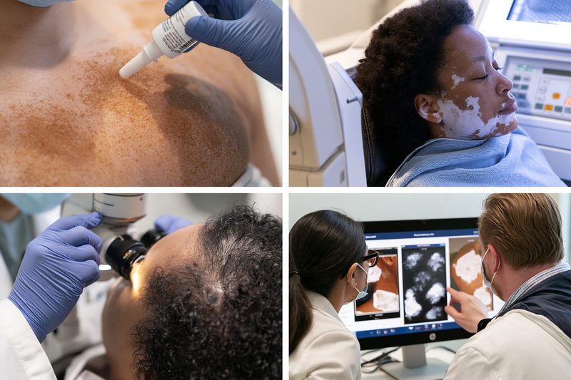
Treatment for white patches on skin depends on the confirmed cause, the extent of pigment loss, the activity of the condition and the presence of scaling, inflammation or other symptoms. Vitiligo, tinea versicolor, pityriasis alba and post-inflammatory hypopigmentation require different management, even when the patches appear similar.
When Tinea Versicolor Is Confirmed
Tinea versicolor is usually treated with topical antifungal creams, lotions, foams or medicated washes. Localised patches may respond to treatment applied directly to the affected skin, while widespread involvement of the chest, back, shoulders or neck may require a formulation that can be distributed across a larger area.[6,7]
The medicine should be used for the prescribed duration even when scaling or itching improves quickly. Incomplete treatment can leave active yeast on the skin and increase the likelihood of early recurrence. Oral antifungal treatment may be considered for extensive, recurrent or treatment-resistant disease but requires medical supervision because of potential drug interactions, contraindications and liver-related adverse effects.[6,7]
Recurrence is common during hot or humid weather. Patients with repeated episodes may require intermittent preventive treatment during high-risk periods. Keeping the skin dry, changing sweat-soaked clothing and avoiding prolonged use of heavy, oily products may reduce conditions that favour yeast overgrowth.
Skin colour often recovers more slowly than the infection itself. Treatment response should therefore be assessed through resolution of scale, itching and active spread rather than expecting immediate repigmentation. Persistent or expanding lesions should be re-examined to confirm that the original diagnosis was correct.[6,7]
When Vitiligo Is Confirmed
Vitiligo treatment is selected according to the type of vitiligo, current disease activity, body surface area involved, location of the patches, duration of pigment loss and colour of the hair within affected skin. Facial and neck lesions often respond differently from patches on the fingertips, toes, lips or other areas with fewer hair follicles.[3,10]
Topical anti-inflammatory medicines may be used for limited disease, while phototherapy may be considered when patches are extensive, progressive or insufficiently responsive to local treatment. Treatment aims may include controlling new pigment loss, stabilising active disease and stimulating repigmentation from surviving melanocytes in the hair follicles or surrounding skin.[3,10]
Progress should be documented with standardised photographs taken under similar lighting. A reduction in new lesions, absence of enlargement and development of small perifollicular pigment islands can indicate stabilisation or early repigmentation. Failure to respond should prompt reassessment of disease activity, adherence, diagnosis and the anatomical likelihood of repigmentation.
Ayurvedic management may be considered after the diagnosis has been established and the extent and activity of vitiligo have been documented. Shwitra is an Ayurvedic description of disorders presenting with altered skin colour, but it should not be treated as an automatic diagnostic equivalent of every modern vitiligo subtype. Treatment should be individualised according to the clinical pattern, constitution, digestive function, associated symptoms and concurrent medicines, with objective photographic and dermatological monitoring.
When Pityriasis Alba Is Confirmed
Pityriasis alba usually improves gradually with regular moisturisation, gentle skin care and protection from excessive sun exposure. Treating underlying dryness or mild eczema can reduce surface roughness and make the colour difference less noticeable.[5]
Inflamed or itchy lesions may require short-term clinician-directed anti-inflammatory treatment. Strong corticosteroid creams are generally unnecessary for uncomplicated pityriasis alba, particularly on the face. Pigment recovery can take several months even after dryness and inflammation have resolved.
When Pigment Loss Follows Inflammation
Post-inflammatory hypopigmentation is managed by identifying and controlling the preceding skin disorder. Active eczema, allergic dermatitis, psoriasis or infection must be treated before normal pigmentation can recover.
Repeated irritation from cosmetic products, bleaching agents, topical medicines or procedures should be stopped when it is contributing to the colour change. Non-scarring pigment loss often improves gradually, whereas deeper inflammation, burns or scarring may produce prolonged or incomplete recovery.
When the Diagnosis Remains Uncertain
Treatment should not be escalated when the diagnosis is based only on appearance and the patch behaves unexpectedly. Lack of scale, continued enlargement, complete whitening, white hair or failure of an adequate antifungal course increases the need for Wood’s lamp examination, dermoscopy or skin scraping.[3,6–8]
A new biopsy or specialist review may be required when patches are atypical, treatment-resistant, painful, ulcerated, indurated or associated with sensory change. Establishing the correct diagnosis before prolonged therapy reduces unnecessary medicine exposure and prevents delay in treating progressive vitiligo or another clinically important skin disorder.
Ayurvedic Treatment for Vitiligo
When white patches on skin are confirmed as vitiligo rather than a fungal infection, treatment should focus on controlling disease activity, protecting remaining melanocytes, encouraging repigmentation, and monitoring whether new patches are still developing. The treatment approach depends on the type of vitiligo, affected body sites, duration, rate of spread, hair pigmentation within the patches, associated autoimmune conditions, and previous treatment response.
Ayurveda approaches vitiligo through an individualised assessment rather than treating every white patch in the same way. Once the diagnosis is established, treatment may include personalised herbal or herbo-mineral medicines, dietary correction, digestive and metabolic assessment, external applications, Rasayana therapy, and selected Panchakarma procedures when clinically appropriate. Progress should be monitored objectively through standardised photographs, lesion measurements, disease activity, and evidence of perifollicular or marginal repigmentation.
For a detailed explanation of diagnosis, Ayurvedic medicines, treatment planning, diet, classical Ayurvedic principles, repigmentation monitoring, and long-term management, read the complete guide on Ayurvedic treatment for vitiligo.
White Patches on Skin Consultation

A consultation for white patches on skin is more accurate when the clinician can review how the patches began, how quickly they changed and which treatments have already been used. Preparing photographs, dates, symptoms and previous reports helps distinguish vitiligo from fungal infection, eczema-related pigment loss and other causes.
Photographs and Timeline
Clear photographs should be taken in natural daylight without filters, makeup or flash glare. Images from different dates can show whether a patch is enlarging, whether new patches are appearing and whether the borders are becoming more sharply defined.
The patient should record when the first patch appeared, where it began and whether it followed a rash, injury, burn, infection, cosmetic procedure or period of intense sun exposure. Rapid progression, appearance at sites of friction or trauma and whitening of hair within a patch may support active vitiligo, while seasonal recurrence with scaling on the upper trunk is more suggestive of tinea versicolor.[3,6,7]
Symptoms and Surface Changes
The clinician should be told whether the patch itches, peels, scales, burns, becomes red or changes after sweating. Fine scale and mild itching support a superficial fungal disorder, whereas uncomplicated vitiligo usually changes colour without altering skin texture.[3,6,7]
Numbness, reduced sweating, altered temperature sensation, pain, bleeding or ulceration should be reported immediately. These findings are not typical of uncomplicated vitiligo or tinea versicolor and may require sensory examination, biopsy or investigation for another disorder.
Previous Treatments
All creams, tablets, shampoos, oils and home remedies used on the patch should be listed with their duration and observed effect. Combination creams may contain corticosteroids, antifungals and antibiotics, even when the patient does not recognise these ingredients.
Temporary improvement in redness or itching does not confirm the diagnosis. Antifungal treatment may clear active tinea versicolor while residual pigment change remains, whereas vitiligo will not respond to antifungal therapy because it is not caused by yeast overgrowth.[3,6,7]
Medical and Family History
Relevant history includes eczema, psoriasis, thyroid disease, autoimmune disorders, diabetes, recurrent fungal infections and previous episodes of unexplained pigment change. A family history of vitiligo or autoimmune disease can increase clinical suspicion but does not establish the diagnosis.[3,10]
The clinician should also know about pregnancy, immune-suppressing medicines, recent antibiotics, occupational chemical exposure and regular contact with hair dyes, adhesives, disinfectants or rubber products. These factors may alter treatment choices or suggest an exposure-related cause of pigment loss.
Previous Tests and Reports
Patients should bring results from Wood’s lamp examinations, skin scrapings, potassium hydroxide microscopy, dermoscopy, biopsy or thyroid testing. Repeating a test may still be necessary when the earlier sample was taken after treatment or from an inactive part of the lesion.[3,6–8]
A complete consultation should end with a documented working diagnosis, an explanation of whether the pigment loss is partial or complete, an assessment of current activity and a plan for follow-up photographs. Treatment response should be judged by changes in scale, spread, border activity and repigmentation rather than by colour alone.
When White Patches on Skin Need Medical Care
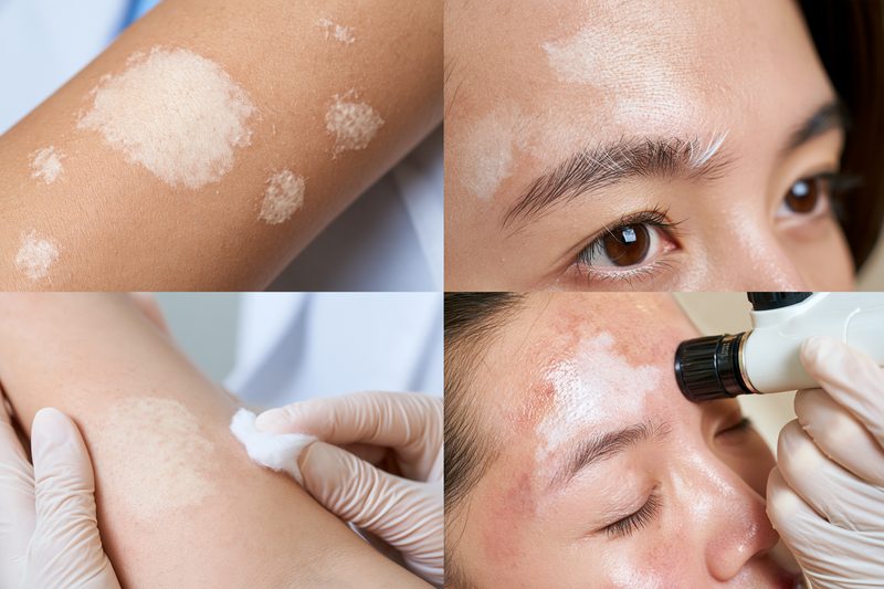
White patches on skin should be medically assessed when they are spreading, changing rapidly, affecting sensitive areas or accompanied by symptoms that are not typical of uncomplicated vitiligo or tinea versicolor. Early evaluation can confirm the cause, document disease activity and prevent prolonged treatment with unsuitable creams.
Rapidly Spreading or Newly Appearing Patches
A patch that enlarges over days or weeks, develops sharply defined white borders or is followed by new patches elsewhere requires examination for active vitiligo or another progressive pigmentary disorder. Small depigmented spots appearing around a larger patch, new lesions after minor skin injury and whitening of hair within the affected area may indicate active pigment loss.[3,10]
Progression should be documented with photographs taken in similar lighting and from the same distance. Changes in size, number and distribution are more clinically useful than relying on memory or comparing photographs taken under different lighting conditions.
Patches on the Face, Hands or Genital Skin
White patches around the eyes, mouth, lips, fingers or hands should be assessed early because these areas are highly visible and may affect confidence, social interaction and quality of life. Treatment response can also vary by location, with facial areas often behaving differently from fingertips, toes and other regions containing fewer hair follicles.[3,10]
Genital white patches require direct examination when they cause itching, burning, painful intercourse, fissures, bleeding, fragile skin or anatomical change. These symptoms may indicate lichen sclerosus or another inflammatory disorder rather than vitiligo or fungal infection. Untreated genital lichen sclerosus can cause scarring and functional complications.[14]
Numbness or Altered Sensation
A pale or white patch associated with numbness, reduced pain sensation, altered temperature perception, weakness or loss of sweating requires prompt in-person assessment. Vitiligo and tinea versicolor do not normally damage cutaneous sensation.
Sensory change may indicate involvement of peripheral nerves or another neurological or infectious process. Continued treatment with antifungal, steroid or pigment-restoring creams without neurological examination may delay the correct diagnosis.
Pain, Ulceration or Bleeding
Uncomplicated vitiligo usually does not cause pain, ulceration, crusting or bleeding. Tinea versicolor may produce mild itching or fine scale but should not cause deep wounds, persistent tenderness or tissue breakdown.[3,6,7]
A white patch that becomes painful, thickened, ulcerated, persistently inflamed or prone to bleeding requires examination for an inflammatory, infectious or neoplastic skin disorder. Biopsy may be considered when the surface, border or texture is atypical or when the lesion does not respond as expected.
Persistent Scale or Itching
Fine scaling and mild itching may support tinea versicolor, especially when lesions affect the chest, back, shoulders or neck. Medical review is appropriate when symptoms persist despite correctly used antifungal treatment, repeatedly return or spread to new areas.[6,7]
Treatment failure may result from an incorrect diagnosis, incomplete application, reinfection, recurrence or another skin disorder that resembles fungal disease. Skin scraping and potassium hydroxide microscopy can help determine whether fungal elements are still present before additional treatment is prescribed.[6–8]
White Patches in Children
Children with persistent, enlarging or multiple white patches should be examined rather than treated repeatedly with over-the-counter creams. Pityriasis alba is common in childhood, but vitiligo, tinea versicolor, post-inflammatory hypopigmentation and congenital pigmentary conditions can produce similar appearances.[3,5–7]
Assessment is particularly important when the patches are completely white, involve the eyelids, lips, hands or genital area, develop white hair or are associated with a family history of vitiligo or autoimmune disease. Photographs and growth-adjusted follow-up can help determine whether a childhood patch is stable or progressive.
Failure of Pigment to Return
Persistent paleness after antifungal treatment does not always mean that infection remains active. Pigment recovery following tinea versicolor may take weeks or months after scale and fungal overgrowth have resolved.[6,7]
Reassessment is needed when the patch continues to enlarge, becomes more clearly depigmented, develops new borders or remains unchanged despite resolution of fungal signs. Wood’s lamp examination and repeat microscopy may distinguish residual post-infectious colour change from vitiligo or another pigmentary disorder.[3,6–8]
Significant Emotional Distress
White patches can affect self-image, relationships, employment and social confidence even when they are not physically painful. Anxiety, avoidance of social situations or persistent fear that the condition is contagious should be addressed during the consultation.
A confirmed diagnosis and clear explanation of transmission, expected progression and treatment options can reduce uncertainty. Vitiligo is not contagious, and tinea versicolor is generally not transmitted through ordinary social contact.[3,6,7]
White Patches on Skin FAQs
White patches on skin can result from vitiligo, fungal infection, eczema-related pigment loss or several less common conditions, so diagnosis should be based on clinical features and targeted testing rather than colour alone.
Can a fungal infection look like vitiligo?
Yes. Tinea versicolor can cause pale patches that resemble vitiligo, especially after sun exposure. Fine scaling, mild itching and involvement of the chest, back, neck or upper arms are more suggestive of fungal infection.
Does vitiligo cause itching or scaling?
Vitiligo usually causes smooth pigment loss without scaling. Mild itching may occasionally occur in active disease, but persistent scale, redness or peeling suggests fungal infection, eczema or another inflammatory condition.
Are fungal white patches contagious?
Tinea versicolor is generally not considered contagious because it results from overgrowth of yeast that normally lives on the skin. It does not usually spread through routine touching or sharing living spaces.
Can a Wood’s lamp confirm vitiligo?
A Wood’s lamp can make vitiligo more clearly visible and reveal subtle areas of depigmentation. The result must still be interpreted with the clinical examination because other pigmentary disorders may also change under ultraviolet light.
How can a doctor confirm fungal white patches?
A clinician may gently scrape surface scale and examine it under a microscope after adding potassium hydroxide. Finding characteristic yeast cells and short fungal filaments supports tinea versicolor.
Why is the skin still white after antifungal treatment?
The fungal overgrowth may clear before normal pigment production returns. Pale areas can remain visible for several weeks or months even after scaling, itching and active infection have resolved.
Can antifungal cream treat vitiligo?
No. Vitiligo is not caused by fungus, so antifungal medicines cannot stop melanocyte loss or restore pigment. A smooth, spreading white patch that does not respond to antifungal treatment requires reassessment.
Are white facial patches in children always vitiligo?
No. Pityriasis alba commonly causes pale, mildly dry patches on the cheeks of children. Tinea versicolor, post-inflammatory hypopigmentation and vitiligo are other possibilities that may require clinical examination.
Can vitamin deficiency cause white patches?
Nutritional deficiency is not the most common cause of isolated white patches. Vitiligo, tinea versicolor, pityriasis alba and post-inflammatory pigment change should be assessed before attributing the patches to a vitamin deficiency.
Does every completely white patch mean vitiligo?
No. Marked depigmentation increases suspicion of vitiligo, but chemical exposure, inflammatory disorders, congenital pigment conditions and other diseases can also produce white areas. Distribution, texture, progression and test findings help establish the diagnosis.
When should white patches be examined urgently?
Prompt assessment is needed when a patch spreads rapidly, develops numbness, loses sweating, becomes painful, ulcerates, bleeds or affects genital skin with fissuring or scarring. These findings are not typical of uncomplicated vitiligo or tinea versicolor.
Can vitiligo and fungal infection occur together?
Yes. A person with vitiligo can also develop tinea versicolor or another fungal infection. New itching or scaling within or around an established vitiligo patch should be examined rather than assumed to represent progression.
References
[1] American Academy of Dermatology Association. (n.d.). Tinea versicolor: Diagnosis and treatment. Retrieved October 27, 2026, from
https://www.aad.org/public/diseases/a-z/tinea-versicolor-treatmentBrief: Explains clinical examination, skin-scraping microscopy and antifungal treatment. Clarifies that normal skin colour may return gradually after the infection has cleared.
[2] American Academy of Dermatology Association. (n.d.). Tinea versicolor: Signs and symptoms. Retrieved October 27, 2026, from
https://www.aad.org/public/diseases/a-z/tinea-versicolor-symptomsBrief: Describes lighter or darker patches, fine scaling, itching and increased visibility after tanning. These features help distinguish tinea versicolor from other causes of white patches.
[3] American Academy of Dermatology Association. (n.d.). Vitiligo: Diagnosis and treatment. Retrieved October 27, 2026, from
https://www.aad.org/public/diseases/a-z/vitiligo-treatmentBrief: Explains assessment of pigment loss and treatment options, including topical medicines, phototherapy and selected surgical procedures. Emphasises confirming the diagnosis before choosing treatment.
[4] American Academy of Dermatology Association. (2026, April 14). Vitiligo: Frequently asked questions.
https://www.aad.org/public/diseases/a-z/vitiligo-overviewBrief: Addresses common questions about vitiligo, its autoimmune basis, pigmentation changes and effects on daily life. Provides accessible background for patients newly concerned about white patches.
[5] Oakley, A. (2020, September). Pityriasis alba. DermNet.
https://dermnetnz.org/topics/pityriasis-albaBrief: Describes pale, poorly defined, mildly scaly patches commonly affecting children’s faces. Explains their association with dry skin and eczema, helping distinguish them from vitiligo.
[6] Oakley, A. (2021, August). Pityriasis versicolor. DermNet.
https://dermnetnz.org/topics/pityriasis-versicolorBrief: Explains Malassezia-associated skin discolouration, typical body sites, diagnosis and treatment. Discusses recurrence and delayed pigment recovery after successful treatment.
[7] Leung, A. K. C., Barankin, B., Lam, J. M., Leong, K. F., & Hon, K. L. (2022). Tinea versicolor: An updated review. Drugs in Context, 11, Article 2022-9-2.
https://pmc.ncbi.nlm.nih.gov/articles/PMC9677953/Brief: Reviews causes, clinical features, differential diagnosis and treatment of tinea versicolor. Covers topical antifungals, oral treatment for selected cases and management of recurrent disease.
[8] Łabędź, N., Navarrete-Dechent, C., Kubisiak-Rzepczyk, H., Bowszyc-Dmochowska, M., Pogorzelska-Antkowiak, A., & Pietkiewicz, P. (2023). Pityriasis versicolor—A narrative review on the diagnosis and management. Life, 13(10), Article 2097.
https://pmc.ncbi.nlm.nih.gov/articles/PMC10608716/Brief: Reviews clinical assessment and diagnostic techniques for pityriasis versicolor, including atypical presentations. Discusses treatment selection and recurrence despite initially effective therapy.
[9] National Health Service. (2026, March 11). Pityriasis versicolor.
https://www.nhs.uk/conditions/pityriasis-versicolor/Brief: Provides patient guidance on symptoms, common affected areas, antifungal treatment and recurrence. Explains when persistent skin changes require further assessment.
[10] van Geel, N., Speeckaert, R., Taïeb, A., Ezzedine, K., Lim, H. W., Pandya, A. G., Passeron, T., Wolkerstorfer, A., Abdallah, M., Alomar, A., Bae, J. M., Bekkenk, M., Benzekri, L., Böhm, M., Eleftheriadou, V., Esmat, S., Ghia, D., Goh, B. K., Grimes, P., … Seneschal, J. (2023). Worldwide expert recommendations for the diagnosis and management of vitiligo: Position statement from the International Vitiligo Task Force—Part 1: Towards a new management algorithm. Journal of the European Academy of Dermatology and Venereology, 37(11), 2173–2184.
https://pubmed.ncbi.nlm.nih.gov/37746876/Brief: Presents international expert consensus on assessing vitiligo subtype, extent and activity. Distinguishes stopping disease progression from restoring pigmentation and emphasises individualised goals and shared decision-making.
[11] Rao, M., Young, K., Jackson-Cowan, L., Kourosh, A., & Theodosakis, N. (2023). Post-inflammatory hypopigmentation: Review of the etiology, clinical manifestations, and treatment options. Journal of Clinical Medicine, 12(3), Article 1243.
https://pmc.ncbi.nlm.nih.gov/articles/PMC9917556/Brief: Reviews reduced pigmentation following inflammation or skin injury. Explains how the preceding rash, physical examination and clinical course help distinguish these changes from other pigment disorders.
[12] Juntongjin, P., & Laosakul, K. (2016). Idiopathic guttate hypomelanosis: A review of its etiology, pathogenesis, findings, and treatments. American Journal of Clinical Dermatology, 17(4), 403–411.
https://pubmed.ncbi.nlm.nih.gov/27206417/Brief: Reviews small, discrete white spots commonly found on sun-exposed forearms and shins, particularly with increasing age. Describes their benign course and variable responses to available treatments.
[13] Ullah, F., & Schwartz, R. A. (2019). Nevus depigmentosus: Review of a mark of distinction. International Journal of Dermatology, 58(12), 1366–1370.
https://pubmed.ncbi.nlm.nih.gov/30801693/Brief: Describes a congenital pigment disorder characterised by a generally nonprogressive pale patch with irregular borders. Helps distinguish longstanding birthmark-like hypopigmentation from acquired vitiligo.
[14] De Luca, D. A., Papara, C., Vorobyev, A., Staiger, H., Bieber, K., Thaçi, D., & Ludwig, R. J. (2023). Lichen sclerosus: The 2023 update. Frontiers in Medicine, 10, Article 1106318.
https://pmc.ncbi.nlm.nih.gov/articles/PMC9978401/Brief: Reviews the presentation, diagnosis and treatment of lichen sclerosus. Explains why white patches with scarring or structural changes require assessment, with biopsy considered for diagnostic uncertainty, treatment failure or suspected malignancy.