


- The Autoimmune Connection
- Vitiligo and Thyroid Disease Prevalence
- Thyroid Disorders Associated With Vitiligo
- Vitiligo and Thyroid Disease: When to Test
- Vitiligo and Thyroid Disease Testing
- Vitiligo and Thyroid Disease Screening
- Can Thyroid Disease Worsen Vitiligo?
- Diagnosing Vitiligo and Thyroid Disease
- Vitiligo and Thyroid Disease Treatment
- Ayurvedic View of Vitiligo and Thyroid Disease
- Diet for Vitiligo and Thyroid Disease
- Monitoring Vitiligo and Thyroid Disease
- Specialist Review for Vitiligo and Thyroid Disease
- Frequently Asked Questions
- References
Vitiligo and thyroid disease frequently occur together because both can develop within a broader background of autoimmune susceptibility. The relationship is strongest with autoimmune thyroid disorders, particularly Hashimoto’s thyroiditis and Graves’ disease, rather than with every thyroid abnormality. The presence of one condition does not prove that it directly caused the other, but it increases the clinical value of reviewing thyroid symptoms, family history and appropriate laboratory findings.[1–6]
Thyroid disease can remain clinically silent during the early stages. Some patients with vitiligo have positive anti-thyroid peroxidase antibodies while thyroid-stimulating hormone and free thyroxine remain within the reference range. Others first present with fatigue, constipation, unexplained weight change, palpitations, menstrual disturbance, neck swelling or temperature intolerance. For this reason, thyroid assessment should distinguish thyroid autoimmunity from actual thyroid hormone dysfunction.[1,2,6–8]
The association is particularly relevant in nonsegmental vitiligo, adults, patients with greater disease extent, those with acral involvement and people with a personal or family history of autoimmune disease. Published studies consistently demonstrate an increased frequency of thyroid disease and thyroid-specific antibodies among patients with vitiligo, although the exact prevalence varies between populations and study methods.[3–6]
Diagnosis and treatment should therefore follow two parallel clinical pathways. Vitiligo requires assessment of disease activity, distribution, follicular pigment, progression and repigmentation, while thyroid disease requires interpretation of thyroid-stimulating hormone, free thyroid hormones, relevant antibodies and structural findings when indicated. Improvement in thyroid function does not automatically mean that vitiligo will repigment, and improvement in skin pigmentation does not establish that thyroid disease has resolved.[1,2,7,8]
The Autoimmune Connection
Vitiligo and thyroid disease are connected mainly through shared autoimmune susceptibility rather than a simple process in which an abnormal thyroid directly removes pigment from the skin. Vitiligo develops when immune activity contributes to the loss of melanocytes, the cells responsible for producing skin pigment. Autoimmune thyroid disorders develop when the immune system reacts against structures within the thyroid gland or interferes with thyroid hormone regulation. Hashimoto’s thyroiditis and Graves’ disease are the thyroid conditions most consistently associated with vitiligo, although not every thyroid abnormality found in a person with vitiligo is autoimmune.[1–4]
This relationship has practical importance because thyroid autoimmunity may be present before obvious symptoms or abnormal thyroid hormone levels develop. A person may therefore have normal thyroid-stimulating hormone and free thyroxine results while anti-thyroid antibodies are already detectable. Conversely, fatigue, hair loss, dry skin, weight change, menstrual disturbance, anxiety or poor concentration may be attributed to vitiligo-related stress when an accompanying thyroid disorder also requires investigation.[2,6]
The association does not mean that everyone with vitiligo will develop thyroid disease. It also does not mean that every person with Hashimoto’s thyroiditis or Graves’ disease will develop white patches. The connection indicates an increased probability within certain patient groups, making clinical history, physical examination and appropriately selected laboratory testing more valuable than assumptions based on either diagnosis alone.[3–6]
Why the Immune System May Affect Both Skin and Thyroid
Autoimmunity occurs when immune defences that normally recognise and remove infections begin reacting against the body’s own cells or proteins. In vitiligo, immune mechanisms involving cytotoxic T lymphocytes and inflammatory signalling contribute to melanocyte injury. Once melanocytes are lost from an affected area, the skin can become distinctly white, and hair growing within the patch may also lose colour in some patients.[1,2]
In Hashimoto’s thyroiditis, immune cells gradually damage thyroid tissue, frequently accompanied by antibodies against thyroid peroxidase or thyroglobulin. This process can eventually reduce thyroid hormone production and lead to hypothyroidism. In Graves’ disease, antibodies stimulate the thyroid-stimulating hormone receptor, which may cause excessive thyroid hormone production, palpitations, tremor, heat intolerance, weight loss and, in some patients, thyroid eye disease.[7,8]
These mechanisms are related through immune dysregulation, but they are not identical. Anti-thyroid antibodies do not directly diagnose vitiligo, and the immune activity responsible for a white patch cannot be measured by thyroid-stimulating hormone alone. Similarly, a high thyroid-stimulating hormone result does not prove that thyroid dysfunction caused the patient’s depigmentation. Each condition requires its own diagnostic assessment.[1,2,7,8]
Patients should also understand the difference between thyroid function and thyroid autoimmunity. Thyroid-stimulating hormone, free thyroxine and, when indicated, triiodothyronine evaluate how the gland is functioning. Anti-thyroid peroxidase, anti-thyroglobulin and thyroid-stimulating hormone receptor antibodies provide information about autoimmune thyroid activity. A patient may therefore have positive antibodies with currently normal thyroid function, or abnormal thyroid function without the antibody pattern typically expected in autoimmune thyroid disease.[7,8]
Shared Genetic and Inflammatory Susceptibility
Vitiligo and autoimmune thyroid disease can occur together because some individuals inherit a greater tendency toward loss of immune tolerance. This does not involve one single “vitiligo gene” or “thyroid gene.” Multiple genes regulating immune recognition, inflammatory signalling and the control of immune cells may contribute, while environmental and biological factors influence whether disease becomes clinically apparent.[1–5]
Family history can therefore provide important information. A person with vitiligo may have relatives with Hashimoto’s thyroiditis, Graves’ disease, type 1 diabetes, alopecia areata, pernicious anaemia or another autoimmune condition. The relative’s disease does not predict exactly what the patient will develop, but it may lower the threshold for investigating new symptoms or repeating thyroid assessment when clinically appropriate.[1,2,6]
Oxidative stress is also being investigated in both conditions. Oxidative stress describes an imbalance between reactive molecules and the body’s protective antioxidant systems. In vitiligo, this imbalance may make melanocytes more vulnerable and may interact with inflammatory pathways. In autoimmune thyroid disease, oxidative mechanisms may contribute to tissue injury and altered immune responses. However, oxidative stress is only one part of a complex disease process and should not be used to justify untested detoxification plans, high-dose antioxidants or the belief that either disease has one nutritional cause.[1,2]
Inflammatory messengers, including interferon-related pathways, help immune cells communicate and may support continuing melanocyte-directed activity in vitiligo. Autoimmune thyroid disorders involve additional immune pathways, B-cell activity and disease-specific antibodies. Their overlap supports a shared autoimmune background, but it does not establish that reducing one blood marker will necessarily stop both diseases.[1,2]
For this reason, an improvement in anti-thyroid antibody levels should not be treated as proof that vitiligo has stabilised. Vitiligo activity must be assessed through new patch formation, enlargement of existing lesions, changes under Wood’s lamp examination, standardised photographs and measurable repigmentation. Thyroid status must be assessed separately through symptoms, thyroid function tests, antibody results when indicated and clinical examination.[2,7,8]
Nonsegmental and Segmental Vitiligo
The association with autoimmune thyroid disease appears stronger in nonsegmental vitiligo than in segmental vitiligo. Nonsegmental vitiligo commonly affects more than one body region and may develop on both sides of the body, although its distribution is not always perfectly symmetrical. Segmental vitiligo usually develops within a more limited area, often on one side, and frequently begins at a younger age.[2,5,6]
This distinction matters because nonsegmental vitiligo is more consistently associated with systemic autoimmune conditions. Patients with extensive, progressive or long-standing nonsegmental disease may therefore require closer evaluation for thyroid symptoms and other autoimmune risk factors. Greater body-surface involvement has also been associated with a higher prevalence of thyroid disease in adults with vitiligo, although disease extent alone cannot diagnose thyroid dysfunction.[3,6]
A single patch on one side of the body should not automatically be labelled segmental vitiligo. Early nonsegmental disease, focal vitiligo, chemical leucoderma, post-inflammatory hypopigmentation and other disorders can initially appear localised. Classification should consider the distribution, rate of progression, duration, Wood’s lamp findings, hair involvement and subsequent development of lesions elsewhere.[2]
The stronger thyroid association in nonsegmental disease does not mean that patients with segmental vitiligo can never develop thyroid disease. Thyroid testing may still be appropriate when symptoms, neck swelling, an abnormal pulse, eye signs, menstrual changes, childhood growth concerns, a strong family history or another autoimmune disorder is present.[2,6]
Which Condition Usually Appears First?
There is no fixed sequence in which vitiligo and autoimmune thyroid disease develop. Vitiligo may appear years before thyroid dysfunction, thyroid disease may be diagnosed first, or both conditions may become apparent within the same period. Thyroid antibodies can also become detectable before the patient develops symptoms or an abnormal thyroid-stimulating hormone result.[2,6,8]
The order of diagnosis should not be mistaken for proof of causation. A patient whose vitiligo appeared after hypothyroidism cannot assume that low thyroid hormone caused the white patches. Similarly, a patient who develops thyroid dysfunction several years after vitiligo should not assume that skin depigmentation damaged the thyroid gland. Both may reflect an underlying susceptibility that becomes clinically visible at different times.[1–6]
Patients already taking levothyroxine, antithyroid medication or other prescribed thyroid treatment should continue it according to their endocrinologist’s instructions. Correcting thyroid dysfunction is important for cardiovascular health, metabolism, menstrual and reproductive health, childhood growth, cognition and general wellbeing, but normalisation of thyroid hormone levels does not automatically repigment established vitiligo.[7,8]
Ayurvedic assessment should also preserve this clinical distinction. Vitiligo may be examined within the classical framework of Śvitra or Kilāsa, while thyroid enlargement, altered thyroid function and autoimmune antibody findings require separate clinical and laboratory interpretation. These conditions should not be compressed into one universal doṣic diagnosis or treated with one fixed formulation. An individualised Ayurvedic plan should remain coordinated with dermatological assessment, thyroid testing and prescribed endocrine care.
Vitiligo and Thyroid Disease Prevalence
Vitiligo and thyroid disease occur together more often than would be expected by chance, but the frequency differs substantially between populations. Published estimates are influenced by patient age, sex, geographic region, vitiligo subtype, disease extent, laboratory criteria and whether researchers counted thyroid antibodies, subclinical dysfunction or only clinically diagnosed thyroid disease. Population-level percentages can therefore guide screening decisions, but they cannot predict whether an individual patient currently has or will later develop a thyroid disorder.[3–6]
Prevalence in Adults
A meta-analysis of studies involving adults with vitiligo in the United States estimated that 13.6% had a thyroid disorder. Hypothyroidism, including cases classified with Hashimoto’s thyroiditis, was present in approximately 9.9%, while hyperthyroidism, including Graves’ disease, was present in approximately 2.1%.[3]
The combined thyroid-disease estimate varied considerably between the included studies. This variation indicates that the percentage should not be applied as a universal rate for every country, ethnicity or clinical setting. A specialist dermatology clinic may include patients with more extensive or treatment-resistant vitiligo than a community population, while studies that perform antibody testing in every participant are likely to detect more thyroid autoimmunity than studies based only on previously recorded diagnoses.[3–5]
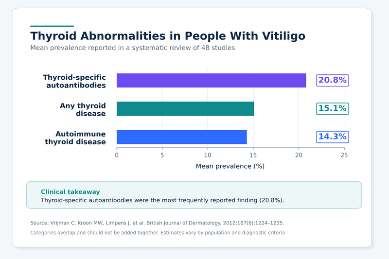
An earlier systematic review reported thyroid disease in approximately 15.1% of people with vitiligo, autoimmune thyroid disease in 14.3% and thyroid-specific antibodies in 20.8%. The antibody percentage was higher because thyroid autoimmunity may be detectable before thyroid hormone production becomes abnormal or before the patient develops recognisable symptoms.[5]
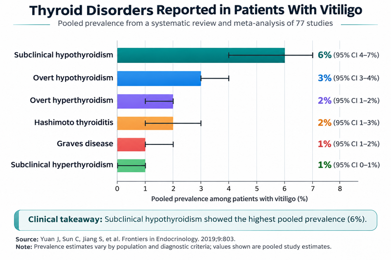
A separate meta-analysis examining specific thyroid disorders found that subclinical hypothyroidism was the most frequently reported functional abnormality, with a pooled prevalence of approximately 6%. Overt hypothyroidism occurred in approximately 3%, overt hyperthyroidism in approximately 2%, and Graves’ disease and subclinical hyperthyroidism in approximately 1% each. These figures were derived from studies conducted in different regions and should be interpreted within the patient’s clinical context.[4]
Hypothyroid disorders are reported more frequently than hyperthyroid disorders among people with vitiligo. This makes Hashimoto’s thyroiditis, elevated thyroid-stimulating hormone and subclinical hypothyroidism especially relevant during assessment. Graves’ disease remains clinically important because untreated hyperthyroidism may affect heart rhythm, bone health, muscle strength, fertility and eye health even though it is less common.[3,4]
The prevalence of thyroid disease also appears to rise with greater vitiligo extent. Adults with more extensive depigmentation have shown a higher frequency of hypothyroidism and other autoimmune conditions than those with limited disease. Extent alone is not sufficient to establish thyroid disease, but it can strengthen the reason for testing when accompanied by symptoms, family history or other autoimmune findings.[3]
Thyroid Antibodies With Normal Hormone Levels
Some patients with vitiligo have positive anti-thyroid peroxidase or anti-thyroglobulin antibodies while their thyroid-stimulating hormone and free thyroxine remain within the laboratory reference range. This pattern is often described as euthyroid thyroid autoimmunity because evidence of an immune reaction is present without current biochemical thyroid failure.[5,6]
A positive antibody result does not mean that hypothyroidism is already causing the patient’s fatigue, weight change, hair loss or white patches. Symptoms must be assessed alongside thyroid-stimulating hormone, free thyroxine, medical history, medication use and other possible causes. Iron deficiency, vitamin B12 deficiency, sleep disorders, depression, menopause and chronic inflammatory disease can produce symptoms that overlap with thyroid dysfunction.
Thyroid antibodies may precede functional disease, particularly when anti-thyroid peroxidase levels are positive. The future risk is not identical for every patient. The probability of progression may be influenced by the thyroid-stimulating hormone level, antibody pattern, age, sex, pregnancy status, family history, thyroid examination and the presence of another autoimmune disease.[6–8]
Antibody positivity alone does not automatically require levothyroxine. Thyroid hormone treatment is generally guided by thyroid function, symptoms, clinical circumstances and established endocrine recommendations rather than by the antibody value in isolation. Pregnancy, pregnancy planning, childhood, markedly abnormal thyroid-stimulating hormone levels and evolving symptoms may require a different threshold for intervention or closer monitoring.[7,8]
Repeated measurement of antibody concentrations is not always necessary after thyroid autoimmunity has been established. Changes in anti-thyroid peroxidase or anti-thyroglobulin levels do not consistently correspond with changes in thyroid function, vitiligo activity or repigmentation. Monitoring usually places greater emphasis on thyroid-stimulating hormone, free thyroxine, symptoms and relevant clinical circumstances.[7,8]
A patient with positive antibodies and normal thyroid function may remain euthyroid for years, develop subclinical hypothyroidism or progress to overt hypothyroidism. Follow-up should therefore be individualised rather than based on the assumption that every antibody-positive patient will experience thyroid failure.
Thyroid Abnormalities in Children
Thyroid abnormalities can occur in children with vitiligo, particularly in those with nonsegmental disease, post-pubertal onset, additional autoimmune conditions or a family history of thyroid disease. The overall likelihood is generally lower in younger children than in adults, but clinically important thyroid disease should not be overlooked simply because the child has no obvious neck swelling.[2,6]
Possible hypothyroid features in children include slowed height gain, delayed puberty, persistent constipation, unusual cold intolerance, reduced energy, dry skin, declining school performance or unexplained weight gain. Hyperthyroidism may present with poor weight gain, a rapid pulse, tremor, heat intolerance, anxiety, increased bowel frequency, sleep disturbance or deterioration in concentration.
These symptoms are not specific to thyroid disease. Growth patterns, nutritional status, sleep, emotional health, school stress, puberty and other medical conditions must be considered before attributing them to thyroid dysfunction. Laboratory results should also be interpreted using age-appropriate reference intervals rather than adult ranges.
Children with segmental, limited and stable vitiligo who have no symptoms, family history, examination abnormalities or other autoimmune conditions may have a lower diagnostic yield from repeated broad laboratory testing. In contrast, nonsegmental vitiligo accompanied by growth disturbance, pubertal delay, neck enlargement, abnormal pulse, extensive depigmentation or another autoimmune disorder provides a stronger clinical reason for thyroid evaluation.[2,6]
When thyroid antibodies are positive but thyroid function remains normal, follow-up should be coordinated with the child’s paediatrician or paediatric endocrinologist. Monitoring should consider growth velocity, pubertal progression, symptoms and thyroid function rather than focusing only on antibody values.
Patients With Greater Thyroid Risk
Thyroid disease is not distributed evenly across all people with vitiligo. A higher frequency has been reported among females, adults, post-pubertal patients and people with nonsegmental rather than segmental vitiligo. These characteristics increase clinical suspicion but do not confirm thyroid dysfunction without appropriate assessment.[3,5,6]
Acral involvement, particularly vitiligo affecting the hands and wrists, has been associated with thyroid abnormalities in several studies. Extensive or actively progressive disease may also reflect a greater autoimmune burden. Patients who are developing new patches, experiencing rapid enlargement of existing lesions or showing involvement across several body regions may therefore merit closer assessment when additional thyroid risk factors are present.[3,6]
Longer vitiligo duration has been associated with increased thyroid antibody positivity in some populations. This may partly reflect the additional time available for another autoimmune condition to become clinically apparent. A normal test performed near the onset of vitiligo does not guarantee that thyroid dysfunction will never develop, especially when symptoms or risk factors emerge later.[5,6]
Personal and family autoimmune history materially alters risk assessment. A patient with vitiligo who also has alopecia areata, type 1 diabetes, pernicious anaemia, coeliac disease, rheumatoid arthritis or another autoimmune condition may have a lower threshold for thyroid evaluation. A first-degree relative with Hashimoto’s thyroiditis, Graves’ disease or another autoimmune disorder also supports closer clinical attention.[1,2,6]
Symptoms remain important regardless of vitiligo type or extent. Persistent fatigue, unexplained weight change, palpitations, tremor, heat or cold intolerance, constipation, menstrual disturbance, neck swelling, eye symptoms, diffuse hair thinning or muscle weakness can justify thyroid testing even in a patient with limited vitiligo.
Female sex, nonsegmental disease, acral distribution, extensive involvement and family history are risk associations rather than diagnostic criteria. Some high-risk patients have normal thyroid results, while thyroid disease can still occur in patients without these characteristics. Clinical examination and appropriately selected laboratory testing remain necessary to distinguish increased probability from an established thyroid diagnosis.[3,6]
Thyroid Disorders Associated With Vitiligo
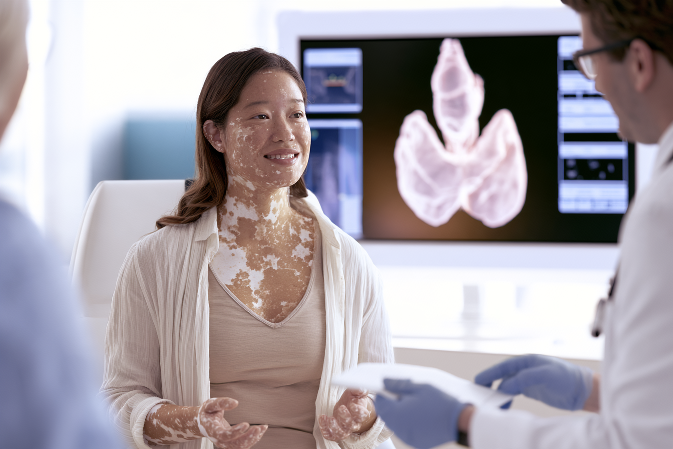
Vitiligo is most strongly associated with autoimmune thyroid disorders rather than with every form of thyroid dysfunction. Hashimoto’s thyroiditis, hypothyroidism, Graves’ disease and subclinical thyroid abnormalities are the principal conditions encountered during clinical assessment. Their symptoms and laboratory patterns differ, so a diagnosis should be based on thyroid function tests, antibody findings, physical examination and the patient’s medical history rather than on vitiligo alone.[3–8]
| Thyroid condition | Typical clinical features | Common TSH pattern | Free T4/T3 pattern | Relevant antibodies | Clinical significance |
|---|---|---|---|---|---|
| Hashimoto’s thyroiditis with normal thyroid function | May be asymptomatic; possible goitre or family history of thyroid disease | Normal | Normal | Anti-TPO commonly positive; anti-Tg may also be positive | Thyroid autoimmunity is present without current hormone deficiency; periodic functional monitoring may be appropriate.[5–8] |
| Overt primary hypothyroidism | Fatigue, cold intolerance, constipation, dry skin, menstrual disturbance, diffuse hair loss, modest weight gain | High | Free T4 low | Anti-TPO often positive when autoimmune | Requires assessment for thyroid hormone replacement and continued biochemical monitoring.[7,8] |
| Subclinical hypothyroidism | Frequently asymptomatic; symptoms may overlap with overt hypothyroidism | High | Free T4 normal | Anti-TPO may be positive | Treatment depends on degree and persistence of TSH elevation, symptoms, age, pregnancy and other clinical factors.[7,8] |
| Graves’ disease | Palpitations, tremor, heat intolerance, sweating, weight loss, anxiety, muscle weakness; eye disease may occur | Low or suppressed | Free T4 and/or T3 elevated | TRAb or TSI may be positive | Requires determination of the severity of hyperthyroidism and selection of antithyroid treatment, radioactive iodine or surgery where appropriate.[7,8] |
| Overt hyperthyroidism from another cause | Similar systemic symptoms to Graves’ disease | Low or suppressed | Free T4 and/or T3 elevated | Depends on cause | Thyroiditis, autonomous nodules, excess thyroid hormone and iodine-related causes must be distinguished from Graves’ disease.[7,8] |
| Subclinical hyperthyroidism | Often asymptomatic; possible palpitations, tremor or heat intolerance | Low or suppressed | Free T4 and T3 normal | Depends on cause | Persistent suppression may be clinically important in older patients and those with cardiovascular or bone risk.[7,8] |
| Euthyroid thyroid autoimmunity | Usually no thyroid-specific symptoms | Normal | Normal | Anti-TPO and/or anti-Tg positive | Indicates autoimmune susceptibility but does not establish hypothyroidism or justify thyroid hormone treatment by itself.[5,7,8] |
| Structural thyroid enlargement or nodule | Neck swelling, asymmetry, pressure symptoms or incidental finding | May be normal, high or low | Depends on function | Variable | Requires structural assessment when clinically indicated; normal thyroid blood tests do not exclude a significant nodule.[7] |
Hashimoto’s thyroiditis, hypothyroidism and subclinical hypothyroidism are particularly relevant because hypothyroid abnormalities are reported more frequently than hyperthyroid disorders among patients with vitiligo. A large systematic review and meta-analysis found subclinical hypothyroidism to be the most frequently reported thyroid dysfunction, with a pooled prevalence of approximately 6%.[4]
Hashimoto’s Thyroiditis and Hypothyroidism
Hashimoto’s thyroiditis is the thyroid disorder most frequently reported in people with vitiligo. The immune response may gradually reduce the gland’s ability to produce thyroid hormones, although thyroid function can remain normal during the early stages. Anti-thyroid peroxidase antibodies are commonly detected, while anti-thyroglobulin antibodies may provide additional evidence of thyroid autoimmunity in selected patients.[3–8]
A patient may therefore have Hashimoto’s thyroiditis before overt hypothyroidism develops. During this phase, thyroid-stimulating hormone and free thyroxine may remain within the reference range even though thyroid antibodies are positive. The presence of antibodies indicates autoimmune susceptibility but does not establish that current fatigue, weight gain, hair loss or vitiligo progression is being caused by thyroid hormone deficiency.[7,8]
When thyroid hormone production declines, thyroid-stimulating hormone commonly rises as the pituitary gland attempts to stimulate the thyroid. Overt primary hypothyroidism is usually characterised by an elevated thyroid-stimulating hormone level accompanied by a reduced free thyroxine level. Patients may experience cold intolerance, constipation, dry skin, slower thinking, menstrual disturbance, muscle weakness, diffuse hair loss or unexplained weight gain, but symptoms can be mild or absent.[7,8]
A goitre may occur, particularly earlier in the autoimmune process, although many patients with Hashimoto’s thyroiditis have no visible thyroid enlargement. A normal neck appearance therefore does not exclude the condition. Conversely, an enlarged thyroid does not confirm Hashimoto’s thyroiditis because nodular disease, iodine-related enlargement and other thyroid disorders can produce similar findings.
Levothyroxine is used when thyroid hormone replacement is clinically indicated. Its dose is adjusted according to thyroid function, age, body weight, pregnancy status, cardiovascular risk and other clinical factors. The antibody concentration alone is not used to determine the replacement dose.[7,8]
Correcting hypothyroidism can improve symptoms caused by thyroid hormone deficiency, including fatigue, constipation, dry skin and menstrual disturbance. It should not be presented as a direct treatment for melanocyte loss, and established vitiligo may remain unchanged even after thyroid-stimulating hormone levels return to the target range.
Graves’ Disease and Hyperthyroidism
Graves’ disease is an autoimmune disorder in which antibodies stimulate the thyroid-stimulating hormone receptor. This stimulation can cause excessive production of thyroid hormones and suppression of thyroid-stimulating hormone. Free thyroxine, triiodothyronine or both may become elevated.[7,8]
Patients may report palpitations, tremor, heat intolerance, excessive sweating, anxiety, reduced exercise tolerance, muscle weakness, increased bowel frequency or unexplained weight loss. Older adults may have less typical symptoms and can present with fatigue, reduced appetite, atrial fibrillation or worsening cardiovascular disease rather than obvious tremor or hyperactivity.
Thyroid eye disease can occur with Graves’ disease and may cause gritty eyes, eyelid retraction, light sensitivity, double vision or prominence of the eyes. New visual disturbance, eye pain, reduced colour perception or difficulty closing the eyelids requires prompt medical assessment because eye involvement does not always parallel the severity of the thyroid hormone abnormality.
Thyroid-stimulating hormone receptor antibodies or thyroid-stimulating immunoglobulins can support the diagnosis when Graves’ disease is suspected. These tests are particularly useful when the biochemical pattern confirms hyperthyroidism but the clinical cause remains uncertain.[7,8]
Treatment may include antithyroid medication, radioactive iodine or thyroid surgery, depending on disease severity, patient age, pregnancy plans, thyroid size, eye involvement, relapse risk and personal preference. Vitiligo does not determine which Graves’ disease treatment is appropriate.
Untreated hyperthyroidism can affect heart rhythm, bone density, muscle strength and reproductive health. A patient with vitiligo and persistent palpitations, tremor or unexplained weight loss should therefore not attribute these symptoms solely to stress or dietary changes.
Subclinical Hypothyroidism
Subclinical hypothyroidism is generally identified when thyroid-stimulating hormone is above the laboratory reference range while free thyroxine remains normal. It is commonly detected during screening because many patients have no symptoms or report complaints that are not specific to thyroid disease.[4,7,8]
A single mildly elevated thyroid-stimulating hormone result does not always confirm persistent thyroid dysfunction. Temporary changes can occur after illness, medication changes, laboratory variation or recovery from another medical condition. Repeat testing may therefore be required before long-term treatment is considered, particularly when the elevation is modest and the patient is clinically stable.[7,8]
Anti-thyroid peroxidase positivity increases the likelihood that the abnormality reflects autoimmune thyroiditis and may indicate a greater risk of progression to overt hypothyroidism. The probability of progression also depends on the degree of thyroid-stimulating hormone elevation, age, sex, family history, pregnancy status and the presence of thyroid enlargement.
Not every person with subclinical hypothyroidism requires immediate levothyroxine. Treatment decisions may consider persistent thyroid-stimulating hormone elevation, symptoms, antibody status, age, cardiovascular factors, pregnancy, fertility planning and whether the value continues to rise on repeat testing.[7,8]
Vitiligo alone should not be used as the sole reason to begin thyroid hormone therapy when free thyroxine remains normal. The decision should be based on the complete endocrine assessment.
Subclinical Hyperthyroidism
Subclinical hyperthyroidism is characterised by a low or suppressed thyroid-stimulating hormone level while free thyroxine and triiodothyronine remain within their reference ranges. The abnormality may be temporary or persistent and can result from early Graves’ disease, autonomous thyroid nodules, thyroid inflammation or excessive thyroid hormone replacement.[7,8]
Patients may have no symptoms, although some experience palpitations, tremor, anxiety, reduced exercise tolerance or heat intolerance. Persistent subclinical hyperthyroidism can be clinically important in older adults and in people with atrial fibrillation, osteoporosis or cardiovascular disease.
A suppressed thyroid-stimulating hormone result should be confirmed and interpreted alongside free thyroxine, triiodothyronine, medication history and clinical examination. Biotin supplements can interfere with some thyroid laboratory assays and may produce misleading results, so supplement use should be disclosed before testing.
Management depends on the degree and duration of thyroid-stimulating hormone suppression, the underlying cause, patient age, cardiac risk and bone health. Vitiligo does not alter the need to identify the specific cause of the abnormal thyroid pattern.
Thyroid Antibodies With Normal TSH
Some patients have positive anti-thyroid peroxidase or anti-thyroglobulin antibodies while thyroid-stimulating hormone and free thyroxine remain normal. This pattern indicates thyroid autoimmunity without current biochemical hypothyroidism.[5–8]
These patients do not automatically require levothyroxine because thyroid hormone production remains adequate. Follow-up is based on symptoms, thyroid-stimulating hormone trends, pregnancy status, family history and the presence of other autoimmune conditions.
Anti-thyroid peroxidase antibodies are more commonly used than anti-thyroglobulin antibodies when autoimmune thyroiditis is suspected. Anti-thyroglobulin testing may be helpful in selected cases, but ordering both tests repeatedly does not necessarily improve clinical decision-making.[7,8]
Antibody concentrations may fluctuate and do not consistently correspond with thyroid function or vitiligo activity. A fall in antibody levels does not establish that thyroid autoimmunity has resolved, and a rise does not prove that vitiligo will spread.
Patients with positive antibodies should be advised to report new symptoms such as persistent fatigue, constipation, cold intolerance, menstrual changes, neck swelling or unexplained weight change. Repeat thyroid function testing may be required when symptoms develop or when clinical circumstances change.
Ayurvedic assessment should distinguish between structural thyroid enlargement, altered thyroid hormone production, autoimmune antibody positivity and vitiligo activity. These findings should not be merged into one classical diagnosis or treated with a uniform formulation. Thyroid medication, laboratory monitoring and dermatological treatment should continue according to the patient’s documented clinical condition.
Vitiligo and Thyroid Disease: When to Test
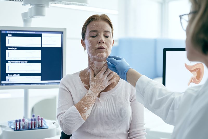
Vitiligo and thyroid disease can produce overlapping complaints, including fatigue, dry skin, hair changes, menstrual disturbance and reduced concentration. Thyroid testing becomes particularly important when these symptoms are persistent, progressive or accompanied by abnormal weight change, altered heart rate, temperature intolerance, neck swelling or a personal or family history of autoimmunity. No single symptom confirms thyroid dysfunction, and biochemical testing is required before symptoms are attributed to an underactive or overactive thyroid.[2,6–8]
Symptoms of an Underactive Thyroid
Hypothyroidism develops when thyroid hormone production becomes insufficient for the body’s needs. Symptoms frequently emerge gradually, and patients may initially interpret them as consequences of ageing, stress, poor sleep, restrictive dieting or emotional distress related to visible vitiligo.
Persistent fatigue is one of the most frequently reported symptoms, but it has limited diagnostic value when it occurs alone. Thyroid evaluation becomes more relevant when fatigue is accompanied by cold intolerance, constipation, unexplained weight gain, reduced exercise capacity, muscle weakness, slowed thinking or increasing sleepiness.[7,8]
Cold intolerance may be clinically significant when a patient feels unusually cold in environments that are comfortable for others or requires substantially more clothing than before. This symptom should be distinguished from temporary sensitivity caused by low body weight, anaemia, inadequate calorie intake or circulatory problems.
Weight gain associated with hypothyroidism is usually modest and may partly reflect fluid retention. Rapid or substantial weight gain should not automatically be attributed to the thyroid because medication effects, reduced physical activity, menopause, metabolic disease and dietary changes may also contribute. Thyroid-stimulating hormone and free thyroxine are necessary to determine whether thyroid hormone deficiency is present.[7,8]
Constipation may become more persistent as intestinal movement slows. Patients should report a sustained change from their usual bowel pattern, particularly when it occurs with fatigue, cold intolerance or dry skin. Constipation accompanied by severe abdominal pain, vomiting, bleeding or marked abdominal swelling requires separate medical assessment.
Dry, coarse or cool skin can occur with hypothyroidism, but dryness of normally pigmented or depigmented skin does not establish a thyroid diagnosis. Patients with vitiligo may also experience dryness from climate, frequent washing, topical treatment or phototherapy. The distribution, onset and accompanying systemic symptoms help determine whether thyroid testing is appropriate.
Hair changes may include diffuse thinning, increased shedding, coarser texture or loss of the outer part of the eyebrows. Diffuse thyroid-related shedding differs from the sharply defined patches of alopecia areata that can also accompany vitiligo. Examination is required because iron deficiency, nutritional restriction, androgen-related hair loss, menopause, recent illness and medication use can produce similar patterns.
Reduced concentration, slower information processing, low mood and memory complaints may occur in hypothyroidism. These symptoms are nonspecific and can also result from depression, anxiety, sleep disorders, vitamin B12 deficiency, iron deficiency or the psychological burden of a visible skin condition. Thyroid testing can identify or exclude one clinically treatable contributor but should not replace a broader assessment when symptoms persist.
Menstrual cycles may become heavier, prolonged or irregular. Some patients experience difficulty conceiving, reduced libido or worsening premenstrual symptoms. Thyroid assessment is especially important when menstrual changes occur with infertility, recurrent pregnancy loss, galactorrhoea, marked fatigue or a family history of autoimmune thyroid disease.
Muscle cramps, stiffness, proximal weakness and slower recovery after exercise may accompany hypothyroidism. Proximal weakness can become noticeable when climbing stairs, rising from a low chair or lifting objects above shoulder level. New or progressive weakness requires evaluation because neurological, muscular and medication-related conditions may produce the same limitation.
In children and adolescents, hypothyroidism may present through slowing of height gain, delayed puberty, reduced school performance, constipation, excessive tiredness or unexplained weight gain. Height velocity may be more informative than weight alone. Thyroid function should be interpreted with age-appropriate reference ranges, and persistent growth or pubertal concerns warrant paediatric assessment.[6–8]
Symptoms of an Overactive Thyroid
Hyperthyroidism increases metabolic and cardiovascular activity. Symptoms may develop gradually or become noticeable over a shorter period, depending on the underlying cause and the degree of thyroid hormone excess.
Palpitations may be experienced as a racing, pounding, fluttering or irregular heartbeat. A persistently elevated resting pulse, new irregular rhythm, chest discomfort, breathlessness or fainting requires timely medical assessment. Older patients may develop atrial fibrillation without the tremor, anxiety or obvious hyperactivity commonly associated with Graves’ disease.[7,8]
Unexplained weight loss despite a normal or increased appetite may indicate excessive thyroid hormone activity. Some patients do not lose weight, particularly when appetite and calorie intake increase substantially. Weight change must therefore be interpreted alongside pulse, temperature tolerance, bowel pattern, muscle strength and thyroid function tests.
Heat intolerance and excessive sweating may cause discomfort in ordinary indoor temperatures, disturbed sleep or a new preference for cooler environments. Menopause, infection, medication effects and anxiety can produce similar symptoms, so heat intolerance alone cannot identify hyperthyroidism.
A fine tremor may be most visible when the hands are held out in front of the body. Patients may also notice difficulty writing, holding a cup, applying makeup or performing precise manual tasks. Caffeine, anxiety, inhaled bronchodilators and several medicines can also cause tremor and should be considered during evaluation.
Nervousness, irritability, restlessness and difficulty sleeping may occur with excess thyroid hormone. These symptoms can resemble an anxiety disorder, but an accompanying rapid pulse, tremor, weight loss, increased sweating or frequent bowel movements strengthens the reason for thyroid testing.
Hyperthyroidism may increase bowel frequency without producing true diarrhoea. Persistent diarrhoea, blood in the stool, abdominal pain or dehydration requires investigation for gastrointestinal causes rather than being assumed to result from the thyroid.
Muscle weakness commonly affects the shoulders, upper arms, hips and thighs. Patients may find it difficult to climb stairs, rise from a seated position or carry routine objects despite retaining normal sensation. Prolonged untreated hyperthyroidism can also contribute to loss of bone density and reduced physical endurance.[7,8]
Menstrual bleeding may become lighter, less frequent or irregular. Fertility may be affected, and uncontrolled hyperthyroidism during pregnancy can create risks for both mother and fetus. Women with vitiligo who are pregnant, planning pregnancy or receiving fertility treatment should report thyroid symptoms and any previous positive thyroid antibody results.
Graves’ disease may produce eye irritation, dryness, redness, light sensitivity, eyelid retraction, double vision or prominence of the eyes. Eye symptoms can occur even when thyroid hormone abnormalities are mild. New double vision, reduced visual clarity, impaired colour perception, eye pain or inability to close the eyelids requires prompt ophthalmic and endocrine assessment.[7,8]
A sudden combination of very rapid heart rate, high fever, severe agitation, confusion, vomiting or marked weakness can indicate a rare but life-threatening hyperthyroid emergency. Immediate medical care is required rather than routine outpatient testing.
Physical Signs Requiring Assessment
Visible or palpable swelling at the front of the neck may represent general thyroid enlargement, one or more nodules or inflammation. Many thyroid enlargements are benign, but examination is required to determine whether thyroid function tests, ultrasound or specialist referral are appropriate.[7]
A rapidly enlarging neck swelling, persistent hoarseness, difficulty swallowing, shortness of breath or a sensation of pressure in the neck requires timely investigation. These symptoms may indicate compression of surrounding structures and should not be managed solely through dietary changes, external applications or observation.[7]
A thyroid nodule does not necessarily alter thyroid hormone production. A patient can have a palpable nodule with normal thyroid-stimulating hormone and free thyroxine results. Structural assessment and functional testing answer different clinical questions, and normal blood results do not remove the need to evaluate a suspicious neck finding.
An unusually slow pulse may accompany hypothyroidism, while a persistently rapid or irregular pulse may accompany hyperthyroidism. Pulse findings should be interpreted in relation to age, fitness, cardiovascular medication, fever, anaemia, dehydration and anxiety. Chest pain, fainting, severe breathlessness or a sustained irregular rhythm requires urgent medical evaluation.
Changes in the eyes, including lid retraction, prominence, restricted eye movement or double vision, increase concern for Graves’ eye disease. Puffiness around the eyes can occur with hypothyroidism but is less specific and may also result from allergy, kidney disease, sleep disturbance or local inflammation.
Diffuse scalp hair thinning, eyebrow loss, brittle nails, unusually dry skin or increased sweating may support thyroid assessment when combined with systemic symptoms. These physical changes should not be attributed to the thyroid without laboratory confirmation because nutritional deficiencies, dermatological disease, medication use and hormonal changes can produce similar findings.
Children require assessment when vitiligo is accompanied by slowed linear growth, delayed or unexpectedly early puberty, persistent changes in school performance, a neck swelling or an abnormal pulse. Parents may notice reduced stamina or altered behaviour before the child describes temperature intolerance or palpitations.[6–8]
Older adults may not develop classical thyroid symptoms. Hypothyroidism may present with cognitive decline, weakness, constipation or reduced mobility, while hyperthyroidism may present with weight loss, atrial fibrillation, breathlessness or unexplained deterioration in general health. A lower threshold for testing may be appropriate when these changes are new and no clear cause has been identified.[7,8]
Clinical suspicion is also strengthened by type 1 diabetes, coeliac disease, pernicious anaemia, alopecia areata, rheumatoid arthritis or another autoimmune disorder. A first-degree relative with Hashimoto’s thyroiditis, Graves’ disease or another autoimmune condition further supports testing when compatible symptoms are present.[1,2,6,7]
Ayurvedic examination may identify changes in Agni, bowel function, sleep, strength and other individual features, but these observations cannot determine thyroid-stimulating hormone, free thyroxine or triiodothyronine levels. Persistent systemic symptoms or physical thyroid signs require laboratory and medical assessment before an Ayurvedic treatment plan is finalised.
Vitiligo and Thyroid Disease Testing
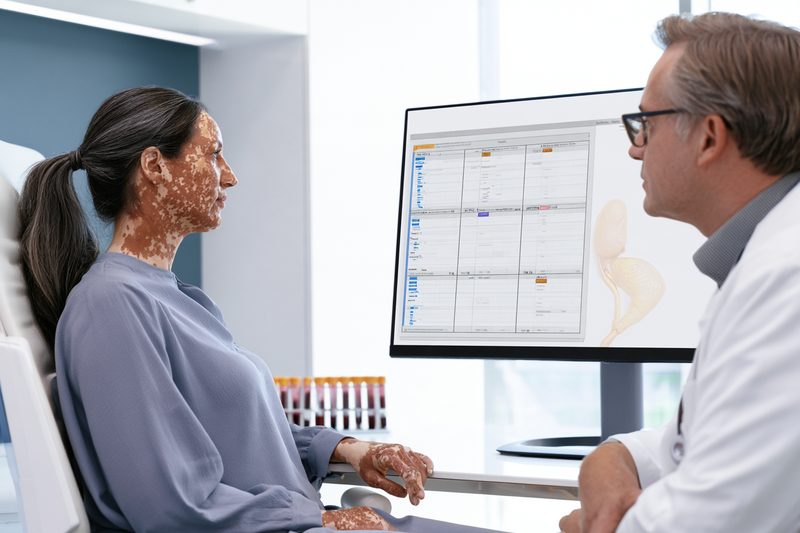
Testing for vitiligo and thyroid disease should distinguish thyroid function from thyroid autoimmunity. Thyroid-stimulating hormone, free thyroxine and triiodothyronine assess hormone regulation, while thyroid antibodies help identify an autoimmune cause. A positive antibody result does not necessarily mean that thyroid function is abnormal, and an abnormal hormone result does not by itself establish Hashimoto’s thyroiditis or Graves’ disease.[7,8]
| Test | What it assesses | When it is useful | Common interpretation | Important limitation |
|---|---|---|---|---|
| TSH | Pituitary regulation of the thyroid | Principal initial thyroid function test in most adults | High TSH suggests primary thyroid underactivity; low TSH may indicate thyroid overactivity | Can be misleading in pituitary disease, pregnancy, severe illness and assay interference.[7,8] |
| Free T4 | Available circulating thyroxine | Used with abnormal TSH and when pituitary disease is possible | High TSH + low free T4 supports overt primary hypothyroidism; low TSH + high free T4 supports hyperthyroidism | Must be interpreted with TSH and clinical context.[7,8] |
| T3 | Circulating triiodothyronine | Most useful when hyperthyroidism is suspected | T3 may rise while free T4 remains normal in some hyperthyroid patients | A normal T3 does not exclude hypothyroidism.[7,8] |
| Anti-TPO antibodies | Autoimmune response against thyroid peroxidase | Evaluation of autoimmune thyroiditis and thyroid autoimmune susceptibility | Positive result supports thyroid autoimmunity | Does not measure current thyroid hormone production; serial levels usually add little once autoimmunity is established.[7,8] |
| Anti-thyroglobulin antibodies | Autoimmune response against thyroglobulin | Selected cases of suspected autoimmune thyroid disease | May support autoimmune thyroid disease | Generally provides less routine diagnostic value than anti-TPO in suspected Hashimoto’s thyroiditis.[8] |
| TRAb/TSI | Antibodies directed at the TSH receptor | Suspected Graves’ disease | Positive result can support Graves’ disease | Not a routine vitiligo screening test; mainly used when hyperthyroidism or Graves’ disease is suspected.[7,8] |
| Thyroid ultrasound | Thyroid structure | Palpable nodule, enlargement, asymmetry or other structural concern | Shows nodules, cysts, enlargement and tissue characteristics | Does not determine whether a patient is hypothyroid or hyperthyroid.[7] |
| Reverse T3 | Inactive thyroid hormone metabolite | Generally not required in routine thyroid assessment | Not recommended as a routine test for hypothyroidism | Does not reliably establish thyroid dysfunction in otherwise stable patients.[8] |
For adults in whom pituitary disease is not suspected, thyroid-stimulating hormone can usually be measured first. If TSH is above the reference range, free thyroxine should be assessed; when TSH is below the reference range, free thyroxine and triiodothyronine help characterise the abnormality.[7,8]
Test selection depends on the patient’s age, symptoms, examination findings, vitiligo subtype, family history, pregnancy status, current medication and previous thyroid results. Adults without evidence of pituitary disease are commonly assessed initially with thyroid-stimulating hormone. Children, adolescents and adults with possible pituitary dysfunction generally require thyroid-stimulating hormone and free thyroxine together because thyroid-stimulating hormone alone may not identify central hypothyroidism.[7]
Thyroid-Stimulating Hormone
Thyroid-stimulating hormone is produced by the pituitary gland and regulates thyroid hormone production. It usually changes before circulating thyroid hormone levels move outside their reference ranges, making it the principal initial test for primary hypothyroidism and hyperthyroidism in most adults.[7,8]
A raised thyroid-stimulating hormone level commonly indicates that the pituitary gland is increasing stimulation because the thyroid is not producing sufficient hormone. The result must be interpreted with free thyroxine. A raised thyroid-stimulating hormone level with reduced free thyroxine supports overt primary hypothyroidism, whereas a raised thyroid-stimulating hormone level with normal free thyroxine is consistent with subclinical hypothyroidism.[7,8]
A low or suppressed thyroid-stimulating hormone level may occur when the thyroid produces excessive hormone. Free thyroxine and triiodothyronine are then required to determine whether overt hyperthyroidism or subclinical hyperthyroidism is present. A low thyroid-stimulating hormone level can also result from thyroid medication, pregnancy-related physiological changes, severe illness, pituitary disease or interference with the laboratory assay.[7,8]
A result near the edge of the reference range should not be interpreted in isolation. Laboratories may use different analytical methods and reference intervals, while age, pregnancy, acute illness and medication can influence the result. Comparison with the laboratory’s stated range and previous results is more informative than comparing values obtained from unrelated laboratories.
Temporary thyroid-stimulating hormone changes can occur during or after acute illness. Mild abnormalities may therefore require repeat testing after recovery when the patient is clinically stable. Repeating the test immediately may reproduce a transient result without clarifying whether persistent thyroid dysfunction exists. When symptoms worsen or new symptoms appear, repeat testing may be considered, but thyroid function tests are generally not repeated within six weeks unless a specific clinical reason requires earlier assessment.[7]
Thyroid-stimulating hormone alone may be misleading when pituitary or hypothalamic disease is suspected. A low or inappropriately normal thyroid-stimulating hormone level with reduced free thyroxine may indicate central hypothyroidism rather than an overactive thyroid. Previous pituitary surgery, head irradiation, pituitary tumour, unexplained visual-field symptoms, adrenal dysfunction or multiple pituitary hormone abnormalities strengthens the need for specialist assessment.
Biotin contained in hair, skin and nail supplements can interfere with some thyroid laboratory methods. This interference may produce falsely low thyroid-stimulating hormone and falsely elevated thyroid hormone results, creating an apparent biochemical pattern of hyperthyroidism. Patients should disclose all supplements before testing and follow the laboratory or clinician’s instructions regarding temporary discontinuation.[7,8]
Free Thyroxine
Thyroxine is the principal hormone released by the thyroid gland. Most circulating thyroxine is attached to transport proteins, while free thyroxine represents the unbound fraction available to enter tissues. Measuring the free fraction reduces the influence of changes in binding proteins and allows thyroid function to be interpreted more accurately with thyroid-stimulating hormone.[7,8]
A raised thyroid-stimulating hormone level with reduced free thyroxine supports primary hypothyroidism. In a patient with vitiligo, anti-thyroid peroxidase testing may then help determine whether autoimmune thyroiditis is the likely cause. Other causes, including thyroid surgery, radioactive iodine exposure, medication effects and iodine-related thyroid dysfunction, remain possible and must be considered from the clinical history.
A raised thyroid-stimulating hormone level with normal free thyroxine indicates a subclinical biochemical pattern. This does not automatically require thyroid hormone treatment. Persistence of the abnormality, degree of thyroid-stimulating hormone elevation, symptoms, antibody status, age, pregnancy, fertility plans and cardiovascular factors influence subsequent management.[7,8]
A suppressed thyroid-stimulating hormone level with elevated free thyroxine supports overt hyperthyroidism. Graves’ disease is one possible cause, but thyroid inflammation, autonomous nodules, excessive thyroid hormone replacement and iodine-related dysfunction can produce similar biochemical findings. Thyroid antibodies, examination and additional investigations may be required to identify the cause.
Low free thyroxine with a low or normal thyroid-stimulating hormone level requires careful interpretation. This combination may occur with pituitary disease, severe non-thyroidal illness, medication effects or laboratory interference. It should not be labelled primary hypothyroidism solely because free thyroxine is reduced.
Pregnancy alters thyroid physiology and the concentrations of hormone-binding proteins. Results should be interpreted using pregnancy-specific or trimester-appropriate reference ranges where available. Women with vitiligo who are pregnant or planning pregnancy require particular attention when thyroid-stimulating hormone is abnormal, thyroid antibodies are positive or there is a previous history of thyroid disease.
Oestrogen-containing medication, pregnancy and changes in binding proteins can alter total thyroxine without producing corresponding changes in free hormone activity. For this reason, total thyroxine and free thyroxine are not interchangeable in every clinical setting. The laboratory method and the patient’s hormonal status should be considered when results do not match the clinical presentation.
Triiodothyronine
Triiodothyronine is the more biologically active thyroid hormone. Most circulating triiodothyronine is produced by conversion of thyroxine in peripheral tissues rather than direct secretion from the thyroid gland. Testing is most useful when hyperthyroidism is suspected.[8]
When thyroid-stimulating hormone is suppressed, free thyroxine and either free or total triiodothyronine may be measured. Some patients with early Graves’ disease or another form of thyrotoxicosis have elevated triiodothyronine while free thyroxine remains within the reference range. This pattern is sometimes described as T3-predominant thyrotoxicosis and would be missed if assessment stopped after a normal free thyroxine result.[7,8]
Triiodothyronine testing has limited value for diagnosing hypothyroidism. The body may maintain triiodothyronine within the reference range even when thyroid-stimulating hormone is raised and free thyroxine is reduced. A normal triiodothyronine level therefore does not exclude clinically significant hypothyroidism.[8]
Free triiodothyronine assays may be less reliable than thyroid-stimulating hormone and free thyroxine in some laboratory settings. Total triiodothyronine can also be influenced by changes in binding proteins. The selected test should be interpreted according to the laboratory method and the clinical question rather than treated as an independent measure of total thyroid health.[8]
Triiodothyronine values may fall during severe illness, prolonged calorie restriction or systemic inflammation without indicating primary thyroid failure. This pattern should not lead to thyroid hormone treatment without assessment of thyroid-stimulating hormone, free thyroxine and the patient’s broader medical condition.
Thyroid Antibodies
Anti-thyroid peroxidase antibodies are the most frequently used antibodies when autoimmune thyroiditis is suspected. Thyroid peroxidase is involved in thyroid hormone synthesis, and antibodies directed against it are commonly found in Hashimoto’s thyroiditis. They may also occur in some people with Graves’ disease or in individuals whose thyroid function remains normal.[7,8]
Testing anti-thyroid peroxidase antibodies is particularly relevant when thyroid-stimulating hormone is above the reference range. A positive result supports an autoimmune cause and may indicate a greater likelihood of future progression from subclinical to overt hypothyroidism. The antibody result does not show how much hormone the gland is currently producing and cannot replace thyroid-stimulating hormone or free thyroxine measurement.[7,8]
Anti-thyroid peroxidase positivity does not automatically require levothyroxine. A patient may remain euthyroid for a prolonged period, and treatment decisions depend on thyroid function, symptoms and clinical circumstances. Repeatedly measuring the antibody concentration usually provides less useful information than monitoring thyroid-stimulating hormone and free thyroxine once autoimmune thyroiditis has been identified.[7,8]
Anti-thyroglobulin antibodies react against thyroglobulin, a protein produced within the thyroid. They may be present in autoimmune thyroid disease, but they generally provide less additional value than anti-thyroid peroxidase antibodies during routine assessment of suspected Hashimoto’s thyroiditis. They may be considered when autoimmune thyroid disease remains strongly suspected despite a negative anti-thyroid peroxidase result or when another thyroid-specific indication exists.[8]
Thyroglobulin antibodies can interfere with thyroglobulin measurement. This issue is particularly relevant in the surveillance of differentiated thyroid cancer, where thyroglobulin may be used as a tumour marker. It does not make thyroglobulin antibody testing a routine investigation for every patient with vitiligo.
Thyroid-stimulating hormone receptor antibodies are used when Graves’ disease is suspected. These may be reported as thyrotropin receptor antibodies or thyroid-stimulating immunoglobulins, depending on the assay. A positive result can support Graves’ disease in a patient with suppressed thyroid-stimulating hormone and elevated free thyroxine or triiodothyronine.[8]
Thyroid-stimulating hormone receptor antibodies can have additional value during pregnancy because they may cross the placenta. Women with current or previous Graves’ disease require specialist monitoring even when definitive thyroid treatment occurred before pregnancy. The timing and interpretation of antibody testing should be coordinated with endocrinology and obstetric care.[8]
Thyroid antibody concentrations should not be used as direct measures of vitiligo activity. A high anti-thyroid peroxidase level does not prove that white patches are spreading, while a falling antibody level does not demonstrate melanocyte recovery. Thyroid autoimmunity and cutaneous depigmentation require separate clinical endpoints.
Thyroid Ultrasound
Thyroid ultrasound evaluates the structure of the gland rather than its hormone-producing function. It can identify enlargement, asymmetry, nodules, cysts and changes in tissue appearance, but it cannot determine whether a patient is hypothyroid or hyperthyroid without corresponding blood tests.
Ultrasound is not required solely because a person has vitiligo, positive thyroid antibodies or a mildly abnormal thyroid-stimulating hormone result. It becomes more relevant when examination identifies a thyroid nodule, visible enlargement, asymmetry, cervical lymph-node abnormality or persistent neck swelling.
Difficulty swallowing, pressure in the neck, persistent voice change, shortness of breath or rapid enlargement of a thyroid mass also supports structural assessment. These symptoms require timely evaluation because normal thyroid hormone levels do not exclude a clinically significant thyroid nodule or compressive enlargement.
Ultrasound findings such as reduced echogenicity or heterogeneous texture may occur in autoimmune thyroiditis, but these changes are not interpreted independently of thyroid function, antibody results and clinical examination. An ultrasound appearance alone should not be used to diagnose the cause of fatigue, hair loss or vitiligo progression.
A thyroid nodule detected on ultrasound may require risk assessment according to its size and sonographic characteristics. Some nodules require only observation, while others may require fine-needle aspiration. The presence of vitiligo does not increase or reduce the importance of established nodule-assessment criteria.
Interpreting Thyroid Results
Thyroid results should be interpreted as patterns rather than isolated values. Raised thyroid-stimulating hormone with reduced free thyroxine supports overt primary hypothyroidism. Raised thyroid-stimulating hormone with normal free thyroxine supports subclinical hypothyroidism. Suppressed thyroid-stimulating hormone with raised free thyroxine or triiodothyronine supports overt hyperthyroidism, while suppressed thyroid-stimulating hormone with normal free hormone levels supports subclinical hyperthyroidism.[7,8]
A normal thyroid-stimulating hormone result usually makes clinically significant primary thyroid dysfunction less likely in a stable adult without pituitary disease. It does not exclude thyroid autoimmunity, structural thyroid disease or central hypothyroidism. Symptoms, examination and medical history determine whether additional testing is required.
Reference intervals vary between laboratories and may differ for children, older adults and pregnant patients. Small numerical differences between laboratories do not necessarily indicate genuine deterioration or improvement. Serial monitoring is easier to interpret when testing conditions and laboratory methods remain reasonably consistent.
Medication history can materially alter thyroid results. Levothyroxine, antithyroid medicines, amiodarone, lithium, glucocorticoids, dopamine-related medicines, immune therapies and iodine-containing products may affect thyroid function or its laboratory interpretation. The timing of levothyroxine in relation to blood collection can also influence free thyroxine levels.
Patients should not change levothyroxine or antithyroid medication because of a single result without clinical review. Dose adjustment depends on the complete biochemical pattern, treatment duration, symptoms, adherence, drug interactions, pregnancy status and the reason the medication was prescribed.
Tests Not Routinely Required
Reverse triiodothyronine does not help diagnose hypothyroidism in otherwise stable, non-hospitalised patients and is not a routine test for the thyroid association with vitiligo. Its concentration can change during illness and altered metabolism without providing a reliable basis for thyroid hormone treatment.[8]
Thyroglobulin is not a general thyroid function test. Its principal clinical use is in selected patients being monitored after treatment for differentiated thyroid cancer. A thyroglobulin value cannot establish whether vitiligo is autoimmune, whether Hashimoto’s thyroiditis is active or whether levothyroxine is required.[8]
Calcitonin testing is reserved for specific clinical situations involving concern for medullary thyroid carcinoma or selected thyroid nodules. It is not part of routine thyroid screening in people with vitiligo.
A full thyroid antibody panel is not required for every patient. Test selection should answer a defined clinical question. Anti-thyroid peroxidase antibodies are relevant when autoimmune hypothyroidism is suspected, while thyroid-stimulating hormone receptor antibodies are relevant when Graves’ disease is suspected. Repeated broad testing can produce incidental findings without changing management.[7,8]
Thyroid ultrasound, radionuclide uptake scanning and fine-needle aspiration are not substitutes for thyroid function tests. Uptake scanning may help identify the cause of thyrotoxicosis in selected patients, while fine-needle aspiration evaluates specific nodules. These investigations are chosen according to structural findings or an established biochemical abnormality rather than the presence of vitiligo alone.
Ayurvedic assessment may evaluate appetite, digestion, bowel function, sleep, temperature tolerance, strength and disease progression, but these observations cannot replace thyroid-stimulating hormone, free thyroxine, triiodothyronine or antibody testing. Formulation selection and any purification or Rasāyana plan should be based on the patient’s complete clinical status while prescribed thyroid treatment and endocrine monitoring continue.
Vitiligo and Thyroid Disease Screening
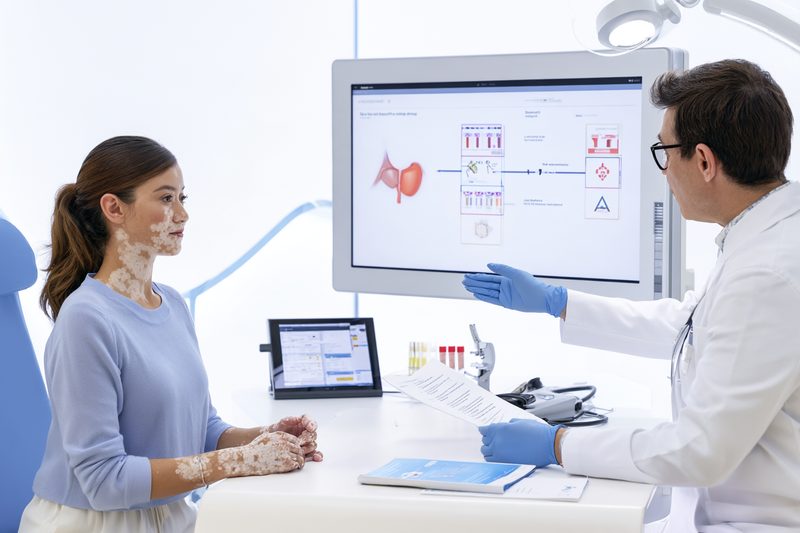
Vitiligo and thyroid disease screening is intended to identify clinically important thyroid dysfunction or autoimmune thyroid susceptibility before complications develop. The higher prevalence of thyroid abnormalities in people with vitiligo supports active clinical assessment, but screening recommendations are not completely uniform. Some international guidelines recommend routine thyroid function and antibody testing, while a risk-based approach may be appropriate in healthcare systems that reserve laboratory screening for patients with symptoms, examination findings or higher-risk vitiligo characteristics.[1,2,6]
Screening does not diagnose the cause of vitiligo, measure melanocyte loss or determine whether white patches are active. It answers a separate clinical question: whether the patient has normal thyroid function, subclinical dysfunction, overt thyroid disease or evidence of thyroid autoimmunity that may require monitoring.
Routine Screening Recommendations
The British Association of Dermatologists recommends routine screening of thyroid function and antithyroid antibodies in people with vitiligo, including children when testing is appropriate for their age. This recommendation reflects the increased prevalence of Hashimoto’s thyroiditis, Graves’ disease, subclinical hypothyroidism and thyroid-specific antibodies among patients with vitiligo.[1]
The International Vitiligo Task Force similarly recommends thyroid-stimulating hormone and anti-thyroid peroxidase antibodies as routine screening investigations after vitiligo has been diagnosed. Additional thyroid tests are selected according to the initial results, symptoms and examination findings rather than ordering every available thyroid investigation for every patient.[2]
Routine screening does not require repeated comprehensive thyroid panels at every dermatology visit. A practical baseline assessment may include thyroid-stimulating hormone and anti-thyroid peroxidase antibodies. Free thyroxine is added when thyroid-stimulating hormone is abnormal or when central thyroid dysfunction is possible. Triiodothyronine and thyroid-stimulating hormone receptor antibodies are generally reserved for patients with suppressed thyroid-stimulating hormone, symptoms of hyperthyroidism or clinical features suggesting Graves’ disease.[2,7,8]
The principal benefit of baseline screening is the detection of thyroid abnormalities that may not yet have produced obvious symptoms. Subclinical hypothyroidism may be present when thyroid-stimulating hormone is elevated but free thyroxine remains normal. Thyroid antibodies may also become positive before thyroid hormone production declines. Identifying these patterns allows the patient’s primary-care clinician or endocrinologist to determine whether observation, repeat testing or treatment is appropriate.[1,2]
A normal baseline thyroid-stimulating hormone result makes clinically important primary thyroid dysfunction less likely at that time, but it does not provide lifetime protection against thyroid disease. Autoimmune thyroid dysfunction can develop after vitiligo has been present for several years, particularly in patients with positive antibodies, nonsegmental disease, additional autoimmune conditions or a family history of thyroid disease.[5,6]
Screening should remain proportionate to the clinical question. Anti-thyroid peroxidase antibodies help identify autoimmune thyroid susceptibility, but repeating antibody concentrations frequently does not usually provide a reliable measure of thyroid function or vitiligo activity. Once thyroid autoimmunity has been identified, thyroid-stimulating hormone, free thyroxine, symptoms and clinical circumstances generally provide more useful follow-up information.[7,8]
Risk-Based Screening
A risk-based approach recognises that the probability of thyroid disease is not identical in every patient with vitiligo. Diagnostic yield is generally higher in patients with nonsegmental vitiligo, female patients, adults or post-pubertal adolescents, extensive or progressive depigmentation, acral involvement, longer disease duration, additional autoimmune disorders or a family history of thyroid disease.[3–6]
Symptoms or physical signs should lower the threshold for testing regardless of the size or distribution of the vitiligo patches. Persistent fatigue, unexplained weight change, heat or cold intolerance, constipation, palpitations, tremor, menstrual disturbance, muscle weakness, diffuse hair loss, neck swelling or eye symptoms justify thyroid assessment even when vitiligo is limited and stable.[6–8]
Nonsegmental vitiligo carries a stronger association with autoimmune thyroid disease than segmental vitiligo. Baseline thyroid assessment is therefore particularly relevant when depigmentation is bilateral, affects several body regions, continues to spread or is accompanied by another autoimmune condition. Segmental vitiligo does not exclude thyroid disease, but repeated laboratory testing may have a lower diagnostic yield in an otherwise healthy patient without symptoms or additional risk factors.[2,6]
Acral lesions affecting the hands, fingers or wrists have been associated with thyroid abnormalities in some patient populations. Acral involvement alone does not establish thyroid dysfunction, but it may support testing when combined with adult onset, female sex, progressive disease, thyroid symptoms or a positive family history.[6]
Patients with alopecia areata, type 1 diabetes, coeliac disease, pernicious anaemia, rheumatoid arthritis or another autoimmune disorder may warrant a lower threshold for thyroid evaluation. The coexistence of more than one autoimmune disease suggests broader immune susceptibility, although it does not determine which thyroid disorder, if any, is present.[1,2,6]
Family history should include Hashimoto’s thyroiditis, Graves’ disease, goitre, thyroid surgery and other autoimmune disorders in first-degree relatives. A family member’s diagnosis does not mean that the patient will develop the same disease, but it increases the value of investigating compatible symptoms and may influence the frequency of follow-up.
Screening is especially important during pregnancy planning and pregnancy because untreated thyroid dysfunction can affect maternal health, fertility and pregnancy outcomes. Women with current or previous Graves’ disease may require thyroid-stimulating hormone receptor antibody testing under specialist supervision, even when definitive thyroid treatment occurred before pregnancy.[7,8]
Children with vitiligo require age-appropriate assessment rather than the automatic use of an adult testing schedule. Nonsegmental vitiligo, post-pubertal onset, extensive disease, slowed growth, delayed puberty, unexplained weight change, constipation, abnormal pulse, neck swelling or a strong family history strengthens the indication for thyroid testing.[1,2,6]
Stable and limited vitiligo in an asymptomatic patient without family history, examination abnormalities or additional autoimmune disease represents a lower-risk clinical pattern. Some clinicians may use observation and symptom education rather than repeated laboratory screening in this group, particularly when previous thyroid results were normal. This approach should be reconciled with the guideline followed in the patient’s country and the availability of primary-care follow-up.[1,2,6]
Screening Pathway
Screening begins with confirmation that the white patches are consistent with vitiligo. Thyroid blood tests cannot distinguish vitiligo from tinea versicolor, pityriasis alba, post-inflammatory hypopigmentation, chemical leucoderma or other causes of reduced pigmentation. An uncertain skin diagnosis should be evaluated clinically, with Wood’s lamp examination or other investigations when indicated.[2]
The thyroid assessment begins with symptoms, medication history, supplement use, pregnancy status, personal autoimmune history and family history. Examination may identify thyroid enlargement, a nodule, abnormal pulse, tremor, eye signs, proximal muscle weakness or changes in weight and growth.
Thyroid-stimulating hormone is the most practical initial functional test for most adults without suspected pituitary disease. A result within the reference range generally makes significant primary hypothyroidism or hyperthyroidism less likely at the time of testing. Further investigation may still be required when pituitary disease, pregnancy-related thyroid dysfunction, structural thyroid disease or laboratory interference is suspected.[6–8]
When thyroid-stimulating hormone is elevated, free thyroxine helps distinguish overt hypothyroidism from subclinical hypothyroidism. Anti-thyroid peroxidase antibodies can help determine whether autoimmune thyroiditis is the probable cause and whether the patient may have a greater likelihood of future progression.[7,8]
When thyroid-stimulating hormone is low or suppressed, free thyroxine and triiodothyronine help determine whether overt or subclinical hyperthyroidism is present. Thyroid-stimulating hormone receptor antibodies may be required when Graves’ disease is suspected from the biochemical pattern, thyroid enlargement, eye findings or compatible symptoms.[7,8]
A positive anti-thyroid peroxidase result with normal thyroid-stimulating hormone and free thyroxine indicates thyroid autoimmunity without current biochemical thyroid failure. This pattern usually requires clinical monitoring rather than automatic thyroid hormone treatment. The follow-up interval depends on symptoms, thyroid-stimulating hormone trends, pregnancy status, age, family history and other autoimmune conditions.[7,8]
A palpable thyroid nodule, asymmetric enlargement, persistent hoarseness, swallowing difficulty or neck pressure requires structural assessment even when thyroid hormone levels are normal. Ultrasound is selected because of the neck finding, not because vitiligo itself requires routine thyroid imaging.
Broad autoimmune testing should not be ordered solely because a patient has vitiligo. Additional investigations are guided by the history, examination and laboratory findings. Gastrointestinal symptoms, unexplained anaemia, neurological changes, joint inflammation or endocrine symptoms may justify evaluation for another disorder, but indiscriminate antibody panels can produce incidental results without clarifying the patient’s diagnosis.[2]
Ayurvedic assessment may document Agni, meaning digestive and metabolic capacity, as well as appetite, bowel pattern, sleep, strength, temperature tolerance and disease progression. These findings can guide individualised Ayurvedic care but cannot determine whether thyroid function is normal. Thyroid-stimulating hormone, free thyroid hormones and appropriate antibody testing remain necessary when thyroid disease is suspected.
Repeat Thyroid Testing
There is no single repeat-testing interval suitable for every patient with vitiligo. Follow-up depends on the baseline results, age, symptoms, antibody status, pregnancy, family history, medication use and whether another autoimmune disorder is present.[6–8]
A patient with normal thyroid-stimulating hormone, negative thyroid antibodies, stable limited vitiligo and no symptoms may not require frequent repeat testing. Reassessment becomes appropriate when new symptoms, progressive vitiligo, thyroid enlargement, an abnormal pulse, pregnancy or another autoimmune diagnosis develops.
Positive anti-thyroid peroxidase antibodies with normal thyroid function justify periodic thyroid-stimulating hormone monitoring because autoimmune thyroiditis may remain stable or progress over time. Antibody concentrations do not need to be repeatedly measured merely to determine whether thyroid function has changed. Thyroid-stimulating hormone and free thyroxine provide more direct information about hormone production.[7,8]
A mildly abnormal thyroid-stimulating hormone result may require confirmation before a long-term diagnosis or treatment decision is made. Repeat testing should allow sufficient time for thyroid regulation to stabilise and is generally not performed sooner than six weeks unless symptoms, pregnancy or another clinical circumstance requires earlier assessment.[7]
Patients taking levothyroxine or antithyroid medication follow a treatment-monitoring schedule rather than a screening schedule. Their testing frequency depends on medication changes, treatment response, pregnancy status and the stability of previous results. Thyroid medication should not be stopped or adjusted solely because vitiligo improves, worsens or begins to repigment.
Children with positive thyroid antibodies, abnormal thyroid-stimulating hormone, growth changes or pubertal concerns require follow-up coordinated with paediatric care. Height velocity, weight pattern, pubertal progression and school functioning add clinical information that cannot be obtained from antibody concentrations alone.
A new pregnancy, fertility treatment, substantial weight change, initiation of medication that affects thyroid function or development of another autoimmune disease may require earlier reassessment. Biotin and other supplements should be disclosed because assay interference can create a false biochemical appearance of thyroid dysfunction.[7,8]
Repeat testing identifies changes in thyroid function but does not establish whether thyroid disease caused vitiligo progression. Thyroid outcomes should be monitored through hormone levels and systemic symptoms, while vitiligo outcomes should be monitored through disease stability, new-lesion formation, standardised photographs and measurable repigmentation.
Can Thyroid Disease Worsen Vitiligo?
Vitiligo and thyroid disease may appear during the same period because both can arise within a shared autoimmune background. This association does not establish that abnormal thyroid hormone levels directly destroy melanocytes or that every change in thyroid function will alter the course of vitiligo. Thyroid disease and vitiligo should therefore be assessed as related but clinically distinct conditions, each with its own symptoms, laboratory markers, complications and treatment goals.[1–6]
Does Hypothyroidism Cause White Patches?
Hypothyroidism does not usually produce the sharply defined, completely depigmented patches characteristic of vitiligo. Thyroid hormone deficiency can cause dry, coarse, pale or cool skin, but these changes differ from the loss of pigment caused by melanocyte destruction. Some patients with hypothyroidism may appear pale because of reduced skin blood flow, anaemia or fluid-related changes, yet this generalised pallor should not be mistaken for vitiligo.[7,8]
True vitiligo commonly presents as well-demarcated white macules or patches that may become more evident under Wood’s lamp examination. The affected skin usually retains its normal texture unless another dermatological condition is also present. Hair growing within a patch may remain pigmented or become white, depending on follicular melanocyte involvement.[1,2]
Hashimoto’s thyroiditis and vitiligo frequently coexist because the same patient may be susceptible to more than one autoimmune disease. This relationship explains why thyroid autoimmunity is detected more often in people with vitiligo, but it does not prove that hypothyroidism itself initiated the depigmentation.[3–6]
The timing of the two diagnoses can be misleading. Vitiligo may develop before thyroid-stimulating hormone becomes abnormal, while Hashimoto’s thyroiditis may be diagnosed years before the first white patch appears. The condition recognised first is not necessarily the cause of the condition identified later.
Patients who notice new white areas should not assume that changing their thyroid medication will restore pigment. The skin diagnosis should first be confirmed because tinea versicolor, pityriasis alba, post-inflammatory hypopigmentation, chemical leucoderma and other disorders can resemble early or limited vitiligo. Thyroid blood tests cannot determine whether a white patch is vitiligo.
A patient can also have abnormal thyroid function without autoimmune thyroid disease. Previous thyroid surgery, radioactive iodine treatment, medication effects, thyroid inflammation, pituitary disease and iodine-related dysfunction can alter hormone levels. These causes do not carry the same autoimmune relationship with vitiligo as Hashimoto’s thyroiditis or Graves’ disease.
Can Thyroid Disease Make Vitiligo Spread?
Active autoimmune thyroid disease and progressive vitiligo may occur in the same patient, but current evidence does not show that a particular thyroid-stimulating hormone or antibody value reliably predicts when vitiligo will spread. New white patches, enlargement of existing lesions and loss of pigment after skin injury are direct indicators of vitiligo activity, whereas thyroid-stimulating hormone, free thyroxine and triiodothyronine measure thyroid regulation.[1,2,7,8]
Patients with extensive or active nonsegmental vitiligo have a higher reported prevalence of autoimmune thyroid disease than patients with some limited or segmental patterns. This finding supports thyroid assessment in higher-risk patients but does not establish that thyroid dysfunction caused the greater skin involvement.[3–6]
Uncontrolled hypothyroidism may contribute to fatigue, low mood, constipation, reduced exercise capacity, menstrual disturbance and dry skin. These effects can increase the overall disease burden and make adherence to phototherapy, topical treatment, sleep routines or follow-up appointments more difficult. Correcting hypothyroidism may therefore improve the patient’s ability to participate in vitiligo treatment without directly producing repigmentation.
Uncontrolled hyperthyroidism may cause anxiety, insomnia, weight loss, palpitations, muscle weakness and heat intolerance. The physiological stress created by untreated Graves’ disease can affect general wellbeing, but a period of worsening vitiligo during hyperthyroidism does not prove a direct hormonal cause.
Vitiligo progression should be evaluated through clinical evidence rather than inferred from thyroid symptoms. Important signs include the appearance of new lesions, visible enlargement of established patches, development of lesions at sites of friction or injury, increasing body-surface involvement and changes documented through standardised photography.[1,2]
A stable thyroid result does not guarantee stable vitiligo. A patient may remain biochemically euthyroid while new patches continue to appear because cutaneous autoimmune activity follows a separate course. Conversely, a patient may develop worsening thyroid function while vitiligo remains unchanged for years.
The distinction is important when treatment results are assessed. If thyroid-stimulating hormone normalises but depigmentation continues, the thyroid treatment has not necessarily failed. It may have corrected thyroid hormone deficiency while the patient still requires active dermatological treatment for vitiligo.
Similarly, stabilisation of vitiligo does not confirm that thyroid autoimmunity has resolved. Thyroid monitoring must continue according to the patient’s hormone levels, antibody status, symptoms, pregnancy plans and endocrine treatment rather than according to skin appearance alone.[7,8]
Will Thyroid Treatment Repigment Vitiligo?
Levothyroxine replaces thyroid hormone in patients with hypothyroidism. Its purpose is to restore appropriate thyroid hormone availability, reduce symptoms of deficiency and prevent complications. It is not a melanocyte-directed treatment and should not be expected to repigment established vitiligo by itself.[7,8]
After appropriate thyroid replacement, patients may notice improvement in fatigue, constipation, cold intolerance, hair shedding, menstrual disturbance or general skin dryness. These improvements can be clinically valuable, but they should not be described as vitiligo repigmentation unless objective pigment return is visible within the depigmented lesions.
Repigmentation requires surviving or regenerating melanocyte sources and control of the cutaneous inflammatory process. Depending on disease activity, distribution and patient characteristics, management may involve topical corticosteroids, topical calcineurin inhibitors, narrowband ultraviolet B phototherapy, excimer treatment, topical Janus kinase inhibition where available, or other specialist-directed treatment.[1,2]
Vitiligo on the face and neck generally has greater repigmentation potential than lesions on the fingers, toes, palms, soles or areas where hairs within the patches have also become white. Normalisation of thyroid function does not remove these anatomical differences in treatment response.
Treatment of Graves’ disease also does not function as a stand-alone vitiligo therapy. Antithyroid medicines, radioactive iodine and thyroid surgery are selected to control excessive thyroid hormone production or remove the source of hyperthyroidism. Any subsequent change in vitiligo should be evaluated independently.
Some patients may experience simultaneous improvement in thyroid function and vitiligo because both conditions are being treated during the same period. Temporal improvement does not establish that thyroid treatment caused repigmentation, particularly when the patient is also receiving topical therapy, phototherapy, nutritional correction or another intervention.
Patients should not increase levothyroxine beyond the prescribed dose in an attempt to accelerate skin recovery. Excess thyroid hormone can cause palpitations, tremor, sleep disturbance, loss of bone density and abnormal heart rhythms. Thyroid hormone dosing is guided by endocrine requirements rather than the colour or extent of vitiligo patches.[7,8]
Prescribed thyroid medication should not be stopped when vitiligo stabilises or begins to repigment. A patient may continue to require levothyroxine even after substantial skin improvement because autoimmune thyroid damage and melanocyte recovery represent different biological processes.
Ayurvedic treatment should follow the same distinction. An individualised plan may address digestion, bowel function, sleep, strength, stress responses and the clinical pattern of Śvitra, but it should not replace required thyroid hormone or antithyroid treatment. Changes in prescribed thyroid medication require repeat laboratory assessment and supervision by the treating clinician.
Does Lowering Anti-TPO Cure Vitiligo?
Anti-thyroid peroxidase antibodies indicate an immune response against thyroid peroxidase, an enzyme involved in thyroid hormone production. They support the diagnosis of autoimmune thyroiditis but do not measure thyroid hormone availability, melanocyte number or the activity of vitiligo.[7,8]
A high anti-thyroid peroxidase value can occur in a patient whose thyroid-stimulating hormone and free thyroxine remain normal. The same patient may have stable, progressive or partially repigmenting vitiligo. The antibody value alone cannot distinguish between these skin outcomes.
Anti-thyroid peroxidase concentrations may rise or fall over time without a corresponding change in thyroid function. Laboratory methods and reference ranges may also differ. Repeated antibody testing therefore provides less information about current thyroid performance than thyroid-stimulating hormone and free thyroxine after autoimmune thyroiditis has already been established.[7,8]
Lowering an antibody concentration has not been validated as a treatment endpoint for vitiligo. A reduced anti-thyroid peroxidase result does not demonstrate that melanocytes have returned, that immune activity in the skin has stopped or that future lesions cannot develop.
The response of vitiligo should be measured through visible clinical outcomes. These include cessation of new-lesion formation, stabilisation of existing borders, reduction in affected surface area, perifollicular or marginal repigmentation, standardised photographs and validated severity assessment.[1,2]
Thyroid treatment outcomes should be measured separately through thyroid-stimulating hormone, free thyroid hormones, symptom control, medication safety and relevant examination findings. Patients with Graves’ disease may also require thyroid-stimulating hormone receptor antibody monitoring in specific circumstances, particularly during pregnancy, but this test does not measure vitiligo activity.[7,8]
Claims that a supplement, restrictive diet or herbal product has cured both diseases solely because anti-thyroid peroxidase levels decreased should be interpreted cautiously. Antibody fluctuation, laboratory variation, concurrent medication and natural disease variability must be considered before attributing the result to one intervention.
High-dose iodine should not be used to lower thyroid antibodies without medical supervision because excessive iodine can aggravate thyroid dysfunction in susceptible individuals. Selenium and other nutrients may be appropriate in selected patients with documented deficiency or a specific clinical indication, but they should not be used as universal antibody-lowering treatment for all patients with vitiligo.
Ayurvedic outcomes should also be documented through separate thyroid and skin measures. Improvement in Agni, sleep, bowel function, energy or tolerance of treatment may provide meaningful clinical value, but these changes do not replace thyroid laboratory monitoring or objective assessment of repigmentation.
A patient can achieve normal thyroid function while anti-thyroid peroxidase antibodies remain positive. Another patient may show lower antibody levels while thyroid-stimulating hormone becomes abnormal. Treatment decisions must therefore be based on the complete clinical and biochemical pattern rather than on a single antibody target.[7,8]
Diagnosing Vitiligo and Thyroid Disease
Vitiligo and thyroid disease require separate diagnostic confirmation even when they occur in the same patient. Thyroid blood tests cannot confirm that a white patch is vitiligo, and the appearance of depigmented skin cannot establish whether thyroid function is normal. Diagnosis should combine dermatological examination, assessment of disease activity, thyroid-focused history, physical examination and appropriately selected laboratory testing.[1,2,7,8]
Confirming Vitiligo
The skin assessment begins with the age at which the first patch appeared, its original location, the speed of progression and whether new patches have developed. The clinician should ask whether depigmentation followed friction, burns, repeated scratching, surgery, pressure from clothing, occupational chemical exposure or another inflammatory skin disorder. Development of vitiligo at sites of injury is known as the Koebner phenomenon and may indicate active disease.[1,2]
Distribution helps distinguish nonsegmental vitiligo from segmental and focal patterns. Nonsegmental vitiligo may affect both sides of the body and commonly involves the face, hands, feet, elbows, knees, body folds or areas around the eyes and mouth. Segmental vitiligo usually remains concentrated within a more restricted region, frequently on one side, although early disease cannot always be classified confidently from a single examination.[1,2]
The complete skin examination should assess visible lesions and less obvious areas rather than focusing only on the patch that prompted consultation. The scalp, eyebrows, eyelashes, beard area, hands, feet and skin around body openings may provide information about disease extent. Examination of intimate areas should occur only when clinically necessary, with consent and appropriate privacy.
The clinician should assess whether hairs within the depigmented area remain normally coloured or have become white. Loss of hair pigment can indicate involvement of follicular melanocytes and may reduce the likelihood of complete repigmentation in that site. It does not mean that treatment is impossible, but it helps establish realistic expectations.
Wood’s lamp examination can make depigmentation more distinct, particularly in lighter skin tones or when lesions are subtle. Under ultraviolet illumination, vitiligo usually appears more clearly demarcated because the affected area lacks normal epidermal melanin. Wood’s lamp examination can also reveal lesions that are not readily visible in ordinary lighting and can improve documentation of the true extent of disease.[1,2]
Wood’s lamp findings should be interpreted with the clinical appearance. Enhancement under the lamp supports pigment loss but does not independently distinguish every cause of hypopigmentation. Scale, inflammation, lesion distribution, medical history and additional testing may still be required when the diagnosis remains uncertain.
Disease activity should be recorded at the initial assessment because treatment decisions differ between stable and progressive vitiligo. New patches, enlargement of existing lesions, development of lesions after injury, trichrome areas with several shades of pigment or small confetti-like macules can indicate active disease. Stable vitiligo is characterised by the absence of meaningful progression over a defined period, although the duration required before surgical treatment is considered may vary.[1,2]
Standardised clinical photographs provide a more reliable baseline than memory. Images should be taken under similar lighting, distance, camera settings and body positioning during follow-up. Photographing the same area inconsistently can make unchanged disease appear better or worse.
Validated assessment tools may be used to document body-surface involvement, disease activity and treatment response. These measures are particularly useful when several body areas are affected or when phototherapy, systemic treatment or a prolonged course of care is planned. Patient-reported quality of life should also be assessed because limited facial or genital vitiligo may cause substantial distress despite involving a small surface area.[1,2]
Skin biopsy is not routinely required when the history, examination and Wood’s lamp findings are typical. It may be considered when lesions are inflamed, scaly, symptomatic, unusually distributed, treatment-resistant or difficult to distinguish from another pigmentary disorder. Histological findings must be interpreted with the clinical pattern because reduced melanocytes can occur in conditions other than vitiligo.
Excluding Other Causes of White Patches
Tinea versicolor can produce lighter or darker patches, commonly on the chest, back, neck or upper arms. The lesions may show fine surface scale that becomes more noticeable when the skin is gently scraped. Microscopic examination of skin scrapings or another fungal test may be used when the diagnosis is uncertain. Antifungal treatment can remove the infection before normal pigmentation fully returns, so persistent colour change immediately after treatment does not necessarily indicate continuing fungal disease.
Pityriasis alba commonly affects children and adolescents and often appears as poorly defined pale patches on the cheeks, upper arms or trunk. Mild dryness or fine scale may be present, and the colour difference can become more visible after surrounding skin darkens in sunlight. The borders are generally less sharply defined than those of established vitiligo.
Post-inflammatory hypopigmentation develops after eczema, psoriasis, injury, infection, burns or another inflammatory process. The patient may recall redness, itching, scaling or treatment at the same site before pigment became lighter. Pigment often returns gradually after inflammation is controlled, although recovery may take several months.
Chemical leucoderma should be considered when depigmentation develops at sites repeatedly exposed to adhesives, dyes, rubber products, disinfectants, cosmetics, hair products or occupational chemicals. Lesions may initially match the contact pattern before appearing at distant areas in susceptible patients. Identifying the exposure is important because continued contact may contribute to further depigmentation.[1,2]
Nevus depigmentosus usually appears during infancy or early childhood and remains relatively stable as the body grows. It is commonly limited to one region and may not show the same bright enhancement or progressive pattern expected in vitiligo. A stable childhood lesion should not be classified as active vitiligo solely because it remains lighter than surrounding skin.
Idiopathic guttate hypomelanosis produces multiple small, pale or white macules, most often on sun-exposed forearms and lower legs of adults. The lesions are usually only a few millimetres in diameter and do not generally merge into the larger symmetrical patches typical of extensive nonsegmental vitiligo.
Hypopigmented mycosis fungoides is an uncommon form of cutaneous lymphoma that may resemble eczema, pityriasis alba or vitiligo. Persistent patches with subtle scale, itching, altered sensation, skin thickening or an unusual distribution may require biopsy, particularly when lesions do not respond as expected to routine treatment.
Reduced sensation within a light patch, nerve-related symptoms or a history of residence in or travel to an endemic region may require assessment for infectious neuropathic causes of hypopigmentation. Sensory loss is not a typical feature of uncomplicated vitiligo and should not be ignored.
Halo naevi consist of a pigmented mole surrounded by a depigmented ring. They can occur in patients with or without vitiligo. The central mole should be examined for asymmetry, irregular borders, colour variation, change in size or other concerning features. A changing or clinically atypical central lesion may require dermoscopic assessment or biopsy.
Scarring, lichen sclerosus and other inflammatory disorders can also produce pale areas. Changes in skin texture, thinning, thickening, pain, fissuring, itching or loss of normal anatomical structures suggest that the condition is not pigment loss alone. Genital white patches with symptoms or structural change require examination rather than treatment based only on photographs.
Clinical photographs sent remotely can support triage but may not reliably show scale, texture, sensation, Wood’s lamp enhancement or the exact border of a lesion. An in-person dermatological examination is valuable when the diagnosis remains uncertain, lesions are changing rapidly or treatment has failed.
Evaluating the Thyroid
Thyroid assessment begins with symptoms, previous test results and the patient’s medical history. Relevant details include earlier thyroid disease, thyroid surgery, radioactive iodine treatment, neck irradiation, pregnancy, fertility treatment, pituitary disease and use of medicines or supplements that may affect thyroid function.[7,8]
Medication review should include levothyroxine, antithyroid drugs, lithium, amiodarone, glucocorticoids, iodine-containing products and high-dose biotin. The time at which thyroid medication was taken relative to blood collection may influence interpretation, particularly when free thyroxine appears unexpectedly high.
The physical examination may include pulse rate and rhythm, blood pressure, weight trend, tremor, proximal muscle strength, reflexes and inspection of the eyes. The neck should be examined for general thyroid enlargement, asymmetry, nodules, tenderness and enlarged lymph nodes. A normal neck examination does not exclude autoimmune thyroid disease, while a palpable nodule can be present despite normal thyroid hormone levels.
For most adults without suspected pituitary disease, thyroid-stimulating hormone is the principal initial functional test. An elevated result is interpreted with free thyroxine to distinguish overt from subclinical hypothyroidism. A low or suppressed thyroid-stimulating hormone result requires free thyroxine and triiodothyronine to determine whether overt or subclinical hyperthyroidism is present.[7,8]
Anti-thyroid peroxidase antibodies can support autoimmune thyroiditis when thyroid-stimulating hormone is elevated or when thyroid autoimmunity is being assessed according to the applicable vitiligo guideline. Positive antibodies with normal thyroid-stimulating hormone and free thyroxine indicate autoimmune susceptibility without current biochemical hypothyroidism.[1,2,7,8]
Thyroid-stimulating hormone receptor antibodies are considered when Graves’ disease is suspected because of suppressed thyroid-stimulating hormone, elevated thyroid hormones, diffuse thyroid enlargement or compatible eye findings. They are not routine tests for every patient with vitiligo.
Free thyroxine should be measured with thyroid-stimulating hormone when pituitary disease is possible. A low free thyroxine level with a thyroid-stimulating hormone result that is low or inappropriately normal can indicate central hypothyroidism and requires specialist assessment rather than routine primary thyroid treatment.[7,8]
Pregnant patients require interpretation using pregnancy-specific reference ranges where available. A result considered acceptable in a non-pregnant adult may require different assessment during pregnancy. Previous Graves’ disease, thyroid surgery or radioactive iodine treatment should be reported because thyroid-stimulating hormone receptor antibodies may remain clinically relevant even after thyroid function has been altered by treatment.[7,8]
Thyroid ultrasound is used to evaluate structure rather than hormone production. It is appropriate when examination identifies a nodule, asymmetry, enlargement or another structural concern. Positive thyroid antibodies or vitiligo alone do not make ultrasound necessary when the neck examination is normal.
Abnormal thyroid results should be interpreted in relation to illness, medication, pregnancy, laboratory method and previous values. Acute illness can temporarily alter thyroid tests, while biotin can create a false pattern resembling hyperthyroidism in some assays. Unexpected results may require repeat testing after the interfering factor has been addressed.[7,8]
Endocrinology referral is particularly important when overt thyroid dysfunction is present, results suggest pituitary disease, Graves’ eye disease is suspected, pregnancy complicates thyroid management or a structural thyroid abnormality requires further assessment. Rapid heart rate, chest pain, severe weakness, marked agitation, visual change or compressive neck symptoms require more urgent evaluation.
Assessing Associated Autoimmune Disease
Vitiligo and autoimmune thyroid disease can occur with other autoimmune conditions, but laboratory investigation should be guided by symptoms and examination rather than by ordering a broad panel for every patient. Indiscriminate screening can identify low-level antibodies that do not explain the patient’s symptoms or alter management.[1,2]
Frequent urination, excessive thirst, unexplained weight loss or recurrent infections may justify blood glucose or glycated haemoglobin testing for diabetes. Type 1 diabetes is more relevant when symptoms develop rapidly, the patient is younger or there is a strong personal or family autoimmune history.
Persistent fatigue, pallor, breathlessness, tongue soreness, numbness or balance disturbance may require a full blood count, iron studies, vitamin B12 and folate assessment. Pernicious anaemia can coexist with autoimmune thyroid disease and vitiligo, but fatigue should not be attributed to vitamin deficiency without appropriate testing.
Chronic diarrhoea, abdominal bloating, unexplained iron deficiency, weight loss, reduced growth in a child or a family history of coeliac disease may justify coeliac serology. Testing should usually occur while the patient is consuming gluten because removing gluten before investigation can reduce test sensitivity.
Patchy scalp or body-hair loss should prompt examination for alopecia areata rather than being assumed to result from thyroid disease. Alopecia areata typically produces smooth, well-defined areas of hair loss and can coexist with both vitiligo and autoimmune thyroid disease.
Joint swelling, prolonged morning stiffness, persistent dry eyes, dry mouth, recurrent mouth ulcers or unexplained inflammatory symptoms may require targeted rheumatological assessment. Generalised joint pain or fatigue alone is not sufficient to diagnose systemic autoimmune disease.
Unexplained weight loss, low blood pressure, persistent nausea, salt craving, marked weakness or darkening of normally pigmented skin can indicate adrenal dysfunction and requires timely medical investigation. Increased pigmentation from adrenal insufficiency should not be confused with repigmentation of vitiligo.
Psychological assessment is also clinically relevant. Visible depigmentation can affect self-esteem, relationships, employment, social participation and willingness to expose affected skin. Anxiety or depression may coexist with endocrine symptoms and can influence adherence to treatment. Mental health symptoms should be evaluated directly rather than attributed solely to appearance or thyroid function.[1,2]
Ayurvedic assessment should follow confirmation of the dermatological and endocrine diagnoses. The pattern of Śvitra may be documented alongside Agni, bowel function, sleep, strength, diet and individual doṣic features, but these observations should not be used as substitutes for Wood’s lamp examination, thyroid function tests or antibody interpretation. Thyroid enlargement, autoimmune thyroiditis and altered thyroid hormone production remain separate findings that require their own monitoring and treatment.
Vitiligo and Thyroid Disease Treatment

Vitiligo and thyroid disease treatment should be coordinated, but each condition requires its own therapeutic plan. Vitiligo treatment is selected according to disease activity, body area, extent, duration, patient age and repigmentation potential. Thyroid treatment is determined by thyroid-stimulating hormone, free thyroid hormone levels, antibody findings, symptoms, underlying cause, pregnancy status and risk of complications.[1,2,7,8]
Treating Each Condition According to Activity
Active vitiligo is characterised by new patches, enlargement of existing lesions, confetti-like depigmentation or development of lesions after friction or injury. The immediate treatment priority is to slow or arrest continuing pigment loss. Once the disease becomes stable, treatment can focus more strongly on repigmentation and, in selected cases, surgical restoration.[1,2]
Stable vitiligo does not require the same approach as rapidly progressive disease. A patient with one unchanged facial patch may be managed with local treatment, while a patient developing multiple new lesions over several weeks may require phototherapy or specialist-directed systemic treatment. Using the same treatment intensity for both patterns can expose one patient to unnecessary risk and provide insufficient control for another.[1,2]
Thyroid activity is assessed through a different set of findings. Overt hypothyroidism, overt hyperthyroidism, subclinical dysfunction and thyroid antibody positivity with normal hormone levels do not require identical management. A positive anti-thyroid peroxidase result alone does not automatically require thyroid hormone replacement, while a markedly abnormal thyroid-stimulating hormone result with altered free thyroid hormone levels may require active endocrine treatment.[7,8]
Treatment priorities can differ at the same consultation. A patient may have rapidly progressive vitiligo but stable thyroid function, requiring active dermatological treatment and routine thyroid monitoring. Another patient may have long-standing stable vitiligo but newly diagnosed severe hyperthyroidism, making control of the thyroid disorder the more urgent medical priority.
Baseline documentation helps prevent one condition from being used as an inaccurate measure of the other. Vitiligo should be monitored through new-lesion formation, lesion enlargement, standardised photographs and repigmentation. Thyroid disease should be monitored through symptoms, thyroid-stimulating hormone, free thyroid hormones, medication response and relevant examination findings.[1,2,7,8]
Treating Hypothyroidism
Levothyroxine is the standard first-line replacement treatment for primary hypothyroidism when thyroid hormone therapy is indicated. It provides synthetic thyroxine to replace the hormone that the thyroid gland can no longer produce adequately. The dose is individualised according to age, body weight, cardiovascular health, severity of hormone deficiency, pregnancy status and remaining thyroid function.[7]
The treatment goal is to restore an appropriate thyroid-stimulating hormone level, relieve symptoms of hormone deficiency and prevent complications. Dose requirements cannot be determined from the size of the vitiligo patches, the anti-thyroid peroxidase concentration or the amount of repigmentation achieved.
Thyroid-stimulating hormone is usually monitored after levothyroxine is started or its dose is changed. Sufficient time must be allowed for hormone regulation to stabilise before the result is interpreted. Once the dose is stable, testing can generally become less frequent, although pregnancy, major weight change, new medication or recurrent symptoms may require earlier reassessment.[7]
Free thyroxine may also be required when symptoms and thyroid-stimulating hormone do not correspond, when pituitary disease is possible, during selected stages of pregnancy or when adherence and absorption are uncertain. Patients with central hypothyroidism require specialist monitoring because thyroid-stimulating hormone may not provide a reliable treatment target.
Levothyroxine should be taken consistently because food, timing and several medicines can affect its absorption. Iron, calcium, some antacids and certain gastrointestinal medicines may reduce absorption when taken too close to the thyroid tablet. Patients should follow a consistent schedule and discuss all medicines, supplements and dietary products with the treating clinician before changing the timing or dose.[7]
A patient who continues to have fatigue, hair loss or weight concerns after thyroid-stimulating hormone has normalised may require assessment for other causes. Iron deficiency, vitamin B12 deficiency, sleep disorders, depression, menopause, medication effects and restrictive diets can produce symptoms similar to hypothyroidism.
Improvement in thyroid-related dry skin or diffuse hair shedding should not be recorded as repigmentation unless pigment is returning within the vitiligo lesions. Standardised photographs and direct examination are required to distinguish general improvement in skin quality from melanocyte recovery.
Subclinical hypothyroidism does not always require immediate levothyroxine. Management depends on whether thyroid-stimulating hormone remains elevated on repeat testing, the degree of elevation, symptoms, age, antibody status, cardiovascular risk, pregnancy and fertility plans. Mild temporary abnormalities may normalise without long-term treatment, while persistent or more pronounced abnormalities may justify therapy.[7]
Pregnant patients and those planning pregnancy require closer thyroid control because thyroid hormone needs and treatment targets can change. Levothyroxine requirements may increase during pregnancy, and monitoring should be coordinated with obstetric and endocrine care rather than postponed until routine postnatal follow-up.[7]
Children with hypothyroidism require paediatric assessment because thyroid hormone is important for growth, puberty and neurological development. Treatment and monitoring must consider age, weight, growth velocity and developmental stage rather than applying adult dosing or reference ranges.
When permanent thyroid hormone deficiency is present, Ayurvedic treatment, diet, supplements or improvement in vitiligo cannot replace the missing hormone. Ayurvedic care may be used alongside endocrine management when appropriate, but levothyroxine changes require laboratory reassessment and supervision.
Treating Graves’ Disease and Hyperthyroidism
Graves’ disease treatment aims to control excessive thyroid hormone production, reduce symptoms and prevent complications affecting the heart, bones, muscles, fertility and eyes. The principal treatment options are antithyroid medication, radioactive iodine and thyroid surgery. Selection depends on age, severity, thyroid size, eye involvement, pregnancy plans, previous relapse, other medical conditions and patient preference.[7]
Antithyroid medicines reduce the thyroid gland’s ability to produce new hormone. They may be used as the initial treatment, during preparation for definitive therapy or for a longer course in selected patients. Thyroid function requires regular monitoring because an insufficient dose may leave the patient hyperthyroid, while an excessive dose may produce hypothyroidism.[7]
Patients taking antithyroid medication should report fever, severe sore throat, mouth ulcers, jaundice, dark urine or unusual illness promptly. These symptoms can indicate rare but serious adverse effects affecting white blood cells or liver function and require urgent medical assessment rather than waiting for the next routine appointment.
Medicines that slow the heart rate may be prescribed temporarily to reduce palpitations, tremor and anxiety while thyroid hormone production is brought under control. These medicines improve symptoms but do not treat the autoimmune cause of Graves’ disease or restore pigment in vitiligo.
Radioactive iodine reduces excessive thyroid activity by damaging hormone-producing thyroid cells. It is unsuitable in pregnancy and may not be appropriate in patients with active thyroid eye disease. Many patients eventually develop hypothyroidism after treatment and require lifelong levothyroxine replacement.[7]
Thyroid surgery may be considered when there is a large goitre, compressive neck symptoms, suspicious nodules, significant eye disease, relapse after other treatment or a need for rapid definitive control. Surgery also results in the need for thyroid hormone replacement when the entire gland is removed.
Thyroid eye disease requires separate assessment because eye activity does not always correspond with thyroid hormone levels. Eye irritation, prominence, double vision, reduced colour perception, visual loss or difficulty closing the eyelids may require ophthalmological treatment in addition to endocrine control.
Smoking increases the risk and severity of thyroid eye disease. Smoking cessation is therefore particularly important for patients with Graves’ disease, especially before radioactive iodine treatment or when eye symptoms are already present.
Pregnancy changes the choice and timing of hyperthyroidism treatment. Radioactive iodine is contraindicated, medication selection may differ by trimester and thyroid-stimulating hormone receptor antibodies may require monitoring because they can affect the fetus. Treatment should be coordinated between endocrinology and obstetric care.[7]
Control of Graves’ disease may improve palpitations, tremor, heat intolerance, sleep and muscle weakness, but it does not replace vitiligo treatment. A patient may require simultaneous endocrine therapy and dermatological treatment even when both disorders developed during the same period.
Dermatological Treatment for Vitiligo
Topical corticosteroids are commonly used for limited vitiligo, particularly on areas away from the eyelids and other thin skin. Potency, application schedule and treatment duration depend on the body site, age and risk of adverse effects. Excessive or prolonged application can cause skin thinning, visible blood vessels, stretch marks or acne-like eruptions, making scheduled review important.[1,2]
Topical calcineurin inhibitors, including tacrolimus or pimecrolimus, may be considered for the face, neck, eyelids, flexures and other areas where prolonged corticosteroid exposure is less suitable. Temporary warmth, burning or irritation may occur during the early treatment period. These medicines do not cause the same skin-thinning effect associated with topical corticosteroids.[1,2]
A topical medicine should be applied to the depigmented area according to the prescribed schedule rather than spread indiscriminately over large areas of normal skin. Photographic review after an adequate treatment period helps determine whether the lesion is stable, repigmenting or continuing to expand.
Narrowband ultraviolet B phototherapy is used for more extensive, progressive or treatment-resistant vitiligo. Treatment is usually administered repeatedly over several months because melanocyte migration and pigment production occur gradually. Facial and trunk lesions generally respond better than patches on the fingers, toes, palms and soles.[1,2]
Phototherapy dosing must be supervised to limit excessive redness, burns and cumulative ultraviolet exposure. Missed sessions, changes in medication, recent sunburn and use of photosensitising products should be reported because they can alter treatment tolerance.
Targeted excimer light or laser treatment may be considered when vitiligo is limited to selected areas. It delivers ultraviolet treatment to the affected skin while reducing exposure of uninvolved areas. Its suitability depends on lesion number, location, accessibility and local availability.[2]
Ruxolitinib cream, a topical Janus kinase inhibitor, may be considered for eligible patients with nonsegmental vitiligo in countries where it is licensed and available. Age restrictions, treated body-surface area, treatment duration, medical history and local prescribing requirements must be reviewed before use. Repigmentation generally requires sustained treatment rather than brief application.[2]
Rapidly progressive nonsegmental vitiligo may require short-term systemic corticosteroid treatment under specialist supervision, often combined with narrowband ultraviolet B phototherapy. The purpose is to arrest disease activity rather than provide immediate complete repigmentation. Systemic corticosteroids are not suitable for unsupervised or prolonged use because they can affect blood glucose, blood pressure, mood, infection risk, bone health and the eyes.[1,2]
Known thyroid disease should be included in the risk assessment before systemic treatment. Active hyperthyroidism, cardiac rhythm abnormalities, osteoporosis risk, diabetes and other medical conditions may alter the safety of corticosteroids or influence the monitoring plan.
Surgical procedures, including grafting or melanocyte transplantation, are reserved for carefully selected patients with stable vitiligo that has not responded adequately to medical treatment. Surgery is generally unsuitable when new lesions are appearing or existing lesions are expanding because active disease can compromise the durability of the result.[1,2]
Surgical response also depends on the treated site, scar tendency, available donor skin and whether hairs within the patch remain pigmented. Acral areas and lesions with white hairs may have lower repigmentation potential than facial or trunk lesions with preserved follicular pigment.
Sun protection reduces burning of depigmented skin and limits contrast caused by tanning of surrounding skin. Camouflage products, self-tanning preparations and colour-correcting cosmetics can provide immediate visual improvement while medical treatment is taking effect. These measures do not interfere with treatment when products are selected and used appropriately.
Psychological care is clinically relevant when vitiligo affects relationships, work, social participation or mental health. Counselling, cognitive behavioural therapy and patient-support resources may be considered alongside medical treatment, particularly when distress is disproportionate to the measured body-surface area.[1,2]
Coordinated Specialist Care
A dermatologist should know the patient’s thyroid diagnosis, recent laboratory results and current endocrine medication before selecting systemic treatment or prolonged phototherapy. An endocrinologist should also know when a patient is receiving systemic corticosteroids, supplements or other medicines that may affect symptoms, metabolism or thyroid test interpretation.
Endocrinology review is appropriate when overt hypothyroidism or hyperthyroidism is identified, thyroid results are difficult to interpret, pituitary disease is suspected, pregnancy complicates management or a patient has Graves’ eye disease. A thyroid nodule, enlarging goitre or compressive neck symptoms may require structural evaluation and surgical assessment.[7,8]
Dermatology review is particularly important when the diagnosis of vitiligo is uncertain, patches are spreading rapidly, a large body area is involved, topical treatment has failed or systemic treatment, phototherapy or surgery is being considered.[1,2]
Primary-care follow-up can coordinate thyroid testing, cardiovascular risk, nutritional deficiencies, mental health and medication interactions. This is particularly valuable when the patient receives care from several practitioners or uses both conventional and Ayurvedic treatment.
All Ayurvedic medicines, powders, tablets, oils and supplements should be recorded in the shared medication history. Products containing iodine-rich ingredients or unverified mineral components may complicate thyroid management or create avoidable safety concerns. Selection should follow the confirmed thyroid diagnosis, current medication and relevant laboratory findings rather than the presence of vitiligo alone.
Follow-up should preserve separate treatment endpoints. Stable thyroid-stimulating hormone does not prove that vitiligo has stopped spreading, and visible repigmentation does not prove that thyroid hormone requirements have changed. Skin photographs, disease-activity assessment and thyroid laboratory results should be reviewed independently.[1,2,7,8]
Continuing Prescribed Thyroid Medication
Levothyroxine should not be stopped because vitiligo begins to repigment, anti-thyroid antibodies decrease or general wellbeing improves. Permanent thyroid hormone deficiency usually continues even when skin symptoms change, and withdrawal can allow hypothyroidism to recur.[7]
Antithyroid medication should not be stopped merely because palpitations or tremor have improved. Symptoms may settle before thyroid hormone levels normalise, and premature withdrawal can lead to recurrent hyperthyroidism.
A thyroid dose should not be increased in an attempt to accelerate metabolism, reduce weight or promote repigmentation. Excess levothyroxine can produce palpitations, abnormal heart rhythm, anxiety, sleep disturbance and loss of bone density.
Changes in thyroid medication require biochemical assessment and clinical review. Skin appearance, appetite, bowel pattern, energy level or an Ayurvedic assessment cannot independently determine the correct levothyroxine or antithyroid dose.
Dietary changes and supplements should be disclosed before repeat thyroid testing. High-dose biotin may interfere with laboratory assays, while iron, calcium and some gastrointestinal medicines may reduce levothyroxine absorption. Apparent deterioration in thyroid control may therefore result from timing, adherence or interference rather than true progression of thyroid disease.[7,8]
Treatment remains most reliable when the patient follows the prescribed thyroid regimen, documents vitiligo objectively and allows each condition to be adjusted according to its own clinical evidence.
Ayurvedic View of Vitiligo and Thyroid Disease

Vitiligo and thyroid disease require two confirmed diagnoses within an individualised Ayurvedic assessment. Classical descriptions can guide evaluation of the skin pattern, digestion, bowel function, strength, disease duration and treatment tolerance, but they do not replace Wood’s lamp examination, thyroid-stimulating hormone, free thyroid hormones, antibody testing or structural thyroid assessment. Hashimoto’s thyroiditis, Graves’ disease, subclinical thyroid dysfunction and thyroid enlargement cannot be treated as one Ayurvedic condition merely because they occur with vitiligo.[1,2,7–10]
Śvitra and Kilāsa in the Classical Texts
The principal classical discussion of depigmenting disease appears in the Charaka Samhita, Chikitsa Sthana, Chapter 7, verses 162–177. The text discusses Śvitra and Kilāsa within the broader therapeutic context of Kuṣṭha while describing them as clinically recognisable pigmentary disorders with their own treatment and prognostic considerations.[9]
Charaka differentiates clinical patterns according to lesion colour, apparent depth of involvement, chronicity, extent, coalescence of patches and changes in the hair growing within affected skin. More numerous or confluent lesions, long-standing disease and altered hair colour are described as less favourable features, while recent and more limited disease with preserved hair pigmentation is described as more amenable to treatment.[9]
These classical observations can support prognosis but should not be treated as exact equivalents of modern vitiligo classifications. Current evaluation also considers segmental or nonsegmental distribution, active or stable disease, affected body surface, anatomical site, Koebner phenomenon, follicular pigment, previous response and validated disease-activity measures.[1,2]
The classical text describes Śodhana, bloodletting, drying measures, internal preparations and external applications for selected presentations. Śodhana refers to physician-supervised purification procedures undertaken only after assessing strength, digestion, bowel tendency, age, comorbidities and suitability. Its presence in the classical chapter does not mean that every patient with vitiligo requires purgation, bloodletting or a standard Panchakarma programme.[9]
A patient with severe anaemia, uncontrolled thyroid disease, cardiovascular instability, pregnancy, low body weight, active infection, advanced age or reduced physiological reserve may be unsuitable for intensive purification. Even in an otherwise suitable patient, preparation, procedure selection, dose, hydration, post-procedure diet and monitoring require individual planning.
Charaka also records multiple external preparations for Śvitra and Kilāsa. Some contain Bakuchi, mineral substances or ingredients derived from historical pharmaceutical practices. These recipes should not be copied directly from translated verses or prepared at home because identity, purification, processing, concentration, skin tolerance and modern regulatory suitability must be established before clinical use.[9]
The text mentions Viruddhāhāra, meaning incompatible dietary combinations, among the classical etiological considerations. This concept can be explored through the patient’s digestive response, food tolerance and habitual diet, but it should not be used to impose extensive food restrictions without clinical justification. Autoimmune susceptibility, genetics and melanocyte-directed immune activity remain central to the modern understanding of vitiligo.[1,2,9]
Historical descriptions of conduct and past actions also occur within the classical chapter. These descriptions do not establish that a patient caused vitiligo through personal behaviour and should not be used to assign guilt. The patient’s current clinical findings, autoimmune risk, disease activity, exposures and treatment response provide the basis for contemporary assessment.
The classical Kuṣṭha framework also considers Rakta, Māṃsa and Meda involvement. These terms represent Ayurvedic tissue-level concepts and should not be translated directly as circulating blood tests, skeletal muscle pathology or measured body fat. They guide pattern recognition within Ayurveda but do not replace examination of melanocyte loss, thyroid hormone production or autoimmune antibodies.[9]
Galagaṇḍa Is Not Every Thyroid Disorder
The Sushruta Samhita, Nidana Sthana, Chapter 11 describes Galagaṇḍa within the diagnostic discussion of neck swellings, while Chikitsa Sthana, Chapter 18 discusses its management together with Granthi, Apacī and Arbuda. The classical description is centred on a gradually developing swelling in the neck with features interpreted according to the involved Doṣa and tissue factors.[10]
Galagaṇḍa can provide an Ayurvedic framework for selected patients with visible or palpable thyroid enlargement, but it is not an exact synonym for Hashimoto’s thyroiditis, Graves’ disease, subclinical hypothyroidism, subclinical hyperthyroidism or isolated thyroid antibody positivity. Many patients with autoimmune thyroid disease have no visible neck swelling, while many neck swellings arise from nodules, cysts, multinodular goitre, thyroiditis or non-thyroid structures.[7,8,10]
A patient with elevated thyroid-stimulating hormone and low free thyroxine may have overt hypothyroidism without Galagaṇḍa. A patient with positive anti-thyroid peroxidase antibodies may remain biochemically euthyroid and have no structural thyroid enlargement. A patient with Graves’ disease may have diffuse enlargement, eye disease or no obvious goitre. These presentations require distinct endocrine interpretation.[7,8]
Structural and functional thyroid assessments answer different questions. Neck examination and ultrasound evaluate enlargement, asymmetry and nodules, while thyroid-stimulating hormone, free thyroxine and triiodothyronine evaluate hormone regulation. Thyroid antibodies help identify an autoimmune mechanism. None of these findings can be inferred reliably from an Ayurvedic pulse assessment or neck appearance alone.[7,8]
Galagaṇḍa-based treatment should therefore not be applied automatically to every patient with an abnormal thyroid report. Management must first establish whether the patient has hypothyroidism, hyperthyroidism, euthyroid autoimmunity, a structural thyroid lesion, a previous thyroidectomy or medication-induced dysfunction.
A rapidly enlarging swelling, persistent hoarseness, swallowing difficulty, breathing difficulty, enlarged cervical lymph nodes or a firm fixed nodule requires prompt medical investigation. External applications or internal Ayurvedic medicines should not delay ultrasound, fine-needle aspiration or specialist referral when clinically indicated.
The absence of a neck swelling does not exclude thyroid disease in a patient with vitiligo. Persistent fatigue, constipation, temperature intolerance, menstrual disturbance, palpitations, tremor, unexplained weight change or abnormal growth in a child can justify thyroid testing even when the neck appears normal.[6–8]
Individual Ayurvedic Assessment
Ayurvedic assessment begins after the skin diagnosis and thyroid status have been established. The record should include the vitiligo subtype, age of onset, duration, rate of spread, affected sites, follicular hair colour, previous treatment, chemical exposure, friction-related lesions and response to sunlight or phototherapy.
The thyroid record should distinguish Hashimoto’s thyroiditis, Graves’ disease, overt dysfunction, subclinical dysfunction, euthyroid antibody positivity, structural enlargement and post-treatment hypothyroidism. Recent thyroid-stimulating hormone, free thyroxine, triiodothyronine when indicated, thyroid antibodies, ultrasound findings and prescribed medication should be reviewed before selecting Ayurvedic treatment.[7,8]
Prakṛti refers to the patient’s constitutional baseline, while Vikṛti refers to the current pattern of disturbance. Neither concept is a substitute for the modern diagnosis. Two patients with the same Prakṛti may have different thyroid disorders, different vitiligo activity and different treatment risks.
Assessment of Agni, appetite, post-meal discomfort, bloating, reflux, bowel frequency and stool consistency can identify digestive problems that may affect dietary tolerance and medicine adherence. These findings should not be used to claim that impaired digestion alone caused autoimmune melanocyte loss or thyroid autoimmunity.
Koṣṭha describes the patient’s bowel tendency and is relevant when oral medicines or purification procedures are considered. A patient with chronic diarrhoea, severe constipation, inflammatory bowel symptoms, previous gastrointestinal surgery or unexplained weight loss requires evaluation of the underlying disorder before treatment is intensified.
Bala represents the patient’s functional strength and capacity to tolerate treatment. It is assessed through age, nutritional status, exercise tolerance, sleep, recovery, comorbidities and previous reactions. A stronger procedure is not necessarily more effective when the patient’s physiological reserve is limited.
Dietary history should identify inadequate calorie intake, excessive fasting, iodine-rich supplements, high-dose biotin, restrictive elimination diets and products that interfere with levothyroxine absorption. The patient’s actual nutritional pattern is more clinically useful than assuming that every person with vitiligo must avoid the same foods.
Medication timing requires particular attention. Levothyroxine absorption can be reduced by iron, calcium and some gastrointestinal products. An Ayurvedic formulation containing mineral ingredients or substantial fibre may require a separate administration time, determined with the treating clinician rather than through unsupervised dose adjustment.[7,8]
Patients with hyperthyroidism require assessment of pulse, weight loss, tremor, sleep, muscle strength and cardiovascular symptoms before stimulating, heating or intensive procedures are considered. Persistent tachycardia, atrial fibrillation, chest discomfort or severe weakness requires endocrine stabilisation.
Pregnancy, pregnancy planning and fertility treatment alter the safety threshold for both thyroid and Ayurvedic treatment. Thyroid targets, medication requirements and permissible herbs or procedures may change, making coordinated obstetric and endocrine supervision necessary.[7,8]
Children require weight-based dosing, age-appropriate assessment and attention to growth, puberty and school functioning. Adult formulations, intensive purification and strong photosensitising applications should not be transferred to a child without paediatric and dermatological review.
Baseline photographs should be taken before Ayurvedic treatment begins. Thyroid laboratory results should be recorded separately. This prevents improvement in digestion, sleep or energy from being misclassified as repigmentation and prevents visible pigment return from being interpreted as normalisation of thyroid function.
Ayurvedic Treatment Principles
Nidāna Parivarjana means avoiding factors that aggravate the patient’s condition. In vitiligo, this may include repeated friction, uncontrolled sun exposure, chemical contact, skin injury and inconsistent treatment. In thyroid disease, it may include unmonitored iodine products, supplement interference, irregular medication timing and abrupt withdrawal of prescribed treatment.
Dietary correction should be based on nutritional adequacy, digestive tolerance, thyroid status, medication schedule and documented deficiency. Universal avoidance of dairy, gluten, citrus, white-coloured foods or multiple food groups is not justified solely by the coexistence of vitiligo and thyroid disease.
When appetite, digestion or bowel function is impaired, treatment may initially focus on restoring regular intake and tolerance before stronger medicines are introduced. A patient who is undernourished, dehydrated or losing weight should not undergo aggressive purification merely because the condition is chronic.
Śamana refers to individualised pacifying treatment using selected internal and external medicines. Selection depends on the skin pattern, thyroid diagnosis, disease activity, strength, digestive tolerance, medication use and comorbidities. The same Śamana prescription is not appropriate for active Graves’ disease, stable Hashimoto’s hypothyroidism and normal thyroid function with positive antibodies.
Śodhana may be considered only after confirming that the patient is suitable. The procedure, intensity and timing must reflect age, strength, bowel tendency, cardiovascular status, endocrine stability and previous treatment. It should not interrupt necessary thyroid medication or destabilise fluid, electrolyte or nutritional balance.
Raktamokṣaṇa, or controlled bloodletting, appears within classical skin-disease management but is not required for every case of Śvitra. Anaemia, anticoagulant use, bleeding disorders, low blood pressure, pregnancy and reduced physiological reserve are relevant contraindications or precautions. The procedure should never be performed as unsupervised home treatment.[9]
External treatment can be selected according to lesion site, skin thickness, sensitivity, hair involvement and exposure to ultraviolet light. Facial, eyelid, genital and flexural skin require more cautious concentrations than thicker areas. Acral lesions may respond differently from facial or trunk lesions and should not automatically receive more aggressive applications.
Rasāyana denotes a restorative phase intended to sustain strength and longer-term care after the patient’s digestion, treatment tolerance and active clinical problems have been assessed. It does not mean that one rejuvenative product can correct every autoimmune pathway, replace thyroid hormone or guarantee repigmentation.
Ayurvedic treatment can be coordinated with topical corticosteroids, topical calcineurin inhibitors, narrowband ultraviolet B, excimer treatment or other dermatologist-directed care. The timing of external products may need adjustment to reduce irritation or unpredictable photosensitivity.
Thyroid medication should continue according to the documented endocrine condition. Levothyroxine replacement is required when the gland cannot produce sufficient hormone, while Graves’ disease may require antithyroid medication or definitive treatment. Ayurvedic improvement in appetite, sleep or bowel function does not independently justify changing these medicines.[7,8]
Treatment response should be reviewed through separate endpoints. Vitiligo assessment includes new-lesion formation, border stability, affected surface area, hair pigmentation and objective repigmentation. Thyroid assessment includes symptoms, thyroid-stimulating hormone, free thyroid hormones, medication requirement and relevant cardiovascular or eye findings.[1,2,7,8]
Bakuchi and Photosensitising Treatment
Bakuchi, also called Avalguja in several classical passages, appears in external Śvitra formulations described in Charaka Samhita, Chikitsa Sthana, Chapter 7. Its presence in the text does not establish one concentration, vehicle, exposure time or ultraviolet dose as suitable for every patient.[9]
Bakuchi-containing preparations can increase the skin’s response to sunlight or therapeutic ultraviolet exposure. Excessive concentration, prolonged application or uncontrolled sun exposure may cause intense redness, burning, blistering, crusting and subsequent darkening or lightening of the skin.
Application should begin only after the skin type, lesion location, current phototherapy, previous photosensitivity and concurrent topical medicines have been reviewed. A small supervised test area may be required before a larger surface is treated.
Bakuchi should not be combined casually with narrowband ultraviolet B, excimer treatment, psoralen photochemotherapy or prolonged sunlight. The combined ultraviolet effect may be greater than expected, particularly when the concentration and absorption of a traditional preparation are not standardised.[1,2,9]
The face, eyelids, neck folds, genital skin and other sensitive areas require greater caution because a preparation tolerated on the trunk may cause severe irritation on thinner skin. Children, pregnant patients and people with a history of photodermatitis require additional safety assessment.
Patients should not increase the application frequency because pigment has not appeared within a few days. Repigmentation develops gradually, while phototoxic injury can occur rapidly. Strong inflammation may interrupt treatment and produce post-inflammatory colour changes that complicate assessment.
Severe burning, marked swelling, blistering, open skin or widespread redness requires discontinuation and clinical review. Additional sunlight should be avoided until the reaction has been assessed.
Some historical external formulations contain substances that require purification or are unsuitable for routine contemporary use. Substitution, omission or modernisation of an ingredient changes the original formulation and requires pharmaceutical and clinical justification rather than informal modification at home.[9]
Why One Formulation Is Unsuitable
A patient with vitiligo and treated Hashimoto’s hypothyroidism has different needs from a patient with uncontrolled Graves’ disease. A patient with normal thyroid function and positive anti-thyroid peroxidase antibodies also differs from a patient who is permanently hypothyroid after thyroid surgery. One formulation cannot address these distinct endocrine states safely.
Vitiligo itself varies by activity, distribution and prognosis. Rapidly progressive nonsegmental disease, stable focal disease, segmental vitiligo, widespread acral involvement and lesions with white hair do not have identical treatment priorities or repigmentation potential.[1,2]
Age, pregnancy, body weight, cardiovascular health, liver and kidney function, gastrointestinal tolerance, anaemia and concurrent medication influence dose and formulation selection. A prescription appropriate for a healthy adult may be unsuitable for a child, an older patient or someone with substantial comorbidity.
Internal treatment should account for whether the patient is taking levothyroxine, antithyroid medication, corticosteroids, immunomodulating therapy or nutritional supplements. Possible interference with absorption, laboratory testing and treatment tolerance must be reviewed before medicines are combined.
Mineral or herbo-mineral preparations require authenticated ingredients, appropriate purification and processing, batch quality control and exact dosing. They should not be selected merely because the disease is autoimmune or long-standing, and they should not be added to an existing prescription without reviewing renal, hepatic and haematological status.
External treatment must also be individualised. A highly photosensitising preparation may be inappropriate for a patient already receiving supervised phototherapy, while a strongly irritant application may worsen lesions through skin injury in a patient demonstrating the Koebner phenomenon.
A formulation should not be judged solely by changes in thyroid antibodies. Anti-thyroid peroxidase levels may fluctuate without corresponding improvement in thyroid function or vitiligo. The prescription should be reviewed against thyroid-stimulating hormone, free thyroid hormones, symptoms, lesion activity, objective photographs and treatment tolerance.[1,2,7,8]
Changes in the Ayurvedic prescription should be documented separately from changes in conventional treatment. When several treatments are started or adjusted simultaneously, attributing improvement or deterioration to one intervention becomes unreliable.
The treatment plan may require modification when vitiligo becomes stable, thyroid function changes, pregnancy begins, a new medicine is introduced or adverse skin sensitivity develops. Individualisation is therefore a continuing clinical process rather than a single constitution-based prescription.
Diet for Vitiligo and Thyroid Disease

Diet for vitiligo and thyroid disease should support adequate nutrition, stable energy, healthy body weight, medication absorption and correction of confirmed deficiencies. No single food plan has been shown to cure both conditions, reverse autoimmune susceptibility or reliably restore pigment. Patients should therefore avoid extreme elimination diets and use laboratory findings, digestive tolerance, thyroid status and current treatment to guide dietary changes.[1,2,7,8]
Is There One Diet for Both Conditions?
Vitiligo and autoimmune thyroid disease can coexist, but they do not require one universal therapeutic diet. Hashimoto’s thyroiditis, Graves’ disease, treated hypothyroidism, subclinical thyroid dysfunction and normal thyroid function with positive antibodies have different clinical needs. Vitiligo also varies according to activity, distribution, body area and treatment response.[1–4]
A balanced eating pattern should provide adequate protein, vegetables, fruits, whole grains or suitable alternatives, healthy fats and sufficient calories. The exact pattern may differ according to age, body weight, cultural preferences, metabolic health, gastrointestinal symptoms and other medical conditions.
Undernutrition can complicate treatment by contributing to fatigue, muscle loss, hair shedding, menstrual disturbance and poor recovery. A patient who has removed several food groups because of vitiligo may develop symptoms that resemble thyroid dysfunction even when thyroid hormone levels are stable.
Rapid weight-loss diets are particularly unsuitable for patients with uncontrolled hyperthyroidism, low body weight, reduced muscle strength or active gastrointestinal symptoms. A substantial calorie deficit can worsen weakness and nutrient deficiency while making it more difficult to distinguish disease symptoms from the effects of dietary restriction.
Patients with hypothyroidism may gain weight, but thyroid correction does not make extreme dieting necessary. Weight management should consider calorie intake, physical activity, sleep, menopause, insulin resistance, medication effects and fluid retention rather than assuming that all weight change is caused by the thyroid.
A patient with Graves’ disease may require increased nutritional attention during the hyperthyroid phase because excessive thyroid hormone can increase energy expenditure and contribute to muscle loss. Once thyroid function is controlled, calorie needs may fall, making reassessment of portion size and activity appropriate.
In Ayurveda, Āhāra refers to the patient’s overall food pattern. Dietary selection may consider appetite, bowel function, tolerance, strength and the clinical pattern of Śvitra, but these observations should not be used to impose the same restrictions on every patient. The classical discussion of dietary incompatibility does not establish that one food combination directly causes autoimmune melanocyte loss or autoimmune thyroid disease.[9]
Iodine and Thyroid Safety
Iodine is required for thyroid hormone production, but both inadequate and excessive intake can disturb thyroid function. Patients should aim for nutritionally adequate intake rather than using high-dose iodine as an immune treatment or thyroid stimulant.[7,8]
Iodised salt can provide a predictable dietary source when used in ordinary quantities. Patients who avoid all salt, follow highly restrictive diets or use only non-iodised specialty salts may have a lower iodine intake, particularly when dairy products, eggs or seafood are also excluded.
More iodine is not necessarily better. High-dose iodine drops, kelp tablets, seaweed concentrates and products marketed as thyroid cleanses may expose patients to unpredictable quantities. Excessive iodine can precipitate or aggravate thyroid dysfunction in susceptible individuals, including patients with autoimmune thyroid disease.[7,8]
The iodine content of seaweed varies widely between species, harvests and products. Eating an occasional ordinary food portion is different from taking concentrated seaweed capsules or consuming large amounts daily. Patients with Hashimoto’s thyroiditis, Graves’ disease or abnormal thyroid function should discuss concentrated iodine products with their treating clinician.
Iodine supplementation should not be started solely because a patient has vitiligo, positive anti-thyroid peroxidase antibodies or fatigue. These findings do not demonstrate iodine deficiency. Assessment should consider diet, pregnancy status, thyroid diagnosis, geographical iodine availability and current medication.
Pregnant and breastfeeding patients have specific iodine and thyroid requirements, but supplementation should be coordinated with obstetric or endocrine care. A product intended for the general adult population may not provide an appropriate dose during pregnancy.
Patients receiving radioactive iodine assessment or treatment may be instructed to follow a temporary low-iodine diet. This is a time-limited medical protocol and should not be continued indefinitely. A low-iodine diet is not a routine treatment for vitiligo or Hashimoto’s thyroiditis.
Ayurvedic products containing seaweed, iodine-rich marine ingredients or several mineral components should be included in the medication record. Their iodine content may not be apparent from the product name, and combining several preparations can produce a higher total exposure than expected.
Vitamins, Minerals and Supplements
Vitamin D, vitamin B12, folate, iron and zinc are relevant to skin, neurological, haematological and immune health, but routine high-dose supplementation is not justified solely by the coexistence of vitiligo and thyroid disease. Testing is more useful when symptoms, dietary restriction, malabsorption risk or a previous abnormal result suggests deficiency.[1,2]
Vitamin B12 assessment may be appropriate when a patient has anaemia, numbness, balance disturbance, tongue soreness, persistent fatigue or a history suggesting pernicious anaemia. Correcting deficiency can improve deficiency-related symptoms but should not be presented as a guaranteed method of repigmenting vitiligo.
Iron deficiency can cause fatigue, breathlessness, restless legs, reduced exercise tolerance, brittle nails and diffuse hair shedding. Menstrual blood loss, gastrointestinal bleeding, restrictive diets and impaired absorption are common considerations. Iron supplementation should be based on appropriate blood tests because unnecessary iron can cause gastrointestinal symptoms and obscure the real cause of fatigue.
Folate deficiency may occur with inadequate intake, malabsorption, alcohol excess or certain medicines. Folate should not be given in a way that masks an unrecognised vitamin B12 deficiency, particularly when neurological symptoms are present.
Vitamin D testing may be considered in patients with limited sun exposure, darker skin, malabsorption, osteoporosis risk or other clinical indications. Correction of deficiency supports bone and muscle health, but vitamin D should not be described as a stand-alone cure for vitiligo or autoimmune thyroid disease.
Zinc deficiency can affect wound healing, taste, appetite, skin and hair, but high-dose zinc taken without indication may interfere with copper balance. The presence of white patches does not prove zinc deficiency.
Selenium has a role in thyroid hormone metabolism, but supplementation should not be prescribed universally to lower thyroid antibodies. The potential benefit, baseline dietary intake, total dose and risk of excessive exposure should be considered. High-dose or prolonged unsupervised use can produce adverse effects rather than additional thyroid protection.
Antioxidant supplements should not replace established vitiligo treatment. Oxidative stress is one component of vitiligo biology, but taking several high-dose antioxidants does not guarantee that immune activity will stop or that melanocytes will return.[1,2]
Biotin is frequently included in hair and nail supplements and may interfere with some thyroid laboratory assays. It can create results that appear consistent with hyperthyroidism even when thyroid function is not genuinely abnormal. Patients should disclose biotin use and follow laboratory instructions before thyroid testing.[7,8]
Patients taking several supplements should bring the complete product list, ingredient labels and doses to clinical review. Duplicate ingredients may be present in multivitamins, thyroid products, hair supplements and Ayurvedic formulations, increasing the risk of excessive intake.
Herbal or nutritional products should not be judged solely by a change in anti-thyroid peroxidase levels. Antibody concentrations can fluctuate, while thyroid-stimulating hormone, free thyroid hormones, symptoms and objective skin findings provide more clinically useful information.[7,8]
Food and Levothyroxine Absorption
Levothyroxine works most reliably when taken consistently. Patients should follow the prescribed schedule rather than changing between fasting, mealtime and bedtime administration from one day to another.[7,8]
A common schedule is to take levothyroxine with water on an empty stomach before breakfast. Bedtime administration may also be suitable when it occurs consistently several hours after the last meal. The same routine should be maintained before thyroid blood tests unless the treating clinician provides different instructions.
Iron and calcium supplements can reduce levothyroxine absorption when taken too close to the thyroid tablet. They are usually separated by several hours according to the prescriber’s instructions. Multivitamins, antacids and some gastrointestinal products may contain iron, calcium or other binding ingredients even when this is not obvious from the product name.[7,8]
Coffee, soy products and very high-fibre meals can also influence absorption in some patients when consumed close to levothyroxine. These foods do not always need to be eliminated. Consistent timing and appropriate separation are generally more useful than unnecessary lifelong avoidance.
A patient who changes from a low-fibre diet to a very high-fibre diet may require reassessment if thyroid-stimulating hormone changes unexpectedly. The same applies after starting calcium, iron, antacids or certain weight-loss products.
Levothyroxine should not be taken together with a large number of herbal powders or mineral preparations unless their effect on absorption has been reviewed. Separating administration times may reduce interaction, but the plan should be coordinated with the clinician managing thyroid replacement.
Persistent abnormal thyroid-stimulating hormone despite an apparently adequate dose may result from missed doses, inconsistent timing, impaired absorption, gastrointestinal disease or interactions. Increasing the levothyroxine dose without reviewing these factors can lead to overtreatment when the interfering factor is later removed.
Patients should not stop eating nutritious foods solely because they appear on internet lists of thyroid-interfering foods. Ordinary portions of cruciferous vegetables, soy or high-fibre foods can usually be incorporated into a balanced diet. Medication consistency and adequate iodine intake are more important than labelling common foods as universally harmful.
Common Food Myths
White-coloured foods do not cause white skin patches. Rice, milk, yoghurt, cauliflower, garlic and other pale foods do not remove melanocytes because of their colour. Eliminating them solely on this basis can reduce dietary variety without treating vitiligo.
Citrus fruits are not universally prohibited in vitiligo. A patient with reflux, oral allergy, gastrointestinal intolerance or another individual reason may need to limit a particular fruit, but citrus does not need to be removed from every vitiligo diet.
Dairy products are not a recognised universal cause of vitiligo or autoimmune thyroid disease. Patients with lactose intolerance, milk-protein allergy or another diagnosed condition may need alternatives, but unnecessary dairy avoidance can reduce protein, calcium, iodine and vitamin intake.
Gluten restriction is appropriate for confirmed coeliac disease and may be required during a supervised diagnostic or therapeutic plan. Removing gluten before coeliac testing can reduce test sensitivity. Vitiligo or positive thyroid antibodies alone do not establish that every patient requires a gluten-free diet.
A patient with chronic diarrhoea, abdominal bloating, unexplained iron deficiency, weight loss, reduced growth or a family history of coeliac disease may need testing before making a long-term dietary change. A gluten-free diet should not be used as a substitute for investigation of persistent gastrointestinal symptoms.
Nightshade vegetables, tomatoes, aubergine and peppers are not established universal triggers of vitiligo. Individual intolerance can occur, but broad exclusion should be based on a reproducible symptom pattern rather than a general autoimmune list.
Avoiding all fermented foods is also unnecessary for most patients. Yoghurt, kefir, fermented vegetables or other foods may be included when tolerated and appropriate for the patient’s nutritional and medical needs.
Food-combining rules should not lead to inadequate protein or calorie intake. A patient who develops weakness, weight loss, menstrual disturbance or worsening fatigue after starting a restrictive plan requires nutritional reassessment.
No food can be expected to normalise thyroid-stimulating hormone when the thyroid gland is unable to produce adequate hormone. Levothyroxine remains necessary when permanent hormone deficiency is present.[7,8]
Similarly, no food can replace antithyroid medication or definitive treatment in uncontrolled Graves’ disease. Palpitations, tremor, weight loss and eye symptoms require medical management rather than dietary treatment alone.
Lifestyle Measures
Regular physical activity supports cardiovascular health, muscle strength, bone health, sleep and weight management. The intensity should reflect thyroid control, cardiovascular status, age and current fitness.
Patients with uncontrolled hyperthyroidism, marked muscle weakness, severe palpitations or abnormal heart rhythm may need to limit strenuous exercise until the condition is stabilised. Exercise should be resumed progressively after medical review.
Patients with treated hypothyroidism can generally participate in regular aerobic and resistance exercise once thyroid status and other medical conditions are stable. Persistent exercise intolerance despite normal thyroid results may require assessment for anaemia, cardiac disease, respiratory disease, sleep disorders or deconditioning.
Adequate sleep supports concentration, appetite regulation and adherence to treatment. Hyperthyroidism can cause insomnia, while hypothyroidism may produce excessive tiredness. Sleep disturbance that persists after thyroid correction may require a separate sleep or mental health assessment.
Psychological stress does not provide a complete explanation for vitiligo or autoimmune thyroid disease. It can, however, worsen sleep, anxiety, self-consciousness and treatment adherence. Stress-management strategies may include counselling, structured relaxation, mindfulness, regular activity or cognitive behavioural therapy when clinically appropriate.[1,2]
Smoking cessation is particularly important in Graves’ disease because smoking is associated with a greater risk and severity of thyroid eye disease. Patients with eye symptoms should avoid both active smoking and regular exposure to tobacco smoke.[7,8]
Depigmented skin is more susceptible to sunburn because it lacks normal melanin protection. Broad-spectrum sunscreen, protective clothing and avoidance of uncontrolled intense exposure can reduce burning and minimise contrast caused by tanning of surrounding skin.[1,2]
Controlled sunlight exposure and medical phototherapy are not interchangeable. Narrowband ultraviolet B and excimer treatment use planned doses, schedules and monitoring. Prolonged sun exposure combined with Bakuchi or another photosensitising preparation can cause severe phototoxic reactions.
Repeated friction, tight clothing, burns, scratching and skin injury may contribute to new lesions in patients with active Koebner phenomenon. Gentle skin care, appropriate moisturisation and protection from occupational chemical exposure can reduce avoidable skin trauma.[1,2]
Alcohol intake should be considered in relation to liver health, medication use, sleep, nutrition and cardiovascular symptoms. Alcohol does not serve as a treatment for either disease and may complicate adherence or aggravate palpitations and sleep disturbance.
Follow-up should use separate measures for each condition. Diet-related improvement in energy or bowel function does not prove that vitiligo is repigmenting, while visible pigment return does not show that thyroid hormone levels are controlled. Skin photographs, disease-activity assessment, thyroid-stimulating hormone, free thyroid hormones and medication review remain necessary.[1,2,7,8]
Monitoring Vitiligo and Thyroid Disease
Monitoring vitiligo and thyroid disease requires two parallel records because improvement in one condition does not reliably indicate improvement in the other. Skin monitoring should document disease activity, affected surface area, repigmentation, treatment tolerance and quality of life. Thyroid monitoring should document hormone levels, symptoms, medication response and condition-specific complications. Keeping these outcomes separate prevents normal thyroid results from being mistaken for vitiligo control and prevents visible repigmentation from being mistaken for recovery of thyroid function.[1,2,7,8]
Monitoring the Skin
A baseline skin assessment should record the vitiligo subtype, body areas involved, approximate extent, activity during the preceding months and whether hairs within the patches remain pigmented. Particular attention should be given to the face, hands, feet, body folds, genital area and sites repeatedly exposed to friction because anatomical location influences both treatment selection and repigmentation potential.[1,2]
Standardised digital photography provides one of the most practical methods of monitoring vitiligo. Photographs should be taken before treatment and repeated at regular intervals, commonly every three to six months. More frequent images may be useful when disease is spreading rapidly, but daily photography can exaggerate minor differences caused by lighting, camera angle or temporary skin redness.[1,2]
Follow-up photographs should use similar lighting, distance, body position, camera settings and background. The skin should be free from camouflage products, self-tanning preparations and temporary stains that could alter the apparent border or colour of a lesion. A wider orientation photograph and a closer image of the affected area can make later comparison more reliable.
Changes in seasonal tanning can create the appearance of either improvement or deterioration. When normally pigmented skin becomes darker during summer, unchanged vitiligo may appear more prominent. When surrounding skin becomes lighter during winter, the same patches may appear smaller or less noticeable. Photographs should therefore be interpreted alongside examination rather than by colour contrast alone.
Wood’s lamp examination can improve monitoring when patch borders are difficult to identify in ordinary lighting. It may reveal subtle depigmentation, clarify whether an apparently improved border still lacks pigment and identify lesions that are less visible in lighter skin tones. The examination should be performed under comparable conditions when it is being used to assess change over time.[1,2]
Disease activity and treatment response are different outcomes. Vitiligo may become stable when no new lesions appear and existing patches stop enlarging, even before visible repigmentation begins. This stabilisation is clinically valuable because continuing melanocyte loss has been controlled, but it should not be reported as complete pigment recovery.
Progression is suggested by new patches, enlargement of established lesions, confetti-like depigmentation, lighter zones around existing borders or lesions developing at sites of friction or injury. A patient who notices several small new macules over a short period may have active disease even when the total affected surface area has changed only slightly.[1,2]
Repigmentation may begin around hair follicles as small islands of colour, advance inward from the margins or develop in a more diffuse pattern. Early perifollicular dots can be clinically meaningful even when the patch remains visible. Photographs taken too far apart or under inconsistent lighting may fail to capture this gradual response.
The extent of vitiligo may be recorded through body-surface estimation or a validated instrument such as the Vitiligo Area Scoring Index. The score estimates both the surface area involved and the degree of remaining depigmentation. It is more useful for monitoring widespread disease than relying on a general impression that the skin appears better or worse.[1,2]
A score does not replace examination of individual high-impact areas. Limited periocular, facial, hand or genital vitiligo may cause considerable psychological or functional concern despite producing a low total body-surface score. These areas should be documented separately when they influence treatment decisions or quality of life.
Hair colour within each patch should also be monitored. Development of white hairs can indicate reduced follicular pigment reserves and may affect the expected response at that site. Pigmented hairs within a lesion can provide a source for perifollicular repigmentation, although their presence does not guarantee a complete response.
Different body sites should not be compared as though they have equal repigmentation potential. Facial and neck lesions commonly respond more readily than patches on the fingers, toes, palms or soles. A slow acral response does not necessarily mean that treatment is ineffective when facial or trunk lesions are stabilising or repigmenting.[1,2]
The response to topical treatment should be assessed after an adequate trial rather than after a few days. Review should include whether the medicine was applied to the correct area, how frequently it was used, whether applications were missed and whether irritation limited adherence.
Topical corticosteroid monitoring should include examination for skin thinning, stretch marks, visible blood vessels, easy bruising, acne-like eruptions and changes around sensitive areas. Risk depends on potency, treated site, duration and patient age. Eyelids, face, flexures and genital skin require greater caution than thicker skin on the trunk or limbs.[1,2]
Topical calcineurin inhibitors may produce temporary warmth, burning or irritation, particularly during the first days of use. Persistent inflammation, crusting or worsening discomfort requires review because uncontrolled irritation can make continued treatment difficult and may complicate assessment of the vitiligo border.
Patients receiving narrowband ultraviolet B or excimer treatment should record missed sessions, redness, burning, blistering and use of any new photosensitising medicine. Mild transient pinkness may occur within a supervised protocol, but painful erythema, prolonged redness or blistering indicates excessive exposure and requires dose reassessment.[1,2]
Progress during phototherapy should be reviewed through serial photographs and lesion-specific examination rather than through the number of sessions alone. A patient may complete many sessions without adequate response at acral sites, while facial lesions may begin repigmenting earlier. Treatment is generally reassessed every three to six months to determine whether the benefit justifies continuation.[1,2]
Bakuchi-containing external preparations and other photosensitising products should be included in the monitoring record. The exact product, concentration, application time, treated area and ultraviolet exposure should be documented. Uncontrolled combination with sunlight, narrowband ultraviolet B or excimer therapy can produce phototoxic injury that may be mistaken for treatment activity.
New itching, scaling, pain or textural change within a white patch should prompt reassessment of the diagnosis or treatment reaction. Uncomplicated vitiligo usually alters pigment without producing persistent scale, ulceration or marked discomfort. These features may indicate dermatitis, infection, chemical irritation or another skin disorder.
Quality of life should be reviewed alongside the physical skin findings. Vitiligo may affect confidence, intimate relationships, clothing choices, employment and willingness to participate in social activities. Psychological distress can remain severe even when the measured surface area is small, particularly when visible or sensitive sites are involved.[1,2]
Improvement in quality of life may occur before substantial repigmentation when treatment stabilises the disease, camouflage becomes effective or the patient gains greater confidence. Conversely, measurable repigmentation does not guarantee resolution of anxiety or social avoidance. Psychological outcomes require direct discussion rather than inference from photographs.
Monitoring Thyroid Function
Thyroid monitoring depends on whether the patient has normal thyroid function with positive antibodies, untreated subclinical dysfunction, established hypothyroidism, Graves’ disease, previous radioactive iodine treatment or thyroid surgery. One schedule cannot be applied safely to every thyroid condition.[7,8]
A patient with positive anti-thyroid peroxidase antibodies but normal thyroid-stimulating hormone and free thyroxine does not require repeated antibody measurement at every visit. Follow-up generally focuses on thyroid-stimulating hormone, symptoms and clinical changes because antibody concentrations do not directly measure the gland’s current hormone output.[7,8]
New fatigue, constipation, cold intolerance, palpitations, tremor, unexplained weight change, menstrual disturbance, neck swelling or eye symptoms may justify earlier reassessment. Symptoms should be interpreted carefully because anaemia, nutritional deficiency, menopause, sleep disorders, anxiety and other conditions can produce similar complaints.
Adults taking levothyroxine for primary hypothyroidism may have thyroid-stimulating hormone measured approximately every three months until the level has stabilised. Stability can be confirmed through two similar measurements within the reference range taken about three months apart. Once stable, annual thyroid-stimulating hormone monitoring is commonly appropriate unless symptoms, pregnancy, medication changes or another clinical factor requires earlier testing.[7]
Free thyroxine may be measured with thyroid-stimulating hormone when symptoms persist despite treatment, results are difficult to interpret, adherence or absorption is uncertain, or pituitary disease is suspected. Thyroid-stimulating hormone alone is not a reliable treatment target in central hypothyroidism.[7,8]
Patients who began treatment with a very high thyroid-stimulating hormone level or after a prolonged period of untreated hypothyroidism may require additional time before the value returns to the reference range. Repeated dose increases made too rapidly can produce overtreatment once the regulatory system has fully responded.[7]
The timing of levothyroxine, meals, supplements and blood collection should remain reasonably consistent between tests. A blood sample collected soon after the thyroid tablet may produce a different free thyroxine result from one collected before the daily dose. Consistent testing conditions make trends easier to interpret.
Iron, calcium, antacids and some gastrointestinal products can reduce levothyroxine absorption when taken too close to the thyroid medicine. A rising thyroid-stimulating hormone level may reflect a new interaction or inconsistent administration rather than progression of thyroid failure. Medication timing and adherence should be reviewed before the dose is automatically increased.[7,8]
Major weight change, pregnancy, initiation or withdrawal of oestrogen-containing treatment, gastrointestinal disease and changes in interacting medicines can alter levothyroxine requirements. A previously stable dose may therefore require reassessment even when the patient has taken it correctly.
During pregnancy, thyroid testing occurs more frequently because hormone requirements and treatment targets differ from those of non-pregnant adults. Women with treated hypothyroidism, current Graves’ disease or a history of definitive Graves’ treatment require coordinated endocrine and obstetric monitoring.[7,8]
Children receiving levothyroxine require age-appropriate thyroid tests together with monitoring of height, weight, growth velocity, puberty and developmental functioning. A laboratory result within range does not remove the need to assess growth and clinical progress.
Patients taking antithyroid medicines for hyperthyroidism require more frequent early testing than patients with stable treated hypothyroidism. Thyroid-stimulating hormone, free thyroxine and free triiodothyronine may be measured approximately every six weeks until thyroid-stimulating hormone returns to the reference range. Thyroid-stimulating hormone can then be monitored at longer intervals, commonly every three months while antithyroid treatment continues.[7]
Thyroid-stimulating hormone may remain suppressed during the early response to treatment even after free thyroxine and triiodothyronine begin to improve. Dose decisions during this phase should therefore consider the full hormone pattern rather than thyroid-stimulating hormone alone.[7,8]
Routine blood counts and liver tests are not required at every visit for all patients taking antithyroid medication. They become important when clinical symptoms suggest a serious medicine-related adverse effect. Fever, severe sore throat, mouth ulcers, jaundice, dark urine or marked illness requires prompt assessment rather than waiting for the next scheduled thyroid test.[7]
After antithyroid medication is stopped, thyroid-stimulating hormone may be checked within approximately eight weeks, every three months during the following year and annually thereafter when results remain stable. This schedule allows recurrent hyperthyroidism or subsequent hypothyroidism to be detected after treatment has ended.[7]
Following radioactive iodine treatment, thyroid-stimulating hormone, free thyroxine and free triiodothyronine may be monitored approximately every six weeks during the early months until thyroid status stabilises. Hypothyroidism can develop after treatment and requires levothyroxine when thyroid hormone production becomes inadequate.[7]
After total thyroidectomy, lifelong thyroid hormone replacement is generally required. Monitoring follows the principles used for treated hypothyroidism. After hemithyroidectomy, thyroid-stimulating hormone and free thyroxine may be checked during the first months and thyroid-stimulating hormone monitored periodically thereafter because the remaining thyroid tissue may or may not maintain adequate hormone production.[7]
Thyroid ultrasound is not repeated routinely merely because thyroid antibodies remain positive. Follow-up imaging is determined by structural findings such as a nodule, enlargement, suspicious sonographic features or compressive symptoms. Hormone tests and ultrasound continue to answer different clinical questions.
Pulse, weight, blood pressure, tremor, muscle strength and eye symptoms can add clinically useful information during thyroid follow-up. A stable laboratory result should still be reviewed when the patient develops an irregular heartbeat, progressive weakness, new eye symptoms or an enlarging neck swelling.
Thyroid eye disease requires its own monitoring because activity can change independently of thyroid hormone levels. New double vision, reduced colour perception, eye pain, visual loss or inability to close the eyelids requires prompt ophthalmological assessment rather than waiting for routine endocrine follow-up.
Monitoring Combined Treatment
When conventional and Ayurvedic treatments are used together, every internal and external product should be recorded by name, ingredients, dose, frequency, start date and administration time. This allows new symptoms, laboratory changes and skin reactions to be assessed against a clear treatment timeline.
Changes should be introduced in a way that preserves the ability to identify benefit or harm. Starting several oral medicines, supplements, external applications and dietary restrictions simultaneously can make it difficult to determine which intervention caused improvement, irritation, gastrointestinal symptoms or an abnormal laboratory result.
Ayurvedic treatment response should not be evaluated only through appetite, bowel regularity, sleep or energy. These outcomes may be clinically meaningful, but vitiligo still requires objective skin monitoring and thyroid disease still requires appropriate hormone testing.
A patient reporting greater energy may have improved sleep or digestion while remaining biochemically hypothyroid. Another patient may achieve normal thyroid-stimulating hormone but continue to experience fatigue because of anaemia, depression, menopause, medication effects or another condition. Symptoms and laboratory results should therefore be reviewed together.
Any Ayurvedic or nutritional product containing iron, calcium, concentrated fibre, iodine-rich ingredients or mineral substances should be assessed for its timing relative to levothyroxine. Unexpected deterioration in thyroid control should prompt review of administration timing, adherence and possible interaction before thyroid medication is changed.[7,8]
Herbo-mineral preparations require verified sourcing, documented processing, exact dosing and safety monitoring appropriate to their ingredients and treatment duration. New nausea, abdominal pain, altered bowel pattern, rash, neurological symptoms or unexplained systemic illness should not be attributed automatically to detoxification or disease correction.
External Ayurvedic applications should be monitored for erythema, swelling, blistering, crusting, pain and post-inflammatory colour change. A severe local reaction is not evidence that pigment treatment is working. Treatment-induced skin injury can interrupt therapy and may provoke additional lesions in patients with an active Koebner response.
Patients undergoing phototherapy should disclose all Ayurvedic external products before each treatment phase. A photosensitising preparation that was tolerated without ultraviolet exposure may produce an excessive reaction when combined with medical phototherapy or intense sunlight.
Prescribed levothyroxine or antithyroid medication should continue according to endocrine findings while Ayurvedic treatment is assessed. Improvement in digestion, sleep, skin texture or repigmentation does not independently justify reducing or stopping thyroid medicine.
Follow-up should record adverse effects as carefully as desired outcomes. A treatment that produces early pigment change but causes repeated burns, severe irritation, thyroid instability or systemic toxicity does not provide an acceptable long-term balance of benefit and risk.
Separate Treatment Endpoints
The principal early endpoint for active vitiligo is stabilisation. This means that new lesions have stopped appearing and established lesions are no longer enlarging. Repigmentation is a separate subsequent endpoint and may continue gradually after disease activity has been controlled.[1,2]
Vitiligo outcomes include reduction in disease activity, change in affected surface area, perifollicular or marginal repigmentation, recovery of pigment at individual sites, treatment tolerance and improvement in quality of life. Complete repigmentation is not the only clinically meaningful response.
A patch can become stable without repigmenting, particularly on an acral site or where hairs have become white. This result may still represent control of progression, but the limited pigment response should be documented accurately rather than described as complete recovery.
Thyroid endpoints depend on the underlying disorder. In hypothyroidism, the goals include appropriate thyroid-stimulating hormone and free thyroxine levels, symptom control and avoidance of excessive replacement. In Graves’ disease, the goals include control of thyroid hormone excess, prevention of relapse, cardiovascular safety and assessment of eye involvement.[7,8]
Positive anti-thyroid peroxidase antibodies are not an independent treatment endpoint for vitiligo. A decrease in the antibody concentration does not establish skin stability, and persistent positivity does not prove that dermatological treatment has failed.
Normal thyroid-stimulating hormone does not mean that autoimmune thyroid susceptibility has disappeared. It indicates that thyroid hormone regulation is currently within the intended range under the existing treatment or monitoring plan.
Visible repigmentation does not show that levothyroxine is no longer required. A patient with permanent thyroid hormone deficiency may need lifelong replacement even after substantial improvement in vitiligo.
Thyroid symptoms may improve before laboratory values fully stabilise, or laboratory values may normalise while nonspecific symptoms persist. Vitiligo may also stabilise before pigmentation returns. These differences in timing should be documented so that treatment is not discontinued prematurely or intensified without clinical justification.
Long-term monitoring remains necessary because either disease can change after a stable period. New skin lesions, recurrent hyperthyroid symptoms, evolving hypothyroidism, pregnancy, medication changes or development of another autoimmune condition can alter the follow-up plan. Separate skin and thyroid records allow each change to be recognised and treated according to its own clinical evidence.[1,2,7,8]
Specialist Review for Vitiligo and Thyroid Disease

Vitiligo and thyroid disease often require coordinated specialist review when either diagnosis is uncertain, symptoms are progressing, treatment is not producing the expected response or complications are developing. Referral does not mean that every patient has severe disease. It allows the skin condition, thyroid status and treatment risks to be evaluated by the appropriate clinician before permanent pigment loss, cardiovascular complications, eye damage, growth disturbance or avoidable treatment reactions occur.[1,2,7,8]
Urgent Medical Assessment
A rapidly enlarging neck swelling, difficulty breathing, increasing difficulty swallowing, persistent hoarseness or a firm fixed thyroid mass requires prompt medical assessment. Thyroid hormone results may remain normal despite a structurally important nodule or compressive enlargement, so normal thyroid-stimulating hormone does not remove the need for examination and imaging.
Chest pain, fainting, severe breathlessness, a persistently rapid or irregular heartbeat or marked muscle weakness requires urgent assessment, particularly when hyperthyroidism is known or suspected. Excess thyroid hormone can affect cardiac rhythm and cardiovascular function, while older adults may develop atrial fibrillation without prominent tremor or heat intolerance.[7,8]
High fever, marked agitation, confusion, vomiting, severe diarrhoea and a very rapid heart rate in a patient with uncontrolled hyperthyroidism can indicate a rare thyroid emergency. Routine outpatient testing is not sufficient when these features occur.
Severe drowsiness, confusion, low body temperature, slowed breathing or profound weakness in a patient with advanced untreated hypothyroidism also requires emergency care. These findings are uncommon but can indicate severe physiological decompensation.
Patients taking antithyroid medication should seek prompt assessment if they develop fever, severe sore throat, mouth ulcers, jaundice, dark urine or marked systemic illness. These symptoms can signal rare but serious effects on white blood cells or liver function and should not be attributed to detoxification, infection alone or an Ayurvedic medicine without investigation.[7]
New double vision, reduced visual clarity, impaired colour perception, severe eye pain or inability to close the eyelids requires urgent ophthalmic assessment. Thyroid eye disease can threaten vision even when thyroid hormone levels are improving, and its activity cannot be judged by thyroid-stimulating hormone alone.[7,8]
A severe reaction to a topical or photosensitising treatment also requires timely review. Widespread redness, intense burning, blistering, open skin, facial swelling or rapidly increasing pain after Bakuchi, phototherapy or another external preparation indicates treatment injury rather than expected repigmentation.
New neurological symptoms within a pale patch, including reduced sensation, weakness, persistent pain or nerve thickening, require reassessment of the diagnosis. Sensory loss is not typical of uncomplicated vitiligo and may indicate another disorder.
Dermatology Referral
Dermatology review is appropriate when the diagnosis of vitiligo remains uncertain after clinical examination. Pale patches associated with scale, itching, pain, altered sensation, textural change, scarring or inflammation may represent another dermatological condition and can require Wood’s lamp examination, fungal testing, dermoscopy or biopsy.
Rapidly progressive vitiligo should be reviewed early because treatment priorities differ from those of stable disease. The appearance of multiple new lesions, rapid enlargement of established patches, confetti-like macules or lesions developing at sites of friction or injury can indicate active disease that may require treatment to limit further pigment loss.[1,2]
Extensive nonsegmental vitiligo warrants specialist assessment when a large surface area is affected, multiple body regions are involved or topical treatment alone is impractical. Narrowband ultraviolet B, targeted phototherapy or specialist-directed systemic treatment may be considered according to disease activity, age and comorbidities.[1,2]
Facial, periocular, genital, acral and mucosal lesions may require specialist guidance because treatment response and safety differ by site. Thin eyelid or genital skin is more vulnerable to corticosteroid adverse effects, while fingers, toes, palms and soles commonly respond more slowly than the face and neck.
White hairs within a patch, long-standing lesions and extensive acral involvement can reduce repigmentation potential. Dermatological review helps distinguish limited response caused by anatomy and follicular involvement from treatment failure caused by inadequate adherence, active inflammation or an incorrect diagnosis.
Referral is also appropriate when an adequate trial of topical treatment has not stabilised the disease or produced measurable response. The review should assess application technique, medicine potency, treatment duration, adherence, irritation, disease activity and whether phototherapy or another modality is required.
Surgical treatment should be considered only by clinicians experienced in vitiligo procedures. Grafting and melanocyte transplantation are generally reserved for selected patients with stable disease. Continuing new-lesion formation or enlargement of existing patches can compromise the result.[1,2]
Children with facial, rapidly spreading or psychologically distressing vitiligo benefit from early dermatological review because treatment selection must account for age, body site, school routine, growth and long-term exposure to topical medicines or ultraviolet therapy.
A biopsy may be considered when hypopigmented mycosis fungoides, inflammatory disease, scarring, chemical injury or another atypical process remains possible. Biopsy is not routinely required for clinically typical vitiligo.
Endocrinology Referral
Endocrinology review is appropriate when thyroid-stimulating hormone and free thyroid hormone results confirm overt hypothyroidism or hyperthyroidism, particularly when symptoms are significant, results are markedly abnormal or the cause remains uncertain.[7,8]
Persistent subclinical thyroid dysfunction may also require specialist assessment when thyroid-stimulating hormone is substantially abnormal, symptoms are difficult to interpret, antibodies are positive, cardiovascular or bone risk is present or the patient is pregnant or planning pregnancy.
A low free thyroxine result with a low or inappropriately normal thyroid-stimulating hormone level raises concern for central hypothyroidism. This pattern can indicate pituitary or hypothalamic disease and should not be managed as ordinary primary hypothyroidism without further assessment.[7,8]
Referral is appropriate when thyroid results remain unstable despite apparently consistent levothyroxine use. The evaluation may need to consider medication timing, gastrointestinal malabsorption, interactions with iron or calcium, major weight change, pregnancy, assay interference and whether the original diagnosis is correct.
Patients with suppressed thyroid-stimulating hormone and elevated free thyroxine or triiodothyronine require investigation of the cause of thyrotoxicosis. Graves’ disease, thyroiditis, autonomous nodules, excess thyroid replacement and iodine-related dysfunction require different management.
Thyroid-stimulating hormone receptor antibody positivity, diffuse goitre, eye involvement or persistent hyperthyroidism strengthens the need for endocrinology care. Decisions regarding antithyroid medication, radioactive iodine and surgery require consideration of age, thyroid size, pregnancy plans, eye disease and relapse risk.[7,8]
A thyroid nodule, asymmetrical gland enlargement or persistent neck pressure may require endocrine, radiological or surgical assessment even when thyroid function is normal. Ultrasound and, when indicated, fine-needle aspiration evaluate structural risk rather than hormone production.
Patients who have undergone radioactive iodine, partial thyroid surgery or total thyroidectomy may require specialist follow-up when thyroid function remains unstable, symptoms persist or replacement requirements change unexpectedly.
Positive anti-thyroid peroxidase antibodies with normal thyroid function do not automatically require endocrinology referral. Referral becomes more relevant when thyroid-stimulating hormone begins to change, symptoms develop, pregnancy is planned, another autoimmune endocrine disorder is suspected or interpretation remains uncertain.
Ayurvedic treatment should not delay endocrinology assessment when thyroid dysfunction is overt, progressive or complicated. Agni, bowel function, strength and other Ayurvedic findings can guide complementary care but cannot determine thyroid hormone replacement, antithyroid dosing or the need for definitive thyroid treatment.
Eye and Thyroid Specialist Care
Thyroid eye disease may require combined endocrinology and ophthalmology care because ocular inflammation can progress independently of thyroid hormone control. Eye symptoms should therefore be recorded separately from palpitations, weight change and other systemic features.
Dryness, redness, grittiness, excessive tearing, eyelid retraction or light sensitivity may be early features. These symptoms warrant assessment when they persist, particularly in a patient with Graves’ disease or thyroid-stimulating hormone receptor antibodies.
Double vision, reduced eye movement, increasing prominence of the eyes, altered colour perception or declining vision requires more urgent review. Compression of the optic nerve is uncommon but can threaten sight if not recognised promptly.[7,8]
Smoking cessation is particularly important because smoking is associated with a higher risk and greater severity of thyroid eye disease. Exposure to tobacco smoke should also be reduced where possible.
Radioactive iodine may require additional consideration in patients with active thyroid eye disease. Treatment selection should be made with endocrine input rather than based solely on convenience or the presence of vitiligo.
Topical skin treatment near the eyelids also requires care. Strong corticosteroids, irritant applications and photosensitising preparations should not be applied around the eyes without appropriate supervision.
Children and Adolescents
Paediatric review is appropriate when vitiligo is spreading rapidly, involves a large surface area, affects the face or genital region or is associated with substantial distress, bullying or school avoidance. Early treatment may help stabilise active disease while reducing the psychological effect of visible lesions.[1,2]
Children with slowed height gain, unexplained weight change, delayed puberty, persistent constipation, abnormal pulse, neck swelling, reduced school performance or marked fatigue require thyroid evaluation. Growth velocity can provide more clinically useful information than a single body-weight measurement.
Thyroid results in children must be interpreted using age-appropriate reference ranges. Adult cut-offs, medication doses and monitoring schedules should not be applied without paediatric assessment.
Positive thyroid antibodies with normal hormone levels may require periodic follow-up rather than immediate medication. Monitoring should include growth, puberty, symptoms and thyroid function rather than repeated antibody measurement alone.
Strong photosensitising preparations, intensive purification procedures and adult herbo-mineral doses are not appropriate for automatic transfer to a child. Treatment selection must consider body weight, skin sensitivity, nutritional status and the child’s ability to report adverse effects.
Parents should be taught to recognise new lesions, rapid enlargement, burns from external applications and symptoms of thyroid dysfunction without repeatedly examining the child in a way that increases anxiety or self-consciousness.
Pregnancy and Fertility
Women with vitiligo and thyroid disease who are planning pregnancy should have thyroid status reviewed before conception. Hypothyroidism and hyperthyroidism can affect fertility, pregnancy health and fetal development, while thyroid medication requirements may change after conception.[7,8]
Women taking levothyroxine may require closer monitoring during pregnancy because hormone requirements can increase. Thyroid-stimulating hormone and free thyroxine should be interpreted using pregnancy-appropriate targets and reference ranges.
Current or previous Graves’ disease requires specialist assessment even when the thyroid has been treated with surgery or radioactive iodine. Thyroid-stimulating hormone receptor antibodies can remain present and may affect the fetus despite maternal hypothyroidism or apparently stable thyroid hormone levels.[7,8]
Radioactive iodine is contraindicated during pregnancy and breastfeeding. Antithyroid medication selection and dosing may also differ by trimester, requiring coordinated endocrine and obstetric care.
Pregnancy alters the safety profile of Ayurvedic medicines and procedures. Strong purification, bloodletting, unverified herbo-mineral preparations and photosensitising external treatments should not be used without specific obstetric and clinical assessment.
Infertility, recurrent pregnancy loss, irregular menstruation or fertility treatment provides a reason to review thyroid function, but vitiligo itself does not establish that thyroid disease is responsible. Reproductive assessment should consider age, ovulation, ovarian reserve, semen factors, uterine conditions and other causes.
Multiple Autoimmune Conditions
Specialist review may be required when vitiligo and autoimmune thyroid disease occur with symptoms suggesting another autoimmune condition. The presence of two autoimmune diagnoses increases clinical awareness but does not justify broad laboratory panels without a defined question.[1,2]
Persistent diarrhoea, weight loss, iron deficiency, abdominal bloating or reduced growth may justify evaluation for coeliac disease. Testing should generally occur before gluten is removed from the diet because dietary exclusion can reduce diagnostic sensitivity.
Numbness, balance disturbance, tongue soreness, anaemia or severe fatigue may require assessment for vitamin B12 deficiency or pernicious anaemia. Diffuse fatigue should not be attributed automatically to thyroid disease when thyroid function is stable.
Frequent urination, excessive thirst and unexplained weight loss require assessment for diabetes. Rapidly developing symptoms, vomiting, abdominal pain or drowsiness require urgent medical evaluation.
Patchy hair loss may indicate alopecia areata and should be distinguished from diffuse shedding caused by thyroid dysfunction, iron deficiency, recent illness or medication.
Persistent joint swelling, prolonged morning stiffness, recurrent mouth ulcers, dry eyes, dry mouth or inflammatory systemic symptoms may justify rheumatological assessment. Nonspecific pain alone is insufficient to diagnose an additional autoimmune disease.
Low blood pressure, salt craving, unexplained weight loss, nausea, marked weakness or increased pigmentation outside the vitiligo patches can indicate adrenal dysfunction and requires prompt endocrine investigation.
Psychological and Quality-of-Life Support
Psychological referral is appropriate when vitiligo contributes to persistent anxiety, depression, social withdrawal, relationship difficulty, avoidance of work or education, disturbed sleep or excessive checking of skin changes. The emotional impact is not determined solely by body-surface area.[1,2]
Facial, hand and genital vitiligo may cause substantial distress even when clinically limited. Patients may also experience stigma, unwanted questions, fear of contagion or concern that the condition reflects poor hygiene or a serious internal disease.
Cognitive behavioural therapy, counselling and structured psychological support can improve coping, treatment adherence and social functioning. These interventions do not imply that vitiligo is psychologically caused.
Children and adolescents may require school-based support when bullying, teasing or reduced participation develops. Parents should avoid blaming diet, behaviour or stress, as this can increase guilt and reduce trust.
Appearance-related distress can persist after repigmentation begins. Psychological recovery and pigment recovery may proceed at different rates and should be assessed separately.
Coordinated Referral
The dermatologist, endocrinologist, primary-care clinician and Ayurvedic physician should work from the same confirmed diagnoses, medication list and recent laboratory results. Conflicting instructions about thyroid medication, ultraviolet exposure, supplements or external applications can create avoidable risk.
Referral information should include the vitiligo subtype and activity, current skin treatment, recent thyroid-stimulating hormone and free thyroid hormone results, antibody findings when relevant, current medication, supplements, pregnancy status and any previous adverse reactions.
Ayurvedic medicines should be documented by formulation, ingredients, dose and timing. External preparations should be identified before phototherapy because photosensitising effects may alter ultraviolet tolerance.
Specialist review is particularly valuable when one treatment appears to improve one condition while destabilising the other. New palpitations, weight loss, abnormal thyroid tests, severe skin irritation or rapidly progressive depigmentation should be assessed according to objective findings rather than attributed automatically to healing, detoxification or temporary adjustment.
Coordinated care allows thyroid disease to be treated to endocrine targets while vitiligo is assessed through stability, lesion progression, repigmentation and quality of life. Each condition can then be managed according to its own evidence without overlooking their shared autoimmune context.[1,2,7,8]
Frequently Asked Questions
Is vitiligo linked to thyroid disease?
Yes. Vitiligo is associated particularly with autoimmune thyroid disorders such as Hashimoto’s thyroiditis and Graves’ disease. Both conditions can occur because of shared autoimmune susceptibility. However, thyroid disease does not directly cause every case of vitiligo, and many people with vitiligo have normal thyroid function.
Can thyroid problems cause white patches on the skin?
Thyroid disorders can cause dry, pale or coarse skin, but they do not usually produce the sharply defined depigmented patches typical of vitiligo. When true white patches develop, vitiligo and other causes of pigment loss should be evaluated separately from thyroid function.
What thyroid disease is most commonly associated with vitiligo?
Hashimoto’s thyroiditis and hypothyroidism are among the thyroid disorders most commonly associated with vitiligo. Graves’ disease and hyperthyroidism can also occur. Thyroid autoantibodies may sometimes be present even when TSH and thyroid hormone levels remain within the normal range.
Should people with vitiligo get thyroid tests?
Thyroid testing is often considered in people with vitiligo, especially those with nonsegmental, extensive or progressive disease, thyroid symptoms, another autoimmune disorder or a family history of thyroid disease. TSH and anti-TPO antibodies are commonly considered during thyroid assessment.
Which thyroid test is most important for vitiligo patients?
TSH is usually the principal initial thyroid function test in adults. If TSH is abnormal, free T4 and sometimes T3 are added. Anti-TPO antibodies can help identify autoimmune thyroid disease, but antibody testing does not replace thyroid hormone testing.
Can TSH be normal with Hashimoto’s thyroiditis?
Yes. A person can have positive anti-TPO or anti-thyroglobulin antibodies while TSH and free T4 remain normal. This is sometimes called euthyroid thyroid autoimmunity. Thyroid function may remain normal for years or change later, so follow-up depends on individual risk and symptoms.
Does high anti-TPO cause vitiligo?
High anti-TPO antibodies do not directly prove that thyroid autoimmunity caused vitiligo. Anti-TPO reflects an immune response against the thyroid, while vitiligo involves immune-mediated melanocyte loss. The two conditions can coexist because of shared autoimmune susceptibility.
Can Hashimoto’s thyroiditis make vitiligo spread?
Hashimoto’s thyroiditis and progressive vitiligo can occur together, but anti-TPO or TSH levels cannot reliably predict when vitiligo will spread. Vitiligo activity is assessed through new patches, enlargement of existing lesions, Koebner phenomenon and objective skin photographs.
Does treating hypothyroidism improve vitiligo?
Treating hypothyroidism can improve fatigue, constipation, dry skin, hair shedding and other thyroid-related symptoms. However, levothyroxine is not a direct vitiligo treatment. Established vitiligo usually requires its own dermatological or appropriately supervised integrative treatment plan.
Can levothyroxine repigment vitiligo?
Levothyroxine replaces thyroid hormone when the thyroid cannot produce enough. It does not directly stimulate melanocyte recovery and should not be expected to repigment vitiligo on its own. Vitiligo repigmentation depends on disease activity, body location, follicular pigment and the treatment used.
Does lowering anti-TPO cure vitiligo?
No. A reduction in anti-TPO antibodies does not prove that vitiligo has resolved. Vitiligo improvement should be assessed by stopping new lesion formation, stabilising existing patches and documenting measurable repigmentation. Thyroid antibodies and skin pigmentation represent different clinical outcomes.
Can thyroid disease cause vitiligo to worsen suddenly?
Thyroid dysfunction may coexist with active vitiligo, but a sudden rise or fall in thyroid hormones does not automatically explain rapid pigment loss. New or rapidly enlarging white patches should be assessed directly for vitiligo activity and other potential contributing factors.
Is vitiligo more common with Hashimoto’s or Graves’ disease?
Vitiligo is associated with both Hashimoto’s thyroiditis and Graves’ disease, but hypothyroid and Hashimoto’s-related patterns are reported more frequently in many studies. The exact prevalence varies between populations, age groups and methods used to diagnose thyroid disease.
Can thyroid antibodies be positive before thyroid symptoms appear?
Yes. Anti-TPO or anti-thyroglobulin antibodies may become detectable before a person develops symptoms or abnormal thyroid hormone levels. Antibody-positive patients may remain euthyroid or later develop thyroid dysfunction, so monitoring is based on TSH, free thyroid hormones and clinical risk.
Is thyroid ultrasound necessary for every vitiligo patient?
No. Thyroid ultrasound is not routinely required simply because a person has vitiligo or positive thyroid antibodies. It is more useful when there is a thyroid nodule, neck enlargement, asymmetry, persistent hoarseness, swallowing difficulty or another structural concern.
Is nonsegmental vitiligo more associated with thyroid disease?
Yes. Nonsegmental vitiligo has a stronger association with autoimmune thyroid disease than segmental vitiligo. Patients with extensive, progressive, acral or long-standing nonsegmental disease may therefore receive greater consideration for thyroid assessment.
Can children with vitiligo have thyroid disease?
Yes. Children and adolescents with vitiligo can develop autoimmune thyroid disease, particularly when vitiligo is nonsegmental or when there is a strong family history or another autoimmune condition. Growth changes, constipation, fatigue, weight changes or delayed puberty can strengthen the reason for thyroid testing.
Is there a special diet for vitiligo and thyroid disease?
There is no single diet proven to cure both vitiligo and autoimmune thyroid disease. A balanced diet should maintain adequate protein, vitamins, minerals and calories while considering thyroid medication timing, confirmed deficiencies, digestive tolerance and individual medical conditions.
Should people with vitiligo avoid gluten because of thyroid disease?
Not automatically. Gluten restriction is important for confirmed coeliac disease, but vitiligo or thyroid antibodies alone do not mean that every patient requires a gluten-free diet. Persistent digestive symptoms, iron deficiency or family history may justify coeliac testing before dietary restriction.
Can iodine make autoimmune thyroid disease worse?
Excessive iodine can aggravate thyroid dysfunction in susceptible individuals. High-dose iodine drops, kelp supplements and concentrated seaweed products should therefore not be used routinely in patients with Hashimoto’s thyroiditis or Graves’ disease without appropriate medical guidance.
Can Ayurveda treat vitiligo when thyroid disease is also present?
Ayurvedic management can be individualised according to the patient’s vitiligo pattern, digestion, strength, disease activity and overall clinical status. Thyroid dysfunction should still be monitored through TSH, free thyroid hormones and relevant antibodies, and prescribed thyroid medication should not be changed without appropriate medical supervision.
What is vitiligo called in Ayurveda?
Vitiligo is commonly discussed under Śvitra and Kilāsa in classical Ayurvedic literature. Charaka Samhita, Chikitsa Sthana, Chapter 7, verses 162–177 describes clinical features, prognosis and treatment principles. These classical terms should not be treated as exact equivalents of every modern vitiligo subtype.
Is Galagaṇḍa the Ayurvedic name for thyroid disease?
Not exactly. Galagaṇḍa is described in the Sushruta Samhita primarily as a swelling in the neck region. It may provide an Ayurvedic framework for selected goitre-like presentations but should not be considered an exact equivalent of Hashimoto’s thyroiditis, Graves’ disease or every thyroid abnormality.
Can Ayurvedic medicines be taken with levothyroxine?
They may be used together in selected patients, but timing and ingredients need review. Iron, calcium, concentrated fibre and some mineral preparations can interfere with levothyroxine absorption. The thyroid medicine should therefore remain on its prescribed schedule, with adequate separation from potentially interacting products.
When should a vitiligo patient see an endocrinologist?
Endocrinology review is appropriate when TSH or thyroid hormone levels are clearly abnormal, Graves’ disease is suspected, thyroid results remain unstable, pregnancy complicates management, a thyroid nodule or goitre is present, or symptoms such as palpitations, unexplained weight change or eye problems develop.
When should vitiligo with thyroid disease be reviewed urgently?
Urgent assessment is needed for severe palpitations, fainting, chest pain, difficulty breathing, rapidly enlarging neck swelling, new double vision, reduced vision, severe eye pain or significant reactions such as blistering after photosensitising vitiligo treatments.
References
[1] Eleftheriadou, V., Atkar, R., Batchelor, J., McDonald, B., Novakovic, L., Patel, J. V., Ravenscroft, J., Rush, E., Shah, D., Shah, R., Shaw, L., Thompson, A. R., Hashme, M., Exton, L. S., Mohd Mustapa, M. F., & Manounah, L. (2022). British Association of Dermatologists guidelines for the management of people with vitiligo 2021. British Journal of Dermatology, 186(1), 18–29. PubMed
Brief. Provides guidance on vitiligo diagnosis, disease activity, thyroid screening, treatment selection and psychological assessment. Discusses the increased prevalence of thyroid disease and thyroid autoantibodies in people with vitiligo.
[2] van Geel, N., Speeckaert, R., Taïeb, A., Ezzedine, K., Lim, H. W., Pandya, A. G., Passeron, T., Wolkerstorfer, A., Abdallah, M., Alomar, A., Bae, J. M., Bekkenk, M., Benzekri, L., Böhm, M., Eleftheriadou, V., Esmat, S., Ghia, D., Goh, B. K., Grimes, P., … Seneschal, J. (2023). Worldwide expert recommendations for the diagnosis and management of vitiligo: Position statement from the International Vitiligo Task Force Part 1: Towards a new management algorithm. Journal of the European Academy of Dermatology and Venereology, 37(11), 2173–2184. PubMed
Brief. Provides an international consensus framework for classifying vitiligo, assessing activity and stability, and selecting treatment based on disease extent and patient goals.
[3] Liu, J., Matangi, S., Malempati, Y., Nasir, A., & Rosmarin, D. (2025). Prevalence and association of autoimmune comorbidities among adults with vitiligo: A systematic literature review and meta-analysis of USA-based studies. Dermatology and Therapy, 15(11), 3109–3124. PubMed
Brief. Synthesises US studies on autoimmune conditions in adults with vitiligo, including hypothyroidism and hyperthyroidism. Also examines the relationship between vitiligo extent and autoimmune comorbidity.
[4] Yuan, J., Sun, C., Jiang, S., Lu, Y., Zhang, Y., Gao, X.-H., Wu, Y., & Chen, H.-D. (2019). The prevalence of thyroid disorders in patients with vitiligo: A systematic review and meta-analysis. Frontiers in Endocrinology, 9, Article 803. PubMed
Brief. Analyses 77 studies and compares pooled prevalence across six thyroid disorders in people with vitiligo. Subclinical hypothyroidism had the highest pooled prevalence among the disorders evaluated.
[5] Vrijman, C., Kroon, M. W., Limpens, J., Leeflang, M. M. G., Luiten, R. M., van der Veen, J. P. W., Wolkerstorfer, A., & Spuls, P. I. (2012). The prevalence of thyroid disease in patients with vitiligo: A systematic review. British Journal of Dermatology, 167(6), 1224–1235. PubMed
Brief. Reports mean prevalences of 15.1% for thyroid disease, 14.3% for autoimmune thyroid disease and 20.8% for thyroid-specific antibody positivity across the vitiligo populations reviewed.
[6] Naidnur, S., & Martínez-Ortega, J. I. (2026). Targeted thyroid screening in patients with vitiligo: A practical narrative review. Cureus, 18(7), Article e111952. Full text · PubMed
Brief. Discusses risk-based thyroid screening in vitiligo, considering clinical pattern, extent, duration, family history, other autoimmune conditions and thyroid symptoms. Identifies TSH as a practical initial screening test.
[7] National Institute for Health and Care Excellence. (2023, October 12). Thyroid disease: Assessment and management (NICE Guideline NG145). NICE recommendations
Brief. Sets out when to measure TSH, free T4 and free T3 in adults and children. Also addresses biotin interference, subclinical hypothyroidism and treatment monitoring. The guideline was originally published in 2019.
[8] American Thyroid Association. (n.d.). Thyroid function tests. American Thyroid Association
Brief. Explains how TSH, free T4 and T3 assess thyroid function and how anti-TPO, anti-thyroglobulin and TSH receptor antibodies help investigate autoimmune thyroid disease.
[9] Agniveśa. (n.d.). Caraka Saṃhitā: Cikitsāsthāna, Chapter 7, Kuṣṭhacikitsita (Verses 162–177). Caraka Saṃhitā, Chapter 7
Brief. Verses 162–172 describe classical management of Śvitra and Kilāsa. Verses 173–177 discuss classification, Doṣa and tissue involvement, prognosis and classical causes.
[10] Suśruta. (n.d.). Suśruta Saṃhitā: Nidānasthāna, Chapter 11, Granthi-Apacī-Arbuda-Galagaṇḍa Nidāna, and Cikitsāsthāna, Chapter 18, Granthi-Apacī-Arbuda-Galagaṇḍa Cikitsita. Nidānasthāna, Chapter 11 · Cikitsāsthāna, Chapter 18
Brief. Nidānasthāna 11 describes Galagaṇḍa as a swelling in the throat or neck region. Cikitsāsthāna 18 describes its classical management according to presentation. The passages provide historical context for neck swelling. Hashimoto’s thyroiditis and Graves’ disease require separate diagnostic criteria.