




- Thalassemia Symptoms at a Glance
- Why Thalassemia Symptoms Differ Between Patients
- Early Thalassemia Symptoms in Babies and Toddlers
- Thalassemia Symptoms in School-Age Children
- Thalassemia Symptoms in Teenagers
- Thalassemia Symptoms in Adults
- Thalassemia Symptoms in Carriers and Thalassemia Minor
- Thalassemia Symptoms by Type and Severity
- Thalassemia Symptoms Versus Iron-Deficiency Anaemia
- Can Thalassemia Symptoms Be Absent?
- When Thalassemia Symptoms Need Urgent Medical Review
- How Doctors Confirm Thalassemia After Symptoms Appear
- From Thalassemia Symptoms to a Personalised Recovery Plan
- What to Do After Recognising Possible Thalassemia Symptoms
- Frequently Asked Questions About Thalassemia Symptoms
- Early Recognition of Thalassemia Symptoms Supports Better Decisions
- References
Thalassemia Symptoms at a Glance
Thalassemia symptoms range from no noticeable health problems in a carrier to severe anaemia, poor growth, abdominal enlargement and organ complications in clinically significant disease. Common early signs include persistent tiredness, weakness, pale skin, breathlessness, dizziness, headaches, a rapid heartbeat and difficulty concentrating. Children may also show poor feeding, reduced playfulness, slow weight gain or declining exercise tolerance, while adults may notice that routine work, walking or climbing stairs has become unusually tiring.[1,2]
Table: Thalassemia Symptoms at a Glance
| Age or patient group | Symptoms and clinical changes to watch | Appropriate next assessment |
|---|---|---|
| Babies and toddlers | Persistent pallor, poor feeding, unusual sleepiness, irritability, slow weight gain, jaundice, dark urine or abdominal enlargement | Paediatric examination, CBC, ferritin and iron studies, haemoglobin analysis and family-history review |
| School-age children | Low stamina, frequent tiredness, headaches, reduced concentration, slow growth, early satiety or increasing abdominal size | CBC trends, growth-chart review, spleen and liver examination, iron assessment and haematology review |
| Teenagers | Reduced growth spurt, delayed puberty, menstrual changes, fatigue, bone or back pain and reduced exercise tolerance | Growth and pubertal assessment, endocrine tests, bone-health evaluation and iron-burden monitoring when indicated |
| Adults | Persistent fatigue, breathlessness, palpitations, reduced work capacity, abdominal fullness, bone pain, menstrual changes or fertility concerns | CBC, iron studies, haemoglobin testing and symptom-directed cardiac, liver, endocrine or bone assessment |
| Thalassemia carriers | Frequently no symptoms; sometimes mild anaemia, pallor or tiredness | Confirm iron status and carrier type; arrange partner testing and genetic counselling when relevant |
The symptom pattern depends on the type of thalassemia, the amount of functioning haemoglobin, the severity of anaemia, the degree of red blood cell destruction and whether the patient requires regular transfusions. Alpha- or beta-thalassemia trait is usually asymptomatic or associated with mild anaemia, whereas non-transfusion-dependent and transfusion-dependent forms can produce progressively greater clinical effects.[1,2]
Symptoms alone cannot determine whether a person has thalassemia minor, an intermediate form or transfusion-dependent disease. Fatigue, pallor and breathlessness are also common in iron deficiency, vitamin deficiencies, chronic inflammation, blood loss and other blood disorders. Persistent symptoms therefore require a complete blood count, red cell indices, ferritin testing and appropriate haemoglobin analysis rather than treatment based only on appearance or symptoms.[2]
Symptoms Caused by Anaemia
Anaemia develops when the body cannot produce enough effective haemoglobin and healthy circulating red blood cells. Because haemoglobin carries oxygen, reduced oxygen delivery can make a child or adult feel tired, weak, dizzy or short of breath. Headaches, leg cramps, a fast heartbeat, reduced concentration and pale skin may also occur.[1]
In babies and toddlers, anaemia may not be described as tiredness. Parents may instead notice poor feeding, irritability, excessive sleepiness, reduced interest in play or difficulty gaining weight. A school-age child may stop frequently during physical activity, avoid sports, struggle to concentrate or appear unusually exhausted after an ordinary school day.
Adults may first recognise the problem when their physical capacity declines. Activities that were previously comfortable, such as walking uphill, carrying groceries, exercising or completing a full workday, may begin to cause disproportionate fatigue, palpitations or breathlessness. These symptoms should not automatically be attributed to ageing, stress or poor fitness when pallor, persistent microcytosis or a family history of anaemia is also present.
Severe breathlessness at rest, fainting, chest pain, confusion or a very rapid heartbeat requires urgent medical assessment. These symptoms may indicate severe anaemia, cardiovascular strain or another acute condition and should not be managed through home remedies or routine supplementation alone.
Signs of Increased Red Blood Cell Breakdown
In clinically significant thalassemia, red blood cells may be destroyed more rapidly than normal. This process, known as haemolysis, can increase bilirubin and cause yellowing of the eyes or skin. Some patients may also develop dark urine or recurrent gallstones.[2]
Yellow eyes should not be dismissed as a harmless feature of thalassemia. New, rapidly worsening or deeply coloured jaundice may also reflect liver disease, infection, increased haemolysis or another medical problem. It should be assessed alongside bilirubin levels, liver function tests, reticulocyte count and the patient’s recent transfusion and medication history.
The spleen may enlarge because it is filtering abnormal red blood cells and participating in blood-cell production outside the bone marrow. A patient may notice fullness beneath the left ribs, a prominent abdomen, reduced appetite or feeling full after eating a small amount. Liver enlargement can also contribute to abdominal swelling in more severe disease.[1,2]
A rapidly enlarging abdomen, severe upper abdominal pain, sudden weakness or pain following an injury requires prompt medical review. An enlarged spleen is more vulnerable to injury, and acute abdominal symptoms should not be assumed to be part of the patient’s usual thalassemia pattern.
Growth, Bone and Puberty-Related Symptoms
Severe beta thalassemia commonly becomes clinically apparent between approximately six and 24 months of age as fetal haemoglobin declines. Pallor, poor weight gain, reduced growth, feeding difficulty and abdominal enlargement may become increasingly noticeable during this period.[2,3]
Growth concerns are best identified by following the child’s height, weight and growth velocity over time. A single measurement may appear normal even when the child is gradually falling away from an established growth curve. Parents should therefore bring previous growth records to appointments whenever poor growth or delayed development is suspected.
Long-standing, inadequately controlled anaemia can stimulate the bone marrow to work harder and expand. In severe disease, this may contribute to thinning of bones, altered facial or skeletal development, reduced bone density and an increased risk of fractures. These changes are associated with clinically significant thalassemia and should not be presented as expected features of an otherwise healthy carrier.[2]
Teenagers may experience delayed puberty, short stature, late onset of menstruation, irregular or absent periods, or delayed sexual development. These changes can be related to chronic anaemia, iron-related endocrine injury, nutritional factors or several interacting complications. Growth and puberty concerns therefore require structured assessment rather than observation alone.[2,4]
Bone pain, recurrent fractures, persistent back pain or loss of height may indicate reduced bone strength or vertebral involvement. Appropriate evaluation may include vitamin D status, calcium-related tests, hormone assessment and bone-density imaging, depending on the patient’s age, transfusion history and clinical findings.
Symptoms Associated With Iron Overload and Long-Term Complications
Iron overload can develop after repeated blood transfusions because the body has no natural mechanism for removing the large amount of iron delivered in transfused red blood cells. Patients with non-transfusion-dependent thalassemia may also accumulate iron through increased intestinal absorption, even when they receive few or no transfusions.[2,4]
Early iron overload may produce no obvious symptoms. A patient can feel reasonably well while iron is accumulating in the liver, heart and endocrine organs. For this reason, the absence of symptoms does not confirm that iron levels are safe, and routine monitoring remains necessary even when energy and appetite appear satisfactory.[4]
As complications develop, patients may experience worsening fatigue, reduced exercise tolerance, palpitations, irregular heartbeat, ankle swelling or breathlessness. Liver involvement may be associated with abnormal liver tests, abdominal discomfort or progressive liver damage. Endocrine effects may contribute to delayed puberty, menstrual irregularity, reduced fertility, thyroid dysfunction, impaired growth or abnormal glucose regulation.[2,4]
These symptoms are not specific enough to measure iron burden. Serum ferritin is useful for following trends but can be influenced by inflammation, infection and liver disease. Liver iron concentration and cardiac iron may therefore require validated magnetic resonance imaging when clinically indicated.[4]
A patient whose symptoms worsen despite apparently stable haemoglobin needs reassessment for iron overload, enlarged spleen, endocrine dysfunction, cardiac disease, infection, nutritional deficiency or another coexisting condition. Changes in symptoms should be interpreted together with laboratory trends, transfusion requirements, chelation history, growth measurements and organ-specific monitoring rather than attributed automatically to thalassemia itself.
Why Thalassemia Symptoms Differ Between Patients
Thalassemia symptoms differ because the condition includes several inherited haemoglobin disorders rather than one uniform disease. Two patients with the same broad diagnosis may have very different haemoglobin levels, transfusion requirements, growth patterns and complication risks. The clinical picture is shaped by the affected genes, the amount of functioning haemoglobin produced, the degree of red blood cell destruction and the body’s ability to compensate for chronic anaemia.[1,2]
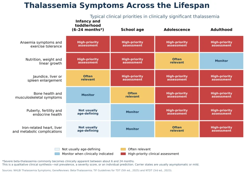
Alpha and Beta Thalassemia Produce Different Clinical Patterns
Alpha thalassemia results from reduced production of alpha-globin chains, whereas beta thalassemia results from reduced or absent production of beta-globin chains. The number and type of affected genes influence how early symptoms appear and how severe they become.[2]
A person with a mild alpha-thalassemia genotype may have no symptoms apart from small red blood cells on a routine blood count. Haemoglobin H disease can cause more persistent anaemia, jaundice, spleen enlargement and episodes of worsening haemolysis during infection, pregnancy or other physiological stress. The most severe alpha-thalassemia syndromes can present before birth and require highly specialised maternal and fetal care.[2]
Beta-thalassemia trait usually causes mild microcytosis with few or no symptoms. More clinically significant beta thalassemia may become apparent in infancy as fetal haemoglobin declines. Poor feeding, pallor, reduced growth and abdominal enlargement can then emerge, although the age and intensity of presentation vary between children.[2,3]
Symptoms cannot reliably identify whether alpha or beta thalassemia is present. Haemoglobin analysis is particularly helpful in beta thalassemia, while genetic testing may be required when alpha thalassemia is suspected because routine electrophoresis can be normal in some carriers.[2]
Carrier States and Clinically Significant Disease
People with thalassemia trait usually remain healthy and do not develop the severe childhood disease seen in transfusion-dependent thalassemia. Some carriers report mild fatigue or reduced exercise tolerance, but significant breathlessness, recurrent jaundice, marked spleen enlargement or major growth problems should prompt investigation for another or an additional condition.[2]
A carrier may also have iron deficiency, vitamin B12 or folate deficiency, thyroid disease, menstrual blood loss, gastrointestinal blood loss or another cause of anaemia. These problems can make symptoms more noticeable without changing the underlying thalassemia genotype. Assuming that every symptom is caused by the carrier state may therefore delay an important diagnosis.
Non-transfusion-dependent thalassemia, commonly abbreviated as NTDT, includes patients who do not require lifelong scheduled transfusions for survival but may still develop clinically important anaemia and long-term complications. Some remain relatively stable for years, while others develop worsening fatigue, spleen enlargement, bone disease, leg ulcers, blood-clotting complications, pulmonary hypertension or iron overload as they age.[2,4]
Transfusion-dependent thalassemia, or TDT, requires regular red blood cell transfusions to maintain an adequate haemoglobin level and suppress ineffective blood formation. Symptoms may improve substantially with a well-planned transfusion programme, but the patient then requires careful monitoring for iron overload, transfusion reactions, alloantibodies and treatment-related complications.[4]
The distinction between NTDT and TDT is based on the patient’s clinical course and transfusion needs rather than a single haemoglobin result. Infection, surgery, pregnancy, rapid growth or another acute stress may temporarily increase transfusion requirements without permanently changing the patient’s category.[4]
Factors That Change Symptom Severity
Haemoglobin level is important, but it does not fully explain how a patient feels. Some people adapt gradually to chronic anaemia and remain active at levels that would cause substantial symptoms in someone whose haemoglobin has fallen quickly. A sudden decline therefore deserves investigation even when the numerical result appears similar to a previous value.
Spleen size can significantly influence symptoms and blood counts. An enlarged spleen may remove red cells, white cells and platelets more rapidly, contributing to worsening anaemia, frequent infections, easy bruising or abdominal discomfort. Increasing transfusion needs together with progressive splenomegaly should be reviewed by an experienced thalassemia team.[2,4]
Infections can temporarily intensify anaemia, jaundice and fatigue. Certain viral illnesses may suppress red blood cell production, while fever and inflammation can increase the body’s metabolic demand. A patient who becomes suddenly pale, weak or breathless during an infection may require an urgent complete blood count and medical assessment.
Nutritional status also changes the symptom burden. Iron deficiency can coexist with thalassemia, particularly in menstruating adults, pregnant patients or people with limited dietary intake. Folate, vitamin B12, vitamin D and other deficiencies may contribute to fatigue, neurological symptoms, bone weakness or impaired growth and should be assessed according to the clinical context rather than supplemented indiscriminately.
Transfusion timing affects how patients feel between appointments. Increasing fatigue, headaches, poor concentration, reduced school attendance or declining exercise tolerance before the next scheduled transfusion may indicate that the pre-transfusion haemoglobin target or transfusion interval needs review. Treatment decisions should be made from documented trends rather than one difficult day.
Chelation adherence influences the risk of iron-related complications. Missing doses may not produce immediate symptoms, but prolonged under-treatment can allow iron to accumulate in the liver, heart and endocrine organs. Conversely, excessive or poorly monitored chelation can cause adverse effects involving the kidneys, liver, hearing, vision or blood counts, depending on the medicine used.[4]
Age also changes the clinical pattern. Young children are more likely to present with pallor, poor feeding, delayed growth and abdominal enlargement. Teenagers may experience reduced growth velocity, delayed puberty and bone concerns. Adults may report occupational fatigue, fertility problems, endocrine dysfunction, chronic pain or complications related to cumulative iron exposure.
Pregnancy can reveal previously mild disease because blood-volume expansion and increased nutritional demands may worsen anaemia. Women with thalassemia also require assessment of cardiac function, iron burden, endocrine health, medication safety and partner carrier status before or early in pregnancy.[4]
These differences explain why symptom comparison between patients is often misleading. A person should not assume that their disease is mild because another family member has few symptoms, or severe because someone with the same label requires frequent transfusions. Individual assessment should integrate genotype, haemoglobin trends, transfusion history, spleen size, iron studies, growth, organ monitoring and the patient’s actual functional capacity.[2,4]
Early Thalassemia Symptoms in Babies and Toddlers

Thalassemia symptoms in babies and toddlers may develop gradually, making them easy to mistake for feeding difficulty, frequent childhood illness or naturally variable growth. Severe beta thalassemia often becomes clinically apparent between approximately six and 24 months of age as fetal haemoglobin declines and the child becomes increasingly dependent on adult haemoglobin production.[2,3]
Some children are identified through newborn screening or family testing before symptoms appear. However, screening programmes and reporting practices differ between countries and regions, and a normal newborn-screening result does not exclude every form of alpha or beta thalassemia. A baby with persistent microcytosis, anaemia, poor growth or a strong family history may still need targeted haemoglobin and genetic assessment.[2]
Persistent Pallor and Reduced Activity
Paleness may be visible in the lips, tongue, palms, nail beds or the inner surface of the lower eyelid. Skin tone, lighting and natural pigmentation can make pallor difficult to judge, so parents should not rely on appearance alone. Persistent pallor should be assessed with a complete blood count rather than treated automatically as iron deficiency.
A baby with worsening anaemia may become less active, sleep more than usual or show reduced interest in surroundings. A toddler may stop playing sooner, ask to be carried more frequently or become breathless during activities that previously caused no difficulty. These changes are often more informative when they represent a clear decline from the child’s usual behaviour.
Parents may also notice irritability, poor sleep or difficulty settling. These features are nonspecific and can occur with many common childhood conditions, but they deserve attention when accompanied by pallor, poor appetite, slow growth, jaundice or a family history of thalassemia.
Feeding Difficulty and Poor Weight Gain
Anaemia can make feeding physically demanding. An infant may suck for a short period, pause frequently, sweat during feeding or fall asleep before taking an adequate amount. Older babies may show reduced interest in solid foods, while toddlers may eat only small portions or become tired during meals.
Poor weight gain may appear before obvious weight loss. The child may continue gaining weight but move progressively downward across established growth percentiles. Height may also increase more slowly than expected when anaemia remains clinically significant over time.[2,4]
Growth assessment should consider the child’s previous measurements, parental height, nutritional intake, feeding pattern and general development. A single low weight does not establish thalassemia, but a persistent decline in growth velocity together with microcytic anaemia requires structured evaluation.
Repeatedly changing formula, adding supplements or increasing dietary iron without confirming the cause may delay diagnosis. Iron deficiency and thalassemia can coexist, but iron treatment should be based on ferritin and appropriate iron studies rather than low haemoglobin or low mean corpuscular volume alone.[2]
Yellow Eyes, Dark Urine and Abdominal Enlargement
Increased destruction of red blood cells can raise bilirubin and produce yellowing of the whites of the eyes or skin. Mild jaundice may be intermittent, while a more noticeable change may occur during infection or another period of physiological stress.
Dark urine may reflect concentrated urine from dehydration, increased red blood cell breakdown, liver dysfunction or another urinary problem. Tea-coloured, brown or unusually dark urine accompanied by jaundice, fever, marked weakness or reduced urine output requires prompt medical assessment.
The abdomen may become progressively prominent when the spleen or liver enlarges. Parents may initially assume that the child is gaining abdominal fat or experiencing bloating. Enlargement caused by the spleen may also reduce appetite because the child feels full after eating a small amount.[2,3]
A visibly enlarged abdomen should be examined rather than monitored only at home. Clinicians may assess liver and spleen size, haemoglobin level, reticulocyte count, bilirubin and other indicators of red blood cell destruction. Ultrasound may be used when physical examination does not provide sufficient detail.
Recurrent Anaemia That Does Not Behave Like Simple Iron Deficiency
Some children are first investigated after a routine blood count shows low haemoglobin, low mean corpuscular volume and low mean corpuscular haemoglobin. These findings can occur in both iron deficiency and thalassemia, so the red cell pattern must be interpreted with ferritin, iron studies, family history and haemoglobin analysis.[2]
A child may receive iron because microcytosis is assumed to represent dietary deficiency. When iron deficiency is genuinely present, treatment should improve the relevant laboratory findings over time. Persistent microcytosis despite confirmed adherence and correction of iron deficiency should prompt investigation for thalassemia or another haemoglobin disorder.
Parents should provide copies of previous blood counts whenever possible. Comparing haemoglobin, red blood cell count, mean corpuscular volume, mean corpuscular haemoglobin and red cell distribution width over time may reveal a persistent pattern that is more informative than a single test result.
Haemoglobin electrophoresis or high-performance liquid chromatography can help identify beta-thalassemia patterns and other haemoglobin variants. Alpha thalassemia may require genetic testing when routine haemoglobin analysis is nondiagnostic but clinical suspicion remains high.[2]
Developmental and Behavioural Changes
A toddler with chronic anaemia may have reduced stamina, limited interest in active play or slower recovery after minor infections. Some children become unusually quiet, while others appear irritable because ordinary activities require more effort.
Developmental delay is not specific to thalassemia and should never be attributed to anaemia without a broader assessment. Hearing, vision, nutrition, thyroid function, neurological development and environmental factors may also need consideration when a child is not meeting expected milestones.
Children with clinically significant disease may experience repeated medical visits, transfusions or illness-related interruptions that affect sleep, appetite, play and social development. Early multidisciplinary support can help protect growth, developmental progress and family wellbeing while the haematological diagnosis and treatment plan are being established.[4]
When a Baby or Toddler Needs Urgent Assessment
Marked lethargy, difficulty waking, rapid breathing, severe breathlessness, poor feeding with signs of dehydration, fainting, bluish lips, chest retractions or a very rapid heartbeat requires urgent medical care. These signs may indicate severe anaemia, infection, respiratory illness or another acute condition.
Urgent evaluation is also appropriate when jaundice worsens rapidly, urine becomes very dark, the abdomen enlarges suddenly or the child develops severe abdominal pain. A fever accompanied by increasing pallor, weakness or reduced responsiveness should not be managed as an ordinary viral illness without clinical assessment.
A child with known thalassemia who becomes suddenly much weaker than usual may have an acute fall in haemoglobin, increased haemolysis, infection or temporary suppression of red blood cell production. The appropriate assessment may include a complete blood count, reticulocyte count, bilirubin, liver tests, infection testing and review of recent transfusions or medicines.[3,4]
When symptoms are persistent but not immediately dangerous, parents should arrange a paediatric or haematology evaluation and bring the child’s newborn-screening record, growth chart, previous blood reports, family history and details of any previous iron treatment. This information helps distinguish thalassemia from nutritional anaemia and supports earlier classification of disease severity.
Thalassemia Symptoms in School-Age Children

Thalassemia symptoms in school-age children may become noticeable through reduced stamina, slower growth, frequent tiredness or difficulty keeping up with ordinary school activities. Some children have already been diagnosed and are receiving regular treatment, while others with milder or non-transfusion-dependent disease may first be investigated after persistent microcytosis, pallor, spleen enlargement or an unexpected decline in physical performance.[1,2]
Reduced Stamina During Play and Physical Education
A child with clinically significant anaemia may run more slowly, stop playing earlier than classmates or need longer recovery after exercise. Parents may notice reluctance to climb stairs, ride a bicycle, walk longer distances or participate in sports that the child previously enjoyed.
Teachers may describe the child as avoiding physical education or repeatedly asking to rest. These observations should not be interpreted automatically as poor motivation. Reduced oxygen delivery, an enlarged spleen, musculoskeletal discomfort or a haemoglobin level that falls too low before the next transfusion may all limit activity.[2,4]
Exercise remains valuable for cardiovascular health, bone strength and emotional wellbeing, so children should not be excluded from all physical activity simply because they have thalassemia. The appropriate intensity depends on haemoglobin stability, heart function, bone health, spleen size and overall treatment status. A child with substantial spleen enlargement may need to avoid contact sports or activities that could cause abdominal injury because an enlarged spleen is more vulnerable to rupture.[4]
A clear decline in exercise tolerance deserves reassessment even when the child has a known diagnosis. The treatment team may need to review pre-transfusion haemoglobin, transfusion timing, iron burden, chelation adherence, cardiac status, nutritional factors and possible infection.
Persistent Tiredness During the School Day
Fatigue may appear as difficulty waking for school, reduced alertness in morning lessons, exhaustion after returning home or an increased need for daytime sleep. Some children remain active for part of the day but become noticeably tired before the next transfusion.
The timing of symptoms can provide useful clinical information. Fatigue that repeatedly worsens during the final days of a transfusion cycle may indicate that the child’s haemoglobin is falling below the level needed for normal daily function. Recording the relationship between symptoms and transfusion dates can help the haematology team evaluate whether the schedule remains appropriate.[4]
Fatigue should not be attributed to thalassemia without considering other causes. Poor sleep, anxiety, infection, thyroid dysfunction, nutritional deficiency, medication effects and iron-related endocrine complications can produce similar symptoms. A child whose tiredness persists despite apparently adequate haemoglobin control may require broader assessment.
Headaches, Dizziness and Difficulty Concentrating
Anaemia can contribute to headaches, light-headedness and difficulty sustaining attention, particularly during prolonged classroom work or physical exertion. A child may need more time to complete assignments, lose concentration late in the day or complain of feeling dizzy when standing quickly.
These symptoms are not specific to thalassemia. Vision problems, dehydration, migraine, sleep disruption, stress and other medical conditions should also be considered. Persistent headaches, fainting, weakness affecting one side of the body, confusion or visual changes require prompt medical evaluation rather than routine observation.
Parents and teachers should focus on changes from the child’s usual performance rather than assuming that academic difficulty is caused by the blood disorder. Formal educational assessment may be appropriate when learning concerns persist independently of fatigue or treatment timing.
Slow Height or Weight Gain
Growth problems may become clearer during the school years when the child’s height and weight are compared over several annual measurements. The most informative sign is often reduced growth velocity rather than a single measurement below the average range.
Chronic anaemia, increased energy requirements, poor appetite, endocrine dysfunction, iron overload and delayed bone maturation may all influence growth in clinically significant thalassemia. Repeated hospital visits and illness-related absences can also affect eating routines, sleep and physical development.[2,4]
Growth should be plotted on an appropriate chart at regular intervals. Clinicians may evaluate height velocity, weight, body mass index, pubertal development, nutritional intake, thyroid function, glucose regulation, vitamin D status and other endocrine factors when growth slows unexpectedly.
Parents should not rely only on comparisons with classmates because normal children mature at different rates. A sustained fall across growth percentiles or a clear slowing of annual height gain is more clinically meaningful and should be discussed with the treatment team.
Abdominal Fullness, Early Satiety and Spleen Enlargement
An enlarged spleen may cause fullness or discomfort beneath the left ribs, a visibly prominent abdomen or a feeling of being full after eating a small amount. Reduced food intake caused by early satiety can further affect weight gain and energy.
Parents may also notice that clothing feels tight around the abdomen even when the child has not gained overall body weight. Progressive abdominal enlargement should be examined because the liver, spleen or both may be involved.
Increasing spleen size can be associated with a greater rate of red blood cell removal and worsening anaemia. In some children, white blood cells and platelets may also fall when the enlarged spleen retains or destroys more blood cells than usual.[2,4]
Sudden severe pain in the upper abdomen, shoulder pain after abdominal injury, faintness or rapid worsening of abdominal swelling requires urgent assessment. Children with known splenomegaly should have an individualized school and sports plan so that teachers and coaches understand any necessary precautions.
Pallor, Jaundice and Dark Urine
Pallor may remain visible in the lips, tongue, palms or nail beds, although it can be difficult to assess accurately across different skin tones. A complete blood count is more reliable than visual judgement when anaemia is suspected.
Yellowing of the eyes may indicate increased red blood cell breakdown or liver involvement. Dark urine can occur with dehydration but may also accompany haemolysis, liver dysfunction or another medical problem. New or worsening jaundice should be evaluated rather than accepted as a routine feature of thalassemia.
A child with jaundice, fever, sudden weakness and dark urine may be experiencing increased haemolysis or an acute illness. Blood counts, reticulocyte count, bilirubin and liver tests may be required, together with assessment for infection and recent medication exposure.
Bone Pain and Skeletal Changes
Long-standing ineffective blood production can stimulate expansion of the bone marrow. In inadequately controlled severe disease, this may contribute to changes in facial bones, thinning of the bone cortex, altered skeletal development and increased fracture risk.[2]
These changes are not expected in every child with thalassemia and are particularly unlikely in an otherwise healthy carrier. Their presence suggests clinically significant disease or inadequate suppression of ineffective blood formation and requires specialist review.
Bone or back pain may also result from low bone density, vitamin D deficiency, physical deconditioning or another musculoskeletal condition. Persistent pain, recurrent fractures, reduced height or difficulty walking should prompt assessment rather than repeated use of pain medicines without identifying the cause.
Evaluation may include nutritional testing, endocrine assessment, imaging and bone-density measurement when appropriate for the child’s age and clinical history. Regular weight-bearing activity may support bone health when medically safe, but exercise recommendations should be adapted for children with severe anaemia, cardiac concerns or substantial splenomegaly.[4]
School Attendance and Treatment-Related Effects
Children receiving regular transfusions or specialist monitoring may miss school for appointments, laboratory tests or treatment-related complications. Frequent absence can affect academic progress, friendships and emotional wellbeing even when the child’s physical symptoms are reasonably controlled.
A coordinated school plan can help the child receive missed work, attend medical appointments without unnecessary academic penalty and rest when symptoms are temporarily worse. School staff should know whom to contact if the child develops fever, marked weakness, breathlessness, fainting or abdominal pain.
Confidentiality should be respected, and the child should be involved in deciding how much information is shared with classmates. The aim is to support normal participation without overlooking genuine medical limitations.
When School-Age Symptoms Need Medical Reassessment
A child should be reassessed when fatigue, pallor or breathlessness becomes progressively worse, school participation declines, growth slows, the abdomen enlarges or symptoms begin occurring earlier in each transfusion cycle. These changes may reflect inadequate haemoglobin control, splenic enlargement, iron-related complications, infection, nutritional deficiency or another coexisting condition.[2,4]
Urgent medical evaluation is required for breathlessness at rest, chest pain, fainting, severe weakness, rapidly worsening jaundice, very dark urine, high fever, severe abdominal pain or symptoms following abdominal trauma. A child who has undergone splenectomy requires particularly prompt assessment of fever because of the increased risk of severe bacterial infection.[4]
Parents can improve the clinical assessment by bringing recent blood counts, transfusion records, chelation details, growth measurements, school observations and a brief record of when symptoms occur. This allows the treatment team to identify patterns and adjust monitoring or treatment according to the child’s documented needs rather than relying on a single symptom report.
Thalassemia Symptoms in Teenagers
Thalassemia symptoms in teenagers may become more noticeable as rapid growth, puberty, school demands and increasing independence place additional strain on the body. Even when childhood treatment has been stable, adolescence can reveal delayed growth, reduced exercise tolerance, bone problems, hormonal changes or complications related to cumulative iron exposure. Regular assessment is therefore important even when the teenager reports feeling generally well.[2,4]
Slower Growth and Delayed Puberty
A teenager with clinically significant thalassemia may grow more slowly than classmates or fail to experience the expected adolescent growth spurt. Reduced growth velocity is more informative than short stature alone because family height, nutrition and the normal timing of puberty vary considerably between individuals.
Chronic anaemia, iron overload, nutritional deficiencies and impaired function of the pituitary, thyroid or other endocrine glands may all contribute to delayed growth. A teenager who was previously following an established growth curve but begins to fall behind requires assessment rather than reassurance based only on family body size.[2,4]
Delayed puberty may become apparent when expected physical changes do not begin or progress. In girls, this can include delayed breast development, late onset of menstruation, irregular periods or the absence of periods after they have previously begun. In boys, delayed enlargement of the testes, limited facial or body hair, lack of voice deepening or slow muscle development may be noticed.
Puberty should be evaluated according to the teenager’s developmental stage rather than by comparing them informally with friends. Assessment may include growth measurements, bone age, thyroid function, sex hormones, pituitary hormones, nutritional status and iron-burden evaluation. Early endocrine referral can help identify treatable contributors and support more normal physical development.[4]
Fatigue, Breathlessness and Reduced School Performance
Teenagers may describe persistent tiredness, difficulty waking, reduced concentration or exhaustion after a full school day. Some remain active immediately after transfusion but develop headaches, breathlessness, palpitations or reduced stamina as the next transfusion approaches.
A repeated pattern of declining energy before transfusion should be documented and discussed with the haematology team. It may indicate that the transfusion interval, pre-transfusion haemoglobin target or overall treatment plan needs review. The teenager should not be expected to tolerate progressive functional decline simply because it has become familiar.
Fatigue that continues despite apparently adequate haemoglobin control may reflect iron-related endocrine dysfunction, sleep problems, anxiety, infection, nutritional deficiency or another medical condition. Persistent fatigue deserves broader investigation rather than automatic attribution to thalassemia.
Reduced concentration may affect examination performance, homework completion and attendance. Parents and teachers should distinguish a change in cognitive stamina from lack of motivation. Flexible appointment planning, access to missed work and appropriate rest periods can reduce academic disruption without lowering educational expectations.
Reduced Exercise Capacity and Palpitations
A teenager may notice that running, dancing, swimming, cycling or climbing stairs has become more difficult. They may stop frequently, experience disproportionate breathlessness or avoid activities that previously felt manageable.
Some reduction in endurance may accompany anaemia, but a clear decline should be assessed for changes in haemoglobin, spleen size, heart function, iron burden and general conditioning. Palpitations, dizziness or chest discomfort should not be dismissed as anxiety without clinical evaluation.
Regular physical activity can support cardiovascular fitness, bone strength and emotional health when adapted to the teenager’s medical status. Exercise recommendations should consider haemoglobin stability, cardiac findings, bone density and spleen enlargement. Contact sports may be unsuitable for teenagers with substantial splenomegaly because of the risk of abdominal injury.[4]
Breathlessness at rest, fainting, persistent chest pain or a sustained rapid or irregular heartbeat requires urgent assessment. These symptoms may indicate severe anaemia, arrhythmia, cardiac dysfunction or another acute condition.
Bone, Back and Joint Symptoms
Adolescence is a critical period for building peak bone mass. Teenagers with clinically significant thalassemia may be vulnerable to low bone density because of chronic anaemia, marrow expansion, delayed puberty, reduced physical activity, nutritional deficiencies and iron-related endocrine dysfunction.[2,4]
Persistent back pain, bone pain, reduced height or recurrent fractures should not be regarded as unavoidable consequences of thalassemia. These symptoms may indicate osteopenia, osteoporosis, vertebral compression or another musculoskeletal problem requiring targeted evaluation.
Bone-health assessment may include vitamin D, calcium and phosphate-related testing, endocrine evaluation and bone-density imaging when clinically appropriate. Treatment decisions should address the underlying contributors rather than relying only on analgesics.
Weight-bearing exercise may be beneficial when medically safe, but the programme should be individualized. A teenager with severe anaemia, cardiac involvement, fractures or marked splenomegaly may require specialist guidance before beginning intensive activity.
Menstrual and Reproductive Symptoms
Girls with thalassemia may experience delayed menarche, irregular cycles, very infrequent periods or secondary amenorrhoea. These changes can reflect delayed puberty, pituitary or ovarian dysfunction, thyroid disease, nutritional factors or the effects of chronic illness and iron overload.[4]
Menstrual blood loss can also contribute to iron deficiency in a teenager who already has thalassemia. Fatigue or a fall in haemoglobin should therefore not automatically be attributed to the inherited disorder. Ferritin and appropriate iron studies may be needed to identify coexisting deficiency.
Boys may experience delayed sexual maturation, reduced testosterone-related development or slower muscle gain. These changes can affect confidence and body image, but they also provide clinically important information about endocrine function.
Fertility cannot be predicted from outward pubertal development alone. Teenagers and families should receive age-appropriate counselling about reproductive health, future fertility and the importance of partner testing when the patient reaches adulthood. Discussions should be private, respectful and appropriate to the teenager’s maturity.
Symptoms Suggesting Endocrine or Metabolic Complications
Excessive thirst, frequent urination, unexplained weight loss or recurrent infections may indicate abnormal glucose regulation and require prompt testing. Unusual sensitivity to cold, constipation, dry skin, slowed growth or persistent fatigue may raise concern about thyroid dysfunction, although these symptoms are not specific.
Delayed growth, delayed puberty and menstrual irregularity may reflect pituitary or gonadal involvement. Because early endocrine changes can be clinically subtle, scheduled screening may identify complications before they produce obvious symptoms.[4]
A teenager may feel well despite significant iron accumulation. Symptom absence should therefore never replace ferritin trends, liver iron assessment, cardiac monitoring and endocrine screening when these are indicated by the transfusion history and treatment guidelines.[4]
Abdominal Fullness and Spleen-Related Symptoms
An enlarged spleen may cause pressure beneath the left ribs, early satiety, reduced appetite or a visibly prominent abdomen. Teenagers may conceal abdominal enlargement because of embarrassment or concern about body image, so clinicians should ask sensitively about discomfort, clothing fit and eating patterns.
Progressive spleen enlargement can contribute to worsening anaemia and reductions in white blood cells or platelets. Increasing fatigue, frequent infections, easy bruising or greater transfusion requirements may therefore require reassessment of spleen function and overall disease control.[2,4]
Sudden severe upper abdominal pain, left shoulder pain, faintness or weakness after an impact to the abdomen requires emergency evaluation. Schools, coaches and family members should understand any sport restrictions when splenomegaly is present.
Emotional and Social Effects of Symptoms
Visible differences in height, delayed puberty, scars, abdominal enlargement or frequent treatment can affect confidence and social participation. Teenagers may avoid changing rooms, sports, relationships or medical discussions because they feel different from peers.
Persistent low mood, withdrawal, sleep disturbance, loss of interest, declining school performance or refusal of treatment may indicate emotional distress rather than simple adolescent resistance. Psychological support should be integrated into care when these changes persist.
Adolescents often begin taking greater responsibility for chelation and appointment schedules. Missed doses may occur because of side effects, treatment burden, embarrassment or difficulty integrating therapy into school and social life. A nonjudgmental discussion is more likely to identify the practical barrier than repeated warnings alone.
Increasing independence should include gradual education about the teenager’s diagnosis, medicines, transfusion schedule, emergency symptoms and monitoring results. This prepares the patient for adult services and reduces the risk of treatment disruption during transition.
When Teenage Symptoms Need Reassessment
Medical review is appropriate when growth slows, puberty is delayed, periods become absent, exercise tolerance declines, bone pain persists or fatigue begins affecting school and daily activities. These changes may reflect inadequate haemoglobin control, iron overload, endocrine dysfunction, nutritional deficiency or another complication requiring targeted investigation.[2,4]
Urgent assessment is required for chest pain, fainting, breathlessness at rest, sustained palpitations, severe weakness, rapidly worsening jaundice, very dark urine, high fever or sudden abdominal pain. A teenager who has undergone splenectomy requires particularly prompt evaluation for fever because serious infection can progress rapidly.[4]
The most useful clinical review combines the teenager’s symptom history with haemoglobin trends, transfusion records, chelation adherence, growth measurements, pubertal staging, ferritin trends, magnetic resonance iron assessment and relevant endocrine, cardiac and bone investigations. This approach helps distinguish normal adolescent variation from treatable thalassemia-related complications.
Thalassemia Symptoms in Adults

Thalassemia symptoms in adults vary from no noticeable problems in a carrier to chronic fatigue, breathlessness, abdominal fullness, bone pain, hormonal disturbance and organ complications in clinically significant disease. Some adults were diagnosed during childhood and have received long-term transfusion or chelation therapy, while others are investigated later after persistent microcytosis, unexplained anaemia, pregnancy-related testing or family screening.[2,4]
Symptoms that first appear in adulthood do not necessarily mean that the inherited disorder has suddenly developed. Mild alpha or beta thalassemia may remain unrecognised for years, whereas non-transfusion-dependent disease can produce complications gradually even when the person has never required regular transfusions. Adults with a known diagnosis also need reassessment when their usual symptom pattern changes because a new problem may be present alongside thalassemia.[2,4]
Persistent Fatigue and Reduced Work Capacity
Chronic fatigue is one of the most commonly reported problems in adults with clinically significant thalassemia. The patient may manage basic daily activities but feel exhausted after commuting, household work, childcare, exercise or a full working day. Some describe mental fatigue, reduced concentration or a need for prolonged recovery after routine tasks.
Anaemia can contribute to these symptoms by reducing oxygen delivery, but haemoglobin level does not always explain the full burden. Iron overload, thyroid dysfunction, diabetes, sleep disturbance, chronic pain, infection, nutritional deficiency, anxiety and treatment-related effects can also reduce energy. Persistent fatigue therefore requires more than a single complete blood count.
In transfusion-dependent thalassemia, a repeated decline in energy before the next transfusion may suggest that the current interval or pre-transfusion haemoglobin target needs review. Patients can help by recording when fatigue begins, how it affects activity and whether it improves after transfusion. A consistent pattern is more useful than describing symptoms only during a clinic visit.[4]
Adults with thalassemia trait usually have no symptoms or only mild anaemia. Severe exhaustion, inability to complete ordinary activities or progressively worsening weakness should not automatically be attributed to the carrier state. Iron deficiency, blood loss, thyroid disease, vitamin deficiency and other causes must be investigated.
Breathlessness, Palpitations and Reduced Exercise Tolerance
Adults may notice breathlessness when climbing stairs, walking quickly, carrying shopping bags or exercising. Activities that were previously comfortable may begin to require frequent pauses. A rapid heartbeat, pounding sensation in the chest or dizziness may occur when the body is compensating for anaemia.
A gradual decline in exercise tolerance can also indicate cardiac dysfunction, pulmonary hypertension, deconditioning or iron-related organ involvement. Symptoms should be interpreted with haemoglobin trends, transfusion history, iron assessment and cardiac evaluation rather than assumed to reflect anaemia alone.[2,4]
Palpitations may be brief and harmless, but persistent, irregular or exercise-related palpitations require medical assessment. The clinician may consider an electrocardiogram, ambulatory rhythm monitoring, echocardiography and cardiac magnetic resonance imaging depending on the patient’s history and risk profile.
Breathlessness at rest, fainting, chest pain, new ankle swelling or a sustained rapid or irregular heartbeat requires urgent evaluation. These symptoms may indicate severe anaemia, arrhythmia, heart failure, pulmonary embolism or another acute condition.
Pallor, Jaundice and Dark Urine
Pallor may be visible in the lips, tongue, palms or nail beds, but it is not always easy to recognise across different skin tones. A complete blood count provides a more reliable assessment than visual appearance alone.
Yellowing of the eyes or skin can result from increased red blood cell breakdown. Some adults also develop gallstones, which may cause pain in the right upper abdomen, nausea or discomfort after meals. Persistent or worsening jaundice should be evaluated because liver disease, infection and medication-related injury can produce a similar appearance.[2]
Dark urine may occur during increased haemolysis, dehydration or liver dysfunction. New tea-coloured or brown urine accompanied by fever, worsening jaundice, back pain, weakness or reduced urine output requires prompt medical review.
A sudden increase in pallor, jaundice or fatigue may indicate an acute fall in haemoglobin. Appropriate testing may include a complete blood count, reticulocyte count, bilirubin, liver function tests and evaluation for infection, bleeding or increased haemolysis.
Abdominal Fullness and Spleen Enlargement
An enlarged spleen may cause pressure beneath the left ribs, early satiety, reduced appetite or a visibly prominent abdomen. Some adults report that they cannot complete a normal meal because they feel full after a few bites.
Progressive splenomegaly can increase the removal of red blood cells and may also reduce white blood cells or platelets. The patient may then experience worsening anaemia, recurrent infections, easy bruising or increasing transfusion requirements.[2,4]
Liver enlargement or chronic liver disease can also contribute to abdominal swelling or discomfort. Assessment may include physical examination, liver tests, ultrasound and iron evaluation according to the clinical context.
Sudden severe pain in the left upper abdomen, shoulder pain, faintness or weakness after abdominal trauma requires emergency assessment because an enlarged spleen is vulnerable to injury. Adults with significant splenomegaly should discuss safe exercise and contact-sport restrictions with their clinical team.
Bone Pain, Back Pain and Fracture Risk
Low bone density is a recognised complication of clinically significant thalassemia. Adults may experience persistent back pain, generalized bone discomfort, loss of height or fractures after relatively minor trauma. Contributing factors include chronic anaemia, marrow expansion, delayed puberty, hormonal dysfunction, nutritional deficiencies and reduced physical activity.[2,4]
Back pain should not automatically be classified as a routine thalassemia symptom. Disc disease, muscular strain, vertebral compression, kidney disease and other common adult conditions may produce similar pain. Persistent pain, neurological symptoms or loss of height requires targeted evaluation.
Bone-health assessment may include vitamin D, calcium and phosphate-related tests, thyroid and sex-hormone evaluation and bone-density imaging. Treatment should address the contributing mechanism rather than relying only on pain relief.
Weight-bearing and resistance exercise may support bone health when medically appropriate. The programme should be individualized for patients with severe anaemia, cardiac disease, fractures, joint problems or substantial splenomegaly.
Hormonal, Menstrual and Fertility Symptoms
Women may experience irregular periods, absent periods, reduced fertility or symptoms associated with early ovarian dysfunction. These problems can arise from pituitary or ovarian effects, iron overload, thyroid disease, nutritional factors or chronic illness.[4]
Heavy menstrual bleeding can cause iron deficiency in a woman who also has thalassemia. A decline in haemoglobin, increasing fatigue or worsening breathlessness should therefore prompt assessment for menstrual blood loss and iron deficiency rather than being attributed automatically to the inherited disorder.
Men may experience reduced libido, erectile difficulty, reduced facial or body hair, low muscle mass or infertility when testosterone production or pituitary function is impaired. These symptoms should be evaluated with appropriate hormonal testing rather than assumed to be psychological.
Fertility may remain possible in many adults with thalassemia, particularly when iron burden and endocrine complications are well controlled. Preconception assessment should include cardiac function, liver health, iron status, endocrine evaluation, medication review and partner carrier testing.[4]
Pregnancy places additional demands on the cardiovascular and haematological systems. Women with thalassemia should receive coordinated care involving haematology and obstetric specialists because transfusion needs, cardiac risk, iron management and thrombosis risk may change during pregnancy.
Symptoms of Iron-Related Endocrine and Metabolic Complications
Iron can accumulate in the pituitary gland, thyroid, pancreas, liver and heart. Because early organ injury may produce few symptoms, scheduled monitoring remains essential even when the patient feels stable.[4]
Excessive thirst, frequent urination, unexplained weight loss, blurred vision or recurrent infections may indicate abnormal glucose regulation or diabetes. Cold intolerance, constipation, dry skin, slowed thinking or increasing fatigue may suggest thyroid dysfunction, although these symptoms overlap with many other conditions.
Reduced libido, menstrual changes, infertility, loss of body hair or reduced muscle strength may indicate pituitary or gonadal dysfunction. Hormonal complications often develop gradually and may be overlooked when fatigue is already considered part of thalassemia.
Symptoms alone cannot measure iron burden. Ferritin trends are useful but can be altered by infection, inflammation and liver disease. Liver iron concentration and cardiac iron may require validated magnetic resonance imaging when clinically indicated.[4]
Liver and Gallbladder Symptoms
Adults with thalassemia may be vulnerable to liver complications related to iron accumulation, previous transfusion-transmitted infection, fatty liver disease, medication effects or other common metabolic conditions. Many liver problems remain silent until laboratory abnormalities or imaging changes are detected.
Right upper abdominal discomfort, persistent nausea, increasing jaundice, abdominal swelling or unexplained itching may require liver assessment. These symptoms are not specific and should be interpreted with liver enzymes, bilirubin, viral screening, ultrasound findings and iron measurements.
Chronic red blood cell breakdown can increase the risk of pigment gallstones. Sudden right upper abdominal pain, fever, vomiting or worsening jaundice may indicate gallbladder or bile-duct obstruction and requires prompt evaluation.
Routine monitoring is important because the absence of pain does not exclude liver iron accumulation or progressive fibrosis. Early identification provides a better opportunity to modify chelation, address metabolic risk and prevent advanced disease.[4]
Leg Ulcers, Blood Clots and Circulatory Symptoms
Some adults with non-transfusion-dependent thalassemia develop chronic ulcers around the ankles. These wounds may heal slowly, recur and significantly affect mobility. Persistent skin breakdown requires wound assessment, infection control and review of the underlying circulatory and haematological factors.[2]
Thalassemia can also be associated with an increased risk of thrombosis in selected patients, particularly those with non-transfusion-dependent disease, previous splenectomy or additional risk factors. One-sided leg swelling, sudden chest pain, unexplained breathlessness or coughing blood requires emergency assessment.
Headache, visual disturbance, weakness affecting one side of the body, speech difficulty or sudden confusion may indicate a neurological emergency. These symptoms should never be assumed to result from routine anaemia.
The individual risk of clotting varies considerably. Preventive treatment is not appropriate for every patient and should be decided from the person’s diagnosis, splenectomy status, transfusion pattern, pregnancy status and other clinical factors.
Symptoms After Splenectomy
Adults who have had their spleen removed may experience fewer symptoms related to splenic enlargement, but they face increased risks of severe infection and thrombosis. A fever can progress rapidly and requires urgent assessment according to the patient’s emergency plan.[4]
Patients should understand their vaccination schedule, any prescribed antibiotic strategy and the need to inform healthcare professionals that they are asplenic. An emergency medical alert card or identification can be valuable when urgent treatment is needed.
Fever, shaking chills, confusion, severe weakness or rapidly worsening illness should not be managed through routine home observation. The patient should follow the emergency instructions provided by the treating team.
Emotional, Cognitive and Quality-of-Life Effects
Long-term treatment can affect employment, relationships, travel, finances and emotional wellbeing. Adults may experience anxiety before transfusions, frustration with chelation, concern about fertility or fear of future organ complications.
Difficulty concentrating may reflect fatigue, sleep disturbance, low mood, hormonal dysfunction or the practical burden of chronic illness. Cognitive complaints should be explored rather than dismissed as stress.
Persistent sadness, social withdrawal, loss of interest, treatment avoidance or feelings of hopelessness deserve professional support. Psychological care does not imply that symptoms are imagined; it helps patients manage the real emotional and behavioural demands of long-term disease.
Treatment plans should consider function as well as laboratory values. The ability to work, exercise, sleep, maintain relationships and complete daily activities provides clinically important information about whether the current plan is meeting the patient’s needs.
When Adult Symptoms Need Medical Reassessment
Reassessment is appropriate when fatigue progressively worsens, exercise tolerance declines, transfusion benefit becomes shorter, abdominal fullness increases, bone pain persists or menstrual and hormonal symptoms develop. These changes may indicate inadequate anaemia control, iron overload, splenic enlargement, endocrine dysfunction, infection, nutritional deficiency or another coexisting disorder.[2,4]
Urgent evaluation is required for chest pain, fainting, breathlessness at rest, sustained palpitations, high fever after splenectomy, rapidly worsening jaundice, very dark urine, sudden abdominal pain, one-sided leg swelling or new neurological symptoms.
An effective review combines the patient’s symptom history with haemoglobin trends, transfusion records, chelation adherence, ferritin patterns, magnetic resonance iron measurements and relevant cardiac, liver, endocrine and bone assessments. This prevents potentially treatable complications from being dismissed as an expected part of adult life with thalassemia.[4]
Thalassemia Symptoms in Carriers and Thalassemia Minor

Thalassemia symptoms are usually absent or mild in people with alpha- or beta-thalassemia trait, also known as thalassemia minor or carrier status. Many carriers feel completely healthy and discover the condition only after a routine blood count, antenatal screening, family testing or evaluation of persistently small red blood cells. Mild anaemia may occur, but pronounced breathlessness, recurrent jaundice, marked spleen enlargement, poor growth or severe weakness are not typical of an uncomplicated carrier state.[1,2,5]
Most Thalassemia Carriers Have No Noticeable Symptoms
A person with thalassemia trait usually produces enough functional haemoglobin to maintain normal daily activity. The complete blood count may show a low mean corpuscular volume and mean corpuscular haemoglobin even when the total haemoglobin level is normal or only mildly reduced.[2]
Some carriers report mild tiredness, reduced stamina or occasional pallor, particularly during pregnancy, infection, heavy menstrual bleeding or periods of inadequate nutrition. When symptoms are more severe than expected, another cause should be investigated rather than assuming that the carrier state alone is responsible.
Children with uncomplicated thalassemia trait generally grow, learn and participate in physical activity normally. Persistent poor growth, delayed puberty, recurrent jaundice, abdominal enlargement or substantial exercise intolerance requires further evaluation for a more clinically significant haemoglobin disorder or another medical condition.
Adults with thalassemia minor should usually be able to work, exercise and perform routine activities without major limitation. Progressive fatigue, fainting, chest discomfort, worsening breathlessness or a marked decline in exercise capacity warrants a broader medical assessment.
Thalassemia Minor and Iron Deficiency Are Different Conditions
Thalassemia trait and iron-deficiency anaemia can both produce small, pale red blood cells. This similarity can lead to carriers receiving iron without confirmation that iron deficiency is present. Thalassemia-related microcytosis does not correct simply by taking additional iron.[2,5]
Ferritin is commonly used to assess iron stores, although infection, inflammation and liver disease can increase ferritin and complicate interpretation. Transferrin saturation, serum iron, total iron-binding capacity and the patient’s dietary, menstrual and gastrointestinal history may provide additional information when the diagnosis remains uncertain.[2]
Iron deficiency can coexist with thalassemia trait. Menstruating adults, pregnant patients, people with restricted diets and individuals with gastrointestinal blood loss may develop genuine deficiency in addition to inherited microcytosis. In these situations, medically supervised iron therapy may improve haemoglobin and iron stores, although the MCV can remain low because the thalassemia trait persists.
Repeated or prolonged iron supplementation without confirmed deficiency is unlikely to improve uncomplicated thalassemia minor. It may also cause gastrointestinal adverse effects and unnecessary iron exposure. Continued treatment should therefore be guided by laboratory evidence and follow-up testing.
A substantial fall in haemoglobin from the patient’s usual baseline requires investigation for blood loss, nutritional deficiency, infection, inflammation, kidney disease, thyroid dysfunction or another haematological condition. Carrier status should not be used to explain every future episode of anaemia.
Thalassemia Minor Does Not Become Thalassemia Major
Thalassemia trait is an inherited carrier state that remains stable throughout life. It does not gradually transform into thalassemia major, and a healthy carrier does not later become transfusion-dependent simply because of ageing.[1,5]
Symptoms may become more noticeable when another condition places additional strain on the body. Pregnancy, heavy bleeding, surgery, infection, iron deficiency or chronic disease can lower haemoglobin and increase fatigue without changing the underlying thalassemia genotype.
A person previously diagnosed with thalassemia minor who later develops worsening anaemia, jaundice, spleen enlargement or a need for transfusion should have the diagnosis reassessed. A more complex genotype, a co-inherited haemoglobin variant or an additional medical disorder may need to be considered.[2]
Different family members can have very different clinical outcomes. One person may inherit a single carrier variant and remain asymptomatic, while another may inherit clinically significant variants from both parents. The severity seen in one relative does not mean that a carrier’s own condition will progress.
Confirming Alpha- or Beta-Thalassemia Trait
A complete blood count may suggest thalassemia trait when microcytosis is persistent and greater than expected for the degree of anaemia. The red blood cell count may remain relatively preserved, but no single blood-count formula can confirm the diagnosis in every patient.[2]
Haemoglobin analysis using high-performance liquid chromatography or electrophoresis can help identify beta-thalassemia trait and other haemoglobin variants. Beta-thalassemia trait is commonly associated with an increased haemoglobin A2 level, although iron status, age, recent transfusion and other haemoglobin findings must also be considered.[2]
Alpha-thalassemia trait can be more difficult to confirm because routine haemoglobin electrophoresis may be normal. Genetic testing may be appropriate when persistent microcytosis remains unexplained, the family background suggests alpha thalassemia, the patient’s partner is also a carrier or precise reproductive-risk assessment is required.[2]
Confirmation is particularly useful for people who have repeatedly received iron for presumed deficiency without the expected response. An accurate diagnosis can prevent unnecessary treatment and help future clinicians interpret lifelong microcytosis correctly.
Patients should retain a copy of their haemoglobin or genetic report and provide it during pregnancy care, surgery, hospital admission or future evaluation of anaemia. This reduces repeated testing and helps prevent inappropriate iron prescriptions.
Pregnancy and Partner Testing
Pregnancy increases blood volume and nutritional requirements, making mild anaemia more noticeable in some carriers. Pregnant patients still require assessment for iron deficiency and other causes of anaemia rather than receiving iron solely because thalassemia trait is present.
The most important long-term implication of carrier status is the possibility of passing a clinically significant haemoglobin disorder to a child. When one biological parent carries a thalassemia variant, the other parent should be offered appropriate testing.[1,5]
When both parents carry beta-thalassemia trait, each pregnancy has a one-in-four probability of beta-thalassemia major, a one-in-two probability of producing a carrier child and a one-in-four probability of producing a child who inherits neither variant. These probabilities apply independently to every pregnancy.
Alpha-thalassemia inheritance depends on the number and arrangement of affected alpha-globin genes. Certain parental combinations can result in haemoglobin H disease or severe fetal alpha thalassemia. Genetic testing can clarify the risk even when both parents are healthy and have no symptoms.[2]
Carrier couples benefit from genetic counselling before pregnancy or early in pregnancy. Precise parental genotyping helps clarify reproductive risk and supports informed decisions about prenatal diagnosis and other available reproductive options.
When Symptoms in a Carrier Need Further Investigation
Mild, stable anaemia without functional limitation may not require treatment beyond confirmation of the diagnosis and appropriate reproductive counselling. New or worsening symptoms should still be evaluated according to their clinical pattern.
Progressive fatigue may be related to iron deficiency, vitamin B12 or folate deficiency, thyroid dysfunction, sleep disturbance, chronic inflammation or another systemic illness. Heavy menstrual bleeding, black stools, visible blood loss or an unexplained fall in haemoglobin requires investigation for the source of blood loss.
Breathlessness at rest, chest pain, fainting, severe weakness or a sustained rapid heartbeat requires urgent medical assessment. These symptoms are not typical of uncomplicated thalassemia trait and may indicate severe anaemia, cardiovascular disease, pulmonary disease or another acute condition.
Yellow eyes, very dark urine, abdominal enlargement or pain beneath the ribs also requires medical review. These findings may indicate haemolysis, liver or gallbladder disease, spleen enlargement or a haemoglobin disorder more clinically significant than simple trait.
A complete evaluation should compare the current blood count with the patient’s previous baseline, assess iron status and review haemoglobin or genetic findings when necessary. This helps distinguish stable carrier-related microcytosis from a new and potentially treatable cause of symptoms.[2,5]
Thalassemia Symptoms by Type and Severity
Thalassemia symptoms depend on which globin genes are affected, how much functional haemoglobin the body can produce and whether anaemia, haemolysis, spleen enlargement or iron accumulation has developed. The clinical range extends from an asymptomatic carrier state to severe transfusion-dependent disease beginning in infancy. Symptoms can indicate that further testing is needed, but they cannot establish the exact thalassemia type or genotype.[2,3]
Table: Thalassemia Symptoms by Type and Severity
| Thalassemia category | Typical clinical pattern | Important monitoring priority |
|---|---|---|
| Silent carrier or thalassemia trait | Usually no symptoms or mild microcytic anaemia; daily activity is generally unaffected | Confirm the diagnosis, avoid unnecessary iron and assess reproductive risk |
| Haemoglobin H disease | Chronic anaemia, pallor, jaundice, dark urine, spleen enlargement and worsening haemolysis during illness | Haemoglobin, haemolysis, spleen size, transfusion needs and iron burden |
| Non-transfusion-dependent thalassemia | Variable fatigue, jaundice, splenomegaly, bone changes, growth or pubertal problems and age-related iron accumulation | Iron burden, liver health, bone density, endocrine function and thrombosis risk |
| Transfusion-dependent thalassemia | Severe anaemia beginning early in life, poor growth or reduced activity without adequate transfusion support | Transfusion adequacy, chelation, liver and cardiac iron, growth, endocrine and bone health |
| Clinically significant alpha thalassemia | Severity ranges from chronic haemolytic anaemia to major fetal disease, depending on the affected alpha-globin genes | Precise genetic diagnosis, anaemia severity, transfusion needs, iron burden and family counselling |
Alpha-Thalassemia Silent Carrier and Trait
A person with an alpha-thalassemia silent-carrier state usually has no symptoms and may have a normal haemoglobin level. The complete blood count may be normal or may show mildly reduced red-cell size. Many people remain unaware of the carrier state unless genetic testing is performed because of family history, partner screening or pregnancy planning.[2]
Alpha-thalassemia trait generally causes persistent microcytosis with no symptoms or only mild anaemia. Some patients report tiredness or reduced stamina, but marked breathlessness, recurrent jaundice, severe abdominal enlargement or transfusion dependence is not expected from uncomplicated trait alone.[2]
Routine haemoglobin electrophoresis may be normal in alpha-thalassemia carriers. Persistent microcytosis with normal iron stores may therefore require molecular testing, particularly when both prospective parents may carry alpha-globin gene changes. Precise testing is important because reproductive risk depends on the number and arrangement of the affected alpha-globin genes.[2]
A carrier who develops a substantial fall in haemoglobin should be evaluated for iron deficiency, blood loss, infection, nutritional deficiency or another medical condition. Stable carrier-related microcytosis should not be assumed to explain every episode of fatigue or anaemia.
Haemoglobin H Disease
Haemoglobin H disease is a more clinically significant form of alpha thalassemia. Its severity varies, but patients may experience chronic anaemia, pallor, fatigue, jaundice, dark urine and enlargement of the spleen or liver. Some individuals remain relatively stable, while others have more pronounced anaemia and reduced exercise tolerance.[2]
Infections, fever, pregnancy, certain medicines or other physiological stresses can increase red-cell destruction and cause a sudden fall in haemoglobin. A patient may become rapidly more pale, weak, jaundiced or breathless during these episodes. Dark urine and reduced activity may accompany increased haemolysis.[2]
Some patients with Haemoglobin H disease require occasional transfusions during acute illness, pregnancy or surgery. A smaller group may develop a more persistent transfusion requirement, particularly when the underlying genetic changes cause a more severe reduction in alpha-globin production.
Progressive spleen enlargement can cause abdominal fullness, early satiety and worsening blood counts. Increasing fatigue, recurrent infections or easy bruising may indicate that the enlarged spleen is affecting red cells, white cells or platelets and requires specialist assessment.
Iron overload can occur even in patients who have received relatively few transfusions because ineffective blood formation may increase intestinal iron absorption. Absence of obvious symptoms does not exclude iron accumulation, so monitoring should be based on laboratory trends and validated imaging when indicated.[4]
Beta-Thalassemia Trait
Beta-thalassemia trait is usually asymptomatic or associated with mild anaemia and persistent microcytosis. Many adults are diagnosed during routine testing, antenatal screening or evaluation of a family member. Daily activity, growth and life expectancy are generally not limited by uncomplicated trait.[2,3]
The blood count may show a low mean corpuscular volume and mean corpuscular haemoglobin despite a relatively preserved red-cell count. Haemoglobin analysis commonly identifies an increased haemoglobin A2 level, although results must be interpreted with iron status, age and recent transfusion history.[2]
Mild fatigue may occur, but severe weakness, breathlessness at rest, recurrent jaundice, progressive spleen enlargement or a need for transfusion should not be attributed automatically to beta-thalassemia trait. Such findings warrant investigation for iron deficiency, blood loss, a co-inherited haemoglobin variant or a more clinically significant form of thalassemia.
Beta-thalassemia trait does not progress into beta-thalassemia major. Its main long-term implication is reproductive risk when the other biological parent also carries beta thalassemia or another clinically important haemoglobin variant.[3]
Non-Transfusion-Dependent Thalassemia
Non-transfusion-dependent thalassemia describes clinically significant disease in which regular lifelong transfusions are not required for survival. Symptoms vary widely. Some patients experience mild chronic fatigue, while others develop substantial anaemia, reduced exercise capacity, jaundice, spleen enlargement, bone changes or growth and pubertal problems.[2,4]
Symptoms may become more apparent during childhood growth, adolescence, pregnancy, infection or another period of increased physiological demand. A patient who previously managed without transfusion may require temporary transfusion support during surgery, pregnancy or acute illness without necessarily becoming permanently transfusion-dependent.[4]
Chronic ineffective blood formation can expand the bone marrow and contribute to facial or skeletal changes, reduced bone density and fractures. Persistent back pain, loss of height or recurrent fractures should prompt evaluation of bone health, endocrine function and nutritional status.[2,4]
An enlarged spleen may cause abdominal fullness, early satiety and discomfort beneath the left ribs. It may also worsen anaemia by increasing blood-cell removal. Progressive spleen enlargement, declining blood counts or increasing transfusion requirements requires specialist reassessment.
Patients with non-transfusion-dependent disease can accumulate iron even without regular transfusions because the body may absorb more iron from the intestine. Iron overload may affect the liver and endocrine organs before clear symptoms appear. Ferritin trends and liver iron measurement may therefore be needed even when the patient feels clinically stable.[4]
Some adults develop leg ulcers, pulmonary hypertension or an increased tendency toward thrombosis, particularly when additional risk factors are present. One-sided leg swelling, sudden breathlessness, chest pain, coughing blood or new neurological symptoms requires emergency assessment.[2,4]
Transfusion-Dependent Thalassemia
Transfusion-dependent thalassemia usually presents during infancy or early childhood with severe anaemia, pallor, poor feeding, reduced activity, inadequate weight gain and progressive abdominal enlargement. Without adequate treatment, chronic anaemia can lead to impaired growth, bone-marrow expansion, skeletal changes and enlargement of the liver and spleen.[3,4]
Regular red-cell transfusions improve oxygen delivery, suppress ineffective blood formation and support normal growth and activity. A child who becomes increasingly tired, pale or breathless before the next scheduled transfusion may need review of the transfusion interval, pre-transfusion haemoglobin target or overall treatment plan.[4]
Repeated transfusions inevitably deliver excess iron. Without effective chelation, iron can accumulate in the liver, heart, pituitary gland, thyroid, pancreas and reproductive organs. Early iron overload may be silent, so patients should not wait for palpitations, liver symptoms, delayed puberty or hormonal problems before undergoing recommended monitoring.[4]
Children and teenagers may develop reduced growth velocity, delayed puberty or low bone density. Adults may experience menstrual irregularity, reduced fertility, thyroid dysfunction, diabetes, low libido, cardiac symptoms or chronic liver disease. These complications are influenced by cumulative iron exposure, transfusion history, chelation effectiveness and individual susceptibility.[4]
Symptoms following a transfusion require immediate attention. Fever, chills, rash, breathlessness, chest or back pain, dark urine, facial swelling or sudden weakness may indicate an acute transfusion reaction. Delayed reactions can also occur after the patient has left the transfusion centre, so patients and caregivers should follow the emergency instructions provided by their clinical team.
Why Symptoms Cannot Determine the Exact Thalassemia Type
Fatigue, pallor, breathlessness, jaundice and abdominal enlargement can occur across several thalassemia types and in many unrelated conditions. The apparent severity of symptoms is also affected by age, rate of haemoglobin decline, infection, pregnancy, nutritional status, spleen size and the presence of organ complications.
A person with relatively low haemoglobin may report few symptoms after gradual physiological adaptation, while another patient may become severely unwell after a rapid but smaller decline. Symptom intensity should therefore be interpreted together with haemoglobin trends and functional changes rather than a single laboratory value.
Accurate classification may require a complete blood count, ferritin and iron studies, haemoglobin analysis and genetic testing. Patients with clinically significant disease may also need bilirubin, reticulocyte count, liver and kidney tests, growth assessment, endocrine screening, cardiac evaluation and magnetic resonance measurement of organ iron.[2,4]
The purpose of classification is not merely to assign a diagnostic label. Identifying the correct type and severity helps determine whether the patient needs observation, genetic counselling, intermittent transfusion support, regular transfusions, chelation, organ monitoring or evaluation for potentially curative treatment.
Thalassemia Symptoms Versus Iron-Deficiency Anaemia

Thalassemia symptoms and iron-deficiency anaemia can appear very similar because both conditions may cause fatigue, weakness, pallor, headaches, dizziness, breathlessness and reduced exercise tolerance. Both can also produce microcytosis, meaning that the red blood cells are smaller than expected. Symptoms and red-cell size alone cannot reliably distinguish them, and some patients have both conditions at the same time.[2]
Table: Thalassemia Symptoms Versus Iron-Deficiency Anaemia
| Clinical or laboratory feature | Thalassemia trait | Iron-deficiency anaemia |
|---|---|---|
| Mean corpuscular volume | Low, often disproportionately low compared with the degree of anaemia | Low |
| Red blood cell count | Often relatively preserved or increased | Commonly reduced or normal |
| Red cell distribution width | Often normal or mildly increased | Frequently increased |
| Serum ferritin | Usually normal or increased when iron deficiency is absent | Usually low; may appear falsely normal during inflammation |
| Haemoglobin analysis | HbA2 is commonly increased in beta-thalassemia trait; adult testing may be normal in alpha-thalassemia trait | No characteristic thalassemia pattern |
| Genetic testing | May confirm alpha or beta thalassemia and define reproductive risk | Does not diagnose routine nutritional iron deficiency |
| Response to iron treatment | Microcytosis persists unless coexisting iron deficiency is also present | Haemoglobin and iron markers should improve when the deficiency and its cause are treated |
| Family implications | Partner testing may be clinically important | Not inherited as a globin-gene carrier state |
Why Thalassemia and Iron Deficiency Are Commonly Confused
A complete blood count in either condition may show reduced haemoglobin, a low mean corpuscular volume and a low mean corpuscular haemoglobin. These overlapping findings can lead to a patient with thalassemia trait being treated repeatedly for presumed iron deficiency without confirmation of depleted iron stores.
Iron-deficiency anaemia develops when the body does not have enough available iron to produce haemoglobin. Common causes include inadequate dietary intake, heavy menstrual bleeding, pregnancy-related requirements, gastrointestinal blood loss, reduced absorption and repeated blood donation. Thalassemia, by contrast, is an inherited disorder in which production of one or more globin chains is reduced.
The two conditions require different clinical responses. Confirmed iron deficiency requires identification of its cause and appropriate iron replacement. Thalassemia trait does not improve simply because iron is prescribed, although a carrier who also has iron deficiency can benefit from medically supervised replacement.
The distinction is especially important in adults with persistent microcytosis. Repeated iron prescriptions may temporarily reassure the patient while delaying carrier identification, partner testing or investigation of another cause of anaemia. Conversely, avoiding iron solely because a patient has thalassemia can leave genuine deficiency untreated.
Symptoms More Suggestive of Iron Deficiency
Iron deficiency commonly causes gradually worsening fatigue, weakness, reduced concentration, headaches, dizziness, breathlessness on exertion and palpitations. Pallor may become noticeable as haemoglobin falls.
Some patients develop unusual cravings for non-food substances, known as pica. Craving or chewing ice is a recognised presentation. Restless legs, brittle nails, hair shedding, soreness of the tongue or cracks at the corners of the mouth may also occur, although none of these symptoms is diagnostic on its own.
Heavy or prolonged menstrual bleeding is an important clue in adolescents and adults. Gastrointestinal symptoms, black stools, visible blood in the stool, unexplained weight loss or regular use of medicines that can cause gastrointestinal bleeding require further evaluation rather than iron treatment alone.
Children with iron deficiency may have poor appetite, irritability, reduced attention, tiredness and declining school performance. Severe or prolonged deficiency may affect growth and development, making early identification particularly important.
Symptoms can be mild even when iron stores are significantly depleted. Ferritin may fall before haemoglobin becomes abnormal, so a person can have iron deficiency without established anaemia. Clinical assessment therefore includes both symptoms and laboratory markers of iron status.
Features That May Suggest Thalassemia Trait
Thalassemia trait is more likely when microcytosis has been present for years, the patient feels generally well, iron stores are adequate and the red blood cell count remains normal or relatively high despite a low mean corpuscular volume.[2]
A family history of thalassemia, unexplained microcytosis or a severe haemoglobin disorder increases suspicion. Ancestry from regions where thalassemia is more prevalent may also be relevant, although carrier status can occur in people from any ethnic or geographic background.
Many carriers have no symptoms beyond mild tiredness or occasional pallor. Severe breathlessness, recurrent jaundice, marked spleen enlargement or major functional impairment is not typical of uncomplicated trait and requires assessment for a more significant haemoglobin disorder or another illness.
Persistent microcytosis after iron deficiency has been corrected is another important clue. The haemoglobin level may improve with iron treatment, while the mean corpuscular volume remains below the usual range because the inherited red-cell pattern persists.
A previous diagnosis of iron deficiency does not exclude thalassemia. A patient may have responded partially to iron because deficiency was present but continue to show microcytosis because an underlying carrier state was not recognised.
How the Complete Blood Count Helps
The complete blood count provides useful patterns but does not confirm the diagnosis by itself. Mean corpuscular volume and mean corpuscular haemoglobin are commonly reduced in both conditions, so they must be interpreted with iron studies and haemoglobin testing.[2]
The red blood cell count is often reduced or normal in iron-deficiency anaemia. In thalassemia trait, it may remain relatively preserved or be higher than expected for the degree of anaemia. This pattern can raise suspicion but is not sufficiently accurate to replace confirmatory testing.
Red cell distribution width measures variation in red-cell size. It is often increased in iron deficiency because newly produced cells become progressively smaller as iron depletion worsens. It may remain normal or rise less markedly in thalassemia trait, although considerable overlap exists.
Blood-count calculations such as the Mentzer index are sometimes used to estimate whether microcytosis is more likely to reflect iron deficiency or thalassemia trait. These indices can support initial screening, particularly where specialist testing is not immediately available, but they can be misleading when both conditions coexist, during pregnancy or in children with complex anaemia. They should not determine treatment without iron studies and appropriate haemoglobin assessment.
A peripheral blood smear may show small, pale red cells in both conditions. Target cells can occur in thalassemia, while pronounced variation in cell size may support iron deficiency, but neither appearance is diagnostic when considered alone.
Ferritin and Iron Studies
Serum ferritin is commonly used as the initial laboratory marker of iron stores. A low ferritin level strongly supports iron deficiency in most clinical settings.[2]
Ferritin is also an acute-phase reactant and may increase during infection, inflammation, liver disease or other systemic illness. A ferritin result within or above the laboratory reference range may therefore fail to exclude deficiency when inflammation is present.
Transferrin saturation, serum iron and total iron-binding capacity can provide additional information. Iron deficiency commonly produces reduced transferrin saturation and increased iron-binding capacity, although these results can also be influenced by inflammation, recent meals, supplements and chronic illness.
C-reactive protein or other markers of inflammation may help interpret ferritin when systemic inflammation is suspected. In selected cases, soluble transferrin receptor testing or specialist haematology assessment may be useful when standard results remain inconclusive.
Ferritin results must also be interpreted differently in patients receiving repeated transfusions. A high ferritin level in transfusion-dependent thalassemia may reflect iron overload, inflammation, liver injury or a combination of factors. It does not identify the distribution of iron within the liver or heart, and magnetic resonance assessment may be required when organ iron needs to be measured.[4]
Haemoglobin Analysis and Genetic Testing
Haemoglobin electrophoresis or high-performance liquid chromatography can identify patterns suggestive of beta-thalassemia trait and other haemoglobin variants. Beta-thalassemia trait is commonly associated with an elevated haemoglobin A2 level.[2]
Iron deficiency can sometimes reduce haemoglobin A2 and make beta-thalassemia trait more difficult to recognise. When clinical suspicion remains high, confirmed iron deficiency may need to be corrected before haemoglobin analysis is repeated.
Recent blood transfusion can also alter haemoglobin fractions because the test measures both the patient’s cells and transfused donor cells. The laboratory and treating clinician need to know the date of the last transfusion before interpreting the result.
Alpha-thalassemia trait may not produce an abnormal adult haemoglobin electrophoresis pattern. Genetic testing may therefore be required when persistent microcytosis remains unexplained despite adequate iron stores, particularly when reproductive risk needs to be clarified.[2]
Genetic testing can also distinguish simple carrier status from more complex combinations of globin-gene variants. This is important when the blood count, symptoms or family history do not fit the expected pattern of uncomplicated trait.
When Both Conditions Occur Together
Thalassemia does not protect a person from iron deficiency. A carrier can develop depleted iron stores because of heavy menstrual bleeding, pregnancy, gastrointestinal blood loss, restricted diet, impaired absorption or another recognised cause.
When both conditions are present, the haemoglobin may be lower and fatigue more pronounced than expected from carrier status alone. The red-cell count may no longer show the relatively preserved pattern often associated with thalassemia trait, making blood-count formulas less reliable.
Medically supervised iron treatment can correct the deficiency component. Follow-up testing is important because haemoglobin and ferritin may improve while the mean corpuscular volume remains low due to the underlying thalassemia.
A lack of complete normalization of the blood count after iron replacement does not necessarily mean that treatment failed. The expected response depends on the original haemoglobin level, adherence, ongoing blood loss, absorption and whether thalassemia-related microcytosis is also present.
Failure of haemoglobin or iron stores to improve requires reassessment of adherence, dose, diagnosis, gastrointestinal absorption and ongoing blood loss. Repeated treatment without investigating the cause can allow clinically important bleeding or gastrointestinal disease to remain undetected.
Iron Treatment When Thalassemia Is Suspected
Iron supplementation is appropriate when deficiency has been demonstrated or is strongly supported by a clinician’s assessment. The presence of thalassemia trait does not prevent treatment of genuine iron deficiency.
Routine iron use is not expected to correct thalassemia-related microcytosis when iron stores are adequate. Continuing supplements indefinitely because the MCV remains low may lead to unnecessary treatment and obscure the patient’s stable baseline blood pattern.
Patients receiving regular transfusions require particular caution because transfused red blood cells deliver substantial iron. Iron supplementation is generally not used unless a specialist identifies a specific indication, while iron overload is managed through monitoring and prescribed chelation.[4]
Iron-containing multivitamins, over-the-counter tonics and fortified products can contribute additional iron exposure. Patients with transfusion-dependent disease or known iron overload benefit from reviewing non-prescription products with their treatment team.
Pregnant carriers require individual assessment. Iron deficiency is common during pregnancy and should be treated when confirmed, but microcytosis alone does not establish deficiency. Ferritin and other relevant tests help distinguish pregnancy-related iron depletion from the inherited blood-count pattern.
When Further Investigation Is Necessary
A patient with persistent microcytosis, normal iron stores or an incomplete response to appropriate iron treatment requires evaluation for thalassemia and other causes of small red blood cells. Haemoglobin analysis and, when indicated, genetic testing can prevent years of repeated empirical treatment.
A significant fall in haemoglobin requires investigation even when thalassemia trait is already known. Heavy menstrual bleeding, gastrointestinal blood loss, nutritional deficiency, infection, inflammation, kidney disease and other haematological disorders may be responsible.
Adults with newly diagnosed iron deficiency need assessment of the underlying cause according to age, sex, medical history and symptoms. Iron replacement can improve the blood count, but it does not address ongoing bleeding, impaired absorption or another untreated condition.
Chest pain, fainting, severe breathlessness, a sustained rapid heartbeat, profound weakness or confusion requires urgent assessment. These symptoms may reflect severe anaemia or another acute cardiovascular or respiratory condition.
Accurate differentiation relies on the complete blood count, ferritin and iron studies, haemoglobin analysis and genetic testing when necessary. This prevents unnecessary iron exposure in patients with adequate stores while ensuring that genuine iron deficiency is identified, treated and investigated appropriately.[2,4]
Can Thalassemia Symptoms Be Absent?

Thalassemia symptoms can be completely absent in some people, particularly those with a carrier state or mild disease. Others may have laboratory abnormalities for years before they notice fatigue, reduced exercise tolerance or other clinical changes. Feeling healthy therefore does not exclude thalassemia, especially when a blood count repeatedly shows microcytosis or there is a family history of a haemoglobin disorder.[2,5]
Why a Person Can Feel Healthy Despite Abnormal Blood Tests
Mild thalassemia can produce small red blood cells without reducing haemoglobin enough to interfere with normal daily activity. The body may maintain adequate oxygen delivery despite a persistently low mean corpuscular volume and mean corpuscular haemoglobin, allowing the patient to work, exercise and live normally without recognising any symptoms.[2]
This is particularly common in alpha-thalassemia silent carrier states and alpha- or beta-thalassemia trait. The abnormality may first be noticed when a complete blood count is performed for an unrelated reason, such as an annual health examination, pregnancy assessment, surgery, blood donation or investigation of another illness.[2]
A person may therefore have had a thalassemia-related blood pattern since birth without knowing it. Previous reports sometimes provide an important clue: when several blood counts taken years apart repeatedly show small red blood cells despite adequate iron stores, an inherited haemoglobin disorder becomes more likely.
The absence of fatigue or pallor does not make laboratory abnormalities unimportant. Identifying the reason for persistent microcytosis can prevent repeated unnecessary iron prescriptions and can provide important information for family and reproductive planning.
Mild Disease May Become Noticeable During Physiological Stress
Some people remain minimally symptomatic until the body experiences increased physiological demand. Infection, pregnancy, surgery, rapid growth, nutritional deficiency or another illness may lower haemoglobin or make an existing degree of anaemia more difficult to tolerate.
A patient may then notice fatigue, dizziness, breathlessness or reduced exercise capacity for the first time. The appearance of symptoms does not necessarily mean that the underlying thalassemia has suddenly become more severe. An additional problem may have reduced the body’s ability to compensate.
Pregnancy is a common situation in which previously unrecognised thalassemia trait is discovered. Routine antenatal testing may show microcytosis, prompting iron studies and haemoglobin analysis. Pregnancy also increases iron and folate requirements, so a carrier can simultaneously develop nutritional deficiency that makes anaemia more symptomatic.
Infection can produce a similar change. A person who normally functions well may temporarily experience greater weakness, jaundice or a fall in haemoglobin during an acute illness. Patients with more clinically significant forms of thalassemia may be particularly vulnerable to changes in red-cell production or destruction during infection.[2,3]
A new symptom should therefore be investigated rather than assumed to represent the natural progression of thalassemia. Comparing the current haemoglobin with the patient’s established baseline can help determine whether a meaningful change has occurred.
Non-Transfusion-Dependent Thalassemia Can Remain Subtle for Years
Some patients with non-transfusion-dependent thalassemia maintain haemoglobin levels that allow relatively normal childhood or adult activity and do not require scheduled lifelong transfusions. Their condition may therefore appear mild when judged only by symptoms.[2]
Clinically important complications can nevertheless develop gradually. Chronic ineffective blood production, increased intestinal iron absorption, spleen enlargement, bone changes and endocrine problems may progress before the patient experiences severe symptoms.
Iron accumulation is particularly important because it can occur in non-transfusion-dependent disease even without regular transfusions. The body increases intestinal iron absorption in response to ineffective erythropoiesis, allowing iron to accumulate slowly over many years.[4]
A patient may consequently feel well while liver iron is increasing. Waiting for abdominal discomfort, severe fatigue or other obvious signs before investigating iron burden can delay identification of clinically important accumulation. Laboratory surveillance and magnetic resonance measurement of liver iron may be indicated according to the patient’s diagnosis, age and previous results.[4]
The same principle applies to bone and endocrine health. Reduced bone density, delayed hormonal changes or early metabolic abnormalities can precede prominent symptoms. Regular surveillance provides information that symptoms alone cannot supply.
Normal Appearance Does Not Exclude Thalassemia
Children with mild thalassemia may have normal height, weight, activity and school performance. Adults may appear healthy, participate in demanding work and have no obvious pallor. Physical appearance cannot reliably identify or exclude a carrier state.
This is especially important within families. Parents may assume that a healthy-looking child could not have inherited thalassemia because another affected family member required transfusions. The genetic combination inherited by each child can be different, producing very different clinical outcomes.
Similarly, brothers and sisters may share a carrier state but have slightly different haemoglobin levels and subjective symptoms. One person may report occasional fatigue while another notices nothing at all. These differences do not necessarily indicate that one carrier state is progressing.
A healthy appearance also cannot distinguish thalassemia from iron deficiency. Both conditions may be discovered incidentally through microcytosis before the patient experiences symptoms. Laboratory testing is required to determine the cause.[2]
How Thalassemia Is Found Before Symptoms Develop
A complete blood count is often the first indication. Persistent reduction in mean corpuscular volume and mean corpuscular haemoglobin can lead to further investigation, particularly when ferritin does not show iron deficiency.[2]
Haemoglobin analysis can detect characteristic patterns associated with beta thalassemia and several other haemoglobin variants. An increased haemoglobin A2 level commonly supports a diagnosis of beta-thalassemia trait when interpreted in the appropriate clinical context.[2]
Alpha-thalassemia carrier states may be less obvious because routine adult haemoglobin electrophoresis can remain normal. Persistent unexplained microcytosis, family history or reproductive risk may therefore lead to genetic testing.[2]
Family screening can identify an asymptomatic person before routine medical testing would otherwise have found the condition. When one family member is diagnosed with thalassemia or a clinically relevant carrier state, testing biological relatives can provide important information about their own health and reproductive risk.
Newborn screening can identify certain haemoglobin abnormalities before symptoms develop, although programmes differ between countries and not every form of thalassemia is reliably detected through routine newborn screening. Children with persistent unexplained microcytosis or a strong family history may still require targeted evaluation even when newborn screening did not identify a problem.
Why an Asymptomatic Diagnosis Still Matters
Knowing that a person carries thalassemia can prevent repeated investigation and inappropriate treatment of lifelong microcytosis. A documented diagnosis allows future clinicians to recognise the patient’s expected blood-count pattern and investigate only meaningful changes.
The diagnosis is also important before pregnancy. A healthy carrier can have a child with clinically significant thalassemia when the other biological parent carries a compatible globin-gene variant. Partner testing and genetic counselling can clarify the probability for each pregnancy.[5]
An asymptomatic diagnosis can also distinguish patients who need no disease-specific treatment from those who require long-term surveillance. A carrier with stable mild microcytosis has very different monitoring needs from a patient with non-transfusion-dependent thalassemia who currently feels well but remains at risk of iron, bone, splenic or endocrine complications.[2,4]
Accurate classification therefore matters more than symptom intensity alone. A person who feels completely healthy may need only reassurance and reproductive counselling, while another apparently well patient may require periodic assessment of haemoglobin, spleen size, iron burden, bone health or organ function.
When a Previously Asymptomatic Patient Should Seek Reassessment
New persistent fatigue, breathlessness, dizziness, reduced exercise tolerance or palpitations should be evaluated rather than attributed automatically to the existing thalassemia diagnosis. A fall in haemoglobin may reflect iron deficiency, bleeding, infection, nutritional deficiency or another medical condition.
New jaundice, dark urine or abdominal enlargement can indicate increased haemolysis, liver disease, gallstones or spleen enlargement and warrants clinical assessment. Bone pain, unexplained fractures, menstrual changes, delayed puberty or fertility concerns may require evaluation for bone or endocrine complications in clinically significant disease.[2,4]
Chest pain, fainting, breathlessness at rest, severe weakness, rapidly worsening jaundice or a sustained rapid or irregular heartbeat requires urgent medical assessment regardless of whether the patient was previously asymptomatic.
The absence of symptoms is reassuring only when it agrees with the patient’s diagnosis, laboratory pattern and recommended monitoring. Regular comparison of current findings with previous results helps distinguish a stable inherited blood pattern from a new problem that requires treatment.[2,4]
When Thalassemia Symptoms Need Urgent Medical Review
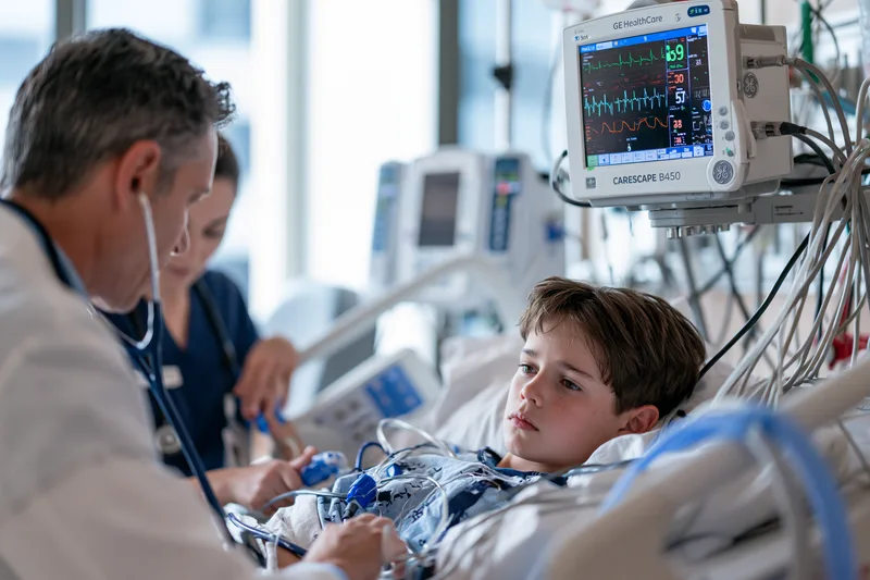
Thalassemia symptoms are often chronic and predictable, but some changes require urgent medical assessment because they may indicate severe anaemia, infection, increased haemolysis, transfusion complications, cardiac problems, splenic injury or thrombosis. Patients and caregivers should pay particular attention to symptoms that are sudden, rapidly worsening or clearly different from the person’s usual pattern.[2,4]
Severe Breathlessness, Chest Pain or Fainting
Breathlessness during strenuous activity can occur with anaemia, but breathlessness at rest or with minimal movement is more concerning. It may indicate a substantial fall in haemoglobin, cardiac dysfunction, pulmonary disease, infection or another acute complication.
Chest pain, fainting, near-fainting or a sustained rapid or irregular heartbeat requires prompt assessment. These symptoms should not be assumed to result from routine anaemia, even in a person with longstanding thalassemia.
Patients with transfusion-dependent disease may have cardiac risk related to cumulative iron exposure, while non-transfusion-dependent patients can develop pulmonary hypertension or other circulatory complications. Cardiac symptoms therefore require investigation according to the individual disease pattern and treatment history.[2,4]
A clinician may assess haemoglobin, oxygen saturation, heart rhythm, blood pressure, cardiac biomarkers and imaging depending on the presentation. When symptoms are severe or rapidly progressing, emergency care is appropriate.
Sudden Severe Weakness or a Rapid Fall in Haemoglobin
A patient who becomes markedly weaker, more pale, dizzy or breathless over hours or days may have experienced an acute fall in haemoglobin. Possible causes include infection, increased red blood cell destruction, bleeding, temporary suppression of bone marrow activity or a delayed transfusion reaction.
Children may become unusually sleepy, stop playing, feed poorly or appear less responsive. Adults may notice that ordinary walking becomes difficult, standing causes dizziness or they cannot complete normal daily activities.
A complete blood count should be compared with the patient’s previous baseline rather than interpreted in isolation. Reticulocyte count, bilirubin, liver tests and other investigations may be required to determine whether red cells are being destroyed, inadequately produced or lost through bleeding.[2,4]
Profound weakness, confusion, fainting or difficulty breathing requires urgent assessment rather than waiting for the next routine clinic appointment.
Rapidly Worsening Jaundice or Very Dark Urine
Mild intermittent jaundice can occur in haemolytic forms of thalassemia, but a sudden increase in yellowing of the eyes or skin may indicate accelerated red blood cell destruction, liver dysfunction, infection or a transfusion-related complication.
Very dark, brown or tea-coloured urine may accompany haemolysis, although dehydration, liver disease and urinary conditions can produce a similar appearance. Dark urine associated with fever, weakness, back pain, reduced urine output or rapidly worsening jaundice requires prompt medical review.
The assessment may include haemoglobin, reticulocyte count, bilirubin fractions, liver enzymes, kidney function and testing for haemolysis. Recent transfusions, infections and new medicines should also be reviewed.
A patient should not increase fluid intake or use home treatment as the sole response to sudden jaundice or dark urine when other systemic symptoms are present.
Severe Abdominal Pain or Sudden Abdominal Enlargement
An enlarged spleen can cause chronic fullness beneath the left ribs, but sudden severe pain in the upper abdomen is different and requires assessment. Splenic injury or rupture can occur after trauma and may cause abdominal pain, left shoulder pain, dizziness, weakness or fainting.
Patients with known splenomegaly should seek urgent care after significant abdominal trauma even if the initial pain appears manageable. Internal bleeding may not always be immediately obvious.
Rapid abdominal enlargement can also reflect increasing liver or spleen size, fluid accumulation or another abdominal problem. New severe pain, vomiting, fever or marked abdominal tenderness should not be attributed automatically to thalassemia.
Right upper abdominal pain with fever, vomiting or worsening jaundice may suggest gallbladder or bile-duct disease, which can occur in patients with chronic haemolysis.[2]
High Fever, Especially After Splenectomy
Fever requires particular attention in patients whose spleen has been removed. The spleen plays an important role in protection against certain bacterial infections, and severe infection can progress rapidly after splenectomy.[4]
A high fever, shaking chills, confusion, severe weakness, rapid breathing or a sudden deterioration in general condition requires urgent medical evaluation. Patients should follow the emergency infection plan provided by their haematology team rather than waiting to see whether the fever resolves spontaneously.
Vaccination and, in selected patients, preventive antibiotics reduce risk but do not eliminate it. Adults and families should ensure that emergency clinicians know about the previous splenectomy because this information can influence the urgency of treatment.
Children without a spleen also require rapid assessment for fever. Parents should keep the child’s vaccination history, medication list and emergency instructions readily available.
Symptoms During or After a Blood Transfusion
Fever, chills, flushing, itching, hives, back pain, chest discomfort, breathlessness, nausea or sudden weakness during a transfusion should be reported immediately to the transfusion team. The transfusion may need to be stopped while the reaction is assessed.[4]
Severe breathlessness, facial swelling, low blood pressure, chest pain or collapse may indicate a serious acute transfusion reaction and requires emergency treatment.
Some reactions occur after the patient has left the transfusion centre. Increasing jaundice, dark urine, unexplained fever, worsening fatigue or an unexpected fall in haemoglobin days after transfusion can be associated with delayed haemolysis or other transfusion complications.
Patients should know whom to contact if symptoms develop after returning home. Keeping a record of transfusion dates, blood-group antibodies and previous reactions can help future transfusion teams reduce risk.
High Fever With Sudden Pallor or Weakness
Infection can increase metabolic demand and may worsen anaemia or haemolysis. A child or adult who develops fever together with rapidly increasing pallor, weakness, breathlessness or jaundice needs clinical assessment.
Certain infections can temporarily suppress red blood cell production and cause a significant decline in haemoglobin, particularly in patients who already depend on increased red-cell production to maintain their baseline.
Reduced oral intake and dehydration can further worsen dizziness, rapid heartbeat and weakness. The severity of the patient’s overall condition is more important than the fever temperature alone.
A patient who is difficult to wake, confused, breathing rapidly or unable to drink should receive urgent care.
One-Sided Leg Swelling or Sudden Unexplained Breathlessness
Some patients with thalassemia have an increased risk of thrombosis, particularly in certain non-transfusion-dependent states and after splenectomy.[2,4]
Painful swelling affecting one leg, sudden chest pain, unexplained shortness of breath, rapid breathing or coughing blood may indicate a blood clot in the leg or lungs. These symptoms require emergency medical assessment.
Risk is influenced by age, splenectomy status, transfusion pattern, pregnancy, immobility and other medical factors. Not every patient with thalassemia requires anticoagulant treatment, but suspected thrombosis should never be managed by observation alone.
Long-distance travel, surgery and prolonged immobilisation may temporarily increase clotting risk in susceptible patients, making individual preventive planning important.
New Neurological Symptoms
Sudden weakness or numbness affecting one side of the body, facial drooping, difficulty speaking, loss of vision, severe unexplained headache, confusion or loss of coordination requires emergency evaluation.
These symptoms may indicate a stroke or another neurological emergency and should not be attributed to fatigue, anaemia or stress.
Patients with additional thrombotic risk factors, including previous splenectomy or a history of blood clots, should be particularly alert to abrupt neurological changes.[2,4]
Rapid assessment is important because treatment options for neurological emergencies are often time-sensitive.
Severe Symptoms in Babies and Young Children
Babies and toddlers cannot reliably describe breathlessness, dizziness or palpitations. Parents may instead notice rapid breathing, poor feeding, difficulty waking, unusual limpness, persistent crying, reduced urine output or a sudden loss of interest in play.
Bluish lips, severe breathing difficulty, chest retractions, fainting or markedly reduced responsiveness requires emergency care.
Rapidly worsening pallor, jaundice, abdominal swelling or dark urine also deserves prompt assessment, particularly when accompanied by fever or reduced feeding.
A child with known thalassemia who suddenly behaves very differently from usual should be assessed on the basis of that change, even if the most recent haemoglobin result was considered acceptable.
Symptoms That Require Prompt Reassessment Even Without an Emergency
Not every important change requires emergency treatment. Progressive fatigue, declining school or work performance, increasing transfusion requirements, slower growth, delayed puberty, worsening abdominal fullness, persistent bone pain or new endocrine symptoms should still trigger timely review.[2,4]
These changes may indicate that disease control, transfusion timing, chelation, iron burden, spleen size, nutritional status or endocrine function needs reassessment.
Patients should report changes in functional capacity as well as laboratory results. Needing more rest, avoiding stairs, missing school or work, eating less because of early satiety or taking longer to recover after ordinary activity can provide early evidence that the current treatment plan is no longer meeting the patient’s needs.
Comparing new symptoms with the individual’s established baseline helps distinguish a predictable chronic pattern from a clinically important change that requires investigation.
How Doctors Confirm Thalassemia After Symptoms Appear
Thalassemia symptoms such as persistent fatigue, pallor, breathlessness, poor growth, jaundice or abdominal enlargement cannot confirm the diagnosis on their own. Doctors combine the patient’s history, physical examination, complete blood count, iron studies and haemoglobin analysis to determine whether thalassemia is present and how clinically significant it may be. Genetic testing is sometimes required to identify the precise alpha- or beta-globin gene changes.[2]
Table: How Doctors Confirm Thalassemia After Symptoms Appear
| Test | What it assesses | Why the result matters |
|---|---|---|
| Complete blood count | Haemoglobin, red blood cell count, MCV, MCH, RDW, white cells and platelets | Identifies anaemia and persistent microcytosis and provides a baseline for future comparison |
| Ferritin and iron studies | Iron stores and available circulating iron | Helps distinguish iron deficiency from inherited microcytosis and detects coexisting deficiency |
| Peripheral blood smear | Red-cell size, colour, shape, target cells and other abnormalities | Supports the differential diagnosis but does not independently confirm thalassemia |
| Haemoglobin electrophoresis or HPLC | HbA, HbA2, HbF and haemoglobin variants | Helps detect beta-thalassemia patterns and other haemoglobin disorders |
| Genetic testing | Alpha- or beta-globin gene deletions and sequence variants | Confirms uncertain diagnoses, defines subtype and clarifies reproductive risk |
| Reticulocyte count and bilirubin | Bone-marrow response and red-cell breakdown | Helps investigate jaundice, haemolysis or an acute fall in haemoglobin |
| Liver and cardiac MRI iron measurement | Iron concentration in the liver and heart | Detects organ iron that may not be reflected accurately by symptoms or one ferritin result |
| Growth, endocrine and bone assessment | Height velocity, puberty, thyroid, glucose, sex hormones and bone density | Identifies age-related complications in clinically significant disease |
Medical and Family History
The assessment begins with the age at which symptoms appeared, how quickly they developed and whether the patient has previously been treated for anaemia. Symptoms beginning during infancy, persistent microcytosis from childhood or repeated anaemia that does not correct as expected with medically supervised iron therapy can increase suspicion of thalassemia.
Doctors review fatigue, breathlessness, jaundice, dark urine, abdominal fullness, bone pain, growth, pubertal development and exercise tolerance. For patients already receiving transfusions, the timing of symptoms in relation to the transfusion cycle, previous reactions, chelation treatment and changes in transfusion requirements are also important.[4]
Family history may reveal relatives with thalassemia trait, unexplained microcytosis, transfusion-dependent anaemia, jaundice, splenectomy or a known haemoglobin variant. The absence of an established family diagnosis does not exclude thalassemia because mild carriers may remain undetected for generations.
Ancestral background may provide additional context because thalassemia is more prevalent in people with origins in the Mediterranean region, Middle East, South Asia, Southeast Asia and parts of Africa. Testing should not be restricted by ethnicity, however, because migration and mixed ancestry mean that thalassemia can occur in any population.[2]
Physical Examination
The physical examination assesses whether anaemia or chronic haemolysis has produced visible clinical changes. Doctors may look for pallor, yellowing of the eyes or skin, rapid heartbeat, delayed growth, pubertal delay or signs of reduced physical development.
The abdomen is examined for enlargement of the spleen or liver. Splenomegaly can contribute to abdominal fullness, early satiety and worsening blood counts, while hepatomegaly may reflect increased blood-cell production, iron accumulation, infection or another liver condition.
In children and teenagers, height, weight and growth velocity are compared with previous measurements. Pubertal development may also be assessed when growth has slowed or expected sexual maturation is delayed.
Bone tenderness, spinal changes, loss of height or previous fractures may prompt investigation of bone density and endocrine health. These findings are associated with clinically significant disease and are not expected in an uncomplicated carrier state.[2,4]
Complete Blood Count and Red-Cell Indices
The complete blood count measures haemoglobin, red blood cell count, mean corpuscular volume, mean corpuscular haemoglobin, red cell distribution width, white blood cells and platelets.
A low mean corpuscular volume indicates microcytosis, while a low mean corpuscular haemoglobin means that each red blood cell contains less haemoglobin than expected. These findings are common in both thalassemia and iron deficiency, so they cannot determine the cause by themselves.[2]
In thalassemia trait, the red blood cell count may remain relatively preserved or be higher than expected for the degree of anaemia. In iron deficiency, the red blood cell count is often lower or normal. This pattern can guide further investigation but is not sufficiently reliable to replace iron studies and haemoglobin analysis.
Red cell distribution width reflects variation in red-cell size. It is often increased in iron deficiency and may be less markedly elevated in thalassemia trait, although overlap is common, particularly when both conditions are present.
White blood cell and platelet counts are also clinically relevant. Low counts may occur when an enlarged spleen retains or removes blood cells more rapidly. Abnormal results require interpretation alongside spleen size, infection history, medicines and the wider clinical picture.
The haemoglobin result should be compared with previous values whenever possible. A patient with chronic stable anaemia may tolerate a haemoglobin level that would cause severe symptoms if reached rapidly, while a sudden fall from the individual baseline can indicate bleeding, infection, increased haemolysis or temporary bone-marrow suppression.
Peripheral Blood Smear
A peripheral blood smear allows the laboratory to examine the size, colour and shape of red blood cells. Thalassemia may produce microcytosis, hypochromia, target cells, variation in red-cell shape and nucleated red blood cells in more severe disease.[2]
These appearances support the investigation but are not diagnostic on their own. Iron deficiency and several other disorders can produce overlapping changes.
The smear may also reveal findings that suggest an alternative or additional diagnosis. This is important when the severity of the anaemia, jaundice or blood-count abnormalities does not fit the expected pattern of uncomplicated thalassemia trait.
Ferritin and Iron Studies
Ferritin and related iron studies help distinguish thalassemia-related microcytosis from iron deficiency. A low ferritin level strongly supports depleted iron stores in most clinical settings.[2]
Ferritin can rise during inflammation, infection and liver disease, so a normal or elevated result does not always exclude iron deficiency. Transferrin saturation, serum iron, total iron-binding capacity and inflammatory markers may provide additional information when the result is difficult to interpret.
Iron deficiency and thalassemia can occur together. In this situation, iron replacement may improve haemoglobin and iron stores, but the mean corpuscular volume may remain low because the inherited thalassemia pattern persists.
Patients with repeated transfusions require a different interpretation. An elevated ferritin may reflect transfusional iron accumulation, inflammation, liver injury or several factors together. Ferritin trends are useful for monitoring, but they do not accurately show how much iron is present in the liver or heart.[4]
Iron should not be prescribed solely because the haemoglobin, mean corpuscular volume or mean corpuscular haemoglobin is low. Treatment is appropriate when deficiency has been demonstrated or is strongly supported by the full clinical assessment.
Haemoglobin Electrophoresis and High-Performance Liquid Chromatography
Haemoglobin electrophoresis and high-performance liquid chromatography measure the proportions of different haemoglobin types. These tests help identify beta-thalassemia trait, clinically significant beta thalassemia and several other haemoglobin variants.[2]
Beta-thalassemia trait is commonly associated with an increased haemoglobin A2 level. Haemoglobin F may also be elevated in some patients, although the pattern depends on age, genotype and coexisting haemoglobin variants.
Clinically significant beta thalassemia can produce reduced or absent haemoglobin A together with increased haemoglobin F. The exact pattern varies, so interpretation should be performed in conjunction with the complete blood count, transfusion history and clinical presentation.
Iron deficiency can sometimes reduce haemoglobin A2 and make beta-thalassemia trait less obvious. When suspicion remains high, confirmed deficiency may need to be treated before haemoglobin analysis is repeated.
Recent transfusion can also alter the result because the sample contains both the patient’s red cells and transfused donor cells. The laboratory and clinician should know the date of the most recent transfusion before interpreting haemoglobin fractions.
Routine adult haemoglobin electrophoresis may be normal in alpha-thalassemia trait. A normal result therefore does not exclude alpha thalassemia when persistent microcytosis, normal iron stores and family history continue to support the diagnosis.[2]
Genetic Testing
Genetic testing can identify deletions or sequence changes affecting alpha- or beta-globin genes. It is particularly useful when blood tests are inconclusive, alpha thalassemia is suspected or precise classification is needed for reproductive counselling.[2]
In alpha thalassemia, genetic testing can determine how many alpha-globin genes are affected and whether two affected genes are located on the same chromosome or on opposite chromosomes. This distinction can substantially change the reproductive risk for a couple.
In beta thalassemia, molecular testing can identify the specific beta-globin variants and help distinguish carrier status from compound or clinically significant disease. It may also clarify unexpected differences between symptoms and haemoglobin-analysis findings.
Genetic results should be interpreted alongside the clinical phenotype. Two patients with related mutations can still have different haemoglobin levels, transfusion requirements and complication patterns because additional genetic and environmental factors influence disease expression.
Testing may also be offered to biological relatives once a clinically relevant variant has been identified. This can detect healthy carriers and support informed partner testing before pregnancy.
Reticulocyte Count and Markers of Haemolysis
Reticulocytes are young red blood cells released by the bone marrow. The reticulocyte count helps show whether the marrow is responding to anaemia by increasing red-cell production.
A raised reticulocyte count may support ongoing haemolysis, while an unexpectedly low count in a patient with worsening anaemia can suggest temporary suppression of red-cell production or another marrow problem. Interpretation depends on the haemoglobin level and the degree of anaemia.
Bilirubin, lactate dehydrogenase and haptoglobin may be assessed when increased red-cell destruction is suspected. Thalassemia can produce chronic haemolysis, but a sudden change in these markers may indicate an acute infection, transfusion reaction or another cause of accelerated destruction.
Liver function tests are important because jaundice can arise from haemolysis, liver injury, gallstone-related obstruction or several processes occurring together. New or worsening jaundice should not be attributed to thalassemia without appropriate evaluation.
Tests Used to Determine Disease Severity
Once thalassemia is confirmed, doctors assess whether the patient has a carrier state, non-transfusion-dependent disease or transfusion-dependent disease. This classification is based on the overall clinical course, haemoglobin pattern, transfusion requirement, growth, spleen size and complications rather than one test result.[2,4]
Patients with clinically significant disease may undergo repeated complete blood counts, bilirubin testing, liver and kidney function tests and assessment of transfusion requirements. Children require regular measurement of height, weight and growth velocity.
Endocrine testing may include thyroid function, glucose regulation, pubertal hormones and other assessments guided by age and symptoms. Menstrual changes, delayed puberty, reduced libido, infertility or impaired growth can indicate endocrine complications that require targeted evaluation.[4]
Bone-density assessment may be appropriate for patients with fractures, persistent bone pain, delayed puberty or recognised risk factors for low bone mass. Vitamin D and other nutritional factors may also be reviewed.
Cardiac assessment can include an electrocardiogram, echocardiography and cardiac magnetic resonance imaging according to the patient’s transfusion and iron history. Palpitations, declining exercise tolerance, breathlessness or ankle swelling may require more urgent evaluation.
Liver assessment may include blood tests, ultrasound, elastography or other imaging when iron accumulation, fibrosis, viral infection or another liver disorder is suspected.
Measuring Iron Burden
Patients receiving repeated transfusions accumulate iron because the body cannot actively remove the large amount delivered in transfused red blood cells. Non-transfusion-dependent patients may also accumulate iron through increased intestinal absorption.[4]
Ferritin is monitored over time but is influenced by infection, inflammation and liver disease. A single ferritin value cannot accurately measure organ iron or determine whether the heart is affected.
Magnetic resonance imaging can estimate liver iron concentration and detect cardiac iron using validated techniques. These results help guide chelation therapy and identify clinically important accumulation before irreversible organ damage develops.[4]
A patient may have significant iron overload without noticeable symptoms. Monitoring should therefore follow the person’s transfusion exposure, disease category, ferritin trend and previous imaging results rather than waiting for liver, heart or endocrine symptoms to appear.
Distinguishing a Carrier From Clinically Significant Thalassemia
A carrier usually has stable microcytosis, normal or mildly reduced haemoglobin and little or no functional limitation. Regular transfusions, progressive spleen enlargement, substantial jaundice, poor growth, delayed puberty or thalassemia-related organ complications are not expected from uncomplicated trait.[2]
Non-transfusion-dependent disease produces a broader clinical range. The patient may live without scheduled transfusions but still experience chronic anaemia, haemolysis, splenomegaly, bone disease, iron accumulation or endocrine complications.
Transfusion-dependent disease generally requires scheduled red-cell transfusions to maintain adequate haemoglobin, support growth and suppress ineffective blood formation. Classification can change with the clinical course, but temporary transfusion during infection, surgery or pregnancy does not automatically establish permanent transfusion dependence.[4]
Accurate diagnosis therefore combines laboratory findings with the patient’s actual function, growth, transfusion history and evidence of complications. This allows treatment and monitoring to match the severity of the disease rather than the diagnostic label alone.[2,4]
From Thalassemia Symptoms to a Personalised Recovery Plan

Thalassemia symptoms such as fatigue, pallor, poor growth, abdominal enlargement, delayed puberty or reduced exercise tolerance are not merely problems to suppress; they are clinical signals that help define the patient’s haemoglobin status, transfusion needs, iron burden, organ health and long-term treatment priorities. A meaningful recovery plan begins by identifying the exact thalassemia type and measuring how the condition is affecting the patient rather than relying on symptoms alone.[2,4]
What Recovery Can Mean in Thalassemia
Recovery does not have one identical meaning for every patient. For a healthy carrier, the priority may be confirming the diagnosis, avoiding unnecessary iron treatment and understanding reproductive risk. For non-transfusion-dependent thalassemia, the goals may include improving stamina, reducing anaemia-related limitations, protecting the bones and organs, monitoring iron accumulation and preventing avoidable complications.
For transfusion-dependent thalassemia, recovery may involve maintaining an appropriate pre-transfusion haemoglobin level, supporting normal growth and puberty, preventing iron-related organ injury and evaluating eligibility for potentially curative treatment. Haematopoietic stem-cell transplantation and approved gene-based therapies can offer the possibility of transfusion independence in selected patients, but suitability depends on age, genotype, organ function, donor availability, previous treatment and access to specialist care.[4]
Symptom improvement is valuable, but it should be interpreted with measurable clinical results. A patient may feel more energetic while iron burden continues to rise, or may remain tired despite an acceptable haemoglobin level because of thyroid dysfunction, nutritional deficiency, sleep disturbance, bone disease or another complication. Recovery therefore requires regular comparison of symptoms with blood tests, transfusion records, imaging and functional progress.
Families seeking a broader understanding of cure, transfusion independence, long-term disease control and integrative care can read Curing Thalassemia: Fact, Fiction and Future. This detailed guide explains how conventional haematology, stem-cell transplantation, gene-based treatment, complication monitoring and individualised Ayurveda-centred care can be considered within one clinically accountable treatment pathway.
Building the Plan Around the Patient’s Confirmed Needs
The first step is to establish whether the patient has a carrier state, non-transfusion-dependent thalassemia or transfusion-dependent disease. This classification determines whether the patient primarily needs counselling, periodic monitoring, intermittent transfusion support, scheduled transfusions, iron chelation or assessment for a potentially curative intervention.[2,4]
The plan should document the patient’s baseline haemoglobin, mean corpuscular volume, mean corpuscular haemoglobin, reticulocyte count, bilirubin, ferritin and haemoglobin-analysis findings. Genetic results, when available, can clarify the precise diagnosis and support family testing and reproductive counselling.
In children, height, weight, annual growth velocity, appetite, school attendance, exercise capacity and pubertal development provide essential information. A child whose haemoglobin appears stable may still require treatment reassessment if growth is slowing, the spleen is enlarging or participation in ordinary activities is declining.
In adults, the assessment should include work capacity, breathlessness, palpitations, menstrual or reproductive symptoms, bone pain, abdominal fullness and changes in daily function. Liver, cardiac, endocrine and bone investigations may be necessary when symptoms or treatment history suggest cumulative complications.
Transfusion-dependent patients require review of transfusion frequency, pre-transfusion haemoglobin, reactions, blood-group antibodies and the duration of benefit after each transfusion. Increasing fatigue before the next appointment or rising transfusion requirements can indicate that the current schedule, spleen status or disease control needs reassessment.[4]
Iron management must remain central even when the patient feels well. Ferritin trends, liver iron concentration and cardiac iron assessment can reveal clinically important accumulation before obvious liver, heart or endocrine symptoms develop. Chelation should be adjusted according to transfusion exposure, organ iron and treatment tolerance rather than a single ferritin result.[4]
Integrating Ayurveda With Objective Monitoring
Ayurvedic care can be individualised around the patient’s appetite, digestion, bowel pattern, sleep, strength, exercise tolerance, growth and recovery after illness or transfusion. These areas influence quality of life and can be followed alongside haematological treatment without replacing the established diagnosis or necessary specialist care.
The Ayurvedic concept of Agni, meaning digestive and metabolic capacity, can be clinically relevant when poor appetite, early satiety, irregular digestion or inadequate nutritional intake is limiting strength and growth. Management should remain adapted to the patient’s constitution, symptoms, age, liver status, iron burden, transfusion schedule and concurrent medicines.
Rasayana, the Ayurvedic approach concerned with nourishment, resilience and restoration, can provide a framework for supporting strength, recovery capacity and long-term wellbeing. Classical Rasayana principles are described in the Charaka Samhita, Chikitsa Sthana, Chapter 1. Their use in thalassemia should be individualised and evaluated through measurable outcomes rather than assumed from the traditional category alone.
Pallor, weakness, reduced appetite and diminished functional strength may also be considered through the classical clinical framework of Pandu Roga, described in the Charaka Samhita, Chikitsa Sthana, Chapter 16. This comparison can guide symptom-oriented Ayurvedic reasoning, but Pandu Roga should not be treated as an exact classical equivalent of a genetically confirmed thalassemia diagnosis.
Any Ayurvedic formulation should be selected only after reviewing the patient’s haemoglobin pattern, ferritin, liver and kidney function, spleen status, transfusion history, chelation medicines and other treatments. Products containing iron or other metals require particular caution in patients with known or possible iron overload, and unsupervised supplementation should be avoided.
Progress can be measured through changes in appetite, energy, sleep, bowel function, weight, growth, school or work participation and recovery after exertion. These outcomes should be reviewed together with haemoglobin trends, transfusion frequency, ferritin, liver tests, kidney tests and relevant organ assessments.
A responsible integrated plan does not ask the patient to choose between Ayurveda and essential haematology care. It coordinates supportive Ayurvedic treatment with transfusions, chelation, infection prevention, endocrine care, bone-health management and specialist assessment for curative options when appropriate.
Preparing for an Individualised Thalassemia Consultation
A useful consultation requires more than a recent haemoglobin result. The patient should provide the complete blood count with red-cell indices, haemoglobin electrophoresis or high-performance liquid chromatography, ferritin and iron studies, bilirubin, liver and kidney tests and any available genetic report.
Patients receiving transfusions should provide the dates of recent transfusions, pre-transfusion haemoglobin results, the amount of blood received, previous reactions and any known red-cell antibodies. The current chelation medicine, dose, adherence pattern and adverse effects should also be documented.
Children benefit from bringing previous height and weight records, growth charts, school observations and details of appetite, physical activity and pubertal development. Adults should include information about exercise tolerance, employment-related fatigue, menstrual history, fertility concerns, bone pain and changes in daily function.
Cardiac magnetic resonance imaging, liver iron assessment, echocardiography, endocrine reports, bone-density scans and specialist summaries should be included when available. Reviewing these records together helps identify the most urgent priorities and prevents treatment decisions from being based only on symptoms.
A personalised plan can then define specific goals for the next stage of care. These may include improving daily energy, supporting appetite and growth, maintaining safer haemoglobin levels, reducing pre-transfusion symptoms, strengthening chelation adherence, protecting the liver and heart, addressing endocrine or bone complications and determining whether the patient should be evaluated for a potentially curative treatment.
Progress should be reassessed at predefined intervals using the same clinical and laboratory measures. This allows patients and families to see whether the plan is producing meaningful improvement and enables treatment to be adjusted before complications become advanced.
What to Do After Recognising Possible Thalassemia Symptoms
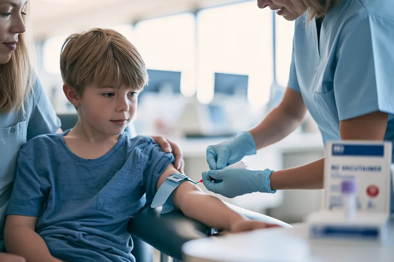
Thalassemia symptoms such as persistent pallor, unexplained fatigue, reduced exercise tolerance, poor growth, jaundice or abdominal enlargement should lead to structured medical assessment rather than self-treatment. The immediate goal is to determine whether the symptoms are caused by thalassemia, iron deficiency, another form of anaemia or more than one condition occurring together.[2]
Arrange the Appropriate Blood Tests
A complete blood count with red-cell indices is usually the starting point. Haemoglobin, red blood cell count, mean corpuscular volume, mean corpuscular haemoglobin and red cell distribution width help identify the pattern of anaemia and whether persistent microcytosis is present.[2]
Ferritin and appropriate iron studies are needed because iron deficiency can produce a similar blood-count pattern. A low ferritin commonly supports iron deficiency, while persistent microcytosis with adequate iron stores increases suspicion of thalassemia or another haemoglobin disorder.[2]
Haemoglobin electrophoresis or high-performance liquid chromatography can help identify beta-thalassemia trait, clinically significant beta thalassemia and other haemoglobin variants. A normal haemoglobin electrophoresis result does not exclude alpha thalassemia, and genetic testing may be needed when clinical suspicion remains high.[2]
Patients who have recently received a blood transfusion should tell the clinician and laboratory because donor red blood cells can affect haemoglobin-analysis results. Previous laboratory reports are also valuable because a longstanding pattern of microcytosis may be more informative than one isolated blood count.
Avoid Treating Every Low MCV as Iron Deficiency
Low mean corpuscular volume and low mean corpuscular haemoglobin are common in both iron deficiency and thalassemia. These findings do not prove that the patient needs iron.[2]
Iron supplementation is appropriate when depleted iron stores have been confirmed or when the treating clinician has strong evidence of deficiency. Thalassemia trait itself does not improve with iron when iron stores are already adequate.
A patient can have thalassemia and iron deficiency at the same time. In that situation, treating the deficiency may improve haemoglobin and fatigue, while the mean corpuscular volume remains low because the inherited thalassemia pattern continues.
Long-term use of iron-containing tablets, tonics or multivitamins without laboratory confirmation should be avoided, particularly in patients who receive transfusions or already have evidence of iron accumulation. Patients should review all non-prescription products with their clinical team because iron may be included in combination supplements without being obvious from the product name.[4]
Collect Relevant Family Information
A family history can help identify inherited risk even when no relative has a formal thalassemia diagnosis. Useful information includes unexplained lifelong anaemia, persistent low MCV, previous blood transfusions, splenectomy, jaundice, gallstones, known haemoglobin variants or children born with severe anaemia.
Parents should bring newborn-screening results, previous paediatric blood counts and any haemoglobin or genetic reports available within the family. Adults should include previous antenatal screening, blood-donation findings and laboratory results from earlier medical examinations.
The absence of a known family history does not exclude thalassemia. Mild carriers may remain healthy and undiagnosed, allowing the variant to pass through several generations before two carriers have a child with clinically significant disease.
When one person is confirmed to carry a thalassemia variant, testing biological relatives can identify other carriers and prevent repeated misdiagnosis of inherited microcytosis as iron deficiency.
Arrange Partner Testing When Carrier Status Is Confirmed
A healthy carrier may have no treatment needs but can still pass a clinically significant globin-gene variant to a child. The reproductive risk depends on the exact variant carried by each biological parent.[2,5]
When one partner has beta-thalassemia trait, the other partner should be tested for beta thalassemia and other relevant haemoglobin variants. When both partners carry beta-thalassemia trait, each pregnancy has a one-in-four probability of producing a child with beta-thalassemia major.[5]
Alpha-thalassemia inheritance requires precise evaluation because risk depends on the number and arrangement of affected alpha-globin genes. Two healthy adults with mild microcytosis can have a pregnancy at risk of Haemoglobin H disease or severe fetal alpha thalassemia if their genetic findings form a high-risk combination.[2]
Genetic counselling can explain the result in practical terms and discuss available reproductive and prenatal diagnostic options. Counselling is most useful before pregnancy or early in pregnancy, when families have sufficient time to understand the implications.
Continue Essential Thalassemia Treatment
A patient with established thalassemia should not stop transfusions, chelation or other prescribed treatment solely because symptoms have improved or an additional supportive therapy has been started. A person may feel well while haemoglobin is falling or iron is continuing to accumulate.[4]
Transfusion schedules are based on disease severity, growth, symptoms, pre-transfusion haemoglobin and the need to suppress ineffective blood production. Missing or delaying transfusions can contribute to worsening fatigue, bone-marrow expansion, spleen enlargement and reduced growth in patients who depend on regular transfusion support.[4]
Chelation therapy reduces excess iron accumulated from repeated transfusions. Missing doses may not cause immediate symptoms, but prolonged under-treatment can increase the risk of liver, cardiac and endocrine injury. Adverse effects should be reported so that the medicine, dose or monitoring plan can be reviewed rather than discontinued without guidance.[4]
Patients using Ayurvedic or other complementary preparations should provide the complete ingredient list to the haematology team. This is particularly important when a formulation contains iron, metallic preparations or ingredients that may affect the liver, kidneys, blood glucose, clotting or absorption of prescribed medicines.
Record Symptoms in Relation to Treatment
A brief symptom record can help identify patterns that are difficult to recall during a consultation. Patients can document when fatigue, headache, breathlessness, palpitations, abdominal fullness or reduced concentration occurs and whether the symptoms change after transfusion.
Children may show symptoms through behaviour rather than verbal complaints. Parents can record changes in play, feeding, school attendance, sleep, appetite and the amount of rest required after ordinary activity.
Adults can note changes in walking distance, stair climbing, work capacity, exercise and recovery after exertion. Functional changes often provide clinically useful information even when the patient cannot describe the exact intensity of fatigue.
Symptoms should be interpreted together with laboratory results. A repeated decline before each transfusion may indicate that the transfusion plan needs review, while persistent fatigue despite stable haemoglobin may lead to assessment of thyroid function, iron burden, sleep, nutrition, cardiac health or another cause.[4]
Monitor Growth and Development in Children
Children with clinically significant thalassemia need regular measurement of height, weight and growth velocity. A gradual fall across growth percentiles may be detected before parents notice an obvious difference in body size.[4]
Pubertal development should also be reviewed at an age-appropriate stage. Delayed breast development, late menstruation, absent periods, limited testicular development or failure to experience the expected adolescent growth spurt may require endocrine evaluation.
Parents should bring earlier growth records whenever possible. Comparing measurements over time is more useful than relying on one clinic visit or comparing the child informally with classmates.
School performance, physical activity and attendance also provide information about the child’s functional health. Increasing absence, inability to participate in ordinary play or worsening concentration may indicate that anaemia or another complication is affecting daily life.
Request Specialist Review for Clinically Significant Disease
A haematology referral is appropriate when haemoglobin analysis suggests thalassemia, genetic testing is needed, anaemia is more severe than expected or the patient has jaundice, spleen enlargement, growth problems or a transfusion requirement.[2]
Patients with established non-transfusion-dependent or transfusion-dependent thalassemia benefit from coordinated assessment of anaemia, transfusion needs, iron burden, liver health, cardiac function, endocrine status and bone health.[4]
Children may require collaboration between paediatric haematology, endocrinology, nutrition and other specialists according to their growth and complication profile. Adults may need reproductive, cardiac, liver, endocrine or bone-health assessment based on symptoms and previous treatment exposure.
Evaluation for stem-cell transplantation or gene-based therapy should occur through an experienced thalassemia centre. Eligibility depends on several clinical factors, and early referral can provide families with accurate information before age, organ injury or disease complications narrow the available options.
Maintain Routine Preventive Care
Thalassemia care does not replace ordinary preventive medicine. Vaccination, dental care, nutrition, physical activity, infection assessment and age-appropriate screening remain important.
Patients who have undergone splenectomy need particular attention to vaccination, fever management and any prescribed antibiotic strategy. They should carry medical identification and ensure that emergency clinicians know they do not have a functioning spleen.[4]
Exercise can support cardiovascular fitness, bone strength and emotional wellbeing, but activity should be adapted for severe anaemia, cardiac disease, fractures or substantial spleen enlargement. Patients with splenomegaly should discuss contact sports and abdominal-injury risk with their treatment team.
Dietary changes should be individualised rather than based on broad restrictions. Patients should not remove nutritious iron-containing foods automatically because thalassemia is present. The effect of normal dietary iron differs from transfusional iron exposure, and dietary advice should consider iron burden, growth, nutrition and the wider clinical context.
A confirmed diagnosis, accurate classification and consistent monitoring allow symptoms to be interpreted correctly. This protects healthy carriers from unnecessary treatment while ensuring that patients with clinically significant thalassemia receive timely transfusion support, iron management, complication screening and assessment of long-term treatment options.[2,4]
Frequently Asked Questions About Thalassemia Symptoms
Thalassemia symptoms differ according to the genetic type, severity of anaemia, age, transfusion requirement and presence of complications. Symptoms can indicate that testing is needed, but no individual symptom can confirm thalassemia or determine whether a patient has a carrier state, non-transfusion-dependent disease or transfusion-dependent disease.
What Are the First Symptoms of Thalassemia?
Early symptoms may include persistent tiredness, weakness, pale skin, reduced exercise tolerance, breathlessness, headaches, dizziness or a rapid heartbeat. In children, poor feeding, reduced activity, slow weight gain or abdominal enlargement may be more noticeable. Mild carriers may have no symptoms and may be identified only through a blood test.
At What Age Do Thalassemia Symptoms Begin?
Severe beta thalassemia often becomes clinically apparent between approximately six and 24 months of age as fetal haemoglobin declines. Thalassemia trait may remain unnoticed throughout life, while non-transfusion-dependent forms can become more apparent during childhood, adolescence, pregnancy, infection or another period of increased physiological demand.
What Are the Early Signs of Thalassemia in Babies?
Possible early signs include persistent pallor, poor feeding, excessive sleepiness, irritability, reduced interest in play, slow weight gain, jaundice and progressive abdominal enlargement. These findings are not specific to thalassemia, so a complete blood count, iron studies and appropriate haemoglobin testing are needed to identify the cause.
What Are the Most Common Thalassemia Symptoms in Adults?
Adults with clinically significant thalassemia may experience chronic fatigue, breathlessness on exertion, palpitations, pallor, jaundice, abdominal fullness, bone pain or reduced exercise capacity. Menstrual, fertility, thyroid, glucose or cardiac symptoms may develop when endocrine or iron-related complications are present. Many adults with thalassemia trait remain asymptomatic.
Does Thalassemia Minor Cause Symptoms?
Thalassemia minor usually causes no symptoms or only mild anaemia, occasional tiredness or pallor. Significant breathlessness, severe weakness, recurrent jaundice, spleen enlargement or major limitation of daily activities is not typical of uncomplicated trait and should prompt investigation for iron deficiency, blood loss or another medical condition.
Can a Thalassemia Carrier Develop Thalassemia Major Later?
A carrier does not gradually develop thalassemia major. Carrier status is determined by the inherited globin-gene pattern and remains stable throughout life. Pregnancy, infection, blood loss or iron deficiency may make anaemia more noticeable, but this represents an additional clinical problem rather than progression from trait to major disease.
Can Thalassemia Be Mistaken for Iron Deficiency?
Thalassemia and iron deficiency can both cause low haemoglobin, low mean corpuscular volume and low mean corpuscular haemoglobin. Ferritin and other iron studies help distinguish them, while haemoglobin analysis supports the diagnosis of beta thalassemia. Alpha-thalassemia trait may require genetic testing when routine haemoglobin analysis is normal.
Can Thalassemia Cause a Swollen Stomach?
Clinically significant thalassemia can enlarge the spleen or liver, producing abdominal fullness, a visibly prominent abdomen, discomfort beneath the ribs or early satiety. A rapidly enlarging abdomen, severe pain, faintness or symptoms after abdominal trauma requires urgent medical assessment because an enlarged spleen is vulnerable to injury.
Can Thalassemia Cause Bone or Back Pain?
Bone and back pain can occur in clinically significant thalassemia because chronic ineffective blood formation, marrow expansion, delayed puberty, endocrine dysfunction and reduced bone density can weaken the skeleton. Persistent pain, loss of height or fractures require assessment for osteoporosis, vertebral compression, vitamin deficiency and other musculoskeletal causes.
Can Someone Have Thalassemia With Normal Haemoglobin?
A person can carry alpha or beta thalassemia while maintaining a haemoglobin level within the laboratory reference range. The complete blood count may still show persistent microcytosis and reduced mean corpuscular haemoglobin. Normal haemoglobin therefore does not exclude a carrier state, particularly when family history or partner screening raises concern.
Which Blood Test Confirms Thalassemia?
Diagnosis usually begins with a complete blood count, ferritin and iron studies. Haemoglobin electrophoresis or high-performance liquid chromatography can identify beta-thalassemia patterns and other haemoglobin variants. Genetic testing may be required to confirm alpha thalassemia, clarify an uncertain result or determine precise reproductive risk.
When Do Thalassemia Symptoms Require Emergency Care?
Emergency assessment is required for breathlessness at rest, chest pain, fainting, severe weakness, rapidly worsening jaundice, very dark urine, sudden abdominal pain, one-sided leg swelling or new neurological symptoms. High fever after splenectomy and fever, chills, breathing difficulty or back pain during or after transfusion also require urgent care.
Early Recognition of Thalassemia Symptoms Supports Better Decisions

Thalassemia symptoms become clinically useful when they are compared with the patient’s established haemoglobin level, growth pattern, transfusion history, iron burden and daily function. Persistent fatigue, pallor, reduced stamina, poor growth, jaundice or abdominal fullness should lead to appropriate testing rather than repeated treatment based on symptoms alone.[2,4]
An early and accurate diagnosis can distinguish a healthy carrier from a patient who needs continuing haematology care. This prevents unnecessary iron supplementation in people with adequate iron stores while helping patients with clinically significant disease receive timely transfusions, chelation, growth monitoring and assessment of organ complications.[2,4]
Children benefit when parents and clinicians recognise changes in feeding, play, growth, school participation and pubertal development before these problems become advanced. Adults should report declining work capacity, worsening breathlessness, palpitations, bone pain, menstrual changes or reduced exercise tolerance even when previous results were considered stable.
Patients who feel well still require monitoring appropriate to their disease category. Iron overload, endocrine dysfunction, reduced bone density and early cardiac or liver involvement may develop before obvious symptoms appear. Regular blood tests and recommended imaging therefore remain important even during periods of good energy and normal daily activity.[4]
A confirmed carrier diagnosis also creates an opportunity for family and partner testing. Because carriers are often asymptomatic, reproductive risk may remain unrecognised until both biological parents are tested. Genetic counselling can explain the likely outcomes for future pregnancies and clarify whether more detailed molecular testing is needed.[2,5]
Treatment decisions should be based on measurable goals such as stable haemoglobin, appropriate growth, improved functional capacity, controlled iron burden, protected organ function and reduced disruption to school, employment and family life. Symptom improvement is most meaningful when it is supported by objective laboratory and clinical progress.
Integrated care can include individualised nutritional and Ayurvedic support when it is coordinated with necessary transfusions, chelation, specialist monitoring and evaluation for potentially curative treatment. Any additional therapy should be reviewed for safety in relation to iron status, liver and kidney function, prescribed medicines and the patient’s underlying thalassemia type.
Sudden breathlessness, chest pain, fainting, rapidly worsening jaundice, very dark urine, severe abdominal pain, neurological symptoms or fever after splenectomy requires urgent medical assessment. Recognising these changes promptly can protect the patient from avoidable complications while maintaining a clear, evidence-based pathway for long-term recovery and care.[2,4]
References
[1] Centers for Disease Control and Prevention. (2024, May 15). About thalassemia.
https://www.cdc.gov/thalassemia/about/index.html
Brief: Provides an overview of thalassemia, including its genetic causes, types, symptoms, diagnosis, and treatment options.
[2] Baird, D. C., Batten, S. H., & Sparks, S. K. (2022). Alpha- and beta-thalassemia: Rapid evidence review. American Family Physician, 105(3), 272–280.
https://www.aafp.org/pubs/afp/issues/2022/0300/p272.html
Brief: Reviews the clinical presentation, diagnostic methods, genetic inheritance, and evidence-based management of alpha- and beta-thalassemia.
[3] Langer, A. L. (2026, February 12). Beta-thalassemia. In M. P. Adam, S. Bick, G. M. Mirzaa, R. A. Pagon, S. E. Wallace, & A. Amemiya (Eds.), GeneReviews®. University of Washington, Seattle.
https://www.ncbi.nlm.nih.gov/books/NBK1426/
Brief: Discusses the genetic basis, clinical manifestations, diagnosis, treatment, and genetic counseling of beta-thalassemia.
[4] Farmakis, D., Porter, J., Taher, A., Cappellini, M. D., Angastiniotis, M., & Eleftheriou, A. (2022). 2021 Thalassaemia International Federation guidelines for the management of transfusion-dependent thalassemia. HemaSphere, 6(8), Article e732.
Brief: Presents international clinical recommendations for transfusion therapy, iron chelation, complication monitoring, and comprehensive care of transfusion-dependent thalassemia.
[5] National Health Service. (2022, October 17). Thalassaemia carriers.
https://www.nhs.uk/conditions/thalassaemia/carriers/
Brief: Explains thalassemia carrier status, inheritance patterns, reproductive risks, carrier screening, and genetic counseling.
[6] Taher, A. T., Musallam, K. M., & Cappellini, M. D. (2023). Guidelines for the management of non-transfusion-dependent β-thalassaemia (3rd ed.). Thalassaemia International Federation.
https://www.ncbi.nlm.nih.gov/books/NBK599489/
Brief: Provides recommendations for the diagnosis, monitoring, prevention, and treatment of complications associated with non-transfusion-dependent beta-thalassemia.
[7] Tamary, H., Greenberg-Kushnir, N., & Dgany, O. (2026, April 23). Alpha-thalassemia. In M. P. Adam, S. Bick, G. M. Mirzaa, R. A. Pagon, S. E. Wallace, & A. Amemiya (Eds.), GeneReviews®. University of Washington, Seattle.
https://www.ncbi.nlm.nih.gov/books/NBK1435/
Brief: Describes the genetic causes, clinical spectrum, diagnostic approaches, management, and reproductive counseling related to alpha-thalassemia.
[8] Musallam, K. M., Cappellini, M. D., Porter, J. B., Farmakis, D., Eleftheriou, A., Angastiniotis, M., & Taher, A. T. (2025). TIF guidelines for the management of transfusion-dependent β-thalassemia. HemaSphere, 9(3), Article e70095.
Brief: Presents updated international recommendations for managing transfusion-dependent beta-thalassemia, including blood transfusion, iron overload, long-term complications, and emerging treatments.
[9] Singh, R. H., Sodhi, J. S., & Dixit, U. (2020). Rasayana Adhyaya. In U. Dixit, Y. S. Deole, & G. Basisht (Eds.), Charak Samhita New Edition (Chikitsa Sthana, Chapter 1). Charak Samhita Research, Training and Skill Development Centre.
https://www.carakasamhitaonline.com/index.php/Rasayana_Adhyaya
Brief: Explains the Ayurvedic concept of Rasayana therapy, emphasizing rejuvenation, nourishment, longevity, and the promotion of general health and vitality.
[10] Kar, A. C., Rai, S., Aladoriya, N., & Deole, Y. S. (2020). Pandu Chikitsa. In G. Singh, M. Goyal, Y. S. Deole, & G. Basisht (Eds.), Charak Samhita New Edition (Chikitsa Sthana, Chapter 16). Charak Samhita Research, Training and Skill Development Centre.
https://www.carakasamhitaonline.com/index.php/Pandu_Chikitsa
Brief: Describes the Ayurvedic understanding of Pandu Roga, including its causes, clinical features, pathogenesis, dietary recommendations, and traditional management approaches. Pandu Roga should not be considered medically equivalent to thalassemia.