




- Bulbar ALS Symptoms at a Glance
- What Bulbar ALS Symptoms Mean and What Bulbar Onset Describes
- When Bulbar ALS Symptoms Need Urgent Medical Assessment
- How Bulbar ALS Symptoms Are Diagnosed
- Speech Support for Bulbar ALS Symptoms
- Speech Support for Bulbar ALS Symptoms
- Swallowing Care for Bulbar ALS Symptoms
- Saliva Care for Bulbar ALS Symptoms
- Nutrition and Weight Support
- Feeding Tubes in Bulbar ALS
- Breathing Support for Bulbar ALS Symptoms
- Multidisciplinary Bulbar ALS Care
- Ayurvedic Support for Bulbar ALS Symptoms
- 30-Day Bulbar ALS Symptom Monitoring
- Bulbar ALS Symptoms: Clinic Questions
- FAQs
- References
Bulbar ALS Symptoms at a Glance
Bulbar ALS symptoms affect the muscles used for speaking, chewing, swallowing, controlling saliva, protecting the airway and producing an effective cough. Early changes may include slurred speech, an altered voice, coughing while drinking, longer mealtimes, saliva pooling, unexplained weight loss or difficulty clearing mucus. Because these functions are closely connected, even a mild change should be discussed with the multidisciplinary ALS team rather than monitored in isolation.[1,2]
The pattern is not identical in every patient. Some people first notice that speech becomes less clear when they are tired, while others develop difficulty managing liquids, chewing firm foods or swallowing tablets. Bulbar dysfunction may be present at the beginning of ALS or develop after weakness has already appeared in the arms, legs or breathing muscles. Regular assessment helps identify clinically important change before communication, hydration, nutrition or respiratory safety is substantially affected.[1,2]
Table : Bulbar ALS Symptoms at a Glance
| Area affected | What the patient may notice | Clinical concern | Useful assessment |
|---|---|---|---|
| Speech | Slurred, nasal, strained, softer or slower speech | Reduced intelligibility and communication fatigue | Speech-language assessment |
| Swallowing | Coughing, choking, wet voice, food sticking, longer meals | Aspiration, dehydration and reduced intake | Clinical swallow assessment, VFSS or FEES when indicated |
| Saliva | Drooling, pooling, sticky mucus or dry mouth | Choking, discomfort and difficult secretion clearance | Saliva, swallowing and respiratory review |
| Nutrition | Weight loss, smaller meals, eating fatigue | Inadequate calories, protein and fluids | Weight trend and dietetic assessment |
| Cough | Weak or ineffective cough | Retained secretions and reduced airway clearance | Cough assessment and peak cough flow where appropriate |
| Breathing | Orthopnoea, morning headache, disturbed sleep, daytime sleepiness | Nocturnal hypoventilation and respiratory impairment | FVC/VC, SNIP/MIP and sleep-related testing |
Speech and Voice Changes
Speech may become slower, softer, slurred, nasal, hoarse, strained or breathy. Weakness of the tongue and lips can reduce the precision of consonants, while reduced movement of the soft palate may create a nasal quality. Weak respiratory support can shorten sentences, lower speaking volume and make sustained conversation tiring. Patients may remain understandable in quiet surroundings but struggle during telephone calls, group conversations or later in the day.[3]
Frequent requests to repeat words, difficulty being understood by unfamiliar listeners and avoidance of conversation may indicate that speech support is needed. Early referral to a speech-language pathologist allows speaking rate, intelligibility, voice quality and communication fatigue to be assessed while the patient can still participate fully in decisions about voice banking, message banking and future communication devices.[1–3]
Swallowing and Chewing Changes
Swallowing changes can begin with coughing after a sip of water, difficulty controlling mixed textures, food collecting inside the cheeks or the sensation that tablets are becoming harder to swallow. Weak tongue movement may make it difficult to form and move a mouthful of food, while reduced lip closure can allow liquids or saliva to escape. Chewing may become slow or tiring, particularly with dry, fibrous or firm foods.[2,4]
A wet or gurgling voice after eating or drinking may indicate that material remains near the airway. Other clinically important signs include repeated throat clearing, nasal regurgitation, breathlessness during meals, avoidance of particular foods and progressively longer mealtimes. Coughing does not always occur when material enters the airway, so the absence of choking does not by itself confirm that swallowing is safe.[2,4]
Changes in swallowing should lead to assessment by a speech-language pathologist and dietitian. Food consistency, fluid texture, posture and pacing need to be selected according to the individual swallowing pattern. Patients should not automatically thicken all liquids or remove major food groups without assessment, because unnecessary restriction can reduce fluid intake, nutritional adequacy and enjoyment of eating.[1,2,4]
Saliva and Secretion Changes
Drooling in bulbar ALS usually develops because saliva is not moved and swallowed efficiently, rather than because the salivary glands are necessarily producing excessive amounts. Weak lip closure may allow saliva to escape, while reduced tongue and throat movement can cause secretions to pool inside the mouth. Saliva may become particularly difficult to manage while speaking, concentrating, eating or lying back.[2,5]
Thick saliva is a different problem from thin, watery drooling. Sticky or stringy secretions may be associated with inadequate fluid intake, mouth breathing, medication effects or excessive drying treatment. A weak cough can make mucus feel trapped in the throat even when the amount of secretion is not unusually high. Treatment therefore depends on whether the principal problem is thin saliva, thick secretions, dry mouth or impaired clearance.[2,5]
Patients and caregivers should report changes in saliva consistency, choking on secretions, disturbed sleep, difficulty using non-invasive ventilation or an increasing need for suction. Excessive saliva-reducing treatment can worsen dry mouth and thick mucus, so medication and supportive measures require repeated review as swallowing and respiratory function change.[1,2,5]
Nutrition and Hydration Changes
Weight loss may occur before a patient recognises severe swallowing difficulty. Longer meals, chewing fatigue, reduced appetite, fear of choking, difficulty preparing food and breathlessness while eating can all reduce calorie and fluid intake. Some patients gradually choose only a narrow range of easy-to-swallow foods, which may provide insufficient energy, protein, fibre or micronutrients.[1,2]
Weight trend is more informative than a single measurement. Families should also notice whether meals are taking substantially longer, portions are becoming smaller, drinks are being avoided or the patient is too exhausted to finish eating. Reduced hydration can worsen constipation, fatigue, dry mouth and thick secretions, creating a cycle that makes swallowing and airway clearance more difficult.[2,4,5]
Early dietetic care can increase the energy and protein content of meals without simply increasing portion size. Nutritional supplements, modified food textures and alternative methods of giving fluids or medicines may be considered according to swallowing safety and patient preference. Discussions about gastrostomy are most useful when started before nutrition, hydration or respiratory function has deteriorated to the point of crisis.[1,2]
Breathing and Cough Changes
Bulbar symptoms can weaken airway protection and make coughing less effective. A patient may cough during meals but still be unable to generate enough force to clear food, saliva or mucus. Recurrent chest infections, a persistently congested-sounding voice, difficulty clearing the throat or prolonged coughing after meals should prompt review of both swallowing and respiratory function.[1,2,6]
Respiratory weakness may initially appear during sleep rather than as obvious daytime breathlessness. Disturbed sleep, morning headache, vivid dreams, daytime drowsiness, reduced concentration, breathlessness when lying flat and difficulty speaking in full sentences are clinically important symptoms. A person may also notice that coughing, laughing or projecting the voice has become weaker.[2,6]
Respiratory assessment should include symptoms as well as objective measurements because no single test captures every aspect of breathing impairment. Depending on the patient’s condition, the ALS team may assess vital capacity, inspiratory muscle strength, cough effectiveness and overnight breathing. Planned introduction of non-invasive ventilation, cough assistance, suction or secretion-management support can improve comfort and reduce the burden of weak airway clearance.[1,2,6]
Speech, swallowing, saliva, nutrition and breathing should be reviewed together because deterioration in one area can affect the others. A weaker swallow may reduce fluid intake, dehydration may thicken secretions, and a weak cough may then make those secretions harder to clear. Coordinated monitoring helps the care team respond to these linked changes before they lead to avoidable communication loss, nutritional decline or respiratory complications.[1,2]
What Bulbar ALS Symptoms Mean and What Bulbar Onset Describes

Bulbar ALS symptoms indicate that motor pathways controlling speech, chewing, swallowing, voice production and airway protection are becoming impaired. The term “bulbar” refers to functions governed mainly by motor nuclei in the brainstem and the nerves that activate the tongue, lips, jaw, palate, pharynx and larynx. It describes the anatomical pattern of weakness; it does not, by itself, determine how quickly the condition will progress or which symptom will become most troublesome.[1,2]
Which Muscles Are Affected by Bulbar Weakness?
Clear speech depends on precisely timed movement of several muscle groups. The tongue shapes consonants and moves food within the mouth, the lips maintain closure and produce sounds, the jaw supports chewing, and the soft palate separates the mouth from the nasal cavity during speech and swallowing. The larynx produces voice and helps close the airway, while the throat muscles move food and liquid towards the oesophagus.[1,3]
When these muscles weaken, speech may lose clarity before the patient experiences complete loss of movement. Small reductions in tongue speed, lip strength or palatal elevation can make pronunciation effortful and alter voice quality. Weakness may not be equal on both sides, and some patients experience stiffness or slowed movement in addition to loss of strength. This combination can produce strained, nasal, slurred or slow speech rather than one uniform voice pattern.[1,3]
Swallowing also depends on coordinated movement across several stages. Food must be prepared and controlled in the mouth, transferred backwards by the tongue, guided through the throat and kept away from the airway. Bulbar weakness can disturb one or several of these stages. A person may therefore manage soft food but struggle with thin liquids, chew adequately but retain food in the cheeks, or swallow safely at the beginning of a meal but lose control as fatigue increases.[2,4]
The same muscles contribute to saliva clearance and cough protection. Saliva normally enters the throat and is swallowed automatically throughout the day. When swallowing frequency or efficiency declines, saliva may pool even when production is normal. If laryngeal closure and cough strength also weaken, the patient may have greater difficulty clearing material that approaches or enters the airway.[2,5]
Bulbar-Onset ALS Versus Limb-Onset ALS
Bulbar-onset ALS means that speech or swallowing difficulty is the first clearly recognised manifestation of the disease. Limb-onset ALS begins with weakness, wasting, cramping or loss of function in an arm or leg. These terms describe the initial site of noticeable symptoms, not completely separate diseases.[1]
A person with bulbar-onset ALS may later develop weakness affecting the limbs, trunk or respiratory muscles. Similarly, someone whose first symptoms involve the hand, shoulder, foot or leg may later develop speech and swallowing difficulties. The sequence differs between patients, which is why bulbar function should be assessed even when the disease began in the limbs.[1,2]
The distinction matters clinically because bulbar-onset disease may require earlier involvement from speech-language pathology, nutrition, respiratory care and assistive-communication services. Patients should not wait for speech to become severely impaired before discussing voice banking or communication technology. Swallowing and weight should also be reviewed before choking, dehydration or substantial nutritional decline develops.[1,2]
The initial symptom pattern can influence the practical priorities of care, but it should not be used alone to predict an individual prognosis. Age, respiratory function, nutritional status, rate of functional change, associated cognitive or behavioural symptoms, genetic factors and response to supportive interventions can all affect the clinical course. Individual monitoring is more informative than comparisons with average progression patterns.[1]
Bulbar ALS and Progressive Bulbar Palsy
Progressive bulbar palsy is a clinical term traditionally used when progressive weakness predominantly affects speech and swallowing muscles. In some patients, the disease remains mainly bulbar for a period before weakness becomes apparent elsewhere. In others, examination or electrophysiological testing identifies wider motor-neuron involvement even when the patient’s main difficulties remain speech and swallowing.[1,2]
The term may appear in older medical records, referral letters or patient information, but its use varies between specialists and healthcare systems. A patient should ask whether the clinician is using it to describe the current symptom pattern or as part of a broader motor-neuron-disease diagnosis. The practical care needs remain centred on communication, swallowing safety, nutrition, secretion control and respiratory monitoring.[1,2]
Bulbar Palsy Versus Pseudobulbar Palsy
Bulbar palsy usually refers to weakness caused by damage to lower motor neurons supplying the muscles of the tongue, palate, throat and larynx. Examination may show reduced muscle bulk, weakness, impaired movement and, in some patients, visible tongue fasciculations. Speech may sound breathy, nasal or slurred, and the gag or palatal response may be reduced.[1]
Pseudobulbar palsy results from disruption of upper motor-neuron pathways controlling many of the same functions. It may produce slow, strained speech, increased jaw reflexes, stiffness and difficulty coordinating swallowing. ALS can involve both upper and lower motor-neuron systems, so an individual may show a mixed bulbar pattern rather than fitting neatly into one category.[1,2]
Pseudobulbar palsy should not be confused with pseudobulbar affect. Pseudobulbar affect causes involuntary or difficult-to-control episodes of laughing or crying that may not match the person’s actual emotional state. It can occur in ALS but is not caused simply by weak speech muscles, and it requires separate clinical assessment because targeted treatment may reduce its impact on communication and social life.[1,2]
These distinctions help the clinical team interpret examination findings, but patients and families benefit most from understanding the functional consequences. Regardless of the terminology used, changes in speech, swallowing, saliva control, cough or breathing should lead to coordinated assessment so support can be introduced before preventable complications develop.[1,2]
When Bulbar ALS Symptoms Need Urgent Medical Assessment

Bulbar ALS symptoms usually develop gradually, but certain changes require urgent or emergency assessment. Sudden speech loss, rapidly worsening swallowing, inability to clear saliva, severe choking or new breathing difficulty should not be treated as routine progression. Patients and caregivers need a clear escalation plan because airway obstruction, aspiration, dehydration and respiratory failure can become dangerous before the next scheduled ALS-clinic appointment.[1,2]
Sudden Speech, Facial or Swallowing Changes
ALS generally causes progressive change over weeks or months rather than a new neurological deficit appearing within minutes. Sudden slurred speech, facial drooping, arm or leg weakness, loss of balance, confusion or abrupt difficulty swallowing may indicate a stroke or another acute neurological emergency. Emergency services should be contacted immediately, even when the patient already has diagnosed bulbar ALS, because an existing diagnosis does not exclude stroke.[1,2]
A rapid deterioration over several hours or days also warrants prompt medical review. Infection, dehydration, medication effects, retained respiratory secretions, fatigue or another intercurrent illness can temporarily worsen speech and swallowing. Assuming that every sudden decline represents irreversible ALS progression may delay treatment of a potentially reversible complication.[1,2]
Choking and Airway Obstruction
Coughing during a meal does not always mean that the airway is completely blocked. A person who can still breathe, speak or produce an effective cough should usually be encouraged to keep coughing while assistance is obtained. By contrast, inability to breathe, speak or cough, rapidly increasing distress, blue or grey lips, reduced responsiveness or collapse indicates severe airway obstruction and requires immediate emergency assistance and standard choking first aid from someone trained to provide it.[2,4]
Caregivers should not place their fingers blindly into the mouth or throat because this can push food deeper into the airway or cause injury. Any visible obstruction should be managed according to recognised first-aid guidance, and the patient’s ALS team should provide an individual choking plan that considers weak cough, limited mobility, communication impairment and any respiratory equipment already in use.[2,4]
Even when a choking episode resolves, prolonged coughing, chest discomfort, wheezing, breathlessness, fever or a new change in voice may indicate that food or liquid has entered the airway. The person may need urgent clinical assessment, particularly when cough strength is poor or respiratory reserve is already reduced.[2,4,6]
Aspiration Without Obvious Choking
Material can enter the airway without producing a strong cough. This is sometimes described as silent aspiration and may occur when throat sensation, laryngeal closure or cough effectiveness is impaired. The absence of coughing therefore does not prove that eating and drinking remain safe.[2,4]
Possible warning signs include a wet or gurgling voice after swallowing, repeated throat clearing, breathlessness during meals, unexplained temperature elevation, increasing chest congestion, recurrent respiratory infections or oxygen levels that are lower than usual. A patient with these changes should receive prompt swallowing and respiratory review rather than relying only on food modification at home.[2,4,6]
Until specialist advice is obtained, patients should not be pressured to continue eating or drinking through repeated choking, marked fatigue or respiratory distress. Any temporary change in oral intake must be coordinated with the clinical team because abruptly restricting food and fluids can cause dehydration, medication interruption and nutritional decline.[1,2]
Inability to Manage Saliva or Secretions
An urgent review is needed when saliva repeatedly enters the airway, secretions cannot be cleared, suction is required much more often than usual or the patient becomes breathless while managing mucus. A weak cough may allow saliva and respiratory secretions to accumulate even when the quantity produced has not increased.[5,6]
Sudden thickening of secretions may reflect dehydration, fever, mouth breathing, infection or excessive drying from saliva-reducing medication. Patients should not independently increase anticholinergic treatment when mucus is already thick, because further drying can make secretions harder to mobilise and may worsen dry mouth, discomfort and airway-clearance problems.[2,5]
Noisy breathing, repeated choking on saliva, inability to tolerate the usual sleeping position or difficulty using non-invasive ventilation because of pooled secretions requires same-day contact with the respiratory or ALS team. Severe distress, inability to move air effectively, altered consciousness or blue-grey discolouration requires emergency care.[1,2,5,6]
Dehydration and Inadequate Intake
Difficulty swallowing liquids can cause dehydration before the patient develops obvious thirst. Warning signs include very dark urine, substantially reduced urination, dry mouth, dizziness, increasing drowsiness, constipation, confusion, rapid decline in energy or inability to take essential medicines. A patient who cannot maintain fluid intake should receive urgent clinical advice rather than waiting for the next routine review.[1,2]
Repeatedly missing meals, taking several hours to complete limited food, rapidly losing weight or becoming too exhausted to eat also requires prompt assessment. These changes may indicate that current food textures, nutritional supplements or meal strategies are no longer sufficient. The swallowing, dietetic and medical teams may need to adjust the plan and reconsider enteral nutrition before malnutrition or respiratory weakness makes intervention more difficult.[1,2,4]
An inability to swallow tablets safely should be reported immediately when the medicine is essential. Tablets should not automatically be crushed, opened or mixed with food because altered-release, enteric-coated and other specialised preparations may become unsafe or ineffective when modified. A pharmacist or prescriber can identify liquid, dispersible, transdermal or feeding-tube-compatible alternatives.[2]
Breathing Symptoms Requiring Emergency Care
Severe breathlessness at rest, gasping, inability to speak because of breathing difficulty, new confusion, unusual drowsiness, collapse or blue-grey lips are emergency signs. These changes may indicate inadequate ventilation, airway obstruction, retained secretions, infection or another acute cardiopulmonary problem and require immediate medical assessment.[1,2,6]
Difficulty lying flat, repeated awakening with breathlessness, morning headaches, markedly reduced cough strength and increasing daytime sleepiness may develop more gradually but still require urgent contact with the ALS respiratory team. These symptoms can indicate nocturnal hypoventilation or declining respiratory-muscle strength even when daytime oxygen readings appear acceptable.[1,2,6]
Supplemental oxygen should not be started or increased without appropriate clinical direction solely because a person with ALS feels breathless. Respiratory-muscle weakness can cause inadequate ventilation and carbon-dioxide retention, which may not be corrected by oxygen alone. Assessment may instead identify a need for non-invasive ventilation, cough augmentation, secretion clearance or treatment of an infection.[1,2,6]
Patients already using non-invasive ventilation need urgent advice if they suddenly cannot tolerate the mask, remain breathless despite their usual settings, develop repeated alarms, cannot clear secretions or become unusually sleepy or confused. Equipment malfunction, mask leakage and disease-related deterioration require different responses, so the respiratory service should provide daytime and out-of-hours contact instructions.[1,6]
When to Contact the ALS Team the Same Day
Same-day clinical contact is appropriate when choking becomes more frequent, meals become substantially longer, fluid intake declines, saliva treatment stops working, secretions become difficult to clear, speech deteriorates rapidly or new breathlessness appears. Early review may allow the team to modify food texture, medication form, suction, cough assistance, nutritional support or ventilation before hospital treatment becomes necessary.[1,2]
Families should keep emergency contact numbers, the current medication list, respiratory-support details and any documented swallowing recommendations readily available. Because impaired speech may make emergency communication difficult, the patient should also have an accessible communication card or device stating the ALS diagnosis, usual communication method, respiratory equipment and key care preferences.[1,2]
How Bulbar ALS Symptoms Are Diagnosed

Bulbar ALS symptoms are diagnosed by identifying a progressive pattern of motor weakness, confirming compatible neurological findings and excluding other disorders that can affect speech, swallowing or the brainstem. No single blood test, scan or symptom proves ALS. Assessment is most reliable when a neurologist experienced in motor neuron disease reviews the clinical history, examination, electrodiagnostic findings and changes observed over time.[1,2]
Clinical History and Neurological Examination
The clinician first establishes when the symptoms began, which function changed first and whether the difficulty has progressed steadily. Important details include changes in speech clarity, voice volume, chewing, swallowing, saliva control, meal duration, weight, cough strength, sleep and breathing. Weakness, stiffness, cramps or muscle wasting in the limbs may indicate that motor-neuron involvement extends beyond the bulbar region.[1,2]
The pattern of progression helps distinguish ALS from disorders that fluctuate markedly during the day or improve substantially with rest. Clinicians also ask about double vision, drooping eyelids, sensory loss, pain, bladder symptoms, medication exposure, previous stroke, neck disease, cancer history and autoimmune illness because these features may suggest another diagnosis or an additional condition requiring treatment.[1,2]
The neurological examination assesses the tongue, lips, jaw, soft palate, face, throat and voice. The clinician may observe tongue bulk, involuntary twitching, movement speed and strength, as well as lip closure, palatal elevation, facial movement and the ability to cough. Speech is assessed for slurring, nasal resonance, strain, reduced volume and loss of articulation precision.[1,3]
ALS can produce both lower and upper motor-neuron signs. Lower motor-neuron involvement may cause weakness, wasting and fasciculations, while upper motor-neuron involvement may produce stiffness, slowed movement, brisk jaw or limb reflexes and strained speech. Some patients show a predominantly lower motor-neuron bulbar pattern initially, whereas others have mixed findings.[1,2]
The arms, legs, neck and trunk are examined even when the patient reports only speech or swallowing difficulty. Subtle weakness, altered reflexes or muscle wasting outside the bulbar region may help establish that the disorder is affecting more than one anatomical area. Respiratory effort, cough strength and the ability to speak in complete phrases should also be observed during the same assessment.[1,2]
EMG and Other Diagnostic Tests
Electromyography records electrical activity within selected muscles and can identify evidence of active and chronic motor-neuron loss. In suspected bulbar ALS, the neurologist may examine muscles supplied by the bulbar region together with muscles in the limbs or trunk. Sampling several regions helps determine whether the process is more widespread than the patient’s symptoms currently suggest.[1,2]
Nerve-conduction studies are usually performed alongside EMG. These tests help evaluate the function of motor and sensory nerves and may identify neuropathies, nerve entrapment or other neuromuscular disorders that can resemble parts of the ALS presentation. Electrodiagnostic findings support the diagnosis, but they must be interpreted with the history and examination rather than used as an isolated yes-or-no test.[1,2]
An early or technically limited EMG may not answer every diagnostic question, particularly when symptoms remain confined to speech and swallowing. The neurologist may recommend repeat clinical examination or additional electrodiagnostic testing if the symptoms continue to progress but the first assessment is not conclusive. Continued observation is an active diagnostic process rather than an indication that the symptoms are unimportant.[1,2]
Magnetic resonance imaging of the brain, brainstem or cervical spine may be used to exclude structural causes such as stroke, tumour, inflammation, compression or other neurological disease. Imaging does not usually diagnose ALS directly, but it can identify alternative explanations that require different treatment.[1,2]
Blood tests are selected according to the patient’s presentation and may investigate metabolic, nutritional, endocrine, inflammatory, infectious, autoimmune or muscle-related conditions. In selected cases, the specialist may recommend lumbar puncture, antibody testing, respiratory studies or genetic counselling and testing. The extent of testing depends on age, family history, examination findings and how typical or atypical the progression appears.[1,2]
Conditions that may resemble bulbar ALS include myasthenia gravis, structural brainstem disease, stroke, inflammatory or hereditary neuromuscular disorders and some muscle diseases. These disorders differ in treatment and prognosis, which is why a specialist diagnosis should be established before symptoms are attributed to ALS or an integrative treatment plan is started.[1,2]
Speech and Swallowing Assessment
A speech-language pathologist evaluates how weakness affects articulation, voice, speaking rate, intelligibility and communication endurance. Speech may be tested during sustained sounds, repeated words, reading and spontaneous conversation because impairment can become more apparent during longer or more demanding speaking tasks.[3,4]
The assessment should document how well familiar and unfamiliar listeners understand the patient, how frequently repetition is needed and whether fatigue reduces communication later in the day. These findings provide a baseline for monitoring progression and help determine when amplification, voice banking, message banking or augmentative communication should be introduced.[1,3,4]
Swallowing evaluation examines lip closure, chewing, tongue control, movement of food through the mouth, initiation of the swallow, voice quality and cough response. The clinician also considers food texture, fluid consistency, posture, meal duration, respiratory effort and whether medication can be swallowed safely.[2,4]
A clinical bedside assessment can identify many swallowing problems, but it may not detect every episode of aspiration. Instrumental testing may therefore be recommended when the cause or severity of swallowing difficulty remains uncertain, when silent aspiration is suspected or when the results are likely to change food, fluid or airway-management recommendations.[2,4]
Videofluoroscopic swallowing study uses moving X-ray images to show how different foods and liquids travel through the mouth and throat. It can identify residue, delayed movement, penetration towards the airway and aspiration. The test may also show whether changes in posture, mouthful size or consistency improve swallowing safety.[2,4]
Fibreoptic endoscopic evaluation of swallowing uses a flexible camera passed through the nose to observe the throat before and after swallowing. It can assess secretion pooling, airway protection, food residue and fatigue across repeated swallows. Selection between endoscopic and radiographic assessment depends on the clinical question, local availability, patient tolerance and respiratory status.[2,4]
Swallowing recommendations should be based on the individual findings rather than diagnosis alone. Two patients with bulbar ALS may need different food textures, fluid consistencies, postures and pacing strategies. Reassessment is necessary when coughing increases, meals become longer, weight falls or the patient develops greater difficulty managing saliva or medication.[1,2,4]
Respiratory and Nutrition Assessment
Respiratory evaluation should begin even when the patient does not report severe breathlessness. Bulbar weakness can reduce cough effectiveness and airway protection, while respiratory-muscle weakness may first appear as disturbed sleep, morning headaches, fatigue, daytime sleepiness or difficulty lying flat.[1,2,6]
Depending on the patient’s condition, testing may include forced or slow vital capacity, inspiratory-pressure measurements, sniff nasal inspiratory pressure, cough-flow assessment and overnight monitoring. No single measurement is sufficient for every patient, so symptoms, test quality and change over time must be considered together.[1,2,6]
Bulbar weakness can make it difficult to maintain a tight seal around a spirometry mouthpiece, which may produce results that underestimate respiratory capacity. The respiratory team may use an adapted interface, additional measurements or repeated testing to obtain a more reliable picture. A normal daytime oxygen reading does not exclude nocturnal hypoventilation or early respiratory-muscle weakness.[2,6]
Nutritional assessment records current weight, previous stable weight, recent percentage weight change, appetite, usual food intake, fluid intake and the time required to complete meals. The dietitian also evaluates whether chewing fatigue, fear of choking, constipation, breathlessness or dependence on others is reducing intake.[1,2]
Weight trend is more clinically useful than one isolated measurement. A patient can experience meaningful nutritional decline even when body weight remains within a conventional reference range. Early recognition allows food fortification, oral supplements, texture modification or enteral-feeding discussions to begin before severe weight loss, dehydration or respiratory deterioration develops.[1,2]
Speech, swallowing, respiratory and nutritional findings should be documented as a shared baseline. Repeating these assessments enables the team to distinguish gradual progression from a sudden complication and to introduce communication support, dietary changes, gastrostomy planning, cough assistance or non-invasive ventilation according to measurable need.[1,2,4,6]
Speech Support for Bulbar ALS Symptoms

Bulbar ALS symptoms can make speech slower, softer, slurred, nasal, strained or increasingly difficult to sustain. Early communication support helps the person remain involved in medical decisions, family conversations, work, relationships and daily activities even as natural speech changes. Referral to a speech-language pathologist should occur when changes are mild, or soon after bulbar involvement is recognised, rather than waiting until speech is consistently difficult to understand.[1–3]
Early Speech-Language Assessment
The first assessment should establish how speech sounds, how much effort it requires and how effectively the person communicates in real situations. The speech-language pathologist may evaluate articulation, voice quality, loudness, speaking rate, breath support, sentence length and intelligibility to familiar and unfamiliar listeners. Telephone conversations, video calls, group discussions and communication in noisy surroundings should also be considered because difficulties may appear in these situations before they become obvious during a quiet clinic conversation.[2–4]
Speech performance can vary with fatigue, emotional strain, respiratory weakness, saliva pooling and the time of day. A person who communicates clearly during a short morning appointment may struggle after a meal or during a longer evening conversation. Assessment should therefore include the patient’s own experience, observations from family members and examples of communication across different settings.[2,3]
The purpose of therapy is not limited to measuring deterioration. Early assessment provides time to identify communication priorities, practise compensatory techniques, record the person’s voice and select equipment before it becomes urgently necessary. It also allows the team to coordinate communication access with hand weakness, neck control, seating, vision, hearing, breathing support and possible cognitive or behavioural changes.[1–3]
Intensive speech-strengthening exercises should not be started without guidance from a clinician experienced in ALS. Repeatedly forcing weakened muscles may increase fatigue without improving everyday communication. Therapy generally emphasises efficient speech, energy conservation, communication-partner training and timely transition to alternative methods when natural speech no longer meets the person’s needs.[1–3]
Making Natural Speech Easier to Understand
Speaking slightly more slowly can improve clarity when it is combined with deliberate pauses between phrases. The aim is not to stretch every sound or produce an unnaturally slow voice, but to allow more time for accurate tongue, lip and jaw movement. Shorter sentences can also reduce breathlessness and help the listener understand the message before speech becomes fatigued.[3]
Important conversations should be scheduled when the person’s energy and voice are usually strongest. Reducing television, music and other background noise allows the listener to focus on speech without asking for unnecessary repetition. Face-to-face positioning improves access to facial expression and lip movement, while adequate lighting supports visual communication.[2,3]
The patient may find it helpful to introduce the subject before giving a longer message. Establishing context narrows the range of possible words and can make partially unclear speech easier to understand. When one word is repeatedly misunderstood, spelling the first letter, writing the word or selecting it from an alphabet board may be more efficient than repeating the entire sentence.[3]
A portable voice amplifier may help when speech remains clear but has become too quiet for conversation. Amplification is less useful when articulation is already severely distorted, because it makes the speech louder without necessarily making it clearer. Device selection should follow assessment rather than being based only on reduced volume.[2,3]
Communication partners should allow sufficient time for the person to finish and should avoid pretending to understand. Repeating the part that was understood, then asking for the missing word or idea, is usually less tiring than requesting the whole message again. Closed questions that can be answered with an agreed eye movement, gesture or yes-and-no signal may be useful when the person is breathless or fatigued, but they should not replace opportunities for independent expression.[2,3]
Family members may begin answering on the patient’s behalf because conversation is taking longer. This can be helpful when requested, but routinely speaking for the person may reduce independence and prevent clinicians from hearing the patient’s own priorities. The preferred level of assistance should be discussed directly and reviewed as communication needs change.[1–3]
Voice and Message Banking
Voice banking involves recording a set of phrases that is processed to create a personalised synthetic voice. The resulting voice can later be used with text-to-speech software or a speech-generating device. It may preserve recognisable features of the person’s voice, although the result may not reproduce every element of natural emotion, accent or expression.[3,10]
Recording is generally easier while speech remains clear and the person can speak without marked fatigue. The speech-language pathologist can help compare available systems, recording requirements, costs, language options, accents, device compatibility and storage arrangements. Several short recording sessions may be more manageable than attempting the entire process at once.[3,10]
People whose speech has already changed may still have options. Some services can work with mildly dysarthric speech, and selected systems may use suitable recordings made before ALS developed. The quality and amount of existing audio influence whether this is feasible, so old videos, voice messages, interviews or family recordings should be preserved rather than edited or discarded.[3,10]
Message banking differs from voice banking because it stores complete recordings in the person’s natural voice. Patients may record greetings, names, affectionate phrases, jokes, prayers, family expressions, instructions, emergency messages and sounds such as laughter. These recordings retain the original rhythm and emotion but can communicate only the messages that were recorded in advance.[3,10]
Voice banking and message banking can be used together. A synthetic voice allows new sentences to be generated, while stored messages preserve familiar expressions in the original voice. Neither process should delay access to a practical communication system, particularly when speech is already changing quickly.[3,10]
Augmentative and Alternative Communication
Augmentative and alternative communication, commonly called AAC, includes methods that supplement or replace speech. AAC does not require the person to stop speaking. Natural speech, gestures, writing, facial expression and electronic communication can be combined, with the most efficient method selected for each situation.[1–3,10]
Low-technology options include pen and paper, alphabet boards, word boards, topic cards, communication books and printed yes-and-no systems. These tools are inexpensive, portable and useful during equipment failure, travel, hospital admission or respiratory emergencies. Every person using an electronic device should retain a reliable low-technology backup that caregivers understand.[2,3,10]
Electronic options include phone or tablet applications, text-to-speech programs and dedicated speech-generating devices. Frequently used phrases can be stored for rapid access, while predictive text may reduce the amount of typing required. The system should support the patient’s preferred languages and allow participation in telephone calls, email, messaging, social media, telehealth and face-to-face conversation where possible.[2,3,10]
A touchscreen may be suitable while hand control remains reliable. When arm or finger movement declines, access can shift to a switch, adapted mouse, joystick, head tracking or eye-gaze control. Eye-gaze systems use eye movement to select letters, words or commands on a screen, but successful use depends on positioning, calibration, lighting, eye control, fatigue and adequate training.[2,10]
The most advanced device is not automatically the most appropriate. Equipment must match the person’s current motor abilities, expected changes, communication environments and willingness to use technology. A system that is too complex, slow or physically demanding may be abandoned even when it has extensive features. Trial use and patient feedback are therefore essential before long-term selection.[2,3,10]
The occupational therapist may help optimise seating, head support, mounting and physical access, while the speech-language pathologist designs the vocabulary and communication layout. Respiratory clinicians may need to ensure that masks, tubing and device positioning allow communication during non-invasive ventilation. Integrated systems may also permit the patient to operate lights, doors, entertainment, alarms or other environmental controls.[1,2]
Training should include the patient, family members, caregivers and relevant healthcare staff. A communication device provides limited benefit when it is left out of reach, not charged, poorly positioned or unfamiliar to those assisting the patient. Chargers, mounts, backup batteries and essential accessories should accompany the person during hospital visits or emergency transport whenever practical.[2,3,10]
Hospitals should document the patient’s reliable yes-and-no response, usual device, access method and level of assistance. Staff should not assume that impaired speech reflects impaired understanding or decision-making capacity. The person must be given sufficient time and appropriate communication support to ask questions, provide consent and express treatment preferences.[1,2]
Monitoring Communication Changes
Speech and communication should be reviewed regularly because needs can change between routine clinic appointments. Monitoring may include speaking rate, intelligibility, repetition frequency, sentence length, voice volume, communication fatigue and the ability to use the telephone or participate in group conversations. The patient’s satisfaction and ability to communicate urgent needs are as important as a formal speech score.[1–4]
Declining speaking rate or increasing reliance on repetition may indicate that AAC preparation should be accelerated even when familiar family members still understand most speech. Waiting until natural speech is no longer functional leaves less time to learn device access, organise vocabulary and develop confidence with communication partners.[2–4]
Communication access must also be reassessed when hand weakness, neck weakness, visual difficulty, respiratory support or fatigue changes. A device selected earlier may require a new mounting position, larger targets, an alternative switch or eye-gaze access. Regular review allows the system to evolve with the patient rather than becoming unusable at the point it is most needed.[1–3,10]
The communication plan should preserve more than basic requests for food, comfort or medical care. Patients need practical ways to express humour, affection, disagreement, privacy, spiritual needs and complex treatment decisions. Maintaining these forms of communication supports identity, relationships and personal control throughout bulbar ALS care.[1–3,10]
Speech Support for Bulbar ALS Symptoms
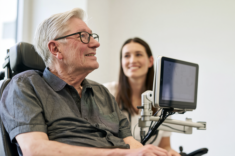
Bulbar ALS symptoms can make speech softer, slower, slurred, nasal, strained or increasingly difficult to sustain. Early communication care helps patients continue participating in medical decisions, relationships, work and everyday activities. Assessment by a speech-language pathologist is most useful while speech remains understandable, because strategies, voice preservation and communication technology can then be introduced before urgent dependence develops.[1–3]
Early Speech Assessment
Speech assessment examines articulation, speaking rate, loudness, voice quality, breath support, sentence length and intelligibility. The clinician also considers whether communication becomes harder during telephone calls, group conversations, noisy situations or later in the day, because a brief clinic conversation may not reveal the full effect of fatigue.[2–4]
Saliva pooling, respiratory weakness, reduced tongue movement and difficulty coordinating speech with breathing can each affect intelligibility. Family observations are valuable when familiar listeners have begun interpreting speech for others, when the patient avoids longer conversations or when repeated attempts to communicate cause visible exhaustion.[2,3]
Early assessment also establishes a baseline for tracking change. Speaking rate, frequency of repetition, listener understanding and communication effort can be reviewed over time so that support is adjusted before natural speech becomes unreliable.[1,3,4]
Speech exercises should be individualised. Forceful or prolonged practice may increase fatigue in weakened muscles, whereas carefully selected techniques can improve efficiency, pacing and communication endurance. The treatment plan should reflect the person’s respiratory capacity, bulbar weakness and daily communication priorities.[1–3]
Improving Speech Clarity
Speaking at a slightly slower pace and pausing between short phrases can improve intelligibility. Shorter sentences reduce the amount of air required and may prevent speech from becoming unclear at the end of a breath. Important conversations are often easier when scheduled during the part of the day when energy and voice are strongest.[2,3]
Background noise should be reduced whenever possible. Face-to-face positioning, adequate lighting and clear visual access to the speaker’s mouth and expression can help listeners understand partially unclear words. Introducing the topic before a longer sentence can also make the message easier to follow.[3]
When a word is repeatedly misunderstood, spelling its first letter, writing it down or pointing to it on an alphabet board may be less tiring than repeating the entire sentence. A portable voice amplifier may help when speech remains clear but has become too quiet, although amplification cannot correct severely reduced articulation.[2,3]
Communication partners should allow enough time for the patient to finish speaking and should confirm what they understood rather than repeatedly asking for the whole message. They should avoid completing sentences unless assistance has been requested. Reliable yes-and-no signals using speech, eye movement, gesture or facial expression can support communication during fatigue or breathlessness.[2,3]
Voice Banking and Message Banking
Voice banking records a structured set of phrases that can be used to create a personalised synthetic voice. This voice may later be connected to text-to-speech software or a speech-generating device. Recording is usually easier while speech remains clear and the person can speak without marked fatigue.[3,10]
Several short recording sessions may be more manageable than one prolonged session. System selection should consider the person’s language, accent, device compatibility, cost, storage arrangements and ability to use the voice across phones, tablets and dedicated communication equipment.[3,10]
Message banking stores complete recordings in the person’s natural voice. Patients may record names, greetings, affectionate phrases, prayers, jokes, instructions and emergency messages. These recordings preserve the original rhythm and emotional expression, while voice banking allows new sentences to be generated later.[3,10]
Patients whose speech has already changed may still have options. Some systems can use mildly affected speech, while suitable recordings from earlier videos, voice messages or interviews may sometimes contribute to voice creation. Existing recordings should therefore be preserved for assessment.[3,10]
Augmentative and Alternative Communication
Augmentative and alternative communication, or AAC, includes methods that supplement or replace speech. A person can continue using natural speech while adding writing, gestures, communication boards or electronic devices according to the situation.[1–3,10]
Low-technology options include pen and paper, alphabet boards, word boards, topic cards and printed yes-and-no systems. These methods remain useful during travel, hospital admission, equipment failure or respiratory emergencies. A dependable non-electronic backup should remain accessible even when the patient normally uses an advanced device.[2,3,10]
Electronic options include mobile applications, text-to-speech programs and dedicated speech-generating devices. Frequently used phrases can be stored for rapid access, while word prediction may reduce the amount of typing required. The system should support the patient’s preferred languages and usual methods of communication, including telephone calls, messaging, email and telehealth.[2,3,10]
A touchscreen may remain practical while hand control is reliable. If arm or finger movement declines, access may shift to switches, adapted mice, joysticks, head tracking or eye-gaze technology. Eye-gaze systems use eye movement to select letters, words or commands, but successful use depends on positioning, calibration, lighting, visual ability and fatigue.[2,10]
Device selection should match the patient’s current movement, expected progression, communication environments and comfort with technology. A complex system may be unsuitable when it is slow, physically tiring or difficult to operate. Trial use allows the patient and clinical team to identify the most practical access method.[2,3,10]
Seating, head support and device mounting can determine whether communication remains accessible throughout the day. Patients using non-invasive ventilation may also need the device positioned so that the mask and tubing do not obstruct the screen or access controls.[1,2]
Family members and caregivers should know how to position, charge and troubleshoot the communication system. The device, charger, mounting equipment and backup method should accompany the patient during hospital visits whenever practical.[2,3,10]
Impaired speech does not necessarily indicate impaired understanding or decision-making ability. Healthcare professionals should provide sufficient time and appropriate communication support so the patient can ask questions, provide consent and express treatment preferences directly.[1,2]
Monitoring Communication Changes
Communication should be reviewed when speech becomes less understandable, conversations take longer, repetition increases or the patient begins avoiding calls and social interaction. Speaking rate, listener understanding, fatigue and the ability to communicate urgent needs provide useful measures of functional change.[1–4]
AAC preparation should be accelerated when speech is declining even if close relatives can still understand most words. Learning a new access method, organising personalised vocabulary and training communication partners become more difficult once speech and limb function have deteriorated substantially.[2–4]
The system may need modification as hand strength, neck control, vision, respiratory support or fatigue changes. Touch access can be replaced by larger targets, switches, head tracking or eye-gaze control so that communication remains available as physical abilities change.[1–3,10]
Effective communication support should allow the patient to express complex choices, humour, affection, disagreement, privacy and spiritual needs, not only basic requests for food or care. Preserving these forms of expression helps maintain autonomy, relationships and personal identity throughout bulbar ALS care.[1–3,10]
Swallowing Care for Bulbar ALS Symptoms

Bulbar ALS symptoms can weaken the coordinated movements required to chew, control food and liquid in the mouth, trigger an effective swallow and protect the airway. Swallowing care should begin when subtle changes appear, because early assessment can reduce choking risk, maintain hydration and nutrition, preserve enjoyment of meals and identify when oral intake is becoming unsafe or excessively tiring.[1,2,4]
Recognising Unsafe Swallowing
Coughing or choking during food and drinks is an important warning sign, but swallowing difficulty may also present less dramatically. Food may remain in the cheeks, liquids may escape through the lips, tablets may feel difficult to move backwards, or the person may require several swallows to clear one mouthful. Meals may become slower, colder food may be left unfinished, and the patient may avoid eating with other people because of embarrassment or fatigue.[2,4]
A wet, gurgling or congested voice after swallowing can indicate that liquid, food or saliva remains near the airway. Repeated throat clearing, watery eyes during meals, nasal regurgitation, breathlessness while eating and coughing several minutes after a meal also require assessment. Recurrent fever, unexplained chest congestion or repeated respiratory infections may indicate aspiration even when the patient does not recall a major choking episode.[1,2,4]
Aspiration can sometimes occur without a forceful cough. Weak laryngeal sensation, impaired airway closure or reduced cough strength may allow material to enter the airway silently. A patient should therefore not be considered safe solely because coughing is absent. Instrumental assessment may be needed when symptoms, weight changes, respiratory complications or clinical findings raise concern despite apparently comfortable eating.[1,4]
Swallowing can deteriorate with fatigue. A person may manage the first part of a meal but lose tongue control, airway protection or cough effectiveness as eating continues. Changes that appear only at the end of meals are clinically relevant and should be reported to the speech-language pathologist rather than attributed simply to slow eating.[2,4]
Safer Eating and Drinking
Meals should be taken while the patient is awake, alert and able to maintain an upright, well-supported position. The head, neck and trunk should be aligned comfortably rather than forced forwards or backwards. Poor seating, inadequate head support and fatigue can reduce control of food and make breathing more difficult during swallowing.[1,2]
Small mouthfuls and a controlled eating pace may reduce the effort required to prepare and swallow food. The patient should be allowed enough time to complete each swallow before taking another mouthful. Rapid alternating between solids and liquids should not be encouraged automatically, because the safest sequence depends on the individual swallowing pattern.[2,4]
Talking while chewing or swallowing can interfere with airway protection and may increase fatigue. Conversation can continue between mouthfuls after the swallow has been completed and the voice sounds clear. Background distractions should be reduced when the person needs to concentrate on coordinating breathing and swallowing.[2,4]
Meals are often safer and more successful when scheduled during periods of better energy. Rest before eating may improve endurance, particularly when the patient has spent considerable effort bathing, dressing or travelling to the dining area. Smaller, more frequent meals can reduce fatigue when a full meal requires excessive chewing or prolonged respiratory effort.[1,2]
Breathlessness during meals should not be ignored. Eating requires temporary pauses in breathing, and respiratory-muscle weakness can reduce tolerance for repeated swallows. A patient who becomes breathless, cannot complete a sentence between mouthfuls or needs prolonged recovery after swallowing should receive combined respiratory and swallowing assessment.[1,2,6]
Patients using non-invasive ventilation may need an individual plan for meals, mask breaks and respiratory recovery. The timing of ventilation should be coordinated with the respiratory and swallowing teams because removing support for long periods may increase fatigue, while eating with certain interfaces may not be practical or safe.[1,6]
Food and Fluid Modification
Food consistency should be selected according to the swallowing assessment rather than according to diagnosis alone. Some patients manage moist, cohesive foods more easily than dry, crumbly or fibrous foods. Others have difficulty with thin liquids but can safely swallow particular solid textures. The appropriate plan depends on tongue control, chewing ability, throat clearance, airway protection and fatigue.[1,2,4]
Dry foods may separate into small particles that are difficult to collect and move through the mouth. Moistening food with suitable sauces, gravies or other compatible liquids may improve cohesion and reduce chewing effort. Tough meat, stringy vegetables, foods with skins and foods that break into crumbs may require modification when they leave residue or repeatedly trigger coughing.[2,4]
Mixed consistencies can be difficult for some patients because the liquid component may reach the throat before the solid component has been fully controlled. Examples include cereal with milk, soup containing pieces, fruit in juice and foods that release liquid during chewing. These foods should not be removed universally, but they should be reviewed when coughing or loss of control occurs.[2,4]
Fluid modification requires particular care. Thickened drinks may slow liquid flow and improve control for some swallowing patterns, but they are not automatically safer for every patient. Excessive thickness can increase residue, require greater tongue effort, reduce fluid enjoyment and contribute to inadequate hydration. The level of thickness should be recommended after swallowing assessment and reviewed as function changes.[1,2,4]
Commercial thickness levels, household descriptions and preparation methods can vary between countries and products. Patients and caregivers need clear instructions regarding the recommended consistency, preparation method and testing procedure. An incorrectly mixed drink may be thinner or thicker than intended, leading to inconsistent swallowing safety and reduced intake.[2,4]
Texture modification should maintain nutritional quality and appearance wherever possible. Simply blending the usual meal with a large volume of water may reduce its energy and protein density. A dietitian can help preserve calorie, protein, fibre and micronutrient intake while the speech-language pathologist determines the safest manageable consistency.[1,2]
A change in texture does not remove the need for monitoring. Increasing meal duration, greater residue in the mouth, repeated coughing, falling fluid intake or continued weight loss may indicate that the previous recommendation is no longer sufficient. Reassessment should occur whenever swallowing function or respiratory status changes.[1,2,4]
Oral Clearance and Positioning
Food residue left in the cheeks, under the tongue or along the gums can later move towards the throat and airway. Patients with reduced tongue movement may not feel or clear this residue effectively. A mirror, gentle tongue movement, an appropriate oral-clearing strategy or assistance from a trained caregiver may be needed after meals.[2,4]
Postural strategies such as changing head direction or chin position can alter how material moves through the throat, but they are not suitable for every swallowing pattern. A posture that helps one patient may increase residue or airway entry in another. These techniques should be prescribed after clinical or instrumental assessment and practised under professional guidance.[2,4]
Remaining upright after eating may support comfort and reduce reflux-related symptoms, although it does not prevent aspiration caused by impaired swallowing. Patients with reflux, regurgitation or coughing after meals should discuss these symptoms with the clinical team because reflux management and swallowing management address different mechanisms.[1,2]
Oral Care and Airway Protection
Regular oral care is an important part of aspiration-risk reduction. Bacteria can accumulate on the teeth, gums, tongue, dentures and oral secretions. When saliva or food enters the airway, poor oral hygiene may increase the bacterial burden carried into the lungs.[2,4]
Teeth, gums and tongue should be cleaned using a method the patient can tolerate safely. A patient with poor lip closure or impaired swallowing may need assistance to prevent excess water or toothpaste from collecting in the mouth. Suction may be appropriate when the person cannot spit or clear oral secretions effectively, but caregivers should receive training in its use.[1,2]
Dentures should be checked for comfort and stability because loose dentures can impair chewing and increase fatigue. Oral pain, ulcers, fungal infection, broken teeth and dry mouth can further reduce food intake and should be treated promptly. Saliva-reducing medicines may also increase dryness and affect oral health, making dental and oral review particularly important.[2,5]
A weak cough reduces the ability to expel material that approaches or enters the airway. Cough effectiveness should therefore be considered alongside swallowing. Manual cough assistance, mechanical insufflation-exsufflation or suction may be introduced when respiratory assessment shows that the patient cannot clear secretions adequately.[1,2,6]
Swallowing Medicines Safely
Difficulty swallowing tablets may appear before the patient has major difficulty with meals. Tablets may remain in the mouth, adhere to the throat or require repeated drinks that are themselves difficult to swallow. Medication form should be reviewed when the patient coughs, needs excessive time or begins avoiding prescribed doses.[2,4]
Tablets and capsules should not be crushed, split or opened without confirmation from a pharmacist or prescriber. Modified-release, enteric-coated, hormonal, cytotoxic and other specialised preparations may become ineffective, irritating or hazardous when altered. Liquid, dispersible, soluble, transdermal or alternative-strength preparations may provide safer options.[2]
Liquid medicine is not automatically easier to swallow. Thin medicinal liquids may flow quickly, while thick syrups may leave residue or be difficult to clear. The volume, viscosity, taste and method of administration should match the patient’s swallowing assessment. A pharmacist can determine whether a medicine is compatible with an advised thickener or feeding tube.[2,4]
Ayurvedic medicines also require swallowing-safety review. Powders, tablets, capsules, oils, decoctions and avaleha preparations differ in particle size, stickiness, volume and flow. A preparation that was manageable earlier may become difficult as tongue control, saliva clearance or cough strength changes. The treating Ayurvedic physician should coordinate dosage form and administration with the swallowing and nutrition teams rather than assuming that every oral preparation remains suitable.
Medicines should not be hidden in food without checking compatibility and obtaining the patient’s agreement. Mixing medication into a full meal may alter absorption, create an unpleasant taste or cause the patient to reject an important source of nutrition. When a feeding tube is present, each medicine requires review for tube compatibility, appropriate dilution and flushing.[2]
Choking and Aspiration Planning
Patients and caregivers should have a written plan describing what to do during coughing, partial obstruction, complete choking and respiratory distress. The plan should reflect the patient’s mobility, cough strength, communication method, respiratory equipment and previously expressed treatment preferences.[1,2]
Caregivers should receive recognised choking-response training appropriate to the patient’s condition. Severe obstruction is suggested when the person cannot breathe, speak or cough effectively, becomes blue or grey, loses responsiveness or collapses. Emergency services should be contacted immediately, and trained first aid should begin according to local guidance.[2]
Blind finger sweeps should be avoided because they can push material deeper into the airway or injure the mouth. Visible food may be removed only when it can be reached safely. A suction machine can clear accessible saliva and loose secretions but does not replace emergency management of a solid airway obstruction.[2,6]
After a significant choking event, persistent coughing, voice change, wheezing, chest discomfort, breathlessness, fever or increasing drowsiness requires prompt medical assessment. A resolved obstruction does not exclude aspiration or respiratory complications, particularly when cough strength is already reduced.[1,2,6]
The patient’s communication system should remain within reach during meals. A simple agreed signal for “stop,” “choking,” “suction,” “breathless” or “call for help” may be essential when speech is weak. Caregivers should know the patient’s reliable yes-and-no response and avoid offering further food or drink until distress has resolved.[1,2]
Increasing choking frequency, prolonged meals, progressive weight loss, dehydration, recurrent chest infections or inability to take essential medication should trigger reassessment of oral intake and discussion of enteral feeding. These conversations are most useful before swallowing or respiratory function deteriorates to the point that decisions must be made during an emergency.[1,2,8]
Saliva Care for Bulbar ALS Symptoms

Bulbar ALS symptoms can interfere with automatic swallowing, lip closure, tongue movement and cough strength, causing saliva to pool in the mouth or throat. The problem is usually reduced clearance rather than excessive saliva production. Management depends on whether secretions are thin and watery, thick and sticky, associated with dry mouth or difficult to clear because the cough has weakened.[1,2,5]
Saliva problems can affect speech, sleep, eating, non-invasive ventilation, social confidence and skin comfort. Secretions that repeatedly enter the throat may also trigger coughing or choking. Early assessment helps distinguish between saliva production, swallowing impairment, dehydration and respiratory secretion retention so that treatment does not improve one problem while worsening another.[1,2,5]
Table : Thin Saliva vs Thick Secretions vs Dry Mouth
| Problem | Typical signs | Main management direction | What may worsen it |
|---|---|---|---|
| Thin saliva | Drooling, pooling, frequent wiping | Improve clearance, positioning and consider saliva-reducing treatment | Weak lip closure and reduced swallowing frequency |
| Thick saliva | Sticky, stringy mucus, repeated throat clearing | Hydration where safe, humidification and secretion clearance | Dehydration and excessive drying medication |
| Dry mouth | Burning, sticky mouth, cracked lips, difficulty speaking | Oral moisture, medication review and dental care | Mouth breathing and anticholinergic treatment |
| Retained respiratory mucus | Congested throat or chest, weak clearance | Cough augmentation, suction where appropriate and respiratory review | Weak cough and respiratory infection |
Thin Saliva and Drooling
Thin saliva may collect inside the mouth, leak from the lips or require frequent swallowing that the patient can no longer perform efficiently. Drooling may become more noticeable while concentrating, speaking, eating, bending forwards or resting with the mouth open. Reduced awareness of saliva and difficulty maintaining an upright head position can add to the problem.[2,5]
A speech-language pathologist can assess lip closure, tongue control, swallowing frequency, posture and the ability to clear secretions. Better head and trunk support may reduce forward leakage in some patients. Swallowing reminders can help when the patient retains sufficient voluntary control, although repeatedly prompting a weak or fatigued swallow may become ineffective as bulbar impairment progresses.[1,2,5]
Soft absorbent cloths, tissues, discreet clothing protection and suitable skin-barrier products can reduce irritation around the lips and chin. The skin should be cleaned gently and dried rather than rubbed repeatedly, because constant moisture and friction may cause soreness or breakdown.[5]
Portable oral suction may help remove saliva that the patient cannot spit out or swallow safely. Suction should be used only where the catheter can be seen and reached without entering deeply into the throat. Deep suction can cause discomfort, gagging, bleeding or airway irritation and should not be performed without specific clinical instruction.[2,5,6]
A patient who repeatedly chokes on saliva, wakes because of secretion pooling or cannot use non-invasive ventilation because the mouth fills with saliva needs prompt review. Treatment may require coordinated changes in swallowing care, medication, suction, positioning and respiratory support rather than simply increasing a drying medicine.[1,2,5,6]
Medicines for Drooling
Anticholinergic medicines reduce salivary secretion and may be prescribed when positioning, swallowing strategies and suction do not provide adequate control. Depending on the patient’s health, route of administration and local practice, treatment may include oral, sublingual or transdermal medicines. Selection should consider swallowing ability, cognition, bowel function, urinary symptoms, heart rhythm, eye disease and the likelihood of adverse effects.[1,2,5]
Possible adverse effects include dry mouth, constipation, urinary retention, blurred vision, drowsiness, confusion and thickening of secretions. Older adults and patients with cognitive or behavioural symptoms may be particularly sensitive. Treatment should begin and be adjusted under clinical supervision, with regular review of both benefit and tolerability.[1,2,5]
The goal is usually comfortable control rather than complete elimination of saliva. Excessive drying can impair speech, increase oral discomfort, promote dental problems and convert manageable watery saliva into thick secretions that are harder to clear. Patients and caregivers should report a new sticky mouth, increasing mucus, constipation, reduced urination or confusion after treatment begins.[2,5]
When swallowing tablets is difficult, the prescriber and pharmacist should consider an alternative formulation or route. Saliva medicines should not be crushed, opened, doubled or applied differently without confirmation, because delivery method and absorption can vary between preparations.[2]
Botulinum Toxin and Salivary-Gland Treatment
Botulinum toxin may be injected into selected salivary glands when drooling remains troublesome despite medication or when systemic adverse effects limit oral treatment. The injections reduce glandular secretion for a period of time and may be guided by ultrasound to improve placement.[1,5,9]
The response is not immediate or permanent, and repeat treatment may be required. Possible complications include dry mouth, thicker secretions and worsening swallowing in some patients. The procedure should therefore be planned by a clinician familiar with ALS, with attention to the patient’s baseline swallowing and respiratory function.[1,5,9]
Targeted radiotherapy to the salivary glands may be considered for persistent, severe drooling when other measures have not provided adequate relief or cannot be tolerated. Treatment aims to reduce saliva production but may cause prolonged dryness, altered secretion consistency or oral discomfort. The expected benefit, treatment burden and potential adverse effects should be discussed individually.[1,5]
Evidence supporting saliva treatments in ALS remains limited compared with evidence available for many common medical conditions. Individual response varies, and repeated review is necessary because a treatment that initially improves drooling may become unsuitable when swallowing, hydration or cough effectiveness changes.[1,9]
Thick Saliva and Sticky Mucus
Thick saliva may feel sticky, stringy or difficult to move from the back of the mouth. Patients may describe constant throat clearing, a sensation of mucus being trapped, difficulty speaking, interrupted sleep or inability to tolerate a ventilation mask. The secretion may originate from the mouth, nose or respiratory tract, so assessment should identify where it is accumulating and whether the patient can cough it out.[2,5,6]
Reduced fluid intake is a common contributor. Patients with dysphagia may drink less because thin liquids cause coughing or because reaching the bathroom has become difficult. Fever, diarrhoea, warm environments, mouth breathing and some medications can further reduce hydration. Fluid intake should be reviewed with the swallowing and nutrition teams so hydration can be improved without introducing an unsafe consistency.[1,2,5]
All current medicines should be reviewed when secretions become thick, particularly saliva-reducing medicines. Reducing or changing a drying treatment may be more appropriate than adding another medication. The prescriber must balance relief from drooling against the patient’s ability to mobilise the remaining secretions.[2,5]
Humidification can reduce dryness caused by mouth breathing, heating systems or non-invasive ventilation. A heated humidifier attached to ventilation equipment may help selected patients, but settings and equipment compatibility should be reviewed by the respiratory team. Poorly maintained humidification equipment can become contaminated and should be cleaned according to the manufacturer’s and clinical service’s instructions.[2,6]
Nebulised saline may be considered when secretions remain thick and difficult to clear. It should be prescribed with attention to respiratory function, bronchial sensitivity and the patient’s ability to cough after secretions loosen. Making mucus thinner without providing an effective method of clearance may increase distress or airway congestion.[2,5,6]
Mucolytic treatment may be appropriate for selected patients, particularly when respiratory mucus is tenacious. The clinical team should determine whether the problem is oral saliva, nasal drainage, reflux-related irritation, infection or lower-airway secretion before choosing treatment. New discolouration, foul smell, fever, chest pain or increasing breathlessness may indicate infection and requires medical assessment.[2,6]
Weak Cough and Secretion Clearance
A patient may produce a coughing sound without generating enough airflow to move mucus. Weakness of the inspiratory, expiratory and bulbar muscles can reduce cough effectiveness, while poor closure of the voice box may prevent pressure from building inside the chest. Saliva treatment alone will not correct this form of airway-clearance failure.[1,2,6]
Respiratory assessment may include cough-flow measurement, inspiratory strength and evaluation of the patient’s ability to clear secretions during illness. The need for support can change during chest infection, dehydration or fatigue even when the patient previously managed without equipment.[1,2,6]
Manual assisted cough uses coordinated pressure on the abdomen or chest to strengthen the expiratory phase of coughing. It should be taught by a respiratory physiotherapist or another trained professional because timing, force and suitability vary according to respiratory status, body shape, pain and feeding-tube placement.[1,6]
Mechanical insufflation-exsufflation, often called cough assist, alternates positive and negative airway pressure to support a stronger cough. Bulbar dysfunction can make the response less predictable because the throat may close or fail to coordinate with the pressure changes. Interface selection, pressure settings and timing therefore require individual adjustment rather than unsupervised use.[1,2,6]
Suction can remove secretions from the mouth and accessible upper airway but cannot mobilise mucus deep inside the lungs. Some patients need a combination of humidification, cough augmentation, positioning and suction. Caregivers should be trained in the sequence of these measures and in recognising when clearance attempts are causing fatigue or respiratory distress.[2,6]
Dry Mouth
Dry mouth may result from saliva-reducing medication, inadequate hydration, mouth breathing, non-invasive ventilation, anxiety or reduced oral intake. Symptoms include burning, difficulty speaking, altered taste, cracked lips, sticky oral surfaces, difficulty wearing dentures and food adhering to the tongue or palate.[2,5]
Dryness can coexist with drooling. A patient may have an uncomfortable dry mouth while saliva still leaks from the lips because the remaining secretion is not swallowed effectively. Increasing drying medication in this situation may worsen oral discomfort without adequately controlling leakage.[5]
Frequent oral care, lip moisturisation and products designed to moisten the mouth may improve comfort. Small sips or ice may be inappropriate when swallowing is unsafe, so moisture strategies should follow the swallowing plan. Alcohol-containing mouthwashes can increase dryness and irritation and may be poorly tolerated.[2,5]
Dental review is important because reduced saliva increases the risk of tooth decay, gum disease, oral infection and mucosal injury. Dentures should be removed and cleaned regularly, and any persistent white patches, ulcers, bleeding, pain or foul odour should be assessed promptly.[2,5]
Patients using non-invasive ventilation should report dryness, mouth leakage, nasal blockage or discomfort to the respiratory team. Mask adjustment, humidification or changes in interface may improve symptoms without reducing the effectiveness of ventilation.[6]
Oral Care and Daily Comfort
The mouth should be inspected regularly for pooled saliva, food residue, dryness, coating, ulceration and infection. Toothbrushing and tongue cleaning may require caregiver assistance when hand weakness or fatigue limits self-care. Excess water and toothpaste should be avoided when the patient cannot spit or swallow safely.[2,5]
Oral care is particularly important after meals and before ventilation. Food residue and thick secretions can interfere with mask comfort and may later move towards the throat. A suction toothbrush or oral suction device may be useful when recommended and demonstrated by the clinical team.[2,5,6]
Strongly flavoured lozenges, sweets or chewing gum may stimulate additional saliva and increase choking risk in patients with impaired swallowing. Products kept in the mouth should not be used unless the patient can control and swallow them safely.[2,5]
Oil pulling, forceful gargling and retaining large volumes of liquid or oil in the mouth can be unsafe when bulbar weakness has impaired airway protection. Nasal administration of oils or liquids also requires caution when swallowing and cough are weak. Any Ayurvedic oral or nasal procedure should be considered only after assessment of swallowing, secretion control and respiratory function.
Monitoring Treatment Response
Response should be assessed according to comfort and function rather than saliva volume alone. Useful changes include less leakage, fewer clothing changes, improved sleep, clearer speech, easier mask use and reduced choking on saliva. Increasing dry mouth, sticky mucus, constipation, urinary difficulty, confusion or greater dependence on suction may indicate that treatment has become excessive or unsuitable.[1,2,5]
Saliva management should be reviewed whenever swallowing, hydration, respiratory function or medication changes. Thin drooling can later become thick secretion retention, and dry mouth can coexist with continued leakage. Regular reassessment allows medication, humidification, suction and cough support to be adjusted before secretion problems interfere with breathing or nutrition.[1,2,5,6]
Same-day clinical contact is appropriate when secretions suddenly become difficult to clear, suction needs increase substantially, non-invasive ventilation cannot be tolerated or the patient repeatedly chokes on saliva. Severe breathlessness, inability to move air, blue-grey discolouration, new confusion, extreme drowsiness or collapse requires emergency medical care.[1,2,6]
Nutrition and Weight Support

Bulbar ALS symptoms can reduce calorie and fluid intake through chewing fatigue, swallowing difficulty, prolonged meals, breathlessness, saliva problems and loss of appetite. Maintaining body weight and hydration is clinically important because nutritional decline can worsen weakness, fatigue, constipation, secretion problems and tolerance of respiratory support. Weight, intake and meal effort should therefore be monitored from the early stages of bulbar involvement.[1,2]
Why Weight Loss Can Occur
Weight loss in ALS may develop even when appetite initially remains normal. Meals often take longer, chewing requires more effort and repeated swallowing can become exhausting. Patients may begin avoiding foods that are difficult to control, reducing portion sizes or stopping before a meal is finished because of fatigue.[1,2,4]
Swallowing problems can also reduce fluid intake. Patients may drink less because thin liquids trigger coughing, because repeated sips are tiring or because getting to the bathroom has become difficult. Reduced hydration can worsen constipation, dry mouth, thick saliva and fatigue, which may further reduce food intake.[2,4,5]
Respiratory weakness can make eating more demanding because breathing must be coordinated with chewing and swallowing. Patients who become breathless during meals may take smaller mouthfuls, pause frequently or stop eating early. Nutritional assessment should therefore consider respiratory symptoms as well as swallowing ability.[1,2,6]
Some patients lose weight despite maintaining a reasonable intake. Changes in energy metabolism and muscle loss may contribute, so nutritional support should not depend only on appetite. A declining weight trend is clinically important even when the patient remains within a conventional “normal” body-mass-index range.[1,2]
Monitoring Weight and Intake
Weight should be measured regularly under similar conditions whenever practical. The trend over several weeks is more useful than a single reading. A persistent fall in weight, looser clothing or visible loss of muscle should prompt dietetic review even if the patient does not report severe swallowing difficulty.[1,2]
Meal duration provides another useful measure. A meal that previously took twenty minutes but now takes forty-five minutes or longer may indicate increasing chewing fatigue, reduced swallowing efficiency or respiratory difficulty. Patients who require prolonged recovery after meals may also be expending more energy than they can replace.[2,4]
Families should note whether portions are becoming smaller, snacks are being skipped, fluids are being avoided or certain textures are disappearing from the diet. Recurrent coughing, food residue, increased saliva difficulty and inability to swallow medicines can all reduce total intake.[2,4]
Hydration can be monitored through fluid intake, urine frequency, urine colour, dry mouth, dizziness and constipation. These signs are not specific, but a consistent pattern can help identify inadequate intake before dehydration becomes severe.[2]
Increasing Calories Without Increasing Meal Size
When appetite or endurance is limited, increasing the energy density of food can be more practical than increasing portion size. Meals can be fortified with ingredients that add calories and protein without substantially increasing volume, provided the final texture remains safe for the patient’s swallowing pattern.[1,2]
Protein-rich foods can help maintain nutritional intake, while calorie-dense additions may reduce the amount that must be eaten to meet energy needs. The exact choices should reflect the person’s food preferences, cultural diet, medical conditions and texture recommendations.[1,2]
Smaller meals eaten more frequently may be better tolerated than three large meals. This approach can reduce chewing fatigue and breathlessness while providing more opportunities to meet daily calorie and protein requirements.[1,2]
Nutritional supplements may be useful when ordinary meals no longer provide enough energy or protein. The consistency of the supplement should match the patient’s swallowing recommendation, because thin liquid supplements may be difficult for some patients even when their nutritional content is appropriate.[1,2,4]
Protein and Nutrient Intake
Protein needs should be met through foods or supplements that are easy to chew and swallow. Soft eggs, dairy foods, smooth nut preparations, legumes, tofu, tender fish or other suitable protein sources may be easier than dry or fibrous foods, depending on the swallowing pattern.[1,2]
Dietary fibre remains important because constipation is common in ALS and may be worsened by immobility, reduced fluid intake and some medications. Fibre intake should be adjusted with hydration, because increasing fibre without sufficient fluid may worsen constipation.[1,2]
Micronutrient intake can fall when the diet becomes restricted to a narrow range of soft foods. A dietitian can review whether the patient is receiving adequate vitamins and minerals from food, fortified products or supplements. High-dose supplementation should not be assumed to improve ALS progression and may interact with medication or other treatments.[1,2]
Hydration Support
Adequate hydration helps maintain circulation, bowel function, oral comfort and secretion consistency. Thick saliva and sticky mucus often become more difficult when fluid intake falls. Improving hydration may therefore reduce secretion burden in some patients, provided fluids can be swallowed safely.[2,5]
Thin liquids are not universally unsafe in bulbar ALS, and thickened fluids are not automatically appropriate. The safest consistency depends on the individual swallowing pattern. If thickened fluids are prescribed, hydration should be reviewed because some patients drink less when beverages become less palatable or more tiring to swallow.[2,4]
Fluid can also be provided through foods with a high water content or through enteral feeding when oral intake is inadequate. The total daily plan should account for water contained in meals, nutritional supplements, medication administration and tube flushes where relevant.[2]
Reducing Meal Fatigue
The patient should begin meals in a comfortable, well-supported position and preferably before severe fatigue develops. Physical tasks such as bathing, dressing or travelling immediately before a meal can reduce the energy available for eating.[1,2]
Foods requiring prolonged chewing may become impractical even when swallowing remains relatively safe. Softer, moist textures can reduce oral effort and shorten mealtime. The objective is to preserve adequate intake without increasing aspiration risk or making eating unnecessarily restrictive.[2,4]
Assistive cutlery, adapted cups, plate guards and supportive seating may reduce the physical effort required to eat when hand, arm or neck weakness is present. Occupational therapy assessment can help maintain independence for longer.[1,2]
Family members should avoid rushing the patient or repeatedly offering another mouthful before the previous one has been cleared. A calm pace allows coordination between chewing, swallowing and breathing and may reduce fatigue.[2,4]
Nutrition During Respiratory Weakness
Breathlessness can substantially reduce food intake. Patients may struggle with large mouthfuls, long conversations during meals or foods requiring extensive chewing. Shorter meals with higher energy density may be better tolerated when respiratory reserve is reduced.[1,2,6]
Patients using non-invasive ventilation may benefit from respiratory support before meals so that they begin eating less fatigued. The respiratory team can advise on mask breaks, timing and whether ventilation support is needed around meals.[1,6]
Increasing daytime sleepiness, morning headache, difficulty lying flat or breathlessness during eating should prompt respiratory reassessment. Nutritional decline should not be treated only with dietary changes when respiratory weakness is contributing to the problem.[1,2,6]
Nutrition When Swallowing Becomes More Difficult
When oral intake falls despite food modification and supplements, the team should assess whether swallowing safety, fatigue or respiratory weakness is the main limiting factor. Persistent weight loss, dehydration, very prolonged meals or inability to take essential medication may indicate that enteral feeding needs to be discussed.[1,2,4]
A feeding tube does not necessarily mean that all oral eating must stop. Some patients continue selected foods and drinks for pleasure when swallowing assessment indicates that this remains acceptably safe. Enteral feeding can provide additional calories, fluids and medicines while reducing the pressure to obtain all nutrition orally.[1,2]
Discussion of gastrostomy is usually easier before severe weight loss or advanced respiratory weakness develops. Earlier planning gives the patient more time to understand the options, consider preferences and undergo respiratory assessment before a procedure is required urgently.[1,2]
Ayurvedic Nutritional Support
Ayurvedic nutritional support can be adapted to the patient’s swallowing ability, digestion, appetite and overall strength. Bṛṃhaṇa refers to nourishing measures intended to support tissue nutrition and physical strength. In classical Ayurvedic practice, such support is individualised rather than determined solely by the neurological diagnosis.
Soft, nutrient-dense preparations may be easier to administer than dry powders or large tablets when chewing and swallowing are impaired. The consistency, stickiness and volume of any preparation should match the patient’s swallowing recommendations. Avaleha preparations, medicated liquids or other oral forms should be reconsidered if tongue control, saliva clearance or cough strength deteriorates.
Heavy, sticky or very viscous preparations may be difficult to clear from the mouth or pharynx in advanced bulbar weakness. Ayurvedic medicines should therefore be selected with attention to swallowing safety, respiratory function, hydration and feeding-tube status rather than being continued in the same form throughout the course of illness.
Any nutritional or Ayurvedic plan should preserve adequate calories, protein and fluids while avoiding unnecessary restriction. Changes in weight, meal duration, choking frequency, bowel function and secretion consistency provide practical indicators of whether the plan remains suitable.[1,2,4,5]
When Nutrition Support Needs Urgent Review
Same-day clinical review is appropriate when the patient can no longer maintain fluids, repeatedly misses meals, is unable to swallow essential medicines or becomes markedly weak, dizzy or drowsy from poor intake.[1,2]
Rapid weight loss, recurrent choking, repeated chest infections, prolonged meals and increasing breathlessness while eating should prompt coordinated review by the neurology, swallowing, dietetic and respiratory teams. These changes may indicate that oral strategies alone are no longer sufficient.[1,2,4,6]
Feeding Tubes in Bulbar ALS
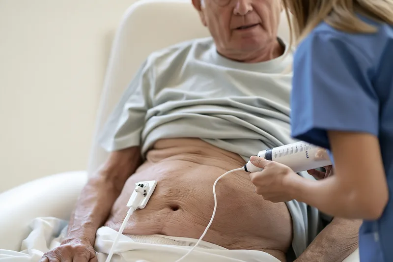
Bulbar ALS symptoms can eventually make eating, drinking and swallowing medicines too slow, tiring or unsafe to provide adequate nutrition and hydration. A gastrostomy tube provides direct access to the stomach for nutrition, fluids and selected medicines when oral intake no longer meets the patient’s needs. Decisions about tube feeding take account of swallowing ability, weight change, respiratory function, meal effort, choking risk, hydration and the patient’s preferences.[1,2]
Table: Feeding Tube Options in ALS
| Method | How it is placed | ProGas participants | 30-day mortality | Practical point |
|---|---|---|---|---|
| PEG | Endoscope passes through the mouth into the stomach | 163 | 3% | Commonly used endoscopic method |
| RIG | Tube placed using radiological imaging guidance | 121 | 3% | Does not require an endoscope through the throat |
| PIG | Image-guided placement using a per-oral technique | 43 | 7% | Available in selected specialist centres |
When a Feeding Tube Is Considered
Gastrostomy may be considered when swallowing difficulty causes persistent weight loss, dehydration, prolonged meals, repeated choking or inability to take sufficient calories and fluids. It may also become useful when the patient can still eat but requires substantial effort to complete meals or cannot reliably swallow essential medicines.[1,2]
The decision is not based on choking alone. Some patients compensate for dysphagia by eating increasingly small portions, restricting food textures or drinking very little. A progressive fall in weight, reduced fluid intake or meals that consume a large part of the day can indicate that oral intake is no longer sustainable even when severe choking has not occurred.[1,2,4]
Respiratory weakness can further reduce eating capacity. A patient who becomes breathless during meals may stop eating before nutritional requirements are met. When respiratory reserve is declining, gastrostomy planning also becomes more complex, making assessment of respiratory function an important part of procedural planning.[1,2,6]
Gastrostomy should be considered before malnutrition, dehydration or respiratory impairment becomes severe. Earlier planning allows swallowing, nutritional and respiratory status to be assessed while the patient has sufficient strength to consider the available procedures and participate fully in decision-making.[1,2]
PEG, RIG and Other Gastrostomy Methods
Percutaneous endoscopic gastrostomy, or PEG, places a feeding tube through the abdominal wall into the stomach using an endoscope passed through the mouth and oesophagus. Sedation and airway management are considered according to respiratory function, secretion burden and the patient’s overall condition.[1,2,8]
Radiologically inserted gastrostomy, often called RIG, uses imaging guidance to place the tube through the abdominal wall without requiring an endoscope to pass through the throat. This approach may be considered when bulbar or respiratory impairment makes endoscopic placement less suitable.[1,2]
Per-oral image-guided gastrostomy is another technique available in some specialist centres. The choice between PEG, RIG and other approaches depends on respiratory function, swallowing impairment, local expertise, anatomy, previous abdominal procedures and the patient’s clinical condition.[1,2]
No single insertion method is appropriate for every person with ALS. The neurologist, gastroenterology or interventional radiology team, respiratory clinician and dietitian consider procedural risk alongside the expected nutritional and hydration benefit.[1,2]
Respiratory Assessment Before Gastrostomy
Respiratory function should be reviewed before gastrostomy because weakness of the diaphragm and other breathing muscles can increase procedural risk. Assessment may include vital capacity, inspiratory-pressure measurements, cough effectiveness, symptoms when lying flat and evidence of nocturnal hypoventilation.[1,2,6]
Bulbar weakness can make some respiratory measurements more difficult to perform because an effective mouth seal may be impossible. Results therefore need to be interpreted together with symptoms and other respiratory tests rather than relying on a single spirometry value.[1,2,6]
Patients already using non-invasive ventilation may require respiratory support before, during or after tube placement. The respiratory team determines the interface, duration and monitoring required according to the patient’s ventilation needs and the type of gastrostomy procedure.[1,6]
Weak cough and retained secretions should also be addressed before the procedure. Cough assistance, suction, humidification or secretion treatment may be needed when saliva or mucus cannot be cleared effectively.[1,2,6]
A recent respiratory infection, marked secretion retention or rapid respiratory deterioration may alter the timing or method of gastrostomy. These factors require reassessment before proceeding.[1,2,6]
Nutrition After Tube Placement
Tube feeding can provide calories, protein, fluids and other nutrients without requiring every nutritional requirement to be met through prolonged oral meals. The dietitian calculates the feeding regimen according to body weight, recent weight change, current intake, gastrointestinal tolerance, activity and overall nutritional needs.[1,2]
Some patients initially use the tube only for supplementary nutrition. For example, oral meals may continue during the day while additional calories and fluids are given through the gastrostomy. As swallowing or fatigue changes, a larger proportion of nutritional requirements can be delivered through the tube.[1,2]
Feeding schedules can include bolus feeds, intermittent feeds or slower pump-assisted feeding. The choice depends on tolerance, reflux, mobility, caregiver availability, sleep patterns and the amount of nutrition required.[1,2]
Feeds may need gradual introduction when the patient has experienced significant weight loss or poor intake. Nausea, abdominal fullness, diarrhoea, constipation, reflux and bloating should be reported because the feeding volume, rate, formula or schedule may require adjustment.[1,2]
Tube feeding should provide adequate water as well as nutrition. Water requirements include routine hydration, medication administration and flushing needed to keep the tube patent. Additional fluid may be required during fever, hot weather, diarrhoea or other conditions that increase fluid losses.[1,2]
Weight should continue to be monitored after gastrostomy. Placement of a tube does not automatically reverse previous weight loss, particularly when nutritional needs, feed tolerance or disease-related metabolic changes are not fully addressed. Ongoing dietetic review is therefore required.[1,2]
Can Eating Continue After Gastrostomy?
A gastrostomy does not automatically require complete cessation of oral eating. Some patients continue foods and drinks for nutrition, pleasure or social participation when swallowing assessment indicates that oral intake remains sufficiently safe.[1,2,4]
The amount and type of oral intake may change over time. A patient may continue selected soft foods while obtaining most fluids and calories through the tube, or use the gastrostomy mainly on days when fatigue makes eating difficult. Swallowing recommendations should be reassessed when coughing, voice changes, chest infections or meal fatigue increase.[1,2,4]
When aspiration risk becomes high, oral intake may need to be reduced or stopped. The decision considers swallowing findings, respiratory reserve, cough effectiveness, the patient’s goals and the consequences of aspiration.[1,2,4]
Food taken for pleasure still requires attention to texture, portion size, posture and fatigue. A very small amount can cause difficulty if the swallowing pattern is unsafe, so tube placement should not be interpreted as protection against aspiration during oral eating.[2,4]
Aspiration Risk After Tube Placement
Gastrostomy reduces the need to swallow food, fluids and medicines orally, but saliva and refluxed stomach contents can still enter the airway. Patients with severe bulbar weakness may therefore continue to experience coughing, secretion problems or respiratory infections after tube placement.[1,2]
Saliva management, oral hygiene, cough support and respiratory monitoring remain necessary. A feeding tube addresses nutritional and hydration access but does not replace treatment of impaired airway protection or weak secretion clearance.[1,2,5,6]
Reflux, regurgitation, coughing during or after feeds and increasing chest congestion should be reported. Feed rate, volume, timing, positioning or formula may need review, and respiratory infection should be considered when fever or increasing breathlessness develops.[1,2]
Medicines Through a Feeding Tube
Many medicines can be administered through a gastrostomy, but each preparation should be checked individually. Modified-release, enteric-coated and certain specialised tablets may not be suitable for crushing or tube administration.[2]
Liquid formulations may be easier to administer but can differ in concentration, viscosity, sugar or sorbitol content. These differences may affect dosing, tube flow and gastrointestinal tolerance. A pharmacist should confirm the appropriate formulation and administration method.[2]
Medicines should generally be administered separately rather than mixing several drugs together in the feeding formula. Appropriate flushing before, between and after medicines helps reduce interactions and tube blockage.[2]
Ayurvedic medicines require the same attention to tube compatibility. Powders, bhasma-containing preparations, thick avaleha, oils and concentrated herbal preparations may obstruct a feeding tube or interact with medicines and feeds if administered without appropriate preparation. The Ayurvedic physician, dietitian, pharmacist and treating medical team should coordinate formulation, dilution, timing and compatibility before administration through the tube.
A preparation that cannot be dispersed uniformly or safely delivered through the tube may require a different dosage form. Tube diameter, feeding regimen, medication burden and flushing requirements should all be considered when modifying treatment.
Gastrostomy Care at Home
The skin around the tube should remain clean and be observed for redness, swelling, pain, discharge, bleeding or leakage. Persistent discomfort, increasing redness, pus, fever or spreading skin inflammation requires medical assessment.[2]
The tube should be secured without excessive tension. Pulling, accidental traction or deterioration of external fixation can damage the skin or alter tube position. Caregivers should know how the tube is normally positioned and whom to contact if it appears displaced.[2]
Tube blockage may occur when feeds, medicines or inadequately dissolved material accumulate inside the lumen. Routine flushing according to the prescribed feeding plan reduces this risk. Sharp objects or excessive force should not be used to clear an obstruction because they can damage the tube.[2]
A newly placed tube that becomes displaced requires urgent professional assessment because the tract may not yet be established. Even with a mature gastrostomy, replacement should follow the instructions provided by the specialist service.[2]
Leakage of stomach contents onto the skin can cause irritation and may indicate problems with tube position, fixation or the gastrostomy tract. Persistent leakage requires clinical review rather than repeated tightening of the external device without assessment.[2]
Monitoring After Gastrostomy
Weight, hydration, bowel function, feed tolerance and respiratory symptoms should be reviewed after tube feeding begins. Continued weight loss may indicate that calorie delivery is insufficient, feeds are not being tolerated or nutritional requirements have changed.[1,2]
Constipation can worsen when mobility falls, fluid intake is inadequate or medications slow bowel function. The total fluid and fibre provided orally and through the tube should therefore be considered together.[1,2]
Increasing reflux, vomiting, abdominal distension or diarrhoea may require modification of feed volume, rate or formula. Sudden severe abdominal pain, persistent vomiting, significant bleeding or inability to use the tube requires urgent medical assessment.[2]
Swallowing should continue to be reviewed even when most nutrition is given through the tube because saliva management, oral comfort and any continued pleasure feeding remain dependent on bulbar function.[1,2,4]
Respiratory monitoring also remains necessary. Progressive breathlessness, weak cough, disturbed sleep or difficulty clearing secretions may require non-invasive ventilation or cough-assistance support independently of nutritional treatment.[1,2,6]
The feeding plan can be adjusted as bulbar ALS progresses so that calories, fluids and medicines remain accessible without requiring exhausting or unsafe oral intake.[1,2]
Breathing Support for Bulbar ALS Symptoms
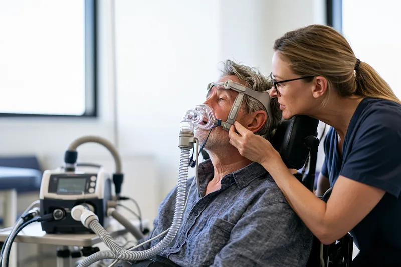
Bulbar ALS symptoms can weaken the muscles responsible for breathing, coughing and protecting the airway. Respiratory decline may begin with disturbed sleep, a weaker cough or breathlessness during speech and meals rather than obvious breathlessness at rest. Regular assessment allows non-invasive ventilation and cough support to be introduced before severe respiratory distress or secretion retention develops.[1,2,6]
Early Breathing Changes
Weakness of the diaphragm and other respiratory muscles can reduce the amount of air moved into and out of the lungs. The earliest effects are often more noticeable during sleep because breathing naturally becomes shallower and respiratory muscles work differently when the person lies down.[1,2,6]
Morning headaches, unrefreshing sleep, frequent waking, vivid dreams, daytime drowsiness and reduced concentration can indicate nocturnal hypoventilation. These symptoms occur when ventilation becomes insufficient during sleep and carbon dioxide begins to accumulate. They may be present even when daytime breathing appears comfortable.[1,2,6]
Breathlessness when lying flat, known as orthopnoea, can develop when the diaphragm becomes weaker. Patients may begin sleeping with additional pillows, raising the head of the bed or avoiding a fully reclined position. Needing progressively greater elevation to sleep comfortably requires respiratory review.[1,2,6]
Breathlessness may also appear while speaking, eating, dressing or walking short distances. Patients may use shorter sentences, pause more frequently for breath or find that their voice fades before the end of a phrase. During meals, difficulty coordinating breathing with chewing and swallowing can increase fatigue and choking risk.[1,2,6]
A weak cough may be one of the first respiratory changes. The cough can sound quiet, soft or incomplete, and the patient may need several attempts to clear saliva or mucus. Frequent throat congestion, prolonged coughing after meals and difficulty clearing secretions during a cold can indicate reduced cough effectiveness.[1,2,6]
Repeated chest infections, unexplained fever, increasing mucus or a persistent change in voice after swallowing may reflect aspiration or impaired secretion clearance. Bulbar weakness and respiratory-muscle weakness can occur together, so swallowing and breathing require coordinated assessment.[1,2,4,6]
Rapid shallow breathing, use of neck muscles to breathe, inward movement of the abdomen during inspiration or difficulty speaking in complete sentences can indicate more advanced respiratory weakness. New confusion, unusual drowsiness or reduced responsiveness may occur when carbon dioxide retention becomes severe and requires urgent medical assessment.[1,2,6]
Respiratory Testing
Respiratory monitoring combines symptoms with objective measurements. No single test identifies every stage of respiratory impairment, and results must be interpreted according to bulbar function, posture, fatigue and the patient’s ability to perform the manoeuvre correctly.[1,2,6]
Forced vital capacity and slow vital capacity measure the volume of air that can be exhaled after a full breath. Measurements may be performed while sitting and, when appropriate, while lying down. A greater decline in the supine position can indicate diaphragmatic weakness that is less apparent during upright testing.[1,2,6]
Weakness of the lips and facial muscles can prevent a tight seal around the mouthpiece. Air leakage may produce a result that appears lower than the patient’s actual respiratory capacity. Adapted mouthpieces, masks or additional respiratory tests may be required when bulbar weakness affects test reliability.[1,2,6]
Sniff nasal inspiratory pressure assesses inspiratory-muscle strength through a short sniff performed through the nose. Maximal inspiratory and expiratory pressure measurements provide additional information about the muscles used to breathe in, breathe out and cough. These tests can supplement vital-capacity measurements when interpreted with clinical symptoms.[1,2,6]
Peak cough flow assesses the airflow generated during coughing. A falling value can indicate that the patient may struggle to clear saliva, food particles or respiratory mucus, particularly during infection. Cough performance also depends on the ability to inhale deeply, close the larynx and generate sufficient expiratory force, all of which may be affected in bulbar ALS.[1,2,6]
Overnight oximetry measures oxygen saturation during sleep, while capnography assesses carbon dioxide. More detailed sleep studies may be used when symptoms suggest nocturnal hypoventilation despite inconclusive daytime testing. A normal daytime oxygen reading does not exclude respiratory-muscle weakness or abnormal overnight ventilation.[1,2,6]
Blood-gas testing may be required when carbon dioxide retention, significant hypoventilation or acute respiratory deterioration is suspected. Results are considered together with symptoms, respiratory measurements and sleep findings rather than used alone to determine treatment.[1,2,6]
Respiratory measurements are repeated according to the rate of clinical change. New orthopnoea, morning headache, daytime sleepiness, weak cough or breathlessness should prompt reassessment even when the previous test result was satisfactory.[1,2]
Non-Invasive Ventilation
Non-invasive ventilation supplies pressurised air through a mask or another external interface without placing a tube into the windpipe. It supports weakened respiratory muscles, increases effective ventilation and can reduce carbon dioxide accumulation. Bilevel positive airway pressure, often called BiPAP, is commonly used because it provides different pressures during inhalation and exhalation.[1,2,6]
NIV may improve sleep quality, morning headaches, daytime alertness, breathlessness and respiratory comfort. Evidence also supports benefits for quality of life and survival in appropriately selected patients. It does not restore weakened motor neurons or stop ALS progression, but it can reduce the physical burden of breathing.[1,2,8]
Treatment may begin during sleep because nocturnal hypoventilation often develops before daytime respiratory failure. The duration of use may increase as respiratory-muscle weakness progresses. Some patients later require support during daytime rest, communication, personal care or other activities that increase breathing effort.[1,2,6]
NIV can be considered when symptoms, examination findings or respiratory investigations indicate respiratory insufficiency. Treatment decisions are not based on one fixed measurement alone. The clinical team considers sleep symptoms, carbon dioxide levels, vital capacity, inspiratory strength, cough effectiveness, rate of decline and the patient’s preferences.[1,2]
Bulbar dysfunction can make NIV more difficult but does not automatically prevent its use. Saliva pooling, thick secretions, weak lip closure and difficulty coordinating the throat with assisted airflow can affect tolerance. Careful adjustment of the interface, pressure settings, humidification and secretion management may improve effectiveness.[1,2,6]
The mask should provide an adequate seal without causing excessive pressure on the nose, cheeks or skin. Nasal masks may be comfortable but can permit air to escape through the mouth when lip closure is weak. Oronasal or full-face interfaces may reduce mouth leakage but can feel more restrictive and require particular attention to saliva, communication and emergency mask removal.[2,6]
Mask fit may change with weight loss, facial weakness or prolonged use. Persistent leakage, eye irritation, skin injury, abdominal bloating, discomfort or repeated device alarms should be reviewed by the respiratory team. Excessively tightening the straps can increase pressure damage without correcting leakage caused by an unsuitable interface.[2,6]
Humidification may relieve nasal or oral dryness and reduce discomfort from thick secretions. The equipment must be maintained and cleaned according to clinical and manufacturer instructions. Humidification does not replace adequate hydration or treatment of impaired secretion clearance.[2,6]
Patients should receive training on applying and removing the interface, operating the ventilator, responding to alarms and recognising equipment failure. Family members and caregivers also need practical training when the patient cannot manage the mask or device independently.[1,2,6]
A backup communication method must remain accessible while the mask is in place. Speech may become more difficult because of airflow, mask coverage or fatigue. Eye signals, communication boards, text-to-speech devices or eye-gaze systems can allow the patient to report discomfort, request suction or ask for the mask to be removed.[1,2]
Eating and drinking usually require an individual plan because most NIV interfaces interfere with oral intake. Meal timing, rest before eating and return to ventilation after meals can reduce respiratory fatigue. Patients requiring prolonged daytime support need coordinated advice from the respiratory and swallowing teams.[1,2]
NIV settings should not be changed without clinical guidance. Increasing pressure may worsen leakage, discomfort, air swallowing or upper-airway obstruction, while inadequate settings may fail to correct hypoventilation. Device data, symptoms and respiratory measurements help determine whether adjustments are required.[1,2,6]
Cough Support
An effective cough requires a sufficiently deep breath, closure of the larynx and forceful contraction of the abdominal and chest muscles. Bulbar ALS can impair laryngeal closure, while respiratory weakness reduces the volume and force available to expel air. The resulting cough may be too weak to clear saliva, food particles or mucus.[1,2,6]
Cough support may be introduced when secretions become difficult to clear, peak cough flow declines or respiratory infections repeatedly cause congestion. Earlier training allows the patient and caregivers to learn the technique before an acute illness increases secretion burden.[1,2,6]
Breath stacking involves taking and holding a series of breaths to increase lung volume before coughing. It may be performed independently or with a lung-volume-recruitment bag when recommended by the respiratory team. Adequate bulbar control is needed to retain the air between breaths, so the technique may be less effective when severe glottic weakness or upper-airway closure is present.[1,2]
Manual assisted cough combines the patient’s cough with carefully timed pressure over the abdomen or lower chest. The technique can increase expiratory airflow when respiratory muscles are weak. It requires professional assessment and caregiver training because excessive or poorly timed pressure can cause discomfort or injury.[1,2,6]
Mechanical insufflation-exsufflation, commonly called cough assist, delivers a positive-pressure breath followed by rapid negative pressure to simulate a stronger cough. It can help move respiratory secretions towards the mouth, where they can be expelled or removed with suction.[1,2,6]
Bulbar dysfunction can reduce the effectiveness of mechanical cough assistance when the throat closes during insufflation or exsufflation. Pressure, timing, flow and interface settings require individual adjustment. Lower pressures, altered timing or coordination with a manually assisted cough may improve tolerance in selected patients.[1,2,6]
Cough-assist sessions may be used routinely and increased during respiratory infections according to the prescribed plan. Treatment should be stopped and reviewed if it causes marked distress, chest pain, severe breathlessness, dizziness, bleeding or inability to coordinate breathing.[2,6]
Oral suction removes saliva and secretions that have reached the mouth or accessible upper airway. It does not replace a cough or remove mucus from deep inside the lungs. Suction is often combined with humidification, positioning, assisted cough and other airway-clearance measures.[2,6]
Suction pressure, catheter depth and technique require training. Deep or forceful suction can injure the mouth or airway, provoke gagging and increase distress. The catheter should not be advanced beyond the area recommended by the clinical team.[2,6]
Adequate hydration can make secretions easier to clear when swallowing and fluid delivery remain safe. Thick mucus may also improve with humidification or prescribed nebulised treatment. Loosening secretions without providing effective cough or suction support may increase congestion, so secretion-thinning measures require an airway-clearance plan.[2,5,6]
A sudden increase in mucus, change in colour or smell, fever, chest pain or worsening breathlessness may indicate respiratory infection. Patients with a weak cough can deteriorate quickly during infection and may require earlier medical assessment, antibiotics, intensified airway clearance or additional ventilatory support.[1,2,6]
Oxygen Use
Breathlessness in ALS commonly results from inadequate ventilation caused by respiratory-muscle weakness. Supplemental oxygen increases the concentration of oxygen being inhaled but does not correct weak ventilation or remove retained carbon dioxide.[1,2,6]
Oxygen given without respiratory assessment can reduce oxygen desaturation while carbon dioxide continues to rise. Increasing drowsiness, headache or confusion may then be overlooked because the oxygen reading appears improved. Oxygen should therefore not replace non-invasive ventilation when hypoventilation is the primary problem.[1,2,6]
Supplemental oxygen may still be required when pneumonia, pulmonary embolism, heart disease or another condition causes low oxygen levels. In these circumstances, oxygen is prescribed with monitoring of ventilation and carbon dioxide, and NIV may also be required.[1,2,6]
Home oxygen should not be started, stopped or increased solely in response to breathlessness without advice from the treating team. A pulse oximeter cannot measure carbon dioxide and may provide false reassurance during neuromuscular respiratory failure.[2,6]
Urgent Respiratory Review
Same-day respiratory review is required when the patient develops new breathlessness, rapidly increasing mask use, inability to clear secretions, repeated ventilation alarms, substantially weaker cough or reduced tolerance of the usual NIV settings.[1,2,6]
Severe breathlessness at rest, gasping, blue or grey lips, inability to speak because of breathing difficulty, new confusion, extreme drowsiness, collapse or inability to move air effectively requires emergency medical care. These signs may indicate acute respiratory failure, airway obstruction, infection or severe secretion retention.[1,2,6]
A patient who cannot tolerate NIV because of saliva, mucus, mask leakage or anxiety requires prompt reassessment rather than simply abandoning support. Interface changes, secretion treatment, humidification, gradual acclimatisation or adjustment of ventilator settings may restore effective use.[1,2,6]
Respiratory equipment, current settings, communication aids and emergency contact details should accompany the patient during hospital transfer whenever possible. Emergency clinicians need to know the usual ventilation pattern, cough-assistance method, secretion plan and the patient’s documented treatment preferences.[1,2]
Multidisciplinary Bulbar ALS Care

Bulbar ALS symptoms affect communication, swallowing, saliva control, nutrition, breathing and emotional well-being at the same time. Care is therefore coordinated across neurology, speech-language pathology, dietetics, respiratory medicine, rehabilitation, nursing, palliative care and primary care. Regular multidisciplinary review allows changes in one function to be addressed before they create complications in another.[1,2]
Neurology and Care Coordination
The neurologist confirms the diagnosis, monitors disease progression and reviews disease-modifying treatment, symptom medicines and emerging functional needs. Changes in speech, swallowing, weight, cough, sleep and breathing are considered alongside limb strength, mobility, cognition and behaviour.[1,2]
An ALS specialist nurse often coordinates referrals, equipment, medication reviews and communication between services. The nurse can help patients and families recognise which symptoms require routine discussion, same-day assessment or emergency care.[1,2]
Care priorities change as bulbar function declines. Early appointments may focus on diagnostic clarification, voice banking and swallowing assessment, while later reviews may involve gastrostomy, non-invasive ventilation, cough assistance, secretion management and communication access. The patient’s goals remain central throughout these transitions.[1,2]
Medication review is required when swallowing tablets becomes difficult or when treatment causes constipation, dry mouth, sedation, confusion or thicker secretions. The neurologist, pharmacist and relevant specialists can change the formulation, route or dose while maintaining essential treatment.[1,2]
Speech and Swallowing Care
The speech-language pathologist assesses articulation, voice, intelligibility, communication fatigue and the ability to use natural speech in daily situations. Early assessment allows communication strategies, voice banking, message banking and augmentative communication to be introduced while the patient can learn and personalise them effectively.[1–3,10]
Communication systems are adjusted when hand weakness, neck weakness, visual changes or respiratory equipment affects access. Touchscreen use may later be replaced by switches, head tracking or eye-gaze control. A low-technology backup remains useful during equipment failure, hospital admission or respiratory emergencies.[2,3,10]
Swallowing assessment examines chewing, tongue control, airway protection, food residue, coughing, voice changes and meal endurance. Clinical assessment may be supplemented by videofluoroscopy or fibreoptic endoscopic evaluation when aspiration risk or the swallowing mechanism requires further clarification.[2,4]
Food texture, fluid consistency, posture and pacing are based on individual findings. Recommendations are reassessed when choking increases, meals become longer, weight declines or respiratory infections occur. A previously suitable strategy may become inadequate as bulbar weakness progresses.[1,2,4]
The speech-language pathologist also contributes to saliva management. Reduced swallowing frequency, weak lip closure and impaired tongue movement can cause saliva pooling even when production is normal. Management may involve positioning, swallowing strategies, suction, medication review and coordination with respiratory care.[2,5]
Dietetic and Gastrostomy Care
The dietitian monitors weight, appetite, meal duration, fluid intake, food texture and the adequacy of calorie and protein intake. Nutritional decline can develop gradually when eating becomes tiring, portions become smaller or fluids are avoided because of coughing.[1,2]
Food fortification, oral supplements and smaller frequent meals can increase intake without requiring large portions. Nutritional recommendations also account for diabetes, gastrointestinal symptoms, constipation, cultural preferences and any restrictions created by swallowing impairment.[1,2]
Persistent weight loss, dehydration, prolonged meals or inability to take medicines may lead to discussion of gastrostomy. The dietitian explains how tube feeding can supplement or replace part of oral intake and develops a feeding plan that provides calories, protein, fluids and medication access.[1,2]
Gastrostomy planning includes swallowing findings, respiratory status, procedural options and the patient’s goals. Earlier discussion allows time to consider the benefits and burdens before severe malnutrition or respiratory weakness makes the procedure more difficult.[1,2]
After tube placement, feed tolerance, bowel function, hydration and weight remain under review. Nausea, reflux, diarrhoea, constipation or continued weight loss may require adjustment of the formula, volume, rate or feeding schedule.[1,2]
Respiratory Care
Respiratory clinicians assess sleep symptoms, breathlessness, cough strength and objective measures of respiratory-muscle function. Monitoring may include vital capacity, inspiratory-pressure testing, cough-flow measurement, overnight oximetry and carbon-dioxide assessment.[1,2,6]
Bulbar weakness can reduce the accuracy of mouthpiece-based respiratory tests because the patient may be unable to maintain an effective lip seal. Alternative interfaces and additional measurements help distinguish true respiratory decline from technically unreliable results.[2,6]
Non-invasive ventilation may be introduced when symptoms or testing indicate inadequate ventilation. Mask type, pressure settings, humidification and usage time are adjusted according to comfort, secretion burden, sleep quality and carbon-dioxide control.[1,2,6]
Respiratory physiotherapy addresses weak cough and secretion retention. Support may include breath stacking, manual assisted cough, mechanical insufflation-exsufflation and suction. Bulbar dysfunction can affect coordination with cough-assist equipment, so settings and technique require individual adjustment.[1,2,6]
Respiratory and swallowing teams work together when meals cause breathlessness, aspiration is suspected or saliva interferes with ventilation. Nutritional decline, secretion retention and respiratory weakness can reinforce one another and require coordinated changes rather than treatment of each symptom in isolation.[1,2,4,6]
Rehabilitation and Assistive Technology
Occupational therapy supports independence in eating, communication, personal care, work and environmental control. Adapted cutlery, cups, seating, head support, communication-device mounts and switches can reduce physical effort and keep essential equipment accessible.[1,2]
Seating affects swallowing, breathing, communication and saliva control. Poor trunk or head support can make chewing and airway protection more difficult, while a well-fitted chair can improve alignment, comfort and access to communication technology.[1,2]
Physical therapy addresses positioning, joint comfort, transfers, mobility and caregiver handling. Gentle movement and range-of-motion care can reduce stiffness and pain without imposing exhausting exercise on weakened muscles.[1,2]
Neck weakness may make it difficult to maintain eye contact, swallow safely or operate an eye-gaze system. Appropriate head support can reduce fatigue while preserving the movement needed for communication and comfort.[1,2,10]
Assistive-technology assessment considers both present ability and expected change. A communication device that depends entirely on finger control may become inaccessible if hand weakness progresses, while a system designed with alternative access options can remain useful for longer.[2,10]
Palliative and Symptom Care
Palliative care can be introduced alongside active neurological, nutritional and respiratory treatment. It addresses breathlessness, anxiety, pain, sleep disturbance, secretion burden, emotional distress and the practical effects of progressive disability.[1,2]
Palliative clinicians also support advance care planning. Patients can document preferences regarding ventilation, hospital treatment, resuscitation, feeding, emergency transfer and end-of-life care while communication remains reliable.[1,2]
Advance planning does not prevent future treatment changes. Preferences can be reviewed as symptoms, family circumstances and personal priorities evolve. Clear documentation reduces uncertainty for relatives and emergency clinicians during acute deterioration.[1,2]
Breathlessness can produce fear even when respiratory equipment is available. Management may include ventilation optimisation, positioning, secretion clearance, treatment of infection and carefully selected symptom medicines. New or rapidly worsening breathlessness still requires assessment for reversible causes.[1,2,6]
Pseudobulbar affect, depression and anxiety can interfere with communication and social interaction. Involuntary laughing or crying should be distinguished from the patient’s actual emotional state. Psychological and pharmacological treatment may reduce distress when these symptoms are clinically significant.[1,2]
Psychological and Social Support
Loss of speech can affect identity, relationships, employment and participation in family life. Patients may withdraw from conversations because speaking is slow or because others interrupt, misunderstand or answer on their behalf. Communication support and counselling can reduce isolation and preserve involvement in decisions.[1–3]
Family members may experience anxiety around choking, respiratory emergencies and equipment use. Training in suction, cough assistance, feeding-tube care, communication methods and emergency response can improve confidence and reduce avoidable crisis-driven care.[1,2]
Social workers can assist with disability benefits, workplace changes, home-care services, equipment funding, transport and legal documentation. Financial and administrative support becomes increasingly relevant when one family member reduces employment to provide care.[1,2]
Spiritual care may support patients who wish to discuss meaning, fear, hope, family responsibilities or end-of-life preferences. Communication aids should remain available during spiritual counselling so the patient can express personal beliefs and choices directly.[1,2]
Primary and Emergency Care
Primary-care clinicians continue to manage common conditions such as hypertension, diabetes, infections, constipation, pain and sleep disturbance. Treatment decisions account for swallowing ability, respiratory reserve, medication interactions and feeding-tube compatibility.[1,2]
Dental and oral care remain important because dry mouth, reduced saliva clearance and limited self-care can increase the risk of decay, gum disease, ulcers and infection. Oral problems can further reduce eating and make ventilation masks uncomfortable.[2,5]
Emergency-care plans record the ALS diagnosis, communication method, ventilation settings, feeding-tube details, secretion-management equipment and the patient’s treatment preferences. A copy can be kept with the patient and shared with family members and home-care staff.[1,2]
Impaired speech does not indicate impaired understanding. Healthcare professionals need to establish a reliable communication method before seeking consent, discussing prognosis or making treatment decisions. The patient requires adequate time to respond without being excluded from the conversation.[1,2,10]
Ongoing Review
Multidisciplinary follow-up assesses speech, swallowing, weight, saliva, cough, ventilation, mobility, cognition, mood and caregiver burden together. The frequency of review increases when symptoms change quickly or when new equipment and treatment are introduced.[1,2]
A decline in one area often signals risk in another. Longer meals may indicate swallowing fatigue and inadequate intake, dehydration may worsen thick secretions, and weak cough may make those secretions harder to clear. Shared clinical records help each professional understand these connections.[1,2,4–6]
Patients and caregivers can report changes between scheduled appointments when speech deteriorates rapidly, choking becomes frequent, weight falls, ventilation becomes difficult or secretions can no longer be cleared. Early adjustment of support may prevent dehydration, malnutrition, aspiration and respiratory crisis.[1,2,6]
Ayurvedic Support for Bulbar ALS Symptoms

Ayurvedic support for bulbar ALS symptoms is individualised according to speech difficulty, swallowing safety, saliva consistency, appetite, bowel function, sleep, respiratory capacity and overall strength. Neurological treatment, speech and swallowing therapy, nutritional planning, ventilation and cough support continue alongside Ayurvedic care because these measures address immediate risks involving communication, aspiration, malnutrition and respiratory weakness.[1,2]
Ayurvedic Clinical Framework
Classical Ayurveda describes progressive neuromuscular weakness within the broad clinical framework of Vāta-vyādhi, disorders in which impaired Vāta affects movement, coordination, strength and neurological function. This framework is described in Charaka Samhita, Chikitsa Sthana, Chapter 28, Vātavyādhi Chikitsa. Vāta-vyādhi is not an exact classical synonym for ALS, and assessment remains based on the patient’s individual symptoms, disease stage and measurable neurological findings.
Prāṇa Vāta is described in Ayurvedic physiology as supporting functions involving swallowing, respiration, sensory coordination and mental activity. Udāna Vāta is associated with speech, voice, effort, expression and upward movement. Their classical locations and functions are described in Ashtanga Hridaya, Sutrasthana, Chapter 12, Doṣabhedīya Adhyāya. These functional concepts can guide symptom assessment without replacing modern neurological localisation or respiratory testing.
Bulbar dysfunction may also affect Agni, the Ayurvedic concept of digestive and metabolic capacity. Reduced appetite, prolonged meals, constipation, abdominal fullness and dependence on modified foods can alter digestion and nutritional tolerance. Ayurvedic nutritional planning therefore considers what the patient can digest and absorb as well as what can be swallowed safely.
Bṛṃhaṇa refers to nourishing and strength-supporting measures. Its principles are described in Charaka Samhita, Sutrasthana, Chapter 22, Langhana-Bṛṃhaṇīya Adhyāya. In bulbar ALS, Bṛṃhaṇa care focuses on maintaining calorie and protein intake, supporting hydration, reducing meal-related exhaustion and preserving body weight without giving volumes or consistencies that increase swallowing risk.
Individualised Treatment Goals
Ayurvedic assessment includes the patient’s constitution, current doṣa pattern, digestive tolerance, bowel function, sleep, fatigue, weight trend and the severity of bulbar and respiratory impairment. Treatment intensity is adapted to the patient’s capacity because a plan suitable for someone with mild speech change may be unsafe for a person with recurrent choking, weak cough or dependence on non-invasive ventilation.
Maintaining appetite and food tolerance can support nutritional care when swallowing-safe foods are available but the patient struggles to eat enough. Symptoms such as early fullness, nausea, constipation, abdominal bloating or poor tolerance of nutritional supplements can reduce total intake and require individual management alongside dietetic review.[1,2]
Bowel regularity is important because constipation can increase abdominal discomfort, reduce appetite and make breathing more difficult. Reduced mobility, inadequate fluid intake, tube feeding and anticholinergic medicines used for saliva control can all contribute. Ayurvedic bowel support must account for hydration, swallowing ability, electrolyte balance and interactions with prescribed laxatives or other medicines.[1,2,5]
Sleep support may be useful when discomfort, anxiety or irregular routines interfere with rest. Morning headache, excessive daytime sleepiness, repeated nocturnal waking or breathlessness when lying flat require respiratory assessment because they may indicate nocturnal hypoventilation rather than primary insomnia.[1,2,6]
Care also addresses comfort, emotional steadiness and preservation of daily routine. Loss of speech, dependence during meals and fear of choking can increase distress. Relaxation practices, counselling, spiritual support and communication aids can be combined with Ayurvedic care without delaying assessment of progressive swallowing or respiratory symptoms.[1–3]
Swallow-Safe Ayurvedic Medicines
The dosage form of an Ayurvedic medicine is as important as its ingredients in bulbar ALS. Powders may scatter inside the mouth or adhere to the throat, tablets may be difficult to move backwards, oils can flow quickly towards the airway, and thick avaleha preparations may leave residue when tongue movement is weak. Each preparation must be matched to the patient’s current swallowing assessment.[2,4]
Avaleha is a semisolid Ayurvedic dosage form. Its thickness, adhesiveness and required swallowing effort vary according to composition and preparation method. A patient who previously tolerated avaleha may later develop difficulty clearing it from the tongue, palate or pharynx. Coughing, a wet voice, repeated swallowing or visible oral residue after administration indicates the need for reassessment.
Chūrṇa, or powdered medicine, can be difficult for patients with poor lip closure, weak tongue control or dry mouth. Mixing a powder into food or liquid changes the texture and may alter the safety of a consistency already prescribed by the speech-language pathologist. Powders should not be mixed into a full meal when the taste could reduce intake of an important source of nutrition.
Kaṣāya, or herbal decoction, may present the same swallowing difficulty as other thin liquids. A small medicinal volume is not automatically safe when thin fluids enter the airway rapidly. Increasing viscosity without professional guidance may also create residue or alter medicine delivery.[2,4]
Medicated oils and ghee preparations require careful selection. Their flow, coating effect and palatability can influence swallowing. Oral administration is avoided when the patient cannot control liquids, repeatedly aspirates secretions or cannot produce an effective protective cough.
Large tablets, capsules and hard pills are unsuitable when the patient struggles to swallow medication. Crushing or opening any preparation requires confirmation that the ingredients can be administered safely in an alternative form. The final preparation must remain chemically stable, accurately dosed and compatible with the patient’s diet, feeding tube and prescribed medicines.
Ayurvedic medicines administered through a gastrostomy require review of particle size, solubility, viscosity and interaction with enteral feed. Thick avaleha, coarse powders and mineral-containing preparations may obstruct narrow tubes. Each medicine requires appropriate dilution and flushing, and preparations should not be mixed directly into enteral formula without compatibility assessment.[2]
Bhasma and other mineral preparations require authenticated manufacturing, contamination testing, accurate dosing and review of kidney and liver function. Product quality is especially important when several medicines are being used or when nutritional intake is poor. Unsupervised use can create avoidable risks involving heavy-metal exposure, drug interactions and organ toxicity.
Food-Based Ayurvedic Support
Ayurvedic dietary care can use warm, soft and nutrient-dense foods when these qualities match the patient’s swallowing recommendations. Temperature, moisture and texture influence comfort and ease of intake, but no food is considered safe solely because it is soft or traditionally nourishing. Airway protection, oral control and pharyngeal clearance remain decisive.[2,4]
Meals can be enriched without markedly increasing volume when appetite and endurance are limited. The additions used must preserve the prescribed texture and provide adequate calories, protein and fluids. Very oily or excessively heavy meals may worsen nausea, reflux or early fullness in some patients, reducing total daily intake.
Spices and digestive ingredients require individual tolerance. Strongly pungent, irritating or very dry preparations may provoke coughing, reflux or oral discomfort. A mild preparation that supports appetite without irritating the throat is more suitable when bulbar symptoms are present.
Fluids are selected according to the swallowing assessment. Warm water, herbal drinks, milk preparations or thin broths are not inherently safe in dysphagia. Their consistency, volume and administration method must correspond to the patient’s demonstrated swallowing ability.[2,4]
Weight, meal duration, fluid intake and swallowing symptoms provide practical measures of whether dietary support is effective. A diet that appears therapeutically appropriate but produces continued weight loss, dehydration or prolonged exhausting meals requires modification.[1,2]
External Therapies
Gentle Abhyanga, external application of oil with light massage, may support comfort, relaxation and skin care when the patient can tolerate positioning and touch. Pressure remains gentle over wasted or painful muscles, and therapy is shortened when fatigue, breathlessness or difficulty clearing saliva develops.
Neck and shoulder support during external therapy is important in patients with head drop or weak trunk control. Lying fully flat may worsen orthopnoea or secretion pooling, so positioning follows the patient’s respiratory requirements.[1,2,6]
Heat-based treatments require caution because prolonged heat can contribute to dehydration, dizziness and fatigue. Patients using non-invasive ventilation, those with respiratory weakness and those unable to reposition independently require close monitoring during any warming procedure.
Strong massage, forceful manipulation and exhausting physical procedures are unsuitable when weakness is advanced. Pain, fatigue, breathing difficulty, prolonged recovery or reduced ability to eat after treatment indicates that the intervention exceeds the patient’s current tolerance.
Oral and Nasal Procedures
Gandūṣa and Kavala involve retaining or moving liquids within the mouth. These procedures can be unsafe when lip closure, tongue control, swallowing or cough protection is impaired. Liquid or oil may escape towards the throat before the patient can expel it.
Nasya involves administration of a substance through the nose. Bulbar weakness, secretion pooling, impaired cough and reduced airway protection increase the risk of discomfort, coughing or aspiration during nasal administration. Nasya is avoided when the patient cannot manage oral and nasal secretions safely.
Forceful gargling presents similar risks because it requires coordinated breathing, laryngeal closure and expectoration. Oral comfort is maintained through gentle cleaning, moisturising products and suction where prescribed rather than procedures that require prolonged liquid retention.[2,5]
Panchakarma Safety
Panchakarma procedures are selected according to strength, hydration, respiratory reserve, mobility and swallowing safety. Intensive purification can cause fatigue, fluid loss, electrolyte disturbance or reduced intake, which may be poorly tolerated in a patient already experiencing weight loss or respiratory weakness.
Vamana, therapeutic emesis, places substantial demands on swallowing, airway protection and respiratory muscles. It is unsuitable when bulbar weakness, aspiration risk, weak cough or respiratory insufficiency is present.
Virechana, therapeutic purgation, can worsen dehydration, weakness and electrolyte imbalance when used intensively. Patients with low fluid intake, constipation related to immobility or dependence on enteral feeding require conservative bowel management coordinated with their medical treatment.
Basti therapy requires consideration of mobility, transfers, bowel control, hydration and caregiver support. It is not undertaken during acute respiratory infection, unstable ventilation, severe weakness or dehydration. Any bowel intervention that causes prolonged fatigue or reduced intake requires reassessment.
Śirodhārā and similar relaxation procedures require safe head and trunk positioning. Patients with orthopnoea, excessive saliva or weak cough cannot remain flat for prolonged periods. Respiratory equipment and communication aids remain accessible throughout treatment.
Interactions and Safety Monitoring
A complete list of prescribed medicines, supplements and Ayurvedic preparations is reviewed before treatment begins. Saliva medicines, riluzole, edaravone, sedatives, anticoagulants, antidepressants, laxatives and medicines for diabetes or blood pressure may interact with herbs or alter tolerance of an Ayurvedic plan.
Liver and kidney function require consideration when concentrated herbal or herbo-mineral preparations are used. Poor oral intake, dehydration and low body weight can change medicine tolerance. New nausea, abdominal pain, jaundice, dark urine, rash, confusion or marked worsening of fatigue requires prompt medical review.
Sedating preparations require caution when the patient already has morning headache, daytime drowsiness or suspected carbon-dioxide retention. Increased sleepiness should not be attributed automatically to relaxation or improved sleep because respiratory hypoventilation can cause similar symptoms.[1,2,6]
Drying herbs can worsen thick saliva, constipation and oral discomfort. Conversely, highly oily or mucus-promoting preparations may increase the burden of secretions in patients with weak cough. Saliva consistency, suction frequency and respiratory comfort are monitored after changes in treatment.[2,5,6]
Blood glucose is monitored when calorie-dense preparations contain substantial sweetening agents, particularly in patients with diabetes or tube feeding. The nutritional value of an avaleha or tonic is included in the daily dietary plan rather than treated separately from total calorie intake.
Measuring Response
Response is measured through speech intelligibility, meal duration, weight, fluid intake, choking frequency, saliva burden, bowel function, sleep quality, respiratory symptoms and caregiver observations. Objective measures help distinguish improved comfort from continued neurological progression.[1,2]
The speech, salivation and swallowing items of the ALS Functional Rating Scale–Revised can support longitudinal monitoring when interpreted with direct clinical assessment. Changes in cough strength, respiratory measurements, non-invasive ventilation use and nocturnal symptoms are recorded alongside Ayurvedic treatment.[1,2,6]
A reduction in fatigue, improved appetite, easier bowel movements or better sleep may represent meaningful supportive benefit even when neurological weakness remains present. Continued weight loss, increasing choking, weaker cough or worsening breathlessness requires immediate adjustment of care regardless of improvement in other symptoms.
Ayurvedic medicines and therapies are modified when the patient’s swallowing, respiratory capacity, feeding method or tolerance changes. Coordination among the Ayurvedic physician, neurologist, speech-language pathologist, dietitian, respiratory team and pharmacist keeps the treatment compatible with the patient’s current clinical needs.[1,2]
30-Day Bulbar ALS Symptom Monitoring

A 30-day review of bulbar ALS symptoms can identify meaningful changes in speech, swallowing, saliva control, nutrition, cough and breathing before they develop into an emergency. Monitoring does not predict an individual rate of progression, but it provides objective information that can guide communication support, swallowing recommendations, nutritional care, secretion management and respiratory assessment.[1,2]
Baseline Assessment
The baseline review records how clearly the patient speaks, how often words must be repeated and whether speech becomes weaker later in the day. Voice quality, speaking rate, sentence length and the ability to communicate by telephone or during group conversation provide practical measures of everyday speech function.[1–4]
Current communication methods are documented, including natural speech, writing, mobile applications, communication boards, text-to-speech devices and eye-gaze systems. Voice banking or message banking status is also recorded so that preparation can be accelerated if speech is changing rapidly.[2,3,10]
Swallowing assessment includes the consistency of foods and fluids currently tolerated, the time needed to complete meals, coughing or choking frequency, food residue in the mouth, wet voice after swallowing and difficulty taking medicines. Any previous videofluoroscopic or endoscopic swallowing findings are considered alongside current symptoms.[2,4]
Weight is measured under consistent conditions, and recent weight change is recorded. Appetite, portion size, fluid intake, nutritional supplements, meal fatigue and dependence on assistance are assessed because weight alone may not show early nutritional decline.[1,2]
Saliva assessment distinguishes between thin drooling, thick sticky secretions, dry mouth and difficulty clearing respiratory mucus. The review includes skin irritation, suction use, sleep disruption, interference with speech and problems tolerating non-invasive ventilation.[2,5,6]
Respiratory baseline assessment includes breathlessness during speech or meals, difficulty lying flat, disturbed sleep, morning headache, daytime drowsiness and cough strength. Available results from vital-capacity testing, inspiratory-pressure measurements, cough-flow assessment, overnight oximetry or carbon-dioxide monitoring are recorded.[1,2,6]
The baseline also documents constipation, sleep quality, fatigue, mobility, head control, current medicines, feeding-tube status, ventilation use and caregiver support. These factors can influence swallowing, hydration, communication and respiratory comfort.[1,2]
Daily Monitoring
Speech is observed for increasing slurring, reduced volume, shorter phrases, greater effort and more frequent repetition. A noticeable decline across several days, particularly when accompanied by breathlessness or saliva difficulty, requires clinical review.[1–3]
Meals are monitored for duration, coughing, choking, breathlessness, food residue and the ability to finish the planned portion. A progressive increase in mealtime or repeated abandonment of meals can indicate declining swallowing efficiency, fatigue or respiratory reserve.[1,2,4]
Fluid intake is tracked because reduced drinking may rapidly worsen constipation, dry mouth and thick secretions. Dark urine, reduced urination, dizziness, marked fatigue or increasing confusion may indicate dehydration and require prompt assessment.[1,2,5]
Saliva and mucus are observed for changes in volume, consistency and ease of clearance. Increasing suction requirements, repeated choking on secretions or inability to tolerate ventilation because of saliva should be reported on the same day.[2,5,6]
Breathing observations include sleep quality, morning symptoms, breathlessness when lying down, speech interrupted by breathing and the effectiveness of cough. New confusion, extreme drowsiness, blue-grey lips or severe breathlessness requires emergency care.[1,2,6]
Weekly Review
Weight is reviewed at least weekly when nutritional decline is a concern. A persistent downward trend is more clinically important than day-to-day fluctuation and should be assessed together with food intake, hydration and meal duration.[1,2]
The number of coughing or choking episodes is compared with the previous week. An increase may indicate that food texture, fluid consistency, positioning or pacing no longer matches the patient’s swallowing function.[2,4]
Speech intelligibility is reviewed using the same familiar and unfamiliar listeners where possible. Increasing dependence on family interpretation or avoidance of conversation indicates that AAC support may need to be expanded.[2–4,10]
Saliva-management burden is reviewed through tissue use, clothing changes, suction frequency, sleep disturbance and interference with communication or ventilation. New dry mouth, thick mucus or constipation may indicate excessive drying from treatment.[2,5]
Non-invasive ventilation use is recorded when prescribed, including hours used, mask tolerance, air leakage, dryness, secretion difficulty and sleep response. Increasing use may reflect benefit, but an unplanned rapid increase can also indicate respiratory deterioration and requires review.[1,2,6]
Day-30 Clinical Review
At day 30, the current findings are compared with the baseline rather than with another patient’s course. Speech, swallowing, weight, hydration, saliva, cough, respiratory symptoms and caregiver burden are considered together.[1,2]
Stable weight with shorter meals and fewer choking episodes may indicate that nutritional and swallowing strategies are helping. Stable speech scores with increasing communication fatigue may still justify earlier use of AAC because formal intelligibility does not capture all functional burden.[2–4]
Reduced drooling accompanied by thick mucus, dry mouth or greater suction dependence may indicate that saliva treatment requires adjustment. Improvement in one secretion problem should not be accepted when airway clearance becomes more difficult.[2,5,6]
Morning headache, daytime sleepiness, difficulty lying flat or weaker cough requires respiratory reassessment even when daytime oxygen readings remain normal. These symptoms may reflect nocturnal hypoventilation or declining respiratory-muscle strength.[1,2,6]
Continued weight loss, dehydration, increasingly prolonged meals or inability to swallow medicines safely may require additional nutritional support and discussion of gastrostomy. These decisions are based on the combined swallowing, nutritional and respiratory assessment.[1,2,4]
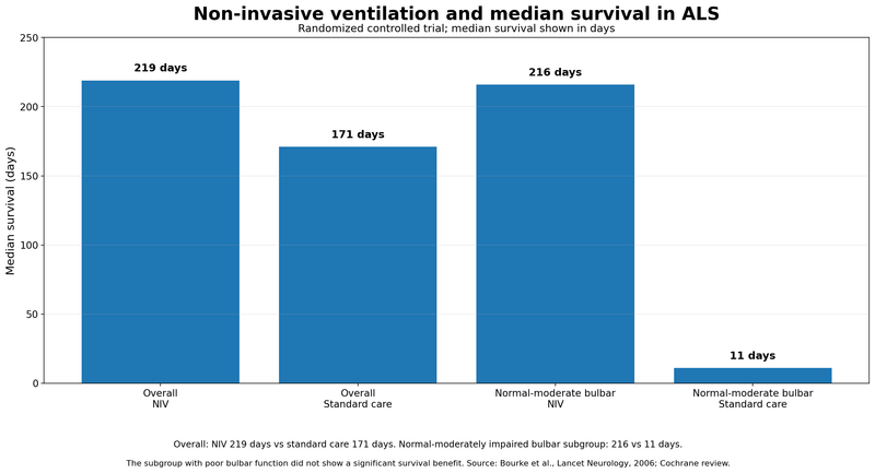
These findings come from a small randomized trial and should not be interpreted as an individual survival prediction. NIV decisions depend on respiratory impairment, bulbar function, secretion management, tolerance and patient preferences.
NICE currently recommends offering NIV to people with MND and respiratory impairment based on multidisciplinary assessment, and states that NIV can improve respiratory symptoms, quality of life and survival without stopping progression of the underlying disease. NICE
Adjusting Care
Speech support is intensified when repetition increases, communication becomes tiring or natural speech is no longer reliable for urgent needs. Changes may include amplification, personalised phrase banks, text-to-speech, alternative access methods or eye-gaze technology.[2,3,10]
Swallowing care is adjusted when coughing, wet voice, food residue or meal fatigue increases. Modification may involve food texture, fluid consistency, posture, mouthful size, pacing or additional instrumental assessment.[2,4]
Nutritional care is changed when weight falls, meals become too long or intake remains inadequate despite fortification. Oral supplements, feeding schedules and enteral nutrition are considered according to swallowing safety, gastrointestinal tolerance and respiratory status.[1,2]
Saliva treatment is modified according to whether secretions are thin, thick, dry or difficult to clear. Medication, hydration, humidification, suction and cough assistance are balanced so that reducing drooling does not create unmanageable mucus.[2,5,6]
Respiratory support is reviewed when sleep symptoms, cough effectiveness, ventilation tolerance or secretion clearance changes. NIV settings, mask type, humidification and cough-assist technique may require reassessment by the respiratory team.[1,2,6]
Ayurvedic medicines and therapies are adjusted when swallowing ability, respiratory capacity, bowel function, feeding route or tolerance changes. Oral dosage forms that leave residue, provoke coughing or interfere with hydration are discontinued or reformulated, while measurable functional and safety outcomes remain under review.
Individual Variation
The same 30-day plan is not suitable for every patient. Monitoring frequency and treatment priorities vary according to bulbar severity, respiratory function, nutritional status, cognition, communication ability, comorbidities and available caregiver support.[1,2]
A patient with mild speech change may focus on voice banking and early communication planning, while a patient with recurrent aspiration and weight loss may require urgent swallowing, respiratory and gastrostomy assessment. A person already dependent on NIV or tube feeding needs a different monitoring schedule from someone who remains fully oral and independently mobile.[1,2]
Sudden deterioration is not recorded for later discussion. Acute speech loss, severe choking, inability to clear secretions, marked dehydration, new confusion or severe breathing difficulty requires immediate medical assessment.[1,2,6]
Bulbar ALS Symptoms: Clinic Questions

When reviewing bulbar ALS symptoms, ask the clinical team to explain which changes require treatment now, which need further assessment and which can be monitored. Bring your recent weight measurements, meal-duration records, swallowing recommendations, medication list and respiratory results so that decisions reflect what is happening at home as well as during the appointment.[1,2]
Do I Need Another Swallowing Assessment?
Ask whether your current food textures, drinks and medication forms remain appropriate, particularly if meals have become longer, coughing has increased or your voice sounds wet after swallowing. Describe difficulties that appear only towards the end of a meal or when you are tired. Ask whether the findings justify further swallowing tests and what you can safely eat and drink while awaiting assessment.[2,4]
Request clear instructions that everyone assisting with meals can follow. These should cover food preparation, fluid consistency, positioning, feeding assistance and the changes that require reassessment. Recommendations need to reflect your swallowing ability rather than a general diet for everyone with ALS.[2,4]
Am I Meeting My Nutrition and Hydration Needs?
Ask the dietitian to compare your current weight with your usual weight and explain whether the change is clinically significant. Request individual calorie, protein and fluid targets, together with practical ways to meet them without making meals excessively large or tiring. Include nutritional supplements and any calorie-containing Ayurvedic preparations when discussing your total intake.[1,2]
Agree on what should trigger contact before the next appointment, such as continuing weight loss, reduced drinking or increasing difficulty finishing meals. Ask whether your current monitoring method is adequate if standing on a scale has become difficult.[1,2]
Should We Discuss a Feeding Tube Now?
Ask what a feeding tube would address in your situation: inadequate calories, dehydration, medication access, exhausting meals or swallowing difficulty. Discuss the likely benefits, procedural risks, daily care requirements and how your respiratory condition affects timing and insertion options. An early discussion allows you to consider these decisions without committing immediately to the procedure.[1,2]
Ask whether selected oral foods or drinks could continue after placement and whether the tube would initially supplement your meals. The answer depends on your swallowing assessment and preferences; tube placement does not automatically mean that every form of oral intake must stop.[4]
Should I Begin Voice Banking or Communication Technology?
Ask whether voice banking or message banking remains practical and which communication system you can begin using now. Request a trial that reflects your usual activities, including conversations at home, telephone calls and medical appointments. A system that works during a brief demonstration may still need adjustment for fatigue, seating or hand weakness.[1,3]
Ask who will provide training, arrange mounting equipment and review access if your movement changes. Establish a simple backup method for communicating pain, breathlessness or an urgent need when the electronic device is unavailable.[1,3]
Which Saliva Treatment Matches My Symptoms?
Ask whether the main problem is watery drooling, thick secretions, dry mouth or difficulty clearing saliva. Describe what happens after each treatment, including changes in mouth comfort, speech, sleep and suction needs. Ask whether medication, hydration, positioning or oral care needs adjustment before increasing a saliva-reducing treatment.[2,5]
Request a clear treatment goal and review date. Less visible drooling is not sufficient evidence of benefit if secretions become harder to clear or interfere more with breathing support.[2,5]
Have My Night-Time Breathing Problems Been Assessed?
Report morning headaches, unrefreshing sleep, daytime sleepiness and any need to sleep more upright. Ask whether your symptoms warrant overnight testing and whether the assessment needs to examine carbon dioxide as well as oxygen. A reassuring daytime oxygen reading does not exclude sleep-related hypoventilation.[2,6]
Ask the respiratory clinician to explain your results and whether they support starting or adjusting non-invasive ventilation. Discuss mask comfort, saliva, leakage and communication difficulties rather than assuming that difficulty using the equipment means it cannot help.[2,6]
Do I Need Cough Assistance, Suction or Both?
Ask whether your cough is strong enough to clear secretions, particularly during a respiratory infection. The team can assess whether assisted coughing, a cough-assist device, suction or a combination is appropriate. Ask for a practical demonstration and supervised practice rather than relying only on written equipment instructions.[1,6]
Your caregiver should understand when to use each prescribed measure, when to stop and whom to contact if clearance becomes ineffective or distressing. Request an individual plan for periods when infection or fatigue increases your need for support.[1,6]
Do My Medicines Need a Different Form?
Ask for a review of medicines that are difficult to swallow, leave residue or require repeated drinks. The prescriber or pharmacist can check whether another formulation or route is appropriate and whether each medicine is suitable for administration through a feeding tube.[2]
Include Ayurvedic medicines, supplements and non-prescription products in this review, bringing their ingredient lists and doses. Ask specifically about interactions, monitoring requirements and swallowing or tube compatibility. Do not assume that a liquid, powder or semisolid preparation is suitable simply because it is not a tablet.
What Should My Family Do During an Emergency?
Request an individual written plan distinguishing a manageable coughing episode from choking, secretion retention or respiratory distress that requires emergency help. Ask who will teach caregivers the appropriate first-aid response and how your communication method, mobility and respiratory equipment affect that response.[2,6]
Confirm how emergency clinicians can access your ventilation details, medication information and treatment preferences. Agree on an accessible way to signal that you cannot breathe, need suction or require immediate assistance when speaking is difficult.[2,6]
Who Should I Contact Between Appointments?
Identify the named professional coordinating your care and obtain the appropriate daytime, out-of-hours and equipment-support contacts. Clarify whether swallowing changes, weight loss, secretion problems and ventilation difficulties should be reported to different services or through one coordinator.[2]
Ask how referrals and equipment requests will be followed up and when your next review is due. Request an earlier assessment when function changes rather than waiting for the planned appointment, and discuss home-based or remote review when travel has become excessively tiring.[1,2]
FAQs
What are usually the first symptoms of bulbar ALS?
Early bulbar ALS symptoms commonly include gradually worsening speech clarity, difficulty pronouncing words, an altered voice or swallowing difficulty. Some people notice coughing with drinks, chewing fatigue or longer meals before obvious limb weakness develops. The first symptom and order of progression vary, and these changes require assessment rather than self-diagnosis.
Can bulbar ALS begin with a hoarse or nasal voice?
A hoarse, nasal, strained or breathy voice can occur with bulbar ALS, but voice quality alone cannot establish the diagnosis. Persistent changes need assessment, particularly when accompanied by progressively unclear speech, tongue weakness or swallowing difficulty. Evaluation considers the entire pattern rather than treating an isolated voice change as proof of ALS.
Does tongue twitching mean someone has bulbar ALS?
Tongue twitching alone does not establish bulbar ALS. Neurologists assess whether it accompanies progressive weakness, muscle wasting, impaired movement, speech changes or swallowing difficulty. Diagnosis depends on the neurological examination and appropriate investigations, not the appearance of the tongue in a photograph or video.
Is constant throat clearing a sign of bulbar ALS?
Constant throat clearing is not a specific sign of bulbar ALS. In someone with established bulbar weakness, it may reflect saliva pooling, retained food or difficulty clearing secretions. Persistent throat clearing needs assessment, particularly when it occurs after swallowing or accompanies a wet voice, weight loss or recurrent chest infections.
Why does ALS cause drooling or saliva pooling?
Drooling usually occurs because weakened lips, tongue and throat muscles cannot control and swallow saliva efficiently, rather than because the glands produce unusually large amounts. Treatment depends on whether saliva is watery, thick or difficult to clear. Excessive drying can make secretions more troublesome, so medicines require regular review.
What are the warning signs of aspiration in ALS?
Possible signs include coughing during meals, a wet or gurgling voice after swallowing, breathlessness while eating and recurrent chest infections. Aspiration can also occur without obvious coughing, so comfortable-looking swallowing does not always establish safety. New or worsening symptoms require swallowing assessment, with urgent medical care for significant breathing difficulty.
Can speech therapy help someone with bulbar ALS?
Speech therapy can help patients communicate more effectively through pacing, shorter phrases, reduced background noise and appropriate communication aids. It also supports voice preservation and preparation for alternative communication. Treatment focuses on practical communication and conserving effort; it does not imply that exercises can reverse the underlying motor-neuron disease.
When should voice banking begin?
Voice banking is best discussed soon after diagnosis, preferably while speech remains clear and recording is not exhausting. It preserves material that may be used to create a personalised synthetic voice. Message banking records complete phrases in the natural voice. A speech-language pathologist can help select an appropriate approach and compatible communication system.
When is a feeding tube considered in bulbar ALS?
A feeding tube is considered when swallowing difficulties, weight loss, dehydration or exhausting meals prevent adequate intake, or when medicines become difficult to administer. Discussions should begin early and include respiratory assessment. Tube feeding may supplement oral intake rather than immediately replacing it, depending on swallowing safety and individual preferences.
Can people with bulbar ALS use NIV or BiPAP?
Yes. Bulbar weakness can make non-invasive ventilation more difficult, but it does not automatically exclude its use. Mask fit, saliva control, airway clearance and ventilator settings need individual attention. Respiratory specialists assess symptoms and test results, then review comfort and effectiveness rather than assuming that one unsuccessful attempt means ventilation cannot help.
What is the difference between bulbar ALS and progressive bulbar palsy?
Bulbar-onset ALS describes ALS that begins with speech or swallowing symptoms. Progressive bulbar palsy describes a motor-neuron presentation dominated by weakness affecting speech, chewing and swallowing. The terms can overlap in clinical use. Neurological examination, testing and follow-up clarify whether involvement remains predominantly bulbar or extends to other regions.
Can bulbar symptoms develop after limb-onset ALS?
Yes. ALS that begins with hand, arm, foot or leg weakness can later affect speech, swallowing and saliva control. “Limb onset” identifies where symptoms started, not every area that may eventually be affected. Communication, nutrition and swallowing therefore need regular review even when they were normal at diagnosis.
What conditions can resemble bulbar ALS?
Other neurological, neuromuscular and structural disorders can cause similar speech and swallowing difficulties. Neurologists use the symptom pattern, examination and selected investigations to distinguish these conditions. No single symptom confirms ALS. Speech or swallowing difficulty that appears suddenly requires emergency assessment rather than being assumed to represent gradual motor-neuron disease.
How quickly do bulbar ALS symptoms progress?
There is no fixed timetable for bulbar ALS symptoms. Speech, swallowing, nutrition and breathing may change at different rates, and one person’s experience cannot predict another’s course. Repeated clinical assessments help guide care. Any abrupt deterioration needs prompt evaluation for an additional problem rather than being attributed automatically to ALS progression.
References
[1] Van Damme, P., Al-Chalabi, A., Andersen, P. M., Chiò, A., Couratier, P., De Carvalho, M., Hardiman, O., Kuźma-Kozakiewicz, M., Ludolph, A., McDermott, C. J., Mora, J. S., Petri, S., Probyn, K., Reviers, E., Salachas, F., Silani, V., Tysnes, O.-B., van den Berg, L. H., Villanueva, G., & Weber, M. (2024). European Academy of Neurology (EAN) guideline on the management of amyotrophic lateral sclerosis in collaboration with European Reference Network for Neuromuscular Diseases (ERN EURO-NMD). European Journal of Neurology, 31(6), e16264. https://doi.org/10.1111/ene.16264
[2] National Institute for Health and Care Excellence. (2019). Motor neurone disease: Assessment and management (NICE Guideline NG42). https://www.nice.org.uk/guidance/ng42
[3] Motor Neurone Disease Association. (2026, April). Dysarthria. https://www.mndassociation.org/professionals/management-of-mnd/dysarthria
[4] Motor Neurone Disease Association. (2026, April). Dysphagia, nutrition and enteral feeding. https://www.mndassociation.org/professionals/management-of-mnd/dysphagia
[5] Motor Neurone Disease Association. (2026, April). Saliva. https://www.mndassociation.org/professionals/management-of-mnd/saliva
[6] Motor Neurone Disease Association. (2026, April). Respiratory symptoms in MND. https://www.mndassociation.org/professionals/management-of-mnd/respiratory-symptoms-mnd
[7] Lee, J., Madhavan, A., Krajewski, E., & Lingenfelter, S. (2021). Assessment of dysarthria and dysphagia in patients with amyotrophic lateral sclerosis: Review of the current evidence. Muscle & Nerve, 64(5), 520–531. https://doi.org/10.1002/mus.27361
[8] Miller, R. G., Jackson, C. E., Kasarskis, E. J., England, J. D., Forshew, D., Johnston, W., Kalra, S., Katz, J. S., Mitsumoto, H., Rosenfeld, J., Shoesmith, C., Strong, M. J., & Woolley, S. C. (2009). Practice parameter update: The care of the patient with amyotrophic lateral sclerosis: Drug, nutritional, and respiratory therapies (an evidence-based review): Report of the Quality Standards Subcommittee of the American Academy of Neurology. Neurology, 73(15), 1218–1226. https://doi.org/10.1212/WNL.0b013e3181bc0141
[9] James, E., Ellis, C., Brassington, R., Sathasivam, S., & Young, C. A. (2022). Treatment for sialorrhea (excessive saliva) in people with motor neuron disease/amyotrophic lateral sclerosis. Cochrane Database of Systematic Reviews, 2022(5), CD006981. https://doi.org/10.1002/14651858.CD006981.pub3
[10] The ALS Association. (n.d.). Augmentative and alternative communication. https://www.als.org/navigating-als/living-with-als/therapies-care/augmentative-alternative-communication