



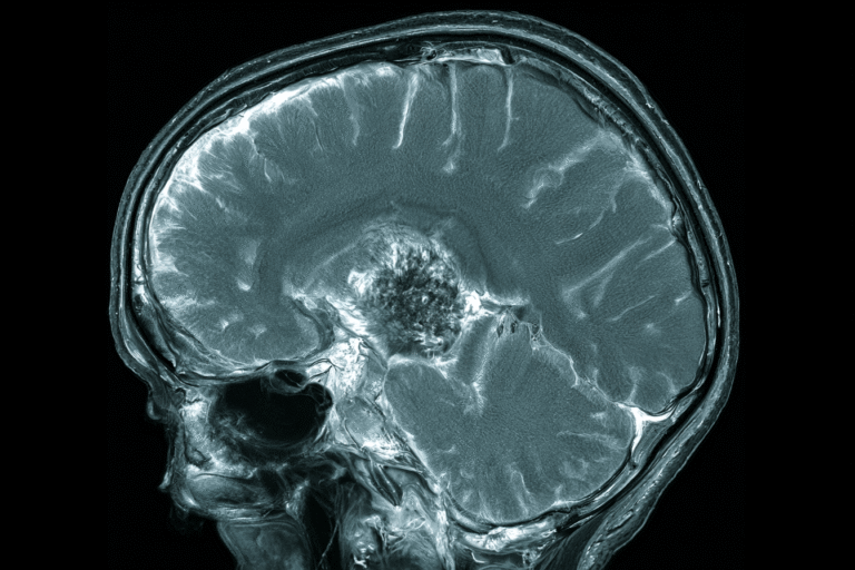
- What Are Small Cell Lung Cancer Brain Metastases?
- Symptoms of Small Cell Lung Cancer Brain Metastases
- How Small Cell Lung Cancer Brain Metastases Are Diagnosed
- Prognosis and Life Expectancy
- Treatment for Small Cell Lung Cancer Brain Metastases
- Ayurveda Led Curative Intent Model for Small Cell Lung Cancer Brain Metastases
- How Treatment Response and Remission Are Confirmed
- Frequently Asked Questions
- Reference
Small cell lung cancer brain metastases develop when cancer cells from a small cell lung tumour travel through the bloodstream and form secondary tumours inside the brain. These tumours are not a new type of brain cancer. They are made of small cell lung cancer cells and must be treated as part of the original lung cancer. Brain involvement may already be present when the cancer is diagnosed, or it may appear later after chemotherapy, immunotherapy or radiation treatment [1–5].
If you or a family member has small cell lung cancer, new headaches, seizures, weakness, confusion, difficulty speaking, loss of balance or changes in vision should not be ignored. Some patients have no early neurological symptoms, which is why contrast enhanced brain MRI is often important during staging and follow up. Early diagnosis may provide more treatment choices and may help prevent severe neurological deterioration [4,5,11].
Why Early Treatment Matters
Small cell lung cancer can grow and spread rapidly. When brain metastases cause surrounding swelling, known as cerebral oedema, the patient may develop vomiting, drowsiness, one sided weakness, seizures or loss of consciousness. In such situations, emergency medical treatment must come first. Corticosteroids, antiseizure medicines, radiation or surgery may be required according to the MRI findings and the patient’s neurological condition [5–9].
In this article, we explain the symptoms, prognosis and established treatment options in a simple manner. We also describe an Ayurveda led curative intent model that aims to address the brain lesions, primary lung tumour, disease outside the brain, neurological impairment and tendency for recurrence together. Curative intent describes the treatment objective and should not be understood as a guaranteed outcome. Any claim of remission must be confirmed through brain MRI, systemic imaging, neurological examination and appropriate laboratory monitoring [5,16].
What Are Small Cell Lung Cancer Brain Metastases?
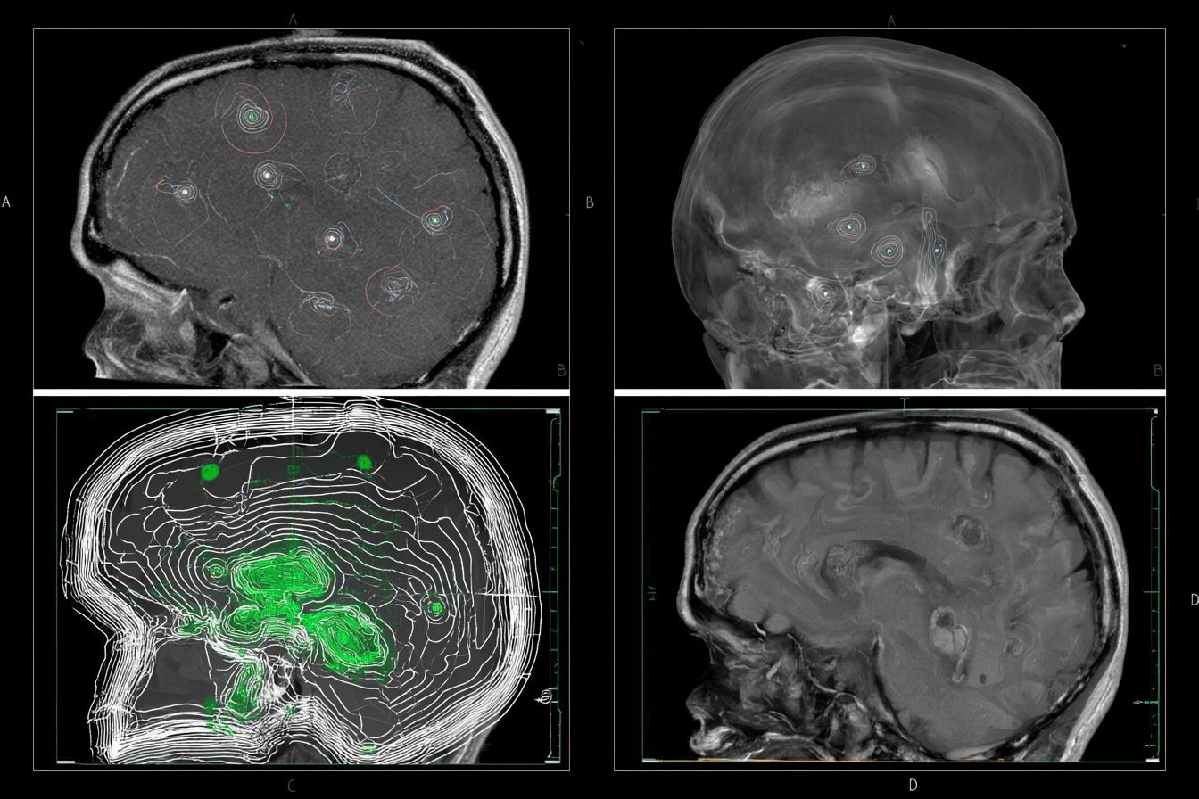
Small cell lung cancer brain metastases are secondary tumours that develop when cancer cells from the lungs reach the brain. They are not primary brain tumours. When doctors examine these lesions, the cells still have the features of small cell lung cancer, so treatment must address both the brain and the cancer elsewhere in the body [1,3,4].
How Small Cell Lung Cancer Reaches the Brain
Small cell lung cancer often grows quickly and may enter the bloodstream at an early stage. Cancer cells can travel through the circulation, reach brain tissue and begin forming new tumours. A patient may have one brain lesion, several separate lesions or more widespread intracranial involvement [1,4,5].
The size and number of lesions are important, but they are not the only factors that guide treatment. Doctors also consider the location of each lesion, surrounding swelling, pressure on nearby brain structures, neurological symptoms and whether cancer is active in other organs [4,5].
When Brain Metastases May Be Found
Brain metastases may be present when small cell lung cancer is first diagnosed. In other patients, they develop later during follow up, even after the lung tumour has responded to chemotherapy, immunotherapy or radiation. Some lesions cause obvious symptoms, while smaller lesions may be found on MRI before the patient notices any neurological change [2,4,5].
If your chest scan shows improvement, it does not automatically confirm that the brain is free from disease. Brain imaging and systemic imaging measure different parts of the cancer and may show different patterns of response.
Does Brain Metastasis Mean Extensive Stage SCLC?
Brain involvement represents distant spread and is generally classified as extensive stage small cell lung cancer. However, this classification does not mean that every patient has the same prognosis. A person with limited brain disease and controlled cancer elsewhere may have different treatment options and outcomes from someone whose cancer is progressing in the brain, liver, bones and other organs at the same time [1–3].
Symptoms of Small Cell Lung Cancer Brain Metastases
The symptoms of small cell lung cancer brain metastases depend on the number, size and location of the tumours. Swelling around a lesion can also disturb nearby brain tissue and raise pressure inside the skull. Some symptoms develop gradually over several days or weeks, while others, such as a seizure or sudden weakness, may appear without warning [5,6].
Not every headache, episode of dizziness or memory problem in a person with small cell lung cancer is caused by brain metastases. Low sodium, infection, dehydration, medicines, stroke and treatment related effects can produce similar symptoms. A new or rapidly worsening neurological problem therefore requires medical assessment rather than assumption [5,10].
Headache, Nausea and Vomiting
A new headache or a clear change in an existing headache may be an early warning sign. The pain may become progressively stronger, feel worse in the morning or increase during coughing, bending or physical strain. Headache may occur when a tumour and the swelling around it increase pressure inside the skull [5,8].
Some patients also develop nausea or repeated vomiting. Vomiting accompanied by severe headache, confusion, weakness or increasing sleepiness requires urgent assessment because it may indicate significant cerebral oedema or raised intracranial pressure.
Seizures and Loss of Awareness
A brain metastasis may irritate the electrical activity of the brain and cause a seizure. A seizure does not always appear as a full body convulsion. The patient may stare without responding, experience repeated movements in one arm or leg, notice an unusual smell or sensation, become temporarily unable to speak or lose awareness for a short period [5,6,9].
If you witness a seizure, protect the person from nearby objects and turn them onto one side when it is safe to do so. Do not force anything into the mouth. A first seizure, a seizure lasting several minutes or repeated seizures without full recovery require emergency medical care.
Weakness, Numbness and Facial Changes
Brain metastases affecting areas that control movement or sensation can cause weakness or numbness. The patient may begin dropping objects, dragging one leg, struggling to raise one arm or finding routine tasks unexpectedly difficult. One side of the face may droop, and the smile may appear uneven [5].
Sudden one sided weakness can also be caused by stroke or bleeding inside the brain. It should therefore be treated as an emergency, even when the patient already has known brain metastases.
Balance and Coordination Problems
Lesions involving the cerebellum or pathways responsible for balance may cause unsteady walking, repeated falls, poor hand coordination or difficulty standing without support. The person may appear dizzy or unable to judge the position of the feet properly.
These changes increase the risk of injury. Family members should not allow the patient to walk alone when balance has recently worsened, and urgent medical evaluation is needed when the loss of coordination progresses rapidly.
Speech, Vision and Swallowing Changes
The patient may develop slurred speech, difficulty finding familiar words or trouble understanding what another person is saying. These changes may be subtle at first and noticed more clearly by family members than by the patient.
Visual symptoms can include blurred vision, double vision or loss of part of the visual field. Difficulty swallowing, repeated choking or coughing while drinking may indicate neurological impairment and can lead to aspiration, dehydration and inability to take medicines safely [5,6].
Memory, Personality and Behavioural Changes
Brain metastases can affect concentration, judgement, memory and behaviour. A previously alert person may become unusually forgetful, irritable, withdrawn or confused. The family may notice inappropriate behaviour, reduced interest in normal activities, difficulty following a conversation or excessive sleepiness.
These changes should not be dismissed as emotional stress or ageing. They may result from the tumour, cerebral oedema, infection, medicine effects or a metabolic disturbance such as low sodium.
Can Brain Metastases Be Present Without Symptoms?
Yes. Small brain metastases may cause no noticeable symptoms and may be discovered only during staging or follow up MRI. A patient may feel well and still have early intracranial disease, which is why brain imaging remains important even when there is no headache, seizure or weakness [4,5,11].
Symptoms That Require Emergency Hospital Care
Emergency assessment is required when the patient develops a first or repeated seizure, sudden weakness, facial drooping, loss of consciousness, severe headache with repeated vomiting, new speech or vision loss, rapidly worsening confusion, unusual drowsiness, inability to walk or difficulty swallowing.
Small cell lung cancer can also cause the syndrome of inappropriate antidiuretic hormone secretion, known as SIADH. The resulting fall in blood sodium may produce headache, weakness, confusion, seizures or reduced consciousness. Serum sodium should therefore be checked rather than assuming that every neurological symptom is caused by tumour progression [10].
| Symptom | Possible explanation | Recommended urgency |
| Progressively worsening headache | Cerebral oedema or rising pressure inside the skull | Urgent medical assessment |
| First seizure | Irritation of brain tissue by a lesion | Emergency |
| Sudden weakness or facial drooping | Brain lesion, stroke or intracranial bleeding | Emergency |
| Confusion or unusual drowsiness | Brain involvement, oedema, infection or low sodium | Emergency |
| Repeated vomiting with headache | Increased intracranial pressure | Emergency |
| Gradual memory or personality change | Frontal or temporal brain involvement | Prompt neurological assessment |
| Difficulty swallowing or repeated choking | Neurological impairment and aspiration risk | Urgent assessment |
How Small Cell Lung Cancer Brain Metastases Are Diagnosed

Small cell lung cancer brain metastases cannot be confirmed from symptoms alone. Headache, weakness, confusion or seizures may also result from low sodium, infection, stroke, medicines or treatment related complications. Your doctor therefore combines brain imaging, neurological examination, blood tests and assessment of the cancer elsewhere in the body before deciding on treatment [1,5,10].
Contrast Enhanced Brain MRI
Contrast enhanced brain MRI is generally the preferred investigation because it can detect small lesions that may not be clearly visible on a routine CT scan. MRI also provides detailed information about the number, size and exact location of the metastases [5,11].
The scan helps doctors identify swelling around the tumour, bleeding, pressure on nearby brain tissue, displacement of the brain’s central structures and blockage of normal fluid circulation. These findings are important because a small lesion in a sensitive area may cause more severe symptoms than a larger lesion in another part of the brain.
Previous MRI images should be compared with the latest scan whenever they are available. This comparison helps determine whether existing lesions are shrinking, remaining stable or increasing and whether new lesions have appeared.
When a CT Brain Scan May Be Used
A CT brain scan may be performed when the patient develops sudden weakness, severe headache, loss of consciousness or another emergency symptom. CT is particularly useful for rapidly detecting major bleeding, hydrocephalus or a large lesion causing pressure inside the skull.
CT may also be used when MRI is unavailable or cannot be performed because of certain implanted devices or other medical restrictions. However, a normal CT scan does not always exclude small brain metastases. Contrast enhanced MRI may still be required when clinical suspicion remains high [5,11].
Assessment of Cancer Outside the Brain
Brain imaging shows only the intracranial part of the disease. Doctors must also determine whether the primary lung tumour, chest lymph nodes and other metastatic sites are controlled or progressing.
CT scans of the chest, abdomen and pelvis are commonly reviewed, while PET CT may be used in selected situations. A patient with limited brain involvement and controlled disease elsewhere may have different treatment options from a patient whose cancer is simultaneously progressing in the liver, bones, adrenal glands or several other organs [1–3].
Blood Tests Before Treatment
Blood tests help identify problems that may worsen neurological symptoms or affect treatment safety. A complete blood count is used to assess anaemia, infection risk and platelet levels, while liver and kidney tests help determine whether the patient can safely receive systemic medicines, contrast imaging or anaesthesia.
Serum sodium is particularly important because SCLC can cause SIADH and severe hyponatraemia. Low sodium may cause confusion, weakness, seizures and reduced consciousness even when the brain lesions have not enlarged [10].
Blood glucose, albumin and other electrolytes may also be checked. These results help the treatment team understand the patient’s nutritional condition, organ reserve and ability to tolerate radiation, surgery, chemotherapy, immunotherapy or an individualised Ayurvedic formulation.
Neurological and Functional Assessment
The doctor evaluates limb strength, speech, vision, balance, coordination, memory, awareness and swallowing. The patient or caregiver should also explain when the symptoms began, whether they are worsening and whether seizures or falls have occurred.
Performance status is assessed to understand how independently the patient can walk, eat, communicate and perform daily activities. This information is considered together with the MRI because treatment decisions should not be based on lesion number alone [5,12].
When the imaging findings are unusual or the diagnosis remains uncertain, a neurosurgeon may consider obtaining tissue from an accessible lesion. In many patients with confirmed SCLC and typical MRI findings, treatment can be planned without a separate brain biopsy, but the final decision depends on the complete clinical picture.
Prognosis and Life Expectancy

Small cell lung cancer with brain metastases is a serious and advanced illness, but the prognosis is not identical for every patient. The outcome depends on how much disease is present in the brain, whether cancer is active elsewhere in the body, the patient’s neurological condition and how well the cancer responds to treatment [12,13].
When you read a survival figure online, it should not be treated as a fixed prediction of how long you or your family member will live. Survival statistics describe groups of patients, including people with very different tumour burdens, general health, access to treatment and responses to therapy.
What Does Median Survival Mean?
Median survival is the point at which half of the patients in a study were alive and half had died. It is not an expiry date, and it cannot predict the exact outcome of one person.
A 2025 United States national analysis included 11,074 patients diagnosed with small cell lung cancer and brain metastases between 2018 and 2020. The median overall survival was 6.6 months. Patients whose metastatic disease was limited to the brain had a median survival of 8.8 months, compared with approximately 6 months when metastases were also present outside the brain [13].
These figures include patients with different ages, performance levels, treatment patterns and disease severity. A newly diagnosed patient should therefore not assume that the national median represents his or her personal survival.
Which Factors Affect the Prognosis?
One of the most important factors is performance status, which describes how independently the patient can walk, eat, communicate and perform daily activities. A person who remains active and neurologically stable is generally more capable of receiving radiation, systemic treatment and further treatment if the disease returns.
The number of brain lesions also matters, but lesion count should not be considered alone. Doctors also assess the size and total volume of the lesions, their location, surrounding oedema, pressure on important brain structures and whether seizures, weakness, speech problems or swallowing difficulties are present.
The SCLC Graded Prognostic Assessment identifies age, performance status, number of brain metastases and the presence of extracranial metastases as important factors influencing survival [12]. This supports a personalised prognosis rather than using one survival estimate for every patient.
Can Some Patients Live Longer Than the Average?
Yes. Longer survival may be seen when the brain tumour burden is limited, neurological function is preserved, disease outside the brain is controlled and the patient can receive effective local and systemic treatment.
In a prospective phase II study of carefully selected patients with one to ten SCLC brain metastases treated with stereotactic radiation, median overall survival was 10.2 months. However, these patients were selected for focused treatment and underwent close MRI surveillance, so the result should not be applied automatically to every patient with brain metastases [15].
The FIRE SCLC study also found that survival differed according to the number of treated brain lesions. Patients with fewer lesions generally had better outcomes, although this was an observational study and the results may partly reflect selection of healthier patients for stereotactic radiosurgery [14].
How Is an Individual Prognosis Estimated?
Your doctor estimates prognosis by reviewing the latest brain MRI, systemic scans, neurological examination, performance status, organ function and previous treatment response. The outlook may change after cerebral oedema is controlled or after brain lesions respond to radiation and systemic treatment.
For this reason, prognosis should be reassessed during treatment rather than decided permanently from the first scan. Reduction or disappearance of brain lesions, absence of new metastases, recovery of neurological function and control of the lung tumour provide more meaningful information than a population survival number alone.
Treatment for Small Cell Lung Cancer Brain Metastases

Treatment for small cell lung cancer brain metastases must address two connected problems. The first is the disease inside the brain, while the second is the cancer in the lungs, lymph nodes or other organs. Treating only the visible brain lesions may not control cancer elsewhere, while systemic treatment alone may not act quickly enough when brain lesions are causing swelling, seizures or neurological weakness [1,2,5,6].
The treatment plan is therefore based on the patient’s symptoms, number and size of brain lesions, surrounding oedema, previous treatment, disease outside the brain and overall physical condition. A person with one small asymptomatic lesion may require a different approach from someone with multiple lesions, repeated seizures and pressure on important brain structures.
How Doctors Decide Which Treatment Should Come First
The urgency of neurological symptoms is one of the most important factors. A patient with a seizure, rapidly worsening weakness, severe headache, repeated vomiting, reduced consciousness or major cerebral oedema generally requires immediate brain directed treatment and medical stabilisation [5,6].
When brain metastases are small and are not causing symptoms, doctors may have more flexibility in deciding whether radiation or systemic treatment should begin first. The decision also depends on whether the cancer has just been diagnosed, whether chemotherapy or immunotherapy has already been used and whether the disease is progressing outside the brain.
The number of lesions is important, but it should not be used alone. Doctors also consider the total volume of the tumours, their location, the amount of surrounding swelling, pressure on nearby structures and the patient’s ability to attend regular MRI follow up.
Corticosteroids for Cerebral Oedema
Brain metastases often produce swelling in the surrounding tissue. This swelling is called cerebral oedema and may cause headache, vomiting, confusion, weakness, speech difficulty or reduced consciousness.
Dexamethasone is commonly prescribed when cerebral oedema is producing significant symptoms. It can reduce swelling and pressure around the tumour, sometimes leading to noticeable neurological improvement within a relatively short period [6,8].
Improvement after dexamethasone does not mean that the metastatic tumour has disappeared. The medicine reduces inflammation and oedema around the lesion, but radiation, surgery or systemic treatment may still be required to control the cancer itself.
The dose and duration should be decided by the treating doctor. Long term or high dose corticosteroid treatment may increase blood glucose, infection risk, gastric irritation, insomnia, mood changes and muscle weakness. It should not be stopped suddenly after prolonged use because the dose may need to be reduced gradually under medical supervision [8].
If you are already taking an Ayurvedic formulation, the treating physician should know the complete composition. This is important because corticosteroids, blood glucose changes, infection risk, liver function and gastrointestinal irritation can influence medicine selection and dosage.
Antiseizure Medicines
A seizure may occur when a metastatic lesion irritates the electrical activity of nearby brain tissue. Antiseizure medicines are usually prescribed after a patient has experienced a seizure or when a neurologist identifies another clear indication [6,9].
Routine preventive antiseizure treatment is not required for every patient who has brain metastases but has never experienced a seizure. The decision should be individual because antiseizure medicines may cause drowsiness, dizziness, imbalance or interactions with other medicines [9].
If a seizure has occurred, the medicine should be taken regularly and should not be stopped without neurological guidance. The patient may also need restrictions on driving, bathing alone, working at heights or using machinery until the seizure risk has been assessed.
A seizure lasting several minutes, repeated seizures without full recovery or a first seizure with loss of consciousness requires emergency hospital care.
Whole Brain Radiation Therapy
Whole brain radiation therapy, commonly called WBRT, delivers radiation to the entire brain. It has traditionally been used frequently in small cell lung cancer because this cancer has a strong tendency to produce multiple visible lesions and microscopic tumour deposits that may not yet be detectable on MRI [1,4,6,7].
WBRT may be considered when there are many brain metastases, diffuse intracranial involvement or a high risk that disease is present beyond the visible lesions. It may also be selected when focused stereotactic radiation is not technically suitable because of lesion number, total tumour volume, location or previous treatment.
The purpose of WBRT is to control existing brain metastases, reduce the risk of neurological progression and treat possible microscopic disease throughout the brain. It is different from prophylactic cranial irradiation, which is given to selected patients without visible brain metastases to reduce the risk of future intracranial spread.
The possible disadvantages include fatigue, hair loss, scalp irritation and effects on memory or concentration. The risk and severity of cognitive changes depend on the patient’s age, baseline neurological condition, radiation plan, previous brain treatment and expected survival.
Doctors must balance the likelihood of intracranial control against the potential effect on cognition and quality of life. For some patients with multiple or rapidly progressing lesions, the broader brain coverage provided by WBRT may be more important than the disadvantages.
Hippocampal Avoidance and Memantine
The hippocampus is a part of the brain involved in memory formation. In selected patients receiving WBRT, radiation planning may be designed to reduce exposure to the hippocampal regions.
Hippocampal avoidance WBRT may help reduce certain cognitive effects when the metastases are not located within or very close to the protected areas. It is not suitable for every patient, and eligibility must be decided after careful review of the MRI and radiation plan [6,7,23].
Memantine may also be prescribed during and after WBRT to help delay cognitive decline. Evidence supporting memantine and hippocampal avoidance includes patients with brain metastases from several different cancers, so these measures should be described as methods of cognitive protection rather than treatments that directly improve SCLC tumour control [22,23].
Stereotactic Radiosurgery and Stereotactic Radiotherapy
Stereotactic radiosurgery, known as SRS, delivers a highly focused dose of radiation to individual brain lesions. Despite the word surgery, no incision is made. Fractionated stereotactic radiotherapy uses a similar focused method but divides the treatment into several sessions.
These treatments reduce radiation exposure to uninvolved brain tissue and may be considered when the number and total volume of lesions are limited. The patient should also be able to undergo regular MRI surveillance because new metastases may later develop in untreated areas of the brain [6,7,14,15].
A prospective phase II study evaluated stereotactic treatment in selected patients with one to ten SCLC brain metastases. The findings supported SRS or fractionated stereotactic treatment as a reasonable option in carefully selected patients, but some later required whole brain radiation or further intracranial treatment [15].
SRS should not be described as universally better than WBRT. Patients selected for SRS often have smaller tumour volumes, better performance status, fewer neurological symptoms and better access to close imaging follow up. These differences can influence survival and treatment outcomes.
The choice between SRS and WBRT therefore depends on lesion number, total tumour volume, location, previous cranial radiation, extracranial disease, neurological symptoms and the patient’s priorities regarding cognition and treatment burden [6,7,14,15].
Brain Surgery
Surgery is less commonly required for small cell lung cancer brain metastases because many patients have multiple lesions or active systemic disease. However, it may be valuable in carefully selected situations [5,6].
A neurosurgeon may consider surgery when one large and accessible lesion is causing severe pressure, neurological deterioration or displacement of brain structures. Surgery may also be useful when the diagnosis is uncertain and tissue is needed to determine whether the lesion represents metastasis, another tumour, treatment related change or infection.
Removing a large lesion can provide rapid decompression, but it does not treat microscopic disease elsewhere in the brain or cancer outside the brain. Radiation or systemic treatment may therefore still be required after surgery.
The decision depends on the location of the lesion, surgical risk, neurological condition, systemic cancer status and the patient’s ability to recover from an operation.
Systemic Chemotherapy and Immunotherapy
Systemic treatment is required because brain metastases are part of a disease that began in the lung and may also involve lymph nodes or other organs. In newly diagnosed extensive stage SCLC, treatment commonly includes platinum based chemotherapy with etoposide and an immune checkpoint inhibitor in suitable patients [1–3].
When brain metastases are producing severe symptoms, local brain treatment usually cannot be postponed while waiting for systemic medicines to work. Radiation, corticosteroids or surgery may need to be given first or coordinated closely with chemotherapy and immunotherapy [5,6].
When the brain lesions are asymptomatic and the patient is otherwise stable, the sequence may be individualised. The treating team considers the extent of extracranial disease, previous medicines, anticipated intracranial response and the feasibility of close MRI monitoring.
Systemic treatment may reduce both the lung tumour and some brain lesions, but intracranial response is not guaranteed. The brain must therefore be reassessed separately rather than assuming that improvement in the chest means that all brain metastases have also responded.
Immunotherapy may cause inflammatory side effects involving the lungs, liver, bowel, endocrine glands, skin or nervous system. Any new symptoms during treatment should be assessed carefully instead of being attributed automatically to the cancer.
For detailed information about chemotherapy, immunotherapy and radiation used for the overall disease, the reader should be directed to the separate small cell lung cancer treatment article rather than repeating every drug regimen here.
Treatment of Recurrent Brain Metastases
Brain metastases may return after WBRT, SRS, chemotherapy or an earlier response. Recurrence may appear as enlargement of a previously treated lesion or as new lesions elsewhere in the brain.
The next treatment depends on what the patient has already received. Selected patients may undergo salvage SRS, surgery for a large accessible lesion, further systemic treatment or carefully planned repeat radiation [1,2,5,6,14,15].
Doctors must also distinguish active tumour recurrence from radiation related injury because both may cause worsening symptoms and similar MRI changes. Additional imaging, repeated MRI or tissue examination may be required when the diagnosis is uncertain.
If the brain disease progresses while cancer outside the brain remains controlled, the treatment may focus mainly on intracranial control. When the brain, lungs and other organs are progressing together, both local and systemic treatment usually need to be reconsidered.
Comparison of the Main Treatment Options
| Treatment | When it may be used | Main purpose | Important limitation |
| Dexamethasone | Symptomatic cerebral oedema | Reduces swelling and pressure symptoms | Does not eliminate the tumour |
| Antiseizure medicine | A seizure has occurred or another indication is present | Reduces the risk of further seizures | Does not treat the cancer |
| Whole brain radiation | Multiple or diffuse brain lesions | Treats visible and possible microscopic intracranial disease | May affect memory and cognition |
| SRS or stereotactic radiotherapy | Selected lesions with limited total volume | Delivers focused radiation to visible tumours | New lesions may develop elsewhere |
| Brain surgery | Large accessible lesion, mass effect or uncertain diagnosis | Provides decompression and tissue diagnosis | Does not treat disease in other areas |
| Systemic treatment | Active disease in the brain and elsewhere | Treats the overall SCLC burden | Intracranial response may vary |
A patient may receive more than one of these treatments. For example, dexamethasone may first control oedema, followed by SRS or WBRT, while systemic treatment addresses the lung tumour and extracranial disease.
Emergency neurological treatment should not be delayed while an Ayurvedic treatment plan is being prepared. Once the patient is stable, the complete MRI findings, systemic disease status, current medicines and organ function can be reviewed to design a coordinated and individualised treatment strategy.
Ayurveda Led Curative Intent Model for Small Cell Lung Cancer Brain Metastases

The Ayurveda led model for small cell lung cancer brain metastases is designed with curative intent rather than being limited to appetite, fatigue, sleep or treatment related weakness. Its wider objective is to work towards complete control of the brain lesions, the primary lung tumour, disease in other organs and the biological tendency for further spread.
Curative intent does not mean that the result can be guaranteed before treatment begins. It means that the treatment is planned with complete radiological response and durable remission as its objectives. We do not consider improvement in headache, appetite, sleep or energy alone as evidence that brain metastases have disappeared. The result must be demonstrated through contrast enhanced brain MRI, systemic scans, neurological examination and laboratory monitoring [5,16].
When you submit a case for Ayurvedic assessment, the brain MRI cannot be reviewed in isolation. We must understand the entire disease pattern because a patient with one small brain lesion and controlled disease in the chest requires a different strategy from a patient with multiple brain lesions, cerebral oedema and simultaneous progression in the liver, bones or adrenal glands.
Why the Brain and the Rest of the Body Must Be Treated Together
Brain metastases are part of small cell lung cancer that began in the lungs. Radiation may control visible lesions inside the brain, but it does not automatically eliminate disease in the lungs, lymph nodes or other organs. In the same way, improvement in the chest does not confirm that every brain lesion has responded.
The Ayurveda curative intent model therefore considers the intracranial and systemic disease as one connected process. The aim is to reduce the measurable tumour burden, control the pathways through which the disease continues to spread, protect neurological function and prevent new lesions from developing.
This does not mean that urgently required radiation, corticosteroids, antiseizure medicines or surgery should be delayed. If a patient has a seizure, major cerebral oedema, reduced consciousness, rapidly worsening weakness or pressure on important brain structures, emergency medical stabilisation must come first [5,6,8,9].
Once the patient is stable, the Ayurvedic plan can be designed around the MRI findings, current oncology treatment, organ function and the patient’s ability to swallow and digest medicine safely.
Individual Disease Mapping Before Ayurvedic Treatment
In our clinical model, treatment begins with a detailed disease map rather than a fixed medicine package. The same formulation should not be prescribed to every patient because the position of the lesions, neurological symptoms, previous radiation and systemic disease burden may be completely different.
The intracranial map records the number, size and location of the brain lesions. It also identifies cerebral oedema, bleeding, mass effect, midline shift, hydrocephalus and whether the patient has previously received whole brain radiation or stereotactic radiation.
The systemic map records the size and activity of the primary lung tumour, chest lymph nodes and metastases involving the liver, bones, adrenal glands or other organs. This assessment helps us understand whether the brain is the only active site or one part of widespread disease.
The neurological map records seizures, headache, speech, vision, memory, behaviour, swallowing, limb strength, coordination and walking ability. A caregiver’s observations are particularly valuable when the patient has confusion, reduced awareness or personality changes.
The Ayurvedic and physiological assessment considers Prakriti, Agni, Ama, Kostha, Bala, Ojas, appetite, bowel function, sleep, weight and physical endurance. Blood counts, sodium, liver function and kidney function must also be reviewed because they influence medicine selection, dosage and safety.
Ayurvedic Interpretation of Brain Metastases
There is no exact classical Ayurvedic diagnosis that is identical to histopathologically confirmed small cell lung cancer brain metastasis. Ayurveda did not describe modern MRI, immunohistochemistry or molecular oncology. The classical concepts should therefore be used to understand the disease process and guide personalised treatment, not to replace the modern diagnosis.
The main concepts relevant to this model are Arbuda, Granthi, Vata, Majja Dhatu, Mastishka, Shotha, Srotorodha, Agni, Bala and Ojas. Apasmara principles may also become relevant when seizures or altered awareness are present.
Arbuda and Abnormal Tissue Growth
Sushruta describes Arbuda as a deep seated and progressively enlarging abnormal tissue formation. The classical description focuses on the involvement of Dosha and Mamsa and describes a mass that is fixed, large, deep rooted and generally non suppurating.
Book name: Sushruta Samhita
Section: Nidana Sthana
Chapter: 11, Granthi Apachi Arbuda Galaganda Nidana
Text number: Sushruta Samhita, Nidana Sthana 11/13–14 [18]
Sanskrit
गात्रप्रदेशे क्वचिदेव दोषाः सम्मूर्च्छिता मांसमभिप्रदूष्य।
वृत्तं स्थिरं मन्दरुजं महान्तमनल्पमूलं चिरवृद्ध्यपाकम्॥१३॥
कुर्वन्ति मांसोपचयं तु शोफं तमर्बुदं शास्त्रविदो वदन्ति॥१४॥
Transliteration
Gātrapradeśe kvacideva doṣāḥ sammūrcchitā māṃsam abhipradūṣya,
vṛttaṃ sthiraṃ mandarujam mahāntam analpamūlaṃ ciravṛddhyapākam.
Kurvanti māṃsopacayaṃ tu śophaṃ tam arbudaṃ śāstravido vadanti.
Translation
When disturbed Doshas become localised in a part of the body and affect Mamsa, they may produce a round, fixed, mildly painful, large, deep rooted, slowly growing and non suppurating swelling. Those knowledgeable in the science describe such a formation as Arbuda.
This passage provides a classical framework for understanding abnormal tissue growth. It should not be interpreted as proof that every feature of Arbuda is identical to the cellular and molecular behaviour of small cell lung cancer.
Sushruta Samhita, Chikitsa Sthana, Chapter 18 describes the classical treatment principles for Granthi and Arbuda. In a modern case, these principles must be adapted according to pathology, MRI findings, systemic disease burden, neurological status and current oncology treatment [18].
Vata and the Spread of Disease
Vata is considered the main functional principle responsible for movement and transportation within the body. In the proposed Samprapti, Vata is relevant to the movement of an established disease process from its original site to a distant vulnerable location.
This does not mean that Vata is biologically identical to cancer cells or the bloodstream. It means that Vata provides a classical framework for understanding movement, dissemination and rapidly changing neurological manifestations.
Charaka Samhita, Chikitsa Sthana, Chapter 28, Vatavyadhi Chikitsa, is relevant when the patient develops weakness, altered movement, facial changes, speech difficulty, loss of coordination or disturbed neurological function [17]. Treatment is selected according to the type of Vata disturbance, associated Doshas, obstruction and the degree of tissue depletion.
Majja Dhatu and Mastishka Involvement
Majja and Mastishka concepts become important when the disease has reached the brain and begins affecting cognition, awareness, coordination, speech or motor control. A patient may show both structural disease and functional neurological disturbance.
The Ayurvedic plan should therefore not target the tumour mass alone. It must also consider the surrounding brain tissue, neurological function, treatment related injury and the patient’s ability to regain speech, movement, memory and independence.
Majja support should not be reduced to prescribing one Medhya herb to every patient. The choice depends on cerebral oedema, seizures, swallowing ability, liver function, current medicines and whether the neurological symptoms are caused by active tumour, radiation effects or a metabolic disturbance.
Shotha and Srotorodha in Cerebral Oedema
The swelling around a brain metastasis may be interpreted through the concepts of Shotha and Srotorodha. The lesion and surrounding oedema may obstruct normal function and place pressure on nearby brain structures.
However, cerebral oedema is a medical emergency when it causes severe headache, repeated vomiting, progressive weakness, confusion or reduced consciousness. Dexamethasone or another urgent intervention should not be replaced by an oral Ayurvedic medicine when rapid pressure reduction is required [6,8].
After stabilisation, the Ayurvedic plan may be adjusted according to the degree of oedema, corticosteroid requirement, blood glucose, sodium level, sleep, appetite and infection risk. Reduction in oedema must be confirmed by MRI and neurological examination rather than assumed from a temporary decrease in headache.
Apasmara Principles When Seizures Are Present
When a patient has seizures, altered awareness or loss of memory surrounding an episode, the principles described under Apasmara may contribute to Ayurvedic clinical reasoning.
The relevant classical sources are Charaka Samhita, Nidana Sthana, Chapter 8, Apasmara Nidana, and Charaka Samhita, Chikitsa Sthana, Chapter 10, Apasmara Chikitsa [17].
A seizure caused by a metastatic brain lesion remains a structural neurological condition. It should not be relabelled simply as Apasmara, and prescribed antiseizure medicines should not be stopped without neurological advice.
Agni, Bala and Ojas
Patients with advanced small cell lung cancer may experience poor appetite, weight loss, weakness, disturbed digestion and reduced treatment tolerance. Chemotherapy, radiation, corticosteroids, infection and the cancer itself may further reduce Bala and disturb Agni.
If Agni is weak, even a well selected formulation may not be digested or assimilated properly. Treatment must therefore consider digestion, bowel function, nausea, vomiting, swallowing and nutritional status rather than prescribing a heavy Rasayana preparation without assessment.
Bala and Ojas are also important because the patient needs sufficient physiological reserve to tolerate radiation, systemic treatment and recovery. Their restoration is part of the treatment strategy, but it should occur alongside measurable control of the cancer rather than being mistaken for tumour treatment by itself.
The Role of Rasayana
Rasayana is relevant to tissue nourishment, memory, strength, sensory function and recovery. It should not be described simply as an immune booster, and it should not be used as a substitute for disease directed treatment.
Book name: Charaka Samhita
Section: Chikitsa Sthana
Chapter: 1, Rasayana Adhyaya
Pada: 1, Abhaya Amalakiya Rasayana Pada
Text number: Charaka Samhita, Chikitsa Sthana 1.1/7–8 [17]
Sanskrit
दीर्घमायुः स्मृतिं मेधामारोग्यं तरुणं वयः।
प्रभावर्णस्वरौदार्यं देहेन्द्रियबलं परम्॥७॥
वाक्सिद्धिं प्रणतिं कान्तिं लभते ना रसायनात्।
लाभोपायो हि शस्तानां रसादीनां रसायनम्॥८॥
Transliteration
Dīrgham āyuḥ smṛtiṃ medhām ārogyaṃ taruṇaṃ vayaḥ,
prabhā varṇa svaraudāryaṃ dehendriya balaṃ param.
Vāk siddhiṃ praṇatiṃ kāntiṃ labhate nā rasāyanāt,
lābhopāyo hi śastānāṃ rasādīnāṃ rasāyanam.
Translation
Rasayana is traditionally described as promoting longevity, memory, intellect, health, vitality, complexion, voice and the strength of the body and senses. It is described as a means of obtaining the best quality of Rasa and the subsequent body tissues.
In small cell lung cancer brain metastases, Rasayana should be selected according to the treatment stage. During rapidly progressive disease, severe Ama, repeated vomiting or major cerebral oedema, the immediate priority may be stabilisation and active disease control. As the patient becomes stable, Rasayana principles can be integrated to support neurological recovery, tissue quality, strength and treatment tolerance.
Stage One: Active Intracranial and Systemic Disease Reduction
The first stage focuses on measurable active disease. The brain lesions, primary lung tumour, lymph nodes and any extracranial metastases are assessed together.
The Ayurvedic formulation is designed according to the dominant disease pattern, neurological symptoms, cerebral oedema, Agni, Bala and organ function. The goal is not only to make the patient feel better but to pursue reduction of visible disease and prevent the appearance of new metastatic sites.
If radiation, dexamethasone, antiseizure medicines, chemotherapy or immunotherapy are being used, the formulation must be coordinated with them. A medicine that is suitable before radiation may need modification when the patient develops vomiting, marrow suppression, liver injury or difficulty swallowing.
Stage Two: Consolidation and Neurological Recovery
The second stage begins after the first measurable response or stabilisation. The formulation is reassessed using the latest MRI, systemic scans, neurological findings and laboratory results.
At this stage, the objectives include addressing residual disease, reducing the chance of early progression, restoring neurological function and improving Agni, Bala and Ojas. If the patient is still dependent on high dose corticosteroids, the treatment plan must continue to consider blood glucose, muscle weakness, infection risk and gastric tolerance.
A decrease in lesion size without complete disappearance is not treated as the final result. Consolidation continues according to the remaining disease burden and the patient’s ability to tolerate treatment.
Stage Three: Remission Monitoring and Recurrence Prevention
The third stage begins only when imaging shows a complete or strong response and the patient is neurologically stable. The purpose is to maintain the response and identify recurrence at the earliest possible stage.
Regular brain MRI remains necessary because a treated lesion may remain controlled while a new lesion appears elsewhere. Systemic imaging is also required because the brain may remain stable while the lung tumour or another metastatic site progresses.
In our model, remission is not declared from one improved scan alone. The response should remain stable across serial imaging, with no new lesions and no unexplained neurological deterioration.
How the Formulation Is Personalised
There should not be one universal Ayurvedic medicine for every patient with small cell lung cancer brain metastases. The formulation and dose depend on the number and location of lesions, cerebral oedema, seizure history, current corticosteroid dose, sodium level, swallowing ability, liver and kidney function and previous radiation.
The treatment phase also matters. A patient receiving active chemotherapy may need a different formulation from a patient recovering after radiation or entering MRI confirmed remission.
Arbuda Pratyanika, Vata regulation, Shotha management, Medhya support, Agni correction and Rasayana may all form parts of the treatment architecture. Their proportions must change according to the dominant clinical need rather than being combined mechanically in the same amount for every patient.
When Rasaushadhi or Bhasma are considered, their source, preparation method, composition and dose must be documented. Periodic complete blood count, liver function and kidney function testing is necessary, particularly when the patient is also receiving chemotherapy, immunotherapy, corticosteroids or antiseizure medicines.
Modern Research Relevant to the Ayurvedic Model
A small randomised pilot trial studied Boswellia serrata in patients receiving radiation for primary or secondary brain tumours. The Boswellia group showed a greater reduction in MRI measured cerebral oedema, but the study included only 44 patients, was not specific to small cell lung cancer and did not prove elimination of brain metastases [19].
A later systematic review concluded that clinical evidence for Boswellia in radiation associated cerebral oedema and radiation necrosis remains limited and varied in quality. Boswellia may therefore be discussed as a possible carefully supervised adjunct in selected patients, not as a replacement for dexamethasone, radiation or neurosurgery [20].
Laboratory research has also reported that curcumin affected proliferation, migration, invasion and JAK STAT3 signalling in small cell lung cancer experimental models [21]. These findings provide a possible biological basis for further research, but they are not human proof that oral curcumin reaches brain metastases, produces remission or improves survival.
The classical Ayurvedic framework and modern laboratory findings can help guide the design of an individualised model. The actual patient result must still be demonstrated through MRI, systemic imaging and sustained clinical response.
Ayurvedic Treatment Objectives and Their Measurement
| Treatment objective | Ayurvedic interpretation | How the result is measured |
| Reduction of brain lesions | Arbuda with Majja and Mastishka involvement | Contrast enhanced brain MRI |
| Control of the lung tumour and other metastases | Deeper Dhatu and Srotas involvement | CT or PET CT |
| Prevention of new metastatic spread | Vata associated dissemination | Absence of new lesions on serial scans |
| Reduction of cerebral oedema | Shotha and Srotorodha | MRI findings and corticosteroid requirement |
| Improvement in seizures and neurological deficits | Vata and Majja disturbance, with Apasmara principles where applicable | Seizure record and neurological examination |
| Recovery of digestion and strength | Agni correction, Bala and Ojas restoration | Appetite, weight, function and laboratory tests |
| Durable remission | Sustained Samprapti Bhanga | Serial brain MRI and systemic imaging |
How Treatment Response and Remission Are Confirmed
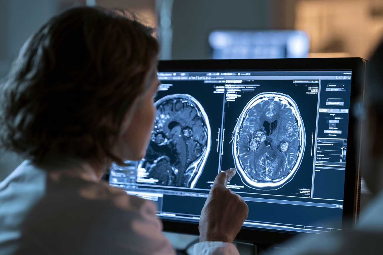
Treatment response in small cell lung cancer brain metastases cannot be judged only by how the patient feels. Headache may improve after dexamethasone because swelling has reduced, while the metastatic lesion may still remain visible. Seizures may stop after antiseizure medicine even when active cancer is still present, and appetite or energy may improve without any change in the brain lesions.
For this reason, doctors assess the brain, neurological function and disease outside the brain separately. In our curative intent model, remission is considered only when clinical improvement is supported by objective imaging and sustained follow up [5,16].
Brain MRI Is the Main Measure of Intracranial Response
Contrast enhanced brain MRI is used to compare the latest findings with the pretreatment scan. Whenever possible, follow up scans should be performed using a similar MRI technique so that changes in lesion size and contrast enhancement can be interpreted more reliably.
The radiologist and treating team assess whether the visible metastases have decreased, disappeared, remained unchanged or increased. They also look for new lesions, changes in cerebral oedema, bleeding, mass effect, midline shift and pressure on surrounding structures [5,11,16].
A complete intracranial response generally means that all measurable brain metastases have disappeared on imaging and no new lesions have developed. A partial response means that the measurable disease has reduced substantially but remains visible. Stable disease means that there is no sufficient reduction to call it a response and no sufficient increase to classify it as progression.
Progressive intracranial disease may be suspected when existing lesions enlarge, new brain metastases appear or neurological deterioration is supported by worsening imaging findings. Formal response assessment also considers neurological status and corticosteroid use rather than relying only on tumour measurements [16].
Cerebral Oedema Must Be Assessed Separately
The swelling around a brain metastasis may change more quickly than the tumour itself. Dexamethasone can reduce oedema and improve headache, vomiting, weakness or confusion before any significant reduction in tumour size is visible.
A lower corticosteroid requirement may support clinical improvement, but steroid reduction must be medically supervised. If the patient requires increasing doses to control the same symptoms, the treatment team should investigate possible progression, recurrent oedema, treatment related injury or another neurological problem [8,16].
Reduction in cerebral oedema is an important treatment achievement because it may restore neurological function and reduce pressure inside the skull. However, it should not be reported as complete tumour remission unless the metastatic lesions have also responded.
Neurological Recovery Is Measured Alongside the Scan
The neurological examination helps determine whether imaging improvement is producing meaningful benefit for the patient. Doctors assess awareness, memory, speech, vision, swallowing, limb strength, coordination, balance and walking ability.
Seizure frequency and duration should also be recorded. If you are caring for the patient, noting the date, approximate duration and appearance of each seizure can help the neurologist understand whether seizure control is improving.
Recovery may occur gradually. A lesion may shrink on MRI while weakness, speech difficulty or poor balance continues because the surrounding brain tissue requires time to recover. In other cases, the neurological symptoms may persist because of permanent injury, treatment effects, stroke, low sodium or another medical condition.
For this reason, a stable neurological condition can sometimes be clinically meaningful even when full recovery has not yet occurred. The scan and the patient’s functional progress must be interpreted together.
The Lung Tumour and Other Metastases Must Also Be Reassessed
A favourable brain MRI does not prove that the entire small cell lung cancer has entered remission. The primary lung tumour, thoracic lymph nodes and metastatic disease in the liver, bones, adrenal glands or other organs must be evaluated separately through appropriate systemic imaging [1–3].
The brain lesions may respond while the chest disease progresses. The opposite can also occur, with good control of the lung tumour but new or enlarging lesions inside the brain. Treatment should therefore not be continued unchanged merely because one part of the disease has improved.
In a whole disease curative intent model, the preferred outcome is control or disappearance of measurable disease in every involved site, together with the absence of new metastases.
Blood Tests Help Confirm Safety and Treatment Tolerance
Laboratory monitoring does not show whether a brain metastasis has disappeared, but it helps determine whether the patient can safely continue treatment. A complete blood count is used to assess haemoglobin, white blood cells and platelets, while liver and kidney tests help identify medicine related toxicity or organ involvement.
Sodium requires particular attention because SCLC associated SIADH can cause weakness, confusion, seizures or reduced consciousness. Blood glucose may also rise during dexamethasone treatment and should be monitored according to the patient’s clinical condition [10].
When an Ayurvedic formulation is being used, blood counts, liver function and kidney function should be reviewed periodically. Any unexpected deterioration should lead to reassessment of the complete treatment plan rather than automatic continuation of the same medicines.
When MRI Findings Are Unclear
An enlarging lesion after stereotactic radiation does not always mean that the cancer has progressed. Treatment related inflammation or radiation injury can sometimes produce increased contrast enhancement and swelling that resembles recurrent tumour.
The treating team may recommend a repeat MRI, advanced imaging, review by a neuroradiologist or, in selected cases, tissue examination. A single uncertain scan should therefore be interpreted cautiously, especially when the neurological condition and other findings do not clearly support progression.
At the same time, worsening symptoms should not be dismissed as a harmless radiation effect without proper evaluation. Rapidly increasing weakness, seizures, vomiting or reduced consciousness requires urgent assessment regardless of the suspected cause.
When Can Remission Be Declared?
Remission should be based on the combined findings of brain MRI, systemic imaging, neurological examination and clinical stability. In our model, an early favourable scan is considered an important response, but not automatically a durable remission.
A stronger remission assessment requires disappearance or sustained control of measurable lesions, no new brain metastases, control of extracranial disease, stable or improving neurological function and no unexplained increase in corticosteroid requirement [16].
Continued surveillance remains necessary even after a complete response because small cell lung cancer can recur in the brain or elsewhere. The interval between scans should be decided by the oncology team according to the treatment received, the previous disease pattern and the patient’s current condition.
Improvement in headache, sleep, appetite, weight or energy is valuable for the patient, but these changes alone cannot confirm elimination of metastatic disease. A curative intent treatment model must ultimately be judged by measurable tumour response, absence of new disease and the durability of that response over time.
You are right. The reference numbers were still appearing inside the answers, and the wording was not sufficiently optimised for featured snippets. Use the following clean FAQ section without numbering, citations, hyperlinks or unnecessary statistics.
Frequently Asked Questions

What Are the First Symptoms of Small Cell Lung Cancer Brain Metastases?
The first symptoms of small cell lung cancer brain metastases may include a worsening headache, nausea, vomiting, seizures, weakness on one side, loss of balance, speech difficulty, vision changes, confusion, memory problems or unusual behaviour. New neurological symptoms in a person with SCLC require prompt medical assessment.
Can Small Cell Lung Cancer Spread to the Brain Without Symptoms?
Yes. Small cell lung cancer can spread to the brain without causing noticeable symptoms. Small brain metastases may be found only during staging or follow up imaging. Contrast enhanced brain MRI is generally more sensitive than CT for detecting small or early brain lesions.
Does Brain Metastasis Mean Extensive Stage Small Cell Lung Cancer?
Brain metastasis generally means that small cell lung cancer has reached the extensive stage because the cancer has spread to a distant organ. However, prognosis and treatment still depend on the number of brain lesions, neurological condition, overall health and whether cancer is active elsewhere in the body.
What Is the Life Expectancy With Small Cell Lung Cancer Brain Metastases?
Life expectancy with small cell lung cancer brain metastases varies from one patient to another. Prognosis depends on brain tumour burden, neurological function, performance status, disease outside the brain and response to treatment. Population survival estimates should not be treated as a fixed prediction for an individual patient.
Can Small Cell Lung Cancer Brain Metastases Be Treated Successfully?
Small cell lung cancer brain metastases may respond to whole brain radiation, stereotactic radiosurgery, systemic treatment or a combination of therapies. Some patients achieve substantial shrinkage or complete disappearance of visible brain lesions, although continued MRI monitoring is necessary because the cancer may return.
Is Stereotactic Radiosurgery Better Than Whole Brain Radiation?
Stereotactic radiosurgery is not automatically better than whole brain radiation for SCLC brain metastases. Focused radiation may suit selected patients with limited brain disease, while whole brain radiation may be preferred for multiple or diffuse lesions. The best option depends on MRI findings and the patient’s overall condition.
Can Ayurvedic Treatment Help Small Cell Lung Cancer Brain Metastases?
An individualised Ayurveda led curative intent model aims to address the brain lesions, primary lung tumour, metastatic activity, neurological impairment and recurrence tendency together. Ayurvedic treatment must be planned around MRI findings, organ function and current oncology treatment, and it should not delay urgent radiation, corticosteroids or seizure management.
Can Ayurveda Be Used With Radiation and Dexamethasone?
Ayurveda may be coordinated with radiation and dexamethasone after reviewing cerebral oedema, seizure history, liver and kidney function, blood sodium, swallowing ability and all current medicines. Dexamethasone should not be stopped suddenly, and urgently required brain directed treatment must always receive priority.
How Are Small Cell Lung Cancer Brain Metastases Monitored?
Small cell lung cancer brain metastases are monitored mainly with contrast enhanced brain MRI and neurological examination. Doctors compare lesion size, new lesions, cerebral oedema, mass effect, seizures, strength, speech, balance and corticosteroid requirement. Improvement in appetite, headache or energy alone cannot confirm tumour clearance.
Can Small Cell Lung Cancer Brain Metastases Come Back After Treatment?
Yes. SCLC brain metastases can return after whole brain radiation, stereotactic radiosurgery or systemic treatment. Recurrence may appear as growth of a previously treated lesion or as a new lesion elsewhere in the brain. Regular MRI surveillance is therefore essential even after a strong response.
When Are Small Cell Lung Cancer Brain Metastases an Emergency?
Small cell lung cancer brain metastases require emergency care when the patient develops a first seizure, sudden weakness, loss of consciousness, severe headache with repeated vomiting, new speech or vision loss, rapidly worsening confusion, difficulty walking or inability to swallow safely.
Reference
1. National Cancer Institute
National Cancer Institute. (2025, May 14). Small cell lung cancer treatment (PDQ®): Health professional version. National Cancer Institute.
https://www.cancer.gov/types/lung/hp/small-cell-lung-treatment-pdq
Used for: Current information on SCLC staging, extensive stage disease, brain metastasis symptoms, diagnostic workup, chemotherapy, immunotherapy, thoracic radiation, brain radiation and recurrent SCLC treatment.
2. NCCN Small Cell Lung Cancer Guidelines Insights
Ganti, A. K. P., Loo, B. W., Jr., Badiyan, S., Bassetti, M., Bestvina, C., Chiang, A., et al. (2026). NCCN Guidelines® Insights: Small cell lung cancer, version 2.2026. Journal of the National Comprehensive Cancer Network, 24(1), e260002.
https://pubmed.ncbi.nlm.nih.gov/41671459
Used for: Current SCLC diagnostic workup, staging, systemic treatment, radiation treatment and recent guideline updates. This is the most current NCCN Guidelines Insights publication included in the article’s evidence base.
3. ESMO Small Cell Lung Cancer Guideline
Dingemans, A. M. C., Früh, M., Ardizzoni, A., Besse, B., Faivre-Finn, C., Hendriks, L. E., et al. (2021). Small-cell lung cancer: ESMO Clinical Practice Guidelines for diagnosis, treatment and follow-up. Annals of Oncology, 32(7), 839–853.
https://pubmed.ncbi.nlm.nih.gov/33864941
Used for: SCLC diagnosis, limited and extensive stage classification, chemotherapy, immunotherapy, radiotherapy, brain imaging, follow up and recurrent disease management.
4. SCLC Brain Metastasis Review
Rittberg, R., Banerji, S., Kim, J. O., Rathod, S., & Dawe, D. E. (2021). Treatment and prevention of brain metastases in small cell lung cancer. American Journal of Clinical Oncology, 44(12), 629–638.
https://pubmed.ncbi.nlm.nih.gov/34628433
Used for: The frequency and timing of SCLC brain metastases, intracranial surveillance, the distinction between prophylactic cranial irradiation and treatment of established lesions, WBRT, SRS and recurrent intracranial disease.
5. EANO–ESMO Brain Metastasis Guideline
Le Rhun, E., Guckenberger, M., Smits, M., Dummer, R., Bachelot, T., Sahm, F., et al. (2021). EANO–ESMO Clinical Practice Guidelines for diagnosis, treatment and follow-up of patients with brain metastasis from solid tumours. Annals of Oncology, 32(11), 1332–1347.
https://pubmed.ncbi.nlm.nih.gov/34364998
Used for: Neurological symptoms, contrast MRI, differential diagnosis, surgery, radiation, systemic treatment, follow up, treatment response and recurrence of brain metastases.
6. ASCO–SNO–ASTRO Brain Metastasis Guideline
Vogelbaum, M. A., Brown, P. D., Messersmith, H., Brastianos, P. K., Burri, S., Cahill, D., et al. (2022). Treatment for brain metastases: ASCO–SNO–ASTRO guideline. Journal of Clinical Oncology, 40(5), 492–516.
https://pubmed.ncbi.nlm.nih.gov/34932393
Used for: The recommendation that symptomatic brain metastases generally require appropriate local therapy, indications for surgery, SRS and WBRT, treatment sequencing, memantine and hippocampal avoidance.
7. ASTRO Radiation Guideline
Gondi, V., Bauman, G., Bradfield, L., Burri, S. H., Cabrera, A. R., Cunningham, D. A., et al. (2022). Radiation therapy for brain metastases: An ASTRO clinical practice guideline. Practical Radiation Oncology, 12(4), 265–282.
https://pubmed.ncbi.nlm.nih.gov/35534352
Used for: Selection of SRS, fractionated stereotactic radiation, WBRT and surgery; radiation for symptomatic brain metastases; cognitive protection; and patient selection for brain directed treatment.
8. Corticosteroids for Metastatic Brain Tumours
Ryken, T. C., Kuo, J. S., Prabhu, R. S., Sherman, J. H., Kalkanis, S. N., & Olson, J. J. (2019). Congress of Neurological Surgeons systematic review and evidence-based guidelines on the role of steroids in the treatment of adults with metastatic brain tumors. Neurosurgery, 84(3), E189–E191.
https://pubmed.ncbi.nlm.nih.gov/30629207
Used for: Dexamethasone and other corticosteroids for temporary relief of symptoms caused by tumour associated cerebral oedema and mass effect.
Important interpretation: Symptom improvement after dexamethasone usually indicates reduced oedema. It does not establish that the metastatic tumour has disappeared.
9. Antiseizure Medicine Guideline
Chen, C. C., Rennert, R. C., & Olson, J. J. (2019). Congress of Neurological Surgeons systematic review and evidence-based guidelines on the role of prophylactic anticonvulsants in the treatment of adults with metastatic brain tumors. Neurosurgery, 84(3), E195–E197.
https://pubmed.ncbi.nlm.nih.gov/30629248
Used for: Antiseizure medicine decisions and the recommendation against routine preventive anticonvulsant treatment for every seizure free, nonsurgical patient with brain metastases. Patients who have experienced a seizure require individual neurological assessment and treatment.
10. Hyponatraemia and SIADH in Lung Cancer
Fiordoliva, I., Meletani, T., Baleani, M. G., Rinaldi, S., Savini, A., Di Pietro Paolo, M., & Berardi, R. (2017). Managing hyponatremia in lung cancer: Latest evidence and clinical implications. Therapeutic Advances in Medical Oncology, 9(11), 711–719.
https://pubmed.ncbi.nlm.nih.gov/29344107
Used for: SCLC associated SIADH, low sodium, confusion, weakness, headache, seizures and reduced consciousness. It supports checking serum sodium instead of assuming that every neurological symptom represents progression of brain metastases.
11. MRI Versus CT in SCLC
Seute, T., Leffers, P., ten Velde, G. P. M., & Twijnstra, A. (2008). Detection of brain metastases from small cell lung cancer: Consequences of changing imaging techniques, CT versus MRI. Cancer, 112(8), 1827–1834.
https://pubmed.ncbi.nlm.nih.gov/18311784
Used for: SCLC specific evidence that MRI identifies more brain metastases than CT and can detect asymptomatic lesions. In this study, detected brain metastases increased from 10% in the CT era to 24% in the MRI era, and some MRI detected lesions were asymptomatic.
12. SCLC Graded Prognostic Assessment
Sperduto, P. W., De, B., Li, J., Carpenter, D., Kirkpatrick, J., Milligan, M., et al. (2022). Graded Prognostic Assessment for patients with lung cancer and brain metastases: Initial report of the small cell lung cancer GPA and update of the non-small cell lung cancer GPA including the effect of programmed death ligand 1 and other prognostic factors. International Journal of Radiation Oncology, Biology, Physics, 114(1), 60–74.
https://pubmed.ncbi.nlm.nih.gov/35331827
Used for: Individualised prognosis according to age, performance status, number of brain metastases and extracranial metastases. It supports explaining why one median survival figure cannot predict the course of every patient.
13. Contemporary United States Survival Analysis
Qidwai, K. A., Sarfraz, Z., Mustafayev, K., Hodgson, L. C., Maharaj, A., Sen, T., Ranjan, T., & Ahluwalia, M. S. (2025). Survival determinants and treatment outcomes of patients with small cell lung cancer and brain metastases: A U.S. national analysis. Cancers, 17(23), 3833.
https://pubmed.ncbi.nlm.nih.gov/41375038
Used for: Contemporary real world prognosis based on 11,074 patients. The study reported a median overall survival of 6.6 months, 8.8 months for brain only disease and 5.95 months when extracranial metastases were also present.
Important interpretation: These are group level estimates from a national database. They are not an individual patient’s fixed life expectancy.
14. FIRE–SCLC Cohort Study
Rusthoven, C. G., Yamamoto, M., Bernhardt, D., Smith, D. E., Gao, D., Serizawa, T., et al. (2020). Evaluation of first-line radiosurgery versus whole-brain radiotherapy for small cell lung cancer brain metastases: The FIRE–SCLC cohort study. JAMA Oncology, 6(7), 1028–1037.
https://pubmed.ncbi.nlm.nih.gov/32496550
Used for: Outcomes after first line SRS, local control, new intracranial disease, neurological death and comparison with WBRT in selected patients.
Important interpretation: This was a retrospective cohort study. Differences between SRS and WBRT groups may partly reflect patient selection and should not be presented as proof that SRS is universally superior.
15. Prospective Phase II SRS Trial
Aizer, A. A., Tanguturi, S. K., Shi, D. D., Catalano, P. J., Shin, K. Y., Ricca, I., et al. (2025). Stereotactic radiosurgery in patients with small cell lung cancer and 1–10 brain metastases: A multi-institutional, phase II, prospective clinical trial. Journal of Clinical Oncology, 43(27), 2986–2997.
https://pubmed.ncbi.nlm.nih.gov/40644657
Used for: Prospective evidence supporting SRS or fractionated stereotactic radiation in carefully selected patients with one to ten SCLC brain metastases and access to frequent MRI surveillance.
The study reported median overall survival of 10.2 months. Twenty two percent of patients later required salvage WBRT, and further intracranial treatment was common. Because the trial was nonrandomised, it does not establish SRS as the best treatment for every patient.
16. RANO Brain Metastasis Response Criteria
Lin, N. U., Lee, E. Q., Aoyama, H., Barani, I. J., Barboriak, D. P., Baumert, B. G., et al. (2015). Response assessment criteria for brain metastases: Proposal from the RANO group. The Lancet Oncology, 16(6), e270–e278.
https://pubmed.ncbi.nlm.nih.gov/26065612
Used for: Objective assessment of intracranial response using measurable lesion size, new brain lesions, neurological status and corticosteroid requirement.
Important interpretation: Improvement in headache, appetite, sleep or energy cannot by itself confirm remission. Brain MRI and neurological assessment are required.
17. Charaka Samhita
Agniveśa. (n.d.). Carakasaṃhitā, with Cakrapāṇidatta’s Āyurvedadīpikā commentary [Electronic Sanskrit text]. National Institute of Indian Medical Heritage, Central Council for Research in Ayurvedic Sciences.
https://niimh.nic.in/ebooks/ecaraka
Used for:
Charaka Samhita, Chikitsa Sthana, Chapter 1, Rasayana Chikitsa: Rasayana, Bala, Ojas, nourishment and recovery principles.
Charaka Samhita, Chikitsa Sthana, Chapter 28, Vatavyadhi Chikitsa: Vata related movement, motor dysfunction, speech disturbance, weakness and neurological manifestations.
Charaka Samhita, Nidana Sthana, Chapter 8, Apasmara Nidana: Classical diagnostic principles related to altered awareness, memory disruption and seizure like episodes.
Charaka Samhita, Chikitsa Sthana, Chapter 10, Apasmara Chikitsa: Classical treatment principles relevant to an Ayurvedic assessment when seizures or altered consciousness are present.
Important interpretation: These chapters provide a classical Ayurvedic framework. They do not describe modern histopathologically confirmed SCLC brain metastases, and they should not replace MRI, neurological diagnosis or emergency seizure management.
18. Sushruta Samhita
Suśruta. (n.d.). Suśrutasaṃhitā, with Dalhaṇa’s Nibandhasaṅgraha and Gayadāsa’s Nyāyacandrikā commentaries [Electronic Sanskrit text]. National Institute of Indian Medical Heritage, Central Council for Research in Ayurvedic Sciences.
https://niimh.nic.in/ebooks/esushruta
Used for:
Sushruta Samhita, Nidana Sthana, Chapter 11, Granthi, Apachi, Arbuda and Galaganda Nidana: Classical diagnostic concepts concerning Granthi and Arbuda.
Sushruta Samhita, Chikitsa Sthana, Chapter 18, Granthi, Apachi, Arbuda and Galaganda Chikitsa: Classical management principles concerning fixed, abnormal and progressively enlarging tissue formations.
Important interpretation: Granthi and Arbuda concepts may support an Ayurvedic clinical interpretation of abnormal tissue growth. They are not anatomically, molecularly or histopathologically identical to modern SCLC brain metastases.
19. Boswellia and Radiation Associated Cerebral Oedema
Kirste, S., Treier, M., Wehrle, S. J., Becker, G., Abdel-Tawab, M., Gerbeth, K., Hug, M. J., Lubrich, B., Grosu, A. L., & Momm, F. (2011). Boswellia serrata acts on cerebral edema in patients irradiated for brain tumors: A prospective, randomized, placebo-controlled, double-blind pilot trial. Cancer, 117(16), 3788–3795.
https://pubmed.ncbi.nlm.nih.gov/21287538
Used for: A small human pilot trial assessing Boswellia serrata for cerebral oedema during radiotherapy for primary or secondary brain tumours.
Important interpretation: The study was small, was not limited to SCLC and did not demonstrate eradication of brain metastases. It cannot be used to claim that Boswellia replaces dexamethasone, radiation or surgery.
20. Systematic Review of Boswellia Evidence
Dejonckheere, C. S., Scafa, D., Käsmann, L., Zeyen, T., Potthoff, A. L., Schäfer, N., et al. (2025). Boswellia serrata for the management of radiation-induced cerebral edema and necrosis: A systematic meta-narrative review of clinical evidence. Advances in Radiation Oncology, 10(4), 101732.
https://pubmed.ncbi.nlm.nih.gov/40092573
Free full text:
https://pmc.ncbi.nlm.nih.gov/articles/PMC11904484
Used for: Evaluation of the total human clinical evidence concerning Boswellia for radiation associated cerebral oedema and cerebral radiation necrosis.
Important interpretation: The review found that the evidence remains limited and heterogeneous. It supports a cautious adjunctive discussion rather than a claim of established antitumour or curative efficacy.
21. Curcumin and SCLC Laboratory Research
Yang, C. L., Liu, Y. Y., Ma, Y. G., Xue, Y. X., Liu, D. G., Ren, Y., Liu, X. B., Li, Y., & Li, Z. (2012). Curcumin blocks small cell lung cancer cell migration, invasion, angiogenesis, cell cycle and neoplasia through the Janus kinase–STAT3 signalling pathway. PLOS ONE, 7(5), e37960.
https://pubmed.ncbi.nlm.nih.gov/22662257
Free full text:
https://pmc.ncbi.nlm.nih.gov/articles/PMC3360669
Used for: Laboratory evidence concerning curcumin and SCLC cell proliferation, migration, invasion, angiogenesis and JAK–STAT3 signalling.
Important interpretation: This is preclinical research. It does not establish an effective human dose, reliable brain penetration, intracranial tumour response, survival improvement or clinical cure.
22. Memantine During Whole Brain Radiation
Brown, P. D., Pugh, S., Laack, N. N., Wefel, J. S., Khuntia, D., Meyers, C., et al. (2013). Memantine for the prevention of cognitive dysfunction in patients receiving whole-brain radiotherapy: A randomized, double-blind, placebo-controlled trial. Neuro-Oncology, 15(10), 1429–1437.
https://pubmed.ncbi.nlm.nih.gov/23956241
Used for: The use of memantine to reduce or delay cognitive deterioration during WBRT.
Important interpretation: This trial included patients with brain metastases from different primary cancers and was not restricted to SCLC. It supports the cognitive protection subsection rather than the tumour control or prognosis section.
23. Hippocampal Avoidance WBRT
Gondi, V., Deshmukh, S., Brown, P. D., Wefel, J. S., Armstrong, T. S., Tomé, W. A., et al. (2023). Sustained preservation of cognition and prevention of patient-reported symptoms with hippocampal avoidance during whole-brain radiation therapy for brain metastases: Final results of NRG Oncology CC001. International Journal of Radiation Oncology, Biology, Physics, 117(3), 571–580.
https://pubmed.ncbi.nlm.nih.gov/37150264
Used for: Hippocampal avoidance WBRT combined with memantine and its effect on cognitive preservation and patient reported neurological symptoms.
Important interpretation: This evidence is not SCLC specific. Hippocampal avoidance is suitable only when lesion location, previous treatment and radiation planning permit it.