




- What Your CBC Can Show and What It Cannot
- Leukemia Blood Test Results That Need Review
- How Is Leukemia Diagnosed?
- Leukemia Blood Test and Blood Smear
- Flow Cytometry for Leukemia
- Leukemia Blood Test and Bone Marrow Testing
- Leukemia Blood Test and Genetic Testing
- Leukemia Blood Test by Type
- Other Causes of an Abnormal Leukemia Blood Test
- When Leukemia Blood Test Results Need Urgent Review
- After a Leukemia Blood Test Leads to Diagnosis
- Questions About Your Leukemia Blood Test
- FAQs
- References
What Your CBC Can Show and What It Cannot
A leukemia blood test usually begins with a complete blood count, commonly called a CBC. This routine test measures the numbers of red blood cells, white blood cells and platelets circulating in the blood. It may provide the first indication that the bone marrow is not producing blood cells normally, but it cannot confirm leukemia or identify the exact leukemia subtype by itself.[1,9]
Some people undergo testing because they have persistent fatigue, unexplained bruising, recurrent infections, fever, night sweats, weight loss or enlarged lymph nodes. Others have no obvious symptoms and first learn that something may be wrong when a routine CBC shows an unexpected change. An abnormal result does not automatically mean leukemia because infections, inflammation, nutritional deficiencies, medications, immune disorders and several non-cancerous conditions can affect the same blood measurements.
Table 1: What Abnormal CBC Results May Mean
| CBC finding | What it may indicate | Other possible causes | Common next step |
|---|---|---|---|
| High WBC count | Infection, inflammation or abnormal white-cell production | Steroids, smoking, stress, pregnancy | Differential, smear and comparison with previous CBCs |
| Low WBC count | Reduced marrow production or increased cell destruction | Viral illness, medicines, autoimmune disease | Absolute neutrophil count, smear and repeat testing |
| Low hemoglobin | Anemia or impaired red-cell production | Iron, vitamin B12 or folate deficiency, bleeding, kidney disease | Reticulocyte count, iron studies and clinical review |
| Low platelets | Reduced production, destruction or sequestration | Infection, medicines, immune disease, liver or spleen disorder | Repeat count, smear and bleeding assessment |
| Pancytopenia | Broad disruption of marrow production | Severe deficiency, infection, aplastic anemia or medication toxicity | Prompt hematology review and possible marrow testing |
| Blasts or abnormal cells | Possible acute leukemia or another marrow disorder | Rarely severe marrow stress or classification error | Urgent smear review, flow cytometry and specialist referral |
What a Complete Blood Count Measures
The white blood cell count reports the total number of leukocytes circulating in the blood. These cells normally protect the body against infection, but the total count does not reveal which type of white blood cell is increased, reduced or abnormal. For this reason, the CBC is commonly performed with a differential count, which separates white blood cells into neutrophils, lymphocytes, monocytes, eosinophils and basophils.
The differential may report both percentages and absolute counts. Absolute counts are often more clinically useful because a percentage can appear high simply because another type of cell has decreased. For example, an elevated lymphocyte percentage does not necessarily mean that the total number of lymphocytes is abnormally high. Doctors therefore interpret the total WBC count, differential and absolute cell counts together rather than relying on one percentage.
A leukemia WBC count may be high, normal or low. Some leukemias release large numbers of abnormal cells into the bloodstream, while others remain mainly within the bone marrow or suppress normal blood-cell production without producing marked leukocytosis. A severe infection, corticosteroid use, inflammation, smoking or physical stress can also produce a high WBC count. There is therefore no single white-cell value that reliably diagnoses or excludes leukemia.[2–4]
The CBC also measures red blood cells, hemoglobin and hematocrit. Red blood cells carry oxygen throughout the body, while hemoglobin is the oxygen-carrying protein inside these cells. Hematocrit represents the proportion of blood composed of red cells. Reduced values may indicate anemia, which can cause tiredness, weakness, dizziness, pale skin, rapid heartbeat or shortness of breath.
Leukemia may contribute to anemia when abnormal cells interfere with normal blood-cell production in the bone marrow. However, anemia is much more commonly caused by iron deficiency, vitamin B12 or folate deficiency, bleeding, kidney disease, chronic inflammation or other medical conditions. A low hemoglobin result must therefore be investigated in context rather than interpreted as evidence of leukemia.
Platelets are small blood-cell fragments that help stop bleeding. A reduced platelet count may contribute to easy bruising, nosebleeds, gum bleeding, prolonged bleeding or pinpoint red spots on the skin. Leukemia can reduce platelet production when abnormal cells occupy or disrupt the marrow, but low platelets may also result from viral illness, medications, immune destruction, liver disease, pregnancy or platelet clumping within the laboratory sample.
The pattern across several blood-cell lines is often more informative than one abnormal value. Doctors may become more concerned when anemia, low platelets and abnormal white-cell findings occur together, especially when the abnormalities are persistent, progressive or accompanied by circulating immature cells. Even this pattern is not specific to leukemia because other bone-marrow disorders, severe infections and certain medications can produce similar changes.
What Automated CBC Flags May Mean
Modern blood analysers count and classify cells rapidly, but they may also generate alerts when a cell population does not fit an expected pattern. Reports may contain terms such as blasts, immature granulocytes, atypical lymphocytes, nucleated red blood cells, left shift, abnormal scattergram or manual smear recommended.
A laboratory flag is not a diagnosis. It indicates that the automated analyser detected an unusual cell size, shape, internal structure or distribution that may require manual review. Reactive cells produced during infection can sometimes trigger the same warning systems as malignant cells. Conversely, an automated count may not fully characterise an abnormal population that is present in small numbers.
Blasts are very immature blood-forming cells that normally remain primarily within the bone marrow. Their presence in peripheral blood can be concerning, particularly when accompanied by anemia, thrombocytopenia or rapidly changing counts. However, the laboratory must confirm that the reported cells are truly blasts and determine their lineage before a leukemia subtype can be assigned.[1,3,4]
A peripheral blood smear is commonly used to investigate these findings. A drop of blood is spread across a glass slide, stained and examined under a microscope. This allows a trained laboratory professional or hematopathologist to assess whether cells appear mature or immature and whether their nuclei, granules, size and shape suggest leukemia, infection, inflammation or another blood disorder.
What a CBC Cannot Determine
A CBC cannot establish whether an abnormal white-cell population is myeloid or lymphoid, whether lymphoid cells are derived from B cells or T cells, or whether the cells carry a defining chromosome or gene abnormality. It also cannot reliably distinguish every leukemia from lymphoma, myelodysplastic disease, a myeloproliferative neoplasm or a severe reactive blood response.
Modern leukemia diagnosis integrates the CBC with cell morphology, flow cytometry, cytogenetic testing and molecular analysis. In many cases, doctors also examine bone marrow because leukemia begins within blood-forming tissue and abnormal cells may not be adequately represented in peripheral blood.[1–5]
A normal or near-normal CBC does not completely exclude leukemia. Some patients have subtle changes, isolated anemia or thrombocytopenia, an abnormal differential or persistent symptoms before the total WBC count becomes clearly abnormal. At the same time, most people with a normal CBC and non-specific symptoms do not have leukemia. Clinical examination and follow-up testing help determine whether further investigation is justified.
The CBC should therefore be understood as the beginning of the diagnostic process rather than a final leukemia test. It identifies patterns that may require closer examination, guides the urgency of referral and helps doctors decide whether a blood smear, flow cytometry, bone marrow examination or molecular testing is needed. A diagnosis should never be made from a single highlighted number without considering the complete report, symptoms, medical history and specialist laboratory findings.[1,9]
Leukemia Blood Test Results That Need Review
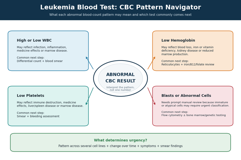
A leukemia blood test does not produce one universally “positive” CBC pattern. Concern increases when an abnormality is unexplained, persists on repeat testing, worsens over time, affects several blood-cell lines or is accompanied by immature or abnormal cells. A single mildly abnormal result is often caused by infection, medication, inflammation or temporary physiological stress, whereas a combination of abnormalities may require prompt hematology assessment.[1–4]
High or Low White Blood Cell Counts
A high white blood cell count is one of the early signs of leukemia in blood work that patients commonly search for, but leukocytosis is not specific to cancer. Bacterial infection, inflammation, corticosteroids, smoking, pregnancy, tissue injury and intense physical stress can all raise the total WBC count.
The type of white blood cell involved is often more informative than the total count. A CBC differential shows whether the increase is mainly due to neutrophils, lymphocytes, monocytes, eosinophils or basophils. Doctors also examine whether the cells are mature, whether several stages of developing cells are present and whether the pattern is consistent with a normal response to infection.
Leukemia can also occur with a normal or low WBC count. In some acute leukemias, abnormal cells remain predominantly inside the bone marrow while normal blood-cell production declines. The circulating blood may therefore show leukopenia, neutropenia, anemia or thrombocytopenia rather than marked leukocytosis.[2,3,6,7]
A low neutrophil count is especially important because neutrophils protect against bacterial and fungal infections. Fever in a person with severe neutropenia requires urgent medical assessment regardless of whether leukemia has been confirmed. The immediate concern is infection risk, not simply the underlying cause of the abnormal count.
The numerical value should always be reviewed together with the differential and peripheral blood smear. A markedly elevated count composed mainly of mature reactive neutrophils has a different significance from a lower count containing blasts or another abnormal cell population. This is why no single leukemia WBC count can reliably confirm or exclude the disease.[1–4]
Anemia and Platelet Changes
Anemia may appear when the bone marrow is unable to produce enough healthy red blood cells. Patients may experience persistent fatigue, reduced exercise tolerance, dizziness, headaches, pale skin, palpitations or shortness of breath. However, these symptoms and blood findings are not unique to leukemia.
Iron deficiency, vitamin B12 deficiency, folate deficiency, kidney disease, chronic inflammation and blood loss are more common causes of anemia. Doctors may therefore order iron studies, vitamin levels, kidney tests, a reticulocyte count and other investigations before attributing a low hemoglobin level to a marrow disorder.
A low platelet count may cause easy bruising, prolonged bleeding, frequent nosebleeds, gum bleeding, unusually heavy menstrual bleeding or small red-purple spots called petechiae. Leukemia may reduce platelet production, but viral infections, immune thrombocytopenia, liver disease, medications and laboratory platelet clumping can produce similar results.
A high platelet count is less typical of acute leukemia but may occur with inflammation, iron deficiency or certain myeloproliferative disorders. It should not be interpreted as reassuring or alarming without considering the remaining CBC findings and the clinical situation.
Concern increases when anemia and thrombocytopenia occur together, particularly when the WBC differential is also abnormal. This pattern may suggest that normal marrow production is being disrupted, although severe vitamin deficiency, aplastic anemia, infection, autoimmune disease and other marrow conditions must also be considered.
Pancytopenia means that red blood cells, white blood cells and platelets are all reduced. It can occur in leukemia, but it is not synonymous with leukemia. The cause may involve reduced marrow production, abnormal destruction of blood cells, medication toxicity, severe infection, nutritional deficiency or enlargement of the spleen. Bone marrow assessment may be needed when the cause remains unexplained.[1–4,9]
Blasts and Persistent Cell Abnormalities
Blasts reported in peripheral blood deserve prompt review because these immature cells normally remain mainly within the bone marrow. Their presence can occur in acute leukemia, but the laboratory must confirm their identity and determine whether they are myeloid or lymphoid. Morphology alone may not be sufficient, particularly when the abnormal cells are few or have overlapping appearances.[1,2,6,7]
A report stating “possible blasts,” “abnormal cells” or “manual differential recommended” is not the same as a confirmed leukemia diagnosis. The next step may include review by a hematopathologist, a repeat specimen, peripheral blood flow cytometry or bone marrow testing, depending on the cell pattern and the patient’s condition.
Persistent absolute lymphocytosis may lead doctors to consider chronic lymphocytic leukemia or another lymphoid disorder. However, viral infections, smoking, stress reactions and other non-cancerous conditions can also increase lymphocytes. CLL is not diagnosed from an elevated lymphocyte percentage alone; a clonal B-cell population must be identified, usually through flow cytometry, and the absolute cell count and duration of the abnormality must be assessed.[5,8]
Persistent monocytosis may require evaluation for infection, inflammation or a myeloid neoplasm. Basophilia accompanied by increased granulocytes and immature myeloid cells may raise concern for chronic myeloid leukemia, but confirmation requires detection of the defining BCR::ABL1 abnormality rather than reliance on the CBC pattern alone.[3,4,10]
The direction of change is also clinically important. A result that remains stable for years may be approached differently from a count that changes rapidly over several days or weeks. Comparing the current report with previous CBCs can show whether the finding is new, temporary, persistent or progressive.
Repeat testing may be reasonable when a mild abnormality occurs during a recent infection and the patient is otherwise well. Repeating the CBC should not delay specialist assessment when blasts are reported, several blood-cell lines are severely reduced, counts are changing rapidly or the patient has fever, active bleeding, severe breathlessness, confusion or marked weakness.
The most useful interpretation of leukemia blood test results therefore comes from the entire pattern rather than one highlighted number. The total WBC count, absolute differential, hemoglobin, platelet count, cell appearance, previous results and current symptoms must be considered together before deciding which further tests are necessary.[1–4]
How Is Leukemia Diagnosed?
How is leukemia diagnosed after an abnormal blood report? Doctors combine the CBC pattern with the patient’s symptoms, medical history, physical examination, blood-cell appearance, immunophenotyping and, when required, bone marrow and genetic testing. No single result is interpreted alone because infections, medications, nutritional deficiencies and other blood disorders can produce findings that overlap with leukemia.[1–5]
Clinical Review After an Abnormal CBC
The first step is to determine whether the abnormality is isolated, temporary or part of a broader pattern. The clinician compares the current CBC with previous reports and reviews whether the white blood cell count, hemoglobin or platelets are stable, recovering or changing progressively.
Recent infections, corticosteroids, antibiotics, immune-suppressing medicines, cancer treatments and nutritional supplements may affect blood counts. The assessment may also include previous chemotherapy or radiation exposure, occupational contact with chemicals such as benzene, a history of another blood disorder and any relevant inherited condition.[1,2]
Symptoms help establish the urgency of further testing. Persistent fever, recurrent infections, unexplained bruising, abnormal bleeding, progressive fatigue, breathlessness, night sweats, unintended weight loss, bone discomfort or abdominal fullness may be clinically important when they occur alongside abnormal blood counts. None of these symptoms confirms leukemia, but the combination may justify more rapid evaluation.
During the examination, the clinician may look for pallor, petechiae, bruising, active bleeding, mouth ulcers, infection, enlarged lymph nodes or enlargement of the liver or spleen. These findings provide context for the blood report and may identify complications requiring treatment before the complete leukemia classification is available.[1,9]
Repeat Testing or Immediate Referral
A repeat CBC with differential may be appropriate when an abnormality is mild, unexpected and potentially related to a recent infection, medication or sample problem. Repeating the test can show whether the count is returning toward the laboratory reference range or remains persistently abnormal.
The timing should be determined by the severity and pattern of the result. Waiting several weeks may be reasonable for selected minor abnormalities in an otherwise well patient, whereas rapidly changing counts may need reassessment within hours or days. Patients should not choose a repeat-testing interval without clinical guidance.
The laboratory may perform a manual differential or peripheral blood smear at the same time. This is particularly important when an automated analyser reports possible blasts, atypical cells, immature granulocytes or an abnormal white-cell distribution. Review by an experienced pathologist can determine whether the finding appears reactive or requires specialised testing.
Repeat testing should not delay hematology referral when blasts are reported, several blood-cell lines are substantially reduced, the WBC count is changing rapidly or the patient has significant bleeding, fever with neutropenia, severe breathlessness, confusion or marked weakness. Suspected acute leukemia requires prompt evaluation because the disease and its complications can progress quickly.[1,2,6,7]
Tests Used to Confirm the Diagnosis
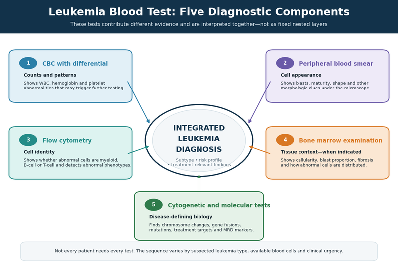
A peripheral blood smear examines the number, maturity and physical appearance of circulating cells. It may reveal blasts, abnormal lymphocytes, altered granulocyte development or other features that narrow the possible diagnosis. The smear can strongly suggest leukemia, but it may not identify the exact lineage or molecular subtype.[1,9]
Flow cytometry evaluates proteins expressed on or within individual cells. The resulting marker pattern can help determine whether an abnormal population is myeloid or lymphoid, whether lymphoid cells are of B-cell or T-cell origin and whether the population has features of clonality. This distinction is essential because leukemias that appear similar on a routine smear may require very different treatment approaches.[1,5]
Bone marrow aspiration provides liquid marrow containing individual cells for microscopic examination, flow cytometry, chromosome studies and molecular analysis. A core biopsy provides a small cylinder of tissue that shows marrow cellularity, architecture, fibrosis and the distribution of abnormal cells.
These procedures are commonly used when acute leukemia is suspected, blood abnormalities remain unexplained, several cell lines are affected or circulating blood does not provide enough abnormal cells for complete testing. Bone marrow examination may also distinguish leukemia from aplastic anemia, myelodysplastic syndromes and other disorders that interfere with normal blood production.[1–4]
Not every patient requires the same sequence of tests. In suspected acute leukemia, morphology, flow cytometry and genetic testing may be initiated in parallel because treatment decisions can depend on obtaining a precise classification quickly. In selected chronic leukemias, peripheral blood may provide enough material for important parts of the diagnostic evaluation.[1,2]
Chronic lymphocytic leukemia can often be established through persistent blood lymphocytosis and peripheral-blood flow cytometry demonstrating a characteristic clonal B-cell population. Bone marrow examination is not routinely required to establish every uncomplicated CLL diagnosis, although it may be useful when cytopenias are unexplained or treatment response must be assessed.[5,8]
When chronic myeloid leukemia is suspected, detection of the BCR::ABL1 fusion is required to confirm the diagnosis. This abnormality may be identified using cytogenetic testing, fluorescence in situ hybridisation or molecular testing. A high WBC count with immature myeloid cells may raise suspicion, but the blood-count pattern alone cannot establish CML.[3,4,10]
Why Genetic Testing Is Part of Diagnosis
Cytogenetic and molecular tests examine the chromosomes and genes within abnormal cells. Karyotyping can detect large chromosome gains, losses or rearrangements, while fluorescence in situ hybridisation looks for selected abnormalities using targeted probes. Polymerase chain reaction and next-generation sequencing can identify specific gene fusions and mutations that may not be visible under a microscope.[2–5]
These findings can define the leukemia subtype, help estimate disease behaviour and reveal whether a targeted treatment may be appropriate. They may also establish a molecular marker that can later be measured to assess treatment response or detect small amounts of remaining disease.
A blast percentage should not be interpreted as a universal diagnostic cutoff without the genetic findings. Current classification systems recognise that some defining chromosome or molecular abnormalities can establish particular acute leukemia categories even when traditional blast thresholds are not met. The hematopathologist therefore integrates morphology, immunophenotype and genetic results rather than applying one numerical rule to every case.[2–4]
The diagnostic process is complete only when the available findings form a coherent explanation for the abnormal blood counts. When results conflict, the clinical team may request another sample, additional marker testing or expert hematopathology review before assigning a final leukemia type. This careful classification helps prevent treatment from being based on an incomplete or incorrect interpretation.[1–5]
Table 2: What Each Leukemia Test Adds
| Test | What it examines | Main question answered |
|---|---|---|
| CBC with differential | Numbers of white cells, red cells and platelets | Are blood-cell counts or proportions abnormal? |
| Peripheral blood smear | Cell appearance and maturity | Are blasts or abnormal-looking cells present? |
| Flow cytometry | Proteins expressed by individual cells | What cell lineage and abnormal population are involved? |
| Bone marrow aspiration | Individual marrow cells | How many abnormal cells are present and how are they developing? |
| Core biopsy | Marrow architecture and cellularity | Is the marrow infiltrated, fibrotic or abnormally cellular? |
| Karyotype and FISH | Chromosome number and structure | Are defining chromosome changes present? |
| PCR and NGS | Specific gene fusions and mutations | What is the molecular subtype, treatment target or monitoring marker? |
Leukemia Blood Test and Blood Smear

When a leukemia blood test shows abnormal cell counts or an automated laboratory flag, a peripheral blood smear can reveal details that the CBC cannot measure. The smear helps a hematopathologist assess the maturity, structure and distribution of blood cells and decide whether the pattern is more consistent with infection, marrow stress, leukemia or another blood disorder.[1,9]
What the Pathologist Looks For
The pathologist evaluates the entire blood-cell population rather than searching for one isolated feature. White cells are assessed for their size, nuclear shape, chromatin pattern, internal granules and stage of maturation. Red cells and platelets are also examined because abnormalities affecting several cell lines may indicate impaired bone marrow function.
Mature white blood cells usually have recognisable features linked to their normal immune role. Blasts appear immature and may have a relatively large nucleus, fine chromatin, visible nucleoli and only a small amount of surrounding cytoplasm. These characteristics can raise strong concern for acute leukemia, but cell appearance alone may not reliably distinguish myeloid blasts from lymphoid blasts.[1,6,7]
The percentage and absolute number of abnormal cells both matter. A patient with a very high WBC count may still have a relatively small proportion of blasts, while another patient with a normal or low WBC count may have a clinically important blast population. Doctors therefore interpret the smear together with the absolute differential, hemoglobin, platelet count and bone marrow findings when available.
Some myeloid blasts contain needle-shaped structures called Auer rods. Their presence supports myeloid differentiation and can help separate acute myeloid leukemia from acute lymphoblastic leukemia. Multiple Auer rods within abnormal promyelocytes may raise concern for acute promyelocytic leukemia, particularly when the patient also has bruising, bleeding or abnormal coagulation results.[1–4]
Suspected acute promyelocytic leukemia requires emergency hematology assessment because severe clotting and bleeding complications can develop before the complete molecular report is available. The smear may create the initial suspicion, but confirmation requires demonstration of the characteristic PML::RARA abnormality.[2–4]
The pathologist also looks for a left shift, meaning that developing myeloid cells are appearing in peripheral blood. A left shift commonly occurs during significant infection or inflammation and does not automatically indicate leukemia. Concern increases when immature cells span multiple developmental stages, basophils are increased or the pattern persists without a clear reactive cause.
Abnormal lymphocytes are assessed differently. Viral infections can produce large, reactive lymphocytes with irregular outlines and abundant cytoplasm. These cells may look alarming on an automated report but often represent an activated immune response rather than malignancy.
A population of small, mature-appearing lymphocytes may occur in chronic lymphocytic leukemia. Smudge cells, which are fragile lymphocytes damaged while preparing the slide, may also be present. Neither finding confirms CLL by itself because similar appearances can occur in other conditions. Peripheral-blood flow cytometry is needed to determine whether the lymphocytes form a characteristic clonal B-cell population.[5,8]
Hairy projections, folded nuclei, unusual granules or other distinctive cell features may suggest less common leukemias, but modern classification does not rely on morphology alone. Similar-looking cells can arise from different biological diseases, while cells belonging to the same leukemia category may not look identical in every patient.[1,3–5]
Understanding Blood Smear Report Terms
A report stating “blasts present” indicates that immature cells have been identified or strongly suspected in the circulating blood. It does not, by itself, establish the leukemia type. Flow cytometry and genetic testing are generally needed to determine the cell lineage and identify defining abnormalities.[1–5]
“Rare blasts” means that only a small number were observed. This finding should not be dismissed simply because the percentage is low. The clinical significance depends on the patient’s symptoms, remaining CBC results, previous blood counts and whether the abnormal cells are confirmed by a hematopathologist.
“Atypical lymphocytes” describes lymphocytes that differ from their usual appearance. The term frequently reflects a reactive response to viral infection, medication or immune stimulation. When atypical lymphocytes are persistent, numerous or unusually uniform, further testing may be required to exclude a clonal lymphoid disorder.
“Immature granulocytes” refers to developing neutrophil-lineage cells such as myelocytes and metamyelocytes. Small numbers may appear during infection, inflammation, pregnancy or recovery from marrow suppression. Persistent or pronounced increases, especially with basophilia, anemia or splenomegaly, may lead to testing for a myeloid neoplasm.
“Nucleated red blood cells” are immature red-cell precursors that normally remain in the marrow. They may enter the bloodstream during severe physiological stress, major bleeding, low oxygen states, marrow infiltration or intense marrow stimulation. Their presence does not specifically diagnose leukemia but may indicate that the marrow is under significant pressure.
“Leukoerythroblastic picture” means that immature white cells and nucleated red cells are circulating together. This pattern may occur when the marrow is infiltrated or severely stressed, but it can also be seen with fibrosis, metastatic cancer, severe infection or major blood loss. Bone marrow testing may be required when the cause is not apparent.
“Manual differential performed” means that laboratory personnel counted and classified cells directly rather than relying only on the automated analyser. “Pathologist review recommended” or “hematopathology review” indicates that the cell pattern requires specialist interpretation.
A smear may occasionally be limited by specimen quality. Delayed processing, cell degeneration, platelet clumping or a poorly prepared slide can distort cell appearance or produce inaccurate counts. When the smear and clinical findings do not agree, the laboratory may request a fresh blood sample.
Why a Blood Smear Is Not the Final Diagnosis
The blood smear provides rapid visual information, but it cannot consistently establish the exact leukemia subtype, genetic risk group or treatment target. Two leukemias may have similar morphology while carrying different chromosome or molecular abnormalities that substantially change treatment decisions.[2–7]
Some leukemias release few or no abnormal cells into peripheral blood. In these cases, the smear may show anemia, thrombocytopenia or reduced normal white cells without displaying a clear leukemic population. A reassuring-looking smear therefore cannot exclude disease confined mainly to the bone marrow.
Flow cytometry adds information about proteins expressed by the abnormal cells. Cytogenetic and molecular tests identify chromosome rearrangements, gene fusions and mutations. Bone marrow aspiration and biopsy show the proportion and distribution of abnormal cells within the tissue where blood production occurs.[1–5]
The smear is most valuable when its findings are integrated with these tests. It can accelerate urgent referral, guide the initial flow-cytometry panel and identify patterns that require immediate action, while definitive classification depends on the combined laboratory and clinical evidence.[1–4]
Flow Cytometry for Leukemia

When a leukemia blood test or peripheral smear identifies a suspicious cell population, flow cytometry can determine which type of blood cell is involved and whether the cells show an abnormal, coordinated pattern. This specialised test is central to diagnosing and classifying many acute and chronic leukemias, but its findings must be interpreted with the CBC, cell morphology and genetic results.[1,5–8]
What Flow Cytometry Detects
Flow cytometry examines proteins located on the surface of cells and, when required, proteins inside them. These proteins are called antigens and change as blood cells develop from immature precursors into mature cells. The pattern of antigens helps the laboratory determine a cell’s lineage and stage of maturation.
The laboratory mixes the patient’s cells with fluorescent antibodies designed to attach to selected antigens. The cells then pass individually through a laser-based instrument. Light signals generated by each cell provide information about its size, internal complexity and antigen pattern.
This process is called immunophenotyping because it identifies cells according to their protein profile. Thousands of cells can be evaluated within a short period, allowing the laboratory to detect an abnormal population that may represent only a fraction of all cells in the sample.[1,5]
In suspected acute leukemia, flow cytometry helps determine whether blasts are myeloid, B-lymphoid or T-lymphoid. This distinction is essential because acute myeloid leukemia, B-cell acute lymphoblastic leukemia and T-cell acute lymphoblastic leukemia have different genetic evaluations, treatment pathways and monitoring strategies.[1,6,7]
Flow cytometry can also show whether abnormal cells are developing normally. Leukemia cells often express combinations of antigens that would not usually appear together at that stage of maturation. They may lack an expected marker, express a marker too strongly or carry a protein normally associated with another developmental stage.
The laboratory does not diagnose leukemia because one marker is positive or negative. Specialists evaluate the entire antigen pattern, the proportion of abnormal cells and how those cells relate to the remaining normal blood populations. A limited panel containing too few markers may not provide enough information for accurate classification.[1,5]
Blood or Bone Marrow Flow Cytometry
Peripheral blood can be used when enough abnormal cells are circulating. This may allow rapid assessment without waiting for a bone marrow procedure, particularly when the CBC and smear show a substantial population of blasts or abnormal lymphocytes.
In chronic lymphocytic leukemia, peripheral-blood flow cytometry is commonly used to demonstrate a clonal B-cell population with a characteristic immunophenotype. A persistently raised lymphocyte count alone is not sufficient because viral infections, smoking and other reactive conditions can also increase lymphocytes.[5,8]
The term clonal means that the cells appear to arise from the same abnormal cell population. Clonality is important evidence of a lymphoid neoplasm, but it does not by itself establish the final disease category, severity or need for immediate treatment. The absolute cell count, symptoms, examination findings and genetic features still matter.
Bone marrow flow cytometry may be required when abnormal cells are scarce or absent in peripheral blood, several blood-cell lines are reduced or acute leukemia remains strongly suspected despite an inconclusive blood sample. Marrow sampling also provides material for morphology, chromosome analysis and molecular testing that may be needed at the same time.[1,6,7]
A negative peripheral-blood flow cytometry result does not completely exclude leukemia. The result may be negative because the abnormal cells are predominantly within the marrow, are present below the test’s detection limit or were not adequately represented in the collected sample. Further testing is guided by the CBC pattern, smear findings and clinical suspicion.
Sample quality can affect reliability. Delayed transport, poor cell viability, clotting or an insufficient number of cells may limit the analysis. A bone marrow aspirate diluted with peripheral blood can also underestimate the proportion of abnormal marrow cells. Reports may therefore state that interpretation is limited by specimen quality or low cellularity.[1]
Understanding the Report
A flow cytometry report may describe an “abnormal immunophenotypic population.” This means that a group of cells has a marker pattern that differs from normal blood-cell development. The report usually states the probable lineage, approximate proportion of abnormal cells and markers supporting the interpretation.
The percentage reported by flow cytometry may not match the blast percentage on the smear or bone marrow differential. The tests use different preparation and counting methods, and some cells may be lost or selected during processing. Clinicians interpret the results together rather than assuming that one percentage is automatically more accurate.
A report may describe findings as “consistent with,” “suggestive of” or “concerning for” a particular leukemia. This wording reflects the fact that flow cytometry identifies the cell phenotype but may not detect the chromosome or gene abnormality required for definitive classification.[1,3–7]
For example, flow cytometry may establish that blasts have myeloid differentiation, but cytogenetic and molecular testing may still be necessary to determine the precise AML category. Similarly, a lymphoblastic population can be assigned to B-cell or T-cell lineage, but genetic findings may further define prognosis and treatment options.[5–7]
Some leukemias do not have a unique immunophenotype. Their marker pattern may overlap with other blood cancers or with regenerating cells after infection or treatment. Expert interpretation is particularly important when the abnormal population is small, the patient has recently received therapy or the sample contains many immature but non-malignant cells.
A “no abnormal population detected” result means that the laboratory did not identify a population meeting its criteria within that specimen. It does not guarantee that leukemia is absent throughout the body. Detection depends on the disease type, number of cells analysed, specimen source, antibody panel and technical sensitivity.
Flow Cytometry After Treatment
Flow cytometry may also be used to look for measurable residual disease, sometimes called MRD. This refers to leukemia cells that remain after treatment but are too few to be recognised through routine microscopy.
Multiparameter flow cytometry can detect an abnormal population among many normal cells when the leukemia has a recognisable immunophenotype. The achievable sensitivity varies according to the leukemia type, sample quality, number of cells examined and expertise of the laboratory. Results should therefore be interpreted according to the assay’s stated detection limit rather than assuming identical sensitivity across centres.[1,6,7]
MRD results can provide information beyond a standard CBC. Blood counts may return toward normal while a small leukemic population remains, or counts may remain abnormal because the marrow is recovering even when no measurable leukemia is detected. The CBC and MRD result answer different clinical questions.
A positive MRD result does not necessarily mean that visible relapse has already occurred. It indicates that abnormal cells were detected above the validated threshold of the assay and may influence risk assessment, treatment intensity or the timing of further monitoring.
A negative MRD result reduces the amount of disease detectable by that particular test but does not prove that every leukemia cell has been eliminated. Leukemia cells may exist below the detection limit, and some diseases are monitored more accurately with a specific molecular test when a suitable genetic marker is available.[2,6,7]
The abnormal antigen pattern can also change during treatment. For this reason, follow-up laboratories may use both the original leukemia-associated phenotype and broader approaches designed to identify cells that differ from normal maturation. Comparing the diagnostic and follow-up samples improves interpretation.
Flow cytometry provides the greatest value when it is integrated with morphology, cytogenetics and molecular testing. It identifies the cellular population involved, while the other tests help define the genetic disease category, prognosis, treatment target and most appropriate method of long-term monitoring.[1,3–8]
Leukemia Blood Test and Bone Marrow Testing

A leukemia blood test may reveal abnormal cells or disrupted blood-cell production, but bone marrow testing allows doctors to examine the tissue where leukemia begins. Bone marrow aspiration and biopsy may be used to confirm or classify leukemia, measure abnormal cells, obtain material for genetic testing and distinguish leukemia from other disorders that can produce similar CBC findings.[1–4]
When Bone Marrow Testing Is Needed
Bone marrow testing is commonly considered when blasts appear in peripheral blood, several blood-cell lines are reduced, unexplained abnormalities persist or acute leukemia remains suspected despite an inconclusive blood sample. It may also be needed when circulating abnormal cells are too few for complete flow cytometry, chromosome analysis or molecular testing.[1,2,6,7]
A normal or nearly normal WBC count does not remove the need for marrow evaluation when other findings remain concerning. Abnormal cells may be concentrated within the marrow, while the peripheral blood shows only anemia, thrombocytopenia, neutropenia or subtle changes in the differential.
The urgency depends on the overall pattern. Severe cytopenias, circulating blasts, active bleeding, fever with neutropenia or rapidly changing counts usually require prompt specialist evaluation. A mild and stable abnormality without concerning symptoms may be investigated differently, particularly when infection, medication or nutritional deficiency offers a plausible explanation.
Not every leukemia diagnosis requires bone marrow testing at the same stage. In some patients with a substantial number of abnormal cells in peripheral blood, flow cytometry and molecular testing can establish important parts of the diagnosis from a blood sample. Bone marrow may still be required to assess disease burden, marrow architecture, additional abnormalities or treatment response.[1–4]
Chronic lymphocytic leukemia can often be diagnosed from persistent lymphocytosis and peripheral-blood flow cytometry showing a characteristic clonal B-cell population. Bone marrow examination may be reserved for unexplained cytopenias, uncertain findings or selected treatment assessments rather than used routinely in every newly diagnosed patient.[5,8]
Chronic myeloid leukemia may be confirmed by detecting BCR::ABL1 in peripheral blood. A marrow examination may still provide additional information about disease phase, blast proportion, chromosome abnormalities and the presence of other marrow changes, particularly when the presentation is atypical or advanced.[3,4,10]
Aspiration and Core Biopsy
Bone marrow aspiration and core biopsy are different but complementary procedures. They are often performed during the same appointment, usually using the back of the pelvic bone as the sampling site.
Aspiration removes a small amount of liquid marrow through a needle. The sample contains individual cells that can be examined under a microscope and used for flow cytometry, cytogenetic studies, fluorescence in situ hybridisation, PCR and next-generation sequencing.
The aspirate helps determine the proportions and maturation stages of blood-forming cells. It may show whether blasts are increased, whether normal cell production has been replaced or suppressed and whether the abnormal population has myeloid or lymphoid features.
A core biopsy removes a narrow cylinder of bone and marrow tissue. Instead of showing only separate cells, it preserves the structure of the marrow. This allows the pathologist to evaluate cellularity, fibrosis, abnormal cell distribution and whether the marrow has been infiltrated by leukemia, lymphoma, metastatic cancer or another disease.
The two samples may provide different information. An aspirate can be excellent for examining individual cells but does not fully show how those cells are arranged within the tissue. A core biopsy may reveal extensive infiltration or fibrosis even when the aspirate contains few usable cells.[1–4]
Occasionally, aspiration produces little or no liquid marrow, a result sometimes called a dry tap. This can occur when the marrow is densely packed with abnormal cells or contains increased fibrosis. A dry tap does not mean that the test has failed completely because the core biopsy may provide the most important diagnostic information.
An aspirate can also become diluted with peripheral blood. This may make the marrow appear less involved than it actually is and can affect blast counts or measurable residual disease assessment. The laboratory evaluates sample quality and may state when haemodilution limits interpretation.
What Bone Marrow Results Can Show
The marrow report describes how cellular the tissue is and whether blood-cell development appears balanced. Cellularity varies with age, so the result must be interpreted against an age-appropriate expectation rather than one universal normal value.
The pathologist estimates the proportion of blasts and examines how myeloid cells, red-cell precursors and megakaryocytes are developing. Megakaryocytes are the marrow cells that produce platelets. Abnormal numbers, shapes or maturation patterns may help distinguish acute leukemia from myelodysplastic syndromes and other marrow disorders.
Blast percentage is important, but it is not interpreted as an isolated universal cutoff. Current leukemia classification also considers defining chromosome rearrangements and molecular abnormalities. Some genetic findings can establish a particular acute leukemia category even when traditional blast thresholds are not reached.[2–4]
Flow cytometry performed on marrow cells identifies the lineage and immunophenotype of the abnormal population. The result is integrated with morphology because a marker pattern alone may overlap with another leukemia or with regenerating marrow cells.
Karyotyping assesses the number and structure of chromosomes in dividing cells. FISH tests selected chromosome regions, while PCR and next-generation sequencing identify gene fusions and mutations. These findings may confirm the subtype, estimate risk, identify a targeted-treatment option and establish a marker for future monitoring.[2–7]
The marrow may also reveal an alternative explanation for the CBC findings. Aplastic anemia, severe nutritional deficiency, myelodysplastic disease, marrow fibrosis, infection, medication-related suppression and lymphoma involvement can produce blood-count patterns that resemble leukemia.
For this reason, a marrow report should not be reduced to the blast percentage alone. The final diagnosis combines cellularity, morphology, flow cytometry, chromosome findings, molecular results and the patient’s clinical presentation.[1–4]
What to Expect During the Procedure
Before the procedure, the clinical team reviews medications, allergies, previous bleeding problems and recent laboratory results. Blood-thinning medicines should not be stopped without specific medical instructions, but the clinician may adjust them when the bleeding risk requires it.
Local anaesthetic is used to numb the skin and tissues over the sampling site. Patients may feel pressure during needle placement and a brief pulling or sharp sensation during aspiration. The core biopsy usually produces pressure rather than the same suction sensation. Sedation may be available depending on the patient’s age, anxiety, medical condition and the practice of the treatment centre.
The procedure itself is usually completed during a short outpatient visit, although preparation and observation add time. After the needle is removed, pressure is applied and a dressing is placed over the site.
Mild soreness or bruising can occur afterward. Patients should follow the centre’s instructions about keeping the dressing dry, bathing, physical activity and pain relief. Persistent bleeding, increasing redness, swelling, drainage, fever or worsening pain should be reported promptly.
Bleeding and infection are recognised complications, although clinicians take precautions to reduce these risks. Patients with very low platelets or coagulation abnormalities may need additional planning, prolonged pressure or blood-product support before or after the procedure.
Results often become available in stages. Cell morphology and flow cytometry may be reported before chromosome cultures and broader molecular panels are complete. An early preliminary result may therefore identify the likely leukemia lineage without yet providing the final subtype, prognostic group or full treatment-relevant profile.
A preliminary diagnosis should not be mistaken for an incomplete evaluation. Waiting for cytogenetic and molecular findings can be essential because these results may change the final classification and treatment plan. When urgent therapy is required, specialists balance the need for rapid intervention with the need to collect adequate diagnostic samples before treatment alters the marrow findings.[1–4,6,7]
Leukemia Blood Test and Genetic Testing

A leukemia blood test can show abnormal cell numbers and may identify a suspicious cell population, but genetic testing helps determine what is driving those cells. Chromosome and molecular findings can confirm a leukemia subtype, clarify prognosis, identify treatment targets and establish a marker that may later be used to measure treatment response.[1–7,10]
Table 3: Blood Findings by Leukemia Type
| Leukemia type | CBC or smear pattern that may occur | Key confirming tests |
|---|---|---|
| AML | WBC may be high, normal or low; anemia, low platelets and blasts may occur | Flow cytometry, marrow evaluation, cytogenetic and molecular testing |
| ALL | Variable WBC count with lymphoblasts, anemia or low platelets | Flow cytometry, marrow evaluation and genetic classification |
| CLL | Persistent absolute lymphocytosis with small mature-appearing lymphocytes | Peripheral-blood flow cytometry and selected genetic tests |
| CML | Increased granulocytes, immature myeloid cells and possible basophilia | Confirmation of BCR::ABL1 by PCR, FISH or cytogenetics |
What Each Genetic Test Examines
Karyotyping examines the number and structure of chromosomes in dividing leukemia cells. The laboratory grows cells, captures them during division and arranges the chromosomes for analysis. This can reveal missing or extra chromosomes, large deletions and rearrangements in which genetic material has moved between chromosomes.
Karyotyping surveys the chromosomes broadly rather than looking for only one predetermined abnormality. Its limitations are that the cells must divide successfully and smaller genetic changes may not be visible. A normal karyotype therefore does not mean that no clinically important mutation or gene fusion is present.[1–4]
Fluorescence in situ hybridisation, commonly called FISH, uses fluorescent probes designed to attach to selected chromosome regions. It can identify specific deletions, gains and rearrangements in both dividing and non-dividing cells and may provide results more quickly than conventional chromosome analysis.
FISH is targeted rather than comprehensive. A negative result means that the abnormalities covered by the selected probes were not detected; it does not exclude genetic changes that were not included in the panel. The laboratory chooses probes according to the suspected leukemia type, cell morphology and flow-cytometry findings.[1–5]
Polymerase chain reaction, or PCR, searches for a specific DNA or RNA sequence. Reverse-transcription PCR is particularly useful for detecting gene fusions formed when pieces of different genes become abnormally joined. These techniques can detect very small amounts of a known abnormal sequence when the correct molecular target has been selected.
For example, BCR::ABL1 testing is essential when chronic myeloid leukemia is suspected and is also relevant in selected acute lymphoblastic leukemias. PML::RARA testing is used to confirm acute promyelocytic leukemia, a subtype requiring urgent specialist management because of its risk of severe bleeding and clotting complications.[2–4,7,10]
Next-generation sequencing, commonly called NGS, examines multiple genes within one test. Depending on the panel, it may detect substitutions, small insertions or deletions and selected gene fusions. Broader panels can reveal abnormalities that would be missed if the laboratory tested only one suspected gene.
In acute myeloid leukemia, mutations involving genes such as NPM1, FLT3, CEBPA, IDH1, IDH2 and TP53 may contribute to classification, risk assessment or treatment planning. The importance of each finding depends on the complete genetic profile rather than the presence of one mutation viewed independently.[2–4,6]
In acute lymphoblastic leukemia, chromosome number, gene rearrangements and molecular changes help define biologically different subtypes. Some findings identify a target for therapy, while others help estimate the likelihood of relapse or determine how closely measurable residual disease should be monitored.[5,7]
No single genetic method detects every relevant abnormality. Karyotyping provides a broad chromosome-level view, FISH examines selected regions, PCR sensitively searches for a known target and NGS analyses multiple genes. Laboratories frequently combine these methods because their strengths and limitations are complementary.[1–7]
Why Genetic Results Can Change the Diagnosis
Leukemias that look similar under a microscope may have different chromosome or molecular abnormalities and may behave differently. Modern classification therefore integrates morphology, flow cytometry, cytogenetics and molecular findings rather than defining disease solely by the CBC, WBC count or blast appearance.[2–5]
Some recurrent genetic abnormalities define a particular leukemia category. This means that identifying the abnormality can be more diagnostically important than relying on a universal blast percentage. The exact criteria vary between leukemia types and between current classification systems, so the result must be interpreted by a hematopathologist familiar with those standards.[2–4]
Genetic findings can also separate leukemia from other marrow disorders. Persistent cytopenias and abnormal cell development may occur in acute leukemia, myelodysplastic syndromes and myeloproliferative neoplasms. A defining fusion, mutation pattern or chromosome abnormality may clarify which disease process is present.
The genetic profile may influence the estimated risk of treatment resistance or relapse. Risk is not determined by one result alone; age, leukemia subtype, chromosome findings, molecular mutations, measurable residual disease and response to initial treatment are considered together.[2,6,7]
Some abnormalities identify a treatment target. BCR::ABL1 can be targeted with tyrosine kinase inhibitors, while selected AML mutations may influence the use of mutation-directed medicines. A potentially targetable result does not automatically determine treatment because the patient’s age, health, disease stage and other genetic findings remain important.[2,6,7,10]
Testing may need to be completed before treatment begins because therapy can rapidly reduce or change the abnormal cell population. When immediate treatment is required, the hematology team usually tries to collect sufficient blood or marrow samples for morphology, flow cytometry and genetic analysis before treatment alters the diagnostic material.[1,2]
Results frequently become available in stages. A preliminary report may identify the likely cell lineage, while karyotyping, specialised FISH studies and broader sequencing take longer. The initial diagnosis may therefore be refined when the final genetic results are issued.
A revised classification does not necessarily mean that the earlier report was incorrect. It may reflect additional information that was not available when the preliminary interpretation was made. Patients should ask whether the diagnosis is provisional, which results remain pending and whether those results could change the treatment plan.
Somatic and Inherited Genetic Changes
Most genetic abnormalities found during leukemia testing are somatic changes. They developed within the leukemia cells and are not necessarily present in the rest of the body. Finding a mutation in blood or bone marrow therefore does not automatically mean that the patient inherited it or will pass it to children.[3–7]
Some patients do have an inherited predisposition to leukemia or another blood disorder. Germline evaluation may be considered when leukemia occurs at an unusually young age, several relatives have related cancers or blood disorders, characteristic physical findings are present, or the leukemia-cell profile suggests an inherited syndrome.
Blood may be unsuitable for confirming a germline change because it contains the abnormal blood-forming cells being investigated. A genetics team may recommend testing a non-blood tissue and interpreting the result alongside the family history. Genetic counselling is valuable because germline findings can affect relatives, stem-cell donor selection and long-term screening.
A variant of uncertain significance means that a genetic change was detected but current evidence cannot establish whether it contributes to disease. It should not be treated as a confirmed hereditary syndrome, a proven leukemia driver or an automatic treatment target.
Genetic reports may also list mutations associated with clonal hematopoiesis. This is an age-associated expansion of blood cells carrying certain mutations and can occur without leukemia. The finding must be interpreted with the blood counts, cell morphology, marrow findings and size of the abnormal clone rather than used as an independent diagnosis.[2–6]
Understanding a Negative Result
A negative genetic panel does not exclude leukemia. The disease may be defined primarily by morphology and immunophenotype, the abnormality may lie outside the tested genes, the sample may contain too few leukemia cells or the laboratory method may not detect that type of chromosome change.
Different laboratories use panels containing different genes and genomic regions. Two tests both labelled “leukemia NGS panel” may not examine identical targets or have the same detection limits. The report should specify which genes were assessed and the minimum level at which an abnormality could be detected.
Sample quality also matters. A marrow aspirate diluted with peripheral blood may reduce the apparent percentage of abnormal cells. Cells may deteriorate during delayed transport, and prior treatment can lower the number of detectable leukemia cells. When the laboratory result conflicts with the clinical picture, repeat sampling or another testing method may be required.[1,2]
A result described as “not detected” is different from a test that could not be completed. Reports may state that the sample was insufficient, failed quality standards or contained too few cells for reliable analysis. In such cases, the clinician may need to submit another blood or marrow specimen.
Genetic Testing During Follow-Up
When a defining molecular abnormality is identified at diagnosis, the same marker may be measured during and after treatment. PCR is commonly used for highly sensitive monitoring when a suitable target, such as BCR::ABL1 or PML::RARA, is present.[2,6,7,10]
Molecular monitoring can detect measurable residual disease below the level visible on a routine blood smear. A patient may have recovering blood counts and no visible blasts while a sensitive molecular test still detects abnormal cells.
A negative molecular result means that the target was not detected above the assay’s stated sensitivity. It does not prove that every leukemia cell has disappeared. A positive result must also be interpreted according to the amount detected, the direction of change and whether the test was performed at the expected stage of treatment.
Results obtained from different laboratories or different testing platforms may not be directly interchangeable. Whenever possible, follow-up testing should use a validated method with consistent reporting so that changes over time reflect the disease rather than differences between assays.
The CBC remains important during monitoring because it shows whether healthy blood-cell production is recovering and may identify infection, bleeding risk or treatment toxicity. Molecular testing answers a different question by assessing the leukemia-specific genetic marker. Neither test should be used as a substitute for the other.[2,6,7,10]
Leukemia Blood Test by Type

A leukemia blood test can look very different depending on whether the disease is acute or chronic and whether it develops from myeloid or lymphoid cells. One patient may have a very high WBC count, while another may have a normal or low count with anemia, low platelets or only a small number of abnormal cells. The CBC can suggest the pattern, but flow cytometry and genetic testing establish the leukemia type.[1–8,10]
Acute Myeloid Leukemia
In acute myeloid leukemia, the WBC count may be high, normal or low. Many patients also have anemia and thrombocytopenia because abnormal myeloid cells interfere with normal blood production inside the marrow. Blasts may appear in peripheral blood, but some patients have few circulating blasts despite substantial marrow involvement.[1–4,6]
The blood smear may show immature myeloid cells, and Auer rods may be visible in some cases. These needle-shaped structures support myeloid differentiation, but their absence does not exclude AML. Flow cytometry is used to confirm that the abnormal cells have a myeloid immunophenotype and to distinguish AML from acute lymphoblastic leukemia or another marrow disorder.[1,2,6]
Bone marrow testing is frequently needed to assess morphology, blast proportion and the distribution of abnormal cells. Cytogenetic and molecular testing then identifies the biological subtype and helps estimate how the disease may respond to treatment.
Mutations and chromosome changes involving NPM1, FLT3, CEBPA, IDH1, IDH2, TP53 and other genes may influence classification, prognosis or treatment selection. These findings are interpreted together because one mutation does not determine the patient’s outlook by itself.[2–4,6]
Acute promyelocytic leukemia is an AML subtype that requires particularly urgent recognition. The smear may show abnormal promyelocytes, while blood tests may reveal low platelets and disturbed coagulation. Confirmation requires detection of PML::RARA, but emergency specialist management may begin when the clinical and laboratory pattern strongly suggests the disease because life-threatening bleeding can develop quickly.[1–4,6]
Acute Lymphoblastic Leukemia
Acute lymphoblastic leukemia may also produce high, normal or low white-cell counts. The CBC commonly shows anemia, low platelets and circulating lymphoblasts, although abnormal cells may be largely confined to the marrow in some patients.[1,5,7]
The blood smear can identify an immature population but cannot consistently determine whether the blasts are of B-cell or T-cell origin. Flow cytometry examines the antigen pattern and is therefore essential for separating B-cell ALL, T-cell ALL and myeloid leukemia.[1,5,7]
Bone marrow evaluation usually provides the material needed to confirm the extent and nature of the abnormal lymphoblast population. Genetic testing then identifies chromosome and molecular subtypes that may influence treatment intensity, targeted therapy and the method used for measurable residual disease monitoring.
Some forms of ALL contain BCR::ABL1 or changes that produce similar signalling activity. Other cases have chromosome-number abnormalities, rearrangements or mutations that place them into different biological risk groups. The CBC cannot reveal these differences, even when two patients have similar WBC counts and blast percentages.[5,7]
In children and adults, age, genetic subtype, measurable residual disease and response to initial therapy all contribute to risk assessment. A high initial WBC count may be clinically relevant in some settings, but it cannot replace immunophenotyping and molecular classification.[5,7]
Chronic Lymphocytic Leukemia
Chronic lymphocytic leukemia is often discovered when routine blood work shows persistent lymphocytosis in a person who feels well. The total WBC count may be elevated, but the important finding is an increased absolute number of clonal B lymphocytes rather than the total white-cell number alone.[5,8]
Reactive lymphocytosis caused by viral infection, smoking or physiological stress can resemble CLL on a CBC. Peripheral-blood flow cytometry is therefore used to determine whether the lymphocytes form a clonal population with a characteristic B-cell marker pattern.[5,8]
The blood smear may show numerous small, mature-appearing lymphocytes and smudge cells. These appearances can support the diagnosis but are not specific enough to confirm CLL without immunophenotyping.
Some patients have a small clonal B-cell population that does not meet the full clinical criteria for CLL and have no enlarged lymph nodes, cytopenias or related symptoms. This may be classified as monoclonal B-cell lymphocytosis and monitored rather than treated as active leukemia. The distinction depends on the clonal B-cell count and clinical findings, not the total WBC count alone.[5,8]
Anemia or low platelets in a person with CLL may result from increasing marrow involvement, immune destruction, bleeding, nutritional deficiency or another medical problem. Bone marrow testing may be considered when the cause is unclear, but it is not required to establish every uncomplicated diagnosis.[5,8]
Genetic tests in CLL are used mainly for prognosis and treatment planning rather than to establish the diagnosis from scratch. Abnormalities detected through FISH and molecular testing can help determine which therapies are more appropriate when treatment becomes necessary.[5,8]
Chronic Myeloid Leukemia
Chronic myeloid leukemia often produces a marked increase in white blood cells, including mature neutrophils and several earlier stages of myeloid development. Basophils may be increased, and platelets may be normal, high or occasionally low. Mild or moderate anemia can also occur.[3,4,10]
This blood pattern may raise strong suspicion for CML, but infection and other myeloproliferative disorders can also produce leukocytosis and immature myeloid cells. The diagnosis requires confirmation of the BCR::ABL1 fusion, usually through PCR, FISH or chromosome analysis.[3,4,10]
The absolute WBC count does not determine whether CML is present or establish the disease phase. Doctors assess blast proportion, basophils, platelet count, symptoms, spleen enlargement and genetic findings together. Bone marrow testing may provide additional information about cellularity, blast percentage and chromosome abnormalities, particularly when the presentation is atypical or advanced.[3,4,10]
After diagnosis, quantitative molecular testing measures the amount of BCR::ABL1 over time. This is more specific than monitoring the WBC count alone. Blood counts may normalise early during treatment, while molecular testing shows how deeply the leukemia-specific signal has fallen.[10]
Why the Pattern Matters
Acute leukemias more often cause rapid marrow failure, producing anemia, thrombocytopenia, infection risk and circulating blasts. Chronic leukemias may be discovered through persistent increases in more mature-looking cells and can remain clinically silent for a period. These are general patterns rather than diagnostic rules.
Less common leukemias, including hairy cell leukemia, chronic myelomonocytic leukemia and large granular lymphocytic leukemia, can produce different combinations of cytopenias, monocytosis, lymphocytosis or spleen enlargement. Their diagnosis also depends on integrating the CBC with morphology, flow cytometry, bone marrow findings and molecular testing.[3–5]
The same CBC pattern can therefore lead to different diagnoses, while patients with the same leukemia type may have very different blood counts. The purpose of the initial blood test is to identify the abnormal pattern and guide the next investigation, not to assign a leukemia subtype from one numerical result.[1–8,10]
Other Causes of an Abnormal Leukemia Blood Test

An abnormal leukemia blood test does not always mean that leukemia is present. Infections, inflammation, medicines, nutritional deficiencies and several non-leukemic blood disorders can change the WBC count, hemoglobin or platelet count. Doctors distinguish these causes by reviewing the complete CBC pattern, blood smear, symptoms, previous results and the way the counts change over time.[1,3,4,9]
Infection and Inflammation
Infection is one of the most common causes of a high WBC count. Bacterial infections frequently increase neutrophils, while viral infections may increase or reduce lymphocytes and can produce atypical lymphocytes on the blood smear. Severe infection can also cause immature granulocytes to enter the bloodstream, creating a left shift that may initially appear concerning.
A reactive increase usually contains white cells that are responding appropriately to infection or tissue inflammation. The cells may be increased in number but retain a broadly normal pattern of maturation. In leukemia, the abnormal population may be more uniform, show disturbed maturation or carry an immunophenotypic or genetic abnormality that cannot be explained by infection alone.
Some viral illnesses can temporarily reduce several blood-cell lines. Influenza, hepatitis viruses, human immunodeficiency virus, Epstein–Barr virus, cytomegalovirus and other infections may cause leukopenia, thrombocytopenia, anemia or a combination of these abnormalities. The clinical history, infection testing and recovery of the counts after the illness help distinguish a temporary viral effect from persistent marrow disease.
Severe infection can occasionally produce pancytopenia through marrow suppression, increased cell consumption or an exaggerated inflammatory response. Conversely, leukemia can weaken normal immune function and make infection more likely. Doctors must therefore determine whether infection explains the abnormal CBC, is a complication of an underlying blood disorder or is occurring at the same time.
Chronic inflammatory and autoimmune conditions can also affect blood counts. Inflammation may increase neutrophils and platelets, while long-standing disease may contribute to anemia. Autoimmune processes can destroy red cells, platelets or neutrophils even when the bone marrow is still producing them.
A single CBC obtained during fever or acute illness may therefore not represent the patient’s usual baseline. Repeat testing after clinical recovery can be useful when the abnormalities are mild and the patient is stable. Repeat testing should not postpone urgent assessment when blasts, severe cytopenias, active bleeding or serious infection are present.
Medicines and Nutritional Deficiencies
Corticosteroids can raise the circulating neutrophil count by changing how neutrophils move between the bloodstream and surrounding tissues. This increase can be substantial, but the pattern and clinical context usually differ from acute leukemia. The prescribing clinician should know when the medicine was started and whether the dose recently changed.
Other medicines can lower blood counts by suppressing marrow production, triggering immune destruction or interfering with essential nutrients. Chemotherapy, immune-suppressing treatments, selected antibiotics, antiepileptic medicines and some antithyroid medicines are recognised examples. The effect may involve one cell line or produce broader cytopenias.
Patients should provide a complete record of prescription medicines, non-prescription products and supplements rather than stopping them independently. The clinician must balance the possibility of a medicine-related abnormality against the risks of abruptly withdrawing an essential treatment.
Iron deficiency commonly causes anemia and may sometimes be accompanied by an increased platelet count. Blood loss from the gastrointestinal tract, heavy menstrual bleeding, pregnancy, restricted diets or impaired absorption may contribute. Iron studies help determine whether the anemia reflects iron depletion rather than marrow replacement by leukemia.
Vitamin B12 or folate deficiency can impair normal blood-cell development and may produce anemia, low white cells and low platelets. Severe deficiency can therefore resemble a primary marrow disorder on the CBC. Red cells are often enlarged, but a normal average cell size does not completely exclude deficiency when several conditions are present together.
The blood smear may show characteristic changes that direct attention toward nutritional deficiency, while measurement of vitamin levels and related metabolic markers helps confirm the cause. When the deficiency is treated appropriately, blood-cell production should begin to recover, although the timing depends on its severity and underlying cause.
Copper deficiency is less common but can cause anemia and neutropenia and may produce marrow changes that resemble myelodysplastic disease. It may occur with malabsorption, previous gastrointestinal surgery, prolonged artificial nutrition or excessive zinc exposure. This possibility is particularly relevant when unexplained cytopenias persist despite adequate iron, vitamin B12 and folate status.
Nutritional deficiencies and leukemia are not mutually exclusive. Finding low iron or vitamin levels should not automatically end the investigation when blasts, severe thrombocytopenia, progressive abnormalities or other concerning findings remain present.
Non-Leukemic Blood and Marrow Disorders
Aplastic anemia can reduce red cells, white cells and platelets because the marrow is unable to produce enough healthy cells. Unlike acute leukemia, the marrow is often markedly hypocellular rather than filled with blasts. Bone marrow biopsy may be required to distinguish these conditions when pancytopenia is unexplained.
Myelodysplastic neoplasms can cause anemia, neutropenia, thrombocytopenia and abnormal cell maturation. Some patients have increased blasts, but the diagnosis depends on marrow morphology, chromosome findings and molecular features. These disorders can overlap clinically with leukemia and, in some cases, may later progress to acute myeloid leukemia.[3,4]
Myeloproliferative neoplasms can increase white cells, red cells or platelets. Chronic myeloid leukemia belongs to this broader biological group but is specifically defined by BCR::ABL1. Other myeloproliferative disorders have different molecular drivers and cannot be identified from the blood count alone.[3,4,10]
Immune thrombocytopenia can produce a very low platelet count while hemoglobin and white cells remain relatively preserved. Autoimmune hemolytic anemia can lower hemoglobin through premature red-cell destruction. These conditions may occur independently, alongside infection or autoimmune disease, or as a complication of a lymphoid malignancy.
Liver disease and an enlarged spleen can alter several blood-cell lines. The spleen may retain increased numbers of platelets, white cells and red cells, while impaired liver function can contribute to abnormal clotting and bleeding. These findings require evaluation of liver tests, physical examination and, when indicated, imaging.
Kidney disease commonly contributes to anemia because the kidneys produce erythropoietin, a hormone that stimulates red-cell formation. Chronic illness, blood loss during dialysis, inflammation and iron deficiency may add to the problem. This pattern differs from leukemia but can coexist with other CBC abnormalities.
Recent bleeding can lower hemoglobin, while the marrow response may temporarily increase immature red cells and platelets. Major surgery, trauma, burns, intense exercise and severe physiological stress can also raise the WBC count without creating a malignant white-cell population.
Pregnancy changes plasma volume and can produce dilutional anemia. Mild neutrophilia is also common, while gestational thrombocytopenia can modestly lower platelets. These physiological changes must be interpreted using pregnancy-appropriate expectations, but severe or progressive abnormalities still require investigation.
Laboratory artefacts should also be excluded. Platelets may clump inside the collection tube and create a falsely low platelet count. Poor sample handling, delayed processing or an inadequate specimen can distort cell appearance and trigger automated warnings. A fresh sample or an alternative collection tube may resolve the discrepancy.
Why Persistence and Progression Matter
A temporary abnormality that improves as an infection resolves has a different significance from a count that remains abnormal across repeated tests. Doctors compare the absolute values, cell proportions and blood-smear appearance rather than deciding solely whether a result falls inside or outside the printed laboratory range.
Progressive changes are particularly important. A steadily rising lymphocyte count, increasing basophils, worsening anemia, falling platelets or the appearance of blasts may justify flow cytometry, molecular testing or bone marrow examination even when the initial abnormality was mild.
Stable abnormalities can still require assessment when they remain unexplained. Some chronic leukemias develop slowly and are discovered before symptoms occur, while some non-cancerous conditions also remain persistent. The duration of an abnormal result does not determine its cause without cell-specific testing.
The most reliable distinction comes from combining clinical context with objective laboratory evidence. A reactive process should have an identifiable trigger and an expected pattern of recovery, whereas a clonal blood disorder is supported by a persistent abnormal cell population, characteristic immunophenotype or defining genetic change.[1–5]
Patients should therefore avoid interpreting an isolated high WBC count, low hemoglobin or low platelet result as proof of leukemia. The appropriate response is timely clinical review, comparison with previous reports and targeted testing based on the complete pattern rather than fear generated by one highlighted value.
When Leukemia Blood Test Results Need Urgent Review

A leukemia blood test requires urgent medical review when the report shows blasts, critically abnormal cell counts, rapidly worsening results or abnormalities accompanied by bleeding, infection, breathlessness or neurological symptoms. The urgency depends on the complete clinical picture rather than one WBC value, because serious complications can occur with high, normal or low total white-cell counts.[1,2,6,7]
Results Requiring Same-Day Assessment
A laboratory report mentioning blasts, abnormal promyelocytes, suspected acute leukemia or a critical hematology result should be reviewed promptly by the ordering clinician or a hematologist. Patients should not wait for a routine follow-up appointment or attempt to interpret the terminology without professional review.
Severe neutropenia becomes particularly urgent when fever is present. Neutrophils are essential for controlling bacterial and fungal infections, and a person with very low neutrophils may deteriorate quickly even when the initial symptoms appear mild. Fever, chills, confusion, unusual weakness or rapidly worsening illness in this setting warrants immediate medical assessment.
Marked thrombocytopenia increases the risk of bleeding, but the platelet count alone does not reveal where or whether bleeding is occurring. New petechiae, extensive bruising, persistent nosebleeds, bleeding gums, blood in urine or stool, unusually heavy menstrual bleeding or bleeding that does not stop requires prompt evaluation.
A falling hemoglobin level may cause progressive weakness, dizziness, palpitations, chest discomfort or shortness of breath. Severe symptoms can occur even when the cause has not yet been established. Patients with fainting, chest pain, breathlessness at rest or difficulty remaining alert should seek emergency care rather than wait for repeat outpatient blood testing.
Pancytopenia can create several risks simultaneously because red cells, white cells and platelets are all reduced. The patient may be vulnerable to anemia-related symptoms, infection and bleeding at the same time. The need for urgent care depends on the severity of each reduction, current symptoms and how quickly the counts have changed.
A rapidly changing CBC may be more concerning than a single stable abnormal value. A sharp rise in abnormal white cells, falling platelets, worsening anemia or newly appearing blasts can indicate an evolving marrow process that requires expedited testing.
Symptoms Requiring Emergency Care
Breathing difficulty, chest pain, confusion, severe headache, visual disturbance, weakness affecting one side of the body, seizures or reduced consciousness require emergency assessment. These symptoms have many possible causes, but in a patient with a markedly abnormal WBC count they may indicate impaired circulation caused by a very large number of abnormal cells.
This complication is sometimes called leukostasis. It is most often associated with acute leukemias and can affect the lungs, brain and other organs. No single WBC cutoff predicts leukostasis in every patient because risk varies according to the leukemia type, cell size, cell behaviour and accompanying symptoms.[2,6]
Patients with very high WBC counts may also develop metabolic disturbances as abnormal cells break down, particularly after treatment begins. Changes in uric acid, potassium, phosphate, calcium and kidney function may occur during tumour lysis syndrome. Reduced urine output, vomiting, severe weakness, muscle cramps, irregular heartbeat, confusion or seizures require urgent clinical attention.
Bleeding into the brain or gastrointestinal tract may initially present with severe headache, vomiting, confusion, black stools, vomiting blood or sudden neurological symptoms. These signs require emergency care regardless of the platelet count shown on the most recent report.
Fever accompanied by shaking chills, low blood pressure, rapid breathing, confusion or severe weakness may indicate sepsis. Leukemia and neutropenia can reduce the body’s ability to contain infection, and patients may not develop the usual local signs such as pus or pronounced swelling.
Suspected Acute Promyelocytic Leukemia
Acute promyelocytic leukemia is a medical emergency because it can cause severe abnormalities in blood clotting. The initial CBC may show low platelets, anemia and an abnormal white-cell count, while the smear may describe abnormal promyelocytes or cells containing multiple Auer rods.[1–4,6]
The clotting disturbance may produce simultaneous bleeding and excessive coagulation. Warning signs include widespread bruising, bleeding from the gums or nose, blood in urine, heavy menstrual bleeding, severe headache, breathlessness or bleeding from intravenous or blood-draw sites.
Confirmation requires genetic evidence of the PML::RARA abnormality, but specialist management should not be delayed when the clinical and laboratory findings strongly suggest acute promyelocytic leukemia. Hematology teams may begin time-sensitive treatment while confirmatory testing is still underway because early complications can be life-threatening.[2–4,6]
When Repeat Testing May Be Appropriate
Not every abnormal CBC requires emergency care. A mildly elevated WBC count during an uncomplicated infection, modest anemia with an identifiable cause or a small isolated abnormality in an otherwise well patient may be managed with planned follow-up and repeat testing.
The repeat interval should be chosen by the clinician after reviewing symptoms, previous reports, medications and the degree of abnormality. Repeating a CBC is useful only when it answers a clinical question, such as whether a reactive change is resolving or a persistent abnormality requires further investigation.
A repeat test should not be used to postpone referral when the original report contains blasts, severe cytopenias, a critical laboratory alert or a pattern strongly suggestive of marrow disease. Delays can be particularly harmful in acute leukemia, where the abnormal cell population and related complications may progress over days rather than months.[1,2,6,7]
Patients should keep a copy of the complete report rather than relying on a verbal summary of the WBC count. The differential, hemoglobin, platelets, laboratory comments and smear findings may be essential for deciding whether same-day assessment, hematology referral or emergency treatment is required.
After a Leukemia Blood Test Leads to Diagnosis

A leukemia blood test that raises suspicion is only the beginning of treatment planning. Once leukemia is confirmed, the clinical team must establish the exact disease type, genetic profile, pace of progression and effect on healthy blood-cell production. These findings determine whether treatment must begin immediately, whether additional tests are needed first or whether careful observation is appropriate.[1–8,10]
Why the Exact Leukemia Type Matters
Leukemia is not one disease. Acute myeloid leukemia, acute lymphoblastic leukemia, chronic lymphocytic leukemia and chronic myeloid leukemia arise from different blood-cell lineages and require different treatment strategies.
Acute leukemias usually require rapid specialist management because abnormal immature cells can expand quickly and suppress normal red-cell, platelet and infection-fighting white-cell production. Treatment decisions may depend on age, overall health, chromosome findings, gene mutations, organ function, infection status and whether leukemia cells are present outside the blood and bone marrow.[2,5–7]
Chronic leukemias may follow a slower course, but they also differ substantially. Some patients with early CLL can be monitored without immediate treatment when they have no progressive symptoms, significant cytopenias or other treatment indications. By contrast, confirmed CML generally requires therapy directed at the BCR::ABL1 abnormality, even when the patient feels well.[8,10]
The WBC count alone cannot determine disease severity or select treatment. A patient with a very high count may have a chronic condition that responds to targeted therapy, while another patient with a normal or low count may have an aggressive acute leukemia requiring urgent treatment. The subtype, genetic findings, symptoms and changes in healthy blood-cell counts provide the clinically meaningful context.[2–8,10]
Genetic results may identify a treatment target or reveal a higher-risk disease pattern. In AML, findings involving FLT3, IDH1, IDH2, NPM1, CEBPA or TP53 may affect classification, prognosis or treatment selection. In ALL, molecular subtypes and measurable residual disease influence risk assessment. In CLL, selected chromosome and gene abnormalities help determine which treatments are more suitable when therapy becomes necessary.[2–8]
Patients should ask whether the diagnosis is final or provisional because chromosome and molecular reports may arrive after the initial morphology and flow-cytometry results. A preliminary diagnosis may identify the broad leukemia category, while later findings refine the subtype and influence the final treatment plan.
Treatment Planning After Diagnosis
Before treatment begins, the care team may evaluate kidney and liver function, clotting, infection risk, cardiac health and the patient’s ability to tolerate intensive therapy. These assessments do not question whether leukemia is present; they help clinicians choose the safest and most appropriate treatment approach.
Treatment may involve chemotherapy, targeted medicines, immunotherapy, antibody-based treatment, cellular therapy, stem-cell transplantation or a combination selected for the specific leukemia. Some chronic leukemias can initially be managed with observation, but observation means structured medical monitoring rather than ignoring the disease.[2,6–8,10]
Supportive care may begin before or alongside leukemia-directed treatment. Blood or platelet transfusions may be required when counts are critically low. Antimicrobial treatment may be needed for infection, while medicines and hydration may be used to reduce metabolic complications when a large number of leukemia cells are expected to break down.
The initial treatment goal also varies. In acute leukemia, clinicians commonly aim to achieve remission and then reduce the risk of relapse. In chronic leukemia, treatment may focus on controlling the abnormal clone, preventing progression and achieving a deep molecular or hematologic response. The meaning of “response” must therefore be interpreted according to the diagnosed leukemia type.
A normalising CBC is encouraging but does not independently prove that leukemia has been eliminated. Bone marrow findings, flow cytometry or molecular testing may still detect measurable disease after the visible blood counts improve. Conversely, delayed recovery of normal counts may reflect treatment-related marrow suppression rather than persistent leukemia.[2,6,7,10]
Where Ayurveda Fits Into Coordinated Care
Ayurvedic care should be considered only after the leukemia diagnosis, subtype and clinical urgency are clearly established. An Ayurvedic assessment cannot replace flow cytometry, bone marrow evaluation or molecular classification because these tests identify biological features that directly influence treatment and prognosis.
An individualized Ayurvedic plan may focus on the patient’s constitution, digestion, appetite, sleep, strength and recovery needs, but it should not be selected from the WBC count alone. Two patients with similar CBC reports may have different leukemia subtypes, genetic findings, treatment exposures and organ-function limitations.
During active leukemia treatment, every Ayurvedic medicine, herbal product, mineral preparation and supplement should be disclosed to the hematology team. The timing and suitability of supportive care must be reviewed carefully when the patient has severe thrombocytopenia, neutropenia, impaired liver or kidney function, active infection or rapidly changing blood counts.
Ayurvedic care should not delay emergency assessment, bone marrow testing or time-sensitive treatment for suspected acute leukemia. It should also not be used to interpret a temporary improvement in hemoglobin, platelets or WBC count as proof of remission. Conventional response testing remains necessary even when symptoms or routine blood counts improve.
For a broader discussion of individualized care, treatment coordination, safety and decision-making after diagnosis, patients can continue to the pillar guide Ayurvedic Treatment for Leukemia. The diagnostic records discussed in this article remain essential because any responsible integrative plan must be based on the confirmed leukemia type rather than an assumed diagnosis.
CBC monitoring continues throughout coordinated care because it identifies anemia, infection risk, bleeding risk, marrow recovery and treatment-related suppression. Flow cytometry, bone marrow testing and molecular monitoring are added when required to determine whether the leukemia-specific population is decreasing, persisting or returning.[1,2,6–8,10]
Questions About Your Leukemia Blood Test

After an abnormal leukemia blood test, asking specific questions can help you understand whether the finding is mild, urgent or still uncertain. The most useful discussion focuses on the complete CBC pattern, blood-cell appearance, changes from previous results and the purpose of any additional testing rather than one highlighted number.[1,9]
Which Parts of My CBC Are Abnormal?
Ask whether the concern involves the total WBC count, absolute neutrophil or lymphocyte count, hemoglobin, platelets or several cell lines together. Clarify whether the abnormality is slight or severe and whether it has appeared for the first time or has been developing across previous reports.
A combination of anemia, thrombocytopenia and abnormal white cells may require a different investigation from an isolated mild increase in one cell type. Understanding the full pattern helps prevent unnecessary fear about a single result while ensuring that more significant abnormalities are not underestimated.
Are the Absolute Counts or Only the Percentages Abnormal?
A white-cell differential may report both percentages and absolute counts. A percentage can look high because another cell type is reduced, even when the actual number of those cells remains within the expected range.
Ask your clinician to explain the absolute neutrophil, lymphocyte, monocyte, eosinophil and basophil counts when relevant. This is particularly important when evaluating lymphocytosis, neutropenia or possible infection because percentages alone can give an incomplete impression of the blood-cell balance.
Was a Blood Smear Reviewed?
Ask whether the laboratory examined a peripheral blood smear or relied only on the automated analyser. Manual review may be needed when the report mentions blasts, atypical lymphocytes, immature granulocytes, nucleated red cells, an abnormal scattergram or a recommendation for pathologist review.
You may also ask whether the reported abnormal cells were confirmed by a hematopathologist. A machine-generated flag can identify a sample that needs closer examination, but it cannot independently establish that leukemia is present.[1,9]
Could Infection, Medication or Deficiency Explain the Result?
Discuss recent fever, infection, inflammation, surgery, physical stress, pregnancy and changes in smoking habits. Provide a complete record of prescription medicines, non-prescription drugs, vitamins, herbal supplements and Ayurvedic preparations because some products may raise or lower blood counts or affect liver, kidney and clotting tests.
Ask whether iron studies, vitamin B12, folate, kidney function, liver function or infection testing is appropriate. A plausible alternative cause should be investigated, but it should not be used to dismiss persistent blasts, progressive cytopenias or rapidly changing counts without further evaluation.
Should the CBC Be Repeated?
Ask whether repeating the CBC would help determine whether the abnormality is temporary, improving or progressive. The appropriate interval may range from immediate reassessment to a planned follow-up after an infection has resolved, depending on the severity of the results and the patient’s symptoms.
Clarify which changes should trigger earlier testing or emergency care. Repeating the CBC should not delay hematology referral when blasts, severe neutropenia, marked thrombocytopenia, worsening anemia or a critical laboratory alert is present.[1,2]
Do I Need Flow Cytometry?
Ask what abnormal cell population the clinician is trying to identify and whether peripheral blood contains enough cells for reliable testing. Flow cytometry may determine whether suspicious cells are myeloid or lymphoid, whether lymphoid cells are B-cell or T-cell derived and whether a clonal population is present.[1,5]
Also ask how a negative result would be interpreted. Peripheral-blood flow cytometry may be inconclusive when abnormal cells are scarce or remain predominantly within the marrow. In that situation, bone marrow testing may still be needed despite a negative blood result.
Is Bone Marrow Testing Necessary?
Ask what information a bone marrow aspiration or core biopsy would add beyond the CBC, smear and flow cytometry. The procedure may be needed to measure blasts, examine marrow structure, investigate unexplained cytopenias or obtain adequate material for chromosome and molecular testing.
Clarify whether both aspiration and biopsy are planned, how discomfort will be managed and whether low platelets or clotting abnormalities require additional precautions. Ask which preliminary results may be available quickly and which genetic studies are likely to take longer.[1–4]
Which Genetic Tests Are Being Ordered?
Ask whether the planned studies include karyotyping, FISH, PCR or next-generation sequencing and what each test is expected to determine. Some tests help confirm the diagnosis, while others classify the subtype, estimate risk, identify a treatment target or establish a marker for future monitoring.[2–7,10]
A negative panel does not always exclude leukemia because tests differ in the genes and chromosome regions they examine. Ask whether the sample was adequate, which abnormalities were actually assessed and whether any pending result could change the provisional diagnosis or treatment plan.
How Urgent Is the Hematology Referral?
Ask whether the results require routine follow-up, expedited specialist review, same-day assessment or emergency care. The answer should consider symptoms, blast findings, the severity of cytopenias, the rate of change and the possibility of infection or bleeding.
Request clear instructions about what to do if fever, active bleeding, worsening breathlessness, chest pain, confusion, severe headache, visual changes or profound weakness develops. Knowing the warning signs in advance can prevent dangerous delays while further test results are pending.
How Will the Diagnosis and Response Be Monitored?
Ask which test will be used as the main baseline for comparison. Monitoring may include serial CBCs, blood smears, flow cytometry, bone marrow examinations or molecular measurement of a leukemia-specific marker.
Clarify what remission, measurable residual disease and molecular response mean for the suspected leukemia type. A normalising WBC count does not necessarily prove that leukemia is absent, while temporarily low counts may reflect treatment-related marrow suppression rather than continuing disease.[2,6,7,10]
FAQs
Can a blood test diagnose leukemia?
A blood test can suggest leukemia, but a complete blood count alone usually cannot confirm it. Doctors may also use a peripheral blood smear, flow cytometry, bone marrow testing and molecular studies to identify the abnormal cell type and establish the exact leukemia subtype.
What blood test is used to check for leukemia?
A complete blood count with a white-cell differential is usually the first blood test used when leukemia is suspected. It measures white cells, red cells, hemoglobin and platelets. Abnormal findings may lead to a blood smear, flow cytometry or bone marrow examination.
What WBC count indicates leukemia?
There is no single WBC count that confirms leukemia. A person with leukemia may have a very high, moderately elevated, normal or low white-cell count. Doctors interpret the total count together with the differential, blood smear, symptoms and results of further testing.
Can leukemia occur with a normal WBC count?
Yes, leukemia can occur with a normal or near-normal WBC count. Abnormal cells may remain mainly inside the bone marrow, or the total count may conceal an abnormal differential. Unexplained anemia, low platelets, persistent symptoms or abnormal cell morphology may still require further investigation.
Can leukemia be missed on a CBC?
Yes, a CBC may not detect every case of leukemia. Some leukemias produce few abnormal cells in the bloodstream, especially when the disease is concentrated in the bone marrow. Persistent unexplained symptoms or worsening blood counts may require flow cytometry, bone marrow testing or molecular analysis.
Does a high lymphocyte count mean CLL?
No, a high lymphocyte count does not automatically mean chronic lymphocytic leukemia. Viral infections, smoking, inflammation and stress can also increase lymphocytes. CLL requires evidence of a persistent clonal B-cell population, usually identified through peripheral-blood flow cytometry.
Do blasts in the blood always mean leukemia?
Blasts in peripheral blood are concerning but do not identify the leukemia type by themselves. The cells must be confirmed and classified through expert blood-smear review, flow cytometry and genetic testing. Prompt hematology assessment is important because circulating blasts may indicate an acute blood or bone marrow disorder.
Can an infection make a CBC look like leukemia?
Yes, infection can produce blood-count changes that resemble leukemia. It may cause a high or low WBC count, immature granulocytes, atypical lymphocytes, anemia or low platelets. The clinical history, blood smear, repeat testing and cell-specific studies help distinguish infection from a clonal blood disorder.
When is a bone marrow biopsy needed?
A bone marrow biopsy may be needed when acute leukemia is suspected, blasts are present, several blood-cell lines are reduced or unexplained abnormalities persist. It can show marrow structure, abnormal cell distribution, fibrosis and disease burden while also providing material for genetic and molecular testing.
Can flow cytometry diagnose leukemia from blood?
Flow cytometry can identify an abnormal leukemia-related cell population when enough suspicious cells are circulating in the blood. It helps determine whether the cells are myeloid, B-cell or T-cell derived. A negative blood result does not exclude leukemia that remains mainly within the bone marrow.
How long do leukemia blood test results take?
A CBC may be available within hours, while blood-smear review and flow cytometry may take longer. Chromosome analysis and broader molecular testing often require additional time. Doctors may provide a preliminary diagnosis before every genetic result is available, especially when urgent treatment decisions are required.
Should an abnormal CBC always be repeated?
An abnormal CBC does not always need to be repeated before further action. Repeat testing may help assess a mild change related to infection, medication or sample error. However, blasts, severe cytopenias, rapidly changing counts, active bleeding or fever with neutropenia require prompt medical review.
Does a normal CBC rule out leukemia?
No, a normal CBC cannot completely rule out leukemia. Some early, chronic or marrow-predominant leukemias may not initially produce obvious changes in every blood-cell line. Persistent symptoms, abnormal examination findings or unexplained changes over time may still justify specialist assessment.
Can improving blood counts prove that leukemia is cured?
No, improving blood counts cannot prove that leukemia has been eliminated. A CBC may show recovery of normal blood-cell production, while flow cytometry or molecular testing still detects measurable residual disease. Remission and treatment response must be assessed using tests appropriate for the diagnosed leukemia subtype.
References
- Arber, D. A., Borowitz, M. J., Cessna, M., Etzell, J., Foucar, K., Hasserjian, R. P., Rizzo, J. D., Theil, K., Wang, S. A., Smith, A. T., Rumble, R. B., Thomas, N. E., & Vardiman, J. W. (2017). Initial diagnostic workup of acute leukemia: Guideline from the College of American Pathologists and the American Society of Hematology. Archives of Pathology & Laboratory Medicine, 141(10), 1342–1393. https://pubmed.ncbi.nlm.nih.gov/28225303/
Used for: Integrated interpretation of blood morphology, flow cytometry, bone marrow specimens, cytogenetics and molecular testing during the initial evaluation of suspected acute leukemia. PubMed - Döhner, H., Wei, A. H., Appelbaum, F. R., et al. (2022). Diagnosis and management of AML in adults: 2022 recommendations from an international expert panel on behalf of the European LeukemiaNet. Blood, 140(12), 1345–1377. https://pubmed.ncbi.nlm.nih.gov/35797463/
Used for: AML diagnosis, genetic risk classification, urgent evaluation of acute promyelocytic leukemia, measurable residual disease and the limitations of using a single blast or WBC threshold. PubMed - Khoury, J. D., Solary, E., Abla, O., et al. (2022). The fifth edition of the World Health Organization Classification of Haematolymphoid Tumours: Myeloid and histiocytic/dendritic neoplasms. Leukemia, 36(7), 1703–1719. https://pubmed.ncbi.nlm.nih.gov/35732831/
Used for: Modern classification of AML, CML, myelodysplastic neoplasms and other myeloid disorders using morphology, immunophenotype and disease-defining genetic changes. PubMed - Arber, D. A., Orazi, A., Hasserjian, R. P., et al. (2022). International Consensus Classification of myeloid neoplasms and acute leukemias: Integrating morphologic, clinical, and genomic data. Blood, 140(11), 1200–1228. https://pubmed.ncbi.nlm.nih.gov/35767897/
Used for: Integrated leukemia diagnosis, blast-threshold exceptions, chromosome and molecular classification, clonal hematopoiesis and differentiation of leukemia from related marrow disorders. PubMed - Alaggio, R., Amador, C., Anagnostopoulos, I., et al. (2022). The fifth edition of the World Health Organization Classification of Haematolymphoid Tumours: Lymphoid neoplasms. Leukemia, 36(7), 1720–1748. https://pubmed.ncbi.nlm.nih.gov/35732829/
Used for: Classification of lymphoid leukemias, interpretation of abnormal lymphocyte populations, immunophenotyping and the distinction between lymphoid leukemia subtypes. PubMed - Shimony, S., Stahl, M., & Stone, R. M. (2025). Acute myeloid leukemia: 2025 update on diagnosis, risk-stratification, and management. American Journal of Hematology, 100(5), 860–891. https://pmc.ncbi.nlm.nih.gov/articles/PMC11966364/
Used for: Current AML diagnostic principles, molecular profiling, risk assessment, treatment-related testing and measurable residual disease monitoring. PubMed - Kantarjian, H., & Jabbour, E. (2025). Adult acute lymphoblastic leukemia: 2025 update on diagnosis, therapy, and monitoring. American Journal of Hematology, 100(7), 1205–1231. https://pmc.ncbi.nlm.nih.gov/articles/PMC12712861/
Used for: ALL immunophenotyping, genetic subtype determination, BCR::ABL1 testing, measurable residual disease and interpretation of treatment response. PubMed Central (PMC) - Hallek, M., Cheson, B. D., Catovsky, D., et al. (2018). iwCLL guidelines for diagnosis, indications for treatment, response assessment, and supportive management of chronic lymphocytic leukemia. Blood, 131(25), 2745–2760. https://pubmed.ncbi.nlm.nih.gov/29540348/
Used for: CLL diagnostic criteria, peripheral-blood flow cytometry, clonal B-cell assessment, monoclonal B-cell lymphocytosis, bone marrow indications and response monitoring. PubMed - National Cancer Institute. (2025). Acute myeloid leukemia treatment (PDQ®): Health professional version. https://www.cancer.gov/types/leukemia/hp/adult-aml-treatment-pdq
Used for: Patient-facing explanations of CBC abnormalities, peripheral blood findings, flow cytometry, bone marrow biopsy, cytogenetic testing and molecular evaluation in AML. Cancer.gov - Apperley, J. F., Milojkovic, D., Cross, N. C. P., et al. (2025). 2025 European LeukemiaNet recommendations for the management of chronic myeloid leukemia. Leukemia, 39(8), 1797–1813. https://pmc.ncbi.nlm.nih.gov/articles/PMC12310532/
Used for: Confirmation of CML through BCR::ABL1, cytogenetic and molecular testing, disease-phase assessment and molecular response monitoring after treatment. PubMed