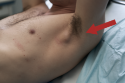



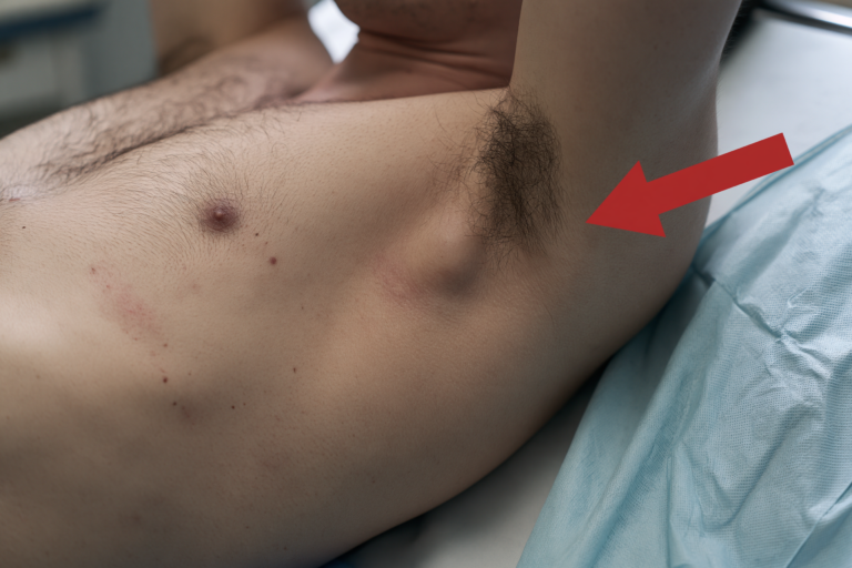
- Desmoid Tumor Symptoms at a Glance
- Desmoid Tumor Symptoms: What Does the Lump Feel Like?
- Desmoid Tumor Symptoms and Pain
- Abdominal Desmoid Tumor Symptoms
- Desmoid Tumor Symptoms Requiring Urgent Care
- Desmoid Tumor Symptoms by Location
- Desmoid Tumor Symptoms and Risk Factors
- Conditions That Mimic Desmoid Tumor Symptoms
- Diagnosing Desmoid Tumor Symptoms
- Desmoid Tumor Symptoms in a Published Case
- Desmoid Tumor Symptoms and Treatment
- Ayurvedic Assessment of Desmoid Tumor Symptoms
- Desmoid Tumor Symptoms and Complete Care
- Desmoid Tumor Symptoms and Specialist Care
- Preparing to Discuss Desmoid Tumor Symptoms
- Desmoid Tumor Symptoms FAQs
- References
Desmoid Tumor Symptoms at a Glance
Desmoid tumor symptoms vary widely because they are determined mainly by the tumor’s location, depth, growth pattern and relationship with nearby muscles, nerves, organs and blood vessels. A desmoid tumor may first appear as a firm, slowly enlarging lump, but deeper tumors may cause pain, pressure, restricted movement or abdominal symptoms before any mass becomes visible. These tumors do not spread to distant organs, yet their locally infiltrative growth can produce significant disability or serious complications when essential structures are compressed. [1,2,4]
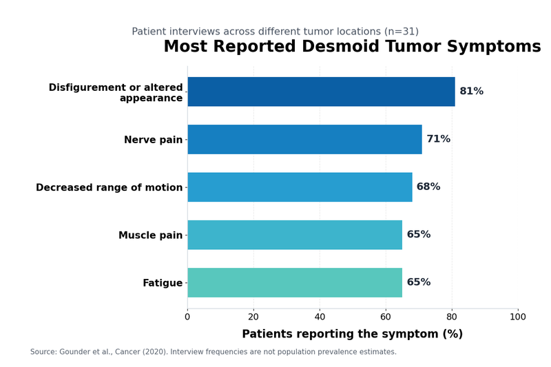
What Are the Most Common Early Symptoms of a Desmoid Tumor?
A new or progressively enlarging lump is one of the most recognizable early findings. The mass may feel firm and fixed within the abdominal wall, shoulder, upper arm, thigh, chest wall, back or another area of soft tissue. Some patients initially notice asymmetry, tightness in a muscle or a change in the contour of the affected body part rather than a clearly defined lump.
The lump may be painless, mildly tender or associated with persistent deep discomfort. Pain can result from pressure on surrounding tissues, stretching of the tissue covering a muscle, restricted movement or irritation of a nearby nerve. A smaller tumor located close to a nerve, joint or organ may therefore cause more severe symptoms than a larger superficial tumor.
When nerves are affected, patients may experience burning pain, tingling, numbness, altered sensation or weakness in the nearby limb. A desmoid tumor around the shoulder or upper arm may make it difficult to lift the arm, dress, drive or sleep on the affected side. A tumor in the thigh, hip or pelvis may alter walking, reduce exercise tolerance or produce pain during prolonged standing.
Abdominal wall tumors may cause a firm swelling that becomes more noticeable when the abdominal muscles tighten. Some patients experience discomfort while bending, coughing, exercising or changing position. The swelling can be mistaken for a hernia, postoperative scar, lipoma or muscular injury, which is why persistent abnormalities require imaging rather than diagnosis by touch alone. [1,2]
Intra-abdominal desmoid tumors can produce less specific symptoms. These may include persistent abdominal pain, pressure, bloating, early fullness after eating, nausea, reduced appetite, constipation or a noticeable change in bowel habits. Symptoms may fluctuate initially, particularly when the tumor intermittently presses on the intestine or surrounding mesentery.
Pain intensity does not always correspond directly with tumor size. A change in pain may reflect nerve irritation, muscle spasm, altered movement, inflammation or pressure on an adjacent structure. Increasing pain should therefore prompt reassessment, but pain alone cannot confirm that the tumor is enlarging. Clinical examination and comparison with previous MRI or CT imaging are needed to determine whether measurable progression has occurred. [5,6]
Can a Desmoid Tumor Develop Without Noticeable Symptoms?
Some desmoid tumors remain asymptomatic for a considerable period, especially when they are small, slow growing or located deep within the abdomen. They may be discovered incidentally during imaging performed for abdominal pain, an unrelated surgical problem or follow-up of another condition.
The absence of pain does not establish that a tumor is inactive, just as the presence of pain does not necessarily prove rapid growth. Imaging provides information about anatomical size and involvement of surrounding structures, while the patient’s symptoms reveal how the disease is affecting movement, sleep, digestion and daily function. Both forms of assessment are important.
A deep tumor may become symptomatic only after it begins to press on the bowel, urinary tract, nerves or blood vessels. For this reason, patients with a confirmed desmoid tumor should report new digestive, urinary, neurological or mobility changes even when an external lump has not changed visibly.
Conversely, some desmoid tumors remain radiologically stable or may regress without immediate intervention. A lack of symptoms can therefore support careful observation in appropriately selected patients, but this decision requires planned imaging and specialist review rather than assuming that no symptoms means no risk. [3,4]
Which Symptoms Need Prompt Medical Assessment?
Any unexplained soft tissue mass that is enlarging, deep, painful or interfering with normal movement should be evaluated by a clinician. A previously diagnosed desmoid tumor also requires reassessment when pain becomes persistent, the lump changes noticeably, movement becomes restricted or new neurological symptoms develop.
Severe or escalating abdominal pain accompanied by repeated vomiting, increasing abdominal distension, inability to pass stool or gas, or marked tenderness may indicate bowel obstruction or another urgent abdominal complication. These symptoms require immediate hospital assessment rather than waiting for a routine appointment.
New limb weakness, progressive numbness, difficulty walking, loss of hand function or severe radiating pain may indicate clinically important nerve compression. Difficulty swallowing, breathing or speaking requires urgent assessment when a tumor is located in the neck, upper chest or another anatomically restricted region.
Pelvic or intra-abdominal tumors may occasionally affect the urinary system. New flank pain, reduced urine output, difficulty urinating, fever or recurrent urinary symptoms should be assessed promptly because compression of the ureter can impair drainage from the kidney.
A rapidly changing symptom pattern should not be attributed automatically to the known tumor. Infection, hernia, bowel disease, musculoskeletal injury and other soft tissue conditions can produce similar complaints. Medical reassessment helps determine whether the symptoms arise from desmoid tumor progression, a treatment effect or an unrelated condition. [1,2,4]
What Should Patients Record Before a Medical Appointment?
A short symptom record can make the consultation more clinically useful. Patients should document when the lump or discomfort was first noticed, whether the area appears to be changing, what activities increase the symptoms and whether pain interferes with sleep, walking, work, eating or bowel function.
Pain is best described by its location, intensity, duration and character. Deep aching, pressure, burning, stabbing and movement-related pain may indicate different patterns of tissue or nerve involvement. Recording whether pain is constant or intermittent and whether it responds to prescribed medication can help the clinical team assess the effect on daily life.
Changes in appetite, abdominal fullness, nausea, bowel frequency, urinary function, limb strength and range of motion should also be documented when relevant. These functional changes may be clinically important even when the patient cannot detect a visible increase in tumor size.
Previous imaging reports, biopsy findings, operative notes and a current medication list should be available for review. Comparisons between earlier and current scans are usually more informative than a single measurement because they show whether the tumor is stable, regressing or progressing over time.
Patient-reported symptoms should be considered alongside imaging rather than treated as secondary information. Studies of people living with desmoid tumors have shown that pain, sleep disruption, fatigue and functional limitation can substantially reduce quality of life, even when the condition is not metastatic. [5,6]
Desmoid Tumor Symptoms: What Does the Lump Feel Like?
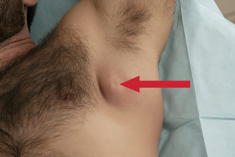
Desmoid tumor symptoms may begin with an unexplained lump or area of swelling before pain or loss of function develops. The mass is often felt beneath the skin or within a muscle, but its texture and mobility can vary according to its depth, anatomical location and involvement of surrounding connective tissue. A desmoid tumor cannot be identified reliably by touch alone because several benign conditions and other soft tissue tumors can feel similar. [1,2,4]
Table : Desmoid Tumor Lump Compared With Common Mimics
| Possible condition | Typical clues | How it is distinguished |
|---|---|---|
| Desmoid tumor | Often firm, deep, slowly enlarging and sometimes fixed within muscle or fascia; may be painful or painless | MRI or CT followed by a planned core needle biopsy |
| Lipoma | Usually soft, mobile and painless, although deep lipomas may feel firmer | Clinical examination, ultrasound or MRI when atypical |
| Abdominal wall hernia | Bulge may become more visible while coughing or straining and may reduce while lying down | Physical examination, ultrasound or CT |
| Cyst | Often smooth and superficial; may become red or tender if inflamed | Ultrasound can determine whether the swelling is fluid-filled |
| Scar or postoperative change | Located near previous surgery or trauma and may remain stable | Comparison with previous records and imaging if it enlarges |
| Other soft-tissue tumor | May be painless or painful and can appear similar to a desmoid tumor | Specialist imaging and pathology are required for accurate classification |
Is a Desmoid Tumor Lump Hard, Soft or Movable?
Many desmoid tumors feel firm because they contain dense fibrous tissue. A superficial tumor may have a clearly noticeable border, while a deeper tumor may feel more like a broad area of fullness, muscular thickening or asymmetry than a rounded lump. Overlying fat and muscle can also make a firm tumor feel softer than expected.
Mobility varies. A tumor arising within or closely attached to muscle and the connective tissue surrounding it may move very little when pressed. Other lesions may appear partially mobile because the skin and superficial tissue move over the mass. Limited mobility can increase clinical concern, but it is not specific to desmoid disease and should not be used to make a diagnosis without imaging and tissue examination.
The skin over the lump is often normal, particularly during the early stages. Redness, warmth or visible surface changes are not required for a desmoid tumor to be present. A normal skin appearance should therefore not delay assessment when a deeper mass is enlarging or causing persistent symptoms.
Repeatedly pressing the area is not a reliable way to determine whether the tumor is growing. Changes in muscle tension, body position, swelling and examination technique can alter how large the lump feels. Clinical measurements and comparison of MRI, CT or ultrasound images provide a more dependable assessment of size and anatomical involvement. [1,2]
Where Can a Desmoid Tumor Lump Appear?
A desmoid tumor can develop wherever connective tissue is present. Common sites include the abdominal wall, shoulders, upper arms and thighs, although tumors may also occur in the chest wall, back, neck, pelvis and other soft tissues. [1]
An abdominal wall desmoid may present as a firm swelling within one of the abdominal muscles. It may be easier to notice while standing, stretching, coughing or tightening the abdomen, although this pattern is not consistent in every patient. Discomfort may occur during exercise, lifting, bending or movements that engage the affected muscle.
A lump near the shoulder or upper arm may initially be mistaken for muscular tension, a sports injury or localized swelling. As the tumor affects nearby structures, the patient may notice reduced shoulder movement, discomfort while reaching overhead or difficulty sleeping on the affected side.
Tumors in the thigh, hip or pelvic region may be noticed as deep fullness, enlargement of one side or a mass that becomes more apparent during walking or exercise. When the tumor is close to a joint, the first clinically important change may be stiffness or reduced range of motion rather than visible growth.
Chest wall and back lesions may produce localized thickening, pressure or discomfort with posture and movement. Tumors in the neck are less common but require careful assessment because even a relatively small mass may affect swallowing, breathing, voice or nearby nerves.
Deep intra-abdominal tumors usually cannot be felt from the outside. In these cases, pain, fullness, nausea, altered bowel function or an incidental imaging finding may appear before a palpable mass develops. The absence of an external lump therefore does not exclude abdominal desmoid disease. [1,2,4]
Does a Desmoid Tumor Lump Always Hurt?
A desmoid tumor lump may be painless, mildly tender or significantly painful. Some patients first notice an unexplained mass without any other symptom, whereas others seek assessment because pain develops before the swelling becomes obvious.
Pain can arise when the tumor stretches surrounding tissue, restricts normal muscle movement or presses on a nerve. The discomfort may be described as aching, pressure, tightness, pulling, burning or pain triggered by a particular movement. Nerve involvement may also cause tingling, numbness, altered sensation or weakness beyond the immediate area of the lump.
A painless lump should not be considered harmless solely because it does not hurt. Similarly, severe pain does not prove that the tumor is large or growing rapidly. A smaller lesion near a nerve, joint or confined anatomical space may cause greater symptoms than a larger tumor located where surrounding tissues can accommodate it.
Pain that changes suddenly also requires clinical interpretation. It may result from tumor progression, muscle spasm, altered posture, treatment effects or an unrelated problem. Imaging is needed when the symptom pattern changes sufficiently to raise concern about anatomical progression. [1,4,5]
How Can a Desmoid Lump Be Distinguished From Other Swellings?
A lipoma usually feels soft and mobile beneath the skin, but some deep lipomas are firmer and difficult to distinguish clinically. A cyst may feel smooth or fluctuant, while scar tissue may feel hard and fixed. A hernia can produce a swelling that becomes more prominent with coughing or straining and may reduce when lying down, although this pattern is not always clear.
Postoperative changes can be especially confusing. A new abdominal wall lump after surgery may represent scar tissue, a hernia, a fluid collection, a suture reaction, another soft tissue lesion or a desmoid tumor. Pregnancy-related stretching of the abdominal wall and previous abdominal procedures can further complicate physical examination.
Clinical examination can identify the mass location, approximate depth, tenderness, mobility and effect on movement, but it cannot reliably confirm the diagnosis. Ultrasound may help determine whether a superficial lesion is solid or fluid-filled. MRI generally provides more detailed information about soft tissue involvement, while CT is often useful for intra-abdominal disease. [1,2]
Definitive diagnosis usually requires a core needle biopsy examined by a pathologist familiar with soft tissue tumors. This is important because desmoid-type fibromatosis can resemble other spindle-cell lesions on imaging and routine examination. An expert pathology review may be appropriate when the diagnosis is uncertain or when treatment decisions depend on distinguishing desmoid tumor from another condition. [1,2]
Any lump that is persistently enlarging, located deep within the tissues, associated with pain or neurological symptoms, or interfering with movement should be assessed without waiting for it to reach a particular size. A stable-looking lump also deserves review when it has not been previously diagnosed, because size and pain alone cannot determine its nature.
Desmoid Tumor Symptoms and Pain

Desmoid tumor symptoms often include persistent pain, pressure or tightness that changes with movement, posture or use of the affected muscle. Pain may arise from tissue infiltration, muscle restriction, nerve irritation or compression of nearby structures, and its severity can differ considerably between patients with tumors of similar size. [1,4,5]
What Does Desmoid Tumor Pain Feel Like?
Desmoid tumor pain is commonly described as a deep ache, pressure, pulling sensation or persistent muscular tightness. Some patients experience tenderness directly over the tumor, while others feel discomfort across a wider area because the affected muscle group compensates during movement.
Movement-related pain may occur when the tumor limits the normal sliding and contraction of muscles and connective tissues. A tumor in the abdominal wall may cause pain when coughing, bending, lifting or rising from bed. A lesion near the shoulder, hip or thigh may become more painful during reaching, walking, climbing stairs or prolonged standing.
Pain can be intermittent during the early stages and become more persistent as the tumor affects surrounding tissues. However, symptom intensity may fluctuate even when imaging remains stable. Physical activity, fatigue, posture, muscle spasm and inflammation can alter the daily pain experience without necessarily indicating measurable tumor growth.
Some patients feel pressure rather than sharp pain. This may present as heaviness, fullness or a sensation that clothing has become tighter over the affected area. Deep abdominal tumors may produce poorly localized discomfort that is difficult to distinguish from gastrointestinal or muscular pain.
The location and character of pain should be documented carefully because these details can help identify which structures may be involved. A local ache suggests a different pattern from burning pain, radiating discomfort or pain associated with numbness and weakness. [1,4]
Why Can Desmoid Tumors Cause Nerve Pain?
Nerve-related pain may develop when a desmoid tumor presses on, surrounds or irritates a peripheral nerve. Patients may describe burning, electric-shock sensations, pins and needles, hypersensitivity or pain that travels along the arm or leg.
Nerve involvement can also cause numbness, reduced grip strength, limb weakness or difficulty performing precise movements. These functional changes may be more clinically important than the pain score alone because they can indicate increasing interference with the nerve’s ability to control sensation or movement.
A tumor near the shoulder or upper arm may produce pain that extends toward the elbow or hand. A lesion in the pelvis, hip or thigh may cause radiating pain, altered sensation or weakness in the leg. Symptoms do not always remain confined to the area where the lump is located.
Neuropathic pain may respond poorly to ordinary non-prescription pain medicines because its mechanism differs from simple muscular soreness. Persistent burning, tingling or radiating pain should therefore be discussed with the treating team so that nerve involvement, appropriate imaging and targeted symptom management can be considered.
Progressive weakness, increasing numbness or loss of coordination requires prompt reassessment. These changes can influence both the urgency of treatment and the choice between continued observation, systemic therapy, local treatment or surgery. [1,4]
Can Desmoid Tumor Pain Become Worse at Night?
Night-time pain may occur when pressure is placed on the affected area or when a patient remains in one position for an extended period. Shoulder, chest wall, back and thigh tumors can make it difficult to lie on the involved side, while abdominal wall pain may increase when turning, stretching or rising from bed.
Sleep disturbance can amplify fatigue, reduce pain tolerance and impair concentration during the day. Over time, disrupted sleep may affect work, mood, physical activity and the ability to cope with ongoing treatment or surveillance.
Pain that repeatedly wakes a patient should be assessed even when daytime symptoms appear manageable. The clinical team may need to review sleeping position, medication timing, muscle spasm, nerve symptoms and whether interval imaging is appropriate.
Night pain is not specific to tumor progression. It may also reflect muscle stiffness, altered posture, reduced movement during sleep or pressure on a sensitive nerve. Its significance depends on whether it is new, steadily worsening, associated with functional loss or accompanied by other changes.
Research examining the lived experience of desmoid tumor has shown that pain and sleep disturbance are major contributors to reduced quality of life. Symptom evaluation should therefore include sleep quality and daily function rather than relying only on tumor measurements. [5,6]
Does Increasing Pain Mean the Tumor Is Growing?
Increasing pain can occur with tumor progression, but it does not prove that the tumor has enlarged. Symptoms may worsen because of nerve irritation, muscular compensation, scar formation, changes in activity, treatment effects or an unrelated condition.
Tumor growth can also occur without a major increase in pain, particularly when the lesion expands into an area with sufficient space and does not compress a sensitive structure. Symptom stability therefore cannot replace scheduled imaging when surveillance has been recommended.
The most reliable assessment combines the patient’s symptom pattern with physical examination and comparison of current imaging against previous scans. MRI is often used to evaluate extra-abdominal soft tissue involvement, while CT may be selected for intra-abdominal tumors or when MRI is unsuitable.
A change in pain becomes more concerning when it is accompanied by measurable loss of movement, new weakness, persistent numbness, increasing abdominal symptoms or inability to perform normal activities. These changes may indicate that the tumor’s anatomical effect has become clinically significant even before a major increase in overall dimensions is documented.
Treatment decisions should consider symptom burden as well as radiological change. A stable tumor that causes severe pain, sleep disruption or progressive functional impairment may require active treatment, while a slowly changing tumor with minimal symptoms may sometimes remain suitable for close observation. [4–6]
Pain improvement should also be interpreted carefully. Reduced discomfort may indicate successful symptom control, decreased pressure on surrounding tissues or improved function, but it does not confirm tumor regression. Objective response should be assessed through imaging alongside changes in pain, mobility, sleep and daily activity.
Abdominal Desmoid Tumor Symptoms

Abdominal desmoid tumor symptoms depend on whether the growth arises within the abdominal wall or deeper inside the abdomen, where it may involve the mesentery, bowel, blood vessels, urinary tract or other nearby structures. Some patients first notice a firm swelling, while others develop pain, pressure, bloating or altered bowel function without any visible lump. [1,2,4]
Table : Abdominal Warning Signs and Required Action
| Symptom pattern | Possible concern | Recommended action |
|---|---|---|
| Mild intermittent pressure or bloating while still eating and passing stool and gas | Early compression, altered bowel movement or another digestive condition | Record the pattern and contact the treating team if it persists or worsens |
| Increasing constipation, meal-related cramping, nausea or early fullness | Partial bowel narrowing or progressive abdominal pressure | Request an earlier clinical review |
| Repeated vomiting with progressive abdominal swelling | Possible bowel obstruction | Seek emergency medical assessment |
| Inability to pass stool or gas with cramping pain | Possible complete obstruction | Seek emergency medical assessment immediately |
| Constant severe abdominal pain, fever, faintness or marked tenderness | Bowel compromise, infection or another acute abdominal condition | Obtain immediate emergency care |
| Flank pain with fever, vomiting, reduced urine or difficulty urinating | Possible urinary tract or ureteric obstruction | Obtain urgent hospital assessment |
Abdominal Wall Desmoid Tumor Symptoms
An abdominal wall desmoid tumor commonly develops within the muscles and connective tissues of the front or side of the abdomen. The first sign may be a firm, slowly enlarging mass that becomes more noticeable while standing, stretching, coughing or tightening the abdominal muscles.
The swelling may be painless initially. As the tumor affects muscle fibres and surrounding fascia, patients may develop localized aching, pulling, tightness or tenderness. Activities that engage the abdominal muscles, including lifting, exercise, bending, coughing or rising from bed, can make the discomfort more noticeable.
Some patients observe a change in abdominal contour rather than a clearly rounded lump. One side may appear fuller, firmer or less flexible than the other. The skin over the area is often normal, and the absence of redness or warmth does not exclude a deeper soft tissue tumor.
An abdominal wall mass can resemble a hernia, lipoma, postoperative scar or muscular injury. Previous abdominal surgery and pregnancy can make the distinction more difficult because both may alter the appearance and strength of the abdominal wall. Imaging is therefore required to define whether the mass is located within muscle, beneath the fascia or inside the abdominal cavity. [1,2]
Pain from an abdominal wall tumor may remain localized or spread across a wider muscular area. Persistent discomfort can affect posture, exercise, sleep and routine tasks, particularly when the tumor lies close to the ribs, pelvis or a surgical scar.
Intra Abdominal and Mesenteric Symptoms
Intra-abdominal desmoid tumors develop inside the abdominal cavity and are often associated with the mesentery, the tissue that supports the intestines and carries their blood vessels. Because these tumors are deep, they may grow without producing an externally visible or palpable mass.
Early symptoms can be vague. Patients may experience intermittent abdominal pain, pressure, bloating, early fullness after eating, nausea or a reduced appetite. These symptoms may initially be attributed to indigestion, irritable bowel syndrome, food intolerance or another common gastrointestinal condition.
The pain may be poorly localized because deep abdominal structures do not always produce a precise pain pattern. Some patients describe a dull ache or persistent heaviness, while others experience cramping that becomes more pronounced after meals or during bowel movements.
As the tumor enlarges, it may displace or compress sections of the intestine. This can slow the movement of food and stool, producing constipation, abdominal distension, altered bowel frequency or a feeling of incomplete evacuation.
Mesenteric tumors may also affect nearby blood vessels or the tissue supporting the bowel. The clinical importance of an intra-abdominal desmoid tumor therefore depends not only on its diameter but also on whether it narrows the bowel, compromises blood flow or obstructs another essential structure. [1,4]
Patients with familial adenomatous polyposis may have an increased risk of intra-abdominal desmoid tumors. In this setting, new abdominal pain, distension or changes in bowel function should be investigated carefully rather than assumed to result from previous intestinal surgery or the underlying inherited condition. [1,4]
Can a Desmoid Tumor Cause Bowel Obstruction?
A desmoid tumor can cause partial or complete bowel obstruction when it compresses, encases or distorts part of the intestine. The risk is greater with intra-abdominal disease than with tumors confined to the abdominal wall.
Partial obstruction may initially cause intermittent cramping, bloating, nausea, reduced appetite and constipation. Symptoms may temporarily improve after passing stool or gas, which can make the condition appear less urgent during its early stages.
As obstruction becomes more severe, the abdomen may become increasingly distended and painful. Vomiting can develop, and the patient may be unable to pass stool or gas. These symptoms require urgent hospital assessment because prolonged obstruction can lead to dehydration, electrolyte disturbance, bowel injury or impaired blood supply.
Pain caused by obstruction is often cramp-like and may occur in waves, but it can become constant when the bowel is severely distended or compromised. Persistent tenderness, fever, faintness, rapid deterioration or a rigid abdomen may indicate a more serious abdominal complication.
Patients with a known intra-abdominal desmoid tumor should not attempt to manage repeated vomiting, severe distension or complete constipation solely with laxatives or dietary changes. Mechanical obstruction must first be excluded because increasing bowel stimulation may be inappropriate when the intestinal passage is physically narrowed. [1,4]
Milder digestive symptoms should also be reported when they become persistent or progressively worse. Earlier reassessment may identify increasing compression before complete obstruction develops.
Pelvic and Urinary Symptoms
A desmoid tumor in the lower abdomen or pelvis may press on the bladder, ureters, bowel, pelvic nerves or major blood vessels. Symptoms can therefore extend beyond abdominal pain and may include pelvic pressure, urinary changes, constipation, leg swelling or radiating discomfort.
Bladder compression may cause urinary frequency, urgency or a sensation that the bladder does not empty completely. These symptoms can resemble a urinary tract infection, but they may occur without burning urination, fever or abnormal urine test results.
Compression of a ureter can interfere with the flow of urine from the kidney to the bladder. Patients may develop flank pain, recurrent urinary infections or reduced kidney function, although obstruction can sometimes progress with few early symptoms.
New severe flank pain, fever, vomiting, markedly reduced urine output or difficulty passing urine requires urgent assessment. Imaging may be needed to determine whether the urinary tract is obstructed and whether kidney drainage has been affected.
Pelvic nerve involvement may cause numbness, burning pain or weakness extending into the hip, groin or leg. Pressure on pelvic blood vessels may contribute to swelling in one leg, particularly when venous return is restricted. These findings require assessment because blood clots and other vascular conditions can produce similar symptoms. [1,4]
Can Abdominal Desmoid Tumors Cause Weight Loss?
Weight loss is not a defining feature of every abdominal desmoid tumor, but it can occur when persistent pain, nausea, early satiety or bowel compression reduces food intake. A patient may begin eating smaller portions because meals intensify pressure, bloating or discomfort.
Repeated vomiting or partial obstruction can also impair nutrition and hydration. Over time, reduced calorie and protein intake may lead to weakness, loss of muscle mass and slower recovery from treatment.
Unintentional weight loss should not automatically be attributed to the desmoid tumor. Infection, endocrine disorders, medication effects, gastrointestinal disease and other malignancies may produce similar changes. Clinical assessment is needed when weight loss is progressive, unexplained or accompanied by fever, bleeding, severe fatigue or altered bowel habits.
Nutritional assessment is particularly important when a patient reports early fullness, reduced appetite or fear of eating because of pain. Monitoring body weight, hydration, meal tolerance and bowel function can help identify deterioration before severe malnutrition develops.
Symptom improvement should be evaluated alongside imaging. Better appetite, reduced bloating or more regular bowel movements may indicate reduced functional pressure, but these changes alone do not prove that the tumor has shrunk. Objective response requires comparison of MRI or CT findings with earlier scans. [3,4]
Desmoid Tumor Symptoms Requiring Urgent Care
Desmoid tumor symptoms require urgent medical assessment when they suggest bowel obstruction, impaired blood flow, urinary blockage or progressive nerve compression. Although desmoid tumors do not metastasize, their local growth can interfere with essential structures and occasionally cause serious complications. [1,4]
Bowel Obstruction Warning Signs
Severe cramping abdominal pain, repeated vomiting, progressive abdominal swelling and inability to pass stool or gas may indicate bowel obstruction. These symptoms are particularly concerning in patients with an intra-abdominal or mesenteric desmoid tumor.
Partial obstruction may begin with intermittent bloating, nausea, constipation and pain after meals. Symptoms can temporarily improve and then return, so repeated episodes should not be dismissed as indigestion or ordinary constipation.
Complete obstruction can rapidly cause dehydration, electrolyte disturbance and injury to the bowel. Persistent vomiting, a markedly distended abdomen or complete inability to pass gas requires immediate emergency assessment.
A sudden change from intermittent cramping to constant severe pain may indicate increasing pressure on the bowel or reduced blood supply. Fever, faintness, rapid heartbeat, marked tenderness or a rigid abdomen may signal a more advanced complication and should be treated as an emergency.
Laxatives should not be taken repeatedly when mechanical obstruction is possible unless a clinician has advised their use. Stimulating the bowel against a fixed narrowing may worsen pain and delay appropriate imaging or surgical assessment. [1,4]
Organ and Blood Vessel Compression
A desmoid tumor can compress nearby organs or major blood vessels, especially when it develops within the abdomen, pelvis, chest or neck. The resulting symptoms depend on which structure is affected and how quickly the compression progresses.
Increasing abdominal pain accompanied by dizziness, weakness, fainting or rapid deterioration requires urgent assessment. These symptoms are not specific to desmoid disease and may indicate bleeding, impaired circulation, infection or another acute condition.
Compression of pelvic veins may contribute to swelling, heaviness or discoloration in one leg. Sudden one-sided leg swelling, chest pain or breathlessness requires emergency evaluation because a blood clot must be excluded.
A tumor near the chest or neck may cause progressive difficulty breathing, swallowing or speaking. New noisy breathing, inability to swallow saliva, rapidly worsening shortness of breath or facial and neck swelling warrants immediate emergency care.
Symptoms caused by vascular or organ compression may become clinically important before the tumor shows a dramatic change in overall size. The relationship between the tumor and surrounding anatomy is therefore as important as its measured diameter. [1,4]
Progressive Nerve Symptoms
New or worsening weakness, numbness, loss of coordination or severe radiating pain may indicate increasing pressure on a nerve or nerve plexus. These symptoms require prompt reassessment because prolonged compression can lead to persistent functional loss.
A tumor affecting the arm or shoulder may cause reduced grip strength, difficulty lifting the arm or loss of hand dexterity. Pelvic or thigh tumors may produce foot weakness, difficulty walking, repeated tripping or altered sensation in the leg.
Pain accompanied by progressive weakness is more concerning than pain alone. Patients should seek urgent medical review when they can no longer perform movements that were previously possible, even if the visible lump appears unchanged.
Rarely, a tumor located near the spine or major pelvic nerves may be associated with loss of bladder or bowel control, numbness around the groin or rapidly worsening weakness in both legs. These symptoms require immediate emergency assessment because severe neurological compression must be excluded. [1,4]
Urinary Obstruction Warning Signs
Pelvic and lower abdominal desmoid tumors may compress a ureter, the tube that carries urine from the kidney to the bladder. Obstruction can cause flank pain, nausea, recurrent urinary infections or a gradual decline in kidney function.
Severe pain in the side or back, fever, chills and vomiting may indicate an infected obstructed urinary system. This combination requires urgent hospital treatment because infection behind a blockage can progress rapidly.
A marked reduction in urine output, inability to urinate or increasing lower abdominal pain should also be assessed immediately. These symptoms may result from urinary obstruction, dehydration, medication effects or another acute condition.
Ureteric obstruction can occasionally progress without severe pain. Patients with a pelvic desmoid tumor should therefore report recurrent urinary infections, unexplained kidney function changes or persistent urinary difficulty even when symptoms seem mild.
Blood tests, urine testing and imaging may be required to determine whether the kidneys and urinary tract are affected. Early recognition can allow drainage or other intervention before permanent kidney damage develops. [1,4]
Desmoid Tumor Symptoms by Location

Desmoid tumor symptoms differ according to the body part involved and the structures surrounding the tumor. A lesion in a limb may mainly restrict movement, while a tumor in the chest, neck or pelvis can affect breathing, swallowing, nerves, blood vessels or organ function. Tumor location may therefore influence symptom severity more strongly than size alone. [1,4]
Table 1: Desmoid Tumor Symptoms by Location
| Tumor location | Common symptoms | Important warning signs |
|---|---|---|
| Abdominal wall | Firm lump, pulling pain, tenderness, discomfort while bending, coughing or lifting | Rapid enlargement, increasing pain or progressive restriction of abdominal movement |
| Intra-abdominal or mesenteric | Deep abdominal pain, bloating, early satiety, nausea, constipation or altered bowel habits | Repeated vomiting, severe distension or inability to pass stool or gas |
| Shoulder or upper arm | Swelling, deep aching, reduced shoulder movement or pain while lifting | Progressive weakness, numbness, loss of grip or marked loss of arm function |
| Hip, thigh or pelvis | Deep mass, stiffness, walking pain, radiating discomfort or altered gait | Increasing leg weakness, one-sided swelling, urinary difficulty or inability to walk normally |
| Chest wall or back | Localised swelling, pressure, posture-related pain or discomfort during deep breathing | New breathlessness, rapidly worsening chest pressure or neurological symptoms |
| Head or neck | Firm neck mass, restricted movement, swallowing difficulty or voice change | Difficulty breathing, rapidly worsening swallowing or increasing facial and neck swelling |
Shoulder, Arm and Thigh Symptoms
A desmoid tumor in the shoulder or upper arm may first appear as a firm swelling, muscular fullness or unexplained difference between the two sides. Pain can increase when lifting, reaching, carrying objects or sleeping on the affected side.
Shoulder involvement may gradually reduce range of motion. Patients may find it difficult to raise the arm overhead, fasten clothing, wash their hair or perform work that requires repeated reaching. The restriction may result from direct involvement of muscle and fascia, protective muscle spasm or pain caused by movement.
When a tumor lies near a nerve, symptoms may extend beyond the visible lump. Burning pain, tingling, numbness, altered sensation or weakness can travel into the forearm or hand. Reduced grip strength, dropping objects or difficulty with fine movements should prompt reassessment.
Thigh and hip tumors may cause deep aching, tightness, swelling or a feeling of heaviness in the leg. Pain may become more noticeable during walking, climbing stairs, exercise or prolonged standing.
A tumor close to the hip or knee can reduce joint movement even when it does not arise within the joint itself. Patients may shorten their stride, favour one side or reduce activity to avoid pain, which can lead to muscle weakness and further functional decline.
New difficulty walking, repeated tripping, progressive leg weakness or increasing numbness requires timely medical review. These symptoms may indicate clinically important nerve or muscle involvement that could influence treatment decisions. [1,4]
Back and Chest Wall Symptoms
Desmoid tumors of the back may present as a firm area of thickening, a deep lump or persistent localized pain. Discomfort can worsen with twisting, lifting, sitting for long periods or lying directly on the affected area.
Muscle involvement may alter posture and place additional strain on nearby muscle groups. Patients may experience stiffness, reduced spinal movement or pain that spreads across the back without a clearly defined point of origin.
A chest wall tumor may cause localized swelling, pressure or pain during movement of the shoulder and upper body. When the tumor lies close to the ribs or intercostal muscles, discomfort may increase with deep breathing, coughing or certain sleeping positions.
Chest wall pain should not automatically be attributed to a known desmoid tumor. New chest pressure, unexplained breathlessness, faintness or pain spreading to the jaw or arm requires urgent assessment because cardiac, pulmonary and vascular causes must also be considered.
Large or strategically positioned chest wall tumors may restrict movement or create a sensation of tightness. Imaging is needed to determine whether the lesion remains confined to soft tissue or is affecting deeper structures.
Persistent pain that disrupts sleep, work or normal movement is clinically important even when the tumor appears radiologically stable. Symptom burden and functional impairment should be considered alongside scan measurements. [4–6]
Head and Neck Symptoms
Head and neck desmoid tumors are uncommon, but symptoms can become significant because many essential structures are concentrated within a small anatomical space. The first sign may be a firm neck mass, facial asymmetry, jaw discomfort or restricted neck movement.
A tumor near the throat can cause difficulty swallowing, a persistent sensation of pressure or discomfort when turning the head. Voice changes may develop when structures involved in speech are compressed or movement becomes restricted.
Breathing difficulty, noisy breathing or rapidly increasing neck swelling requires immediate assessment. Even a relatively small tumor can become clinically important when it narrows the airway or affects nearby vessels.
Tumors near the jaw, face or skull base may cause pain, numbness, weakness or reduced movement depending on the nerves and muscles involved. Some patients may experience difficulty chewing, opening the mouth fully or maintaining normal facial movement.
Because enlarged lymph nodes, thyroid disease, salivary gland disorders and other tumors can also present as a neck mass, clinical examination alone cannot establish the diagnosis. Imaging and biopsy are usually required to determine the nature of the lesion. [1,4]
Why Location Matters More Than Size Alone
A small desmoid tumor located beside a major nerve, blood vessel, airway, ureter or section of bowel may cause greater risk than a larger tumor growing in an area with more space. Treatment decisions should therefore consider anatomical relationships rather than relying only on maximum diameter.
A tumor that grows slowly may still require active treatment if it causes progressive weakness, organ compression, severe pain or major loss of function. Conversely, a larger tumor that remains stable and causes minimal symptoms may sometimes be monitored with planned imaging.
Changes in daily function can reveal clinically meaningful progression before a patient notices visible enlargement. Difficulty lifting an arm, walking normally, swallowing, sleeping comfortably or completing routine tasks should be documented and discussed during follow-up.
Imaging should assess not only whether the tumor has changed in size but also whether its relationship with nearby structures has altered. A small increase in one direction can be important when it brings the tumor closer to a nerve, organ or blood vessel.
Patient-reported pain, mobility, sleep and functional capacity provide information that cannot be captured by imaging alone. Combining these outcomes with MRI or CT findings gives a more accurate picture of how the tumor is affecting health and whether the current management plan remains appropriate. [4–6]
Desmoid Tumor Symptoms and Risk Factors

Desmoid tumor symptoms can occur in people of any age and without an identifiable risk factor, although certain age groups, inherited conditions and clinical circumstances are associated with a higher likelihood of diagnosis. Risk factors cannot confirm that a lump or pain is a desmoid tumor, but they can help clinicians decide when imaging, biopsy or genetic assessment deserves particular attention. [1,4]
Age and Sex Patterns
Desmoid tumors are most often diagnosed between 15 and 60 years of age, with the highest incidence reported in adults in their thirties and forties. They can also occur in children, adolescents and older adults, so age outside the typical range should not be used to dismiss a persistent soft tissue mass. [1,4]
Women are diagnosed more frequently than men, particularly during the reproductive years. This difference may reflect a combination of biological, hormonal and pregnancy-related factors, although sex alone does not predict whether a tumor will develop, grow or cause severe symptoms. [1,4]
The presenting symptoms are determined more by anatomical location than by age or sex. A young adult may notice an abdominal wall lump, while another patient of the same age may present with shoulder pain, restricted movement or an incidental intra-abdominal mass.
A new lump in a younger patient is often initially attributed to exercise, muscle strain, a cyst or another benign condition. Persistent enlargement, fixation within a muscle, unexplained pain or progressive loss of movement should prompt imaging regardless of the patient’s age.
Pregnancy and the Abdominal Wall
Some desmoid tumors are diagnosed during pregnancy or after childbirth, most often within the abdominal wall. Patients may notice a firm area of swelling, asymmetry, pulling pain or discomfort when using the abdominal muscles.
Abdominal wall changes are common during and after pregnancy, and most do not represent a desmoid tumor. Rectus diastasis, hernia, scar tissue and normal postpartum tissue changes can also cause visible or palpable abnormalities.
A mass that persists, becomes progressively larger, feels deeply fixed or causes increasing pain should be assessed rather than assumed to be a normal postpartum change. Ultrasound may be used as an initial examination, while MRI can provide more detailed soft tissue information without ionising radiation when clinically appropriate.
Pregnancy-associated presentation does not establish that hormones caused the tumor. The relationship is complex, and individual tumors can behave differently. Some remain stable, some regress and others require active treatment because of growth, pain or functional effects. [4]
A previous desmoid tumor does not automatically mean that future pregnancy is unsafe. Planning should be individualized according to tumor location, recent imaging, previous growth behaviour, current treatment and the potential effect of pregnancy-related abdominal changes. Patients receiving systemic therapy should also discuss contraception, fertility and treatment timing with their specialist team.
Surgery and Scar-Related Presentations
Desmoid tumors may occasionally arise in areas affected by previous surgery, particularly within the abdominal wall or abdomen. In patients with familial adenomatous polyposis, abdominal surgery is a recognized clinical context in which desmoid disease may subsequently develop.
A postoperative lump is more commonly caused by scar tissue, hernia, fluid collection, suture reaction or another benign process. Examination alone may not reliably distinguish these conditions from a desmoid tumor.
The interval between surgery and the appearance of symptoms can vary. Patients should seek assessment when a scar-associated mass enlarges, becomes increasingly painful, restricts movement or produces abdominal pressure and altered bowel function.
Imaging can determine whether the abnormality lies within the skin, fat, fascia, muscle or abdominal cavity. A solid mass with uncertain characteristics generally requires specialist assessment and, when appropriate, core needle biopsy before definitive treatment is planned.
Physical injury has also been reported before some desmoid diagnoses, but an ordinary injury does not usually mean that a desmoid tumor will develop. A lump that remains after an apparent muscle injury, continues enlarging or fails to improve as expected should be reassessed rather than repeatedly treated as a strain.
Familial Adenomatous Polyposis
Most desmoid tumors are sporadic, meaning that they arise without an inherited desmoid syndrome. More than 90% are associated with acquired changes involving the CTNNB1 gene within the tumor cells rather than a mutation inherited by every cell in the body. [4]
A smaller proportion occurs in people with familial adenomatous polyposis, an inherited condition caused by a germline alteration in the APC gene. Familial adenomatous polyposis leads to numerous colorectal adenomas and a substantially increased risk of colorectal cancer if appropriate surveillance and preventive management are not provided. [1,4]
Desmoid tumors associated with familial adenomatous polyposis are frequently intra-abdominal or located in the abdominal wall. They may be single or multifocal and can develop before or after bowel surgery.
Symptoms may include abdominal pain, pressure, bloating, nausea, constipation, vomiting or a palpable abdominal mass. Because these complaints can also result from previous intestinal surgery, adhesions or other gastrointestinal problems, imaging is needed to identify the cause.
A desmoid tumor does not automatically mean that the patient has familial adenomatous polyposis. Most people with a desmoid diagnosis do not have the inherited condition, and molecular findings from the tumor can help the specialist team distinguish a sporadic CTNNB1-associated tumor from possible APC-related disease. [1,4]
When Genetic Counselling May Help
Genetic counselling may be considered when a patient with a desmoid tumor also has numerous colorectal polyps, colorectal cancer at an unusually young age, a known family history of familial adenomatous polyposis or relatives with multiple colorectal cancers and desmoid tumors.
Assessment may also be appropriate for selected patients with intra-abdominal or multiple desmoid tumors, particularly when the personal or family history raises concern for an inherited APC alteration. The decision should be based on the complete clinical history rather than tumor location alone.
Genetic counselling helps patients understand what testing can and cannot show, how an inherited result could affect colorectal surveillance and whether close relatives may also need assessment. Testing should be accompanied by appropriate pre-test and post-test counselling because the result can have implications beyond the person with the tumor.
When familial adenomatous polyposis is confirmed, care generally includes coordinated colorectal surveillance, assessment for other associated manifestations and specialist monitoring of the desmoid tumor. New abdominal symptoms should not be attributed automatically to the known condition because bowel obstruction, postoperative complications and unrelated gastrointestinal disorders can present similarly.
Patients without a family history should still pursue appropriate investigation of suspicious symptoms. An inherited predisposition is only one part of desmoid biology, and the absence of affected relatives does not exclude a sporadic desmoid tumor. [1,4]
Conditions That Mimic Desmoid Tumor Symptoms
Desmoid tumor symptoms such as a firm lump, abdominal pain, pressure, restricted movement or altered bowel function can resemble several benign and malignant conditions. The similarity is clinically important because the correct diagnosis usually depends on imaging and tissue examination rather than the way a mass feels or the severity of pain alone. [1,2,4]
Lipoma, Cyst and Scar Tissue
A lipoma is a benign growth of fatty tissue that commonly presents as a soft, slowly enlarging lump beneath the skin. Many lipomas move slightly when pressed and remain painless, but deep lipomas can feel firm, fixed and difficult to distinguish from a desmoid tumor during physical examination.
A cyst may feel smooth or rounded and can become tender if it is inflamed or infected. Superficial cysts may cause redness, warmth or drainage, whereas a desmoid tumor usually arises deeper within connective tissue and does not typically produce a skin opening. These differences can guide assessment but are not reliable enough to establish the diagnosis.
Scar tissue can form a firm area after surgery, injury or childbirth. It may remain stable for years or cause pulling discomfort when the surrounding tissue stretches. A scar-associated lump that continues to enlarge, becomes increasingly painful or extends beyond the original scar should be assessed with imaging.
Postoperative fluid collections, suture reactions and localized inflammation can also produce swelling near a surgical site. Ultrasound may help determine whether the area contains fluid or solid tissue, while MRI or CT can define deeper involvement when the findings remain uncertain. [1,2]
Hernia and Abdominal Wall Weakness
An abdominal wall hernia occurs when tissue pushes through a weakness in the muscle or fascia. The swelling may become more noticeable during coughing, lifting or straining and may decrease when the patient lies down.
An abdominal wall desmoid tumor can also become more visible when the abdominal muscles contract, which can make the two conditions difficult to distinguish. Unlike many hernias, a desmoid mass may feel persistently firm and may not reduce with gentle pressure, although examination findings vary.
Hernias can cause discomfort, dragging sensations or intermittent pain, especially during activity. A trapped hernia may produce severe pain, vomiting and bowel obstruction, creating symptoms similar to an intra-abdominal desmoid complication.
A painful abdominal swelling that becomes hard, cannot be reduced or is accompanied by vomiting and inability to pass stool or gas requires urgent assessment. Imaging is often necessary because both hernias and desmoid tumors can occur in people with previous abdominal surgery. [1,4]
Muscle Injury and Fibromatosis
A muscle strain or tear can cause localized pain, swelling and firmness after exercise, lifting or trauma. Symptoms usually improve over time with appropriate rest and rehabilitation, although bruising and scar formation can persist.
A desmoid tumor may initially be mistaken for a chronic muscle injury when pain develops during movement or when the mass lies deep within a muscle. The possibility of a tumor becomes more concerning when the swelling enlarges, pain persists beyond the expected healing period or function continues to decline.
Other forms of fibromatosis can also produce firm tissue growth. Superficial fibromatoses involving the hand or foot differ from deep desmoid-type fibromatosis in their location and behaviour, but accurate classification still requires clinical and pathological assessment.
Persistent symptoms after an apparent sports injury should not be attributed indefinitely to muscle strain. Imaging is appropriate when the lump becomes more prominent, the pain worsens or the affected limb loses strength or range of motion. [1,2]
Desmoid Tumor and Gastrointestinal Stromal Tumor
Desmoid tumors and gastrointestinal stromal tumors can both present with abdominal pain, fullness, a palpable mass or incidental findings on imaging, but they arise from different tissues and behave differently.
A gastrointestinal stromal tumor develops in the wall of the digestive tract, most often in the stomach or small intestine. Symptoms may include abdominal discomfort, early satiety, nausea, gastrointestinal bleeding, black stools, vomiting blood or anaemia. Some patients have no symptoms until the tumor becomes large or is found during imaging or endoscopy. [8]
A desmoid tumor usually develops from connective tissue in the abdominal wall, mesentery or other soft tissues. It does not spread to distant organs, but it can infiltrate locally and compress the bowel, blood vessels, ureters or nerves. Gastrointestinal stromal tumors can metastasize, particularly when their biological risk is high. [1,4,8]
Blood in the stool, black stools, unexplained anaemia or vomiting blood are more suggestive of bleeding from within the digestive tract than of a typical desmoid tumor. These symptoms require prompt medical evaluation because several gastrointestinal conditions, including ulcers and cancers, can cause them.
CT or MRI may suggest whether a mass originates from the bowel wall or adjacent soft tissue, but imaging alone may not always provide a definitive distinction. Biopsy and expert pathological analysis are required when the diagnosis remains uncertain. [1,2,8]
Other Soft Tissue Tumors
Several benign and malignant soft tissue tumors can present as a painless or painful enlarging mass. These include nerve sheath tumors, smooth-muscle tumors, fibrous tumors and soft tissue sarcomas.
A rapidly enlarging mass, deep location, persistent pain or progressive neurological symptoms increases concern but does not identify a specific diagnosis. Some malignant tumors grow slowly, while some benign tumors enlarge quickly or cause severe symptoms because of their location.
Soft tissue sarcomas can arise in the limbs, trunk, abdomen or retroperitoneum. Depending on their location, they may cause a firm lump, pain, nerve symptoms, abdominal pressure or organ dysfunction. Unlike desmoid tumors, many sarcoma subtypes have the potential to spread to distant organs.
Enlarged lymph nodes, metastatic cancer, lymphoma and infections can also produce masses in the neck, abdomen, pelvis or limbs. Fever, night sweats, unexplained weight loss or marked fatigue should be reported, although their absence does not exclude a significant condition.
The diagnostic pathway should therefore focus on identifying the tissue of origin and obtaining an accurate pathological diagnosis rather than assuming that a mass is benign or malignant from symptoms alone. [1,2,4]
Why Biopsy Is Needed
Imaging can show the size, depth and relationship of a mass to nearby structures, but many soft tissue lesions share overlapping appearances. MRI is particularly useful for defining muscle, fascia and nerve involvement, while CT is often preferred for intra-abdominal and pelvic disease.
A core needle biopsy usually provides tissue for microscopic examination without removing the entire mass. The procedure should be planned carefully because the biopsy pathway may affect later surgery or local treatment if another tumor type is diagnosed.
Desmoid-type fibromatosis is composed of spindle-shaped cells and can resemble other fibrous or spindle-cell lesions. Pathologists may use immunohistochemistry, including assessment of nuclear beta-catenin, together with the microscopic pattern and clinical findings.
Testing for a CTNNB1 alteration can support the diagnosis of a sporadic desmoid tumor in appropriate cases. APC-related assessment may be considered when the clinical history raises concern for familial adenomatous polyposis.
A pathology review by a specialist experienced in soft tissue tumors may be valuable when the biopsy findings are uncertain, the imaging and pathology do not agree or the proposed treatment carries substantial risk. Correct classification prevents patients from receiving surgery, systemic therapy or prolonged observation for the wrong condition. [1,2,4]
Diagnosing Desmoid Tumor Symptoms
Desmoid tumor symptoms cannot confirm the diagnosis because lumps, pain, restricted movement and abdominal pressure can occur with many other conditions. Diagnosis usually combines a detailed clinical assessment, anatomical imaging, a core needle biopsy and expert pathological review. Molecular testing may provide additional confirmation and help identify the small proportion of patients who require assessment for an inherited APC-related condition. [1,2,4]
Clinical History and Examination
The diagnostic assessment begins with when the lump or symptoms first appeared, whether they are changing and how they affect daily function. The clinician will ask about pain, sleep disruption, weakness, numbness, bowel changes, urinary symptoms, appetite, weight and any difficulty walking, lifting or moving the affected area.
Previous pregnancy, abdominal surgery, trauma and treatment for another tumor may be relevant, but none of these factors proves that a desmoid tumor is present. A personal history of numerous colorectal polyps or a family history of familial adenomatous polyposis, early colorectal cancer or desmoid tumors should also be reported.
During examination, the clinician assesses the mass’s location, approximate size, depth, firmness, mobility and relationship to muscle contraction. The surrounding skin, nearby joints, circulation, sensation and muscle strength may also be examined.
An abdominal assessment may identify tenderness, distension, a palpable mass or signs of impaired bowel function. A limb examination can reveal reduced range of motion, weakness or nerve-related sensory changes that are not apparent from the size of the lump alone.
The initial consultation should also document baseline pain and function. This provides a comparison for later visits and helps determine whether symptoms are stable, improving or progressing independently of changes seen on imaging.
No routine blood test can confirm a desmoid tumor. Blood tests may still be required to investigate anaemia, inflammation, kidney function, dehydration or another possible cause of the patient’s symptoms, particularly when abdominal or urinary complications are suspected. [1,2]
Ultrasound, MRI and CT
Ultrasound is often used as an initial test for a superficial lump or abdominal wall mass. It can show whether the abnormality is solid or fluid-filled, estimate its depth and assess its relationship with nearby muscles and blood vessels.
Ultrasound alone usually cannot distinguish a desmoid tumor reliably from other solid soft tissue lesions. A suspicious or indeterminate mass therefore requires additional imaging and, when appropriate, tissue sampling.
MRI is generally preferred for desmoid tumors of the abdominal wall, limbs, shoulder, chest wall, pelvis and other extra-abdominal soft tissues. Its strong soft tissue contrast helps define the tumor’s relationship with muscles, fascia, nerves, joints and blood vessels. MRI also avoids ionising radiation, which is valuable when repeated scans are required over several years. [2,4]
Different MRI sequences provide information about the tumor’s internal composition. Areas with higher T2 signal may contain more water-rich, cellular tissue, while lower T2 signal may reflect a greater collagen component. These findings can support assessment of biological change, but they cannot replace measurements, symptom evaluation or pathological confirmation. [2,4]
Contrast-enhanced MRI may help define tumor boundaries and internal characteristics. Patients should inform the imaging team about pregnancy, kidney disease, implanted medical devices, previous contrast reactions or severe claustrophobia so that the examination can be planned safely.
CT is frequently used for intra-abdominal and mesenteric desmoid tumors because it clearly demonstrates the bowel, mesentery, urinary tract and major abdominal vessels. It can identify bowel narrowing, obstruction, ureteric compression and other complications that may require urgent management. [2,4]
CT may also be selected when MRI is unavailable, unsuitable or unable to answer the clinical question. Because CT uses ionising radiation, the need for repeated examinations should be balanced against age, tumor location and the quality of information obtainable from MRI.
A scan should be interpreted in comparison with previous imaging whenever possible. Small differences can result from patient position, image angle or measurement technique, while clinically important progression may involve extension toward a nerve or organ without a dramatic change in maximum diameter.
Imaging can strongly suggest desmoid-type fibromatosis, but it cannot provide definitive confirmation in most newly diagnosed cases. Several benign fibrous lesions, postoperative changes, gastrointestinal stromal tumors and soft tissue sarcomas can produce overlapping appearances. [1,2,4]
Core Needle Biopsy
A core needle biopsy removes small cylinders of tissue for microscopic examination. It is generally preferred because it provides more architectural information than a fine needle sample while avoiding the morbidity of removing an undiagnosed deep mass.
The biopsy may be guided by ultrasound or CT to ensure that representative tissue is obtained while avoiding major blood vessels, nerves, bowel and areas unlikely to provide a useful sample. The approach should ideally be planned with a team experienced in soft tissue tumors so that it does not complicate any future local procedure.
Local anaesthetic is commonly used, and several tissue cores may be collected during the same procedure. Patients should receive instructions about wound care, physical activity and symptoms that require medical review after the biopsy.
Temporary soreness, bruising or swelling can occur. An apparent increase in size or pain immediately after biopsy may result from local bleeding or inflammation rather than true tumor progression. Persistent enlargement, severe pain, fever, neurological changes or signs of significant bleeding require reassessment. [4]
An excisional biopsy, in which the entire lump is removed before a diagnosis is established, is not appropriate for every patient. Attempting to remove a deep or infiltrative mass without specialist planning may cause unnecessary functional damage and can make further treatment more complicated if the lesion proves to be a different tumor.
The pathology report should be reviewed together with the imaging findings. When the microscopic diagnosis does not match the tumor’s location or radiological appearance, the case may require additional tissue, further staining or a second pathological opinion. [2,4]
Pathology and Beta Catenin
Desmoid-type fibromatosis is composed of spindle-shaped fibroblastic cells arranged within a collagen-rich background. The cells often extend between surrounding muscle fibres, which helps explain why the tumor may feel fixed and why complete surgical removal can be difficult.
The cells generally appear relatively uniform and lack the marked atypia seen in many malignant tumors. However, microscopic appearance alone may overlap with scars, nodular fasciitis, nerve sheath tumors, low-grade sarcomas and other spindle-cell lesions.
Immunohistochemistry uses antibodies to detect proteins within the tissue. Nuclear beta-catenin staining is an important supportive feature in many sporadic desmoid tumors, but it is not positive in every case and is not completely specific when interpreted alone.
The pathologist may use a panel of stains selected according to the tumor’s location and appearance. This helps distinguish desmoid-type fibromatosis from gastrointestinal stromal tumor, smooth-muscle tumors, nerve sheath tumors and selected sarcomas.
Because desmoid tumors are rare and the differential diagnosis can be complex, confirmation by a pathologist experienced in soft tissue tumors is particularly valuable. A specialist review may prevent unnecessary surgery, inappropriate systemic treatment or prolonged observation of a different condition. [2,4]
CTNNB1 and APC Testing
Most sporadic desmoid tumors contain an acquired alteration in the CTNNB1 gene within the tumor tissue. This alteration affects beta-catenin regulation and can support the diagnosis when the microscopic and immunohistochemical findings are uncertain.
A somatic CTNNB1 alteration is present only in the tumor and is not usually inherited or passed to children. Its detection can help separate a sporadic desmoid tumor from disease associated with familial adenomatous polyposis. [1,4]
Familial adenomatous polyposis is caused by an inherited alteration in the APC gene. It is associated with numerous colorectal polyps, a high lifetime risk of colorectal cancer and an increased risk of intra-abdominal desmoid tumors.
The absence of a detectable CTNNB1 alteration does not automatically establish an inherited APC condition. It may reflect limitations of the tissue sample or testing method, and some apparently CTNNB1-negative tumors require more sensitive analysis or pathological reconsideration.
Further assessment for familial adenomatous polyposis may be appropriate when a CTNNB1 alteration is not identified, particularly in a patient with an intra-abdominal or multifocal tumor, numerous colorectal polyps, early colorectal cancer or a suggestive family history. This assessment may include genetic counselling, germline APC testing and colorectal evaluation. [1,4]
Molecular findings should be interpreted with the pathology, imaging and family history rather than used as an isolated diagnostic result. Mutation subtype may provide biological information, but it should not by itself determine whether a patient undergoes surveillance, systemic therapy, a local procedure or surgery.
Once the diagnosis is confirmed, the baseline scan should document the tumor’s full anatomical extent and relationship with nearby structures. Subsequent decisions can then consider symptom burden, functional impairment, imaging behaviour and the risk posed to nerves, organs, blood vessels or the bowel. [2,4]
Desmoid Tumor Symptoms in a Published Case

Desmoid tumor symptoms may begin with a persistent swelling rather than severe pain or digestive disturbance. A published abdominal wall case involving a 23-year-old woman demonstrates how a relatively short history of localized swelling led to clinical examination, imaging, surgery and histopathological confirmation of intramuscular desmoid-type fibromatosis. [7]
Presentation and Imaging
The patient reported a two-month history of swelling in the right upper abdomen. The location was clinically important because an abdominal wall mass can arise from skin, fat, fascia, muscle, a previous scar or an underlying abdominal structure. A persistent swelling in this region therefore requires anatomical imaging rather than diagnosis by appearance or palpation alone. [7]
Ultrasonography was used to evaluate the mass before computed tomography provided more detailed anatomical information. CT demonstrated a well-defined soft tissue lesion within the anterior abdominal wall, positioned to the right of the midline along the rectus abdominis muscle. This intramuscular location helped explain why the swelling could feel deeply fixed rather than freely movable beneath the skin. [7]
The case shows why abdominal wall desmoid tumor symptoms can be confused with a hernia, lipoma, scar-related change or muscular lesion. A patient may notice only a localized bulge, while imaging reveals that the abnormality originates within the abdominal musculature. The absence of bowel symptoms does not exclude an abdominal wall tumor because lesions confined to the muscle may not compress the digestive tract.
Imaging can define the depth, size and relationship of a mass to surrounding structures, but it cannot always distinguish desmoid-type fibromatosis from other solid soft tissue tumors. Histopathological assessment remains necessary when the imaging appearance is not sufficiently specific or when a definitive diagnosis will change management. [2,4]
Surgery and Pathological Confirmation
The treating team performed complete surgical resection with a reported two-centimetre margin. Removing the tumor created an abdominal wall defect that required reconstruction with surgical mesh to restore structural support. [7]
Abdominal wall reconstruction may be necessary when surgery removes part of the rectus muscle, fascia or other load-bearing tissue. The planning must consider protection of the abdominal organs, restoration of core stability, wound healing and the risk of postoperative hernia.
Histopathological examination confirmed the diagnosis of desmoid tumor. The reported microscopic findings included a relatively uniform myofibroblastic proliferation arranged in long bundles within collagenous tissue. Immunohistochemical findings included reactivity for vimentin, smooth muscle actin and beta-catenin, interpreted together with the morphology and clinical setting. [7]
The patient was discharged on the fifth postoperative day and was reported to be recovering well. This represents an early postoperative outcome rather than proof of permanent disease control. Desmoid tumors can recur after apparently complete resection, so longer follow-up with clinical assessment and imaging remains important. [4,7]
Pain relief, wound healing and restoration of movement should be assessed alongside surveillance imaging after abdominal wall surgery. Patients may also require graded rehabilitation because removal and reconstruction of abdominal muscle can temporarily affect lifting, posture, coughing and routine physical activity.
What the Case Means for Patients
The case demonstrates that a new abdominal swelling can be the principal symptom of a desmoid tumor even when the patient does not initially report bowel obstruction, weight loss or severe systemic illness. A lump that persists or enlarges should therefore be investigated before it is assumed to be a harmless muscular or postoperative change.
It also shows that the anatomical origin of a mass can determine treatment complexity. A tumor limited to a surgically accessible part of the abdominal wall may be considered for resection when the expected functional consequences are acceptable. A tumor involving critical vessels, nerves, bowel or several abdominal structures may require a different strategy.
Surgery performed in one published case should not be interpreted as the required treatment for every patient with abdominal wall desmoid disease. Current management considers symptom burden, documented growth, anatomical risk, anticipated surgical morbidity and the possibility that the tumor may remain stable or regress during active surveillance. [4]
A symptomatic tumor does not automatically require immediate surgery. Increasing pain, progressive loss of function, rapid radiological growth or threat to an essential structure may support active treatment, but available options can include systemic therapy, selected local procedures or surgery depending on the individual case.
For patients considering surgery, the decision should include the likely size of the resulting defect, the need for mesh or tissue-flap reconstruction, expected recovery, recurrence risk and the effect on abdominal strength. The ability to remove the tumor must be balanced against the possibility of creating long-term functional impairment.
The published report describes one clinical observation and cannot predict another patient’s outcome. Its greatest practical value is showing the diagnostic sequence from persistent swelling to imaging and pathological confirmation, while emphasizing that treatment must be selected according to tumor location, symptoms and the risks of intervention. [4,7]
Desmoid Tumor Symptoms and Treatment
Desmoid tumor symptoms influence treatment decisions alongside imaging findings, tumor location, growth rate and the risk to nearby organs or nerves. A newly diagnosed tumor does not always require immediate intervention, but increasing pain, functional loss, bowel or urinary compression and progressive neurological symptoms can indicate that observation is no longer sufficient. [3,4]
When Active Surveillance Is Appropriate
Active surveillance is often considered for a newly diagnosed desmoid tumor that is not causing severe symptoms, threatening an essential structure or showing clinically significant progression. This approach recognizes that some tumors remain stable for prolonged periods and others may regress without immediate surgery or systemic treatment. [3,4]
Surveillance does not mean that the tumor is being ignored. It involves planned clinical reviews, repeated imaging and assessment of how the condition affects pain, movement, sleep, work, eating and bowel or urinary function. The interval between scans is individualized according to tumor location, recent behaviour and the potential consequences of growth.
MRI is commonly used for extra-abdominal tumors because it provides detailed information about muscles, fascia, nerves and blood vessels without ionising radiation. CT may be preferred for intra-abdominal disease when bowel, mesenteric vessels, ureters or other deep structures need to be assessed.
The first follow-up scans are important because they establish whether the tumor is stable, regressing or progressing. A small change on one scan may require confirmation over time, particularly when symptoms remain mild and no critical structure is threatened.
Patients should not wait for the next scheduled scan when new symptoms develop. Increasing pain, weakness, numbness, reduced movement, persistent vomiting, progressive abdominal distension or urinary difficulty may require earlier assessment.
Active surveillance may remain appropriate when imaging shows stability and the patient can perform normal activities without substantial pain or functional loss. It may also continue when mild symptoms are manageable and treatment risks are greater than the current effect of the tumor.
The decision should be reviewed regularly rather than treated as permanent. A patient who is initially suitable for observation may later require active treatment if symptoms become more disruptive, imaging shows sustained growth or the tumor approaches a vulnerable structure. [3,4]
When Symptoms Prompt Active Treatment
Persistent or worsening pain is one of the most common reasons to reconsider the management plan. The clinical significance depends on whether pain interferes with sleep, walking, work, exercise, eating or routine self-care rather than on the pain score alone.
Progressive functional impairment can justify treatment even when the change in tumor size appears modest. A shoulder tumor that increasingly restricts arm movement, a thigh tumor that alters walking or an abdominal wall tumor that limits lifting may have become clinically significant despite limited radiological progression.
New nerve symptoms require particular attention. Burning pain, tingling, numbness or weakness may indicate increasing contact with a nerve. Progressive loss of strength or coordination can influence the urgency of treatment because prolonged compression may cause persistent disability.
Intra-abdominal symptoms may require treatment when the tumor narrows the bowel, compresses the ureter or affects mesenteric blood vessels. Recurrent cramping, vomiting, worsening constipation, reduced food intake or urinary obstruction may indicate that the anatomical risk has increased.
Rapid or consistent growth across serial scans can also support active treatment, particularly when the direction of growth brings the tumor closer to an organ, nerve or blood vessel. Maximum diameter alone may not fully describe risk because a relatively small extension toward a critical structure can be clinically important.
A stable tumor can still require treatment when symptom burden remains severe. Conversely, radiological growth does not always demand immediate intervention when the tumor remains asymptomatic, distant from vulnerable structures and suitable for close monitoring. The decision depends on the combined pattern rather than a single measurement. [3–6]
Patient preference also matters. Some individuals may tolerate mild symptoms during surveillance, while others experience unacceptable disruption to work, family responsibilities, sleep or mental wellbeing. Shared decision-making should address the expected benefits, limitations and adverse effects of each treatment option.
How Response Is Measured
Treatment response should be evaluated through imaging, symptom change and functional recovery. Tumor shrinkage is important, but it is not the only sign that treatment is helping.
A patient may experience less pain, improved sleep and greater mobility before a major reduction in tumor dimensions becomes visible. These changes can indicate reduced biological activity, decreased pressure on surrounding tissues or improved control of inflammation and muscle spasm.
MRI may show changes in internal signal characteristics as well as size. Increasing collagen content or reduced cellular activity can alter the appearance of the tumor even when the external measurements change slowly. Such findings should be interpreted by clinicians familiar with desmoid imaging. [4]
Pain assessment should include intensity, frequency, character and effect on daily life. A reduction from severe to moderate pain may be meaningful when it allows the patient to sleep, walk or return to work, even if discomfort has not disappeared completely.
Functional measures should be specific to the tumor location. Shoulder tumors may be followed through arm movement and lifting ability, lower-limb tumors through walking and stair climbing, and abdominal tumors through meal tolerance, bowel function and ability to use the core muscles.
Patient-reported outcomes are particularly important because scan measurements cannot capture fatigue, sleep disturbance, fear of movement or the cumulative effect of persistent discomfort. Studies have shown that symptom burden can substantially reduce quality of life in people with desmoid tumors. [5,6]
Improvement in symptoms does not by itself prove that the tumor has regressed. Pain may decrease because of medication, rehabilitation or adaptation, while the tumor remains unchanged. Imaging should therefore continue according to the surveillance plan.
A temporary increase in discomfort after treatment does not always indicate progression. Some therapies can cause inflammation, fluid change, muscle irritation or treatment-related pain. The timing, severity and accompanying findings should be reviewed before concluding that the tumor is worsening.
Choosing Between Treatment Options
Systemic therapy may be considered when a tumor is progressive, symptomatic, unresectable or likely to cause substantial harm if surgery is attempted. The choice depends on previous treatments, comorbidities, fertility considerations, expected adverse effects and the urgency of disease control. [4]
Targeted medicines can provide disease control and tumor response in selected patients, but they require monitoring for treatment-specific toxicities. The balance between symptom improvement and adverse effects should be reviewed throughout therapy rather than assessed only at the end of treatment.
Surgery may be appropriate when the tumor can be removed with acceptable functional consequences and when the expected benefit outweighs the risks of recurrence, reconstruction and long-term disability. It is no longer considered the automatic first treatment for every resectable desmoid tumor. [4]
A technically removable tumor may still be a poor surgical candidate if resection would sacrifice a major nerve, blood vessel, organ or large portion of functional muscle. In such cases, systemic treatment or another local strategy may offer a better balance between disease control and preservation of function.
Local treatments may be considered for selected tumors when expertise is available and the anatomy is suitable. Their role depends on tumor size, accessibility, proximity to vulnerable structures and the patient’s previous treatment history.
Pain medication, physiotherapy, rehabilitation and nutritional support may be needed alongside tumor-directed treatment. These measures do not replace treatment of progressive disease, but they can reduce disability and help preserve strength while the primary therapy takes effect.
The management plan should change when its risks or benefits change. Continued surveillance is reasonable while the tumor remains stable and symptoms are acceptable, whereas increasing pain, functional decline or organ compromise may justify active treatment even before major enlargement occurs. [3–6]
Ayurvedic Assessment of Desmoid Tumor Symptoms

Desmoid tumor symptoms can be assessed in Ayurveda through pain, tissue involvement, digestion, bowel function, sleep, strength and the patient’s ability to perform normal activities. This assessment can support individualized symptom management, but it does not replace histopathology, CT or MRI because classical Ayurvedic categories cannot establish the modern diagnosis or determine whether a tumor is stable, progressing or compressing a critical structure. [1,4–6]
Classical Interpretation of a Persistent Mass
The Sushruta Samhita, Nidana Sthana, Chapter 11 describes Granthi and Arbuda among disorders presenting as persistent swellings or masses. Granthi refers broadly to a knot-like or nodular swelling, whereas Arbuda describes a larger, deeper and progressively developing mass. These descriptions provide a classical framework for examining the consistency, depth, growth pattern, pain and tissue involvement of a swelling. [9]
Granthi and Arbuda should not be treated as direct synonyms for desmoid-type fibromatosis. The classical categories were formulated before modern histopathology, immunohistochemistry, molecular testing and cross-sectional imaging. A desmoid tumor must therefore remain a pathology-based diagnosis, particularly because gastrointestinal stromal tumors, sarcomas, scars, hernias and several benign soft tissue lesions can present with similar symptoms.
Ayurvedic examination may consider whether the mass is firm or soft, superficial or deep, painful or painless, mobile or fixed, stable or enlarging. The physician also examines whether symptoms worsen with movement, meals, bowel activity, sleep position or pressure on the affected area. These observations help define the patient’s functional burden but cannot independently determine tumor biology.
Vata is assessed in relation to pain, restricted movement, tingling, numbness, weakness and disturbed sleep. Mamsa Dhatu, the Ayurvedic concept relating to muscular and structural tissue, becomes clinically relevant when the tumor lies within the abdominal wall, shoulder, thigh or another muscle group. Agni, meaning digestive and metabolic capacity, is considered when the patient has poor appetite, early satiety, bloating, nausea or irregular bowel movements. Bala refers to functional strength and includes walking ability, lifting capacity, endurance and recovery from daily activity.
These findings are used to individualize supportive care rather than to assign every desmoid tumor the same Ayurvedic pattern. Two patients with tumors of similar size may require different approaches when one has severe nerve pain and insomnia while the other has abdominal pressure, constipation and reduced food intake.
Individualized Symptom Management
Pain should be assessed according to its character and anatomical pattern. A deep ache or pulling sensation may accompany muscle and fascial restriction, while burning, radiating pain or numbness raises concern for nerve involvement. Progressive weakness or loss of coordination requires medical reassessment because symptom-relieving treatment alone may conceal clinically important compression.
Abdominal symptoms require particular caution. Mild bloating or reduced appetite may be compatible with individualized digestive support, but repeated vomiting, severe distension, inability to pass stool or gas, fever or constant escalating pain requires urgent hospital assessment. Oral Ayurvedic medicines should not be started or intensified while bowel obstruction is suspected. [1,4]
Bowel function should be recorded before treatment begins. Constipation may result from reduced mobility, analgesic medicines, dehydration, low food intake or mechanical compression. These causes require different management, and strong bowel-stimulating medicines may be inappropriate when an intra-abdominal mass is narrowing the intestine.
Sleep disturbance should be treated as a clinically meaningful symptom rather than an inconvenience. Persistent night pain can worsen fatigue, reduce pain tolerance and impair rehabilitation. Improvements in uninterrupted sleep, comfortable positioning and daytime activity may provide early evidence of symptomatic benefit even before the next scan. [5,6]
Nutritional assessment is important when abdominal pressure, nausea or early satiety limits food intake. The plan should prioritize tolerable meals, adequate protein, hydration and maintenance of body weight. Herbal preparations should not displace food or worsen fullness in a patient who is already struggling to eat.
Physical activity should be adapted to the tumor’s location. Complete inactivity can accelerate weakness and stiffness, but forceful stretching or loading may aggravate pain when a tumor involves muscle, fascia, a joint or a nerve. Rehabilitation should therefore focus on safe movement, preservation of range and gradual strengthening within the limits established by the treating team.
Kanchanara Guggulu Avaleha
Kanchanara Guggulu is described classically in the Sharangadhara Samhita, Madhyama Khanda, Vataka Kalpana, Chapter 7, verses 95–100. Its traditional ingredients include Kanchanara bark, purified Guggulu, Varuna, Triphala, Trikatu and Trijataka. Triphala combines Haritaki, Bibhitaki and Amalaki; Trikatu combines Shunthi, Maricha and Pippali; and Trijataka combines Tvak, Ela and Patra. [10]
The classical preparation is a Vataka or Gulika, meaning a pill-based dosage form. Kanchanara Guggulu Avaleha is therefore a physician-designed adaptation rather than the original classical dosage form. Avaleha is a semi-solid preparation made according to the general pharmaceutical principles described in the Sharangadhara Samhita, Madhyama Khanda, Avaleha Kalpana, Chapter 8.
The rationale for selecting an Avaleha may include easier dose adjustment, incorporation of a patient-specific herbal decoction and improved administration for patients who have difficulty swallowing multiple tablets. Its suitability depends on digestion, blood glucose status, bowel function, appetite and the total volume the patient can comfortably tolerate.
The formulation should not be prescribed solely because imaging has shown a mass. Selection requires review of the biopsy diagnosis, tumor location, current symptoms, prescribed oncology medicines, gastrointestinal tolerance and the risk of obstruction. A patient with a superficial abdominal wall tumor and normal bowel function has different safety considerations from a patient with a mesenteric tumor causing vomiting and constipation.
Classical identity and pharmaceutical standardization do not establish clinical efficacy against desmoid tumors. Available evidence does not demonstrate that Kanchanara Guggulu or its Avaleha adaptation can reliably shrink a desmoid tumor, prevent progression or replace active surveillance, systemic therapy or surgery. Any symptomatic improvement must therefore be separated from objective tumor response. [4,10]
The preparation should use authenticated raw materials, properly purified Guggulu and documented manufacturing controls. Changes to the classical ingredients, addition of mineral preparations or concentration of the formula should be recorded because they alter the safety profile and make the final medicine different from the original formulation.
The dose cannot be inferred from the product name alone. Avaleha concentration, sugar or jaggery content, decoction strength and added ingredients can vary substantially. Dosing should be individualized according to age, digestive tolerance, metabolic health, concurrent medicines and the purpose for which the preparation is being used.
Safety and Coordinated Care
Before starting an Ayurvedic formulation, the physician should review all prescription medicines, non-prescription analgesics, anticoagulants, thyroid medicines, anticancer treatments and supplements. This reduces the risk of duplicated ingredients, gastrointestinal intolerance and unrecognized herb–drug interactions.
Baseline liver and kidney function may be appropriate when treatment is expected to continue for several months, particularly if the patient is also receiving systemic anticancer therapy or medicines requiring laboratory monitoring. New jaundice, dark urine, persistent vomiting, severe diarrhoea, rash, facial swelling or unusual bleeding requires prompt medical review.
A pungent or resin-rich formulation may aggravate reflux, abdominal burning, nausea or loose stools in susceptible patients. Continuing a preparation despite worsening digestive symptoms can reduce nutritional intake and obscure signs of an evolving abdominal complication.
Pregnancy, planned conception and breastfeeding require specific review before treatment. The fact that abdominal wall desmoid tumors can occur during or after pregnancy does not establish the safety of every Ayurvedic ingredient during these periods.
The sarcoma or oncology team should remain informed about all Ayurvedic medicines being taken. Coordinated care is particularly important when imaging shows tumor progression, when systemic therapy is being considered or when symptoms suggest bowel, ureteric, vascular or neurological compression.
Symptom relief should not be used to postpone scheduled imaging. A patient may feel less pain because sleep, muscle spasm or digestion has improved while the tumor remains unchanged or continues to grow. Conversely, temporary discomfort does not always indicate progression because treatment, rehabilitation and activity changes can influence symptoms.
Assessing the First 30 Days
The first 30 days are best used to evaluate tolerability and measurable functional changes rather than to promise tumor shrinkage. Pain intensity, night-time waking, analgesic use, appetite, meal tolerance, bowel frequency, abdominal distension, body weight and ability to perform normal activities should be recorded from the beginning.
For a limb or shoulder tumor, meaningful early changes may include improved range of motion, longer walking distance, easier dressing or reduced pain during ordinary movement. For an abdominal wall tumor, the patient may monitor comfort while coughing, bending, rising from bed and using the core muscles.
For intra-abdominal disease, reduced nausea or improved bowel regularity may be encouraging, but recurrent cramping, vomiting or progressive distension requires immediate reassessment rather than continued observation at home.
Adverse effects should be documented with the same importance as benefits. A preparation that slightly reduces pain but causes persistent diarrhoea, reflux, loss of appetite or medication interaction does not provide a favourable overall response.
Radiological response should be assessed according to the imaging schedule established by the treating specialist. Thirty-day symptom improvement can support continuation of a well-tolerated plan, but reduction in tumor size or biological activity must be demonstrated through appropriate MRI or CT comparison. [4–6]
Desmoid Tumor Symptoms and Complete Care

Desmoid tumor symptoms identify how the disease is affecting the patient, but they do not determine the diagnosis or treatment by themselves. A complete care plan must integrate biopsy findings, tumor location, molecular results, changes across serial imaging, functional impairment and the risk to nearby organs, nerves and blood vessels. [1,4]
Why Symptoms Are Only the Starting Point
A lump, persistent pain or abdominal pressure may lead to the first medical consultation, but similar symptoms can arise from hernias, scar tissue, gastrointestinal stromal tumors, soft tissue sarcomas and other benign or malignant conditions. Treatment should not begin until the diagnosis has been confirmed through appropriate imaging and pathological assessment.
Symptom severity does not always correspond with tumor size. A small desmoid tumor beside a major nerve, ureter or section of bowel may create greater urgency than a larger tumor growing in an area with more anatomical space. Conversely, a sizeable tumor may remain stable and cause limited functional disturbance.
The direction of growth can be more important than a change in maximum diameter. Extension toward the bowel, mesenteric vessels, airway, spinal structures or a major peripheral nerve may alter the risk even when the overall measurement has changed only slightly.
Pain, sleep disruption, restricted movement and reduced ability to work or exercise provide clinically important information that imaging cannot capture. These symptoms should be recorded consistently and considered alongside radiological behaviour when deciding whether surveillance remains appropriate. [5,6]
A treatment review becomes more urgent when pain is progressively worsening, normal movement is being lost or the patient develops weakness, numbness, vomiting, abdominal distension, urinary difficulty or other signs of organ compression. Severe symptoms suggestive of bowel obstruction, urinary blockage, airway compromise or major neurological dysfunction require immediate medical assessment rather than routine follow-up. [1,4]
How Desmoid Tumors Differ From GIST
Desmoid tumors and gastrointestinal stromal tumors can both present with abdominal pain, fullness or a detectable mass, but they are biologically distinct diseases requiring different diagnostic and treatment pathways.
A desmoid tumor arises from fibroblastic connective tissue and may develop in the abdominal wall, mesentery, limbs, shoulder, chest wall or other soft tissues. It does not metastasize to distant organs, but its locally infiltrative growth can damage muscles, compress nerves and obstruct abdominal or pelvic structures. [1,4]
A gastrointestinal stromal tumor arises from the wall of the gastrointestinal tract, most commonly the stomach or small intestine. It may cause abdominal pain, early satiety, fatigue, anaemia, black stools, blood in vomit or other signs of gastrointestinal bleeding. Unlike desmoid disease, GIST can spread to distant sites, particularly the liver and abdominal lining. [8]
The distinction cannot be made reliably from symptoms alone. CT or MRI may suggest the structure from which the mass originates, but biopsy, microscopic examination, immunohistochemistry and appropriate molecular testing are required when the diagnosis remains uncertain.
Treatment recommendations for one condition should not be transferred to the other. A medicine selected for mutation-defined GIST does not automatically treat desmoid-type fibromatosis, while a desmoid treatment strategy may be inappropriate for a gastrointestinal stromal tumor. Accurate classification is therefore essential before surgery, systemic treatment or prolonged observation is chosen.
Patients who have received conflicting opinions should request a review of the original pathology slides, imaging and molecular findings by clinicians experienced in soft tissue tumors. A second review can be particularly valuable when the tumor is intra-abdominal, the biopsy sample was limited or the proposed treatment could cause major functional loss.
Building a Complete Treatment Plan
A complete desmoid tumor plan begins by determining whether the disease is stable, regressing or progressing. For an asymptomatic or mildly symptomatic tumor that is not threatening a critical structure, active surveillance with planned MRI or CT may avoid unnecessary treatment. Surveillance must include symptom assessment because radiological stability does not always mean that the patient’s pain and functional burden are acceptable. [3–6]
Active treatment may be considered when serial imaging confirms meaningful progression, symptoms become difficult to control or the tumor threatens bowel, urinary, neurological, vascular or respiratory function. Available approaches can include systemic therapy, selected local procedures or surgery, depending on the anatomical site and expected treatment morbidity. [4]
Surgery should be evaluated according to the function that may be lost as well as the tissue that can be removed. An operation involving the abdominal wall may require mesh or flap reconstruction, while resection near a major nerve could cause permanent weakness or sensory loss. Technical resectability does not necessarily mean that surgery provides the best overall outcome.
Systemic treatment decisions should consider the urgency of tumor control, previous therapies, other medical conditions, fertility plans and the adverse-effect profile of each medicine. Improvement should be measured through imaging, pain, sleep, movement, nutritional status and the patient’s ability to resume normal activities.
Supportive care remains important whether the patient is under surveillance or receiving tumor-directed treatment. Pain management, rehabilitation, nutritional support and psychological care can reduce disability and help the patient maintain daily function while the tumor’s behaviour is being assessed.
Ayurvedic care may be incorporated for individualized support of pain, sleep, digestion, strength and treatment tolerance when it is coordinated with the specialist team. It should not delay biopsy, scheduled imaging or treatment of progressive disease. Oral preparations are inappropriate when bowel obstruction is suspected until the cause of vomiting, distension or constipation has been medically assessed.
Kanchanara Guggulu Avaleha should be prescribed only after reviewing the confirmed diagnosis, tumor location, bowel function, concurrent medicines and metabolic health. Symptom improvement during its use should be documented separately from tumor response, which requires objective comparison of MRI or CT findings.
Using the Complete Desmoid and GIST Guide
The complete guide to desmoid tumors and gastrointestinal stromal tumors with integrative Ayurvedic care provides the broader context needed after symptoms have been identified. It explains how pathology, molecular findings, imaging behaviour and anatomical risk influence the choice between surveillance, systemic treatment, local intervention and surgery.
Patients can use the guide to understand why two people with apparently similar abdominal masses may receive different recommendations. One may have a stable abdominal wall desmoid tumor suitable for surveillance, while another may have an intra-abdominal lesion threatening the bowel or a mutation-defined GIST requiring systemic treatment.
The guide also helps patients prepare for specialist consultations by clarifying which records are most useful. These commonly include the pathology report, molecular results, original imaging files, previous scan comparisons, operative records, current medicines and a written account of pain, bowel symptoms, sleep and functional changes.
A treatment decision should answer whether the diagnosis is secure, whether the tumor is progressing, which structures are at risk, what benefit is expected from treatment and what lasting harm the intervention could cause. It should also define how response will be measured and which symptoms require earlier reassessment.
For patients combining conventional and Ayurvedic care, the complete plan should identify the role of each treatment, establish laboratory and imaging follow-up and document all formulations being used. This coordinated approach reduces the risk of interactions, duplicated therapy and delayed recognition of progression while keeping symptom relief, functional recovery and objective disease control as separate measurable outcomes.
Desmoid Tumor Symptoms and Specialist Care

Desmoid tumor symptoms should be reviewed by a specialist team when a lump is enlarging, pain is persistent, movement is declining or abdominal, urinary or neurological complications are suspected. Because desmoid-type fibromatosis is rare and can resemble scar tissue, hernia, gastrointestinal stromal tumor or soft tissue sarcoma, accurate diagnosis and treatment planning benefit from clinicians experienced in soft tissue tumors. [1,2,4]
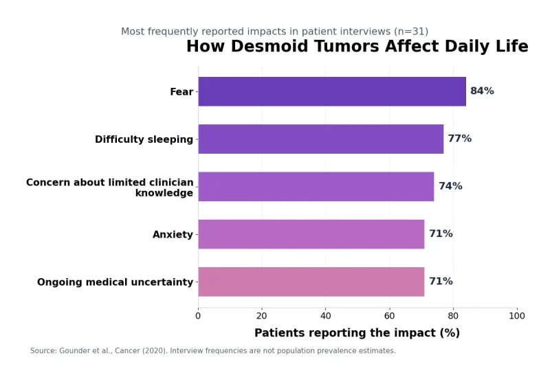
When Specialist Review Is Needed
A newly discovered deep or enlarging mass should be assessed before an unplanned attempt is made to remove it. Surgery performed without appropriate imaging, biopsy planning or pathological diagnosis can create a larger tissue defect, complicate later treatment and expose the patient to functional loss without establishing whether surgery was necessary.
Specialist review is particularly important when the tumor lies near the bowel, ureter, major blood vessel, peripheral nerve, airway or an important joint. In these locations, a relatively small change in growth direction may be more clinically significant than a larger increase in overall diameter.
Patients with a confirmed desmoid tumor should seek specialist reassessment when pain becomes progressively harder to control, sleep is repeatedly disrupted or normal activities become difficult. New weakness, numbness, altered walking, reduced grip strength or loss of joint movement may indicate increasing involvement of muscles or nerves.
Persistent nausea, recurrent vomiting, progressive abdominal distension, severe constipation or reduced urine output requires more urgent evaluation. These symptoms may indicate bowel or urinary compression and should not be managed only through dietary changes, laxatives or symptom-relieving medicines. [1,4]
A second opinion may be valuable when the imaging and pathology do not agree, the biopsy sample was limited or major surgery has been proposed. Review of the original tissue slides and imaging files is often more useful than repeating every investigation immediately.
India and Singapore
In India, patients may enter care through a general physician, surgeon, gastroenterologist, orthopaedic specialist or medical oncologist. Once a deep soft tissue tumor is suspected, referral to a tertiary cancer centre or academic hospital with dedicated soft tissue oncology, musculoskeletal oncology, surgical oncology, radiology and specialist pathology can reduce the risk of fragmented decision-making. [14]
The pathology should be reviewed before definitive treatment when the original diagnosis was made from a small sample, an excised lump without preoperative imaging or a report that does not clearly distinguish desmoid-type fibromatosis from another spindle-cell tumor. Immunohistochemistry and CTNNB1 testing may be considered when the diagnosis remains uncertain.
Patients travelling between Indian cities should carry the original pathology slides or tissue blocks when available, digital imaging files rather than reports alone, details of previous surgery and a complete medication record. Repeating scans without access to earlier images can make it difficult to determine whether the tumor is truly progressing.
In Singapore, evaluation may begin through primary care, a hospital specialist or an oncology service. Complex abdominal, pelvic or limb tumors are best reviewed by a multidisciplinary rare-tumor or sarcoma-focused team that can coordinate radiology, pathology, surgery, medical oncology and supportive care. [15]
Singapore-based patients receiving treatment in another country should maintain one coordinating specialist locally. This clinician can arrange laboratory monitoring, review adverse effects, respond to urgent symptoms and ensure that recommendations from different teams do not conflict.
Patients travelling to India or Singapore for a second opinion should clarify whether the consultation will include formal pathology review, radiology comparison and a multidisciplinary recommendation. A consultation based only on written reports may not answer important questions about biopsy accuracy, anatomical risk or previous tumor growth.
United States and Canada
In the United States, patients with a confirmed or suspected desmoid tumor may benefit from referral to a high-volume academic centre or specialist soft tissue tumor program. The team may include a medical oncologist familiar with desmoid therapies, a surgeon experienced in the affected anatomical region, a radiologist, a soft tissue pathologist, a genetic counsellor and rehabilitation specialists. [1,4]
Insurance requirements may influence which centres are accessible, whether prior authorization is needed and where imaging or treatment can be performed. These administrative issues should be addressed early, but they should not delay emergency assessment for bowel obstruction, severe neurological deterioration, urinary blockage or breathing difficulty.
Before seeking an out-of-network opinion, patients can request electronic copies of MRI or CT images, pathology material and molecular results. A specialist may be able to review these records before recommending whether an in-person examination, repeat biopsy or additional imaging is necessary.
In Canada, access commonly begins through a family physician, emergency department or hospital specialist, followed by referral into a provincial cancer or academic soft tissue tumor program. Provincial pathways differ, but desmoid management should involve a multidisciplinary team with relevant sarcoma and connective-tissue tumor experience. [13]
Canadian patients who live far from a specialist centre may receive imaging and routine blood tests closer to home while major treatment decisions are reviewed centrally. The local and specialist teams should agree on which clinician is responsible for ordering scans, reviewing results and responding to new symptoms.
A patient moving between provinces or changing hospitals should obtain a complete transfer package. This should include the pathology diagnosis, molecular findings, imaging files, measurements from previous scans, details of systemic treatment, surgery records and documented adverse effects.
United Kingdom and Australia
In the United Kingdom, a persistent or suspicious soft tissue mass may be assessed through primary care and referred to a regional specialist service. Once desmoid-type fibromatosis is diagnosed or strongly suspected, management should involve a team able to review imaging, pathology and treatment options collectively. [11]
Patients should ask whether the original biopsy and imaging have been reviewed by the regional multidisciplinary team before agreeing to surgery. This is especially important when an operation could remove abdominal muscle, affect a major nerve or require complex reconstruction.
A general hospital may continue to provide blood tests, pain care and some follow-up imaging, while the specialist centre directs tumor-specific management. Patients should know whom to contact if symptoms worsen between scheduled appointments.
In Australia, a suspicious deep soft tissue mass should be referred to a specialist multidisciplinary service before biopsy or surgery whenever clinically feasible. This approach helps ensure that the biopsy route, imaging and possible future operation are planned together. [12]
Patients living in regional or remote areas may need shared care between a metropolitan specialist centre and local clinicians. Telehealth can support follow-up discussions, but new weakness, severe abdominal symptoms or a rapidly changing mass may still require direct examination and urgent imaging.
Travel distance should be considered when surveillance intervals are planned. The patient should receive clear instructions about which symptoms justify local emergency care, which changes require early contact with the specialist team and which findings can safely wait until the next scheduled review.
What the Specialist Team Should Review
The specialist team should confirm that the pathological diagnosis is secure before selecting treatment. This may require review of tissue morphology, beta-catenin immunohistochemistry, CTNNB1 testing and the possibility of APC-related familial adenomatous polyposis when the clinical history is suggestive. [1,2,4]
Imaging review should determine the tumor’s full anatomical extent rather than reporting only its largest measurement. The team should assess whether the lesion is approaching a nerve, blood vessel, bowel segment, ureter, joint or another structure that could become difficult to protect.
Previous scans should be compared directly using the original images. Reports created at different centres may use different measurement techniques, making apparent changes unreliable unless the radiologist reviews the complete imaging sequence.
The treatment discussion should separate radiological progression from symptom burden. A stable tumor may still require treatment when pain or disability is severe, while a slowly enlarging tumor may remain suitable for surveillance when symptoms are minimal and no vulnerable structure is threatened.
For patients who may receive systemic therapy, the team should address baseline blood tests, cardiovascular and metabolic health, fertility, pregnancy planning, contraception and medication interactions. The monitoring schedule should specify which adverse effects require dose adjustment, temporary interruption or urgent review.
Rehabilitation should be included when pain, surgery or prolonged inactivity has reduced movement and strength. Physiotherapy must be adapted to the tumor’s anatomical location so that function is preserved without forceful loading of painful or compromised tissue.
Patients using Ayurvedic formulations or other complementary treatments should provide the complete ingredient list, dose and manufacturing details. The specialist and Ayurvedic physicians should agree on laboratory monitoring and distinguish improvements in pain, sleep or digestion from objective tumor response on imaging.
A written care plan should identify the confirmed diagnosis, current management strategy, imaging interval, symptom-monitoring priorities and emergency warning signs. It should also state which clinician is responsible for reviewing results and coordinating decisions when several hospitals or countries are involved.
Preparing to Discuss Desmoid Tumor Symptoms

Preparing to discuss desmoid tumor symptoms helps the specialist understand not only where the tumor is located, but also how it is affecting pain, movement, sleep, digestion and daily independence. A well-organized consultation can reduce repeated investigations, improve comparison with previous scans and clarify whether continued surveillance or active treatment is appropriate. [1,2,4–6]
Bring Complete Medical Records and Imaging
The pathology report is one of the most important records because treatment depends on confirming desmoid-type fibromatosis rather than another soft tissue tumor. The report should include the biopsy method, microscopic diagnosis, immunohistochemistry and any available CTNNB1 or APC-related testing.
Patients seeking a second opinion should ask whether the reviewing centre requires the original pathology slides, paraffin tissue block or unstained tissue sections. A written report alone may not be sufficient when the diagnosis is uncertain, the biopsy sample was small or the proposed treatment could cause significant functional loss.
The original CT or MRI images are more useful than printed reports alone. Radiologists need access to the complete imaging series to evaluate the tumor’s boundaries, internal characteristics and relationship with muscles, nerves, bowel, ureters and blood vessels.
Previous scans should be arranged chronologically with the examination dates clearly recorded. Direct comparison can show whether the tumor is stable, regressing or growing toward a vulnerable structure. Differences between written measurements from separate hospitals may be unreliable when image angles and measurement methods are not identical.
Ultrasound images and reports should also be included when the tumor was first identified through examination of a superficial lump. These records can help establish when the abnormality became visible and whether its apparent depth or composition changed before MRI or CT was performed.
Operative notes are important for patients who have undergone previous surgery. They can describe the tissues removed, surgical margins, reconstruction method, mesh placement and whether the tumor involved nearby nerves, muscles, bowel or blood vessels.
A complete medication record should include prescription medicines, non-prescription pain relievers, hormonal medicines, anticoagulants, supplements and Ayurvedic formulations. The exact dose, frequency and treatment start date help the specialist identify possible adverse effects, interactions and reasons for changes in symptoms.
Patients receiving Ayurvedic care should bring the complete ingredient list rather than only the product name. This is particularly important for individualized Avaleha preparations because the concentration, sweetening base, added herbs and dose may differ between prescriptions.
Recent blood results may be required when systemic treatment is being considered or when the patient has vomiting, reduced food intake, urinary obstruction or treatment-related adverse effects. Relevant tests can include blood count, kidney function, liver function and electrolytes, depending on the clinical situation.
A concise treatment history should state when surveillance, surgery, systemic therapy, radiotherapy, local procedures, rehabilitation or complementary treatment began and ended. The reason for stopping each treatment should also be recorded, particularly when the cause was progression, intolerance, pregnancy planning or lack of benefit.
Record Symptoms and Functional Changes
A symptom diary should begin before the consultation whenever possible. Recording symptoms over several days provides a more reliable picture than trying to remember isolated episodes during the appointment.
Pain should be documented by location, intensity, character, duration and trigger. Descriptions such as aching, burning, pressure, pulling, stabbing or electric pain can help distinguish muscular discomfort from possible nerve involvement.
The effect of pain on function is often more clinically useful than the pain score alone. Patients should record whether discomfort interferes with walking, dressing, lifting, driving, sleeping, working, exercising or caring for themselves.
Night-time symptoms deserve specific documentation. The patient should note whether pain prevents sleep, causes repeated waking or makes it impossible to lie on the affected side. Sleep disruption can indicate significant symptom burden even when daytime activity remains possible. [5,6]
For abdominal tumors, the diary should include appetite, early fullness, nausea, vomiting, abdominal distension, bowel frequency and the ability to pass stool and gas. Recording whether symptoms worsen after meals can help determine whether bowel compression or impaired gastrointestinal movement requires further investigation.
Urinary frequency, urgency, difficulty passing urine, flank pain and changes in urine volume should be reported when the tumor is located in the abdomen or pelvis. These symptoms may indicate bladder or ureteric compression and can require kidney function testing or additional imaging.
Patients with limb, shoulder or pelvic tumors should document weakness, numbness, tingling, altered grip, repeated tripping or reduced joint movement. Progressive neurological changes can affect treatment urgency even when the external lump appears stable.
Body weight can be recorded weekly when appetite, early satiety or vomiting is present. Unintentional weight loss may indicate inadequate nutritional intake, persistent gastrointestinal symptoms or another medical problem that requires separate assessment.
Patients may photograph a visible superficial swelling under consistent lighting and body position, but photographs cannot establish whether a tumor is growing internally. Deep tumors and abdominal wall lesions should be assessed through clinical examination and appropriate imaging rather than home measurement alone.
The dates of sudden changes should be recorded accurately. A rapid increase in pain after biopsy, surgery or a new medicine may represent inflammation, bleeding or treatment toxicity rather than immediate tumor progression.
Any emergency episode should be documented, including hospital attendance for vomiting, bowel obstruction, urinary blockage, severe weakness or breathing difficulty. These events can substantially change the risk assessment and future treatment plan.
Prepare Questions About Diagnosis and Treatment
The consultation should clarify whether the diagnosis has been confirmed by a specialist soft tissue pathologist. Patients can ask whether the microscopic findings, beta-catenin staining, molecular results and imaging appearance are fully consistent with desmoid-type fibromatosis. [1,2,4]
The specialist should explain where the tumor begins and which structures it touches, displaces or surrounds. Knowing whether the tumor is close to a nerve, bowel segment, ureter, blood vessel, joint or airway helps patients understand why treatment recommendations may differ from those given to another person with a similar-sized tumor.
Patients should ask whether the tumor is stable, regressing or progressing across serial imaging. The answer should be based on direct comparison of the original images rather than written measurements alone.
When active surveillance is recommended, the patient should understand the imaging interval, preferred scan type and symptoms that justify earlier reassessment. The plan should identify who will review the scan and how the patient will receive the result.
The threshold for starting treatment should be defined clearly. Relevant triggers may include sustained radiological growth, worsening pain, loss of movement, nerve dysfunction, bowel or urinary compression and unacceptable impairment of daily life. [3–6]
When systemic treatment is proposed, the discussion should cover the expected likelihood and timing of symptom improvement, how response will be measured and which adverse effects require laboratory monitoring or dose adjustment.
Fertility, pregnancy planning and contraception should be discussed before starting a medicine that may affect reproductive function or fetal development. These questions are particularly important for younger adults who may remain on treatment for an extended period.
When surgery is being considered, patients should ask what muscle, fascia, nerve, bowel or other tissue may need to be removed. The likely need for mesh, flap reconstruction, rehabilitation and time away from work should be discussed before consent.
The specialist should explain why surgery is preferred over surveillance or systemic therapy in the individual case. A tumor that can technically be removed may still carry an unacceptable risk of long-term weakness, pain or organ dysfunction.
Patients considering complementary care should ask which symptoms can be supported safely and whether any ingredients could interact with systemic therapy, analgesics, anticoagulants or other medicines. Symptom improvement should be monitored separately from tumor response on MRI or CT.
The emergency plan should be clear before the consultation ends. Patients with intra-abdominal disease should know how to respond to repeated vomiting, severe distension or inability to pass stool or gas. Those with tumors near nerves, the neck or chest should understand the urgency of progressive weakness, loss of bladder or bowel control, difficulty swallowing or breathing.
The final treatment plan should identify the confirmed diagnosis, present disease status, immediate clinical priorities, follow-up interval and clinician responsible for coordination. This is especially important when pathology, imaging, oncology, surgery, rehabilitation and Ayurvedic care are provided by different teams or in different countries.
Desmoid Tumor Symptoms FAQs

Desmoid tumor symptoms can include a persistent lump, deep pain, restricted movement, abdominal pressure, bowel changes or nerve-related symptoms. The pattern depends on the tumor’s location and the structures it affects, so diagnosis requires imaging and biopsy rather than symptoms alone.
What Are the First Symptoms of a Desmoid Tumor?
The first sign is often a firm, slowly enlarging lump in the abdominal wall, shoulder, arm, thigh or another soft tissue area. Deep tumors may instead cause persistent pain, pressure, restricted movement, bloating or altered bowel habits without a visible mass.
What Does a Desmoid Tumor Lump Feel Like?
A desmoid tumor often feels firm, deep and less movable than a superficial fatty lump. Some tumors feel like muscular thickening rather than a clearly rounded mass. Texture alone cannot confirm the diagnosis because scars, hernias, lipomas and other tumors may feel similar.
Does a Desmoid Tumor Always Cause Pain?
No. Some desmoid tumors remain painless, especially when small or located away from nerves and joints. Others cause aching, pressure, pulling, burning pain or tenderness when they involve muscles, fascia or nearby nerves.
Can a Desmoid Tumor Cause Severe Abdominal Pain?
Yes. An abdominal desmoid tumor can cause severe pain if it compresses the bowel, ureter, blood vessels or nerves. Severe pain with vomiting, abdominal swelling or inability to pass stool or gas requires urgent medical assessment.
Can a Desmoid Tumor Cause Bloating and Constipation?
An intra-abdominal desmoid tumor may cause bloating, constipation, early fullness, nausea or cramping by pressing on the intestine. Progressive distension, repeated vomiting or inability to pass stool and gas may indicate bowel obstruction.
Can a Desmoid Tumor Cause Back or Nerve Pain?
Yes. Tumors near the back, pelvis, shoulder or limbs may irritate nerves and cause burning pain, tingling, numbness, weakness or radiating discomfort. Progressive weakness, loss of coordination or difficulty walking requires prompt reassessment.
Can a Desmoid Tumor Develop Without a Visible Lump?
Yes. Deep tumors in the abdomen, pelvis, chest or muscles may not produce a visible mass. They may first cause pain, pressure, digestive symptoms, urinary changes, weakness or restricted movement, or they may be discovered incidentally on imaging.
Do Desmoid Tumor Symptoms Always Get Worse?
No. Symptoms may remain stable, improve or fluctuate over time. Some desmoid tumors stop growing or regress without immediate treatment, while others progress. Scheduled imaging is still necessary because symptom improvement does not always mean the tumor has shrunk.
Is a Desmoid Tumor Cancerous?
A desmoid tumor does not spread to distant organs, but it can grow into surrounding tissues and cause serious local complications. Its risk depends on whether it affects the bowel, urinary tract, nerves, blood vessels, airway or other important structures.
Can Desmoid Tumor Symptoms Improve Without Surgery?
Yes. Symptoms may improve during active surveillance, systemic treatment, pain management or rehabilitation. Surgery is not required for every patient, particularly when the tumor is stable or removal would cause substantial functional damage.
How Are Desmoid Tumors Diagnosed?
Diagnosis usually combines clinical examination, imaging and a core needle biopsy. MRI is commonly used for limb and abdominal wall tumors, while CT is often used for intra-abdominal disease. A specialist pathologist confirms desmoid-type fibromatosis from the tissue sample.
When Should Desmoid Tumor Symptoms Be Treated as an Emergency?
Repeated vomiting, severe abdominal distension, inability to pass stool or gas, difficulty breathing, markedly reduced urine output, rapidly worsening weakness or loss of bladder or bowel control requires immediate emergency assessment.
References
[1] National Cancer Institute. (2019, February 27). Desmoid tumor. https://www.cancer.gov/pediatric-adult-rare-tumor/rare-tumors/rare-soft-tissue-tumors/desmoid-tumor
Brief: Provides an overview of desmoid tumors, including their characteristics, clinical presentation, diagnosis, treatment options, and prognosis.
[2] Desmoid Tumor Research Foundation. (n.d.). Diagnosis. Retrieved October 23, 2026, from https://dtrf.org/about-desmoid-tumors/diagnosis/
Brief: Describes diagnostic procedures for desmoid tumors, including clinical evaluation, magnetic resonance imaging (MRI), biopsy, and histopathological examination.
[3] Desmoid Tumor Research Foundation. (n.d.). Treatment & monitoring. Retrieved October 23, 2026, from https://dtrf.org/about-desmoid-tumors/treatment-monitoring/
Brief: Discusses current management approaches for desmoid tumors, emphasizing active surveillance, systemic therapies, selected surgical interventions, and regular monitoring.
[4] Mangla, A., Agarwal, N., & Schwartz, G. (2024). Desmoid tumors: Current perspective and treatment. Current Treatment Options in Oncology, 25(2), 161–175. https://link.springer.com/article/10.1007/s11864-024-01177-5
Brief: Reviews contemporary therapeutic strategies for desmoid tumors, including observation, tyrosine kinase inhibitors, gamma-secretase inhibitors, and emerging targeted treatments.
[5] Kasper, B., Gounder, M., Hernandez, L., Baumgarten, C., & Ratan, R. (2024). Capturing patient voice to improve outcomes that matter to patients with desmoid tumor. Cancer Management and Research, 16, 617–628. https://pmc.ncbi.nlm.nih.gov/articles/PMC11166168/
Brief: Highlights the importance of patient-reported outcomes in assessing symptoms, functional limitations, emotional well-being, and quality of life among patients with desmoid tumors.
[6] Schut, A.-R. W., de Bruin, L. E., de Rooij, B. H., Lidington, E., Timbergen, M. J. M., van der Graaf, W. T. A., van Houdt, W. J., Bonenkamp, J. J., Jones, R. L., Grünhagen, D. J., Sleijfer, S., Gennatas, S., Verhoef, C., & Husson, O. (2023). Physical symptom burden in patients with desmoid-type fibromatosis and its impact on health-related quality of life and healthcare use. Cancer Medicine, 12(12), 13661–13674. https://onlinelibrary.wiley.com/doi/full/10.1002/cam4.5985
Brief: Investigates the physical symptom burden experienced by patients with desmoid-type fibromatosis and its association with reduced health-related quality of life and increased healthcare utilization.
[7] Kumar, N., Patra, S. R., Nishi, & Arya, A. P. (2024). Aggressive fibromatosis in the abdominal wall: A rare case of intramuscular desmoid tumor. Indian Journal of Surgical Oncology, 15(Suppl. 4), 654–658. https://pmc.ncbi.nlm.nih.gov/articles/PMC11846815/
Brief: Presents a rare case of intramuscular desmoid tumor involving the abdominal wall, discussing its clinical presentation, diagnostic findings, surgical treatment, and potential for local recurrence.
[8] PDQ Adult Treatment Editorial Board. (2024, December 13). Gastrointestinal stromal tumors treatment (PDQ®)—Health professional version. National Cancer Institute. https://www.cancer.gov/types/soft-tissue-sarcoma/hp/gist-treatment-pdq
Brief: Provides evidence-based information on the diagnosis and management of gastrointestinal stromal tumors (GISTs), including surgery and targeted therapy. GISTs are distinct from desmoid tumors, making this primarily a comparative reference.
[9] Bhishagratna, K. L. (Ed. & Trans.). (1911). An English translation of the Sushruta Samhita (Vol. 2, Nidana Sthana, Chapter 11). https://archive.org/details/in.ernet.dli.2015.39322
Brief: Presents classical Ayurvedic descriptions of Granthi (nodular swelling) and Arbuda (tumor-like growth), providing historical perspectives on abnormal tissue growth and its clinical characteristics.
[10] Sharangadhara. (2016). Sharangadhara Samhita of Pandita Sharangadharacharya (B. Tripathi, Ed.; Dipika Hindi commentary; Madhyama Khanda, Chapters 7–8). Chaukhamba Surbharati Prakashan. https://archive.org/details/dLhu_sharangadhara-samhita-of-sharangadhara-acharya-containing-anjananidana-of-agnive
Brief: Describes classical Ayurvedic pharmaceutical preparations, particularly Vataka and Avaleha Kalpana, providing traditional references for the preparation and administration of herbal formulations.
[11] Hayes, A. J., Nixon, I. F., Strauss, D. C., Seddon, B. M., Desai, A., Benson, C., Judson, I. R., & Dangoor, A. (2025). UK guidelines for the management of soft tissue sarcomas. British Journal of Cancer, 132(1), 11–31. https://www.nature.com/articles/s41416-024-02674-y
Brief: Presents updated UK clinical guidelines for multidisciplinary management of soft tissue sarcomas, with specific recommendations for the assessment and management of desmoid fibromatosis.
[12] Cancer Council Australia. (2020). Optimal care pathway for people with sarcoma (bone and soft tissue sarcoma). https://www.cancer.org.au/assets/pdf/ocp/sarcoma-bone-soft-tissue-tumours-january-2020
Brief: Outlines an organized multidisciplinary healthcare pathway for patients with bone and soft tissue sarcomas, addressing diagnosis, treatment planning, supportive care, and follow-up. Desmoid fibromatosis is explicitly excluded from this pathway.
[13] Feng, X., Khan, O., Kurien, E., Mack, L., Przybojewski, S., & de Jong, E. (2025). Clinical practice guideline SAR-004: Desmoid tumours (Version 2). Cancer Care Alberta, Alberta Health Services. https://www.albertahealthservices.ca/assets/info/hp/cancer/if-hp-cancer-guide-sar004-desmoid.pdf
Brief: Provides evidence-informed clinical recommendations for desmoid tumor diagnosis and management, including active surveillance, systemic therapy, surgery, local treatment options, and multidisciplinary decision-making.
[14] National Cancer Grid. (2019). Bone and soft tissue tumors management guidelines 2019. https://www.ncgindia.org/assets/ncg-guidelines-2019/ncg-guidelines-for-bst-2019.pdf
Brief: Presents Indian recommendations for the investigation and management of bone and soft tissue tumors, emphasizing diagnostic evaluation, imaging, biopsy, appropriate treatment planning, and multidisciplinary care.
[15] Lim, H. J., Li, Y. X., Chee, M., Tan, J. W. S., Ng, G., Chong, C. Y. L., Seo, C. J., Wong, J. S. M., Abdul, M. R., Soo, K. C., Chia, C. S., & Ong, C.-A. J. (2023). Streamlining multidisciplinary care in sarcoma management. Annals of the Academy of Medicine, Singapore, 52(6), 333–335. https://annals.edu.sg/streamlining-multidisciplinary-care-in-sarcoma-management/
Brief: Discusses the importance of early specialist referral, coordinated diagnosis, and multidisciplinary teamwork in improving sarcoma management and reducing delays in appropriate treatment.