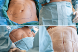




- Why Desmoid Tumor and GIST Must Be Understood Together
- Desmoid Tumor and GIST at a Glance
- The First 72 Hours After a Desmoid Tumor or GIST Diagnosis
- Understanding Desmoid Tumor
- Understanding Gastrointestinal Stromal Tumor
- Pathophysiology of the Desmoid Tumor and GIST Interface
- Clinical Presentation of Desmoid Tumor and GIST
- Common and Rare Disorders Associated With Desmoid Tumor and GIST
- Emergency Red Flags in Desmoid Tumor and GIST That Can Save a Patient’s Life
- Step by Step Diagnostic Roadmap for Desmoid Tumor and GIST
- Diagnostic Overlaps and Differential Diagnosis of Desmoid Tumor and GIST
- Risk Stratification and Prognosis in Desmoid Tumor and GIST
- Desmoid Tumor Treatment
- GIST Treatment
- Real Patient Case Studies in Desmoid Tumor and GIST
- Published Ayurveda Case Study Centred Integrative Model in Advanced GIST
- Treatment Challenges Shared by Desmoid Tumor and GIST
- Financial Planning for Desmoid Tumor and GIST Treatment in the USA & Other Countries
- What Cure Really Means for Desmoid Tumor and GIST Patients
- Ayurvedic Perspective on Desmoid Tumor and GIST
- Avaleha Plus Rasayana as the Clinical Bridge for Root Level Recovery
- Kanchanara Guggulu Avaleha (Medicine)
- Safe Integration of Ayurveda With Desmoid Tumor and GIST Care
- Frequently Asked Questions
- References
Why Desmoid Tumor and GIST Must Be Understood Together
Desmoid tumor and GIST are separate diseases, but patients and caregivers often face similar problems during diagnosis and long term care. Both may appear as deep abdominal masses, both can cause pain or obstruction, and both may require expert radiology, pathology, and multidisciplinary review. Understanding where they overlap and where they differ helps prevent inappropriate surgery, incomplete molecular testing, delayed treatment, and false assumptions about prognosis. [12][41][56] PubMed
Place this section immediately after the article highlights and urgent safety box, before the main introduction.
Published Desmoid Tumor and GIST Cases
A published 2026 case report documented short-term clinical changes in a 57-year-old woman with advanced gastrointestinal stromal tumor and extensive metastatic disease. She received supervised Ayurvedic inpatient care while continuing imatinib and conventional supportive medicines. The case shows how Ayurveda may be integrated to address pain, digestion, abdominal distension, ascites, sleep, weakness, and treatment tolerance without replacing mutation-directed oncology care. [618]
Published Case
| Case detail | Published information |
|---|---|
| Publication | Arbuda With Yakrit Ashrita Roga: An Ayurvedic Case Study on Gastrointestinal Stromal Tumor With Liver Metastasis |
| Year | 2026 |
| Hospital | Jeena Sikho Lifecare Limited Hospital, Andheri, Maharashtra |
| Authors | Acharya Manish, Dr Richa, Dr Sadhana Yadav, and Dr Tanu Rani |
| Patient | An anonymized 57-year-old woman |
| Treatment period | Fourteen days of supervised inpatient care |
Patient Condition
| Clinical area | Patient findings |
|---|---|
| Confirmed condition | Gastrointestinal stromal tumor with extensive peritoneal and omental metastatic disease |
| Abdominal findings | Ascites, abdominal pain, bloating, distension, and multiple metastatic deposits |
| General symptoms | Weakness, poor appetite, breathlessness on exertion, pedal oedema, and disturbed sleep |
| Bleeding symptoms | Intermittent melena with black stools |
| Blood findings | Anaemia was present before treatment and worsened during the observation period |
| Imaging findings | Extensive peritoneal disease and a radiologically suspicious liver lesion |
Treatment Approach
| Treatment component | Published treatment |
|---|---|
| Targeted oncology medicine | Imatinib mesylate 400 mg |
| Conventional supportive medicines | Ondansetron, furosemide, and spironolactone |
| Ayurvedic oral treatment | Individualized tablets, capsules, powders, and syrups selected according to symptoms and clinical assessment |
| Basti treatment | Punarnava Taila and Sahachar Taila Matra Basti |
| External therapies | Abhyanga, Baluka Swedana, abdominal Lepa, Udar Basti, and Patabandhana |
| Diet and lifestyle | Light, digestible food, warm water, selected herbal drinks, moderated activity, and structured daily routine |
The published treatment used several Ayurvedic medicines rather than one single formulation. In clinical practice, compatible herbal actions may be organized into a simpler physician-designed preparation when this is suitable for the patient’s Agni, bowel function, nutritional status, bleeding risk, liver function, kidney function, and ongoing oncology treatment.
Reported Outcome
| Outcome area | Published change |
|---|---|
| Pain | Score improved from 6 to 3 |
| Breathlessness | Score improved from 6 to 1 |
| Sleep | Score improved from 2 to 8 |
| Abdominal girth | Decreased during the inpatient period |
| CA 125 | Decreased from 1,227.20 U/ml to 502.3 U/ml |
| Follow-up imaging | No significant new lesions were reported on short-term CT |
| Disease status | Metastatic disease remained present |
| Haemoglobin | Decreased from 8.9 g/dL to 7.2 g/dL |
| Bleeding | Intermittent melena continued |
| Body weight | Decreased substantially during the observation period |
The findings represent a mixed clinical outcome. Pain, breathlessness, sleep, abdominal girth, and one laboratory marker improved, while anaemia worsened, bleeding continued, body weight declined, and measurable tumor regression was not demonstrated. [618]
Decision Value for Patients and Caregivers
| Clinical decision | Practical value |
|---|---|
| Continue appropriate oncology care | Imatinib remained part of the treatment, showing that Ayurveda was used in an integrative rather than replacement model |
| Monitor more than tumor size | Pain, sleep, appetite, ascites, breathing, bleeding, weight, and laboratory safety all influenced the patient’s condition |
| Do not ignore worsening anaemia | Falling haemoglobin and continued melena require active medical evaluation even when some symptoms improve |
| Simplify the Ayurvedic plan where appropriate | A carefully designed physician-supervised formulation may reduce the burden of taking numerous tablets, powders, capsules, and syrups |
| Keep potent medicines separately adjustable | Mineral or highly potent medicines are safer when prescribed separately in measured doses that can be changed or stopped quickly |
| Separate health improvement from tumor response | Better sleep, pain, appetite, or strength does not by itself prove tumor shrinkage |
| Use objective monitoring | Blood counts, liver function, kidney function, body weight, symptoms, and imaging should guide continuation or modification of treatment |
Possible Benefits to Assess Within 30 Days
| First month assessment | Realistic benefit to monitor |
|---|---|
| Appetite and digestion | Improved meal tolerance, reduced post-meal discomfort, and more consistent food intake |
| Bowel function | More comfortable and regular bowel movements when obstruction is absent |
| Pain and abdominal discomfort | Possible reduction in pain, bloating, or abdominal pressure |
| Sleep | Improved sleep duration and reduced disturbance from pain or anxiety |
| Energy and mobility | Better stamina, walking capacity, and ability to perform daily activities |
| Treatment tolerance | Improved ability to continue imatinib or another prescribed oncology treatment |
| Medicine adherence | Easier daily use when the Ayurvedic regimen is simplified and clearly scheduled |
| Safety | Confirmation that the formulation is not worsening bleeding, diarrhoea, liver function, kidney function, glucose, or blood counts |
| Tumor response | A baseline may be established, but measurable shrinkage or cure should not be promised within 30 days |
The first 30 days are most useful for assessing tolerability, digestive response, symptom change, treatment adherence, nutritional stability, and laboratory safety. Tumor response must be evaluated separately through appropriate imaging and specialist interpretation.
Important Case Study Note
Published case reports are clinical observations. They can show that a particular treatment pathway was used and that certain outcomes occurred, but they cannot prove that the same results will occur in another patient.
Results may differ according to the GIST molecular subtype, stage, metastatic burden, organ function, bleeding status, nutritional reserve, concurrent imatinib, medicine quality, adherence, Ayurvedic assessment, and follow-up duration.
In this case, Ayurveda was provided alongside imatinib, diuretics, antiemetic treatment, external procedures, diet, and lifestyle care. The reported outcome therefore reflects combined integrative management and should not be attributed to one medicine alone. [618]
What Is a Desmoid Tumor
A desmoid tumor, also called desmoid type fibromatosis, is a rare fibroblastic growth arising from connective tissue. It does not metastasize to distant organs, but it can grow into nearby muscles, fascia, nerves, blood vessels, bowel, or urinary structures. Its clinical course is variable. Some tumors remain stable or regress, while others enlarge and cause persistent pain, restricted movement, bowel obstruction, ureteric compression, or loss of organ function. [12] PubMed
Desmoid tumor is therefore better described as nonmetastasizing rather than harmless. The word benign may give families the mistaken impression that no specialist follow up is needed. In reality, the risk depends on the tumor’s location, rate of growth, symptoms, relationship to vital structures, and effect on daily function.
Most sporadic desmoid tumors involve abnormal activation of the WNT beta catenin pathway, commonly through a CTNNB1 alteration. A smaller group is associated with APC related familial adenomatous polyposis. A history of numerous colorectal polyps, early colorectal cancer, or desmoid tumors in close relatives should therefore be discussed with the specialist team. [12] PubMed
What Is Gastrointestinal Stromal Tumor
Gastrointestinal stromal tumor, or GIST, is the most common mesenchymal tumor of the gastrointestinal tract, although it remains uncommon compared with epithelial cancers of the stomach or bowel. It appears to arise from the interstitial cells of Cajal or related precursor cells, which help regulate gastrointestinal movement. Most GISTs develop in the stomach or small intestine, although they may also arise in the duodenum, rectum, colon, oesophagus, omentum, or mesentery. [41][56]
GIST has variable malignant potential. A small gastric tumor with low mitotic activity may behave very differently from a ruptured small intestinal tumor with high mitotic activity. Risk assessment therefore considers the primary site, tumor size, mitotic count, rupture status, stage, and molecular subtype rather than relying on size alone. [41][56] PubMed
Approximately 85 percent of GISTs contain an oncogenic variant in KIT or PDGFRA. Tumors without these variants include distinct groups such as SDH deficient, NF1 associated, BRAF altered, and rare NTRK rearranged GISTs. This molecular diversity explains why one targeted medicine cannot be expected to work equally well for every patient. [56] Cancer.gov
Why Desmoid Tumor and GIST Can Be Confused
The diagnostic overlap is greatest when either tumor develops inside the abdomen. A mesenteric desmoid tumor and an extra gastrointestinal GIST may both appear as soft tissue masses close to bowel loops or abdominal vessels. Both may contain spindle shaped cells on microscopy, and both may cause pain, fullness, obstruction, or an incidental finding on imaging.
Imaging alone may not provide the final diagnosis. The distinction usually requires correlation between tumor location, biopsy morphology, immunohistochemistry, and molecular testing. Nuclear beta catenin supports desmoid tumor in the correct pathological setting. KIT or CD117 and DOG1 support GIST, while SDHB testing may help identify SDH deficient disease. [12][41][56] PubMed
This distinction changes the entire treatment pathway. Many stable desmoid tumors can begin with active surveillance, whereas localized GIST may require surgery followed by recurrence risk assessment. Advanced or unresectable GIST may require mutation directed tyrosine kinase inhibitor therapy. A mistaken diagnosis can therefore expose a patient to an unnecessary operation, an ineffective drug, or delayed molecularly appropriate treatment.
Desmoid tumor and GIST are not different stages of the same disease, and one does not ordinarily transform into the other. Their interface is diagnostic and clinical rather than biological.
How Ayurveda Interprets the Desmoid and GIST Interface
Ayurveda begins with the whole patient rather than the mass alone. The physician evaluates Agni, meaning digestive and metabolic capacity, Dhatu, meaning the body’s structural and functional tissues, Srotas, meaning physiological channels, Bala, meaning functional strength, and Ojas, meaning systemic vitality and resilience.
Classical descriptions of Granthi and Arbuda appear in Sushruta Samhita, Nidana Sthana, Chapter 11. These concepts describe different forms of localized, firm, deep, and persistent tissue growth. They may provide a useful Ayurvedic framework for assessing a patient with a tumor, but they are not exact diagnostic equivalents of desmoid tumor or GIST. Modern pathology, immunohistochemistry, molecular testing, staging, and imaging must remain unchanged. [267] Jain Literature
The Ayurvedic assessment asks why the patient’s digestion, tissue metabolism, elimination, sleep, strength, and recovery capacity have become disturbed. It also examines how previous surgery, chronic medication, poor nutritional intake, pain, stress, bowel dysfunction, and constitutional factors affect the present condition.
This does not mean that every desmoid tumor should automatically be labelled as a Kapha disorder or that every GIST has the same Dosha pattern. Two patients with the same pathology may differ considerably in appetite, bowel function, body strength, treatment toxicity, sleep, pain, and tissue involvement. Ayurvedic treatment must therefore be individualized rather than copied from a fixed tumor formula.
What Patients and Caregivers Need to Understand Early
The first priority is to confirm the diagnosis. The patient should know whether the pathology has been reviewed by a sarcoma experienced pathologist, whether the immunohistochemistry is complete, and whether molecular testing has been performed where clinically relevant.
The second priority is to identify the present treatment goal. The goal may be active surveillance, complete surgical removal, tumor shrinkage, long term control, organ preservation, symptom relief, or prevention of recurrence. These goals are not interchangeable, and the expected benefit of treatment must be weighed against surgical harm, drug toxicity, fertility concerns, financial burden, and effect on daily life.
The third priority is to define how improvement will be measured. Tumor dimensions are important, but they are not the only outcome. Pain, bleeding, appetite, bowel function, weight, mobility, laboratory findings, treatment tolerance, and quality of life must also be monitored.
Ayurveda can be integrated responsibly when its role is clearly defined and coordinated with specialist tumor care. Its practical contribution may include individualized support for digestion, nutrition, bowel function, sleep, strength, recovery, and long term wellbeing. It must not delay emergency treatment, surgery when urgently required, mutation directed GIST therapy, or imaging based assessment.
Desmoid Tumor and GIST at a Glance

Desmoid tumor and GIST are rare stromal conditions that may arise in or around the abdomen, but they differ in biological behaviour, metastatic risk, molecular drivers, and treatment. A desmoid tumor does not spread to distant organs, yet it may infiltrate nearby tissues and cause serious local complications. GIST can recur or metastasize and usually requires risk assessment based on pathology, tumor location, rupture status, and molecular subtype. [12][41][56]
The Essential Difference Between Desmoid Tumor and GIST
A desmoid tumor develops from fibroblastic connective tissue. It may grow through fascia, muscle, mesentery, nerves, blood vessels, bowel, or urinary structures. Its main danger comes from local infiltration, compression, chronic pain, obstruction, and loss of function. It does not usually spread through the bloodstream to distant organs. [12]
GIST develops from the gastrointestinal stromal system, most commonly in the stomach or small intestine. Some GISTs remain localized and can be removed completely, while others recur, spread to the liver or peritoneum, or require long term targeted therapy. Its behaviour cannot be predicted from tumor size alone. [41][56]
Clinical Comparison of Desmoid Tumor and GIST
| Clinical feature | Desmoid tumor | GIST |
|---|---|---|
| Basic classification | Nonmetastasizing fibroblastic tumor | Mesenchymal gastrointestinal tumor with variable malignant potential |
| Common anatomical sites | Abdominal wall, mesentery, pelvis, chest wall, head and neck, extremities | Stomach, small intestine, duodenum, rectum, colon, oesophagus, omentum, mesentery |
| Main molecular pathway | CTNNB1 or APC related WNT beta catenin activation | KIT, PDGFRA, SDH and less common molecular alterations |
| Distant spread | Does not metastasize | May metastasize, particularly to the liver or peritoneum |
| Main clinical danger | Local invasion, organ compression, pain, obstruction, impaired movement | Bleeding, rupture, recurrence, metastasis, drug resistance |
| Common pathology support | Nuclear beta catenin with compatible morphology | KIT or CD117, DOG1 and molecular testing |
| Initial treatment approach | Often active surveillance when clinically safe | Surgery, surveillance, or targeted therapy according to stage and risk |
| Long term concern | Progression, chronic symptoms, local recurrence, functional damage | Recurrence, metastasis, treatment toxicity, acquired resistance |
| Important inherited association | Familial adenomatous polyposis related to APC alterations | SDH related syndromes, NF1, and rare familial KIT or PDGFRA alterations |
Why the Word Benign Can Mislead Desmoid Tumor Patients
Desmoid tumors are often described as benign because they do not metastasize. This description does not reflect the full clinical risk. A mesenteric desmoid tumor may compress or encase bowel loops, vessels, nerves, or ureters. An extremity tumor may restrict movement or cause persistent neuropathic pain. A tumor near a vital structure may become dangerous even without distant spread.
The risk is therefore determined by anatomical location, rate of growth, symptom burden, organ involvement, and functional loss. A small tumor beside a vital structure may require closer attention than a larger tumor in a less dangerous location.
Why GIST Cannot Be Treated as One Uniform Disease
GIST includes several molecular subtypes with different treatment responses. KIT mutated disease may respond differently according to the affected exon. PDGFRA D842V mutated GIST is generally resistant to imatinib. SDH deficient, NF1 associated, BRAF altered, and rare NTRK rearranged tumors may require different diagnostic and therapeutic considerations. [41][56]
The complete diagnosis should therefore include the primary site, tumor size, mitotic activity, rupture status, stage, immunohistochemistry, and molecular findings. A report stating only “GIST positive” may not provide enough information for long term treatment planning.
Why Desmoid Tumor and GIST May Be Confused
The greatest overlap occurs when a tumor develops inside the abdomen. Mesenteric desmoid disease and extra gastrointestinal GIST may both appear as solid soft tissue masses close to bowel loops or major vessels. Both may show spindle shaped cells on biopsy and may produce pain, fullness, constipation, vomiting, obstruction, or an incidental imaging finding.
Imaging alone may not reliably distinguish them. The final diagnosis usually requires correlation between the tumor’s anatomical origin, microscopic appearance, immunohistochemistry, and molecular testing. Nuclear beta catenin supports desmoid tumor in the correct pathological context, while KIT or CD117 and DOG1 support GIST. SDHB testing may be required when SDH deficient GIST is suspected. [12][41][56]
How the Diagnosis Changes Treatment
A stable desmoid tumor may be monitored with structured active surveillance rather than treated immediately. Treatment becomes more appropriate when the tumor grows, causes increasing pain, threatens an organ, restricts function, or significantly reduces quality of life. [12]
Localized GIST may require surgery followed by recurrence risk assessment. High risk or advanced disease may require mutation directed targeted therapy. An incorrect diagnosis can therefore expose a patient to unnecessary surgery, an ineffective drug, or delayed specialist treatment.
Desmoid tumor and GIST are not different stages of one disease. One does not normally transform into the other. Their interface lies in diagnostic appearance, anatomical presentation, pathology, and long term management.
Ayurvedic Understanding of Desmoid Tumor and GIST
Ayurveda evaluates the tumor together with the patient’s digestion, metabolism, tissue condition, strength, bowel function, sleep, pain, nutritional status, and treatment tolerance. Classical concepts such as Granthi and Arbuda may provide a framework for understanding firm, deep, persistent tissue growths, but they should not replace the confirmed modern diagnosis. [267]
A patient with desmoid tumor may show a different Ayurvedic pattern from another patient with the same pathology. One may have marked Vata disturbance with pain, reduced strength, constipation, and weight loss. Another may show greater Kapha and Meda involvement with heaviness, slow growth, poor metabolic activity, and tissue congestion. GIST may similarly involve different combinations of Agni impairment, Rakta and Mamsa disturbance, nutritional decline, bleeding, or treatment related weakness.
The Ayurvedic plan must therefore be individualized according to the patient’s present Dosha, Dhatu, Agni, Ama, Srotas, and Bala. It should remain coordinated with pathology, molecular testing, imaging, surgery, targeted therapy, and emergency care.
The First 72 Hours After a Desmoid Tumor or GIST Diagnosis

The first 72 hours after a desmoid tumor or GIST diagnosis should be used to confirm what has been found, identify any immediate danger, secure the complete medical record, and reach the correct specialist team. A stable patient usually has time for expert review before an irreversible decision is made. A patient with bleeding, obstruction, rupture, infection, or organ compression may need urgent hospital treatment. [12][41][56]
First Determine Whether the Desmoid Tumor or GIST Is Causing an Emergency
The first question is not whether the mass has been called benign or malignant. The immediate question is whether it is threatening the patient’s life or an important organ.
Vomiting blood, black stools with dizziness or weakness, rapidly worsening abdominal pain, repeated vomiting, inability to pass stool or gas, fainting, a rigid or markedly swollen abdomen, fever with severe pain, or sudden reduction in urine output requires urgent medical assessment. These symptoms may indicate gastrointestinal bleeding, complete bowel obstruction, tumor rupture, perforation, infection, or ureteric compression. [12][56]
A caregiver should not wait for the next appointment when these symptoms appear. Avaleha, herbal powders, fasting, laxatives, oil therapy, or Panchakarma must not be used to manage a suspected obstruction, active bleeding, perforation, or acute abdomen. Emergency stabilization must come first.
Secure the Complete Medical Record
Patients often receive only a short summary after imaging or biopsy. That is not enough for expert review. The complete record should include the pathology report, immunohistochemistry results, molecular testing, blood reports, operative notes, discharge summaries, and the original scan images.
The original CT, MRI, or PET images should be obtained in DICOM format rather than relying only on the written radiology report. A specialist radiologist may need to compare the mass with nearby bowel, vessels, nerves, ureters, and other organs.
The original pathology slides or paraffin tissue block should also remain available. A sarcoma pathologist may need to review the tissue before a major operation or long term treatment begins. Rare spindle cell tumors can resemble one another, particularly when the biopsy sample is small or the mass is located in the mesentery or omentum. [12][41]
The patient or caregiver should also prepare one dated treatment history showing previous surgery, medicines, dose changes, supplements, Ayurvedic preparations, adverse effects, and reasons for stopping any treatment. This prevents important information from being lost when care moves between hospitals or countries.
Confirm the Exact Diagnosis Before Accepting a Long Term Plan
A report stating only “abdominal tumor,” “spindle cell tumor,” or “stromal lesion” may not be sufficient. The diagnosis should identify whether the lesion is desmoid type fibromatosis, GIST, or another soft tissue condition.
For a suspected desmoid tumor, the pathologist may assess morphology, nuclear beta catenin, and CTNNB1 status where appropriate. APC related evaluation may be needed when the patient has multiple colorectal polyps, early colorectal cancer, or a family history suggesting familial adenomatous polyposis. [12]
For GIST, the report should include morphology and relevant immunohistochemistry, commonly including KIT or CD117 and DOG1. SDHB assessment may be required in selected patients. Molecular testing should identify the gene, exon, and specific alteration whenever possible because KIT, PDGFRA, SDH deficient, NF1 associated, BRAF altered, and other subtypes may not respond to treatment in the same way. [41][56]
A molecular report stating only “mutation positive” is incomplete for clinical decision making. The treating team should explain the exact alteration and whether it predicts sensitivity or resistance to the proposed medicine.
Request Review by a Sarcoma Specialist Team
Desmoid tumor and GIST should be managed through a team experienced in sarcoma and rare stromal tumors. This usually includes specialist radiology, pathology, surgery, medical oncology, genetics, nursing, nutrition, rehabilitation, and supportive care.
In the United Kingdom, patients should confirm that the case has been referred to the appropriate regional sarcoma service and reviewed through a multidisciplinary team. In the United States, the patient should ask whether the treating centre regularly manages desmoid tumors and GIST and whether expert pathology and molecular testing are available. [83][103]
A second opinion is particularly important when the imaging and pathology do not agree, the diagnosis is based on a very small biopsy, a major organ sacrificing operation has been proposed, molecular testing is incomplete, or the tumor has not behaved as expected.
Seeking a second opinion does not require abandoning the existing doctor. It allows the patient to confirm that diagnosis, timing, and treatment goals are appropriate before an irreversible step is taken.
Do Not Rush Into Irreversible Treatment When the Patient Is Stable
Urgent complications must not be delayed, but a clinically stable patient should not be pressured into immediate surgery or long term medication before the diagnosis and treatment objective are clear.
Many desmoid tumors can begin with active surveillance when they are stable, symptoms are manageable, and no vital structure is threatened. Immediate surgery may create functional damage without improving long term control in every patient. [12]
Localized GIST often requires surgery, but the operation should be planned to avoid tumor rupture and preserve function. In selected mutation sensitive GISTs, preoperative targeted therapy may reduce tumor size and make surgery safer. Starting treatment before molecular testing can be inappropriate when the subtype is resistant to the proposed drug. [41][56]
The patient should ask whether the current recommendation is intended to cure, shrink, stabilize, prevent recurrence, relieve symptoms, or protect an organ. These goals require different treatment strategies and different methods of measuring success.
Clarify What Must Happen Before Treatment Starts
Before surgery, the patient should understand which organs or tissues may need to be removed, whether a stoma may be required, what the risk of tumor rupture is, and what functional limitations may follow.
Before beginning a tyrosine kinase inhibitor, the patient should understand the molecular reason for choosing it, the planned dose, required laboratory tests, important interactions, expected side effects, and the symptoms that require urgent contact.
Before entering surveillance, the patient should receive a written scan schedule, symptom monitoring plan, and clear criteria for starting treatment. “Wait and watch” without defined follow up is not structured surveillance.
Review Every Medicine, Herb, Supplement, and Food Product
The oncology and Ayurvedic teams should receive one complete list of everything the patient takes. This includes prescription medicines, painkillers, antacids, antibiotics, vitamins, herbal powders, teas, juices, Ayurvedic preparations, protein supplements, and over the counter products.
Natural products can affect liver enzymes, drug transporters, bleeding risk, blood pressure, hydration, or the absorption of anticancer medicines. Taking a herb at a different time of day does not automatically prevent an interaction. [134]
A patient should not start a market Avaleha or self prepared tumor formula during the first days after diagnosis. The physician must first understand the pathology, current symptoms, bowel status, liver and kidney function, blood counts, ongoing treatment, and digestive capacity.
Establish Baseline Symptoms and Function
The scan provides only part of the baseline. The patient and caregiver should also record pain location and severity, bowel pattern, appetite, meal capacity, weight, sleep, mobility, urinary symptoms, bleeding, fatigue, and ability to perform normal daily activities.
These observations allow later assessment of whether treatment is improving the patient’s life, even when tumor measurements remain stable. They can also reveal deterioration that may not be obvious from imaging alone.
Body weight, complete blood count, haemoglobin, liver function, renal function, and other clinically relevant tests should be recorded before treatment whenever possible. Future results can then be compared with a clear starting point.
Give the Caregiver a Defined Role
The caregiver should help organize reports, record symptoms, maintain the medicine list, note missed doses, and keep emergency contact details available. The caregiver should not be expected to decide whether a symptom represents progression, but should know which changes require immediate reporting.
New pain, vomiting, black stools, increasing abdominal distension, falling food intake, weight loss, reduced urine output, swelling, jaundice, sudden weakness, or emotional withdrawal should be documented with the date they began.
The patient and caregiver should also decide who will communicate with the hospital, who will carry the medical file, and who will accompany the patient when major decisions are discussed.
Where Ayurveda Fits During the First 72 Hours
Ayurveda should begin with assessment, not immediate prescribing. The Ayurvedic physician should review the confirmed diagnosis, pathology, molecular findings, imaging, organ function, current medicines, nutritional condition, bowel function, and emergency risk before recommending treatment.
The early Ayurvedic assessment may document Agni, meaning digestive and metabolic capacity, Bala, meaning functional strength, Dhatu, meaning the condition of the body’s tissues, and Ojas, meaning systemic vitality and resilience.
During active bleeding, complete obstruction, severe vomiting, dehydration, acute liver injury, kidney impairment, infection, or immediate postoperative instability, intensive oral formulations and Shodhana procedures are generally unsuitable. The patient must first be medically stabilized.
Once the diagnosis and treatment phase are clear, Ayurveda may be introduced in a coordinated manner to support digestion, nutrition, bowel function, sleep, strength, recovery, treatment tolerance, and long term wellbeing. Any effect on the tumor must still be assessed through independent imaging, pathology, laboratory monitoring, and specialist review.
The Decision Every Patient Should Reach by the End of the First 72 Hours
By the end of this initial period, the patient should know the exact working diagnosis, whether an emergency is present, which specialist team is responsible, whether pathology and molecular testing are complete, what the current treatment goal is, and how the response will be measured.
The patient does not need to understand every medical detail immediately. The immediate need is a safe, documented, specialist reviewed plan that separates urgent action from decisions that can wait for complete information.
Understanding Desmoid Tumor

A desmoid tumor is a rare connective tissue growth that does not spread to distant organs but can infiltrate the structures around it. Its behaviour varies considerably. Some tumors remain unchanged for years or gradually regress, while others enlarge, produce persistent pain, compress organs, or reduce movement and independence. This unpredictable natural history is why every desmoid tumor requires an individualized plan rather than automatic surgery or one standard medicine. [12] PubMed
What Is a Desmoid Tumor
A desmoid tumor, also called desmoid type fibromatosis or aggressive fibromatosis, develops from fibroblastic cells that normally help form connective tissue, fascia, ligaments, and scar tissue. The tumor is composed largely of spindle shaped fibroblastic cells and collagen.
It is described as nonmetastasizing because it does not usually travel through the blood or lymphatic system to form distant tumors. This distinguishes it from metastatic sarcoma. However, the tumor may extend into nearby muscles, nerves, bowel loops, blood vessels, or urinary structures through finger like projections that are difficult to separate from normal tissue. [12] PubMed
The term benign may therefore create false reassurance. A desmoid tumor can be clinically serious when its location threatens an organ, causes obstruction, or produces progressive functional loss.
Where Desmoid Tumors Develop
Desmoid tumors may arise almost anywhere connective tissue is present. Common locations include the abdominal wall, mesentery, pelvis, shoulders, upper limbs, lower limbs, chest wall, head, and neck.
An abdominal wall desmoid tumor may present as a firm, slowly enlarging mass. It may be associated with pregnancy, previous abdominal surgery, or scar tissue, although these factors do not explain every case.
An intra abdominal or mesenteric desmoid tumor may develop around bowel loops and major blood vessels. It can remain unnoticed until it causes abdominal fullness, pain, constipation, vomiting, urinary obstruction, or difficulty eating.
An extremity desmoid tumor may restrict joint movement, compress nerves, weaken muscle function, or produce pain during daily activity. Head and neck tumors may be smaller but clinically important because they develop close to the airway, nerves, vessels, and other vital structures.
The significance of a desmoid tumor therefore depends more on its anatomical relationships and behaviour than on its size alone.
Why a Desmoid Tumor Can Be Dangerous Without Metastasis
The main risk from a desmoid tumor is local infiltration. The mass may wrap around or compress structures that cannot be safely removed. Surgery intended to remove the tumor may sometimes require loss of bowel, muscle, nerve, blood vessel, or another organ.
An intra abdominal tumor may cause partial or complete bowel obstruction. Mesenteric disease may compromise blood supply to the bowel or produce fistula, abscess, or perforation in advanced cases. Pelvic disease may compress the ureters and impair kidney drainage.
Pain may arise from tissue stretching, inflammation, nerve compression, muscle involvement, or altered posture. Persistent pain can disturb sleep, reduce movement, weaken appetite, and contribute to anxiety or depression even when imaging shows limited change.
Caregivers should therefore monitor function as closely as tumor size. Increasing difficulty walking, eating, sleeping, passing stool, urinating, or performing normal activity may be clinically important even before the next scan.
CTNNB1 APC and WNT Beta Catenin Biology
Most sporadic desmoid tumors involve abnormal activation of the WNT beta catenin signalling pathway. Many contain an acquired alteration in the CTNNB1 gene, which encodes beta catenin. The altered protein accumulates inside the cell and promotes continued fibroblastic growth.
These CTNNB1 changes are usually somatic. This means they developed within the tumor and are not automatically inherited by the patient’s children.
A smaller group of desmoid tumors is associated with inherited APC gene alterations. APC normally helps regulate beta catenin. Loss of normal APC function can permit excessive WNT signalling and increase the risk of both colorectal polyposis and desmoid disease.
The molecular result can support diagnosis, but it should not be interpreted separately from pathology, immunohistochemistry, imaging, and clinical history. The behaviour of one CTNNB1 altered tumor cannot be predicted with certainty from the mutation alone. [12] PubMed
Desmoid Tumor and Familial Adenomatous Polyposis
Familial adenomatous polyposis is an inherited condition caused by a pathogenic APC variant. Affected individuals may develop numerous colorectal polyps and have a high lifetime risk of colorectal cancer without appropriate surveillance and preventive care.
Desmoid tumors associated with familial adenomatous polyposis commonly arise within the abdomen or mesentery. They may appear after abdominal surgery, although surgery is not the only factor involved.
Genetic assessment should be discussed when a patient has multiple colorectal polyps, colorectal cancer at a young age, several affected relatives, multiple desmoid tumors, or an intra abdominal desmoid tumor without an established explanation.
A sporadic CTNNB1 altered desmoid tumor and an APC associated desmoid tumor do not have identical family implications. This distinction affects colorectal surveillance, genetic counselling, surgical planning, and assessment of relatives.
Why Desmoid Tumor Behaviour Is Unpredictable
A desmoid tumor does not follow a simple pattern of continuous growth. Some enlarge for a period and later stabilize. Some remain unchanged from the time of diagnosis. Others gradually become smaller without active tumor directed treatment.
Spontaneous regression has been documented at different anatomical sites. This is one reason immediate surgery is no longer considered necessary for every newly diagnosed desmoid tumor. [12] PubMed
The opposite pattern is also possible. A previously stable tumor may begin growing or symptoms may worsen without a dramatic increase in its measured diameter. Regular follow up should therefore combine imaging with pain, function, bowel symptoms, urinary symptoms, sleep, and quality of life.
One patient’s course should not be used to predict another patient’s outcome. A dramatic success or recurrence described online may not reflect the biology, location, or treatment context of a different tumor.
Common Desmoid Tumor Symptoms
Symptoms depend mainly on location. A superficial tumor may appear as a firm mass or swelling. It may be painless initially and become uncomfortable as it enlarges or restricts movement.
An abdominal or pelvic tumor may cause pressure, abdominal pain, early satiety, bloating, constipation, nausea, vomiting, urinary symptoms, or unexplained weight loss. Repeated vomiting, inability to pass stool or gas, rapidly increasing pain, or marked abdominal distension requires urgent assessment.
A tumor involving nerves or muscles may cause radiating pain, tingling, weakness, reduced range of movement, altered posture, or difficulty performing normal tasks.
Pain severity does not always correspond directly with tumor size. A relatively small mass beside a nerve may be more symptomatic than a larger tumor in a less sensitive location.
How Desmoid Tumor Is Confirmed
Imaging defines the location, size, internal characteristics, and relationship with nearby structures. MRI is particularly useful for many soft tissue locations and for comparing internal signal changes over time. CT is often useful for intra abdominal anatomy and complications.
Imaging cannot always establish the final diagnosis. A planned core biopsy is usually required when pathology is uncertain or before systemic treatment begins. The biopsy route should be selected by the specialist team so that it does not complicate later surgery or damage important structures.
Pathology commonly shows long fascicles of bland spindle shaped cells within a collagenous background. Nuclear beta catenin staining supports desmoid tumor, but it is not completely specific. CTNNB1 testing may strengthen the diagnosis, particularly when the morphology or staining pattern is uncertain.
Expert pathology review is important when the biopsy is small, beta catenin staining is unclear, the mass is intra abdominal, or GIST and another spindle cell tumor remain possible.
Why Active Surveillance May Be the First Choice
Active surveillance is often appropriate when the desmoid tumor is newly diagnosed, symptoms are mild or manageable, imaging does not show immediate organ danger, and function remains preserved.
This strategy includes planned imaging, regular symptom review, and defined reasons for starting treatment. It does not mean that the tumor is ignored.
Treatment may become necessary when serial imaging confirms clinically meaningful progression, pain becomes difficult to control, mobility declines, nutritional intake is impaired, or the tumor threatens bowel, nerves, vessels, ureters, or another essential structure.
A decision to observe can protect the patient from unnecessary surgery and treatment toxicity. A decision to treat can protect organ function when the disease is clearly active. Both choices can be appropriate when based on specialist review and the patient’s actual condition. [12] PubMed
Ayurvedic Assessment of Desmoid Tumor
Ayurveda evaluates the desmoid tumor together with the patient’s digestion, metabolism, tissue condition, pain, bowel function, sleep, strength, nutritional status, and response to previous treatment.
Agni is the digestive and metabolic capacity responsible for transforming food into usable nourishment.
Ama refers to incompletely processed metabolic material within the Ayurvedic framework.
Mamsa Dhatu represents muscle and structural soft tissue.
Meda Dhatu represents adipose and lipid associated tissue.
Rakta Dhatu represents the blood tissue system.
Bala is the patient’s functional strength, while Ojas represents systemic vitality and resilience.
A firm, deep, persistent connective tissue growth may be assessed through classical principles related to Granthi and Arbuda. These are interpretive frameworks rather than exact equivalents of a pathology confirmed desmoid tumor.
The Ayurvedic physician should not assume that every patient has the same Vata and Kapha pattern. A painful tumor with constipation, sleep disturbance, weight loss, and weakness may require a different approach from a slowly growing mass in a patient with heaviness, poor appetite, metabolic sluggishness, and tissue congestion.
The assessment should also account for active surveillance, systemic therapy, recent surgery, liver and kidney function, blood counts, fertility plans, diabetes, nutritional status, and the risk of bowel or urinary obstruction.
The Safe Role of Ayurveda in Desmoid Tumor Care
Ayurvedic treatment may support digestion, appetite, bowel regularity, sleep, strength, mobility, recovery, and tolerance of long term care when it is individually prescribed and medically monitored.
The role of Avaleha, Rasayana, Shamana, or carefully selected external therapy depends on the disease phase. A patient with stable disease and adequate digestive capacity may tolerate a different plan from a patient with active obstruction, repeated vomiting, severe weakness, or recent surgery.
Shodhana procedures should not be used during active bleeding, suspected obstruction, dehydration, infection, acute organ dysfunction, or significant treatment toxicity.
Ayurvedic improvement should be measured honestly. Better appetite, reduced pain, improved bowel function, stronger sleep, stable weight, and increased mobility are health outcomes. Reduction or stabilization of the tumor must be established separately through specialist imaging and radiological interpretation.
Ayurveda should not delay active surveillance imaging, urgent decompression, necessary surgery, or systemic treatment when organ function is threatened.
What Patients and Caregivers Should Track
A useful record should show the date of every scan, three dimensional tumor measurements, the radiologist’s conclusion, pain pattern, bowel and urinary changes, body weight, appetite, sleep, mobility, analgesic use, and treatment tolerance.
The record should also include the exact dose and dates of every oncology medicine, Ayurvedic formulation, supplement, interruption, and adverse effect.
Changes should be reported before the planned appointment when the patient develops increasing pain, repeated vomiting, black stools, abdominal distension, inability to pass stool or gas, reduced urine output, new weakness, jaundice, fever, or rapid decline in normal activity.
This combined record allows the treating team to judge the desmoid tumor through imaging, symptoms, function, and safety rather than relying on one measurement alone.
Understanding Gastrointestinal Stromal Tumor

Gastrointestinal stromal tumor, commonly called GIST, is a rare mesenchymal tumor that develops in or near the digestive tract. Its behaviour ranges from a very small, slow growing lesion to an aggressive tumor that recurs or spreads. The treatment plan depends on the primary site, tumor size, mitotic activity, rupture status, stage, and molecular subtype rather than on the name GIST alone. [41][56]
What Is Gastrointestinal Stromal Tumor
GIST arises from the gastrointestinal stromal system and is thought to originate from the interstitial cells of Cajal or closely related precursor cells. These cells help coordinate the muscular contractions that move food through the gastrointestinal tract.
GIST is different from the more common epithelial cancers of the stomach, small intestine, or colon. It develops within the deeper muscular layers of the gastrointestinal wall and often grows outward into the abdominal cavity rather than forming an obvious surface lesion inside the bowel.
Microscopically, most GISTs contain spindle shaped cells. A smaller proportion contain epithelioid cells, while some show a mixed pattern. This appearance can overlap with other soft tissue tumors, which is why morphology must be interpreted together with immunohistochemistry and molecular testing. [56][187]
Where Gastrointestinal Stromal Tumor Develops
GIST can arise anywhere along the gastrointestinal tract, but approximately 60 percent develop in the stomach and about 30 percent arise in the jejunum or ileum of the small intestine. Less common sites include the duodenum, rectum, colon, and oesophagus. Rare tumors may appear in the omentum or mesentery without a clearly identifiable primary tumor in the bowel wall. [56]
The location affects symptoms, surgical planning, recurrence risk, and expected drug response. A gastric GIST may cause early fullness, bleeding, or anaemia. A small intestinal GIST may present with pain, obstruction, rupture, or internal bleeding. A rectal GIST may produce pelvic pressure, constipation, or difficulty passing stool.
Small GISTs may remain clinically silent and are sometimes discovered during imaging, endoscopy, or surgery performed for another reason. The discovery of a small lesion does not automatically mean that it will behave aggressively, but it still requires expert assessment because risk cannot be determined from size alone.
How GIST Behaves
GIST has variable malignant potential. Some localized tumors can be removed completely and never recur. Others return after surgery, spread to the liver or peritoneum, or require long term targeted therapy.
The liver and the peritoneal surfaces within the abdomen are the most common sites of metastatic spread. Lymph node involvement is uncommon in typical adult GIST, although it can occur more often in certain pediatric and SDH deficient forms. Lung and other extra abdominal metastases are less common than liver or peritoneal disease. [56]
A large tumor is not automatically aggressive, and a small tumor is not automatically harmless. Biological behaviour depends on the combined effect of anatomical site, mitotic rate, rupture status, molecular subtype, and stage.
Tumor rupture is particularly important because it can release tumor cells into the abdominal cavity and substantially increase recurrence risk. The operative note and pathology report should therefore state clearly whether rupture occurred before or during surgery.
Common GIST Symptoms
Gastrointestinal bleeding is one of the most common presentations of GIST. Bleeding may be sudden and visible as vomiting blood or black stools, or it may occur slowly and produce chronic anaemia. Patients may then experience fatigue, paleness, breathlessness, dizziness, rapid heartbeat, or reduced exercise tolerance. [56]
Other symptoms include abdominal pain, early satiety, nausea, reduced appetite, weight loss, difficulty swallowing, constipation, bowel obstruction, or a palpable abdominal mass. Symptoms depend strongly on the tumor’s site and size.
A tumor growing outward from the bowel wall may become large before it narrows the digestive passage. This is why some patients have few early symptoms despite having a substantial abdominal mass.
Sudden severe abdominal pain, collapse, fainting, low blood pressure, vomiting blood, black stools with weakness, or a rigid abdomen may indicate rupture, internal haemorrhage, perforation, or another emergency. These symptoms require immediate hospital assessment.
Why GIST Is a Molecular Disease
Approximately 85 percent of GISTs contain an oncogenic alteration in KIT or PDGFRA. These genes encode receptor tyrosine kinases that regulate cell growth and survival. When they become continuously activated, stromal cells can multiply without normal control. [56]
KIT alterations are the most common. They may occur in different exons, and the exact exon can influence tumor location, biological behaviour, and sensitivity to targeted treatment.
PDGFRA altered GISTs occur less frequently and often develop in the stomach. The PDGFRA D842V alteration is clinically important because it is generally resistant to imatinib. A patient should therefore know the exact mutation rather than being told only that the tumor is KIT positive or mutation positive. [56][117]
Clinical research has shown that imatinib response differs according to KIT and PDGFRA mutation status. Molecular testing is therefore part of treatment selection and not merely an optional research investigation. [117]
KIT and PDGFRA Wild Type GIST
A GIST without a detectable KIT or PDGFRA alteration is sometimes described as wild type. This term does not identify one uniform disease. It includes several biologically distinct subgroups that require further investigation.
Many wild type tumors are SDH deficient or associated with neurofibromatosis type 1. Other less common alterations may involve BRAF, NTRK, or additional molecular pathways. [56]
SDH deficient GIST commonly affects younger patients, often arises in the stomach, and may be multifocal. It can involve lymph nodes and may show a slower clinical course despite metastatic disease. These tumors are generally less responsive to standard tyrosine kinase inhibitors than common KIT altered GIST.
Loss of SDHB staining on immunohistochemistry supports the diagnosis of SDH deficient disease. Further genetic or epigenetic assessment may then be required.
Hereditary and Syndromic GIST
Most GISTs are sporadic, which means they arise without an inherited family syndrome. A small proportion are associated with hereditary conditions that may affect the patient and relatives.
Neurofibromatosis type 1 associated GIST often develops as multiple tumors in the small intestine. The patient may also have café au lait macules, neurofibromas, or a known family history of NF1.
Carney Stratakis syndrome involves inherited SDH related predisposition to GIST and paraganglioma. Carney triad classically involves gastric GIST, paraganglioma, and pulmonary chondroma, although not every patient develops all three conditions.
Rare families carry inherited KIT or PDGFRA alterations and may develop multiple GISTs. A young age at diagnosis, multiple tumors, unusual molecular findings, paraganglioma, pulmonary chondroma, or a family history of related tumors should prompt genetic counselling. [56]
How GIST Recurrence Risk Is Assessed
The recurrence risk after removal of localized GIST is assessed through several connected factors. These include the primary anatomical site, maximum tumor size, mitotic count, rupture status, surgical findings, margins, and molecular subtype. [41][56][187]
Mitotic count estimates how actively tumor cells are dividing. The pathology report should state how the count was measured because reporting methods may vary.
Tumor site is also important. A nongastric GIST may carry a higher risk than a gastric GIST of similar size and mitotic activity. This is why a universal size based classification can mislead patients.
A complete report should not use broad labels such as low grade or high grade without explaining the underlying measurements. The patient should be told the exact risk category and which features produced that assessment.
How GIST Is Diagnosed
Contrast enhanced CT is commonly used to define the primary tumor, assess liver or peritoneal spread, and plan treatment. MRI may be useful for selected anatomical sites, such as the rectum or liver, or when CT is unsuitable.
Endoscopy can identify a submucosal mass, ulceration, or bleeding. Endoscopic ultrasound may help determine the wall layer from which the tumor arises and may guide biopsy in selected upper gastrointestinal lesions.
Biopsy should be planned by the specialist team. Some apparently resectable tumors with characteristic imaging may proceed directly to surgery, while other lesions require tissue confirmation before treatment. The biopsy route should minimize the risks of bleeding, rupture, and peritoneal contamination. [41][56]
Pathology commonly includes KIT or CD117 and DOG1 immunohistochemistry. CD117 positivity supports GIST but is not completely specific, and a minority of GISTs may be negative or weakly positive. DOG1 can be especially useful when KIT staining is uncertain. [56]
The final diagnosis should combine the anatomical origin, microscopic pattern, immunohistochemistry, molecular findings, and imaging. No single marker should be interpreted in isolation.
Why GIST Can Be Confused With Other Tumors
GIST may resemble leiomyoma, leiomyosarcoma, schwannoma, inflammatory myofibroblastic tumor, desmoid type fibromatosis, or another spindle cell lesion.
Confusion is more likely when the biopsy is small, the mass lies outside the gastrointestinal wall, or the immunohistochemical panel is incomplete. Mesenteric desmoid disease and extra gastrointestinal GIST may look similar on imaging and may both contain spindle shaped cells.
Expert pathology review is especially important when KIT and DOG1 results are unclear, beta catenin is positive, SDHB is lost, molecular testing is incomplete, or the tumor has not responded as expected to the selected treatment.
Ayurvedic Understanding of GIST
Ayurveda does not contain a direct historical diagnosis equivalent to molecularly confirmed GIST. Classical concepts such as Granthi and Arbuda may help the physician interpret firm, persistent, deep seated tissue growths, but they must not replace modern pathology, immunohistochemistry, mutation testing, staging, or radiological follow up.
An Ayurvedic assessment examines the tumor together with Agni, meaning digestive and metabolic capacity, Rakta Dhatu, meaning the blood tissue system, Mamsa Dhatu, meaning muscle and structural soft tissue, Srotas, meaning physiological channels, Bala, meaning functional strength, and Ojas, meaning systemic vitality and resilience.
A bleeding gastric GIST with anaemia, poor appetite, and weight loss presents differently from a stable postoperative GIST in a patient receiving adjuvant therapy. A patient with diarrhoea, oedema, fatigue, and reduced food intake during a tyrosine kinase inhibitor also requires a different Ayurvedic plan from a patient under surveillance without active symptoms.
Ayurvedic treatment must therefore be based on the patient’s present condition rather than on the tumor name alone. A fixed formulation described as a universal GIST medicine does not reflect classical individualized practice.
The Safe Role of Ayurveda in GIST Care
Ayurveda may be integrated to support appetite, digestion, bowel function, sleep, strength, weight maintenance, rehabilitation, and tolerance of long term treatment. These outcomes should be documented separately from changes in tumor measurements.
Avaleha is not automatically suitable for every GIST patient. It may be unsuitable during active obstruction, repeated vomiting, severe early satiety, poor oral intake, uncontrolled diabetes, acute bleeding, liver injury, kidney dysfunction, or immediate postoperative instability.
Patients receiving imatinib, sunitinib, regorafenib, ripretinib, or another targeted medicine require careful review of every herb, mineral preparation, supplement, juice, and over the counter product. Natural substances may alter drug metabolism, blood pressure, bleeding risk, hydration, or liver function.
Ayurvedic medicine should not be introduced in a way that delays mutation directed treatment, necessary surgery, emergency management, or scheduled imaging. Improvement in appetite, energy, sleep, bowel function, or strength may be clinically valuable, but it does not by itself prove that the GIST has reduced.
What Patients and Caregivers Should Track
The patient’s record should include the exact anatomical site, pathology findings, mitotic count, rupture status, mutation and exon, stage, surgery details, current drug and dose, treatment interruptions, laboratory results, and every scan measurement.
The caregiver should also track bleeding, stool colour, abdominal pain, early satiety, vomiting, bowel function, body weight, appetite, swelling, skin changes, fatigue, mobility, missed doses, and emotional withdrawal.
New bleeding, repeated vomiting, inability to pass stool or gas, sudden severe pain, collapse, jaundice, breathing difficulty, fever, or rapidly declining function should be reported urgently rather than waiting for the next scheduled scan.
A complete record allows the team to judge GIST through molecular biology, imaging, symptoms, function, safety, and quality of life rather than relying on one measurement alone.
Pathophysiology of the Desmoid Tumor and GIST Interface
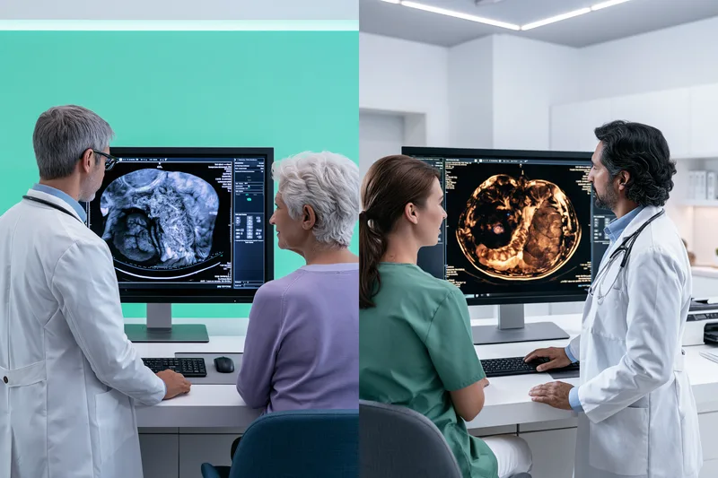
Desmoid tumor and GIST can both arise as deep abdominal masses, but they develop through different biological pathways. Desmoid tumor is mainly driven by abnormal WNT beta catenin signalling in fibroblastic connective tissue, while GIST usually develops through uncontrolled receptor tyrosine kinase signalling in gastrointestinal stromal cells. These differences explain why the tumors may look similar on imaging or biopsy but behave differently, respond to different medicines, and require separate monitoring strategies. [12][41][56] PubMed Central (PMC)
How Desmoid Tumor Develops
Desmoid tumor develops from fibroblastic or myofibroblastic cells that normally contribute to connective tissue, wound repair, fascia, and collagen formation. In most sporadic cases, an acquired alteration in the CTNNB1 gene interferes with the normal breakdown of beta catenin.
Beta catenin is usually regulated by a protein complex that includes APC. When CTNNB1 is altered, beta catenin can accumulate inside the cell and move into the nucleus, where it activates genes involved in cell growth and tissue formation. The result is continued fibroblastic proliferation and excessive production of collagen rich tissue. [12]
A smaller group of desmoid tumors develops in people with an inherited alteration in the APC gene, particularly in familial adenomatous polyposis. Loss of normal APC function also allows beta catenin signalling to remain active. This explains why sporadic CTNNB1 related desmoid tumor and inherited APC related desmoid disease converge on the same WNT beta catenin pathway, even though their family implications are different.
The tumor grows by extending into nearby fascia, muscle, mesentery, nerves, vessels, or organs. It does not usually acquire the biological ability to spread through the bloodstream to distant sites. Its danger comes from persistent local growth and the difficulty of separating infiltrating tumor tissue from essential normal structures.
The amount of collagen within a desmoid tumor can also affect how it appears on MRI and how it responds over time. A tumor may become less biologically active or more fibrotic without showing a large immediate reduction in its outer dimensions. Response assessment should therefore consider symptoms, function, and internal imaging changes as well as size.
How GIST Develops
GIST is believed to arise from the interstitial cells of Cajal or related precursor cells in the gastrointestinal wall. These cells normally help coordinate the muscular contractions that move food through the digestive tract.
Most GISTs are driven by activating alterations in KIT or PDGFRA. These genes produce receptor tyrosine kinases, which normally receive controlled external signals before instructing the cell to grow or survive. An activating mutation can keep the receptor switched on even when no normal growth signal is present.
Continuous KIT or PDGFRA signalling activates several internal pathways involved in cell survival, proliferation, metabolism, and resistance to normal cell death. This allows abnormal stromal cells to accumulate and form a tumor. Additional chromosomal and molecular changes may develop as the tumor progresses, contributing to more aggressive behaviour or treatment resistance. [41][56] PubMed Central (PMC)
The exact mutation affects treatment response. KIT exon 11 altered GIST often behaves differently from KIT exon 9 altered disease. PDGFRA D842V altered GIST is generally resistant to imatinib. This is why molecular testing must identify the gene, exon, and specific alteration rather than simply reporting that a mutation is present. [117]
How SDH Deficient GIST Develops
Not every GIST is driven by KIT or PDGFRA. Some tumors are deficient in the succinate dehydrogenase complex, which normally contributes to mitochondrial energy production and cellular metabolism.
When succinate dehydrogenase function is lost, succinate accumulates within the cell. This can disturb oxygen sensing and epigenetic regulation, allowing abnormal growth related genes to remain active. SDH deficient GIST therefore develops through a metabolic and epigenetic pathway rather than the usual KIT or PDGFRA mechanism.
These tumors often arise in the stomach, may affect younger patients, and can be multifocal. They may also be associated with Carney Stratakis syndrome or Carney triad. Their response to standard tyrosine kinase inhibitors may differ from common KIT altered GIST, making correct classification clinically important. [56] JAMA Network
Why Desmoid Tumor and GIST Can Look Similar
Desmoid tumor and GIST are both mesenchymal tumors, meaning they arise from connective or supporting tissue rather than from the epithelial lining of an organ. Both may contain spindle shaped cells and both may form solid masses within the abdomen.
A mesenteric desmoid tumor may lie beside or surround bowel loops, while an extra gastrointestinal GIST may arise in the mesentery or omentum without an obvious bowel wall origin. On imaging, both may appear as soft tissue masses with variable enhancement and internal composition.
Small biopsy samples can increase the difficulty because they may not show the full architecture of the tumor. The diagnosis must therefore combine anatomical origin, cellular appearance, immunohistochemistry, and molecular testing.
Nuclear beta catenin supports desmoid tumor in the correct morphological setting. KIT or CD117 and DOG1 support GIST, while loss of SDHB staining may indicate SDH deficient GIST. No single stain should be interpreted without the complete pathological and clinical context. [12][41][56]
Why Their Biology Leads to Different Treatment
Desmoid tumor biology is often unpredictable. Some tumors progress, while others stabilize or regress. This natural variation supports active surveillance for selected patients who have manageable symptoms and no immediate organ threat.
Treatment becomes more appropriate when the desmoid tumor shows clinically important progression, increasing pain, functional decline, or risk to bowel, nerves, vessels, ureters, or another vital structure. The treatment objective is often durable control with preservation of function rather than elimination of every visible fibroblastic cell.
GIST treatment is more directly linked to stage and molecular subtype. Localized disease may require surgery, while high risk or advanced disease may require mutation directed targeted therapy. A drug that inhibits KIT may be effective in one molecular subtype and ineffective in another.
Secondary mutations can emerge during long term treatment and alter the structure of the kinase target. This may reduce drug binding and produce acquired resistance after an initial period of response. Apparent progression must therefore be assessed in relation to adherence, drug exposure, mutation profile, and the possibility of new resistant clones.
Ayurvedic Interpretation of the Desmoid Tumor and GIST Interface
Ayurveda does not describe CTNNB1, KIT, PDGFRA, or SDH pathways. It approaches disease through changes in Dosha, Agni, Dhatu, Srotas, Bala, and Ojas.
A firm and persistent tissue mass may be evaluated through the classical frameworks of Granthi and Arbuda. These concepts describe abnormal localized growth involving disturbed Doshas and affected tissues, but they should not be treated as exact substitutes for a modern pathological diagnosis. [267]
Mamsa Dhatu represents muscle and structural soft tissue, while Rakta Dhatu represents the blood tissue system. Meda Dhatu represents adipose and lipid associated tissue. Agni refers to digestive and metabolic transformation, and Srotas refers to the channels through which nourishment, waste, and physiological activity move.
In a desmoid tumor patient, the Ayurvedic physician may assess firmness, local fixation, pain, restriction of movement, bowel disturbance, nutritional decline, and the condition of Mamsa and surrounding tissues. Kapha may be considered where density, heaviness, and persistent tissue accumulation dominate, while Vata may be considered where pain, displacement, obstruction, dryness, disturbed movement, or functional loss is prominent.
In a GIST patient, the assessment may place greater attention on Agni, Rakta, Mamsa, bleeding, appetite, bowel function, weight, treatment toxicity, and tissue strength. A bleeding gastric GIST with anaemia cannot be treated through the same Ayurvedic plan as a stable postoperative patient receiving surveillance or targeted therapy.
These Ayurvedic interpretations describe the patient’s functional and tissue state. They do not replace molecular mechanisms or establish that Dosha imbalance directly caused a specific genetic mutation.
What Root Level Ayurvedic Care Means in Practice
Root level Ayurvedic care means identifying and correcting the patient’s ongoing disturbances in digestion, nutrition, bowel function, sleep, pain, strength, tissue recovery, and treatment tolerance. It does not mean assuming that improvement in Agni or Ojas has automatically removed the tumor.
The Ayurvedic plan may use individualized Ahara, Vihara, Shamana, Avaleha, or Rasayana according to the patient’s disease phase and capacity. Shodhana should be considered only in carefully selected stable patients and is unsuitable during active bleeding, obstruction, repeated vomiting, dehydration, infection, severe weakness, or acute organ toxicity.
Health improvement should be documented through appetite, weight, bowel pattern, sleep, pain, mobility, strength, and quality of life. Tumor response must be documented separately through imaging, pathology, laboratory monitoring, and specialist interpretation.
This separation protects patient trust. Ayurveda remains centred on restoring the patient’s internal balance and functional resilience, while modern oncology continues to define tumor biology, progression, treatment response, and emergency risk.
Clinical Presentation of Desmoid Tumor and GIST

The clinical presentation of desmoid tumor and GIST depends on the tumor’s location, size, growth pattern, and relationship with nearby organs. Both conditions may cause abdominal pain, fullness, a palpable mass, vomiting, or bowel obstruction. However, gastrointestinal bleeding is more characteristic of GIST, while progressive local infiltration and functional restriction are more typical of desmoid tumor. Symptoms must be interpreted together with imaging, pathology, molecular findings, laboratory results, and changes in daily function. [12][41][56]
Common Clinical Presentation of Desmoid Tumor
A desmoid tumor may first appear as a firm, slowly enlarging mass, localized swelling, persistent pain, or increasing restriction of movement. Superficial tumors may be visible or palpable, while deeper tumors can remain unnoticed until they compress a nerve, muscle, bowel loop, blood vessel, ureter, or another organ.
Pain may develop when the tumor stretches fascia, infiltrates muscle, compresses nerves, or alters normal posture and movement. Pain severity does not always correspond with tumor size. A relatively small tumor beside a nerve or joint may cause greater disability than a larger tumor in a less sensitive location.
Some desmoid tumors are found incidentally during imaging performed for another condition. An asymptomatic tumor does not automatically require immediate treatment. Its anatomical risk, behaviour on serial imaging, and effect on function must be considered before treatment is started. [12]
Abdominal Wall Desmoid Tumor Symptoms
An abdominal wall desmoid tumor commonly presents as a firm lump or area of thickening within the abdominal muscles. The mass may become more noticeable when the patient stands, coughs, stretches, or contracts the abdominal wall.
Patients may experience tightness, tenderness, or discomfort during exercise, lifting, bending, or movements that engage the abdominal muscles. The tumor may also alter abdominal symmetry or interfere with daily movement.
An abdominal wall mass is not automatically a desmoid tumor. Hernia, scar tissue, hematoma, endometriosis, lipoma, and other soft tissue tumors may produce a similar appearance. Imaging and pathology are therefore required before treatment is planned.
Intra Abdominal Desmoid Tumor Symptoms
An intra abdominal desmoid tumor may arise in the mesentery or pelvis, where it can grow around bowel loops, vessels, nerves, or ureters. These tumors may remain clinically silent until they become large enough to cause compression or obstruction.
Symptoms may include abdominal pain, bloating, fullness, early satiety, reduced appetite, constipation, nausea, vomiting, or unexplained weight loss. Partial bowel obstruction may cause intermittent symptoms, with periods of relative comfort between episodes of cramping, distension, or vomiting.
Compression of a ureter may cause flank pain, recurrent urinary infection, reduced urine output, hydronephrosis, or deterioration in kidney function. Advanced intra abdominal disease may rarely contribute to bowel ischaemia, fistula, abscess, perforation, or severe infection.
Repeated vomiting, inability to pass stool or gas, rapidly increasing abdominal distension, severe continuous pain, fever, fainting, or reduced urine output requires urgent hospital assessment. These symptoms should not be treated at home as ordinary constipation or indigestion.
Extremity Desmoid Tumor Symptoms
A desmoid tumor in an arm, shoulder, thigh, leg, hand, or foot may present as a firm swelling or deep tissue mass. Symptoms develop when the tumor restricts a joint, compresses nerves, or infiltrates muscles and fascia.
The patient may notice reduced range of movement, weakness, altered gait, reduced grip, difficulty lifting an arm, or avoidance of activities that were previously comfortable. Tingling, numbness, burning pain, or pain radiating along a limb may indicate nerve involvement.
Caregivers may notice protective posture, slower walking, reduced arm movement, or difficulty rising from a chair before the patient reports significant disability.
Head Neck and Chest Wall Desmoid Tumor Symptoms
Head and neck desmoid tumors may cause a visible mass, facial asymmetry, restricted neck movement, difficulty opening the mouth, swallowing discomfort, voice change, or nerve related symptoms. Even a small tumor may be clinically important in this region because the airway, major vessels, and nerves are closely positioned.
A chest wall desmoid tumor may cause localized pain, a firm mass, discomfort during deep breathing, or restricted shoulder movement. Detailed anatomical imaging is necessary before surgery because removal may affect muscles, ribs, nerves, or respiratory function.
Common Clinical Presentation of GIST
The clinical presentation of GIST depends on the primary site, tumor size, rate of growth, ulceration, bleeding, rupture, and presence of metastatic disease. Gastrointestinal bleeding is one of the most common presentations. It may be sudden and visible or slow and clinically silent. [41][56]
Visible bleeding may appear as vomiting blood, black tarry stools, or blood in the stool. Slow bleeding may cause chronic iron deficiency anaemia without obvious external blood loss.
Patients with chronic anaemia may experience fatigue, pallor, breathlessness during mild activity, dizziness, headaches, rapid heartbeat, reduced concentration, or declining physical stamina. Anaemia should not be treated repeatedly without investigating the source of blood loss.
Other GIST symptoms include abdominal pain, early satiety, nausea, reduced appetite, weight loss, difficulty swallowing, constipation, bowel obstruction, or a palpable abdominal mass. Some tumors grow outward from the gastrointestinal wall and may become large before they interfere with the passage of food or stool.
Gastric GIST Symptoms
A GIST arising in the stomach may cause upper abdominal discomfort, early satiety, nausea, reduced appetite, bloating, or pressure after meals. The patient may gradually reduce portion size without recognizing this as an important symptom.
Ulceration of the tumor into the stomach may cause chronic or acute bleeding. Some patients present with unexplained anaemia before they develop pain or a palpable mass.
Vomiting blood, black stools, fainting, marked weakness, or rapid heartbeat requires urgent medical assessment because significant gastrointestinal bleeding may be present.
Small Intestinal GIST Symptoms
A small intestinal GIST may cause intermittent abdominal pain, anaemia, bleeding, nausea, vomiting, bowel obstruction, or internal haemorrhage. These tumors may grow outward into the abdominal cavity and remain unnoticed until they become large, rupture, or narrow the intestine.
Cramping pain that comes and goes may indicate partial obstruction. Increasing abdominal distension, repeated vomiting, constipation, or inability to pass gas may indicate complete obstruction.
Sudden severe pain with fainting, low blood pressure, or collapse may indicate tumor rupture or internal bleeding and requires emergency care.
Non gastric GIST may carry a different recurrence risk from gastric GIST of similar size and mitotic activity. The primary site must therefore be recorded accurately in the pathology and surgical reports. [41][56][187]
Oesophageal and Rectal GIST Symptoms
Oesophageal GIST is uncommon and may cause progressive difficulty swallowing, discomfort behind the breastbone, regurgitation, or weight loss. Difficulty swallowing that continues to worsen should not be attributed automatically to reflux.
Rectal GIST may cause pelvic pressure, constipation, pain during bowel movements, incomplete evacuation, altered stool calibre, urinary symptoms, or a palpable mass during examination.
Because the pelvis contains several closely positioned organs, treatment planning must consider bowel, urinary, sexual, and functional outcomes.
Silent and Incidentally Detected GIST
Small GISTs may be discovered during endoscopy, imaging, or surgery performed for another reason. Some remain indolent and never produce clinically significant disease.
The discovery of a small lesion does not automatically mean that major surgery is required. It also does not mean the lesion can be ignored. Site, size, growth pattern, endoscopic ultrasound features, biopsy findings, age, comorbidities, and specialist assessment determine the appropriate plan. [56]
Symptoms of Advanced or Metastatic GIST
Advanced GIST may cause progressive abdominal pain, increasing fullness, reduced appetite, weight loss, fatigue, or declining function. Liver metastases may initially cause no symptoms but can later contribute to upper abdominal discomfort, liver enlargement, jaundice, or abnormal liver tests.
Peritoneal disease may cause abdominal distension, altered bowel function, pain, early satiety, or fluid accumulation. These symptoms are not specific to GIST and require imaging and laboratory assessment.
Lymph node and lung metastases are uncommon in typical adult GIST but may occur in selected pediatric or molecular subtypes. The absence of enlarged lymph nodes does not exclude metastatic disease because the liver and peritoneum are more common sites of spread. [56]
Why Desmoid Tumor and GIST Symptoms May Overlap
Both desmoid tumor and GIST may cause abdominal pain, fullness, nausea, vomiting, constipation, reduced appetite, a palpable mass, or bowel obstruction.
A mesenteric desmoid tumor may compress the bowel from outside, while a GIST may arise from the bowel wall and grow outward. Imaging may suggest the likely origin, but expert pathology is often required to distinguish them.
Gastrointestinal bleeding is more suggestive of GIST than desmoid tumor, but bleeding alone cannot establish the diagnosis. Other gastrointestinal diseases may also cause anaemia, black stools, or vomiting blood.
Symptoms Do Not Always Measure Tumor Progression
Increasing symptoms may indicate tumor progression, but pain can also worsen because of nerve irritation, muscle spasm, scar tissue, constipation, infection, treatment toxicity, anxiety, or another unrelated condition.
A stable scan does not guarantee that the patient is clinically stable. Anaemia, malnutrition, partial obstruction, dehydration, liver toxicity, kidney dysfunction, thyroid disturbance, muscle loss, or depression may worsen without a major change in tumor dimensions.
The opposite may also occur. A patient may feel relatively well while imaging shows tumor progression. Symptoms, laboratory findings, function, and imaging must therefore be assessed together.
Emergency Warning Signs for Patients and Caregivers
Severe or rapidly increasing abdominal pain requires urgent assessment, particularly when accompanied by vomiting, abdominal rigidity, fever, dizziness, or collapse.
Repeated vomiting and inability to pass stool or gas may indicate complete bowel obstruction. Vomiting blood, black stools with weakness, fainting, marked pallor, rapid heartbeat, or breathlessness may indicate significant gastrointestinal bleeding.
New flank pain, reduced urine output, urinary infection, or rising creatinine may indicate ureteric compression or kidney obstruction.
Sudden weakness, jaundice, confusion, breathing difficulty, severe swelling, or chest pain during treatment also requires urgent medical review.
Ayurvedic medicines, fasting, laxatives, massage, or Panchakarma must not be used to delay emergency evaluation.
Ayurvedic Assessment of Desmoid Tumor and GIST Symptoms
Ayurveda evaluates the symptom pattern together with the confirmed diagnosis, disease stage, imaging findings, organ function, and current treatment.
Agni refers to digestive and metabolic capacity. Poor appetite, early satiety, bloating, nausea, and irregular bowel function may indicate impaired digestion, but they can also result from mechanical obstruction or tumor pressure. Structural causes must be excluded before these symptoms are treated as a functional digestive imbalance.
Mamsa Dhatu represents muscle and structural soft tissue. Pain, restriction of movement, muscle weakness, and deep tissue involvement may be evaluated through this framework, but anatomical imaging remains necessary.
Rakta Dhatu represents the blood tissue system. Bleeding, anaemia, pallor, dizziness, and reduced stamina require modern investigation and stabilization. Rakta supporting herbs cannot replace control of active blood loss.
Bala refers to functional strength, while Ojas represents systemic vitality and resilience. Weight loss, disturbed sleep, poor mobility, reduced treatment tolerance, and emotional withdrawal indicate reduced systemic reserve and should influence the intensity of Ayurvedic treatment.
The same tumor diagnosis may produce different Ayurvedic patterns. A painful obstructive desmoid tumor associated with constipation and weight loss requires a different plan from a stable, dense mass without significant symptoms. A bleeding gastric GIST also requires a different approach from a postoperative GIST under surveillance.
Safe Ayurvedic Support for Clinical Symptoms
Ayurvedic care may support appetite, bowel regularity, sleep, strength, mobility, recovery, and treatment tolerance when it is individualized and coordinated with specialist care.
Avaleha should be considered only when the patient can digest and tolerate it safely. It may be unsuitable during active bleeding, repeated vomiting, bowel obstruction, severe early satiety, uncontrolled diabetes, liver injury, kidney impairment, or immediate postoperative recovery.
Shodhana is not appropriate during acute abdominal symptoms, dehydration, severe weakness, infection, significant anaemia, active bleeding, or suspected obstruction.
Symptom improvement must be documented separately from tumor response. Better appetite, reduced pain, improved bowel function, stable weight, and increased mobility are meaningful health outcomes. Reduction or stabilization of desmoid tumor or GIST must still be confirmed through specialist imaging and clinical interpretation.
What Patients and Caregivers Should Record
The symptom record should include the date and location of pain, relationship of symptoms to meals or movement, stool colour, bowel frequency, vomiting, appetite, meal capacity, body weight, sleep, mobility, urinary changes, bleeding, swelling, fatigue, medication adherence, and analgesic use.
Caregivers should record changes from the patient’s usual baseline rather than isolated symptoms. A gradual decline in walking, eating, communication, self care, or treatment adherence may be clinically important even when severe pain is absent.
This record helps the oncology and Ayurvedic teams distinguish tumor progression, mechanical complications, treatment toxicity, nutritional decline, and functional imbalance.
Common and Rare Disorders Associated With Desmoid Tumor and GIST

Common and rare disorders associated with desmoid tumor and GIST may arise from the tumor itself, pressure on nearby organs, internal bleeding, impaired food intake, inherited syndromes, surgery, or long term targeted therapy. Recognizing these problems early helps patients and caregivers seek treatment before anaemia, obstruction, kidney injury, infection, malnutrition, or drug toxicity becomes severe. Ayurveda can support digestion, strength, tissue recovery, and treatment tolerance after urgent structural causes have been identified and medically stabilized. [12][41][56]
Gastrointestinal Bleeding and Chronic Anaemia
Gastrointestinal bleeding is a common presentation of GIST, particularly when the tumor ulcerates into the stomach or intestine. Bleeding may be visible as vomiting blood or black stools, but it may also occur slowly without obvious external blood loss. Chronic bleeding can produce iron deficiency anaemia, fatigue, pallor, dizziness, breathlessness, rapid heartbeat, headaches, and reduced ability to work or exercise. [56][502]
Repeated treatment for low haemoglobin without investigating the cause may delay diagnosis. A patient with unexplained anaemia, black stools, fainting, worsening weakness, or breathlessness requires prompt gastrointestinal and oncological assessment.
Ayurvedic care may support appetite, digestion, strength, and recovery after the source of bleeding has been identified and controlled. Rakta Dhatu describes the blood tissue system, but herbs intended to support Rakta cannot replace endoscopy, imaging, transfusion, surgery, or tumor directed treatment when active bleeding continues.
Abdominal Pressure Early Satiety and Malnutrition
Desmoid tumor and GIST may press on the stomach, bowel, mesentery, liver, or surrounding abdominal structures. Patients may feel full after a few bites, lose interest in food, develop nausea, or reduce meal size without recognizing the importance of the change.
Persistent early satiety can lead to weight loss, protein deficiency, muscle wasting, impaired wound healing, reduced immunity, and poor treatment tolerance. A falling body weight may also be hidden by fluid retention during targeted therapy.
Caregivers should monitor food intake, portion size, body weight, muscle strength, and the patient’s ability to complete daily activities. A dietitian should become involved when weight is falling, meals are becoming progressively smaller, or weakness is increasing.
Ayurvedic treatment may support Agni, meaning digestive and metabolic capacity, but early satiety caused by mechanical pressure cannot be corrected through digestive stimulants alone. Imaging must first determine whether the stomach or bowel is being compressed.
Intestinal Obstruction and Sub Obstruction
An intra abdominal desmoid tumor may compress or surround bowel loops, while GIST may narrow the intestinal passage or produce obstruction through its size, location, or associated complications.
Partial obstruction may cause intermittent bloating, cramping pain, constipation, nausea, and vomiting. Symptoms may improve temporarily and then return. Complete obstruction may cause repeated vomiting, severe distension, inability to pass stool or gas, dehydration, and escalating pain.
Intra abdominal desmoid tumors have been associated with bowel obstruction, vascular encasement, tumor rupture, perforation, and other gastrointestinal complications. GIST can also present as acute obstruction, perforation, rupture, or peritonitis. [401][502]
Suspected obstruction requires urgent medical assessment. Laxatives, high fibre diets, strong digestive herbs, oil administration, or Panchakarma may worsen symptoms or delay necessary intervention. Ayurveda should be resumed only after the obstruction risk has been evaluated and the patient can safely tolerate oral treatment.
Chronic Pain and Neuromuscular Dysfunction
Desmoid tumors may infiltrate muscle, fascia, nerves, or joints. Patients can develop deep aching pain, burning pain, numbness, reduced range of movement, muscle weakness, altered gait, or protective posture.
The caregiver may notice slower walking, difficulty rising from a chair, reduced arm movement, avoidance of stairs, or disturbed sleep before the patient describes a major change.
Pain does not always mean that the tumor has enlarged. Scar tissue, nerve irritation, muscle spasm, postoperative change, treatment toxicity, constipation, or another unrelated condition may contribute. Imaging and examination are needed when the pain pattern changes.
Ayurvedic support may include individualized pain management, sleep support, gentle rehabilitation, external treatment, and Vata balancing care. Strong massage, heat, or pressure should not be applied over an unstable mass, recent surgical site, bleeding area, or structure at risk without medical clearance.
Ureteric Compression Hydronephrosis and Kidney Injury
Pelvic or mesenteric desmoid tumors may compress one or both ureters, preventing urine from draining normally from the kidney to the bladder. This can cause hydronephrosis, infection, flank pain, rising creatinine, or gradual loss of kidney function.
The patient may notice reduced urine output, pain in the back or side, fever, urinary infection, nausea, or leg swelling. Some cases are discovered only through imaging or laboratory changes.
Persistent obstruction can cause permanent kidney damage or sepsis. It may require a stent, nephrostomy, surgery, systemic tumor treatment, or another specialist intervention.
Ayurvedic medicines must be reviewed when kidney function is impaired because altered clearance can increase the risk of toxicity. Diuretic herbs should not be used as a substitute for relieving a mechanical blockage.
Vascular Compression and Bowel Ischaemia
A large intra abdominal desmoid tumor may encase or displace mesenteric vessels. Reduced blood supply to the bowel can cause severe pain, bowel injury, ischaemia, perforation, or infection.
Pain that becomes continuous, severe, or disproportionate to previous symptoms requires urgent assessment, particularly when accompanied by vomiting, fever, abdominal rigidity, blood in the stool, fainting, or low blood pressure.
This is not a situation for outpatient detoxification or dietary experimentation. Emergency imaging, surgical review, infection control, and organ support take priority.
Fistula Abscess Perforation and Sepsis
Deep infiltrative desmoid disease may rarely erode into bowel or contribute to fistula formation, abscess, necrosis, rupture, or perforation. GIST may also rupture or perforate, causing leakage of blood or gastrointestinal contents into the abdominal cavity.
Warning signs include sudden severe pain, fever, chills, vomiting, a rigid abdomen, confusion, rapid heartbeat, low blood pressure, or collapse.
These complications can progress rapidly to sepsis and require emergency hospital management. Ayurveda may support rehabilitation after stabilization, but oral Avaleha, Shodhana, fasting, or home treatment must not delay surgery, drainage, antibiotics, or intensive care.
GIST Rupture and Internal Haemorrhage
GIST may outgrow its blood supply, develop internal necrosis, bleed, or rupture. Rupture may occur spontaneously or during surgery and is an important adverse prognostic factor because tumor cells may spread within the abdominal cavity. [41][56]
The patient may develop sudden severe pain, weakness, dizziness, fainting, abdominal swelling, rapid pulse, or low blood pressure. Internal haemorrhage may occur without visible blood in the stool.
Tumor rupture requires urgent specialist treatment. The operative report should document whether rupture occurred because this information can influence recurrence assessment and postoperative planning.
Liver and Peritoneal Metastatic Disease
GIST most commonly spreads to the liver and peritoneal surfaces. Early metastatic disease may cause no symptoms and may be found only through imaging.
As disease progresses, patients may develop abdominal fullness, pain, reduced appetite, weight loss, ascites, or declining function. Liver involvement may cause right upper abdominal discomfort, liver enlargement, jaundice, itching, or abnormal liver tests.
These symptoms can also arise from drug toxicity, infection, gallbladder disease, or another condition. Laboratory testing and imaging are needed before the cause is assumed.
Ayurvedic liver support should never be prescribed from symptoms alone. The physician must distinguish metastatic disease, biliary obstruction, viral hepatitis, fatty liver, and TKI related liver injury because each requires different management.
Rare Consumptive Hypothyroidism
Consumptive hypothyroidism is a rare paraneoplastic disorder reported in a small number of patients with GIST. The tumor may overexpress an enzyme that inactivates thyroid hormones, creating unusually high thyroid hormone requirements. [56][468] Cancer.gov
Patients may experience severe fatigue, cold intolerance, constipation, dry skin, slowing of movement, weight change, or cognitive difficulty. These symptoms may be mistaken for ordinary cancer related fatigue or treatment toxicity.
Thyroid function should be evaluated when symptoms are unexplained or persistent. Treatment requires endocrine assessment and management of both the thyroid disturbance and the underlying tumor.
Familial Adenomatous Polyposis and Gardner Syndrome
A proportion of desmoid tumors occur in people with familial adenomatous polyposis caused by an inherited APC alteration. These patients may develop numerous colorectal polyps, early colorectal cancer, and intra abdominal or abdominal wall desmoid tumors.
Desmoid disease is an important cause of illness in familial adenomatous polyposis, particularly when tumors involve the mesentery or develop after abdominal surgery. [416] PubMed
Genetic evaluation should be discussed when the patient has multiple colorectal polyps, colorectal cancer at a young age, multiple desmoid tumors, an intra abdominal desmoid tumor, or a family history of related disease.
This diagnosis affects more than the individual patient. Relatives may require counselling, genetic testing, and colorectal surveillance.
Ayurveda may support digestion, strength, recovery, and long term wellbeing, but it cannot replace colonoscopic surveillance, genetic counselling, preventive surgery when indicated, or specialist management of APC related disease.
Neurofibromatosis Type 1 Associated GIST
GIST may occur in patients with neurofibromatosis type 1. These tumors often arise in the small intestine, may be multiple, and commonly lack the typical KIT or PDGFRA mutations despite expressing KIT on immunohistochemistry. [56]
The patient may have café au lait macules, neurofibromas, freckling in characteristic areas, or a known family history of neurofibromatosis type 1.
Recognizing the association is important because the tumor biology and treatment response may differ from common KIT mutated GIST. Molecular testing and specialist review should guide treatment.
SDH Deficient GIST and Carney Stratakis Syndrome
SDH deficient GIST usually arises in the stomach and often affects younger patients. It may be multifocal, involve lymph nodes, and follow a different course from common adult KIT mutated GIST.
Carney Stratakis syndrome is an inherited condition associated with GIST and paraganglioma. Patients and relatives may require genetic counselling and surveillance beyond the gastrointestinal tract.
Paraganglioma can produce episodes of severe headache, palpitations, sweating, anxiety, or high blood pressure, although some tumors do not release hormones.
The presence of a gastric GIST in a young person, multiple tumors, loss of SDHB staining, or a family history of paraganglioma should prompt further assessment. [56] Cancer.gov
Carney Triad
Carney triad is a rare association involving gastric GIST, paraganglioma, and pulmonary chondroma. Not every patient develops all three tumors at the same time.
The condition often affects younger individuals and may require long term surveillance of the abdomen, chest, and paraganglionic system.
A lung lesion in this setting should not automatically be assumed to represent GIST metastasis because pulmonary chondroma may form part of the syndrome. Specialist radiology, pathology, and genetic evaluation are needed.
Familial KIT and PDGFRA Related GIST
Rare families carry inherited KIT or PDGFRA alterations and may develop multiple GISTs. Some affected individuals may also show skin pigmentation changes, swallowing problems, or other features depending on the inherited alteration.
A young age at diagnosis, multiple GISTs, or several relatives with GIST should prompt genetic counselling. Family screening should be guided by a genetics team rather than informal testing.
Disorders Related to Long Term TKI Therapy
Imatinib and other tyrosine kinase inhibitors may control GIST for prolonged periods, but they can also produce chronic adverse effects that influence nutrition, mobility, adherence, and quality of life.
Imatinib may cause nausea, diarrhoea, abdominal discomfort, fatigue, muscle cramps, rash, fluid retention, swelling around the eyes or legs, blood count changes, and liver dysfunction. Later line TKIs may also contribute to hypertension, hand and foot skin reactions, thyroid disturbance, bleeding, liver injury, or cardiovascular effects depending on the medicine used. [41][518] Cancer Research UK
Patients should not reduce, divide, or stop a TKI independently. A treatment may still be controlling the tumor even when side effects require dose adjustment or supportive care.
Ayurvedic medicines must be reviewed for interactions and overlapping toxicity. A herb described as liver supporting may still alter drug metabolism, and a mineral preparation may add risk when liver or kidney function is impaired.
Malnutrition Sarcopenia and Functional Decline
Poor intake, bleeding, early satiety, surgery, chronic inflammation, diarrhoea, pain, and long term treatment can lead to sarcopenia, meaning loss of muscle mass and strength.
Patients may lose the ability to walk normally, climb stairs, carry objects, or recover after routine activity even when body weight changes little.
Nutritional support and safe resistance based rehabilitation are important. Severe dietary restriction, prolonged fasting, or attempts to starve the tumor can worsen weakness and treatment tolerance.
Ayurvedic care may support Agni, appetite, sleep, and Bala, but the diet must still provide adequate energy and protein according to the patient’s medical needs.
Anxiety Depression and Scan Related Distress
Desmoid tumor and GIST can create long periods of uncertainty. Patients may fear recurrence, drug resistance, surgery, financial burden, or the next scan.
Anxiety may appear as insomnia, irritability, repeated checking of symptoms, inability to concentrate, or avoidance of appointments. Depression may present as withdrawal, loss of interest, reduced self care, hopelessness, or poor medication adherence.
These symptoms deserve direct assessment and treatment. Psychological support, counselling, medication when appropriate, caregiver involvement, and clear clinical communication can improve quality of life and treatment continuity.
Ayurvedic care may support sleep, routine, stress regulation, and mind body stability, but significant anxiety, depression, or suicidal thoughts require qualified mental health care.
Caregiver Burnout
Caregivers may become responsible for transport, medicine supply, insurance communication, symptom monitoring, food preparation, and emotional support while also maintaining work and family duties.
Burnout may appear as exhaustion, irritability, sleep disturbance, poor concentration, resentment, anxiety, or declining physical health.
The caregiver should not be expected to manage every responsibility alone. Clear emergency contacts, shared family roles, respite care, counselling, and practical support protect both caregiver and patient.
How Ayurveda Can Support These Associated Disorders Safely
Ayurveda can support the patient most effectively when it is integrated after the associated disorder has been correctly identified.
Agni focused treatment may help selected patients with poor appetite or treatment related digestive disturbance, but it must not be used to mask bowel obstruction, bleeding, or tumor pressure.
Rakta and Bala supporting care may assist recovery from corrected anaemia, but it cannot replace investigation and control of ongoing blood loss.
Rasayana may support recovery, nutrition, sleep, strength, and quality of life during stable phases, but it should not be described as proof of tumor eradication or recurrence prevention without objective evidence.
Avaleha should be avoided or modified when the patient has severe early satiety, obstruction, repeated vomiting, uncontrolled diabetes, active bleeding, impaired swallowing, acute liver injury, or kidney dysfunction.
Shodhana is unsuitable during active bleeding, obstruction, perforation risk, severe weakness, dehydration, infection, cytopenia, acute organ dysfunction, or immediate postoperative recovery.
The Ayurvedic physician should review pathology, imaging, molecular findings, current drugs, laboratory trends, and the oncology plan before prescribing. Health improvement and report improvement should always be recorded separately.
What Patients and Caregivers Must Report Immediately
New black stools, vomiting blood, fainting, repeated vomiting, inability to pass stool or gas, rapidly increasing abdominal pain, fever with a rigid abdomen, sudden reduction in urine output, jaundice, breathing difficulty, chest pain, severe swelling, or marked confusion requires urgent medical review.
A patient with these symptoms should not wait for an Ayurvedic appointment, the next scan date, or the next scheduled oncology visit.
Emergency Red Flags in Desmoid Tumor and GIST That Can Save a Patient’s Life

Desmoid tumor and GIST can remain stable for long periods, but bleeding, bowel obstruction, tumor rupture, perforation, infection, kidney obstruction, or treatment toxicity may develop suddenly. Patients and caregivers must know which symptoms require immediate hospital assessment rather than waiting for the next scan or consultation. Ayurveda can support recovery and long term stability, but emergency complications must first be treated through urgent imaging, resuscitation, surgery, drainage, transfusion, or oncology care when indicated. [12][41][56]
Vomiting Blood or Passing Black Stools
Vomiting fresh blood, material resembling coffee grounds, or passing black tarry stools may indicate gastrointestinal bleeding. GIST can bleed when its surface ulcerates into the stomach or intestine. Slow bleeding may cause anaemia, while rapid bleeding may lead to low blood pressure, collapse, or circulatory shock. [41][56]
Paleness, dizziness, breathlessness, rapid heartbeat, cold sweating, confusion, or sudden weakness increases the urgency even when visible blood appears limited. Some patients develop significant internal blood loss before a large quantity of blood is seen externally.
The patient should attend an emergency department immediately. Food, iron supplements, haemostatic herbs, or Rakta supporting medicines should not be used as substitutes for locating and controlling active bleeding. Ayurveda may support recovery only after the source of bleeding has been investigated and stabilized.
Repeated Vomiting and Inability to Pass Stool or Gas
Repeated vomiting, progressive abdominal swelling, cramping pain, constipation, and inability to pass gas may indicate complete bowel obstruction. An intra abdominal desmoid tumor can compress or surround bowel loops, while GIST may narrow the gastrointestinal passage or obstruct it through tumor growth, inflammation, or associated complications. [12][56]
Partial obstruction may initially produce intermittent symptoms. The patient may have several hours or days of bloating, nausea, reduced appetite, or colicky pain followed by temporary improvement. This fluctuation can create false reassurance.
Complete obstruction can rapidly cause dehydration, electrolyte disturbance, bowel ischaemia, perforation, infection, and shock. The patient should not be given laxatives, high fibre food, strong digestive medicines, oil administration, or Panchakarma during suspected obstruction. Emergency imaging and surgical assessment take priority.
Sudden Severe Abdominal Pain or a Rigid Abdomen
Sudden severe abdominal pain that differs from the patient’s usual discomfort may indicate tumor rupture, internal bleeding, bowel perforation, ischaemia, abscess, or another acute abdominal emergency.
A rigid abdomen, pain worsened by movement, fever, vomiting, rapid pulse, or marked tenderness increases concern for perforation, peritonitis, or infection. GIST may rupture or bleed into the abdomen, while advanced intra abdominal desmoid disease may compromise bowel or surrounding structures. [12][41][56]
The patient should not wait to see whether the pain improves after rest, herbal medicine, heat application, massage, or fasting. Sudden severe pain requires urgent hospital evaluation even when the previous scan described stable disease.
Fainting Collapse or Sudden Low Blood Pressure
Fainting, collapse, severe dizziness, cold skin, rapid pulse, unusual sleepiness, or confusion may indicate internal bleeding, dehydration, infection, or circulatory shock.
Internal haemorrhage may occur without vomiting blood or passing black stools. A ruptured or bleeding abdominal tumor may release blood into the abdominal cavity, producing weakness and low blood pressure before external bleeding becomes visible.
The caregiver should contact local emergency services. The patient should not be allowed to walk independently or drive. Current medicines, allergies, diagnosis, recent laboratory results, and the treating hospital’s details should travel with the patient whenever possible.
Fever With Abdominal Pain or Confusion
Fever associated with increasing abdominal pain, vomiting, chills, confusion, low blood pressure, or rapid breathing may indicate abscess, bowel perforation, fistula, infection, or sepsis.
Sepsis can progress rapidly, particularly in a patient who is malnourished, postoperative, receiving systemic therapy, or has reduced white blood cell counts. A normal temperature does not completely exclude severe infection in an immunocompromised patient.
Emergency assessment may require blood tests, cultures, imaging, intravenous fluids, antibiotics, drainage, surgery, or intensive care. Ayurvedic antimicrobial formulations should not delay these interventions.
Reduced Urine Output Flank Pain or Recurrent Urinary Infection
A pelvic or mesenteric desmoid tumor may compress the ureter and block urine flow from the kidney. The patient may develop flank pain, reduced urine output, nausea, fever, recurrent urinary infection, leg swelling, or rising creatinine. [12]
Ureteric obstruction may progress silently and cause hydronephrosis or permanent kidney injury. A patient with one functioning kidney, bilateral obstruction, infection, or acute reduction in urine output requires particularly urgent assessment.
Diuretic herbs cannot open a mechanically obstructed ureter. The patient may require imaging, a ureteric stent, nephrostomy, surgery, or tumor directed treatment before Ayurvedic support can be used safely.
Jaundice Dark Urine or Severe Weakness During Treatment
Yellowing of the eyes or skin, dark urine, pale stools, severe itching, persistent nausea, marked fatigue, right upper abdominal pain, or confusion may indicate liver dysfunction.
The cause may be drug related liver toxicity, biliary obstruction, infection, metastatic disease, dehydration, or another medical condition. Targeted therapies used for GIST can affect liver function, and monitoring is necessary throughout treatment. [41][159]
The patient should contact the oncology team promptly. A prescribed TKI should not be stopped, reduced, or restarted independently unless emergency instructions have already been provided. Herbal or mineral preparations should also be paused and reviewed because overlapping liver toxicity or drug interactions may be possible.
Severe Diarrhoea Dehydration or Inability to Eat
Persistent diarrhoea, repeated vomiting, inability to retain fluids, severe weakness, dizziness on standing, reduced urine output, or dry mouth may indicate significant dehydration.
Dehydration can alter kidney function, electrolyte balance, blood pressure, and drug exposure. It may also make an otherwise tolerated medicine unsafe.
The oncology team should be informed early rather than after several days of deterioration. Strong digestive medicines, purgation, fasting, or detoxification procedures are unsuitable until hydration and the cause of symptoms have been assessed.
Chest Pain Breathing Difficulty or Severe Swelling
Chest pain, sudden shortness of breath, fainting, severe leg swelling, or rapid worsening of generalized oedema requires urgent assessment. These symptoms may indicate a cardiovascular event, pulmonary embolism, fluid overload, severe anaemia, infection, or drug related toxicity.
Mild swelling around the eyes or ankles can occur with some targeted therapies, but sudden, painful, one sided, or rapidly progressive swelling must not be assumed to be an ordinary side effect.
The patient should not self treat these symptoms with diuretics, reduced fluid intake, or herbal formulations without medical evaluation.
Sudden Weakness Numbness or Loss of Function
New limb weakness, numbness, loss of balance, difficulty walking, inability to move a joint, sudden bladder or bowel dysfunction, or rapidly worsening nerve pain may indicate nerve compression, spinal involvement, vascular compromise, or an unrelated neurological emergency.
Desmoid tumors near major nerves, the spine, pelvis, or extremities can produce progressive neurological or functional symptoms. Even when distant metastasis is not expected, local compression may still cause permanent disability if treatment is delayed. [12]
The patient should receive urgent examination and imaging. Massage, strong stretching, heat, or manipulation should not be applied over the affected area before structural risk has been assessed.
Rapid Weight Loss and Progressive Inability to Eat
Rapid weight loss, visible muscle wasting, increasing early satiety, weakness, and progressive inability to complete meals may indicate mechanical pressure, obstruction, treatment toxicity, depression, infection, or advancing disease.
Malnutrition reduces healing capacity, muscle strength, immunity, and tolerance of surgery or systemic treatment. A patient may also lose substantial muscle despite a relatively stable body weight when fluid retention is present.
The patient should receive nutritional, medical, and radiological assessment. Ayurveda may support Agni, appetite, sleep, and Bala after obstruction, bleeding, infection, and organ dysfunction have been excluded.
What Caregivers Should Do During an Emergency
The caregiver should note when the symptom began, how quickly it changed, whether vomiting, bleeding, fever, collapse, reduced urine output, or inability to pass stool or gas is present, and which medicines were taken that day.
The caregiver should carry the patient’s diagnosis, current oncology drug and dose, allergies, recent laboratory results, pathology summary, treating centre details, and complete Ayurvedic and supplement list.
The patient should not be given food, Avaleha, oil, laxatives, herbal powders, or mineral medicines when obstruction, perforation, active bleeding, or emergency surgery is possible unless the emergency team specifically permits oral intake.
When Ayurvedic Treatment Must Be Paused
Ayurvedic treatment should be paused and medically reviewed during active bleeding, repeated vomiting, suspected bowel obstruction, perforation, severe dehydration, jaundice, acute kidney injury, sepsis, severe cytopenia, sudden neurological loss, or immediate postoperative instability.
Pausing treatment during an emergency does not mean that Ayurveda has failed. It means that the treatment phase has changed and the patient first requires stabilization.
Once the emergency has been controlled, Ayurveda may be reintroduced gradually according to digestion, oral tolerance, liver and kidney function, bowel status, blood counts, nutritional condition, and the updated oncology plan.
How Ayurveda Helps Patients Recognize Deterioration Early
Ayurvedic monitoring pays close attention to changes in Agni, appetite, bowel movement, urine output, Bala, sleep, pain, complexion, mental clarity, and daily function. These observations can help patients and caregivers notice deterioration before the next scheduled scan.
This early recognition is valuable only when it leads to timely medical assessment. Symptoms caused by bleeding, obstruction, rupture, infection, or organ compression must not be interpreted only as Dosha aggravation.
The safest integrative model uses Ayurvedic observation to detect change early, modern emergency care to stabilize life threatening complications, and individualized Ayurvedic treatment to support recovery after the patient becomes clinically stable.
Step by Step Diagnostic Roadmap for Desmoid Tumor and GIST

The diagnosis of desmoid tumor and GIST should not rely on one scan, one stain, or one short pathology summary. These tumors may resemble other abdominal and soft tissue masses, yet their treatment pathways are very different. A reliable diagnosis combines specialist imaging, a carefully planned biopsy when required, expert pathology, immunohistochemistry, molecular testing, assessment of organ risk, and multidisciplinary review. Ayurveda should be introduced only after the modern diagnosis and current disease phase are clear. [12][41][56]
Why Diagnosis Should Begin With a Sarcoma Specialist Team
Desmoid tumor and GIST are uncommon enough that general experience with ordinary abdominal tumors may not be sufficient. The diagnostic process should involve clinicians who regularly manage sarcoma and stromal tumors.
A specialist multidisciplinary team commonly includes a radiologist, pathologist, surgeon, medical oncologist, specialist nurse, genetic professional, dietitian, rehabilitation clinician, and supportive care team. An Ayurvedic physician can participate in the integrative plan after reviewing the same confirmed pathology, imaging, molecular findings, laboratory results, and treatment objectives.
Specialist review becomes particularly important when the tumor is close to bowel, major vessels, nerves, ureters, or reproductive organs. It is also important when the diagnosis is based on a small sample, the imaging and pathology do not agree, the tumor is described only as a spindle cell lesion, or a major operation is being proposed. [83][103]
Clinical History Before Imaging and Biopsy
The diagnostic process begins with a detailed history rather than the scan alone. The clinician should document when symptoms started, whether they are progressing, and how they affect food intake, bowel function, mobility, sleep, work, and daily independence.
A desmoid tumor history should include previous abdominal surgery, pregnancy, trauma, familial adenomatous polyposis, colorectal polyps, early colorectal cancer, and relatives with desmoid tumors or polyposis.
A GIST history should include gastrointestinal bleeding, anaemia, black stools, vomiting blood, early satiety, abdominal pain, weight loss, bowel obstruction, previous gastrointestinal surgery, and family history of GIST, neurofibromatosis, paraganglioma, or unusual tumors.
The patient should bring a complete medicine list. This includes prescription drugs, over the counter medicines, herbal products, Ayurvedic preparations, supplements, powders, teas, juices, and protein products. Some symptoms may be caused or worsened by treatment, bleeding, organ dysfunction, or drug interactions rather than by tumor growth alone.
Imaging for Desmoid Tumor
MRI is commonly used for desmoid tumors arising in the abdominal wall, extremities, chest wall, head, neck, or other soft tissue locations. It shows the tumor’s relationship with muscle, fascia, nerves, vessels, and nearby organs and can help assess internal tissue changes over time.
CT is often useful for intra abdominal and mesenteric desmoid tumors because it provides a clear view of bowel loops, mesenteric vessels, ureters, kidneys, and other abdominal structures. CT may also be necessary during an emergency when obstruction, perforation, abscess, ischaemia, or internal bleeding is suspected.
The baseline imaging report should document three dimensional measurements rather than one diameter alone. It should also describe whether the tumor displaces, compresses, encases, or directly involves nearby structures.
Serial imaging should ideally be compared with the original images, not only with previous written reports. A small difference in measurement may reflect technique, patient position, or image selection rather than true biological progression.
In desmoid disease, treatment response may involve changes in internal MRI signal and tissue composition before a major reduction in overall size becomes visible. Symptoms, function, and internal radiological characteristics should therefore be considered together. [12]
Imaging for GIST
Contrast enhanced CT is central to the evaluation of suspected or confirmed GIST. It helps identify the primary tumor, assess its relationship to nearby organs, and look for spread to the liver, peritoneum, or other sites.
The radiology report should describe the tumor’s site of origin, size, enhancement pattern, ulceration, necrosis, bleeding, rupture, and relationship with surrounding structures. Chest imaging may also be included according to the clinical situation and staging plan.
MRI can be useful when the tumor is located in the rectum, pelvis, or liver, or when CT contrast is unsuitable. MRI may also provide additional soft tissue information in selected patients.
Endoscopy can reveal a submucosal mass, surface ulceration, or bleeding. Endoscopic ultrasound may help determine which layer of the gastrointestinal wall is involved and may guide tissue sampling in selected gastric, oesophageal, or duodenal lesions.
PET imaging is not required for every patient. It may be useful in selected cases when early metabolic response must be assessed, when conventional imaging is uncertain, or when treatment planning requires additional information. [41][56]
Why Imaging Alone Is Not Enough
Imaging may strongly suggest desmoid tumor or GIST, but other diseases can appear similar. Leiomyoma, leiomyosarcoma, schwannoma, inflammatory myofibroblastic tumor, lymphoma, sclerosing mesenteritis, scar tissue, and other soft tissue lesions may produce overlapping appearances.
A mesenteric desmoid tumor and an extra gastrointestinal GIST may both appear as solid abdominal masses near bowel loops and vessels. Imaging can suggest the likely origin, but it may not provide the final diagnosis.
Treatment should not be selected only because a mass resembles GIST or desmoid tumor on a scan. The diagnosis must be supported by pathology whenever tissue confirmation is clinically required.
Planning the Biopsy Safely
The biopsy should be planned by the team that will manage the patient. The route must avoid unnecessary damage to bowel, vessels, nerves, or organs and should not compromise a later operation.
A core needle biopsy usually provides more tissue than fine needle aspiration and is often preferred for soft tissue diagnosis. The sample must be sufficient for morphology, immunohistochemistry, and molecular testing.
A suspected GIST does not always require biopsy before surgery. A clearly resectable lesion with characteristic imaging may sometimes proceed directly to operation when no preoperative systemic treatment is planned. Biopsy becomes important when the diagnosis is uncertain, metastatic disease is suspected, surgery would be extensive, or neoadjuvant or systemic therapy is being considered. [41][56]
The biopsy should be performed in a manner that minimizes bleeding, rupture, and contamination. The patient should not undergo an unplanned excision of a mass before specialist review because this may complicate definitive treatment.
Pathology of Desmoid Tumor
Desmoid tumor commonly shows a proliferation of relatively uniform spindle shaped fibroblastic or myofibroblastic cells arranged within a collagen rich background. The cells often appear less atypical than those of a high grade sarcoma, although the tumor can still behave aggressively at the local level.
Nuclear beta catenin staining supports the diagnosis in the correct morphological and clinical setting. It is not completely specific and should not be interpreted alone.
CTNNB1 molecular testing may help confirm sporadic desmoid tumor when morphology or immunohistochemistry is uncertain. The result may also help distinguish sporadic disease from an APC related process, although family history and genetic assessment remain important.
A pathology report should state whether the tissue is consistent with desmoid type fibromatosis and whether another spindle cell tumor remains in the differential diagnosis. [12]
Pathology of GIST
GIST may show spindle shaped cells, epithelioid cells, or a mixture of both. The pathology report should describe the cellular pattern, tumor size, mitotic activity, necrosis, ulceration, margins, and rupture status when surgical tissue is available.
KIT or CD117 is positive in most GISTs, while DOG1 is another important marker. CD34 may be positive but is not specific enough to establish the diagnosis alone.
A minority of GISTs may show weak or absent KIT staining. DOG1 and molecular testing become especially important in these cases. Loss of SDHB staining can support SDH deficient GIST and should prompt further assessment according to the patient’s age, tumor site, and clinical context. [41][56]
The pathology report should not end with the words spindle cell tumor when adequate testing can establish a more precise diagnosis.
Molecular Testing for GIST
Molecular testing should identify the gene, exon, and specific alteration whenever possible. KIT and PDGFRA testing is central before mutation directed therapy is selected.
KIT exon 11 altered GIST may respond differently from KIT exon 9 altered disease. PDGFRA D842V altered GIST is generally resistant to imatinib. These distinctions affect treatment selection and expected response. [117]
When KIT and PDGFRA alterations are absent, further assessment may include SDH status, NF1, BRAF, NTRK, and other selected alterations. The required testing depends on the patient’s age, anatomical site, immunohistochemistry, family history, and available specialist resources.
A report stating only that GIST is mutation positive or wild type is not enough for complete treatment planning. The treating clinician should explain what was tested, what was found, and whether the result changes the proposed therapy.
Molecular and Genetic Assessment for Desmoid Tumor
CTNNB1 testing may support the diagnosis of sporadic desmoid tumor. An identified CTNNB1 alteration is usually confined to the tumor and does not automatically imply an inherited condition.
APC related evaluation should be considered when the patient has numerous colorectal polyps, colorectal cancer at a young age, multiple desmoid tumors, a strong family history, or clinical features suggesting familial adenomatous polyposis.
Genetic counselling is important because an inherited APC alteration has implications for colorectal surveillance, surgical planning, relatives, and long term care. The presence of a desmoid tumor alone does not prove familial adenomatous polyposis, but the possibility should not be missed.
What a Complete Desmoid Tumor Report Should Explain
The report should identify the anatomical site, tumor dimensions, relationship to nearby structures, imaging characteristics, biopsy method, pathological diagnosis, beta catenin result, and CTNNB1 or APC related findings where clinically relevant.
The multidisciplinary conclusion should also state whether the tumor threatens bowel, nerves, vessels, ureters, mobility, or another important function.
The patient should receive a written surveillance or treatment plan. Active surveillance without a scan schedule and defined escalation criteria is incomplete care.
What a Complete GIST Report Should Explain
A complete GIST report should identify the primary anatomical site, maximum size, histological pattern, mitotic count, rupture status, margins, necrosis, immunohistochemistry, stage, metastatic sites, and molecular subtype.
The mitotic count should be reported using a standardized microscopic area so that recurrence risk can be interpreted correctly.
The surgical record should state whether the tumor ruptured before or during surgery. Rupture can substantially alter recurrence risk and postoperative treatment planning.
The molecular report should identify the exact KIT, PDGFRA, SDH related, NF1, BRAF, NTRK, or other clinically relevant finding when available. [41][56]
When Expert Pathology Review Is Needed
Expert pathology review should be considered when the diagnosis is based on a very small biopsy, the immunohistochemistry is conflicting, the tumor is located outside the usual gastrointestinal sites, beta catenin and KIT results create uncertainty, or the disease has not responded as expected.
Review is also important before a major organ sacrificing operation, before long term systemic treatment, or when the report uses broad terms without a final classification.
The original slides or paraffin block should be transferred to the reviewing centre. A written report without access to the tissue may not allow a complete reassessment.
Staging and Risk Assessment
Desmoid tumor is not staged in the same way as a metastatic cancer because it does not normally spread to distant organs. Its risk is assessed through location, progression, pain, organ involvement, functional impairment, and quality of life.
GIST requires assessment of whether the disease is localized, locally advanced, unresectable, recurrent, or metastatic. Postoperative recurrence risk depends on tumor site, size, mitotic activity, rupture status, and other pathological or molecular features. [41][56]
The patient should ask for the exact risk category and the factors used to determine it. General statements such as low risk or high risk should be explained in relation to the individual report.
The Role of the Ayurvedic Assessment After Diagnosis
Ayurvedic assessment should begin after the modern diagnosis has been confirmed and urgent structural complications have been addressed. Ayurveda does not replace biopsy, immunohistochemistry, molecular testing, staging, or imaging.
The physician should review the pathology report, imaging, molecular findings, current treatment, blood counts, liver and kidney function, nutritional status, bowel function, bleeding risk, and organ involvement.
The Ayurvedic evaluation then assesses Prakriti, meaning constitutional tendencies, Vikriti, meaning the present imbalance, Agni, meaning digestive and metabolic capacity, Ama, meaning incompletely processed metabolic material, Dhatu, meaning body tissues, Srotas, meaning physiological channels, Bala, meaning functional strength, and Ojas, meaning systemic vitality and resilience.
This assessment identifies how the disease and its treatment are affecting the whole patient. It may reveal poor digestion, constipation, diarrhoea, weight loss, sleep disturbance, pain, weakness, anxiety, or impaired recovery that requires individualized support.
Why Ayurveda Must Not Be Used to Confirm the Tumor Diagnosis
Pulse examination, tongue examination, Dosha assessment, or symptom pattern cannot determine whether an abdominal mass is desmoid tumor, GIST, lymphoma, sarcoma, scar tissue, or another condition.
Ayurvedic concepts such as Granthi and Arbuda provide a classical framework for understanding abnormal tissue growth, but they do not identify CTNNB1, KIT, PDGFRA, SDH deficiency, mitotic rate, rupture status, or metastatic disease.
The safest model preserves the modern pathological diagnosis and uses Ayurveda to understand the patient’s individual digestive, metabolic, tissue, functional, and recovery needs.
What Patients and Caregivers Should Keep
The patient should retain the complete pathology report, molecular report, immunohistochemistry, original scan images, radiology reports, laboratory results, operative notes, medication history, and multidisciplinary treatment plan.
The caregiver should maintain one dated timeline that records symptoms, weight, food intake, bowel function, bleeding, pain, mobility, drug doses, dose interruptions, Ayurvedic treatment, adverse effects, and scan results.
This record allows both the oncology and Ayurvedic teams to work from the same clinical information. It also prevents treatment changes from being made on incomplete memory or isolated reports.
Diagnostic Overlaps and Differential Diagnosis of Desmoid Tumor and GIST

Diagnostic overlaps between desmoid tumor and GIST are most important when a deep abdominal mass develops near the bowel, mesentery, omentum, or major blood vessels. Both conditions may appear as solid soft tissue tumors, both may contain spindle shaped cells, and both may cause pain, fullness, obstruction, or an incidental imaging finding. The final diagnosis should therefore combine anatomical origin, imaging, biopsy morphology, immunohistochemistry, and molecular testing rather than depending on one scan or one marker. [12][41][56]
Why Desmoid Tumor and GIST Can Look Similar on Imaging
An intra abdominal desmoid tumor commonly arises in the mesentery and may grow between bowel loops or around mesenteric vessels. An extra gastrointestinal GIST may also develop in the mesentery or omentum without an obvious connection to the stomach or intestine.
On CT, either tumor may appear as a well defined or infiltrative soft tissue mass with variable enhancement. Larger lesions may contain areas of reduced density caused by degeneration, necrosis, haemorrhage, or differences in fibrous content.
MRI may show variation in signal intensity according to the amount of collagen, cellular tissue, haemorrhage, and internal degeneration. A collagen rich desmoid tumor may show relatively low signal in some areas, while a more cellular lesion can appear brighter. GIST can also show mixed signal characteristics, particularly when it contains haemorrhage, cystic change, or necrosis.
These appearances may suggest a diagnosis, but they do not reliably establish it. The radiologist must determine whether the mass arises from the bowel wall, displaces the bowel, surrounds vessels, infiltrates fascia, or develops independently within the mesentery.
Why PET Findings May Overlap
Desmoid tumor and GIST may both show uptake on fluorodeoxyglucose PET imaging because metabolically active tumor cells use glucose. The degree of uptake varies and does not by itself distinguish benign local infiltration from malignant potential.
PET may be useful in selected GIST patients when early metabolic response to targeted therapy is important or when conventional imaging is uncertain. It is not required for every patient and should not replace CT, MRI, pathology, or molecular testing. [41][56]
A metabolically active mass should not automatically be labelled malignant, and low uptake should not automatically be interpreted as harmless. PET findings must be read in the full clinical context.
Spindle Cell Morphology in Desmoid Tumor and GIST
Spindle shaped cells are elongated cells commonly seen in connective tissue tumors. Both desmoid tumor and many GISTs may show spindle cell morphology, creating difficulty when the biopsy is small or fragmented.
Desmoid tumor usually contains relatively uniform fibroblastic or myofibroblastic cells arranged in long sweeping fascicles within a collagen rich background. The cells may appear bland, with limited atypia, even when the tumor is locally aggressive.
Spindle cell GIST may also form intersecting fascicles. It can show variable cellularity, nuclear palisading, cytoplasmic vacuoles, or other features depending on the primary site and molecular subtype. Some GISTs are epithelioid rather than spindle shaped, while others contain both patterns. [56][187]
Microscopic appearance alone may not be sufficient. The pathologist must integrate morphology with anatomical origin, immunohistochemistry, molecular testing, and radiological findings.
Immunohistochemistry That Helps Distinguish the Tumors
Nuclear beta catenin staining supports desmoid tumor when the morphology and clinical setting are compatible. The stain is useful because abnormal WNT beta catenin signalling is central to many desmoid tumors.
Beta catenin staining is not completely specific. A positive result should not override conflicting morphology or molecular findings, and a negative result does not automatically exclude desmoid tumor.
GIST commonly expresses KIT, also called CD117, and DOG1. DOG1 is particularly helpful when KIT staining is weak or absent. CD34 may also be positive, but it is not specific enough to establish GIST by itself. [41][56]
SDHB immunohistochemistry becomes important when SDH deficient GIST is suspected. Loss of SDHB staining may indicate a distinct molecular subgroup that requires further genetic or epigenetic assessment.
A complete panel is more reliable than one isolated stain. Treatment should not be based on a single immunohistochemical result without expert pathological interpretation.
Molecular Testing When the Diagnosis Remains Uncertain
CTNNB1 testing may support sporadic desmoid tumor, particularly when morphology and beta catenin staining are not conclusive. APC assessment may be required when familial adenomatous polyposis is suspected.
For GIST, molecular testing should identify the exact KIT or PDGFRA alteration whenever possible. Tumors without these changes may require evaluation for SDH deficiency, NF1, BRAF, NTRK, or other less common alterations. [41][56]
Molecular testing serves two purposes. It helps confirm the tumor classification and guides treatment. A mass classified incorrectly as GIST may be exposed to an ineffective tyrosine kinase inhibitor, while a true GIST classified as desmoid tumor may miss mutation directed therapy.
Desmoid Tumor Versus Extra Gastrointestinal GIST
This is one of the most difficult diagnostic distinctions in the abdomen. Both tumors may arise in the mesentery, both may surround bowel loops, and both may appear separate from the gastrointestinal wall.
Desmoid tumor often shows an infiltrative relationship with mesenteric tissue, fascia, and vessels. It may encase structures without producing the same pattern of metastatic spread seen in GIST.
Extra gastrointestinal GIST may show a more expansile mass, internal necrosis, haemorrhage, or metastatic deposits in the liver or peritoneum. These features may suggest GIST but are not diagnostic on their own.
Expert pathology, KIT and DOG1 staining, beta catenin assessment, and molecular testing are often required before treatment is selected.
Other Tumors That Can Mimic Desmoid Tumor or GIST
Leiomyoma and leiomyosarcoma may arise from smooth muscle and can resemble spindle cell GIST. Schwannoma may develop in the gastrointestinal tract and can also appear as a well defined spindle cell mass.
Inflammatory myofibroblastic tumor, solitary fibrous tumor, sclerosing mesenteritis, scar tissue, lymphoma, and other sarcomas may produce overlapping imaging or pathological features.
Endometriosis, postoperative fibrosis, haematoma, and inflammatory masses may also resemble desmoid tumor in the abdominal wall or pelvis.
The differential diagnosis becomes especially important when the tumor behaves differently from the initial expectation. Unexpected progression, failure to respond to treatment, or a mismatch between symptoms and imaging should prompt review of the original pathology.
Consequences of an Incorrect Diagnosis
A desmoid tumor misclassified as GIST may lead to unnecessary tyrosine kinase inhibitor treatment, avoidable toxicity, and delay in selecting an appropriate surveillance or systemic strategy.
A GIST misclassified as desmoid tumor may miss mutation testing, targeted therapy, recurrence risk assessment, and appropriate postoperative follow up.
An unplanned operation can also create harm. A surgeon may remove a tumor without protecting against rupture, sacrifice avoidable organs, or create a biopsy tract that complicates definitive treatment.
The correct diagnosis therefore affects not only the tumor name but also surgery, medication, family screening, surveillance, fertility counselling, financial planning, and prognosis.
When Patients Should Request Expert Review
Expert review is appropriate when the pathology report uses broad terms such as spindle cell tumor or mesenchymal lesion without a final classification.
Review is also important when beta catenin, KIT, DOG1, or SDHB findings are incomplete or contradictory, when molecular testing has not been performed before long term GIST therapy, or when the mass lies outside the usual gastrointestinal locations.
A second opinion should also be considered when a major organ sacrificing operation is proposed, the disease has progressed despite treatment, or the clinical course does not match the expected behaviour of the diagnosis.
The original slides, paraffin block, radiology images, operative notes, immunohistochemistry, and molecular reports should be sent to the reviewing centre. A short written summary alone may not allow a complete reassessment.
Ayurvedic Interpretation of Diagnostic Overlap
Ayurveda may classify firm, persistent, deep tissue masses through concepts related to Granthi and Arbuda, but these terms cannot determine whether the patient has desmoid tumor, GIST, leiomyosarcoma, lymphoma, or another condition. [267]
Pulse examination, tongue examination, Dosha assessment, or symptom pattern cannot replace biopsy, immunohistochemistry, molecular testing, and imaging.
The Ayurvedic physician should preserve the exact modern diagnosis and then evaluate the individual disturbance in Agni, Dhatu, Srotas, Bala, and Ojas. This allows Ayurveda to remain patient centred without creating diagnostic confusion.
A patient with a pathology confirmed desmoid tumor may require support for pain, bowel function, sleep, mobility, digestion, or postoperative recovery. A patient with GIST may require support for bleeding related weakness, appetite, treatment tolerance, bowel function, or long term rehabilitation.
These supportive priorities are clinically important, but they do not change the pathology diagnosis or replace mutation directed treatment.
What Patients and Caregivers Should Confirm Before Treatment
The patient should know whether the diagnosis has been reviewed by a sarcoma pathologist, which immunohistochemical markers were tested, whether molecular testing was completed, and whether the imaging and pathology agree.
The caregiver should keep the original pathology report, molecular report, scan images, operative notes, and treatment timeline in one file.
Before surgery or systemic treatment begins, the family should be able to state the exact diagnosis, the current treatment goal, the expected benefit, the main risks, and how the response will be measured.
This level of clarity reduces avoidable treatment, prevents loss of time, and gives both the oncology and Ayurvedic teams a reliable foundation for coordinated care.
Risk Stratification and Prognosis in Desmoid Tumor and GIST

Risk stratification in desmoid tumor and GIST helps the patient and treating team estimate how the disease may behave, what treatment is justified, and how closely the patient should be monitored. The two conditions require different prognostic models. Desmoid tumor risk is based mainly on anatomical danger, documented growth, symptoms, and functional loss. GIST risk is assessed through tumor site, size, mitotic activity, rupture status, stage, and molecular subtype. [12][41][56]
Why Desmoid Tumor and GIST Require Different Risk Models
Desmoid tumor does not normally spread to distant organs. Its prognosis is therefore not determined through the same metastatic staging system used for most cancers. A desmoid tumor may remain stable for years yet still require close monitoring because of its location beside bowel, vessels, nerves, or ureters.
GIST has malignant potential and may recur after surgery or spread to the liver and peritoneum. Its risk assessment must therefore consider both local disease and the probability of future recurrence or metastasis.
The word prognosis does not mean that a doctor can predict exactly what will happen to one patient. It describes the most likely range of outcomes based on known clinical, pathological, molecular, and treatment related factors.
How Desmoid Tumor Risk Is Assessed
The most important desmoid tumor risk factors are anatomical site, rate of progression, symptom burden, organ involvement, and effect on daily function. Tumor size is relevant, but it should never be interpreted alone. [12]
A relatively small tumor beside a major nerve, mesenteric vessel, ureter, or bowel loop may carry greater clinical risk than a larger tumor in a less sensitive location. The treating team should explain not only how large the tumor is but also which structures it touches, displaces, compresses, or encases.
Serial imaging is needed to determine whether the tumor is truly progressing. A difference of a few millimetres between scans may result from positioning, imaging technique, or measurement variation. Progression becomes more convincing when repeated scans show a consistent increase or when imaging changes are accompanied by worsening symptoms or function.
Pain alone should not automatically be interpreted as tumor growth. Nerve irritation, muscle spasm, scar tissue, postoperative change, constipation, anxiety, or another medical problem may contribute. New or changing pain still requires assessment because it may be the first indication of structural pressure.
Anatomical Risk in Desmoid Tumor
An intra abdominal desmoid tumor may carry greater immediate risk when it surrounds bowel loops, mesenteric vessels, or ureters. These relationships can lead to obstruction, impaired blood supply, hydronephrosis, fistula, infection, or loss of organ function.
Head and neck tumors may be clinically important because of their proximity to the airway, major nerves, and blood vessels. Extremity tumors can cause permanent functional loss when they involve a joint, major nerve, or critical muscle group.
The anatomical report should identify whether the tumor is merely adjacent to a structure or is compressing, encasing, or infiltrating it. These distinctions directly affect whether surveillance remains appropriate or treatment should begin.
Symptoms and Function in Desmoid Tumor Prognosis
Desmoid tumor prognosis includes quality of life and functional preservation, not only tumor dimensions. Increasing pain, reduced movement, disturbed sleep, declining food intake, bowel symptoms, urinary obstruction, or inability to work may justify treatment even when the measurable growth appears modest.
The patient’s baseline function should be documented at diagnosis. Walking distance, joint movement, meal capacity, bowel pattern, sleep, analgesic use, body weight, and ability to perform daily activities provide practical measures for future comparison.
A tumor that remains radiologically stable while the patient loses function should not be considered fully controlled until the reason for deterioration is understood.
Familial Adenomatous Polyposis and Desmoid Risk
Desmoid tumors associated with familial adenomatous polyposis may behave differently from sporadic disease and often arise inside the abdomen or mesentery. Previous abdominal surgery, APC related biology, multiple tumors, and complex involvement of bowel or vessels may influence management.
The presence of familial adenomatous polyposis also changes the wider care plan because the patient requires colorectal surveillance, genetic counselling, family assessment, and coordinated surgical decision making.
A desmoid tumor should not be treated as an isolated mass when an inherited APC related disorder is present.
Why Desmoid Tumor Prognosis Cannot Be Predicted From One Scan
Desmoid tumor has an unpredictable natural history. Some tumors enlarge, some stabilize, and some regress without immediate tumor directed treatment. This variability supports active surveillance when the patient is clinically stable and no critical structure is threatened. [12]
One early increase in size does not always mean that immediate aggressive treatment is required. The decision should consider the speed and consistency of growth, symptoms, anatomical risk, and the likely harm of the proposed intervention.
The opposite is also important. A patient should not remain under passive observation when bowel function, mobility, pain, nutrition, or kidney drainage is deteriorating. Active surveillance must have clear escalation criteria.
How GIST Recurrence Risk Is Assessed
The principal pathological factors used to estimate GIST recurrence risk are the primary tumor site, maximum size, mitotic activity, and rupture status. Stage and molecular subtype add further clinical information. [41][56][187]
These variables must be interpreted together. A large gastric GIST with low mitotic activity may have a different risk from a small intestinal GIST of similar size. A ruptured tumor may have a different prognosis from an intact tumor with otherwise similar pathology.
The patient should request the complete pathology report rather than accepting only a broad description such as low grade or high grade. The report should explain which findings produced the assigned risk category.
Why Tumor Site Matters in GIST
Gastric GIST generally has a more favourable prognosis than small intestinal or rectal GIST when tumor size and mitotic activity are otherwise similar. This does not mean that every gastric GIST is low risk or every nongastric GIST is aggressive. Site is one component of the complete model. [41][187]
The primary site should be established from imaging, surgery, and pathology. A large abdominal mass may obscure its origin, particularly when it grows outward from the gastrointestinal wall.
Extra gastrointestinal GIST should be reviewed carefully because some apparent mesenteric or omental GISTs may represent metastasis from an unrecognized gastrointestinal primary or another spindle cell tumor.
Why Tumor Size Matters
Larger GISTs generally carry a higher recurrence risk, but size should not be used alone. A small tumor with high mitotic activity or rupture may still require significant concern, while a larger tumor with low mitotic activity in a favourable site may follow a different course.
The pathology report should record the maximum tumor dimension from the resected specimen whenever possible. Imaging measurements and pathological measurements may differ because of fixation, bleeding, necrosis, or changes after preoperative treatment.
When neoadjuvant therapy has already reduced the tumor, the team should preserve the original pretreatment measurements because they remain important for understanding the disease.
Why Mitotic Activity Matters
Mitotic activity estimates how frequently tumor cells are dividing. A higher mitotic count is generally associated with a greater risk of recurrence or aggressive behaviour. [41][56][187]
The report should state the counted area using a standardized method. Older reports may describe mitoses per fifty high power fields, while contemporary pathology increasingly reports mitoses within a defined square millimetre area. These values should not be compared casually without understanding the reporting method.
Mitotic activity can vary within the tumor. Adequate sampling is therefore important, especially in a large or heterogeneous mass.
Why GIST Rupture Changes Prognosis
Tumor rupture is one of the most important adverse prognostic factors in localized GIST. Rupture may release tumor cells into the abdominal cavity and increase the risk of peritoneal recurrence. [41][56]
Rupture may occur spontaneously before surgery or during an operation. It should be documented clearly in the surgical and pathology records rather than inferred later from incomplete notes.
Not every surface defect or biopsy is classified as true tumor rupture. The specialist team should interpret the operative circumstances carefully because incorrect classification may lead to either undertreatment or unnecessary long term therapy.
Surgical Margins and GIST Prognosis
Complete removal without rupture is the preferred surgical objective for localized GIST. A microscopically positive margin does not carry the same meaning in every clinical situation and should be reviewed in relation to tumor site, rupture, molecular biology, and whether further surgery would cause major functional loss.
A positive margin does not automatically mean that immediate repeat surgery is necessary. The case should be reviewed by the sarcoma multidisciplinary team before additional intervention is planned.
The patient should ask whether residual disease is visible, whether the concern is microscopic or macroscopic, and whether further surgery would improve outcome enough to justify the associated harm.
Molecular Subtype and GIST Prognosis
Molecular testing contributes to prognosis and predicts treatment sensitivity. KIT and PDGFRA alterations are not interchangeable, and even different exons within the same gene may influence treatment response. [41][56]
KIT exon 11 altered GIST often responds to imatinib, although recurrence risk still depends on the complete pathological profile. KIT exon 9 altered disease may follow a different clinical pattern and may require a different treatment strategy in advanced disease.
PDGFRA D842V altered GIST is generally resistant to imatinib. This molecular finding changes treatment selection and prevents the patient from remaining on a drug that is unlikely to provide meaningful control.
SDH deficient and NF1 associated GIST may show a slower but prolonged clinical course in some patients, yet they can still recur, metastasize, or require long term surveillance. A slower pattern should not be mistaken for absence of risk.
Localized GIST and Advanced GIST Have Different Prognostic Questions
For localized GIST, the central question is the probability of recurrence after surgery and whether adjuvant treatment is likely to reduce that risk.
For unresectable or metastatic GIST, the central questions are drug sensitivity, duration of disease control, development of resistance, organ function, symptom burden, and access to later line treatment.
A patient with advanced GIST may live for many years with sequential targeted treatment. Long term control is clinically meaningful, but it is not identical to treatment free cure.
The prognosis should therefore be explained in relation to the patient’s exact stage and treatment goal rather than through one general GIST survival figure.
Why Survival Statistics Must Be Interpreted Carefully
Survival statistics describe groups of patients treated during a particular period. They may combine different tumor sites, molecular subtypes, stages, treatments, ages, and health conditions.
Older survival data may not reflect current molecular testing, modern surgery, newer targeted medicines, or improved supportive care. Statistics from metastatic disease should not be applied to a patient with a completely resected localized tumor.
A percentage cannot determine how long one patient will live. It can support planning, but it must be interpreted with the individual pathology, molecular findings, response to treatment, general health, and follow up.
Patients should ask which population produced the statistic and whether that group resembles their own clinical situation.
Prognostic Meaning of Treatment Response
A reduction in tumor size can indicate response, but stable disease may also represent successful control, particularly during targeted therapy for advanced GIST.
Some GISTs become less dense or less metabolically active before showing substantial shrinkage. Radiologists familiar with GIST should interpret changes in enhancement, internal composition, and new lesions rather than relying only on diameter.
Progression in one lesion while other disease remains controlled may represent focal resistance rather than complete treatment failure. This situation may require local treatment, biopsy, or a change in systemic therapy depending on the overall pattern.
Ayurvedic Assessment of Patient Reserve and Prognosis
Ayurveda does not replace pathological risk classification, molecular testing, or oncological prognosis. Dosha assessment cannot determine mitotic activity, mutation status, rupture risk, or the probability of metastasis.
Ayurveda contributes by evaluating the patient’s capacity to tolerate disease and treatment. Agni describes digestive and metabolic capacity, Bala describes functional strength, Dhatu describes the condition of the body’s tissues, and Ojas describes systemic vitality and resilience.
Poor appetite, progressive weight loss, disturbed bowel function, severe sleep impairment, muscle wasting, reduced mobility, persistent anaemia, and poor recovery indicate reduced physiological reserve. These factors may not alter the tumor’s molecular risk category, but they influence treatment tolerance, healing, function, and quality of life.
The Ayurvedic physician should therefore distinguish tumor prognosis from patient reserve. A biologically high risk GIST may occur in a patient with good strength and nutrition, while a radiologically stable desmoid tumor may occur in a patient whose function and nourishment are deteriorating.
How Ayurveda Can Support a Higher Risk Patient
Ayurvedic care may support appetite, digestion, bowel function, sleep, strength, mobility, rehabilitation, and treatment tolerance when it is coordinated with oncology.
The plan should be conservative during active systemic therapy, postoperative recovery, or organ toxicity. Every herb, mineral preparation, and Avaleha must be reviewed for interaction, digestibility, liver and kidney safety, and compatibility with the current disease phase.
Rasayana may be used as a restorative strategy after the patient is clinically stable. It should not be described as proven prevention of GIST metastasis or desmoid recurrence unless objective clinical evidence supports that conclusion.
Health improvement should be measured through appetite, weight, bowel pattern, sleep, pain, strength, mobility, and quality of life. Tumor control must be measured separately through imaging, pathology, laboratory findings, and specialist assessment.
What Patients and Caregivers Should Ask About Prognosis
The patient should ask which factors determine the current risk category, whether the tumor threatens an organ, whether rupture occurred, what the exact molecular subtype is, and whether the present treatment aims for cure, remission, stability, or symptom control.
The caregiver should ask which new symptoms require earlier imaging, how treatment toxicity will be monitored, and what change would cause the team to alter the plan.
The family should also request a written surveillance schedule and understand whether future risk is expected to fall, remain constant, or change after surgery, systemic treatment, or a period of stability.
A clear prognosis discussion should leave the patient with a practical plan rather than only a percentage.
Desmoid Tumor Treatment

Desmoid tumor treatment is selected according to documented growth, pain, anatomical risk, loss of function, previous treatment, age, fertility plans, and the likely harm of intervention. Many newly diagnosed patients do not need immediate tumor directed therapy. Active surveillance is often the preferred starting approach when symptoms are manageable and no vital structure is threatened. Ayurveda may be integrated to support digestion, strength, sleep, mobility, recovery, and treatment tolerance, but it must not delay imaging, emergency care, surgery, or systemic treatment when progression threatens organ function. [12] PubMed
Goals of Desmoid Tumor Treatment
The goal of treatment should be stated before therapy begins. Depending on the clinical situation, the aim may be to stop progression, reduce tumor size, relieve pain, preserve bowel or kidney function, improve movement, avoid disabling surgery, or maintain long term stability.
Complete disappearance of the tumor is not always necessary to obtain a meaningful clinical result. A patient may benefit substantially when the tumor becomes stable, pain reduces, mobility improves, and organ function remains protected.
The expected benefit must be weighed against adverse effects, fertility concerns, financial burden, treatment duration, and the possibility that the tumor could have stabilized without immediate intervention.
Active Surveillance in Desmoid Tumor Treatment
Active surveillance is appropriate for many patients with newly diagnosed desmoid tumor. It is most suitable when the tumor is not causing severe symptoms, imaging shows no immediate threat to bowel, vessels, nerves, ureters, airway, or other vital structures, and daily function remains preserved. [12]
Active surveillance requires planned clinical reviews and repeat imaging. MRI is commonly used for extra abdominal soft tissue disease, while CT may be more suitable for intra abdominal or mesenteric tumors. The imaging interval depends on location, symptoms, previous growth, and specialist assessment.
The patient should also record pain, bowel function, urinary symptoms, food intake, weight, sleep, mobility, analgesic use, and ability to perform normal activities. A stable measurement does not exclude worsening nerve compression, nutritional decline, or functional loss.
Observation should not continue without reassessment when the tumor shows consistent progression, pain becomes difficult to control, movement declines, or an organ becomes threatened. Active surveillance is a treatment strategy with defined escalation criteria, not passive waiting.
When Desmoid Tumor Treatment Should Begin
Tumor directed treatment becomes appropriate when serial imaging confirms clinically meaningful progression or when symptoms and anatomical risk justify intervention.
Increasing pain alone should be evaluated carefully because pain may also arise from nerve irritation, scar tissue, muscle spasm, constipation, previous surgery, or another condition. Treatment is more strongly justified when pain is accompanied by progressive growth, functional loss, increasing analgesic use, sleep disturbance, or reduced quality of life.
Treatment should be considered promptly when the tumor threatens bowel continuity, mesenteric blood supply, kidney drainage, a major nerve, the airway, or an important joint. Repeated vomiting, inability to pass stool or gas, reduced urine output, severe continuous pain, fever, or collapse requires emergency assessment rather than routine outpatient treatment.
The decision should be made by a sarcoma multidisciplinary team whenever possible. The team should explain why treatment is required now, which outcome is expected, and how long the chosen treatment will be continued before response is assessed.
Nirogacestat in Desmoid Tumor Treatment
Nirogacestat is an oral gamma secretase inhibitor approved by the United States Food and Drug Administration in November 2023 for adults with progressing desmoid tumors who require systemic treatment. Its use is not intended for every newly diagnosed or stable desmoid tumor. [29][64] U.S. Food and Drug Administration
The phase three DeFi trial included adults with progressing desmoid tumors. Nirogacestat improved progression free survival, objective response, pain, symptom burden, physical function, role function, and health related quality of life compared with placebo. Responses may take time, so early stability can still represent meaningful benefit while treatment continues. [29]
Common adverse effects include diarrhoea, nausea, fatigue, low phosphate levels, rash, mouth inflammation, headache, and vomiting. Laboratory monitoring and early management of adverse effects are necessary to prevent dehydration, nutritional decline, or unplanned discontinuation.
Ovarian dysfunction occurred frequently among trial participants of reproductive potential. Menstrual interruption, menopausal symptoms, and altered ovarian function require discussion before treatment begins. Fertility preservation, pregnancy planning, contraception, and reproductive counselling should be addressed early rather than after symptoms appear.
Drug approval, reimbursement, and availability differ between countries. A treatment available in the United States may not be routinely funded through the same pathway in the United Kingdom or another country. Patients should confirm the current local status through their sarcoma team rather than relying on information from another healthcare system.
Tyrosine Kinase Inhibitors for Desmoid Tumor
Tyrosine kinase inhibitors have been used for progressive or symptomatic desmoid tumors when systemic treatment is needed. Sorafenib, pazopanib, and imatinib have been studied, although their evidence, response rates, adverse effects, and availability differ.
A randomized phase three study showed that sorafenib prolonged progression free survival compared with placebo in patients with progressive, symptomatic, or recurrent desmoid tumors. Tumor responses often developed gradually rather than during the first weeks of treatment. [283] New England Journal of Medicine
Sorafenib may cause hand and foot skin reactions, rash, diarrhoea, fatigue, hypertension, weight loss, and liver abnormalities. Blood pressure, skin condition, hydration, food intake, and liver function require monitoring.
Pazopanib has also shown activity in progressive desmoid tumor. It may cause hypertension, diarrhoea, nausea, fatigue, hair colour change, reduced appetite, liver dysfunction, and other adverse effects. Treatment selection should consider the patient’s cardiovascular status, liver function, nutritional condition, other medicines, and ability to attend regular monitoring. [298]
Imatinib may stabilize selected progressive tumors, but objective shrinkage is less consistent than with some newer treatments. It should not be chosen merely because it is a targeted medicine. The specialist should explain the expected benefit and why it is preferred over other available options.
No tyrosine kinase inhibitor should be started, reduced, divided, or stopped without medical guidance. Silent dose reduction can produce inadequate drug exposure, while continuing an intolerable dose can lead to avoidable toxicity and treatment abandonment.
Chemotherapy for Desmoid Tumor
Chemotherapy remains an option for selected patients, particularly when progressive disease is causing major symptoms, threatening an organ, or requiring a response within a clinically important period.
Lower intensity regimens may include methotrexate combined with vinblastine or vinorelbine. These treatments can provide durable control but may require prolonged administration. Blood counts, liver function, infection risk, fatigue, nausea, neuropathy, and effects on fertility must be monitored.
Anthracycline based treatment or liposomal doxorubicin may be considered when a more rapid response is required. These medicines can produce greater short term toxicity and may affect bone marrow, heart function, fertility, and general strength.
Chemotherapy is not automatically required because a desmoid tumor has increased in size. The expected speed of response, anatomical urgency, treatment burden, and availability of less toxic alternatives should be reviewed before it is selected.
Surgery in Desmoid Tumor Treatment
Surgery is no longer the automatic first treatment for every resectable desmoid tumor. The possibility of recurrence, spontaneous stabilization, and major functional harm has shifted management toward active surveillance and selective intervention. [12]
Surgery may still be appropriate when a tumor can be removed with limited morbidity, when it causes an emergency such as bowel obstruction, or when other treatments are unsuitable or unsuccessful.
The surgical team should explain which muscles, nerves, vessels, bowel segments, or organs may need to be removed. A technically complete operation is not necessarily a good outcome if it leaves the patient with severe disability, short bowel syndrome, chronic pain, a permanent stoma, or loss of limb function.
Wide surgical margins should not be pursued at any cost. Margin status must be interpreted together with tumor location, symptoms, functional consequences, and the possibility of postoperative surveillance.
Repeat surgery for recurrent disease should not be performed automatically. The case should be reassessed as a new treatment decision because repeated operations may increase scarring, functional loss, and surgical complexity.
Surgery for Intra Abdominal and Familial Disease
Intra abdominal desmoid tumors require particular caution because they may surround mesenteric vessels, bowel loops, ureters, or other structures that cannot be safely separated.
Patients with familial adenomatous polyposis may have complex mesenteric disease and a history of previous abdominal surgery. Additional surgery can sometimes stimulate further desmoid activity or create short bowel, nutritional dependence, fistula, or vascular complications.
Emergency surgery may still be necessary for complete obstruction, perforation, uncontrolled bleeding, bowel ischaemia, or sepsis. In a clinically stable patient, the team should consider whether systemic treatment, surveillance, decompression, or another approach can preserve more bowel and function.
Cryoablation and Other Local Treatments
Image guided cryoablation may be considered for selected extra abdominal desmoid tumors that are accessible without unacceptable risk to skin, nerves, vessels, or organs.
During cryoablation, probes are placed into the tumor and controlled freezing is used to destroy targeted tissue. The procedure may reduce tumor volume and pain while avoiding major surgery in suitable patients.
Cryoablation is not appropriate for every anatomical site. Tumors surrounding bowel, major nerves, or important vessels may carry a high risk of injury. Treatment should be performed by an interventional radiology team experienced with desmoid disease.
Other local procedures may be considered in specialist centres, but evidence and availability vary. The patient should ask whether the goal is complete ablation, partial volume reduction, pain relief, or protection of function.
Radiotherapy for Desmoid Tumor
Radiotherapy may be considered for selected progressive, unresectable, or recurrent tumors when surgery would cause major morbidity and systemic treatment is unsuitable or ineffective.
The decision requires careful attention to age, anatomical location, expected long term survival, fertility, nearby organs, and the risk of late radiation effects. These effects may include fibrosis, stiffness, impaired growth in younger patients, nerve or vascular damage, and a small long term risk of radiation induced malignancy.
Radiotherapy is usually less suitable for intra abdominal disease because bowel and other sensitive organs may receive harmful exposure. It should be planned by a specialist radiation oncology team familiar with desmoid tumors.
Historical Hormonal Treatment and Anti Inflammatory Medicines
Tamoxifen, other anti oestrogen treatments, and nonsteroidal anti inflammatory medicines were used more commonly in earlier desmoid tumor practice.
The natural tendency of some desmoid tumors to stabilize or regress makes uncontrolled reports difficult to interpret. Contemporary consensus guidance does not support routine hormonal therapy or anti inflammatory medicine as reliable tumor directed treatment for progressive disease. [12]
Anti inflammatory medicines may still be used for pain when they are medically appropriate. Their use should consider gastrointestinal bleeding risk, kidney function, blood pressure, and interactions with other treatment.
A patient should not remain on an ineffective hormonal or anti inflammatory strategy while the tumor continues to threaten an organ or cause progressive functional loss.
Pain Control and Rehabilitation
Pain management should begin even when active surveillance is selected. The patient should not be required to wait for tumor shrinkage before receiving help with pain, sleep, mobility, or daily function.
Pain may contain mechanical, inflammatory, muscular, neuropathic, and psychological components. Treatment may include appropriate analgesics, neuropathic pain medicines, physiotherapy, occupational therapy, sleep support, and psychological care.
Rehabilitation should be adapted to the tumor’s location. Aggressive stretching, deep massage, or high load exercise may worsen pain when a tumor involves muscle, fascia, nerves, or a joint. Complete inactivity can also accelerate weakness and loss of movement.
A physiotherapist familiar with the imaging and anatomical risk can help preserve safe mobility while avoiding pressure on vulnerable structures.
Fertility Pregnancy and Reproductive Planning
Desmoid tumors frequently affect adults of reproductive age. Fertility and pregnancy planning should therefore be part of treatment selection rather than an optional discussion.
Pregnancy does not automatically cause every desmoid tumor to progress, and a history of desmoid tumor is not an absolute reason to avoid pregnancy. The decision should consider tumor site, previous behaviour, current stability, treatment history, and the ability to monitor the patient during pregnancy.
Some systemic treatments can affect ovarian function, sperm production, fetal development, or pregnancy safety. Fertility preservation should be discussed before treatment whenever there is a meaningful risk.
A patient should not stop an effective systemic treatment in order to conceive without coordinated advice from sarcoma, fertility, and maternal medicine specialists.
Measuring the Response to Desmoid Tumor Treatment
Response should be assessed through imaging, symptoms, function, and safety rather than tumor diameter alone.
A tumor may become less cellular or more fibrotic before it becomes substantially smaller. MRI signal and internal tissue characteristics may therefore contribute to response assessment alongside dimensions.
Clinical response includes reduced pain, improved sleep, better bowel or urinary function, increased range of movement, reduced analgesic use, improved food intake, and return to normal activity.
Treatment safety requires monitoring of blood counts, liver function, renal function, blood pressure, electrolytes, menstrual health, body weight, hydration, and treatment specific adverse effects.
A stable tumor accompanied by better function and reduced pain may represent a valuable response. A stable scan accompanied by worsening weakness, obstruction symptoms, or organ dysfunction requires further investigation.
Ayurvedic Assessment Before Desmoid Tumor Treatment
Ayurvedic treatment should begin with a review of the confirmed pathology, anatomical site, imaging, disease behaviour, current oncology plan, blood reports, organ function, nutrition, bowel status, and emergency risk.
The Ayurvedic physician then evaluates Agni, meaning digestive and metabolic capacity, Ama, meaning incompletely processed metabolic material, Mamsa Dhatu, meaning muscle and structural tissue, Srotas, meaning physiological channels, Bala, meaning functional strength, and Ojas, meaning systemic vitality and resilience.
A fixed diagnosis such as Vata Kapha desmoid tumor should not be assigned to every patient. A painful extremity tumor with nerve compression, insomnia, constipation, and muscle weakness presents differently from a stable abdominal wall tumor in a patient with heaviness, poor appetite, and metabolic sluggishness.
The Ayurvedic plan should therefore follow the patient’s current clinical state rather than the tumor name alone.
Ayurveda During Active Desmoid Tumor Treatment
During systemic treatment, Ayurveda should focus on compatibility, nutrition, digestion, bowel stability, sleep, strength, and rehabilitation.
Every Avaleha, herb, supplement, and mineral preparation must be reviewed for possible interaction with nirogacestat, a tyrosine kinase inhibitor, chemotherapy, pain medicine, anticoagulants, or other prescribed treatment.
Diarrhoea, vomiting, liver dysfunction, low blood counts, kidney impairment, dehydration, or severe loss of appetite may require Ayurvedic medicine to be reduced, modified, or paused.
Shodhana is generally unsuitable during active bleeding, obstruction, severe weakness, dehydration, acute infection, major organ toxicity, or immediate postoperative recovery.
The purpose of integration is to help the patient remain nourished, functional, and able to continue necessary care safely. It should not make treatment toxicity harder to interpret.
Ayurveda During Active Surveillance
Active surveillance can provide an appropriate period for individualized Ayurvedic care when the patient is clinically stable and emergency complications have been excluded.
The plan may focus on regular Agni, comfortable bowel function, stable body weight, restorative sleep, reduced pain, preserved mobility, and improved Bala.
Avaleha may be considered when digestive capacity is adequate and the patient does not have active obstruction, repeated vomiting, uncontrolled diabetes, severe early satiety, or impaired swallowing.
Rasayana may be used as a restorative strategy for strength, tissue nourishment, sleep, and long term recovery. It should not be described as proven prevention of desmoid progression or recurrence without objective evidence.
Tumor behaviour must continue to be assessed through scheduled imaging and specialist interpretation.
Ayurveda After Surgery
Postoperative Ayurvedic care should begin only after bleeding is controlled, oral intake is tolerated, bowel function is returning, and the surgical team has excluded infection, obstruction, fistula, or other complications.
The initial focus should be on appetite, digestion, bowel rhythm, sleep, wound recovery, pain, mobility, and restoration of strength.
Heavy Avaleha, potent mineral preparations, oil administration, and Shodhana may be unsuitable during early recovery. Treatment should be introduced gradually according to Agni, organ function, nutritional status, and the updated oncology plan.
Health improvement and tumor outcome must be documented separately. Better appetite, sleep, strength, and mobility represent recovery. Absence of recurrence must be established through long term imaging.
When Ayurvedic Treatment Must Be Paused
Ayurvedic medicine should be paused and reviewed when the patient develops black stools, vomiting blood, repeated vomiting, inability to pass stool or gas, rapidly worsening pain, fever, jaundice, reduced urine output, severe diarrhoea, allergic symptoms, falling blood counts, rising liver enzymes, or imaging confirmed progression.
Progression should not automatically lead to increasing the Avaleha dose, adding more herbs, or introducing stronger mineral medicines. The diagnosis, adherence, treatment exposure, interaction risk, anatomical danger, and oncology options must first be reassessed.
What Patients and Caregivers Should Confirm
The patient should know why treatment is being started, what outcome is expected, when the first response assessment will occur, which adverse effects require urgent contact, and whether fertility may be affected.
The caregiver should maintain a dated record of pain, bowel and urinary function, food intake, weight, sleep, mobility, medication adherence, adverse effects, laboratory results, Ayurvedic treatment changes, and scan findings.
A safe desmoid tumor treatment plan combines specialist assessment, appropriate surveillance or tumor directed therapy, early symptom management, rehabilitation, and carefully monitored Ayurvedic support according to the patient’s actual clinical condition.
GIST Treatment

GIST treatment is determined by the tumor’s anatomical site, stage, size, mitotic activity, rupture status, operability, and molecular subtype. Localized disease may be treated with surgery, while high risk, unresectable, recurrent, or metastatic GIST may require mutation directed tyrosine kinase inhibitor therapy. Ayurveda can be integrated to support appetite, digestion, strength, bowel function, sleep, rehabilitation, and treatment tolerance, but it must not replace surgery, molecular testing, targeted therapy, or emergency care when these are medically indicated. [41][56]
Goals of GIST Treatment
The treatment goal should be defined before therapy begins. For localized GIST, the aim may be complete removal without tumor rupture and reduction of postoperative recurrence risk. For a large tumor in a difficult anatomical location, the immediate goal may be shrinkage before surgery so that more stomach, bowel, rectum, or another organ can be preserved.
For unresectable or metastatic GIST, treatment usually aims to produce durable disease control, protect organ function, relieve symptoms, and delay resistance. Long term control with medication is clinically valuable, but it is different from treatment free cure.
Patients and caregivers should know whether the current plan aims for complete removal, preoperative shrinkage, postoperative recurrence reduction, control of metastatic disease, relief of bleeding or obstruction, or preservation of quality of life.
Treatment of Small and Incidentally Detected GIST
A small GIST may be discovered during endoscopy, imaging, or surgery performed for another reason. Immediate surgery is not required for every very small gastric lesion.
Selected gastric or upper gastrointestinal tumors measuring approximately two centimetres or less and lacking concerning features may be monitored with endoscopic ultrasound or another specialist surveillance strategy. Tumor growth, irregular margins, ulceration, internal heterogeneity, symptoms, or other high risk features may favour biopsy or removal. [56]
This approach should not be applied automatically to small intestinal, rectal, symptomatic, enlarging, or molecularly unusual tumors. The patient’s age, general health, tumor site, imaging appearance, and ability to attend reliable follow up must be considered.
Ayurvedic treatment should not be used to avoid necessary diagnostic surveillance. A small lesion still requires a documented monitoring plan, even when the patient has no symptoms.
Surgery for Localized GIST
Surgery is the main treatment for many localized and resectable GISTs. The surgical objective is complete removal of all visible disease while keeping the tumor capsule intact and preserving as much normal organ function as possible. [41][56]
GIST generally pushes surrounding tissue aside rather than infiltrating it in the same way as some other cancers. Wide removal of large areas of normal tissue is therefore not always necessary. The operation should still be planned by a surgeon familiar with GIST because the tumor can be fragile and may rupture if handled improperly.
Routine removal of normal appearing lymph nodes is usually unnecessary because lymph node spread is uncommon in typical adult GIST. Enlarged or suspicious nodes may require assessment, particularly in SDH deficient, pediatric, or other unusual GIST subtypes.
Laparoscopic or minimally invasive surgery may be suitable for selected gastric and other accessible tumors when the surgeon can remove the tumor safely without rupture. Large, friable, anatomically complex, or organ threatening tumors may require an open operation.
The patient should understand which part of the stomach or bowel may be removed, whether normal digestion may change, whether a temporary or permanent stoma is possible, and what nutritional or rehabilitation support may be needed after surgery.
Why Tumor Rupture Must Be Avoided
GIST may have a fragile pseudocapsule. Tumor rupture can release cells into the abdominal cavity and significantly increase the risk of peritoneal recurrence. Rupture may occur spontaneously before treatment, during biopsy, or during surgery. [41][56]
The operative note should state clearly whether the tumor remained intact. A general statement that the operation was completed successfully does not provide enough information for postoperative risk assessment.
Rupture does not always mean that the tumor visibly burst into many pieces. The specialist team should determine whether bleeding, spillage, perforation, piecemeal removal, or another surgical event meets the accepted definition.
Patients should ask whether rupture occurred because the answer may change the estimated recurrence risk, duration of targeted therapy, and surveillance plan.
Neoadjuvant GIST Treatment Before Surgery
Neoadjuvant treatment is targeted therapy given before surgery. It may be considered when a GIST is very large, difficult to remove safely, or positioned where immediate surgery would require major loss of stomach, bowel, pancreas, rectum, or another important structure. [41][56]
Imatinib is commonly used before surgery when the tumor contains an imatinib sensitive KIT or PDGFRA alteration. Treatment may continue for several months until the tumor reaches its maximum useful response and surgery becomes safer.
Biopsy and molecular testing should be completed before relying on neoadjuvant treatment. A PDGFRA D842V altered GIST is generally resistant to imatinib, while SDH deficient and NF1 associated tumors also respond differently from common KIT altered disease. Starting imatinib without knowing the molecular subtype may delay effective treatment. [56][117]
During neoadjuvant treatment, imaging should assess changes in tumor dimensions, internal density, enhancement, and relationship with nearby organs. A multidisciplinary team should decide when further shrinkage is unlikely to improve surgery.
The patient should not continue preoperative therapy indefinitely without a defined surgical review point. Delaying surgery beyond the period of meaningful response may allow resistance or new complications to develop.
Adjuvant Imatinib After GIST Surgery
Adjuvant treatment is therapy given after complete surgical removal to reduce recurrence risk. Imatinib is recommended for selected patients with a sufficiently high risk of recurrence and an imatinib sensitive molecular subtype. [41][56]
Risk assessment usually considers the primary site, tumor size, mitotic activity, and rupture status. A large nongastric tumor with high mitotic activity or rupture may carry substantially greater risk than a small intact gastric tumor with low mitotic activity.
Three years of adjuvant imatinib is an established standard for many patients with high risk, mutation sensitive GIST. Treatment duration may be individualized according to recurrence risk, mutation, tolerance, evolving evidence, and specialist guidance. [41][56]
The patient should know the planned duration before beginning treatment and understand that stopping early may reduce the intended protective benefit. Fatigue, oedema, nausea, diarrhoea, cramps, skin changes, or financial pressure should be reported early so that supportive care or dose review can be arranged.
Adjuvant imatinib should not be prescribed merely because KIT immunohistochemistry is positive. Immunohistochemistry confirms lineage, while molecular testing helps predict drug sensitivity.
GIST Treatment for Unresectable or Metastatic Disease
Unresectable GIST cannot be removed safely at the time of assessment. Metastatic GIST has spread beyond the primary site, most often to the liver or peritoneum.
Targeted therapy is the main treatment for most patients with unresectable, recurrent, or metastatic disease. Surgery may later become possible when treatment produces sufficient shrinkage, but surgery is not appropriate for every patient.
In advanced GIST, targeted therapy is generally continued while it remains effective and tolerable. Unplanned interruption can permit rapid tumor regrowth, even when scans previously showed good control. Patients should never stop or divide tablets without consulting the oncology team.
The treatment sequence is influenced by the original mutation, secondary resistance mutations, previous response, toxicity, organ function, and local drug availability. Current commonly used options include imatinib, sunitinib, regorafenib, and ripretinib, with avapritinib used for appropriate PDGFRA exon 18 altered disease, including D842V. [56] Cancer.gov
First Line Imatinib for Mutation Sensitive GIST
Imatinib is the usual first targeted treatment for most patients with advanced KIT or imatinib sensitive PDGFRA altered GIST. It blocks abnormal kinase signalling and can produce tumor shrinkage or prolonged stability. [41][56][117]
The dose is selected by the oncology team according to molecular subtype, treatment setting, tolerance, and country specific guidance. Patients with KIT exon 9 altered advanced disease may be considered for a higher dose strategy than patients with common KIT exon 11 altered disease.
Imatinib may cause fluid retention, swelling around the eyes or ankles, nausea, diarrhoea, abdominal discomfort, fatigue, muscle cramps, rash, anaemia, reduced blood counts, or liver dysfunction. These effects should be reported before they become severe. [159]
A treatment response may appear as reduced tumor density or enhancement before major size reduction occurs. The radiologist should therefore interpret the complete imaging pattern rather than using diameter alone.
GIST Treatment for KIT Exon 9 Altered Disease
KIT exon 9 altered GIST frequently arises in the small intestine and may behave differently from common KIT exon 11 altered tumors. Molecular identification matters because the preferred imatinib dosing strategy in advanced disease may differ. [41][56]
The patient should not change the dose based on an online report or another patient’s treatment. A higher dose may increase toxicity and requires specialist selection and monitoring.
After complete surgery, the relevance of the mutation must be interpreted together with the original tumor site, size, mitotic activity, rupture status, and recurrence risk. Molecular subtype does not replace pathological risk assessment.
PDGFRA D842V GIST and Avapritinib
PDGFRA D842V is a distinct molecular subtype that is generally resistant to imatinib. Continuing imatinib after this mutation is confirmed may expose the patient to toxicity without meaningful tumor control. [56][117]
Avapritinib is a mutation directed treatment with substantial activity in unresectable or metastatic PDGFRA D842V altered GIST. Its regulatory indication and funding pathway differ between countries, so access must be confirmed through the treating sarcoma team. [56] PubMed
Avapritinib can cause oedema, anaemia, nausea, fatigue, cognitive changes, dizziness, mood changes, or other neurological symptoms. Rare intracranial bleeding has also been reported. Patients and caregivers should report confusion, memory change, hallucination, unusual sleep disturbance, severe headache, weakness, or altered behaviour promptly.
Older patients and those already experiencing cognitive difficulty may need particularly careful monitoring. The caregiver’s observations can be important because the patient may not recognize early cognitive change.
Second Line Sunitinib
Sunitinib is commonly used when advanced GIST progresses during imatinib or when imatinib cannot be tolerated. It inhibits several kinase pathways and may control tumors carrying particular primary or secondary KIT alterations. [56]
Treatment may be given through a planned treatment cycle or a continuous schedule, depending on the prescribed regimen. The patient should follow the exact schedule because taking tablets on the wrong days can increase toxicity or reduce exposure.
Possible adverse effects include fatigue, nausea, diarrhoea, mouth soreness, skin colour changes, hand and foot skin reactions, hypertension, thyroid dysfunction, reduced blood counts, bleeding, and cardiac effects.
Blood pressure, blood counts, thyroid function, liver function, kidney function, and cardiac symptoms may require monitoring. Chest pain, severe breathlessness, fainting, rapid swelling, or uncontrolled hypertension requires urgent review.
Third Line Regorafenib
Regorafenib is commonly used after progression or intolerance to both imatinib and sunitinib. It inhibits several pathways involved in tumor growth, blood vessel formation, and resistant KIT signalling. [56]
Treatment is commonly given through a repeating schedule that includes treatment days and a planned break. The patient must follow the calendar exactly.
Important adverse effects include hand and foot skin reaction, hypertension, diarrhoea, fatigue, reduced appetite, mouth soreness, voice change, and liver toxicity. Early skin care, blood pressure monitoring, hydration, and liver testing can prevent manageable toxicity from becoming severe.
Painful redness, blistering, or peeling on the hands or feet should be reported early. Waiting until walking becomes difficult may require a longer treatment interruption.
Fourth Line Ripretinib
Ripretinib is used for advanced GIST after progression or intolerance to several previous kinase inhibitors, commonly including imatinib, sunitinib, and regorafenib. It is designed to inhibit a broad range of KIT and PDGFRA resistance alterations. [56]
The INVICTUS trial showed improved progression free survival with ripretinib compared with placebo in heavily pretreated advanced GIST. Its approved position and funding may vary between the USA, UK, and other countries. [56] PubMed Central (PMC)
Possible adverse effects include hair loss, fatigue, nausea, abdominal pain, constipation, muscle pain, hypertension, hand and foot skin reaction, cardiac dysfunction, and skin tumors.
Patients may require blood pressure monitoring, cardiac assessment, skin examination, and attention to wound healing around surgery. The oncology team should provide instructions about temporary interruption before and after an operation.
GIST Treatment for SDH Deficient and NF1 Associated Disease
SDH deficient and NF1 associated GIST often lacks the common KIT and PDGFRA alterations that predict imatinib sensitivity. Imatinib is therefore less likely to provide meaningful benefit in these subtypes. [56]
Some patients may receive another targeted treatment, surgery, surveillance, or a clinical trial depending on disease location, growth rate, symptoms, and previous therapy. The optimal approach remains less clearly defined than for common KIT altered GIST.
SDH deficient disease may occur in younger patients, may be multifocal, and may involve lymph nodes. Genetic or epigenetic evaluation may be required, particularly when paraganglioma or a family syndrome is possible.
A slowly growing tumor can still require long term monitoring. Indolent behaviour should not be interpreted as guaranteed harmlessness.
Focal Progression During Otherwise Effective GIST Treatment
Focal progression occurs when one or a few lesions grow while most disease remains controlled by the current TKI. This may reflect a resistant clone within an otherwise treatment sensitive tumor population.
The specialist team may consider surgery, ablation, radiotherapy, or another local treatment for the progressing area while continuing the TKI that still controls the remaining disease.
Stopping an otherwise effective medicine solely because one lesion has changed may allow sensitive tumors elsewhere to regrow. The entire disease pattern must be reviewed before the systemic treatment is changed.
Biopsy or molecular reassessment may be useful when the result is likely to alter treatment. The risks of biopsy must still be weighed against the potential clinical benefit.
Surgery for Advanced GIST
Surgery may be considered for selected patients whose metastatic or unresectable disease has responded well to targeted therapy and can be removed with acceptable morbidity.
Surgery may also help manage focal progression, bleeding, obstruction, perforation, or another complication. It should not be used automatically when widespread disease is progressing at multiple sites.
The patient should understand whether surgery is intended to remove all visible disease, control a resistant lesion, prevent an emergency, or relieve symptoms. Targeted therapy often continues before and after surgery according to specialist guidance.
Role of Radiotherapy in GIST Treatment
GIST is generally less responsive to conventional radiotherapy than many other cancers, so radiotherapy is not a routine treatment for localized disease.
It may be used selectively to relieve pain, control bleeding, or treat a difficult metastatic site when surgery or ablation is unsuitable. [56]
Modern radiotherapy techniques can deliver focused treatment while limiting exposure to nearby organs, but anatomical location and bowel sensitivity may restrict its use.
Role of Conventional Chemotherapy
Traditional cytotoxic chemotherapy has limited effectiveness in GIST and is not a standard treatment for most patients. The development of mutation directed TKIs changed GIST care because these medicines target the biological pathways that drive the tumor. [41][56]
A treatment described generally as chemotherapy should therefore be clarified. Patients may use this term for any anticancer tablet, although imatinib and related medicines are targeted therapies rather than conventional cytotoxic chemotherapy.
Clinical Trials in GIST Treatment
Clinical trials are important for patients with rare molecular subtypes, multiple resistance mutations, intolerance to approved drugs, or progression after standard treatment.
Trial referral should be considered before the patient becomes too weak to meet eligibility requirements. Entering a trial does not always mean that all standard options have been exhausted.
The patient should ask what phase the trial is in, whether treatment is randomized, what standard care alternative exists, whether repeat biopsy is required, which costs are covered, how often travel is needed, and what treatment is available after the trial ends.
A clinical trial should be evaluated as a treatment decision, not simply as a final attempt.
Measuring Response to GIST Treatment
CT is commonly used to measure response and detect new lesions. MRI may be used for selected sites or when CT is unsuitable. PET may help assess early metabolic response in selected clinical situations. [41][56]
A responding GIST may become less dense, less vascular, or more cystic before its maximum diameter decreases substantially. Measuring size alone may underestimate benefit.
Clinical response includes reduced bleeding, pain, early satiety, vomiting, obstruction, or analgesic use. Functional response includes improved food intake, weight stability, mobility, sleep, work capacity, and daily independence.
A stable scan can represent successful control. A stable scan with worsening anaemia, weight loss, jaundice, diarrhoea, or functional decline requires investigation for bleeding, toxicity, malnutrition, obstruction, or another medical problem.
Treatment Adherence and Silent Dose Reduction
Long term oral therapy works only when the intended dose is taken consistently. Patients may quietly miss doses because of nausea, fatigue, cost, travel, fear of toxicity, or the belief that stable scans mean treatment is no longer needed.
Caregivers should ask about adherence without blame. A missed dose should be recorded, and repeated difficulty should be discussed with the oncology pharmacist or treating clinician.
Tablets should not be divided unless the prescribed product and treatment plan specifically permit it. Dose reductions and interruptions should be medically documented so that progression is not mistaken for biological resistance when drug exposure has been inadequate.
Fertility Pregnancy and GIST Treatment
GIST treatment may affect pregnancy planning, fetal safety, fertility, and breastfeeding. Tyrosine kinase inhibitors can harm a developing fetus, and effective contraception is generally required during treatment and for a drug specific period after the last dose. [41][56]
Patients who may wish to have children should discuss fertility preservation before long term therapy begins. This may include sperm storage, egg or embryo preservation, and referral to a fertility specialist.
Pregnancy should not be attempted by stopping an effective TKI without coordinated advice. The risks of interruption, disease stage, molecular subtype, treatment alternatives, and reproductive goals must be considered together.
Food Herb Supplement and TKI Interactions
Every patient should provide one complete list of prescription drugs, over the counter medicines, Ayurvedic preparations, herbal powders, teas, juices, supplements, protein products, and recreational substances.
Foods and natural products can alter drug metabolizing enzymes, transporters, blood pressure, bleeding risk, liver function, or drug absorption. St John’s wort can substantially reduce imatinib exposure and should not be combined casually with treatment. Other interactions may also be clinically important. [134]
Taking an herbal product two hours apart from a TKI does not automatically prevent an interaction that occurs through liver enzymes or cumulative organ toxicity.
The full ingredient list, dose, manufacturer, start date, and reason for use should be shared with the oncology pharmacist and Ayurvedic physician before a product is introduced.
Ayurvedic Assessment Before GIST Treatment
Ayurvedic care should begin by reviewing the exact GIST diagnosis, primary site, stage, mitotic activity, rupture status, molecular subtype, current oncology treatment, organ function, nutritional status, and bowel symptoms.
The physician then assesses Agni, meaning digestive and metabolic capacity, Rakta Dhatu, meaning the blood tissue system, Mamsa Dhatu, meaning muscle and structural tissue, Srotas, meaning physiological channels, Bala, meaning functional strength, and Ojas, meaning systemic vitality and resilience.
A patient with a bleeding gastric GIST, severe anaemia, and early satiety requires a different plan from a stable postoperative patient receiving surveillance. A patient receiving imatinib also requires a different plan from one experiencing hypertension and hand and foot toxicity during later line therapy.
Ayurvedic treatment should follow the patient’s present condition rather than the tumor name alone.
Ayurveda During Active GIST Treatment
During active TKI therapy, Ayurveda should focus on safe supportive goals such as appetite, bowel stability, sleep, strength, weight maintenance, mobility, and recovery.
The formulation must be simple enough that new toxicity can be identified. Introducing several herbs, supplements, and mineral medicines at the same time makes it difficult to determine which product caused a liver abnormality, rash, diarrhoea, bleeding tendency, or change in drug exposure.
Avaleha may be unsuitable during severe early satiety, repeated vomiting, bowel obstruction, active bleeding, uncontrolled diabetes, poor swallowing, or acute liver or kidney dysfunction.
Herbo mineral medicines require authenticated ingredients, appropriate classical processing, contaminant testing, lawful sourcing, precise dosing, and close laboratory monitoring. They should never be added solely because the disease is advanced.
Ayurveda After GIST Surgery
Postoperative Ayurveda should begin only when bleeding is controlled, bowel function is returning, oral intake is tolerated, and the surgical team has excluded infection, obstruction, leakage, fistula, or another complication.
The initial focus should be on appetite, bowel rhythm, wound recovery, sleep, pain, mobility, and restoration of strength. Heavy Avaleha, strong purgation, oil administration, or multiple potent medicines may be unsuitable during early recovery.
The final pathology and molecular report must still determine whether surveillance or adjuvant imatinib is indicated. Improved appetite or wound healing does not remove the need for recurrence risk assessment.
Rasayana During GIST Surveillance
Rasayana may be considered during a stable surveillance phase to support nutrition, sleep, tissue recovery, strength, and quality of life.
Its outcomes should be measured through body weight, appetite, bowel function, sleep, pain, mobility, daily activity, and laboratory safety. Any claim of reduced recurrence risk or tumor control must be supported separately by serial imaging and specialist interpretation.
Rasayana should not be presented as a reason to stop adjuvant therapy or extend the interval between recommended scans.
When Ayurvedic Treatment Must Be Paused
Ayurvedic medicines should be paused and reviewed when the patient develops active bleeding, repeated vomiting, suspected obstruction, jaundice, severe diarrhoea, acute kidney injury, rapidly rising liver enzymes, falling blood counts, allergic symptoms, severe dehydration, confusion, or unexpected treatment toxicity.
Imaging confirmed progression requires oncological reassessment. The response should not be automatic escalation of Avaleha, addition of stronger herbs, or inclusion of more mineral ingredients.
The diagnosis, adherence, drug exposure, interaction risk, molecular subtype, and available oncology options must first be reviewed.
What Patients and Caregivers Should Confirm
The patient should know the exact molecular subtype, current disease stage, treatment goal, medicine and dose, planned duration, monitoring schedule, major adverse effects, and reason for each treatment decision.
The caregiver should record medication adherence, bleeding, stool colour, vomiting, bowel function, food intake, weight, swelling, blood pressure when required, fatigue, skin changes, cognitive change, laboratory trends, Ayurvedic treatment changes, and scan results.
A safe GIST treatment plan combines mutation directed oncology, careful monitoring, early control of adverse effects, nutritional and functional support, and individualized Ayurvedic care that remains compatible with the patient’s medically necessary treatment.
Real Patient Case Studies in Desmoid Tumor and GIST

Published Ayurveda Case Study Centred Integrative Model in Advanced GIST
A published 2026 case report described a 57 year old woman with gastrointestinal stromal tumor, extensive peritoneal and omental metastatic disease, ascites, intermittent melena, poor appetite, abdominal pain, bloating, weakness, breathlessness, pedal oedema, and disturbed sleep. She received a 14 day supervised inpatient programme that combined ongoing imatinib with Ayurvedic oral medicines, Basti, external therapies, diet, and lifestyle care. [618]
The case did not use one standardized Avaleha as the principal medicine. It used several proprietary tablets, capsules, powders, and syrups. However, the clinical logic of the treatment supports an Avaleha centred model because the Ayurvedic objectives were broader than tumor suppression alone. The programme addressed Agni, abdominal distension, ascites, bowel function, liver support, pain, weakness, sleep, tissue recovery, and treatment tolerance through several oral formulations administered repeatedly throughout the day.
A carefully designed, patient specific Avaleha can potentially bring these related therapeutic objectives into one monitored dosage form, reduce the burden of taking numerous products, and allow the formulation to be modified according to Agni, Dosha, Dhatu, Bala, bowel function, organ safety, and oncology treatment. This is a clinical rationale derived from the case, not proof that Avaleha alone produced the reported outcome.
Published Case Snapshot
| Case detail | Published information |
|---|---|
| Patient | Anonymized 57 year old woman |
| Hospital | Jeena Sikho Lifecare Limited Hospital, Andheri, Maharashtra |
| Diagnosis | GIST with extensive peritoneal and omental metastatic disease, ascites, and a radiologically suspicious liver lesion |
| Modern treatment continued | Imatinib 400 mg, ondansetron, furosemide, and spironolactone |
| Ayurvedic treatment period | 14 days of supervised inpatient care |
| Main Ayurvedic approach | Multiple oral medicines, Basti, Abhyanga, Swedana, Lepa, abdominal oil retention, diet, and lifestyle treatment |
| Reported benefits | Reduced abdominal girth, lower CA 125, improved pain, reduced breathlessness, improved sleep, and no significant new lesions on short term follow up CT |
| Important limitations | Metastatic disease remained present, haemoglobin fell, melena continued intermittently, body weight declined, and measurable tumor regression was not demonstrated |
Why This Published Case Supports an Avaleha Medicine
The patient received a large number of oral products at different times during the day. These included medicines aimed at Granthi and Arbuda, liver support, digestion, fluid balance, pain, immunity, bowel function, anaemia, and general recovery. [618]
This type of regimen can become difficult for a weak patient to follow consistently. Advanced GIST patients may already be taking imatinib, antiemetic medicines, diuretics, pain medicines, and other supportive treatments. Adding many tablets, capsules, syrups, and powders increases pill burden, confusion, cost, and the risk of missed doses.
An Avaleha centred plan can provide a more coherent Ayurvedic treatment structure. The physician can select a classical foundation, add only the herbs required for the individual patient, and deliver them through one measured semisolid preparation. Separate medicines can then be reserved for indications that cannot be incorporated safely into the Avaleha, such as precisely dosed mineral preparations, urgent symptom medicines, or treatments requiring independent adjustment.
The purpose is not to mix every product from the published case into one jar. The purpose is to reduce unnecessary duplication and create a formulation with a clear clinical objective.
How an Avaleha Can Reduce Medicine Burden
The published patient received several oral medicines intended to address overlapping clinical problems. Multiple formulations contained herbs associated with Granthi management, Agni support, liver function, Rasayana, Rakta support, fluid balance, and tissue recovery. [618]
A physician designed Avaleha may combine compatible herbs within one stable preparation when their actions, dose, processing, and safety are suitable. This can reduce the number of daily administrations and make treatment easier for a patient with fatigue, weakness, poor appetite, or a complex oncology schedule.
A measured Avaleha dose can also improve documentation. The patient and caregiver can record the exact morning and evening quantity instead of tracking many differently timed products.
The final formula must still be individualized. A patient with active diarrhoea, severe early satiety, uncontrolled diabetes, repeated vomiting, or obstruction may not tolerate an Avaleha, even when reducing pill burden would otherwise be helpful.
How the Case Supports the Need for Multi Targeted Ayurvedic Treatment
The reported treatment did not focus only on the tumor. It addressed abdominal distension, ascites, appetite, liver related findings, pain, sleep, breathlessness, oedema, bowel symptoms, and systemic weakness. [618]
This whole patient approach is consistent with the logic of a custom Avaleha plus Rasayana strategy. One part of the formula may support Agni and digestion. Another may address Kapha and Mamsa related accumulation. Another may support Rakta, Bala, recovery, and treatment tolerance.
The formulation should therefore be designed around the patient’s dominant clinical needs rather than around the word GIST alone.
A patient with ascites, oedema, poor appetite, and low protein requires a different balance from a postoperative patient with stable weight and constipation. A bleeding patient with severe anaemia requires stabilization and investigation before Lekhana or strongly heating treatment is considered.
Why Kanchanara Guggulu Avaleha Is a Rational Foundation
The published inpatient protocol included a Granthi focused medicine whose reported ingredients broadly resembled the Kanchanara Guggulu group, including Kanchanara, Guggulu, Triphala, Trikatu, Varuna, Ela, Tvak, and Patra. [618]
This supports the relevance of Kanchanara Guggulu principles within the Ayurvedic management framework used in the published case. It does not prove that Kanchanara Guggulu or Kanchanara Guggulu Avaleha independently controlled the GIST.
A physician designed Kanchanara Guggulu Avaleha can use the classical herbal foundation and then be modified according to the patient’s presentation. Guduchi, Haridra, Punarnava, Bhumyamalaki, Manjistha, Ashwagandha, or another adjunct may be considered when there is a clear patient specific reason.
The strongest formulation is not the one with the longest ingredient list. It is the one in which every ingredient has a defined purpose, suitable dose, known interaction profile, and measurable outcome.
Why Rasayana Should Be Built Into the Avaleha Strategy
The published patient had generalized weakness, poor appetite, disturbed sleep, low haemoglobin, low total protein, and advanced metastatic disease. These findings show that tumor care alone was not the only clinical requirement. The patient also needed support for nourishment, sleep, tissue recovery, Bala, and overall resilience. [618]
An Avaleha can provide a practical vehicle for combining selected Granthi focused herbs with suitable Rasayana support. This may help the physician avoid giving one separate product for every symptom.
Rasayana ingredients must still match the patient’s Agni and disease phase. A heavy nourishing medicine may worsen fullness in a patient with severe early satiety. A strongly reducing medicine may worsen weight loss and weakness. The formula must balance Lekhana and Brimhana according to the patient’s actual condition.
Role of Separate Mineral Medicines
The published case included several herbo mineral products. The paper reported ingredients such as Lauha Bhasma, Swarna Makshika Bhasma, Mukta Shukti Bhasma, Tamra Bhasma, Abhraka Bhasma, and other potent substances within proprietary formulations. [618]
This does not support putting all these minerals into one universal Avaleha. Their precise quantities per tablet or capsule were not reported, and the paper did not provide batch specific quality certificates or elemental analysis.
When a qualified Rasa Shastra physician considers a mineral medicine necessary, it is safer to prescribe it separately in a precisely measured patient specific dose. Separate dispensing allows the medicine to be reduced or stopped immediately if liver, kidney, blood count, digestive, neurological, or allergic problems arise.
The Avaleha should remain the stable herbal base, while potent mineral medicines are added separately only when their indication, dose, processing, and monitoring are clear.
Reported Benefits Relevant to an Avaleha Centred Model
During the published observation period, abdominal girth decreased, pain reduced, breathlessness improved, sleep improved, and CA 125 fell. Follow up CT reported no significant new lesions compared with the previous scan. [618]
These findings support the value of supervised integrative care for symptom burden and short term physiological stabilization. They do not prove that one oral formulation reduced the metastatic tumors.
The outcomes that are most relevant to an Avaleha centred model are improved appetite potential, digestive support, better sleep, reduced symptom burden, greater treatment tolerance, and structured long term administration.
Future Avaleha based cases should document these outcomes clearly and also report tumor measurements, laboratory safety, imatinib adherence, adverse effects, and long term follow up.
Findings That Require Caution
The patient’s haemoglobin declined from 8.9 g/dL to 7.2 g/dL, and intermittent melena continued during the treatment period. Body weight also fell substantially, although part of the reduction may have reflected decreased ascites and oedema. Total protein remained low, and metastatic lesions remained visible. [618]
These findings show why an Avaleha formula must not focus only on Lekhana, Granthi reduction, or fluid loss. The patient’s Rakta, nutrition, body weight, muscle strength, bleeding status, and protein reserve must be monitored.
A reduction in abdominal girth should not automatically be interpreted as tumor shrinkage. It may reflect reduced ascites, altered fluid balance, reduced food intake, or weight loss.
A fall in a nonspecific tumor marker should not be treated as proof of GIST regression when imaging does not show measurable reduction.
How a 30 Day Avaleha Protocol Should Be Evaluated
A 30 day Avaleha course should have predefined health, safety, and report outcomes.
Health outcomes may include improved appetite, reduced post meal discomfort, more regular bowel function, better sleep, reduced pain, stable body weight, improved mobility, and better tolerance of imatinib or another oncology treatment.
Safety outcomes should include haemoglobin, complete blood count, liver function, kidney function, glucose where relevant, stool colour, vomiting, hydration, oedema, and any new rash or intolerance.
Report outcomes should remain separate. Tumor response requires CT, MRI, or another appropriate imaging study interpreted by the treating specialist. A 30 day period may be sufficient to evaluate tolerance and supportive benefit but may be too short to demonstrate meaningful tumor shrinkage.
Clinical Meaning of the Published Case for Our Avaleha Model
The published case supports five principles relevant to a patient specific Avaleha strategy.
The Ayurvedic plan should remain integrative rather than replacing imatinib or other indicated oncology care.
Treatment should address the whole patient, including Agni, abdominal symptoms, fluid balance, pain, sleep, weakness, and tissue recovery.
The oral regimen should be simplified wherever possible to improve adherence and reduce pill burden.
Mineral medicines should be prescribed separately and monitored rather than mixed indiscriminately into one Avaleha.
Health improvement, laboratory safety, and tumor response must be measured independently.
Published Case Interpretation
This case provides published clinical support for supervised Ayurveda as part of integrative GIST care. It also provides a rational basis for developing a simpler, patient specific Avaleha centred protocol that combines compatible herbal actions within one monitored preparation.
The case does not prove that Avaleha alone treats metastatic GIST. It supports the clinical logic that a carefully formulated Avaleha may offer a more structured, individualized, and practical method of delivering long term Ayurvedic care alongside molecularly appropriate oncology treatment. [618]
Important Case Study Note
Published case reports and institutional patient stories are clinical observations. They can illustrate possible treatment pathways and important decisions, but they cannot predict or guarantee the same outcome for another patient.
Results vary according to tumor location, molecular subtype, stage, organ involvement, treatment selection, adherence, general health, and follow up duration. A 30 day improvement in appetite, sleep, pain, bowel function, or strength is valuable, but it does not by itself prove tumor shrinkage or cure.

Treatment challenges shared by desmoid tumor and GIST extend beyond choosing a surgery or medicine. Patients may face years of surveillance, repeated imaging, adverse effects, recurrence anxiety, fertility concerns, financial pressure, functional decline, and uncertainty about when treatment should change. Desmoid tumor and GIST have different biology, but both require a long term plan that protects organ function, preserves quality of life, and responds early when the disease or treatment burden changes. [12][41][56]
Long Term Treatment Does Not Mean the Same Thing for Every Patient
Some patients with desmoid tumor remain under active surveillance without immediate medicine. Others require systemic therapy, local treatment, or surgery because the tumor is progressing or threatening an important structure.
A patient with localized GIST may undergo surgery and enter surveillance, with or without postoperative imatinib. A patient with advanced GIST may require continuous targeted therapy and several treatment lines as resistance develops.
The phrase long term treatment can therefore mean observation, a fixed course of postoperative therapy, continuous targeted treatment, repeated procedures, or lifelong surveillance. The treating team should explain which model applies and what would allow the plan to stop, continue, or change.
Uncertainty During Desmoid Tumor Surveillance
Active surveillance protects many desmoid tumor patients from unnecessary surgery and treatment toxicity, but it can also create psychological strain. Patients may feel that nothing is being done while a visible tumor remains inside the body.
Structured surveillance should reduce this uncertainty. The patient should know the scan interval, symptoms that require earlier review, anatomical structures at risk, and the exact findings that would trigger treatment. [12]
A surveillance plan becomes unsafe when follow up is irregular, symptoms are repeatedly dismissed, or the patient does not know whom to contact between appointments. Observation must remain active, documented, and responsive to change.
Ayurveda may support digestion, bowel function, pain, sleep, strength, and daily routine during a stable surveillance phase. These improvements can make the waiting period more manageable, but they do not replace scheduled imaging or prove that the tumor has regressed.
Long Term Drug Dependency in GIST
Advanced or unresectable GIST is often managed as a chronic condition with continuous tyrosine kinase inhibitor therapy. The medicine may control disease for a prolonged period, but stopping an effective treatment can allow rapid regrowth. [41][56]
This creates a practical dependency on medication access, adherence, laboratory monitoring, pharmacy supply, and treatment affordability. Patients must plan around travel, employment changes, insurance changes, pregnancy intentions, and the possibility of future resistance.
Long term control is a meaningful clinical success, but it is not identical to permanent treatment free cure. Patients should be told this distinction early so they can make informed decisions about quality of life, toxicity, and future planning.
Primary and Acquired Drug Resistance
Primary resistance means that the tumor does not respond adequately from the beginning because its molecular subtype is not sensitive to the selected drug. PDGFRA D842V altered GIST is a clear example because it is generally resistant to imatinib. [41][56]
Acquired resistance occurs after an initial response or period of stability. New molecular changes may develop within the tumor and reduce the ability of the drug to control it.
Progression should not automatically be interpreted as complete failure of every targeted treatment. The team should first review the molecular subtype, drug dose, adherence, interactions, treatment duration, imaging pattern, and whether progression is limited to one lesion or occurring throughout the body.
Focal progression may sometimes be managed through surgery, ablation, radiotherapy, or another local intervention while the current medicine continues to control the remaining disease. More widespread progression may require a change in systemic treatment.
Ayurvedic medicine cannot reverse an unrecognized resistance mutation. Its safe role is supportive while the oncology team identifies the biological cause of progression and selects the next tumor directed strategy.
Recurrence After Surgery
Surgery may remove all visible disease, but it does not guarantee that recurrence will never occur.
Desmoid tumor may recur locally because its fibroblastic extensions can blend with surrounding tissues and because the disease has an unpredictable natural history. Repeated surgery may create further scarring, pain, weakness, bowel loss, nerve injury, or functional impairment. [12]
GIST recurrence risk depends on primary site, size, mitotic activity, rupture status, stage, and molecular subtype. A complete operation with negative margins does not remove the need for risk assessment and surveillance. [41][56]
The patient should receive a written postoperative plan explaining the final pathology, recurrence category, scan schedule, need for adjuvant treatment, and symptoms that require early review.
Ayurvedic postoperative care may support appetite, bowel recovery, sleep, wound healing, strength, and rehabilitation. It should not be used to reduce the frequency of specialist surveillance or to replace indicated adjuvant therapy.
Repeated Surgery and Functional Loss
A technically successful operation may still create major long term disability. Surgery for intra abdominal desmoid tumor may require bowel resection, vascular reconstruction, urinary procedures, or removal of other involved structures.
GIST surgery may affect gastric capacity, bowel length, rectal function, nutrition, continence, or sexual function depending on the tumor site.
Before surgery, the patient should understand which organs may be removed, whether a stoma is possible, what nutritional changes may follow, and how the operation may affect mobility, fertility, work, and independence.
A second specialist opinion is reasonable before an organ sacrificing operation when the patient is clinically stable. The purpose is not to delay necessary care but to confirm that surgery, preoperative treatment, surveillance, and less destructive options have been considered.
Adverse Effects and Cumulative Toxicity
Long term treatment can affect several systems at the same time. Imatinib may cause fluid retention, nausea, diarrhoea, muscle cramps, fatigue, rash, blood count changes, and liver abnormalities. Later line GIST medicines may also cause hypertension, hand and foot skin reactions, thyroid disturbance, cardiac effects, bleeding, liver dysfunction, and severe fatigue depending on the drug used. [159]
Nirogacestat may cause diarrhoea, nausea, fatigue, low phosphate levels, rash, mouth inflammation, and reproductive effects, including ovarian dysfunction in some patients. [29][64]
Side effects should not be minimized simply because the treatment is oral. A tablet taken at home may still require frequent laboratory tests, blood pressure monitoring, skin assessment, fertility counselling, nutritional care, and dose modification.
Patients should report adverse effects early. A manageable symptom can become severe when the patient waits until the next scan or silently stops taking the medicine.
Silent Non Adherence
Non adherence is common when treatment causes fatigue, diarrhoea, swelling, nausea, skin pain, or emotional exhaustion. Cost, travel, forgetfulness, fear, and misunderstanding can also lead to missed doses.
Some patients divide tablets, reduce the dose, skip treatment on difficult days, or stop medication after a favourable scan without informing the treating team.
This creates two risks. The tumor may receive insufficient drug exposure, and later progression may be mistaken for biological resistance when the medicine was not taken consistently.
Caregivers should discuss adherence without accusation. The purpose is to identify barriers and obtain help with adverse effects, finances, pharmacy access, or dosing instructions.
Ayurvedic treatment should not add complexity to an already difficult schedule. A simple, clearly documented plan is safer than several simultaneous herbs, supplements, powders, and mineral preparations.
Food Herb Supplement and Drug Interactions
Patients often assume that natural products are automatically compatible with targeted therapy. This is not always true.
Herbs and supplements may affect liver enzymes, drug transporters, blood pressure, bleeding risk, hydration, or organ toxicity. St John’s wort can reduce imatinib exposure, and other natural products may have clinically relevant interactions with anticancer treatment. [134]
Taking an herbal medicine several hours apart from a TKI does not automatically prevent an interaction that occurs through metabolism or cumulative toxicity.
The oncology team and Ayurvedic physician should review one complete list containing every prescription drug, over the counter product, supplement, juice, herbal powder, tea, Avaleha, Bhasma, and recreational substance.
A new product should be introduced only when its purpose, ingredients, dose, monitoring plan, and stop criteria are clear.
Nutritional Decline and Sarcopenia
Pain, bleeding, early satiety, bowel symptoms, surgery, diarrhoea, nausea, depression, and treatment toxicity can reduce food intake.
Sarcopenia means loss of muscle mass and strength. It may develop even when body weight appears stable because fluid retention can conceal muscle loss.
Patients may notice slower walking, difficulty rising from a chair, reduced grip, poor recovery after activity, or loss of independence. These changes affect treatment tolerance, postoperative recovery, and quality of life.
Nutrition should be adapted to the actual clinical situation. High fibre food may be unsafe in partial obstruction. Heavy meals may be poorly tolerated with early satiety. Severe fasting or attempts to starve the tumor can worsen weakness and treatment tolerance.
Ayurvedic support may focus on Agni, appetite, bowel rhythm, and Bala, but the plan must still provide adequate energy and protein. Digestive improvement without sufficient nutrition will not reverse muscle loss.
Fertility and Reproductive Health
Desmoid tumor and GIST often affect adults during reproductive years. Fertility should be discussed before treatment, not after reproductive function has already changed.
Some systemic therapies may affect ovarian function, sperm production, pregnancy safety, or fetal development. Fertility preservation may include sperm storage, egg preservation, embryo preservation, or referral to a reproductive specialist.
Pregnancy planning must also consider the risk of interrupting an effective treatment. Patients should not stop a TKI or another systemic medicine independently in order to conceive.
Ayurvedic Rasayana cannot be assumed to preserve fertility during a gonadotoxic or pregnancy restricted treatment. Reproductive decisions require coordination between sarcoma care, fertility specialists, maternal medicine, and the Ayurvedic physician.
Pain and Functional Disability
Pain may arise from tumor pressure, nerve involvement, muscle infiltration, postoperative scarring, bowel disturbance, treatment toxicity, or fear related muscle tension.
A patient may become progressively less active to avoid pain. This can lead to stiffness, weakness, poor sleep, loss of confidence, and dependence on caregivers.
Pain control should not wait for tumor shrinkage. Appropriate analgesics, neuropathic pain treatment, physiotherapy, occupational therapy, sleep care, and psychological support may all be required.
Ayurvedic external treatment, movement guidance, and Vata balancing care may support selected patients. Strong massage, heat, stretching, or pressure should not be applied over a vulnerable tumor, recent surgical area, bleeding site, or compressed structure without medical approval.
Scan Anxiety and Emotional Distress
Many patients experience increasing anxiety before imaging. This may appear as insomnia, irritability, loss of appetite, repeated symptom checking, difficulty concentrating, or avoidance of appointments.
A favourable scan may provide temporary relief, followed by fear before the next surveillance cycle. Families can become trapped in a repeated pattern of anticipation and distress.
Clear scan schedules, written treatment goals, rapid communication of results, psychological support, and a structured scan to scan plan can reduce uncertainty.
Ayurveda may support sleep, daily routine, stress regulation, and emotional steadiness. Persistent depression, panic, severe anxiety, or suicidal thoughts require qualified mental health assessment and should not be treated only through lifestyle advice.
Financial Toxicity in the United States and United Kingdom
Financial toxicity includes more than the price of a medicine. It can involve insurance authorization, co payments, travel, accommodation, loss of employment, reduced caregiver income, childcare, fertility preservation, and repeated imaging. [199]
In the United States, access may depend on insurance networks, prior authorization, specialty pharmacy arrangements, and out of pocket expenses. High cost can contribute to delayed treatment, dose skipping, and early discontinuation of oral targeted therapy. [214]
In the United Kingdom, the NHS reduces direct drug costs for many patients, but families may still face travel to regional centres, time away from work, delayed appointments, accommodation, and limited access to nonstandard therapies outside trials.
Patients should disclose financial difficulty before treatment is interrupted. Hospital financial counsellors, charitable programmes, manufacturer assistance, NHS pathways, and clinical trials may provide options when the team is informed early.
Caregiver Burden
Caregivers often manage medicines, appointments, transport, insurance communication, food preparation, emergency observation, and emotional support while continuing employment and family duties.
Caregiver burnout may appear as exhaustion, irritability, sleep disturbance, anxiety, resentment, poor concentration, or declining physical health. [239]
A safe care plan should identify which tasks belong to the patient, caregiver, hospital, pharmacy, and wider family. One caregiver should not be expected to carry every responsibility indefinitely.
Caregivers need clear emergency contacts, access to the treatment plan, scheduled rest, and permission to seek help for their own health.
Stable Imaging With a Deteriorating Patient
A stable scan does not always mean that the patient is clinically stable.
Anaemia, occult bleeding, partial obstruction, dehydration, liver toxicity, kidney dysfunction, thyroid disturbance, infection, malnutrition, sarcopenia, depression, or medication toxicity may worsen without a large change in tumor dimensions.
The patient’s weight, appetite, bowel function, pain, sleep, mobility, laboratory results, and daily activity must therefore be reviewed alongside imaging.
Ayurvedic observations of Agni, bowel pattern, Bala, sleep, and daily function can help identify deterioration early. These findings should lead to medical assessment rather than being interpreted only as Dosha imbalance.
Quality of Life as a Treatment Outcome
Tumor shrinkage is not the only meaningful outcome. A treatment that preserves bowel function, mobility, sleep, food intake, employment, fertility, or independence may provide major benefit even when the tumor remains visible.
The patient should discuss which outcomes matter most before treatment begins. One person may prioritize rapid shrinkage because an organ is threatened. Another may prioritize long term stability with minimal toxicity.
Quality of life measures should not be used to excuse uncontrolled progression, but they should influence decisions when several medically reasonable options are available.
Ayurvedic Understanding of the Shared Treatment Burden
Ayurveda evaluates how the disease and its treatment affect Agni, Dhatu, Srotas, Bala, and Ojas.
Agni refers to digestive and metabolic capacity. Persistent nausea, diarrhoea, poor appetite, early satiety, or treatment related indigestion may reduce the patient’s ability to maintain nutrition.
Dhatu refers to the body’s tissues. Muscle loss, anaemia, poor wound healing, and reduced strength indicate that the patient’s tissue reserve requires support.
Bala describes functional strength, while Ojas represents systemic vitality and resilience. Sleep disturbance, repeated infection, emotional exhaustion, poor recovery, and reduced treatment tolerance may indicate declining reserve.
These classical concepts help organize supportive care, but they do not measure tumor mutation, mitotic activity, rupture risk, or drug resistance.
Role of Rasayana in Long Term Recovery
Rasayana is described in Charaka Samhita, Chikitsa Sthana, Chapter 1 as a classical approach concerned with rejuvenation, tissue quality, strength, and healthy longevity. [321]
In desmoid tumor and GIST care, Rasayana should be positioned as a restorative strategy during a stable phase. Its goals may include improved appetite, sleep, strength, weight maintenance, mobility, and recovery.
Rasayana should not be described as proven prevention of desmoid recurrence or GIST metastasis without appropriate human clinical evidence.
The formulation must be selected according to digestion, diabetes status, liver and kidney function, bowel condition, current oncology medicine, and treatment phase.
When Ayurvedic Treatment Must Be Modified or Paused
Ayurvedic treatment should be reviewed when the patient develops active bleeding, repeated vomiting, obstruction symptoms, severe diarrhoea, jaundice, rapidly rising liver enzymes, worsening kidney function, falling blood counts, allergic symptoms, severe dehydration, inability to eat, or unexplained deterioration.
Imaging confirmed progression requires oncology reassessment. The response should not be an automatic increase in Avaleha, addition of more herbs, or introduction of stronger mineral medicines.
The physician should first review the diagnosis, treatment adherence, interaction risk, drug exposure, nutritional status, disease phase, and available oncology options.
Building a Rational Combined Treatment Plan
A rational plan assigns clear responsibilities to each team.
The oncology team manages pathology, imaging, molecular testing, surgery, targeted therapy, resistance, emergency care, and tumor response.
The Ayurvedic physician manages individualized supportive care for digestion, nutrition, bowel function, sleep, strength, recovery, and long term wellbeing within safe limits.
Patients and caregivers should maintain one shared treatment record showing every medicine, dose, start date, change, adverse effect, laboratory result, and scan finding.
This coordinated approach reduces confusion, improves treatment continuity, and allows health improvement and tumor response to be measured honestly.
Financial Planning for Desmoid Tumor and GIST Treatment in the USA & Other Countries

Financial planning for desmoid tumor and GIST treatment should begin at the same time as medical planning. These conditions may require specialist pathology, molecular testing, repeated imaging, surgery, long term medication, rehabilitation, travel, and years of follow up. When costs, insurance approval, medicine supply, or caregiver responsibilities are not addressed early, patients may delay scans, miss doses, postpone second opinions, or discontinue effective treatment without informing the clinical team. Financial toxicity can therefore affect treatment continuity, quality of life, and clinical outcomes. [199][214]
Why Financial Planning Is Part of Desmoid Tumor and GIST Care
The financial burden of treatment is not limited to hospital invoices or medicine prices. It may include transport, accommodation, unpaid leave, childcare, fertility preservation, nutritional support, rehabilitation, private pathology review, laboratory monitoring, and repeated travel to a specialist sarcoma centre.
A patient receiving active surveillance may still face substantial costs because surveillance requires scheduled imaging and specialist review. A patient taking an oral tyrosine kinase inhibitor may avoid infusion centre charges but may still face recurring pharmacy costs, insurance restrictions, blood tests, adverse effect management, and loss of working hours.
Caregivers may also lose income while coordinating appointments, medicine delivery, emergency admissions, insurance communication, and travel. These indirect costs often accumulate slowly and may become more difficult to manage than one large hospital expense. [199][239]
The patient and caregiver should therefore request a written care pathway showing what is likely to be required over the next three months, one year, and longer term. The purpose is not to predict every future cost but to identify the expenses most likely to interrupt care.
Financial Planning for Desmoid Tumor and GIST in the United States
In the United States, access may depend on insurance networks, deductibles, coinsurance, prior authorization, specialty pharmacy arrangements, and whether the treating sarcoma centre is classified as in network.
Before a major test or treatment begins, the patient should confirm whether expert pathology review, molecular testing, contrast imaging, surgery, fertility preservation, rehabilitation, and the prescribed medicine require prior authorization. Approval for one consultation does not always include pathology review, imaging, surgery, or laboratory testing performed at the same institution.
Patients should request access to an oncology financial navigator or hospital financial counsellor. This professional can help clarify insurance benefits, appeal denials, identify assistance programmes, coordinate payment plans, and estimate the patient’s likely responsibility before treatment begins.
A family should not assume that insurance approval for a medicine will remain valid indefinitely. Authorizations may expire, employment may change, insurers may alter formularies, and specialty pharmacies may require repeated documentation. Approval dates and renewal deadlines should be recorded in the personal medical file.
Molecular Testing and Pathology Costs in the United States
GIST treatment may depend on KIT, PDGFRA, SDH, NF1, BRAF, NTRK, or other molecular findings. Incomplete testing can lead to ineffective treatment or loss of time, but comprehensive molecular testing may require specific insurance approval. [41][56]
The patient should ask whether testing will be performed through the hospital, an external laboratory, or a commercial genomic company. The family should confirm whether the laboratory is in network and whether financial assistance is available if insurance denies or only partly covers the test.
Expert pathology review may also generate separate professional and laboratory charges. This review can still be clinically valuable when the biopsy is limited, immunohistochemistry is incomplete, the diagnosis is uncertain, or a major operation is being planned.
Patients should obtain the original pathology slides or tissue block when needed for second review rather than paying for an unnecessary repeat biopsy solely because previous material was not transferred.
Oral Targeted Therapy and Specialty Pharmacy Costs
Many GIST medicines are supplied through specialty pharmacies. The process may require insurance approval, repeated clinical documentation, laboratory results, and scheduled delivery.
Patients should confirm the monthly out of pocket cost before the first prescription is shipped. They should also ask whether the medicine is available as a generic product, whether a manufacturer assistance programme applies, and whether charitable foundation support is available.
High out of pocket costs have been associated with reduced adherence to oral tyrosine kinase inhibitors in other cancer populations. Although such studies are not specific to GIST, they demonstrate how financial pressure can affect long term oral treatment. [214]
A patient should never split doses, take tablets on alternate days, delay refills, or stop treatment because of cost without informing the oncology team. The doctor may be able to appeal an insurance decision, adjust the supply route, identify assistance, or select another medically appropriate option.
Caregivers should monitor the remaining medicine supply and request renewal before the final week. Travel, holidays, weather disruption, address changes, and insurance transitions can interrupt delivery.
Surgery and Hospital Costs in the United States
Surgery may involve several separately billed services, including the surgeon, assistant surgeon, anaesthesiologist, hospital facility, pathology, radiology, intensive care, rehabilitation, and postoperative follow up.
Before planned surgery, the family should confirm whether every major professional and facility is in network. A hospital may be in network while a particular anaesthesia, pathology, or consulting service is not.
The patient should also ask whether the operation may require bowel resection, stoma care, nutritional support, physical rehabilitation, home nursing, or additional equipment after discharge. These needs can create substantial costs beyond the operation itself.
Emergency surgery does not allow the same preparation, but the caregiver should still request early involvement of the hospital financial team once the patient is stable.
Financial Planning for Desmoid Tumor and GIST in the United Kingdom
In the United Kingdom, core diagnosis and treatment are usually delivered through the NHS. Direct charges for specialist consultations, surgery, imaging, and approved systemic therapy are therefore different from those faced by many insured patients in the United States.
The financial burden may still be significant. Patients may need to travel repeatedly to a regional sarcoma centre, take unpaid leave, arrange accommodation, pay for childcare, and coordinate care between local and specialist hospitals.
The family should understand which appointments can be completed locally and which require travel to the regional centre. Blood tests, supportive treatment, and some follow up may sometimes be coordinated with the local hospital or general practitioner, while specialist imaging, surgery, pathology review, or clinical trials may require travel.
The patient should know which consultant and specialist nurse are responsible for the case and how to report worsening symptoms between appointments. Waiting for the next scheduled scan because travel is difficult may allow an urgent complication to progress.
NHS Treatment Access and Regional Variation
Access to medicines may depend on national recommendations, commissioning arrangements, regional pathways, clinical eligibility, and availability through specialist centres or trials.
A medicine available in the United States may not be routinely available through the NHS at the same time. This does not automatically mean that the treatment is medically inappropriate or permanently unavailable. The specialist team may consider another approved medicine, an individual funding process, an early access programme, or a clinical trial.
Patients should ask whether the proposed treatment is routinely commissioned, whether approval is required, what evidence supports the recommendation, and what alternative options exist if funding is delayed or refused.
A private consultation may provide another opinion, but the family should clarify whether private recommendations can be implemented through the NHS and whether private testing must be repeated or reviewed within the NHS pathway.
Travel and Accommodation Costs in the United Kingdom
Regional sarcoma care may require substantial travel for patients living far from the specialist centre. Repeated journeys can affect employment, fatigue, nutrition, caregiver availability, and adherence to follow up.
Families should ask whether travel assistance, hospital accommodation, charitable support, or local monitoring arrangements are available. The specialist nurse or hospital social worker may know which programmes apply.
Appointments should be coordinated where possible so that imaging, blood tests, consultations, and pharmacy collection occur during the same visit. This may reduce repeated travel and lost working days.
The caregiver should keep copies of appointment letters, receipts, travel dates, and treatment schedules because some employers, insurers, charities, or assistance programmes may require documentation.
Employment and Income Protection
Desmoid tumor and GIST can affect employment even when the patient appears physically well. Fatigue, pain, diarrhoea, swelling, repeated appointments, surgery, scan anxiety, and reduced mobility may interfere with full time work.
The patient should discuss whether temporary reduced hours, remote work, medical leave, modified duties, or workplace accommodation are needed. Continuing the same workload despite severe symptoms may worsen exhaustion and reduce treatment adherence.
The family should also review sick leave, disability benefits, income protection, pension implications, and employment rights. These rules differ between countries, employers, insurance policies, and employment categories, so qualified local advice may be necessary.
Caregivers may also need flexible working arrangements or protected leave. Their income loss should be included in the financial plan rather than treated as an invisible family responsibility.
Fertility Preservation and Reproductive Costs
Some treatments may affect ovarian function, sperm production, pregnancy safety, or the timing of family planning. Fertility preservation may involve sperm storage, egg collection, embryo preservation, medication, procedures, and long term storage fees.
Patients should discuss fertility before systemic treatment begins. Delaying the discussion until reproductive function changes may reduce available options.
Coverage varies considerably in the United States, while NHS eligibility can depend on age, location, existing children, treatment risk, and local criteria. The patient should ask for early referral and written clarification of likely costs.
Ayurvedic Rasayana should not be presented as a substitute for evidence based fertility preservation when the proposed oncology treatment carries reproductive risk.
Nutrition Rehabilitation and Supportive Care Costs
Patients with early satiety, bowel symptoms, bleeding, anaemia, muscle loss, or postoperative weakness may require dietetic support, nutritional supplements, physiotherapy, occupational therapy, pain care, psychological support, or home assistance.
Some services may be included within hospital care, while others may require separate insurance approval, private payment, or community referral.
Families should prioritize interventions according to clinical need. Expensive supplements do not automatically provide better nutrition, and unproven anticancer diets may divert money from essential food, medicine, transport, and laboratory monitoring.
A qualified dietitian should guide nutrition when obstruction, significant weight loss, diabetes, kidney disease, liver dysfunction, or postoperative digestive changes are present.
Financial Planning for Ayurvedic Care
Ayurvedic care should have a transparent written cost structure. The patient should understand the consultation fee, medicine cost, preparation period, follow up schedule, laboratory monitoring, shipping, customs, and expected duration before treatment begins.
A custom Avaleha may cost more than a mass produced product because it may involve individualized formulation, authenticated raw materials, small batch preparation, quality testing, and physician monitoring. Higher cost alone does not prove better quality or effectiveness.
The Ayurvedic physician should provide the formulation name, whether it is classical or physician designed, complete ingredient list, dose, Anupana, intended purpose, review date, and monitoring requirements.
Patients should not pay large amounts for a product described only as a secret cancer cure, detoxification medicine, immunity booster, or guaranteed tumor remover. A credible plan should explain what is being treated, how improvement will be measured, what adverse effects are possible, and when treatment must be stopped or modified.
Hidden Costs of Market Avaleha and Self Medication
A market Avaleha may appear less expensive than a supervised formulation, but ineffective or unsuitable treatment can create additional costs through delayed diagnosis, worsening obstruction, drug interactions, liver injury, repeated consultations, or loss of a treatment window.
Self preparation may also require multiple raw materials, equipment, storage, and repeated attempts without reliable dose uniformity or contamination testing.
The least expensive product is not necessarily the safest. The family should evaluate whether the medicine is appropriate for the patient’s pathology, treatment phase, digestive capacity, diabetes status, liver and kidney function, and current oncology medicines.
Ayurvedic care should never consume the money required for essential scans, pathology, molecular testing, surgery, targeted therapy, emergency care, or adequate nutrition.
Warning Signs of Financial Exploitation
Patients should be cautious when a clinic or seller promises guaranteed cure without reviewing pathology, imaging, molecular findings, organ function, or current treatment.
Other warning signs include pressure to stop oncology treatment, refusal to disclose ingredients, demands for large advance payments without a written plan, claims that laboratory monitoring is unnecessary, and explanations that progression is always a healing reaction.
A request for payment is not itself inappropriate. Hospitals, specialists, laboratories, pharmacies, and Ayurvedic clinics all have legitimate costs. The concern arises when charges are not transparent, outcomes are guaranteed, or fear is used to prevent the patient from seeking another opinion.
Caregiver Responsibilities in Financial Planning
Caregivers often become responsible for insurance calls, NHS correspondence, pharmacy delivery, travel booking, receipts, appointment scheduling, and medicine supply.
These tasks should be shared where possible. One person may manage medical records, another finances, and another transport or meals.
The caregiver should document every approval, denial, renewal date, payment, medicine delivery, and pending application. Written records reduce confusion when several organizations are involved.
Caregiver burnout can affect financial decision making. Exhausted families may pay urgently for unproven treatments or miss deadlines for legitimate assistance. Practical support and shared responsibility protect both the patient and household. [239]
What to Do When Treatment Becomes Unaffordable
The patient should tell the oncology and Ayurvedic teams before stopping or reducing any treatment. Financial difficulty is relevant clinical information and should not be hidden because of embarrassment.
In the United States, the team may explore insurance appeals, generic medicines, manufacturer support, nonprofit assistance, in network alternatives, or trial participation.
In the United Kingdom, the team may review NHS commissioning routes, specialist funding processes, local support, travel assistance, or trials.
If Ayurvedic treatment becomes unaffordable, the physician should identify which components are essential, which can be simplified, and whether food, routine, and lower cost supportive measures can be used safely. The patient should not substitute an unknown market product without clinical review.
The Financial File Every Family Should Maintain
The financial file should contain insurance or NHS correspondence, authorization numbers, approval dates, pharmacy contacts, medicine costs, assistance applications, travel receipts, employment letters, treatment estimates, Ayurvedic invoices, customs charges, and renewal deadlines.
It should be stored with the medical file but organized separately so that urgent clinical reports can be found quickly.
The family should review the file before every major treatment change. A new medicine may alter pharmacy arrangements, laboratory requirements, travel frequency, and time away from work.
Why Financial Stability Supports Clinical Stability
Financial stability does not guarantee tumor control, but it reduces avoidable treatment interruption, delayed imaging, missed appointments, poor nutrition, and caregiver crisis.
A rational plan protects the costs most directly connected to safety and disease control. These include emergency access, pathology, molecular testing, indicated treatment, laboratory monitoring, imaging, nutrition, and reliable medicine supply.
Ayurvedic care should strengthen this pathway by supporting the patient’s digestion, function, recovery, and quality of life within a transparent and affordable plan. It should not create a competing financial burden that weakens essential oncology care.
What Cure Really Means for Desmoid Tumor and GIST Patients

Cure in desmoid tumor and GIST means different things to patients, caregivers, oncologists, and Ayurvedic physicians. A patient may understand cure as complete disappearance of the tumor, freedom from continuous medicine, recovery of normal function, and relief from fear of recurrence. Oncology may describe success as complete resection, no evidence of disease, durable remission, stable imaging, or prolonged control with treatment. Ayurveda places greater emphasis on reversing the pathological process while restoring Agni, Dhatu, Mala, Bala, Ojas, and mental wellbeing. These meanings must be discussed openly so that the treatment goal is realistic, measurable, and shared by everyone involved. [12][41][56][267]
What Cure Means to the Patient and Caregiver
For most patients, cure is not only a scan result. It means returning to ordinary life without pain, bleeding, bowel difficulty, repeated hospital visits, or constant dependence on treatment.
Patients often expect cure to mean that no tumor is visible, no continuous medicine is required, and no major restriction remains on work, travel, food, exercise, fertility, or family life. Caregivers may define cure as the point at which they no longer need to monitor every meal, bowel movement, medicine dose, or change in pain.
Psychological recovery is equally important. A patient may have no evidence of active disease yet continue to experience severe scan anxiety, fear of recurrence, reduced confidence, or difficulty planning the future. Complete recovery therefore includes physical, functional, emotional, and social restoration.
The treating team should ask what the patient means when using the word cure. One person may value complete tumor removal above all else. Another may prioritize preservation of bowel function, mobility, fertility, or independence. The safest treatment plan respects the patient’s priorities without creating unrealistic promises.
The Oncology Meaning of Cure in Desmoid Tumor and GIST
In oncology, cure generally implies that the disease has been removed or eradicated and is not expected to return. This conclusion usually cannot be made immediately after surgery or the first favourable scan.
Complete resection means that all visible tumor was removed. It does not automatically prove that microscopic disease is absent or that recurrence is impossible.
No evidence of disease means that current examinations and imaging do not identify active tumor. It describes the present assessment rather than guaranteeing the future.
Complete response means that measurable disease has disappeared according to the assessment method being used. A complete response during medicine does not always mean that treatment can be stopped safely.
Stable disease means that the tumor has not changed enough to qualify as either response or progression. In advanced GIST and some desmoid tumor pathways, prolonged stability can represent a valuable treatment outcome.
Treatment free remission means that the disease remains controlled after active tumor directed treatment has ended. This is closer to what many patients understand as cure, although continued surveillance may still be required.
Long term suppression means that continuous treatment is controlling the disease. This can allow a patient to live a long and functional life, but it is not the same as permanent eradication without medicine.
What Cure Means in Desmoid Tumor
Desmoid tumor does not metastasize, but it may infiltrate nearby structures, recur locally, or remain active for many years. A patient may be considered successfully treated when the tumor remains stable, symptoms improve, organ function is protected, and no further intervention is needed.
Complete surgical removal is not the only route to a good outcome. Surgery may cause major functional loss when the tumor surrounds bowel, vessels, nerves, or muscles. A stable residual tumor that causes no meaningful symptoms may represent a better outcome than an aggressive operation that leaves the patient with permanent disability. [12]
Some desmoid tumors stabilize or regress during active surveillance. In such a case, the absence of medicine does not mean the patient has been neglected. The disease may be controlled safely without exposing the patient to unnecessary toxicity.
Desmoid tumor cure should therefore be judged through several outcomes. The tumor should be absent, regressing, or durably stable. Pain and functional restriction should be minimal. Bowel, urinary, neurological, and musculoskeletal function should be protected. The patient should not require repeated emergency intervention.
A patient should not be told that a desmoid tumor is permanently cured after one operation or one stable scan. Long term follow up is required because recurrence and delayed progression can occur unpredictably.
What Cure Means in Localized GIST
Some localized GISTs can be removed completely and may never recur. The probability of long term cure depends on the primary site, tumor size, mitotic activity, rupture status, surgical findings, stage, and molecular subtype. [41][56]
A small, intact gastric GIST with low mitotic activity may carry a favourable prognosis after complete removal. A ruptured small intestinal GIST with high mitotic activity carries a different risk even when all visible disease has been removed.
For higher risk, mutation sensitive GIST, postoperative imatinib may be used to reduce recurrence risk. The requirement for adjuvant treatment does not mean that surgery failed. It means that pathology indicates a meaningful risk of microscopic disease returning.
The patient should understand whether treatment is intended to increase the chance of cure or to delay recurrence. The duration of adjuvant treatment, molecular sensitivity, and surveillance schedule should be stated clearly.
A patient should not assume that stopping treatment marks the end of care. Long term surveillance remains necessary because recurrence may develop after a period without symptoms.
What Cure Means in Advanced GIST
Advanced or metastatic GIST is commonly managed through long term targeted therapy. The goal is often durable disease control rather than permanent treatment free eradication.
A patient may experience major tumor shrinkage, disappearance of symptoms, and return to normal activity while continuing a tyrosine kinase inhibitor. This is a meaningful clinical success, but stopping an effective medicine may allow the disease to regrow.
The patient should know whether the present medicine is being used with curative intent or as long term suppression. This distinction influences expectations, adherence, fertility planning, financial preparation, and future treatment decisions.
When resistance develops, changing the medicine does not mean that the entire treatment journey has failed. GIST may require several mutation directed treatment lines over time. Each period of disease control can protect organ function and preserve quality of life. [41][56]
Ayurvedic support during advanced GIST treatment should focus on helping the patient remain nourished, functional, mentally stable, and able to tolerate necessary therapy. It must not create the impression that improvement in appetite or energy means that targeted treatment can be stopped.
The Ayurvedic Definition of Health and Cure
Ayurveda does not define health only as the absence of a visible tumor. Classical health includes balanced Doshas, properly functioning Agni, stable Dhatus, normal elimination, and a clear and contented state of mind and senses.
Sanskrit
समदोषः समाग्निश्च समधातुमलक्रियः ।
प्रसन्नात्मेन्द्रियमनाः स्वस्थ इत्यभिधीयते ॥
Transliteration
Samadoṣaḥ samāgniś ca samadhātu malakriyaḥ
Prasannātmendriya manāḥ svastha ityabhidhīyate
Simple English meaning
A person is considered healthy when the Doshas are balanced, Agni functions properly, the Dhatus and elimination processes remain normal, and the mind, senses, and inner self are clear and content.
Classical source
Sushruta Samhita, Sutra Sthana, Chapter 15, Verse 41. [267]
This definition is directly relevant to long term tumor care. A patient may show radiological stability but continue to experience poor appetite, constipation, sleep disturbance, anaemia, weakness, pain, and fear. From an Ayurvedic perspective, the treatment remains incomplete if the patient’s overall physiological and mental stability has not been restored.
The opposite must also be understood. Improved digestion, energy, sleep, and emotional wellbeing do not prove that the tumor has disappeared. Objective tumor status still requires imaging, pathology, laboratory monitoring, and specialist interpretation.
Root Level Recovery Through Samprapti Reversal
Ayurvedic treatment aims to understand and interrupt the full disease process rather than addressing one symptom in isolation. This approach is commonly described through Samprapti Vighatana, meaning interruption or reversal of the pathological sequence.
In a patient with desmoid tumor or GIST, the Ayurvedic physician assesses the confirmed modern diagnosis together with Agni, Ama, Dosha, Dhatu, Srotas, Bala, Ojas, bowel function, nutrition, sleep, pain, treatment toxicity, and organ function.
Agni refers to digestive and metabolic transformation. Persistent nausea, early satiety, diarrhoea, constipation, poor appetite, and reduced assimilation may weaken the patient’s ability to recover.
Dhatu refers to the body’s tissues. Anaemia, muscle loss, poor wound healing, reduced strength, and nutritional decline indicate that tissue support is required.
Srotas refers to physiological channels. Obstruction, impaired bowel movement, disturbed urinary flow, and reduced tissue nourishment may be assessed through this framework, but mechanical obstruction must still be confirmed and treated medically.
Bala refers to functional strength, while Ojas represents systemic vitality and resilience. Restoring these qualities may help the patient tolerate treatment and regain normal activity.
Root level care therefore means correcting the patient’s ongoing digestive, nutritional, functional, and recovery disturbances while the oncology team manages the tumor itself. It does not mean replacing pathology or molecular biology with Dosha terminology.
The Role of Avaleha and Rasayana in Root Level Recovery
Avaleha may provide a sustained Ayurvedic dosage form for selected patients when digestion, bowel function, blood sugar, swallowing, and oral tolerance are adequate.
Its purpose should be defined clearly. During active oncology treatment, it may be designed to support appetite, bowel stability, sleep, strength, and treatment tolerance. After surgery, the focus may shift toward nutritional recovery, tissue healing, mobility, and restoration of Bala. During stable surveillance, it may form part of a longer term individualized plan.
Rasayana is described in Charaka Samhita, Chikitsa Sthana, Chapter 1, Rasayana Adhyaya. It is concerned with rejuvenation, tissue quality, strength, resilience, and healthy longevity. [321]
In desmoid tumor and GIST care, Rasayana should be used as a restorative clinical strategy rather than as an unsupported promise of tumor eradication. Its outcomes should be measured through appetite, weight, bowel function, sleep, strength, mobility, emotional stability, and treatment tolerance.
Avaleha and Rasayana should not be used during active bleeding, complete obstruction, repeated vomiting, perforation, acute kidney injury, severe liver dysfunction, uncontrolled infection, or immediate postoperative instability.
How Ayurveda and Oncology Measure Cure Together
A rational integrative model measures two related but separate outcome groups.
Health improvement includes pain, appetite, bowel function, sleep, weight, strength, mobility, emotional wellbeing, daily activity, and tolerance of treatment.
Report improvement includes tumor measurements, internal imaging characteristics, metastatic findings, haemoglobin, blood counts, liver function, kidney function, and specialist assessment.
True recovery becomes more convincing when both categories improve. A patient who feels stronger while the tumor remains stable may have achieved meaningful health improvement and disease control. A patient whose tumor reduces but whose nutrition and function deteriorate still requires further supportive care.
Neither category should be used to conceal the other. Ayurveda should not claim tumor cure from symptom improvement alone. Oncology should not ignore severe weakness or poor quality of life merely because imaging is stable.
What Would Count as a Credible Cure Claim
A credible cure claim requires a confirmed baseline diagnosis, complete pathology, molecular classification where relevant, pretreatment imaging, dated treatment details, serial objective assessment, independent radiology, documentation of concurrent treatments, and adequate follow up.
The report must disclose whether the patient also underwent surgery, targeted therapy, radiotherapy, chemotherapy, ablation, or surveillance. These interventions may contribute to the outcome and cannot be removed from the narrative.
A single favourable scan is not enough. Temporary symptom relief is not enough. Stopping medicine without adequate follow up is not enough.
A single case can show that a particular outcome occurred. It cannot prove that the same treatment will cure every patient with desmoid tumor or GIST.
Questions Patients Should Ask About Cure
Patients should ask whether the current treatment aims for complete eradication, treatment free remission, recurrence reduction, durable stability, organ protection, or symptom control.
They should ask what evidence will be used to declare success, how long follow up is required, whether treatment can eventually stop, and what risk remains after a favourable scan.
Caregivers should ask what symptoms might indicate recurrence, what monitoring continues after treatment, and which changes require earlier review.
An honest cure discussion should give the patient clarity rather than a guarantee. Modern oncology defines the tumor status, while Ayurveda works to restore the patient’s digestive, tissue, functional, and mental stability within the same long term plan.
Ayurvedic Perspective on Desmoid Tumor and GIST

The Ayurvedic perspective on desmoid tumor and GIST begins only after the diagnosis has been confirmed through imaging, pathology, immunohistochemistry, and molecular testing where relevant. Ayurveda does not replace the modern diagnosis or assume that every tumor has the same Dosha pattern. It evaluates how the disease and its treatment have affected Agni, Dhatu, Srotas, Bala, Ojas, digestion, nutrition, sleep, pain, bowel function, mobility, and mental wellbeing. This allows treatment to remain individualized while modern oncology continues to manage tumor biology, anatomical risk, surgery, targeted therapy, and emergency care. [12][41][56][267]
Classical Correlation Without Replacing the Modern Diagnosis
Desmoid tumor and GIST are modern pathological entities. Classical Ayurvedic texts do not describe CTNNB1 altered desmoid tumor, KIT altered GIST, PDGFRA altered GIST, or SDH deficient GIST as separate diseases.
The concepts of Granthi and Arbuda provide interpretive frameworks for certain firm, persistent, deep, and slowly enlarging tissue masses. They help the Ayurvedic physician understand the pattern of Dosha involvement, affected Dhatus, obstruction of Srotas, local symptoms, and systemic weakness. They should not be treated as exact translations of every benign or malignant tumor.
A small superficial desmoid tumor, a mesenteric desmoid tumor threatening the bowel, a localized gastric GIST, and metastatic KIT altered GIST cannot be placed under one identical Ayurvedic diagnosis. Their pathology, anatomical risk, prognosis, and modern treatment remain different.
The Ayurvedic assessment therefore begins with the exact modern diagnosis and then examines the patient’s individual internal state. This prevents classical terminology from creating diagnostic confusion or delaying necessary tumor directed care.
Classical Description of Arbuda
Sushruta describes a deep, fixed, slowly enlarging, non suppurating tissue mass under the concept of Arbuda. This description is relevant to the Ayurvedic assessment of certain chronic masses, although it does not establish a direct equivalence with desmoid tumor or GIST.
Sanskrit
गात्रप्रदेशे क्वचिदेव दोषाः सम्मूर्च्छिता मांसमभिप्रदूष्य ।
वृत्तं स्थिरं मन्दरुजं महान्तमनल्पमूलं चिरवृद्ध्यपाकम् ॥१३॥
कुर्वन्ति मांसोपचयं तु शोफं तमर्बुदं शास्त्रविदो वदन्ति ॥१४॥
Transliteration
Gātrapradeśe kvacid eva doṣāḥ sammūrcchitā māṃsam abhipradūṣya
Vṛttaṃ sthiraṃ mandarujaṃ mahāntam analpamūlaṃ ciravṛddhyapākam
Kurvanti māṃsopacayaṃ tu śophaṃ tam arbudaṃ śāstravido vadanti
Simple English meaning
When aggravated Doshas become established in a part of the body and disturb Mamsa Dhatu, they may produce a round, fixed, large, deep rooted, mildly painful, slowly enlarging, and non suppurating tissue swelling. Classical authorities describe this as Arbuda.
Classical source
Sushruta Samhita, Nidana Sthana, Chapter 11, Verses 13 and 14. [267]
The description provides a classical understanding of the physical character of a deep mass. It does not distinguish benign from malignant biology, identify molecular mutations, determine metastatic potential, or predict response to targeted medicines. These questions still require modern pathology and oncology.
Granthi and Arbuda as Separate Clinical Frameworks
Granthi generally describes a localized nodular swelling produced by Dosha and Dhatu disturbance. Its consistency, pain, mobility, colour, and rate of growth vary according to the dominant pathological pattern.
Arbuda describes a larger, deeper, more fixed, slowly progressive mass with a broad or deep root and limited tendency to suppurate. The distinction is based on classical clinical characteristics rather than modern histological classification.
A small or limited connective tissue lesion may show greater resemblance to a Granthi pattern, while a large, fixed, deep, infiltrative mass may resemble the classical Arbuda description. This interpretation remains provisional and patient specific.
An Ayurvedic physician should not classify every desmoid tumor as Granthi and every GIST as Arbuda without considering anatomy, pathology, symptoms, disease stage, and the patient’s systemic condition.
Nidan Assessment in Desmoid Tumor and GIST
Nidan refers to the factors associated with the development, aggravation, or continuation of disease. In desmoid tumor and GIST, Ayurvedic Nidan assessment does not claim that diet, stress, or Dosha disturbance directly created a CTNNB1, KIT, PDGFRA, or SDH alteration.
The purpose is to identify factors that may be worsening digestion, nutrition, inflammation, bowel function, sleep, recovery, pain, treatment tolerance, and tissue strength.
The physician should examine food quantity, meal timing, digestibility, constipation, diarrhoea, chronic indigestion, previous surgery, physical trauma, sleep disturbance, emotional stress, inactivity, loss of muscle, repeated antibiotic use, long term medication, alcohol exposure, smoking, metabolic disease, and nutritional deficiency.
For desmoid tumor, previous surgery, pregnancy related changes, familial adenomatous polyposis, and local anatomical stress may be clinically relevant, but they should not be interpreted as universal Ayurvedic causes.
For GIST, gastrointestinal bleeding, early satiety, obstruction, surgery, targeted therapy, diarrhoea, nausea, liver toxicity, and chronic anaemia may become major factors maintaining weakness and disturbed Agni after the tumor has already developed.
Nidan Parivarjana means removing or correcting avoidable aggravating factors. It may include improving meal regularity, stopping unsafe self medication, correcting constipation safely, avoiding nutritionally damaging fasting, restoring sleep, treating anaemia, and reducing activities that worsen pain or mechanical compression.
Samprapti in the Ayurvedic Tumor Framework
Samprapti describes the sequence through which Dosha disturbance affects Agni, Dhatu, Srotas, and a vulnerable anatomical location.
A simplified Ayurvedic tumor framework may begin with persistent aggravating factors and disturbed Agni. Inadequate transformation of food and tissue nourishment may contribute to Ama formation, disturbed Dosha function, impaired circulation through Srotas, and abnormal Dhatu metabolism.
The disturbed Doshas may then become established in a susceptible tissue or anatomical site. This stage is called Sthanasamshraya, meaning localization of the pathological process in a vulnerable region.
When the disturbance affects Mamsa, Rakta, Meda, or related Srotas, the patient may develop a firm, fixed, painful, obstructive, bleeding, or enlarging mass pattern. The exact sequence differs according to the patient and should not be applied as a universal model.
Samprapti Vighatana means interrupting this pathological sequence. In clinical practice, this may involve correcting digestion, reducing inappropriate accumulation, restoring bowel and urinary function, supporting tissue nourishment, managing pain, rebuilding strength, and preventing further physiological decline.
Samprapti Vighatana does not mean that the tumor has been eradicated unless objective imaging, pathology, and specialist assessment confirm that outcome.
Ayurvedic Samprapti of Desmoid Tumor
Desmoid tumor may be assessed through varying combinations of Vata, Kapha, Mamsa Dhatu, Meda Dhatu, and local Srotas involvement.
Kapha may contribute to firmness, density, stability, heaviness, slow growth, and excessive tissue accumulation. Mamsa Dhatu becomes clinically relevant because desmoid tumors arise within connective tissue, fascia, muscle related planes, and mesenteric structures.
Vata may contribute to pain, infiltration along tissue planes, altered movement, nerve symptoms, bowel disturbance, obstruction, dryness, weight loss, and functional decline. When the tumor restricts normal movement or compresses nerves and bowel, Vata disturbance may become clinically dominant even if the mass itself is dense and Kapha influenced.
Meda Dhatu may become relevant when there is metabolic sluggishness, obesity, fatty tissue involvement, or associated Kapha accumulation. It should not be assumed to be involved merely because the tumor occurs within the abdomen.
Rakta may require greater attention when there is inflammation, vascular involvement, postoperative blood loss, or impaired tissue circulation. Desmoid tumor does not ordinarily produce gastrointestinal bleeding in the same manner as GIST, so Rakta involvement must be interpreted through the patient’s actual presentation.
A patient with a painful extremity desmoid tumor, insomnia, constipation, muscle wasting, and nerve compression requires a different Ayurvedic plan from a patient with a stable abdominal wall mass, good strength, slow metabolism, heaviness, and poor appetite.
Ayurvedic Samprapti of GIST
GIST begins within the gastrointestinal stromal system, so Agni, Annavaha Srotas, Purishavaha Srotas, Mamsa Dhatu, and Rakta Dhatu may become clinically important.
Annavaha Srotas refers to the channels involved in food intake, digestion, and movement of food through the upper digestive tract. Early satiety, nausea, poor appetite, vomiting, and pressure after meals may indicate disturbance in this system, but mechanical obstruction and tumor pressure must first be assessed through imaging.
Purishavaha Srotas refers to the channels involved in formation and elimination of stool. Constipation, diarrhoea, altered stool calibre, obstruction, or inability to pass gas may reflect both functional disturbance and structural disease.
Rakta Dhatu becomes particularly relevant when the tumor causes ulceration, gastrointestinal bleeding, chronic anaemia, pallor, dizziness, and loss of stamina. Rakta supporting therapy cannot control active tumor bleeding and should begin only after the source has been identified and medically managed.
Mamsa Dhatu may be considered because GIST is a mesenchymal soft tissue tumor arising within or near the muscular wall of the gastrointestinal tract. This does not mean that all Mamsa disorders are tumors or that GIST can be diagnosed through Dhatu assessment.
Pitta may become prominent in patients with bleeding, inflammation, burning symptoms, liver stress, diarrhoea, or treatment related mucosal irritation. Vata may dominate when there is pain, obstruction, constipation, weight loss, weakness, anxiety, or disturbed intestinal movement. Kapha may contribute to mass formation, heaviness, sluggish digestion, oedema, or excessive tissue accumulation.
The Dosha pattern may change during treatment. A patient may begin with a Kapha influenced mass pattern and later develop severe Vata disturbance after surgery, weight loss, prolonged diarrhoea, or chronic targeted therapy.
Role of Agni in Ayurvedic Care
Agni is the digestive and metabolic capacity responsible for transforming food into usable nourishment and supporting healthy tissue metabolism.
A patient with adequate Agni is more likely to maintain appetite, weight, bowel stability, strength, and tolerance of treatment. A patient with impaired Agni may develop nausea, poor appetite, bloating, irregular bowel function, heaviness, weakness, reduced assimilation, and progressive nutritional decline.
Agni assessment should never be separated from structural evaluation. Early satiety may arise from stomach compression. Constipation may result from bowel narrowing. Vomiting may indicate obstruction. Poor appetite may result from bleeding, infection, liver dysfunction, medication toxicity, or depression.
Deepana and Pachana medicines are traditionally used to support digestive and metabolic function, but strong stimulating medicines may be unsuitable when the patient has active bleeding, gastritis, severe diarrhoea, dehydration, Pitta aggravation, or treatment related mucosal injury.
The purpose is to restore comfortable digestion without increasing irritation, heat, dehydration, or drug toxicity.
Role of Ama
Ama is an Ayurvedic concept describing incompletely processed metabolic material associated with impaired transformation and disturbed physiological function.
Clinical features considered during Ama assessment may include heaviness, coated tongue, poor appetite, bloating, sluggishness, altered stool, body ache, and reduced clarity. These features are not specific to Ama and may also indicate infection, liver disease, drug toxicity, dehydration, or another medical problem.
Ama should not be described as a laboratory proven toxin, a genetic mutation, or the physical tumor itself. It is a functional Ayurvedic concept used to guide digestion, metabolism, and treatment sequencing.
A physician should not prescribe aggressive Ama reducing treatment to a weak, underweight, postoperative, or anaemic patient merely because a tumor is present. Such patients may require nourishment and restoration rather than further depletion.
Dhatu Assessment
Dhatu assessment determines which tissues are weakened, disturbed, obstructed, or inadequately nourished.
Mamsa Dhatu assessment includes muscle mass, connective tissue condition, mobility, strength, local firmness, pain, and tissue recovery.
Rakta Dhatu assessment includes haemoglobin, bleeding, pallor, dizziness, circulation, inflammatory features, and treatment related marrow effects.
Meda Dhatu assessment includes metabolic condition, body composition, lipid related tissue support, heaviness, oedema, and nutritional storage.
Rasa Dhatu assessment includes hydration, early nourishment, appetite, plasma volume, fatigue, and recovery after illness.
Majja Dhatu becomes relevant when there are neurological symptoms, marrow suppression, severe weakness, or treatment related blood count changes. It should not be invoked merely because the disease is advanced.
Dhatu assessment should incorporate modern laboratory findings and body composition rather than relying only on classical examination.
Srotas Assessment
Srotas are functional channels through which nourishment, movement, communication, and elimination occur.
In desmoid tumor, local Srotorodha may be considered when the mass restricts movement, compresses nerves, obstructs bowel passage, or impairs urinary drainage. Mechanical compression requires anatomical treatment and cannot be removed through Srotoshodhana medicines alone.
In GIST, disturbance may involve food passage, bowel elimination, blood loss, liver function, and nutritional transport. The exact Srotas involved depends on the tumor’s site and complications.
Srotoshodhana means restoring appropriate channel function. In a stable patient, this may include supporting bowel regularity, circulation, digestion, and tissue nourishment. In a patient with complete obstruction, ureteric compression, or vascular compromise, surgical or interventional correction may be required first.
Bala and Ojas Assessment
Bala means functional strength and the patient’s capacity to withstand disease and treatment. It includes physical strength, nutritional reserve, mobility, immunity, mental endurance, and recovery after exertion.
Ojas represents systemic vitality, stability, and resilience. It is not identical to a laboratory immune marker and should not be presented as a measurable anticancer substance.
Declining Bala may appear as muscle loss, fatigue, reduced walking, poor appetite, repeated infection, slow wound healing, and inability to tolerate treatment.
Reduced Ojas may be considered when the patient shows persistent weakness, disturbed sleep, emotional exhaustion, poor recovery, and loss of overall stability.
Ayurvedic treatment intensity should reflect Bala. A strong patient with stable disease may tolerate a different plan from a frail patient with anaemia, weight loss, diarrhoea, and postoperative weakness.
Ayurvedic Treatment Principles
Ayurvedic treatment should follow a phased plan rather than beginning with the strongest available medicine.
The first phase establishes safety. Active bleeding, obstruction, perforation, infection, organ failure, severe dehydration, and acute treatment toxicity must be identified and stabilized.
The second phase corrects Agni and bowel function without causing further depletion. The patient’s ability to digest the proposed formulation must be assessed before Avaleha, Ghrita, Guggulu, or herbo mineral medicines are prescribed.
The third phase uses individualized Shamana therapy to address the dominant Dosha, affected Dhatu, symptoms, and treatment burden.
The fourth phase focuses on Brimhana and Rasayana when the patient requires tissue nourishment, strength, recovery, and long term stability.
These phases may overlap, and treatment must change when the disease phase changes.
Shodhana in Desmoid Tumor and GIST
Shodhana refers to planned purification procedures such as Virechana and Basti. It is not mandatory for every tumor patient and should not be presented as an essential cancer detoxification method.
Virechana is therapeutic purgation performed after appropriate preparation and under supervision. It may be considered in selected, stable patients when the physician identifies a suitable Pitta or Kapha related indication and the patient has adequate strength.
Basti is medicated enema therapy selected according to Vata disturbance and the patient’s bowel and systemic condition. It must not be given during bowel obstruction, perforation risk, severe diarrhoea, active bleeding, or unstable postoperative recovery.
Shodhana is generally unsuitable during active gastrointestinal bleeding, complete or partial obstruction, repeated vomiting, severe anaemia, dehydration, infection, acute liver injury, acute kidney injury, major cytopenia, uncontrolled weakness, or immediate postoperative instability. [416]
The decision should be based on Bala, Agni, disease stage, anatomy, current oncology treatment, and laboratory safety. A patient should never attempt Panchakarma at home for desmoid tumor or GIST.
Shamana in Desmoid Tumor and GIST
Shamana uses individualized medicines, diet, and routine to pacify the dominant disturbance without intensive elimination.
The prescription may address pain, digestion, bowel irregularity, bleeding related weakness, sleep disturbance, oedema, treatment intolerance, or tissue depletion according to the patient’s needs.
A Lekhana approach, intended to reduce excessive accumulation, may be considered in a strong patient with clear Kapha and Meda predominance. It may be harmful in a patient with weight loss, muscle wasting, anaemia, poor appetite, or advanced weakness.
A Brimhana approach, intended to nourish and rebuild tissue, may be appropriate for a depleted patient. It must be modified when there is severe early satiety, poor digestion, uncontrolled diabetes, obstruction risk, or fluid retention.
The same patient may require Lekhana principles during one phase and Brimhana principles during another. Static prescribing does not reflect classical individualized treatment.
Rasayana in Long Term Recovery
Rasayana is described in Charaka Samhita, Chikitsa Sthana, Chapter 1, Rasayana Adhyaya. It is concerned with rejuvenation, healthy tissue quality, strength, resilience, cognition, and longevity. [321]
In desmoid tumor and GIST care, Rasayana is most appropriate after urgent complications have been controlled and the patient can digest and tolerate restorative treatment.
The goals may include improved appetite, stable weight, better sleep, stronger mobility, recovery after surgery, reduced treatment fatigue, and improved quality of life.
Rasayana should not be described as proven eradication of GIST, prevention of metastasis, or guaranteed prevention of desmoid recurrence. Tumor outcomes must continue to be measured through imaging and specialist review.
The selected Rasayana should reflect the patient’s Agni, Dhatu, age, diabetes status, liver and kidney function, bowel condition, current medicines, and treatment phase.
Ahara in Desmoid Tumor and GIST
Ahara means diet selected according to digestive capacity, nutritional requirement, bowel function, treatment effects, and disease anatomy.
There is no single Ayurvedic diet suitable for every desmoid tumor or GIST patient. A patient with stable bowel function may tolerate a varied balanced diet. A patient with early satiety may require smaller and more frequent meals. A patient with partial obstruction may require temporary fibre modification under specialist guidance.
Guru Ahara means food that is heavy to digest. Snigdha Ahara means unctuous or oily food. These are not universally harmful. A depleted patient may require nourishing and unctuous food, while a patient with severe heaviness and poor digestion may not tolerate it well.
Prolonged fasting, severe calorie restriction, raw food only diets, and attempts to starve the tumor can worsen sarcopenia, immunity, wound healing, and treatment tolerance.
Diet should provide adequate energy, protein, fluid, and micronutrients according to the patient’s needs. Ayurvedic dietary principles should improve digestibility without compromising nutritional adequacy.
Vihara and Daily Function
Vihara includes sleep, movement, work, emotional regulation, and daily routine.
A stable routine supports appetite, medicine adherence, bowel regularity, sleep, and psychological safety. Irregular meals, severe sleep loss, overwork, and prolonged inactivity can worsen fatigue and functional decline.
Movement should be adapted to tumor location, nerve involvement, surgery, cardiovascular status, and treatment toxicity. Complete inactivity may worsen weakness and stiffness, while aggressive exercise may increase pain or injury.
Gentle movement, breathing practices, restorative sleep, and psychological support may improve wellbeing. They should not be presented as direct tumor reducing treatments.
Ayurvedic Medicine Safety
Every Ayurvedic medicine should have a clear name, complete ingredient list, exact dose, Anupana, start date, review date, and treatment goal.
Herbo mineral preparations require authenticated ingredients, lawful sourcing, appropriate Shodhana and Samskara, precise dosing, contaminant testing, and laboratory monitoring.
The use of mercury, arsenic, lead, or other potent mineral ingredients should never be justified simply because the disease is severe. The physician must explain the classical indication, preparation standard, expected benefit, dose, monitoring plan, and stop criteria.
Patients receiving TKIs or other systemic treatments require interaction review. Herbs and supplements may alter drug metabolism, blood pressure, bleeding risk, liver function, or kidney function. [134]
Ayurvedic treatment should be paused during unexplained jaundice, severe diarrhoea, bleeding, falling blood counts, kidney dysfunction, allergic reactions, repeated vomiting, or rapidly worsening weakness.
Evidence Boundaries in Ayurvedic Oncology
Classical textual use provides historical and therapeutic rationale. Laboratory studies can suggest biological activity. Animal studies can support further investigation. Case reports can document individual outcomes. None of these alone proves that a formulation cures desmoid tumor or GIST in humans.
A controlled clinical study provides stronger evidence because it compares outcomes systematically. Guideline supported evidence requires broader review and expert consensus.
Every Ayurvedic claim should state its evidence level. A herb that affects a cancer cell line in a laboratory should not be described as clinically proven to shrink GIST. A classical formulation used for Granthi should not automatically be presented as a validated desmoid tumor cure.
This distinction protects patient trust and helps identify which treatments deserve formal clinical research.
Measuring Ayurvedic Treatment Outcomes
Ayurvedic progress should be measured through health improvement and report improvement.
Health improvement includes appetite, digestion, bowel function, sleep, pain, energy, weight, strength, mobility, work capacity, emotional wellbeing, and treatment tolerance.
Report improvement includes tumor measurements, radiology interpretation, metastatic findings, haemoglobin, blood counts, liver function, kidney function, and specialist assessment.
A patient may feel better while imaging remains stable. This is a meaningful health outcome but should not be described as tumor eradication.
A tumor may shrink while the patient remains weak, undernourished, or unable to tolerate treatment. This indicates that supportive care remains necessary despite radiological response.
When Ayurvedic Treatment Must Stop or Change
Ayurvedic treatment must be paused and reassessed when the patient develops active bleeding, repeated vomiting, suspected obstruction, severe diarrhoea, jaundice, acute kidney injury, rapidly rising liver enzymes, falling blood counts, allergic symptoms, severe dehydration, infection, or sudden functional decline.
Imaging confirmed progression should trigger oncology review. The response should not be automatic addition of more herbs, stronger Avaleha, or potent mineral medicines.
The physician should first examine whether the diagnosis is complete, whether the patient is taking treatment consistently, whether a drug interaction is present, whether disease resistance has developed, and whether an organ is at risk.
The Patient Centred Integrative Goal
The Ayurvedic goal is to restore digestion, nourishment, tissue strength, bowel function, sleep, mobility, resilience, and mental stability while remaining compatible with the patient’s oncology plan.
The oncology goal is to diagnose and control the tumor, protect organs, manage resistance, perform surgery when necessary, and monitor recurrence or metastasis.
The safest treatment model allows both systems to work from the same pathology, imaging, molecular findings, laboratory results, and symptom record.
Ayurveda remains at the centre of whole patient recovery, while modern oncology remains responsible for tumor specific diagnosis, emergency treatment, and mutation directed care.
Avaleha Plus Rasayana as the Clinical Bridge for Root Level Recovery

Avaleha plus Rasayana can provide a structured Ayurvedic bridge between tumor directed oncology and whole patient recovery in desmoid tumor and GIST. Avaleha offers a sustained oral dosage form that can be individualized according to Agni, Dosha, Dhatu, symptoms, treatment phase, and digestive tolerance. Rasayana provides the later restorative component intended to support tissue nourishment, Bala, Ojas, functional recovery, and long term wellbeing. This combined strategy must remain coordinated with pathology, molecular testing, imaging, surgery, targeted therapy, and emergency care. [12][41][56][321][602]
What Root Level Recovery Means in Ayurveda
Root level recovery does not mean assuming that one medicine can remove every tumor. It refers to identifying and interrupting the patient’s complete Ayurvedic pathological sequence through Samprapti Vighatana.
The physician assesses the confirmed tumor together with impaired Agni, disturbed bowel function, reduced food intake, tissue depletion, pain, sleep disturbance, treatment toxicity, obstruction risk, mental stress, and loss of strength. Treatment then addresses the active disturbances that continue to weaken the patient or interfere with recovery.
In a desmoid tumor patient, this may include pain, restricted movement, constipation, disturbed sleep, loss of muscle strength, postoperative weakness, or anxiety during active surveillance.
In a GIST patient, it may include early satiety, anaemia related weakness, nausea, diarrhoea, poor appetite, weight loss, treatment related swelling, or difficulty tolerating long term targeted therapy.
Correcting these disturbances is clinically valuable, but it should not be described as tumor eradication unless independent imaging and specialist assessment confirm that result.
Classical Basis of Avaleha
Avaleha is a classical semisolid dosage form prepared from a suitable liquid medium such as a decoction or expressed juice, a sweetening base, medicinal powders, and other ingredients according to the formulation.
Sharangadhara Samhita, Madhyama Khanda, Chapter 8, Avaleha Kalpana describes the preparation, consistency, dose, use, storage, and assessment of a properly prepared Avaleha. [602]
The dosage form allows several medicinal substances to be combined in a stable and palatable preparation. It may be useful when treatment must continue for an extended period and when the patient can digest the semisolid base adequately.
Avaleha is not automatically lighter, stronger, or safer than other dosage forms. Its suitability depends on the ingredients, concentration, sugar or jaggery content, lipid content, dose, Anupana, digestive capacity, bowel condition, diabetes status, and concurrent medicines.
A patient with strong digestion and stable bowel function may tolerate an Avaleha well. A patient with repeated vomiting, bowel obstruction, severe early satiety, uncontrolled diabetes, impaired swallowing, or severe diarrhoea may require a different dosage form.
Classical Basis of Rasayana
Rasayana is described in Charaka Samhita, Chikitsa Sthana, Chapter 1, Rasayana Adhyaya. It is concerned with healthy tissue development, strength, resilience, cognition, functional longevity, and preservation of health. [321]
Rasayana does not refer only to a list of rejuvenating herbs or mineral medicines. It includes the condition of the patient, preparation of the body, suitable diet, behaviour, sleep, mental stability, and the ability to digest and utilize the prescribed medicine.
In desmoid tumor and GIST care, Rasayana is most appropriate when urgent complications have been controlled and the patient has sufficient Agni to receive restorative treatment.
The intended outcomes may include improved appetite, stable weight, better sleep, increased strength, improved mobility, postoperative recovery, and greater tolerance of medically necessary treatment.
Rasayana should not be presented as clinically proven prevention of GIST metastasis or desmoid recurrence unless objective human evidence supports that claim.
Why Avaleha Plus Rasayana Forms a Clinical Bridge
Modern oncology defines the tumor through pathology, molecular biology, staging, anatomical risk, and imaging. It determines whether the patient requires surveillance, surgery, targeted therapy, radiotherapy, ablation, or emergency intervention.
Ayurveda evaluates how the tumor and treatment affect digestion, nourishment, tissue strength, elimination, sleep, mobility, pain, emotional stability, and the patient’s ability to recover.
Avaleha plus Rasayana connects these two needs without confusing their roles. The Avaleha can provide the individualized daily therapeutic base, while Rasayana principles guide the longer restorative phase.
The bridge is successful when the patient remains medically safe, receives indicated oncology treatment without avoidable interruption, and shows measurable improvement in nutrition, strength, function, sleep, or treatment tolerance.
It is not successful merely because the patient is taking several expensive herbs or mineral preparations.
Who May Be Suitable for Avaleha Plus Rasayana
A stable patient under active surveillance may be suitable when bowel function is normal, oral intake is adequate, Agni can digest the preparation, and no urgent anatomical complication is present.
A patient receiving a stable oncology treatment may be suitable when the formulation has been reviewed for interactions and the patient’s liver function, kidney function, blood counts, hydration, and bowel pattern are being monitored.
A postoperative patient may become suitable after bleeding is controlled, bowel function has returned, oral intake is tolerated, and infection, leakage, obstruction, or another surgical complication has been excluded.
A patient in long term recovery may be suitable when the primary goals are rebuilding strength, improving appetite, supporting mobility, restoring sleep, and maintaining quality of life.
Suitability must be reassessed whenever the scan result, oncology drug, bowel function, laboratory findings, or disease phase changes.
Who Should Not Begin Avaleha Plus Rasayana Immediately
A patient with active gastrointestinal bleeding, complete or suspected bowel obstruction, repeated vomiting, perforation, acute abdominal pain, sepsis, severe dehydration, acute liver injury, acute kidney injury, severe cytopenia, or immediate postoperative instability should not begin routine Avaleha therapy.
A patient unable to swallow safely or retain oral medicines requires stabilization and an alternative treatment plan.
A patient with uncontrolled diabetes may not tolerate a conventional sweet Avaleha base. The physician must consider a modified dosage form rather than assuming that every classical preparation is appropriate.
A patient with severe early satiety may be unable to tolerate a dense semisolid medicine. Adding Avaleha to already limited food intake may worsen fullness and reduce nutritional consumption.
Shodhana and strong Lekhana treatment are generally unsuitable in a depleted, anaemic, underweight, dehydrated, or unstable patient.
Phase One Stabilizing Safety Agni and Treatment Tolerance
The first phase begins with safety. The physician reviews bleeding, bowel passage, vomiting, urinary output, liver function, kidney function, blood counts, hydration, nutritional condition, and current oncology treatment.
The initial Ayurvedic objective is not aggressive tumor targeting. It is to establish sufficient digestive and functional stability for treatment to be tolerated.
Agni support should be gentle and adapted to the patient’s presentation. A patient with nausea and poor appetite may need a different approach from a patient with diarrhoea, burning symptoms, or partial obstruction.
Strong Deepana and Pachana medicines may worsen mucosal irritation, bleeding, dehydration, or treatment related diarrhoea in an unsuitable patient.
The patient’s ability to digest the proposed Avaleha should be assessed through appetite, post meal comfort, bowel pattern, tongue, abdominal symptoms, and absence of increasing heaviness or nausea.
When this phase is successful, the patient should be able to eat more comfortably, maintain hydration, pass stool without distress, sleep more regularly, and tolerate the necessary oncology plan more consistently.
Phase Two Individualized Granthi and Arbuda Strategy
The second phase considers the classical Granthi and Arbuda framework after the patient is clinically stable.
The treatment is selected according to the dominant Dosha, affected Dhatu, anatomical site, pain pattern, bowel function, nutritional reserve, and current tumor directed treatment.
A strong patient with a firm, slowly progressive, Kapha predominant mass and adequate nutrition may tolerate selected Lekhana and Kapha reducing principles.
A patient with weight loss, anaemia, poor appetite, pain, constipation, disturbed sleep, and muscle depletion may be harmed by an overly reducing treatment. Such a patient may require Vata stabilization and carefully balanced nourishment before stronger Lekhana measures are considered.
The formulation must also differ between a desmoid tumor and GIST. A desmoid patient may require greater attention to Mamsa, fascia, pain, mobility, nerve compression, and local functional restriction.
A GIST patient may require greater attention to gastrointestinal passage, Rakta, anaemia, early satiety, bowel symptoms, liver function, and targeted therapy tolerance.
One formula should not be labelled as universally appropriate for both diseases merely because both may be interpreted through an Arbuda framework.
Phase Three Rasayana and Dhatu Recovery
The third phase begins when the patient is stable enough to receive restorative care and urgent tumor directed decisions have been completed.
The Rasayana plan should address the Dhatus that have been weakened by disease, surgery, bleeding, reduced intake, diarrhoea, inactivity, or long term systemic treatment.
Rasa Dhatu support may be relevant when hydration, early nourishment, and general recovery remain poor.
Rakta Dhatu support may be considered after active bleeding has been controlled and the cause of anaemia has been treated.
Mamsa Dhatu support becomes important when the patient has lost muscle, mobility, and functional strength.
Ojas support focuses on systemic stability, recovery, sleep, mental steadiness, and resilience rather than on an unsupported claim of immune destruction of the tumor.
Rasayana is continued only while the patient can digest it safely and while objective monitoring shows that it is not contributing to toxicity, weight gain from fluid retention, worsening glucose control, or reduced food intake.
Avaleha Plus Rasayana During Desmoid Tumor Surveillance
Active surveillance may provide a suitable period for individualized Ayurvedic treatment when the tumor is stable and no vital structure is threatened.
The plan may focus on regular bowel function, reduced pain, restorative sleep, maintenance of body weight, preserved movement, and reduction of treatment related anxiety.
The patient should continue the planned MRI or CT schedule. Improvement in pain or digestion does not prove that the desmoid tumor has reduced.
A change in pain, mobility, bowel function, urinary function, or neurological symptoms requires earlier specialist review even when Ayurvedic treatment appears to be helping other aspects of health.
Avaleha Plus Rasayana During GIST Targeted Therapy
During imatinib or another TKI, the formulation must be selected with particular attention to drug interactions, liver function, kidney function, blood counts, blood pressure, hydration, and gastrointestinal adverse effects.
The Ayurvedic plan should not contain unnecessary complexity. Several new herbs, supplements, juices, and mineral preparations should not be introduced simultaneously.
A simple formulation with a defined purpose is easier to monitor. The purpose may be appetite support, bowel stabilization, sleep, strength, or recovery from fatigue.
Every ingredient must be disclosed to the oncology team or oncology pharmacist. Natural substances may alter drug metabolizing enzymes, transporters, blood pressure, bleeding risk, or organ toxicity. [134]
Separating the Avaleha and TKI by a few hours does not automatically prevent a metabolic interaction.
The TKI dose must not be reduced or stopped because the patient feels stronger after beginning Ayurveda. Stable or improved health may reflect better supportive care while the targeted medicine continues to control the tumor.
Avaleha Plus Rasayana After Surgery
Postoperative treatment begins with recovery of safe oral intake, bowel function, hydration, wound stability, and mobility.
A dense Avaleha may be unsuitable during the early postoperative period when nausea, ileus, reduced stomach capacity, or bowel dysfunction is present.
The initial Ayurvedic plan may use lighter and more digestible support before progressing to a semisolid restorative preparation.
The physician should know which organs or bowel segments were removed and whether the patient has altered absorption, a stoma, delayed gastric emptying, or risk of short bowel.
Rasayana is introduced gradually when Agni improves and the patient can utilize nourishment without increasing nausea, fullness, constipation, or diarrhoea.
The final pathology must still determine whether the patient requires surveillance or adjuvant treatment. Ayurvedic recovery does not replace postoperative recurrence assessment.
Avaleha Plus Rasayana in Advanced Disease
A patient with advanced disease may still benefit from improved appetite, sleep, bowel function, mobility, pain control, and mental stability.
The formulation should match the realistic treatment goal. In a patient receiving long term targeted therapy, the primary objective may be treatment tolerance and preservation of function.
In a patient with progressive disease and limited oral intake, a large or complex Avaleha may create more burden than benefit.
Palliative and supportive care should be integrated early when pain, nausea, obstruction, fatigue, or emotional distress becomes significant. Ayurveda can contribute to symptom support but should not create false expectations or delay palliative procedures and medical symptom control.
How the Avaleha Formula Is Individualized
The physician must consider Prakriti, current Dosha disturbance, Agni, Ama, affected Dhatu, Srotas, Bala, age, body weight, bowel pattern, diabetes status, liver and kidney function, blood counts, tumor location, disease stage, and current medicines.
The liquid base, principal herbs, Prakshepa Dravya, sweetening medium, lipid component, dose, timing, and Anupana may all require modification.
A patient with Kapha predominance and strong Agni may require a different concentration and supporting medicine from a weak Vata predominant patient with constipation and weight loss.
A patient with Pitta aggravation, active gastritis, mucosal injury, diarrhoea, or liver stress may not tolerate a strongly heating formulation.
A patient with diabetes may require a different dosage form rather than a standard sugar rich Avaleha.
Individualization means more than adding several expensive ingredients. Every substance should have a defined therapeutic purpose and safety justification.
Why There Is No Universal Avaleha Dose
The dose depends on the formulation’s concentration, ingredients, digestive capacity, body strength, age, concurrent medicines, and intended goal.
A dose appropriate during stable surveillance may be excessive during targeted therapy related diarrhoea or postoperative early satiety.
The dose may need to begin low and increase gradually according to tolerance. It may also require reduction or temporary interruption when laboratory abnormalities or digestive symptoms appear.
A printed market dose cannot account for the patient’s changing disease phase. Physician review is necessary throughout treatment.
Anupana Timing and Food Context
Anupana is the accompanying medium used to guide administration and improve suitability. Its selection depends on the formulation, Dosha pattern, digestive capacity, symptoms, and treatment objective.
Warm water, a suitable decoction, milk, or another medium may be selected in different circumstances. No Anupana is appropriate for every patient.
Milk may be unsuitable in lactose intolerance, severe early satiety, diarrhoea, or certain Kapha predominant conditions. Honey should not be heated during preparation and may be unsuitable for some patients with metabolic disease.
The timing before or after food must also be individualized. A patient with poor tolerance on an empty stomach may require administration after food, while another treatment objective may require different timing.
The Ayurvedic medicine and TKI schedule should be reviewed together rather than decided independently.
Monitoring Safety and Clinical Benefit
The monitoring plan should include appetite, bowel function, sleep, pain, body weight, mobility, energy, treatment tolerance, and daily activity.
Laboratory monitoring should reflect the oncology medicine and Ayurvedic ingredients. It may include complete blood count, liver function, kidney function, electrolytes, thyroid testing, glucose, and other relevant markers.
The date of every new formulation or dose change should be recorded beside the laboratory timeline.
If a liver enzyme rises after a new Ayurvedic medicine is introduced, the formulation must be considered as a possible contributor rather than automatically blaming the oncology drug.
Continued treatment requires evidence that the intended benefit outweighs inconvenience, cost, digestive burden, interaction risk, and laboratory toxicity.
Measuring Health Improvement and Report Improvement
Health improvement includes better appetite, comfortable bowel function, restorative sleep, reduced pain, stable weight, improved strength, increased mobility, better emotional wellbeing, and greater tolerance of oncology treatment.
Report improvement includes changes in tumor dimensions, MRI or CT characteristics, metastatic lesions, haemoglobin, blood counts, liver function, kidney function, and the treating specialist’s assessment.
These outcomes should remain separate. Better appetite and sleep are valuable but do not prove tumor shrinkage.
A smaller tumor is encouraging but does not prove that the patient’s nutrition, strength, or quality of life has recovered.
A credible integrative record reports both favourable and unfavourable findings.
When Avaleha Plus Rasayana Must Be Paused
Treatment should be paused and reviewed when the patient develops active bleeding, repeated vomiting, suspected obstruction, severe diarrhoea, jaundice, acute kidney dysfunction, rapidly rising liver enzymes, falling blood counts, allergic symptoms, severe dehydration, infection, confusion, or inability to eat.
New black stools, vomiting blood, severe abdominal pain, inability to pass stool or gas, reduced urine output, fainting, or fever with abdominal rigidity requires urgent hospital care.
Imaging confirmed progression requires oncology reassessment. The response should not be automatic addition of stronger herbs, increased Avaleha dose, or more mineral medicines.
The physician should first review adherence, interactions, molecular resistance, anatomical risk, treatment exposure, and whether the original Ayurvedic goal remains appropriate.
Role of the Patient and Caregiver
The patient should know the name of the formulation, whether it is classical or physician designed, complete ingredients, dose, timing, Anupana, treatment goal, review date, monitoring requirements, and stop criteria.
The caregiver should record appetite, bowel function, vomiting, bleeding, weight, mobility, adherence, laboratory changes, adverse effects, and every modification of the Ayurvedic or oncology plan.
Neither patient nor caregiver should prepare the formulation at home or replace it with a market product without physician review.
The clinical bridge works only when both teams share the same records and the patient receives one coherent plan rather than several disconnected treatments.
Realistic Goal of Avaleha Plus Rasayana
The realistic goal is to improve the patient’s internal stability while the tumor receives appropriate specialist management.
A successful plan may help the patient eat better, maintain weight, pass stool comfortably, sleep more deeply, recover strength, tolerate treatment, and participate more actively in daily life.
Any effect on the desmoid tumor or GIST must be demonstrated independently through imaging and specialist interpretation.
This approach keeps Ayurveda at the centre of whole patient recovery without overstating evidence or weakening necessary tumor directed care.
Kanchanara Guggulu Avaleha (Medicine)

Kanchanara Guggulu Avaleha for desmoid tumor and GIST must be prepared as a patient specific medicine, not as a universal commercial product. The classical Kanchanara Guggulu group provides the foundation, while additional herbs, Rasayana substances, Bhasma, or Pishti may be selected only after reviewing the confirmed diagnosis, tumor location, disease phase, Agni, Dosha, Dhatu, Bala, bowel function, nutritional status, liver and kidney function, blood counts, diabetes, fertility concerns, and ongoing oncology treatment. [349][602]
The strongest formulation is not the one containing the greatest number of expensive herbs and minerals. A medicine becomes stronger when every ingredient has a clear indication, the patient can digest it, the batch is correctly prepared, interactions are reviewed, and the response is measured through symptoms, laboratory tests, and imaging.
The standard 30 day dose of 15 grams twice daily requires 900 grams of finished Avaleha. This dosage should be used only after individual medical clearance. It must not be copied from this article onto a market product or home prepared formulation because different batches can contain very different concentrations of Guggulu, extracts, sweeteners, herbs, and mineral medicines.
Classical Core That Must Remain the Foundation
The classical core is inspired by Kanchanara Guggulu described in Sharangadhara Samhita, Madhyama Khanda, Chapter 7, Gutika Kalpana and Bhaishajya Ratnavali, Galagandadi Roga Chikitsa, Chapter 44. The Avaleha method is inspired by Sharangadhara Samhita, Madhyama Khanda, Chapter 8, Avaleha Kalpana. [349][602]
| Classical ingredient | Principal Ayurvedic purpose |
|---|---|
| Kanchanara | Principal Granthi and glandular swelling drug within the classical formula |
| Shuddha Guggulu | Lekhana, Srotoshodhana, Kapha and Meda reducing support |
| Haritaki | Vata regulation, bowel support, Anulomana and Rasayana |
| Bibhitaki | Kapha related accumulation and metabolic support |
| Amalaki | Rasayana, Pitta balance and tissue support |
| Shunthi | Deepana, Pachana and Vata Kapha balance |
| Maricha | Deepana, Pachana and enhancement of formulation processing |
| Pippali | Deepana, Rasayana and digestive support |
| Varuna | Classical support in Granthi, Gulma and obstructive patterns |
| Ela | Aromatic digestive support and palatability |
| Tvak | Digestive and Kapha balancing support |
| Patra | Aromatic digestive support within Trijataka |
This classical core should not be diluted by adding a long list of substances without recalculating the entire preparation. Every additional herb changes the concentration, digestive burden, final dose, moisture content, stability, and interaction profile.
Potent Herbal Adjuncts That May Be Selected by the Ayurvedic Physician
The following herbs may be considered as patient specific adjuncts. They should not all be combined automatically in one Avaleha. Each requires a defined purpose and measurable clinical goal.
| Potent herb | Possible Ayurvedic purpose | Important clinical caution |
|---|---|---|
| Guduchi | Rasayana, Agni support, recovery and systemic resilience | Not an automatic addition during unexplained liver dysfunction |
| Haridra | Kapha Pitta balance, Rakta support and inflammatory symptom management | May affect bleeding risk or drug handling in susceptible patients |
| Daruharidra | Pitta, Rakta and inflammatory pattern support | Strong bitterness may reduce intake in weak patients |
| Nimba | Pitta Kapha and Rakta related treatment framework | Can aggravate dryness, reduced appetite and Vata in depleted patients |
| Manjistha | Rakta and tissue circulation support | Does not replace treatment of active gastrointestinal bleeding |
| Punarnava | Oedema, fluid balance and Mutravaha support | Cannot relieve mechanical ureteric obstruction |
| Bhumyamalaki | Liver and Pitta related support | Must not conceal TKI related liver injury or biliary obstruction |
| Kalmegha | Strong Tikta and Pitta related digestive support | May worsen poor appetite, diarrhoea or depletion |
| Musta | Digestive support, diarrhoea and Pitta Kapha regulation | Infection and drug toxicity must be excluded first |
| Ashwagandha | Brimhana, sleep, strength and recovery in selected depleted patients | May be too heavy in poor Agni and requires liver and thyroid review |
| Yashtimadhu | Mucosal, Pitta and restorative support | Long use may affect blood pressure, potassium and fluid retention |
| Shatavari | Brimhana, mucosal nourishment and reproductive support | Not an automatic choice in hormone sensitive or Kapha dominant patterns |
| Tulsi | Kapha Vata support, appetite and respiratory resilience | Requires interaction review with anticoagulant or metabolic medicines |
| Shigru | Kapha Meda and inflammatory support | Strong and heating for some Pitta or depleted patients |
| Lodhra | Kapha, Rakta and tissue stabilizing framework | Astringency may worsen constipation or poor intake |
| Chitraka | Potent Deepana and Pachana in selected strong patients | Can irritate the gastrointestinal tract and must not be used during bleeding |
| Bhallataka | Highly potent classical drug used only in selected supervised protocols | Irritant, allergenic and unsuitable for routine inclusion |
| Amalaki Rasayana | Pitta balance, nourishment and Rasayana support | Does not replace treatment of anaemia or active disease |
| Shallaki | Pain and inflammatory symptom support | Interaction and gastrointestinal tolerance require review |
| Gokshura | Mutravaha and urinary support | Cannot correct tumor related ureteric compression |
The dose of these adjuncts cannot be standardized for every desmoid tumor or GIST patient. A weak patient with anaemia, weight loss, severe early satiety, and poor Agni may require fewer reducing herbs and greater restorative support. A strong patient with clear Kapha, Mamsa, and Meda accumulation may require a different balance.
Guduchi, Ashwagandha, Haridra, Nimba, Manjistha, Punarnava, and other widely described herbs should not be added merely because laboratory studies report antioxidant, cytotoxic, or antiproliferative activity. Most such evidence is preclinical and does not prove tumor control in patients with desmoid tumor or GIST.
Highly Potent Herbs That Should Not Be Included in a Public Standard Formula
Bhallataka, Vatsanabha, Jayapala, Kuchala, and other highly potent or toxic drugs should never be added to a general oncology Avaleha merely to describe it as stronger.
Bhallataka requires careful purification, patient selection, dose titration, allergy precautions, and monitoring for mucosal and skin reactions.
Vatsanabha requires exact Shodhana and microdosing because improper use can cause serious cardiac and neurological toxicity.
Jayapala is a powerful purgative and is inappropriate in patients with bowel obstruction risk, dehydration, diarrhoea, poor nutrition, or active oncology toxicity.
Kuchala requires strict purification and dose control because of strychnine related toxicity.
These drugs should not be included in a market product, home preparation, or fixed 900 gram batch without a separate specialist indication.
Potent Mineral and Herbo Mineral Medicines That May Be Considered Separately
Mineral medicines should not be mixed automatically into the full Avaleha jar. When clinically justified, they should preferably be dispensed separately in a precisely measured patient specific dose. Separate dispensing allows immediate withdrawal when liver, kidney, blood count, neurological, gastrointestinal, or allergic toxicity appears.
| Mineral or herbo mineral medicine | Possible traditional role | Safe position in this formulation |
|---|---|---|
| Suvarna Bhasma | Rasayana, Bala and Ojas support | Separate microdose only after full assessment |
| Abhraka Bhasma | Rasayana, respiratory, digestive and Dhatu support | Separate patient specific prescription |
| Heeraka Bhasma | Rare Rasayana described in severe chronic disease frameworks | Not a routine ingredient and no established human GIST or desmoid efficacy |
| Swarna Makshika Bhasma | Agni, Rakta, Pitta and metabolic support | Requires quality testing and liver monitoring |
| Lauha Bhasma | Rakta and iron related support | Only for a documented indication after active bleeding is controlled |
| Mandura Bhasma | Anaemia and liver related classical framework | Not a substitute for investigation of blood loss |
| Yashada Bhasma | Metabolic, wound and selected Prameha support | Requires glucose, renal and interaction review |
| Mukta Pishti | Cooling, Pitta and mucosal support | Supportive only and not a tumor medicine |
| Pravala Pishti | Pitta, burning and mineral support | Separate dose according to calcium and renal status |
| Godanti Bhasma | Pitta and selected pain or fever patterns | No direct tumor treatment role |
| Shankha Bhasma | Amla Pitta and digestive support | Unsuitable as a substitute for structural gastrointestinal assessment |
| Gandhaka Rasayana | Classical Rasayana and skin related support | Separate prescription with quality assurance |
| Tamra Bhasma | Highly potent Lekhana and liver related classical applications | Specialist only because improper preparation can be toxic |
| Rajata Bhasma | Selected Rasayana and neurological frameworks | Not a standard addition |
| Kasis Bhasma | Selected Rakta and iron related indications | Requires strict indication and safety review |
No mineral medicine should be selected only because the tumor is advanced or because the ingredient is expensive. There is no controlled human evidence showing that routine addition of Suvarna, Heeraka, Abhraka, Swarna Makshika, or other Bhasmas to Kanchanara Guggulu Avaleha cures desmoid tumor or GIST.
Mineral Medicines Excluded From the Standard Public Avaleha
Rasasindura, Makaradhwaja, Talasindura, Mallasindura, Naga Bhasma, Vanga Bhasma, and Trivanga Bhasma should not be placed in a universal public recipe or market Avaleha.
Rasasindura and Makaradhwaja contain processed mercury compounds. Talasindura and Mallasindura may involve arsenical or mercurial substances according to the preparation. Naga Bhasma contains lead based material. Vanga and Trivanga preparations may contain tin, zinc, and lead related components.
Traditional processing and modern contamination are not the same issue, but both require rigorous quality assurance. Some Ayurvedic products have been found to contain lead, mercury, or arsenic in potentially toxic amounts, particularly when identity, processing, testing, or labelling is unreliable. [614] NCCIH
These substances require a Rasa Shastra specialist, batch specific elemental analysis, lawful manufacturing, precise dosing, organ function monitoring, and a clear indication. They must never be added by a patient, ordinary retailer, unlicensed seller, or home manufacturer.
Why All Potent Herbs and Minerals Should Not Be Combined Together
Combining every potent herb and mineral would make the medicine less rational rather than stronger. The final formulation could become too heating, too reducing, too heavy, too astringent, or too difficult to digest.
A complex formula also makes adverse effects impossible to trace. When liver enzymes rise, diarrhoea begins, appetite falls, or a rash appears, the physician may not know which of twenty or thirty ingredients caused the problem.
Multiple ingredients can also influence drug transporters, cytochrome enzymes, blood pressure, bleeding risk, glucose, thyroid function, liver function, or kidney function. Cancer medicines and herbal products can interact through both pharmacokinetic and pharmacodynamic mechanisms. Time separation alone does not remove these risks. [134] Cancer.gov
The most defensible model uses the classical core, adds only a few clearly indicated herbal adjuncts, and dispenses any mineral medicine separately.
Critical Warning About Market Kanchanara Guggulu Avaleha
A market product carrying the name Kanchanara Guggulu Avaleha is not automatically equivalent to a physician designed formula. The original classical medicine is a Guggulu based pill, not an Avaleha. A commercial Avaleha may therefore be a proprietary adaptation with a composition, concentration, sweetening base, and manufacturing method that differ completely from the formulation described in this article.
A patient should not buy the medicine from an open marketplace, social media seller, unverified website, ordinary retailer, or overseas reseller merely because the product label contains the words Kanchanara, Guggulu, Granthi, Arbuda, immunity, detoxification, or cancer support.
The product may fail or become unsafe for the following reasons.
| Reason a market Avaleha may fail | Clinical consequence |
|---|---|
| The product is not the same formulation | The ingredients and concentration may not match the prescribed medicine |
| One formula is sold to every patient | It ignores Dosha, Agni, Dhatu, Bala, tumor site and disease phase |
| The diagnosis may be incomplete | A different tumor or emergency may remain untreated |
| Wrong botanical species may be used | The medicine may have little or different pharmacological activity |
| The wrong plant part may be used | Bark, root, fruit, leaf and extract are not interchangeable |
| Rare herbs may be substituted | The label may not reflect the actual raw material |
| Guggulu may be raw or poorly purified | Digestive irritation, contamination and poor tolerance may occur |
| Classical ratios may be altered | The formula may no longer represent Kanchanara Guggulu |
| Extract strength may be very low | The patient receives little meaningful raw drug equivalent |
| The Avaleha may contain excessive sugar | This may be unsuitable for diabetes, fatty liver or poor metabolic tolerance |
| Honey may have been heated | The preparation may not follow classical Avaleha principles |
| Ghee or oil may be poor quality | Oxidation and poor digestibility may reduce safety |
| The Paka may be incorrect | Underprocessing permits microbial growth and overprocessing damages constituents |
| Powders may not be mixed uniformly | Every spoonful may deliver a different dose |
| The product may be old or heat damaged | Shipping and storage can alter moisture, aroma and stability |
| Preservatives, colours or flavours may be undisclosed | Sensitive patients may develop intolerance or allergy |
| Microbial contamination may be present | Mould, bacteria or yeast can develop in semisolid products |
| Aflatoxins or pesticide residues may be present | Long term ingestion may add avoidable toxicity |
| Heavy metal contamination may occur | Lead, mercury, arsenic or cadmium exposure can injure organs |
| Mineral medicines may be undeclared | The patient and oncologist cannot assess dose or interaction risk |
| No batch specific certificate may be available | Identity, purity, strength and contamination cannot be verified |
| The product may be counterfeit | Online packaging and labels can be copied |
| The dose may be copied from another dosage form | Tablet doses cannot be converted directly into Avaleha grams |
| The Anupana may be wrong | The medicine may become poorly tolerated or misdirected |
| The patient may have active obstruction or bleeding | Oral Avaleha may delay emergency treatment |
| The patient may be receiving a TKI | Interaction with imatinib or another drug may alter exposure or toxicity |
| Liver or kidney impairment may be present | Ingredients may accumulate or worsen organ injury |
| Diabetes may not have been considered | A sweet Avaleha may destabilize glucose control |
| Pregnancy or fertility status may be ignored | Some herbs or minerals may be unsuitable |
| No physician monitors the response | Toxicity or disease progression may continue unnoticed |
| No stop criteria are provided | The patient continues an unsuitable medicine |
| Marketing may promise a guaranteed cure | Essential surgery, TKI therapy or surveillance may be delayed |
WHO emphasizes that safety in traditional medicine depends on practitioner competence, product quality, communication, documentation, regulation, and evidence informed integration. WHO quality guidance also requires attention to identity, contaminants, residues, and manufacturing control. [612][613] World Health Organization
In the United States, products sold as dietary supplements are regulated differently from approved medicines, and manufacturers are responsible for safety and labelling before marketing. FDA action commonly occurs after a misbranded or adulterated product reaches the market. [615] U.S. Food and Drug Administration
Why a Market Product May Contain the Right Name but Still Not Work
Two jars may carry the same formulation name while containing different raw material quality, Guggulu concentration, extract strength, moisture content, particle size, sweetening base, and Prakshepa quantity.
A label may list Kanchanara first, but this does not show how much bark was used, whether it was extracted, or how much raw drug equivalent remains in each 15 gram dose.
The manufacturer may use powdered herbs directly instead of preparing a proper Kwatha. Another may use a concentrated extract. A third may dilute the batch with a large quantity of sugar or syrup. These products cannot be expected to act identically.
The words classical, premium, Rasayana, nano, detox, oncology, or immune support do not prove botanical identity, classical processing, safety, or clinical effectiveness.
Why Market Bhasma Cannot Be Added to the Avaleha by the Patient
A patient must never open Bhasma capsules or mix mineral powders into an Avaleha jar. This creates uneven distribution and makes accurate dosing impossible.
If 250 milligrams of a potent substance is added to 900 grams of medicine without pharmaceutical mixing equipment and validation, some portions may contain very little while others contain a concentrated amount.
The patient also cannot verify whether the Bhasma has passed classical tests, elemental analysis, particle characterization, microbial testing, or contaminant limits.
A physician may prescribe a separate microdose after reviewing laboratory results. The patient should take only the individually labelled dose supplied by a licensed pharmacy.
Why Home Preparation Is Unsafe
Kanchanara Guggulu Avaleha must never be prepared at home or without the direct supervision of a qualified Ayurvedic physician and an appropriately licensed Ayurvedic pharmacy.
Home preparation cannot reliably confirm the botanical identity of bark, fruits, spices, or resin. It cannot verify whether Guggulu has undergone correct Shodhana or whether it contains adulterants.
The decoction may be reduced too little, producing a weak and unstable preparation, or too far, changing concentration and digestibility.
Incorrect Paka may leave excess moisture and permit mould growth. Overcooking may damage volatile and heat sensitive constituents.
Honey may be added while the mass is too hot. Powders may be added at the wrong stage. Guggulu may settle at the bottom rather than dispersing uniformly.
Household spoons and kitchen scales cannot assure dose uniformity for potent herbs or Bhasma. Domestic utensils cannot provide validated microbial control or contamination testing.
Home storage may expose the medicine to moisture, heat, sunlight, insects, or repeated hand contact. A semisolid product can appear normal while microbial contamination develops.
The patient cannot perform batch specific tests for heavy metals, pesticides, aflatoxins, microbial count, or chemical fingerprinting.
Mandatory Standards for a Legitimate Patient Specific Batch
A legitimate prescribed batch should clearly state the patient’s name, diagnosis, formulation name, complete ingredients, exact quantities, dosage, Anupana, batch number, manufacturing date, net weight, storage method, review date, and prescribing physician.
The pharmacy should maintain botanical authentication records, Guggulu purification records, manufacturing process records, final yield, in process temperature checks, and batch release documents.
The batch should be assessed for microbial quality, relevant heavy metals, pesticide residues, aflatoxins, moisture, and an appropriate identity or chromatographic profile. WHO guidance recognizes botanical identification, contaminants, residues, and standardization as essential components of herbal medicine quality. [613][617] World Health Organization
When mineral medicines are prescribed, each should have a separate batch number, certificate of analysis, processing documentation, dose, indication, monitoring plan, and stop criteria.
Mandatory Medical Review Before Starting
Before the 15 gram twice daily dose is prescribed, the physician should review the latest scan, pathology, molecular findings, oncology medicine, complete blood count, haemoglobin, liver function, renal function, glucose, bleeding history, bowel function, weight, appetite, and treatment tolerance.
A patient receiving imatinib, sunitinib, regorafenib, ripretinib, avapritinib, nirogacestat, chemotherapy, anticoagulants, diabetes treatment, or blood pressure medicine requires an interaction review.
The oncology team should know the complete Ayurvedic ingredient list. The Ayurvedic physician should know the exact oncology drug and dose.
When the Medicine Must Not Be Started
Kanchanara Guggulu Avaleha should not be started during active gastrointestinal bleeding, black stools, vomiting blood, repeated vomiting, complete or suspected bowel obstruction, severe early satiety, perforation risk, acute abdominal pain, severe diarrhoea, dehydration, acute liver injury, acute kidney injury, severe cytopenia, uncontrolled infection, uncontrolled diabetes, inability to swallow, or immediate postoperative instability.
Pregnancy, breastfeeding, fertility treatment, and planned surgery require separate review.
When the Medicine Must Be Stopped
The medicine should be stopped and reviewed when the patient develops black stools, vomiting blood, repeated vomiting, inability to pass stool or gas, worsening abdominal distension, severe pain, jaundice, dark urine, reduced urine output, severe diarrhoea, rash, facial swelling, breathing difficulty, rapidly rising liver enzymes, worsening kidney function, falling blood counts, severe weakness, or confusion.
Suspected bleeding, obstruction, perforation, sepsis, rupture, or organ failure requires emergency hospital care. The patient must not wait for the next Ayurvedic appointment.
Imaging confirmed progression requires oncology reassessment. The response should not be automatic addition of more herbs, more Guggulu, or stronger Bhasma.
Final Patient Warning
Kanchanara Guggulu Avaleha described in this article is a physician designed clinical formulation, not an over the counter wellness product.
A market Avaleha may carry the same name but may have a different formula, strength, raw material quality, sweetening base, manufacturing method, and safety profile. It may be ineffective for the individual patient and may also interfere with medically necessary treatment.
Never buy this medicine solely from a product name, never add herbs or Bhasma yourself, and never prepare it at home or without direct supervision from a qualified Ayurvedic physician and a licensed Ayurvedic pharmacy.
Ayurvedic treatment should support the patient’s digestion, strength, recovery, and quality of life while the desmoid tumor or GIST continues to receive appropriate imaging, pathology based assessment, molecular treatment, surgery, or emergency care.
Safe Integration of Ayurveda With Desmoid Tumor and GIST Care
Safe integration of Ayurveda with desmoid tumor and GIST care requires one coordinated clinical plan rather than two disconnected treatment systems. Modern oncology remains responsible for confirming the diagnosis, defining anatomical and molecular risk, managing emergencies, selecting surgery or targeted treatment, and measuring tumor response. Ayurveda remains responsible for individualized support of Agni, nutrition, bowel function, sleep, pain, Bala, tissue recovery, treatment tolerance, and long term wellbeing. Neither system should change the other system’s treatment without reviewing the same medical records and safety data. [12][41][56]
Why Desmoid Tumor and GIST Integration Must Be Coordinated
Patients sometimes receive oncology treatment from one hospital and Ayurvedic treatment from another clinician without either team knowing the complete medicine list. This fragmented model increases the risk of drug interactions, duplicated treatment, delayed recognition of toxicity, and confusion when laboratory results or symptoms change.
A coordinated model begins with one verified diagnosis and one shared treatment timeline. The Ayurvedic physician should review the pathology, immunohistochemistry, molecular report, imaging, surgery records, oncology medicine, laboratory results, and current disease phase before prescribing.
The oncologist or oncology pharmacist should receive the complete Ayurvedic ingredient list, exact dose, timing, manufacturing source, and reason for use. A statement that the patient is taking herbal medicine is not sufficiently detailed for interaction assessment.
The patient should not be placed in a position where one clinician advises continuation and another advises stopping without direct clinical reasoning. Conflicting advice should be clarified before either plan is changed.
What Oncology Must Continue to Manage
Modern oncology must continue to manage the tumor specific elements that Ayurveda cannot replace. These include biopsy, expert pathology, immunohistochemistry, molecular testing, risk stratification, staging, surgery, targeted therapy, resistance assessment, imaging surveillance, and emergency complications. [12][41][56]
For desmoid tumor, the oncology team determines whether active surveillance remains safe or whether progression, pain, functional loss, or organ threat requires treatment.
For GIST, the oncology team identifies the molecular subtype, selects the appropriate TKI, determines whether surgery is required, estimates recurrence risk, and manages primary or acquired resistance.
Ayurvedic improvement in appetite, sleep, pain, or strength should never be used as a reason to delay a scheduled scan, stop a TKI, decline necessary surgery, or ignore a new complication.
What Ayurveda Can Responsibly Contribute
Ayurveda can assess how the tumor and its treatment are affecting digestion, nutrition, bowel function, sleep, pain, mobility, emotional stability, tissue strength, and recovery.
Agni describes digestive and metabolic capacity. Dhatu describes the condition of the body’s tissues. Bala describes functional strength. Ojas describes systemic vitality and resilience.
During active oncology treatment, the Ayurvedic objective may be to improve appetite, stabilize bowel function, support sleep, reduce avoidable weakness, and help the patient continue medically indicated treatment more comfortably.
During surveillance, Ayurveda may support long term routine, movement, digestion, weight stability, pain management, and mental steadiness.
After surgery, the focus may shift toward restoration of oral intake, bowel rhythm, wound recovery, mobility, strength, and sleep.
These benefits should be measured as health outcomes. Tumor response must continue to be measured independently through imaging, pathology, laboratory findings, and specialist interpretation.
Baseline Assessment Before Ayurvedic Treatment Begins
Ayurvedic treatment should not begin from the tumor name alone. The physician should first identify whether the patient is in the diagnostic phase, active treatment phase, surveillance phase, progression phase, or postoperative recovery phase.
The review should include the latest scan, pathology, molecular subtype, current oncology treatment, complete blood count, liver function, renal function, glucose when relevant, blood pressure when relevant, body weight, appetite, bowel pattern, bleeding history, urinary function, and treatment adverse effects.
The patient should disclose every prescription drug, over the counter medicine, supplement, tea, juice, powder, protein product, and recreational substance.
A new Ayurvedic formulation should not be started during unexplained jaundice, severe diarrhoea, repeated vomiting, active bleeding, suspected obstruction, severe dehydration, acute kidney injury, severe cytopenia, uncontrolled infection, or immediate postoperative instability.
Integration During Active Desmoid Tumor Treatment
A patient receiving nirogacestat, sorafenib, pazopanib, chemotherapy, radiotherapy, or another active desmoid treatment requires conservative and closely monitored Ayurvedic support.
The immediate priorities are hydration, food intake, bowel stability, sleep, pain, skin health, strength, and reproductive safety where relevant.
Diarrhoea, nausea, low phosphate levels, hypertension, rash, liver abnormalities, menstrual changes, or severe fatigue may be related to the oncology medicine. Introducing several new Ayurvedic products at the same time can make the cause of toxicity difficult to identify.
The Ayurvedic plan should therefore remain simple, clearly documented, and reversible. Strong Shodhana, aggressive Lekhana, and complex herbo mineral combinations are generally inappropriate when the patient is experiencing active treatment toxicity.
Integration During GIST Targeted Therapy
A patient receiving imatinib, sunitinib, regorafenib, ripretinib, avapritinib, or another targeted treatment requires formal drug interaction review.
Herbs and supplements may affect cytochrome enzymes, drug transporters, liver function, blood pressure, bleeding risk, thyroid function, hydration, or cardiac safety. Taking the herbal medicine several hours apart from the TKI does not automatically remove a metabolic interaction. [134]
St John’s wort is an important example because it can reduce imatinib exposure. Piperine containing products may alter the absorption or metabolism of other compounds. Highly concentrated extracts may act differently from ordinary culinary use.
The Ayurvedic physician should know the exact TKI, dose, schedule, and adverse effect profile. The oncologist or pharmacist should know the exact Ayurvedic ingredients and dose.
Ayurvedic treatment should support adherence rather than create reasons for missed doses. A formulation that worsens diarrhoea, nausea, early satiety, swelling, or liver tests is not suitable merely because it is traditionally described as Rasayana.
Integration During Desmoid Tumor Surveillance
Active surveillance can be a suitable phase for individualized Ayurvedic care when the tumor is stable, symptoms are manageable, and no vital structure is threatened.
The goals may include regular appetite, comfortable bowel function, restorative sleep, reduced pain, preserved movement, stable body weight, and improved emotional wellbeing.
The scan schedule must continue unchanged unless the sarcoma team modifies it. A patient who feels better may still have radiological progression, while a patient with stable imaging may still develop functional decline.
New pain, reduced mobility, vomiting, constipation, urinary symptoms, numbness, weakness, or rapid loss of function requires earlier specialist review rather than automatic escalation of the Ayurvedic medicine.
Integration After Surgery
Postoperative Ayurveda should begin only after the surgical team confirms that bleeding is controlled, oral intake is tolerated, bowel function is returning, and there is no infection, leakage, fistula, obstruction, or other acute complication.
The initial Ayurvedic plan should be light, digestible, and focused on recovery. Dense Avaleha, high Guggulu content, potent mineral medicines, strong purgation, and heavy oil administration may be unsuitable during early recovery.
The physician should understand what part of the stomach, bowel, muscle, nerve, or another organ was removed. Altered anatomy can change digestion, absorption, bowel frequency, nutritional requirements, and medicine tolerance.
The final pathology must still determine whether the patient requires surveillance, adjuvant imatinib, or another oncology treatment. Improved wound healing or appetite does not remove the need for recurrence risk assessment.
Drug Herb and Mineral Interaction Safety
Every Ayurvedic medicine should have a complete written ingredient list. Proprietary labels such as immunity formula, detoxification medicine, tumor medicine, or Rasayana are not adequate for safety assessment.
Mineral medicines require particular caution. Suvarna Bhasma, Abhraka Bhasma, Heeraka Bhasma, Swarna Makshika Bhasma, Gandhaka preparations, and other herbo mineral medicines should not be added automatically to an oncology formulation.
Mercurial, arsenical, lead based, or other potent mineral preparations require lawful sourcing, documented Shodhana and Marana, batch specific analysis, precise dosing, and organ function monitoring. They should never be mixed into an Avaleha by the patient.
A patient should not purchase an unidentified Bhasma online or add powder from a capsule into a medicine jar. Uneven distribution can create unpredictable dosing, and the product may not have adequate identity or contaminant testing.
One Medicine List for Both Clinical Teams
The patient should maintain one medicine list containing the exact product name, full ingredients, dose, timing, start date, prescriber, purpose, manufacturer, batch number, and reason for any change.
This list should include oncology medicines, pain medicines, anticoagulants, blood pressure treatment, diabetes medicines, antacids, antibiotics, Ayurvedic formulations, supplements, juices, teas, and powders.
The list should be reviewed whenever a new medicine begins, laboratory findings change, surgery is planned, or unexpected symptoms develop.
An unlabeled powder or secret formula should not be continued during oncology care because neither team can assess its interaction or toxicity risk.
Shared Laboratory Monitoring
Laboratory monitoring should reflect both the oncology treatment and Ayurvedic ingredients. Depending on the clinical situation, this may include complete blood count, haemoglobin, liver function, renal function, electrolytes, glucose, thyroid function, blood pressure, and other treatment specific tests.
The date of every new Ayurvedic medicine or dose change should be recorded beside the laboratory timeline.
If liver enzymes rise after an Ayurvedic product is introduced, the product must be considered as a possible contributor. The oncology medicine should not automatically be blamed.
If blood counts fall, the team should assess bleeding, marrow effects, nutrition, infection, oncology treatment, and all additional medicines.
Ayurvedic treatment should be paused while severe or unexplained laboratory abnormalities are being evaluated.
Measuring Whether Integration Is Helping
Health improvement and tumor response should remain separate.
Health improvement includes appetite, bowel function, sleep, pain, body weight, strength, mobility, daily activity, emotional wellbeing, and treatment tolerance.
Tumor and report outcomes include imaging measurements, internal tumor characteristics, metastatic findings, haemoglobin, blood counts, liver function, kidney function, and specialist assessment.
A patient may feel better while the tumor remains stable. This is a meaningful supportive result and should be described accurately.
A tumor may become smaller while the patient remains weak, malnourished, or unable to tolerate treatment. This means that supportive care still requires improvement.
A credible integrative plan reports both favourable and unfavourable findings.
When Ayurvedic Treatment Must Be Paused
Ayurvedic treatment should be paused and urgently reviewed when the patient develops vomiting blood, black stools, repeated vomiting, inability to pass stool or gas, severe abdominal pain, jaundice, marked diarrhoea, reduced urine output, severe dehydration, allergic swelling, breathing difficulty, rapidly worsening weakness, confusion, falling blood counts, or rising liver or kidney markers.
Suspected obstruction, perforation, internal bleeding, sepsis, tumor rupture, or acute organ failure requires emergency hospital care.
Imaging confirmed progression requires oncology reassessment. The response should not be automatic addition of stronger herbs, more Guggulu, a higher Avaleha dose, or additional Bhasma.
The diagnosis, adherence, drug exposure, interaction risk, molecular resistance, anatomical danger, and treatment objective must first be reviewed.
Integration Within the United States Healthcare System
In the United States, the patient should identify whether integrative medicine consultation, Ayurvedic care, laboratory monitoring, and imported or compounded products are covered by insurance.
The oncology pharmacist should review the Ayurvedic formulation before it begins. This is particularly important when the patient receives a specialty pharmacy TKI or participates in a clinical trial.
A clinical trial may prohibit certain supplements or herbal medicines because they could alter drug exposure or make adverse effects difficult to interpret. The patient should disclose every product rather than withholding information out of fear that Ayurveda will be rejected.
Custom Ayurvedic medicine should not replace the financial resources needed for molecular testing, scans, prescribed therapy, emergency care, adequate food, or treatment related travel.
Integration Within the United Kingdom Healthcare System
In the United Kingdom, the patient should inform the sarcoma consultant, specialist nurse, general practitioner, and oncology pharmacist about Ayurvedic medicines and supplements.
The medicine list should be entered into the clinical record whenever possible, particularly before surgery, anaesthesia, biopsy, radiotherapy, or a change in targeted therapy.
Products purchased privately or imported may vary in ingredient disclosure, manufacturing quality, contaminant testing, and regulatory oversight. A professionally printed label does not guarantee compatibility with oncology treatment.
The Ayurvedic physician should coordinate follow up with the NHS scan and laboratory schedule. New symptoms or abnormal tests should be reviewed before the formulation is continued.
Communication Between the Oncology and Ayurvedic Teams
Direct communication is preferable when the patient permits it. At minimum, both teams should receive the same written summary containing the diagnosis, disease phase, current medicines, treatment goal, laboratory trends, and recent imaging conclusion.
The Ayurvedic physician should document the intended purpose of each formulation. The oncologist should document any interaction or toxicity concern rather than advising against Ayurveda without explanation.
When the teams disagree, the patient should receive a clear account of the specific risk. The decision should be based on anatomy, molecular treatment, organ safety, evidence, and the patient’s informed preferences.
No clinician should promise that their system alone can manage every aspect of desmoid tumor or GIST.
The Caregiver’s Role in Safe Integration
The caregiver can help maintain the shared medicine list, record treatment changes, monitor food intake, track bowel function, observe adverse effects, and carry the medical file during appointments or emergencies.
The caregiver should not alter the dose of either oncology or Ayurvedic medicine independently.
New symptoms should be reported to the appropriate team rather than interpreted as detoxification, healing crisis, Dosha adjustment, or unavoidable cancer progression without assessment.
The caregiver should know which symptoms require emergency care and when the Ayurvedic medicine must be paused.
The Shared Long Term Goal
The shared goal is maximum safe tumor control with minimum avoidable toxicity, preserved organ function, maintained nutrition, better treatment tolerance, functional independence, emotional stability, and timely response to progression.
Modern oncology remains responsible for tumor specific diagnosis, molecular treatment, surgery, resistance assessment, imaging, and emergency management.
Ayurveda remains central to individualized whole patient care through Agni, Dhatu, Bala, Ojas, nutrition, bowel function, sleep, mobility, recovery, and long term wellbeing.
Integration is successful when both systems protect the same patient through one transparent, measurable, and medically coordinated plan.
Frequently Asked Questions
Patients and caregivers often have similar questions after a desmoid tumor or GIST diagnosis. These answers explain the main differences, urgent risks, treatment goals, molecular testing, recurrence, caregiver monitoring, and the safe role of Ayurveda. Individual decisions still depend on pathology, tumor location, disease stage, molecular subtype, symptoms, and specialist assessment.
Is a Desmoid Tumor Cancer
A desmoid tumor is not considered a metastatic cancer because it does not usually spread to distant organs. However, it can grow into nearby muscles, bowel, nerves, blood vessels, or ureters. Its medical importance depends on location, growth, pain, organ compression, and loss of function rather than metastatic potential.
Is GIST Always Malignant
GIST has variable malignant potential. Some small localized tumors behave slowly and may be cured through surgery, while others recur, metastasize, or require long term targeted treatment. Risk depends on the primary site, tumor size, mitotic activity, rupture status, stage, and molecular subtype.
Can a Desmoid Tumor Turn Into GIST
No. Desmoid tumor and GIST are biologically different diseases, and one does not normally transform into the other. They may appear similar as abdominal soft tissue masses or spindle cell tumors, which is why expert pathology, immunohistochemistry, imaging, and molecular testing may be required to distinguish them.
Why Can a Benign Desmoid Tumor Be Dangerous
A desmoid tumor can be dangerous when it surrounds or compresses bowel, nerves, vessels, ureters, muscles, or the airway. It may cause obstruction, pain, impaired movement, kidney damage, or loss of organ function even though it does not metastasize. The word benign should not be interpreted as harmless.
Why Is Active Surveillance Used for Desmoid Tumor
Some desmoid tumors remain stable or become smaller without immediate treatment. Active surveillance avoids unnecessary surgery and drug toxicity when the tumor is not threatening an organ or causing major symptoms. It requires scheduled imaging, symptom monitoring, and clearly defined reasons for beginning treatment.
Can GIST Be Cured With Surgery
Some localized GISTs can be completely removed and may never recur. The likelihood of long term cure depends on tumor site, size, mitotic activity, rupture status, stage, molecular subtype, and surgical findings. Higher risk tumors may require postoperative targeted therapy and continued surveillance after surgery.
Why Is Molecular Testing Important in GIST
Molecular testing identifies the alteration driving the tumor and helps predict whether a targeted medicine is likely to work. KIT, PDGFRA, SDH, NF1, BRAF, and NTRK related tumors may require different treatment strategies. A report stating only GIST or KIT positive may not be sufficient for complete treatment planning.
What Is Wild Type GIST
Wild type GIST usually refers to a tumor without a detected KIT or PDGFRA alteration. It is not one single disease. Further assessment may identify SDH deficient, NF1 associated, BRAF altered, NTRK rearranged, or another uncommon molecular subtype with different clinical and treatment implications.
What Is SDH Deficient GIST
SDH deficient GIST develops through disruption of the succinate dehydrogenase complex rather than a typical KIT or PDGFRA mutation. It often arises in the stomach, may affect younger patients, and can be multifocal. Selected patients require genetic assessment and surveillance for associated syndromes or other tumors.
Can Desmoid Tumor and GIST Look the Same on a Scan
Yes. A mesenteric desmoid tumor and an extra gastrointestinal GIST may both appear as solid abdominal masses near bowel loops and blood vessels. Imaging may suggest the likely diagnosis, but a reliable distinction may require biopsy, morphology, beta catenin, KIT, DOG1, SDHB, and molecular testing.
Does a Stable Scan Mean the Patient Is Doing Well
Not always. A stable scan may coexist with anaemia, occult bleeding, partial obstruction, malnutrition, liver toxicity, kidney dysfunction, muscle loss, or severe treatment adverse effects. Imaging, symptoms, laboratory results, body weight, mobility, food intake, and quality of life should be reviewed together.
How Often Are Follow Up Scans Needed
The scan interval depends on the diagnosis, anatomical site, original risk, treatment phase, previous growth, symptoms, and molecular subtype. A newly diagnosed or recently treated patient may require closer monitoring than someone with prolonged stability. New symptoms can justify earlier imaging before the scheduled date.
Which Symptoms Require Emergency Care
Vomiting blood, black stools with weakness, repeated vomiting, inability to pass stool or gas, sudden severe abdominal pain, fainting, fever with a rigid abdomen, reduced urine output, jaundice, confusion, breathing difficulty, or sudden loss of function requires urgent medical assessment rather than a routine appointment.
Does Pain Always Mean Tumor Progression
No. Pain may result from tumor pressure, nerve irritation, muscle spasm, scar tissue, surgery, constipation, treatment toxicity, or another condition. A new location, night pain, rapidly increasing pain, or pain associated with vomiting, bleeding, weakness, or functional loss requires prompt assessment.
Can GIST Medicine Be Stopped After a Good Scan
A favourable scan does not automatically mean that targeted therapy can be stopped. In advanced GIST, the medicine may be maintaining tumor control, and interruption can allow regrowth. The patient should continue the prescribed treatment until the oncology team reviews the disease setting, mutation, response, toxicity, and treatment goal.
What Happens When Imatinib Stops Working
The team first confirms true progression and reviews adherence, dose exposure, interactions, molecular subtype, and treatment tolerance. Progression may represent primary resistance, acquired resistance, or focal resistance in one lesion. Another TKI, local treatment, surgery, molecular reassessment, or a clinical trial may then be considered.
Can Ayurveda Be Used During GIST Targeted Therapy
Ayurveda may be integrated under coordinated supervision when the complete formulation is reviewed for interactions and organ safety. Its role may include supporting appetite, bowel function, sleep, strength, recovery, and treatment tolerance. It must not replace the prescribed TKI, molecular assessment, laboratory monitoring, or scheduled imaging.
Can Ayurveda Cure Desmoid Tumor or GIST From the Root
Ayurveda aims to correct the patient’s broader disturbance in Agni, Dhatu, Srotas, Bala, Ojas, nutrition, sleep, and function. These goals may support whole patient recovery. A claim of tumor cure still requires confirmed pathology, serial imaging, independent specialist assessment, documented concurrent treatment, and adequate long term follow up.
Can Kanchanara Guggulu Avaleha Replace Surgery or Targeted Therapy
No universal Avaleha can replace emergency surgery, mutation directed GIST treatment, necessary systemic therapy, or structured surveillance. A physician designed Kanchanara Guggulu Avaleha may be considered within an individualized integrative plan, but its supportive benefits and tumor related outcomes must be assessed separately.
Why Should Patients Avoid Market Avaleha
A market product may not match the prescribed ingredients, concentration, classical ratios, digestive capacity, diabetes status, bowel condition, organ function, or oncology medicines. Raw material substitution, poor Guggulu purification, contamination, excessive sugar, undisclosed minerals, and lack of monitoring may make it ineffective or unsafe.
Can Patients Prepare Kanchanara Guggulu Avaleha at Home
No. Professional preparation requires authenticated herbs, purified Guggulu, controlled decoction reduction, correct Avaleha consistency, dose uniformity, microbial safety, contaminant testing, and batch documentation. Patients must never prepare it independently or add herbs or Bhasma without supervision from a qualified Ayurvedic physician and licensed pharmacy.
Is Panchakarma Necessary for Every Patient
No. Panchakarma is not mandatory in desmoid tumor or GIST care. Selected procedures may be considered only when the patient is stable and has adequate strength. Shodhana is unsuitable during bleeding, bowel obstruction, repeated vomiting, severe anaemia, dehydration, infection, acute organ injury, or early postoperative recovery.
What Should Caregivers Monitor Between Scans
Caregivers should observe pain pattern, bleeding, stool colour, vomiting, bowel function, food intake, body weight, mobility, urine output, swelling, treatment adherence, fatigue, skin changes, sleep, and emotional withdrawal. Changes should be dated and reported without waiting for the next scheduled scan.
What Should Be Kept in the Patient Medical File
The file should contain pathology, immunohistochemistry, molecular testing, original scan images, radiology reports, tumor measurements, surgery records, medicine doses, treatment interruptions, adverse effects, laboratory trends, Ayurvedic prescriptions, batch details, financial approvals, specialist contacts, and a concise emergency summary.
When Should a Second Opinion Be Requested
A second opinion is reasonable when pathology remains uncertain, molecular testing is incomplete, imaging and pathology disagree, a major organ sacrificing operation is proposed, the disease progresses unexpectedly, or the treating team has limited experience with desmoid tumor or GIST.
What Does Cure Mean in Desmoid Tumor and GIST
Cure may mean complete removal with no recurrence, durable treatment free remission, or permanent absence of detectable disease. Stable disease maintained by continuous medication is valuable control but is not identical to treatment free cure. The intended goal should be discussed clearly before each treatment phase.
References
General Clinical and Oncology References
[12] Desmoid Tumor Working Group. (2020). The management of desmoid tumours: A joint global consensus-based guideline approach for adult and paediatric patients. European Journal of Cancer, 127, 96–107.
Brief: Major international consensus guideline supporting active surveillance as the initial approach for many desmoid tumors and defining when systemic treatment, surgery, or local intervention is appropriate. PubMed
[29] Gounder, M. M., Ratan, R., Alcindor, T., et al. (2023). Nirogacestat, a γ-secretase inhibitor for desmoid tumors. The New England Journal of Medicine, 388(10), 898–912.
Brief: Phase 3 DeFi trial showing improved progression-free survival, tumor response, pain, symptoms, physical function, and quality of life with nirogacestat in progressing desmoid tumors. PubMed
[41] Casali, P. G., Blay, J. Y., Abecassis, N., et al. (2022). Gastrointestinal stromal tumours: ESMO–EURACAN–GENTURIS clinical practice guidelines for diagnosis, treatment and follow-up. Annals of Oncology, 33(1), 20–33.
Brief: Core international GIST guideline covering expert pathology, mutation testing, risk assessment, surgery, neoadjuvant and adjuvant imatinib, advanced GIST treatment, and surveillance. PubMed
[56] PDQ Adult Treatment Editorial Board. (2024). Gastrointestinal stromal tumors treatment PDQ: Health professional version. National Cancer Institute.
Brief: Comprehensive evidence review covering GIST symptoms, anatomical sites, pathology, KIT and PDGFRA mutations, SDH-deficient disease, hereditary syndromes, prognosis, targeted therapy, and complications. National Cancer Institute
[64] U.S. Food and Drug Administration. (2023, November 27). FDA approves nirogacestat for desmoid tumors.
Brief: Regulatory source confirming the US indication for nirogacestat in adults with progressing desmoid tumors requiring systemic treatment and summarizing efficacy and safety findings. FDA
[83] Hayes, A. J., Nixon, I. F., Strauss, D. C., et al. (2025). UK guidelines for the management of soft tissue sarcomas. British Journal of Cancer, 132(1), 11–31.
Brief: Current UK guidance supporting specialist sarcoma referral, multidisciplinary care, expert pathology, imaging, surgery, systemic therapy, rehabilitation, and follow-up. PubMed
[103] Judson, I., Jones, R. L., Wong, N. A. C. S., et al. (2025). Gastrointestinal stromal tumour: British Sarcoma Group clinical practice guidelines. British Journal of Cancer, 132(1), 1–10.
Brief: UK-specific GIST guideline covering molecular diagnosis, multidisciplinary management, surgery, imatinib, later-line treatment, and surveillance. PubMed
[117] Heinrich, M. C., Corless, C. L., Demetri, G. D., et al. (2003). Kinase mutations and imatinib response in patients with metastatic gastrointestinal stromal tumor. Journal of Clinical Oncology, 21(23), 4342–4349.
Brief: Landmark study demonstrating that GIST response to imatinib differs according to KIT and PDGFRA mutation subtype, supporting molecularly guided therapy. PubMed
[134] National Cancer Institute. (2024). Cancer therapy interactions with foods and dietary supplements PDQ: Health professional version.
Brief: Evidence review showing that foods, herbs, and supplements can alter anticancer drug exposure, supporting careful interaction review before combining Ayurvedic medicines with TKIs. National Cancer Institute
[159] Cancer Research UK. (n.d.). Imatinib Glivec.
Brief: Patient- and clinician-oriented information on imatinib dosing, diarrhoea, oedema, nausea, muscle cramps, liver effects, blood-count changes, and the importance of treatment adherence. Cancer Research UK
[178] Sena, L. (2023, August 23). Desmoid tumor survivor: Why I fly 3,000 miles to get quarterly check-ups at MD Anderson. The University of Texas MD Anderson Cancer Center.
Brief: Medically reviewed real-patient narrative documenting recurrent abdominal pain, near-small-bowel obstruction, emergency surgery, specialist review by Dr Shreyaskumar Patel, and subsequent surveillance. MD Anderson Cancer Center
[187] Miettinen, M., & Lasota, J. (2006). Gastrointestinal stromal tumors: Pathology and prognosis at different sites. Seminars in Diagnostic Pathology, 23(2), 70–83.
Brief: Important pathology review showing why anatomical site, tumor size, and mitotic activity must be considered together when estimating GIST prognosis. PubMed
[199] Carrera, P. M., Kantarjian, H. M., & Blinder, V. S. (2018). The financial burden and distress of patients with cancer: Understanding and stepping-up action on the financial toxicity of cancer treatment. CA: A Cancer Journal for Clinicians, 68(2), 153–165.
Brief: Reviews financial toxicity in cancer care, including treatment costs, employment disruption, psychological burden, and effects on treatment decisions. PubMed
[214] Dusetzina, S. B., Winn, A. N., Abel, G. A., Huskamp, H. A., & Keating, N. L. (2014). Cost sharing and adherence to tyrosine kinase inhibitors for patients with chronic myeloid leukemia. Journal of Clinical Oncology, 32(4), 306–311.
Brief: Demonstrates that higher patient out-of-pocket costs can reduce adherence to oral TKI treatment. This evidence comes from CML and should not be presented as a GIST-specific study. PubMed
[239] Kent, E. E., Rowland, J. H., Northouse, L., et al. (2016). Caring for caregivers and patients: Research and clinical priorities for informal cancer caregiving. Cancer, 122(13), 1987–1995.
Brief: Defines the practical, emotional, physical, and financial burden carried by cancer caregivers and supports integrating caregiver needs into clinical care. PubMed
Classical Ayurvedic References
[267] Suśruta. (2014). Suśruta Saṃhitā with the Nibandhasaṅgraha commentary of Dalhaṇācārya (Y. T. Ācārya & N. R. Ācārya, Eds.; Sūtra Sthāna 15/41; Nidāna Sthāna 11/13–14). Chaukhambha Sanskrit Sansthan.
Brief: Used for the classical description of Arbuda as a deep, fixed, slowly enlarging mass and for the classical definition of health involving balanced Dosha, Agni, Dhatu, Mala, mind, and senses. Digitized classical text
[321] Agniveśa. (2017). Caraka Saṃhitā with the Āyurveda Dīpikā commentary of Cakrapāṇidatta (Y. T. Ācārya, Ed.; Cikitsā Sthāna, Chapter 1, Rasāyana Adhyāya). Chaukhambha Surbharati Prakashan.
Brief: Classical foundation for Rasayana, tissue restoration, Bala, resilience, healthy longevity, and individualized restorative care. Digitized classical text
[349] Śārṅgadhara. (n.d.). Śārṅgadhara Saṃhitā (B. Tripathi, Ed.; Madhyama Khaṇḍa, Chapter 7, Guṭikā Kalpanā). Chaukhambha Surbharati Prakashan.
Brief: Primary classical source for the Kanchanara Guggulu ingredient group and classical Guggulu-based dosage-form preparation. Digitized text
[350] Govinda Dāsa Sena. (2015). Bhaiṣajya Ratnāvalī (S. N. Mishra, Ed.; Chapter 44, Galagaṇḍādi Roga Cikitsā). Chaukhambha Surbharati Prakashan.
Brief: Supports the classical use of Kanchanara Guggulu within Galaganda, Gandamala, Granthi, and related Kapha-associated tissue-enlargement patterns. Digital catalogue
[602] Śārṅgadhara. (n.d.). Śārṅgadhara Saṃhitā (B. Tripathi, Ed.; Madhyama Khaṇḍa, Chapter 8, Avaleha Kalpanā). Chaukhambha Surbharati Prakashan.
Brief: Classical foundation for Avaleha preparation, Paka, Prakshepa addition, consistency, administration, and storage. Digitized text
Kanchanara Guggulu and Ayurvedic Pharmacology Research
[375] Sunny, S. E., & Lakshmi, S. T. (2022). Analytical profile of Kanchanara Guggulu tablets prepared as per references in Sharangadhara Samhita and Bhaishajya Ratnavali. International Journal of Ayurveda and Pharma Research, 10(6), 29–35.
Brief: Compared Kanchanara Guggulu prepared according to two classical references using pharmacognostic, physicochemical, HPTLC, and microbial-quality parameters. It supports standardization, not clinical tumor efficacy. Journal article IJAPR
[603] Garang, Z., Feng, Q., Luo, R., et al. (2023). Commiphora mukul Hook. ex Stocks Engl.: Historical records, application rules, phytochemistry, pharmacology, clinical research, and adverse reaction. Journal of Ethnopharmacology, 317, 116717.
Brief: Comprehensive review of Guggulu history, traditional applications, constituents, pharmacology, clinical research, and reported adverse reactions. PubMed
[605] Hemavathi, K. N., Pervaje, R., Prasad, T. S. K., & Dagamajalu, S. (2026). Untargeted metabolomics and network pharmacology unveil AKT1 inhibition by Kanchanara Guggulu in triple-negative breast cancer. Scientific Reports.
Brief: Preclinical study showing dose-dependent cytotoxicity, reduced migration, and suppression of phospho-AKT and mTOR in MDA-MB-231 breast cancer cells. It is not human GIST or desmoid evidence. Scientific Reports Nature
[606] Singh, S. V., Choi, S., Zeng, Y., Hahm, E. R., & Xiao, D. (2007). Guggulsterone-induced apoptosis in human prostate cancer cells is caused by reactive oxygen intermediate-dependent activation of c-Jun NH2-terminal kinase. Cancer Research, 67(15), 7439–7449.
Brief: Laboratory study demonstrating apoptosis-related effects of isolated guggulsterone in prostate cancer cells. This does not prove that oral Guggulu treats desmoid tumor or GIST. PubMed
[607] Shishodia, S., Sethi, G., Ahn, K. S., & Aggarwal, B. B. (2007). Guggulsterone inhibits tumor-cell proliferation, induces S-phase arrest, and promotes apoptosis through activation of c-Jun N-terminal kinase, suppression of the Akt pathway, and downregulation of antiapoptotic gene products. Biochemical Pharmacology, 74(1), 118–130.
Brief: Preclinical evidence that isolated guggulsterone can affect cell-cycle and apoptosis pathways. It should not be interpreted as clinical Guggulu efficacy in GIST or desmoid tumor. PubMed
[608] Sandhya, T., Lathika, K. M., Pandey, B. N., & Mishra, K. P. (2006). Potential of traditional Ayurvedic formulation, Triphala, as a novel anticancer drug. Cancer Letters, 231(2), 206–214.
Brief: Laboratory and animal research demonstrating apoptosis and experimental tumor-growth effects of Triphala. It is preclinical evidence and not a human GIST or desmoid trial. PubMed
[609] Kung, F. P., Lim, Y. P., Chao, W. Y., et al. (2021). Piperlongumine, a potent anticancer phytotherapeutic, induces cell-cycle arrest and apoptosis in vitro and in vivo through the ROS/Akt pathway in human thyroid cancer cells. Cancers, 13(17), 4266.
Brief: Nonretracted preclinical evidence involving piperlongumine from Piper longum. It does not demonstrate that ordinary Pippali powder treats desmoid tumor or GIST. PubMed
[610] Shoba, G., Joy, D., Joseph, T., Majeed, M., Rajendran, R., & Srinivas, P. S. S. R. (1998). Influence of piperine on the pharmacokinetics of curcumin in animals and human volunteers. Planta Medica, 64(4), 353–356.
Brief: Human pharmacokinetic evidence showing that piperine can substantially alter the bioavailability of another compound, supporting caution about drug interactions with Maricha-containing formulations. PubMed
[611] Behera, S. K., Modi, P. K., Karthikkeyan, G., et al. (2021). From LC-MS/MS metabolomics profiling of Kanchanara Guggulu to molecular docking and dynamics simulation of quercetin pentaacetate with aldose reductase. Bioinformation, 17(11), 911–915.
Brief: Chemical profiling and computational research identifying multiple constituents of Kanchanara Guggulu. It does not provide clinical antitumor efficacy evidence. PubMed
Quality and Safety References
[612] World Health Organization. (2025, November 28). Traditional medicine.
Brief: WHO guidance emphasizing evidence-informed integration, qualified practitioners, communication between healthcare systems, regulation, and product quality. World Health Organization
[613] World Health Organization. (2011). Quality control methods for herbal materials.
Brief: Provides methods for herbal identity, foreign matter, moisture, microbial contamination, residues, and other quality-control parameters relevant to patient-specific Avaleha manufacturing. World Health Organization
[614] National Center for Complementary and Integrative Health. (2019). Ayurvedic medicine: In depth. National Institutes of Health.
Brief: Reviews Ayurveda evidence and safety concerns, including the possibility of lead, mercury, or arsenic exposure from some inadequately controlled Ayurvedic products. NCCIH
[615] U.S. Food and Drug Administration. (2024). Dietary supplements.
Brief: Explains the US regulatory framework for dietary supplements and why market availability should not be interpreted as proof of premarket efficacy or oncology compatibility. FDA
[616] Saper, R. B., Phillips, R. S., Sehgal, A., et al. (2008). Lead, mercury, and arsenic in U.S.- and Indian-manufactured Ayurvedic medicines sold via the Internet. JAMA, 300(8), 915–923.
Brief: Analysis of Internet-sold Ayurvedic medicines documenting potentially concerning lead, mercury, and arsenic content in some sampled products, supporting batch-specific quality testing rather than assuming all products are equivalent. PubMed