


- The Three Main Types of Trigeminal Neuralgia
- What Causes Trigeminal Neuralgia?
- Why Trigeminal Neuralgia Is Often Mistaken for Toothache
- Trigeminal Neuralgia Versus Other Causes of Facial Pain
- Warning indicators That Require Faster Specialist Assessment
- How Trigeminal Neuralgia Is Diagnosed
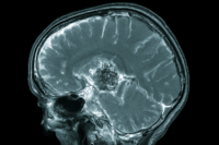
- Why MRI Is Important in Trigeminal Neuralgia
- Trigeminal Neuralgia Treatment: What Are the Main Goals?
- First-Line Medicines for Trigeminal Neuralgia
- What to Do During a Severe Trigeminal Neuralgia Attack
- Second-Line and Add-On Medicines for Trigeminal Neuralgia
- Trigeminal Neuralgia Procedures Compared: How the Options Differ
- What Many Trigeminal Neuralgia Treatment Pages Do Not Explain
- The Hidden Nutritional and Oral Consequences of Trigeminal Neuralgia
- Ayurvedic Understanding of Trigeminal Neuralgia
- How Ayurveda Can Be Integrated Responsibly
- Trigeminal Neuralgia Diet and Daily Living: Protecting Nutrition Without Unnecessary Restrictions
- Oral Hygiene and Dental Care During Trigeminal Neuralgia
- Work, Driving and Daily Safety
- Travel Planning for a Patient With Trigeminal Neuralgia
- A 30-Day Trigeminal Neuralgia Action Plan
- How to Measure Whether Trigeminal Neuralgia Treatment Is Working
- Who May Benefit From Integrative Ayurvedic Care?
- What If Trigeminal Neuralgia Treatment Is Not Working?
- When Should You Seek a Second Opinion?
- How to Choose a Trigeminal Neuralgia Treatment Centre
- A Structured Integrative Trigeminal Neuralgia Pathway
- What Results Can a Patient Realistically Expect?
- Can Ayurveda Cure Trigeminal Neuralgia?
- Best Classical-Inspired Avaleha Name
- Patient-Friendly Preparation Method
- Dosage and 30-Day Course
- Safety, Screening and Monitoring
- Important Warning: Do Not Buy a Ready-Made Trigeminal Neuralgia Avaleha From the Market
- Why a Market-Bought Avaleha May Not Work
- Why Doctor-Supervised Customisation Gives the Best Chance of Response
- Which Tests Are Actually Useful in Trigeminal Neuralgia?
- Trigeminal Neuralgia After Shingles or Other Nerve Injury
- Choosing Trigeminal Neuralgia Treatment According to the Patient Profile
- How to Use a Trigeminal Neuralgia Pain Diary Clinically
- Avoiding Both Overtreatment and Undertreatment
- Understand Trigeminal Neuralgia Treatment Ethically
- The Next Decision Should Solve the Patient’s Main Problem
- Ayurvedic Medicines Used in Trigeminal Neuralgia
- Ayurvedic Management When Carbamazepine Is Already Being Taken
- Ayurvedic Treatment After Failed Dental Procedures
- Ayurveda After Microvascular Decompression, Gamma Knife or Radiofrequency
- What Modern Research Says About Ayurvedic Herbs for Neuropathic Pain
- What Published Evidence Exists for Ayurveda in Trigeminal Neuralgia?
- Frequently Asked Questions About Trigeminal Neuralgia
- Reference
Ayurvedic cure for trigeminal neuralgia begins with identifying the exact cause and pattern of the patient’s facial pain. Trigeminal neuralgia can cause sudden, severe and electric-shock-like pain while eating, speaking, brushing the teeth, washing the face or touching a sensitive area. Although the pain may be felt in the teeth, gums, cheek, jaw, lips, forehead or around the eye, the underlying problem usually involves abnormal activity within the trigeminal nerve pathway.
The Ayurvedic cure for trigeminal neuralgia is planned according to the patient’s pain pattern, MRI findings, affected trigeminal nerve division, disease duration, previous dental procedures, current neurological medicines, digestion, sleep, strength and associated chronic disorders. Treatment is not limited to temporary pain suppression. It aims to reduce electric-shock attacks, improve trigger tolerance, restore eating and speaking, support sleep and nutrition, and help the patient achieve sustained functional recovery.
In Ayurveda, selected trigeminal neuralgia presentations may be clinically correlated with Anantavāta, described in the Suśruta Saṃhitā, Uttara-tantra, Śiroroga Vijñānīya Adhyāya 25. Treatment principles are described in Śiroroga Pratiṣedha Adhyāya 26, while broader Vāta-management principles are discussed in Caraka Saṃhitā, Cikitsā-sthāna 28, Vātavyādhi Cikitsā [19,52].
A personalised Ayurvedic cure for trigeminal neuralgia may include carefully selected Vāta-Śāmaka, Vedanāsthāpana, Bṛṃhaṇa, Medhya and Rasāyana medicines, together with appropriate dietary and procedural support. MRI, neurological evaluation and neurosurgical advice remain important when neurovascular compression, multiple sclerosis, tumour or another secondary cause is suspected.
Trigeminal neuralgia is a neurological pain disorder that causes sudden, severe and usually one-sided facial pain. Patients commonly describe the pain as an electric shock, stabbing jolt, sharp current or shooting sensation across the cheek, jaw, teeth, gums, lips, forehead or area around the eye. A single attack may last only a few seconds, but repeated attacks can make eating, speaking, smiling, washing the face or brushing the teeth frightening [1,2,6,17,18].
The condition is more than an occasional facial pain. A light touch, cool breeze, movement of the lips or contact with a toothbrush may trigger pain severe enough to interrupt an ordinary activity immediately. Some patients experience separated attacks with completely pain-free intervals, while others develop an aching, burning or throbbing background pain in the same area between the electric-shock episodes [1,2,6,18].
One of the greatest clinical problems is that trigeminal neuralgia can be felt in the teeth, gums or jaw. Patients may therefore undergo dental consultations, root-canal procedures or even tooth extraction before the neurological nature of the pain is recognised. Dental disease must be properly investigated when suspected, but irreversible dental treatment should not be used as an experiment when no dental abnormality adequately explains a brief, shock-like and touch-triggered pain pattern [2,8,18].
The objective of this guide is not to label every facial pain as trigeminal neuralgia. It is to help you understand whether the pattern resembles the disorder, recognise warning signs, prepare for the correct investigation and compare the available treatment pathways. It will explain medicines, MRI findings, microvascular decompression, radiosurgery, percutaneous procedures and the potential supportive role of Ayurveda within coordinated neurological care.
An MRI report alone does not diagnose trigeminal neuralgia. The diagnosis begins with a detailed description of the pain, its duration, location, triggers and accompanying symptoms. Imaging is then used to investigate neurovascular compression and exclude conditions such as multiple sclerosis, tumours and other structural causes [2,3,5,6].
When Facial Pain Requires Urgent Medical Assessment
Most facial pain is not a medical emergency, but certain findings require faster evaluation because they may indicate secondary trigeminal neuralgia or another neurological disorder.
Seek prompt medical or neurological assessment when facial pain is accompanied by facial numbness, altered sensation, muscle weakness, hearing loss, persistent ear symptoms, visual disturbance, optic neuritis, pain on both sides of the face, fever, unexplained weight loss, a previous history of cancer or other new neurological symptoms. Presentation below 30 years of age is also regarded as a reason for more urgent specialist assessment [2].
Urgent care is also appropriate when the pain prevents adequate eating or drinking, causes dehydration or makes it impossible to take prescribed medicine. A person experiencing thoughts of self-harm or suicide should contact emergency medical and mental-health services immediately. Research has documented substantial anxiety, depression, sleep disturbance and suicidal ideation among some patients with trigeminal neuralgia and related neuralgias, making psychological safety an essential part of treatment rather than an optional addition [15,16].
Does Your Facial Pain Fit the Pattern of Trigeminal Neuralgia?
The pattern of the pain is usually more informative than pain intensity alone. Many conditions can cause severe facial pain, but trigeminal neuralgia has a distinctive combination of short duration, electric-shock quality, trigeminal distribution and provocation by harmless stimulation.
The following self-check can help you prepare for a medical consultation. It cannot confirm the diagnosis.
| Clinical feature | Pattern commonly reported in trigeminal neuralgia | Why it matters |
| Pain quality | Electric shock, stabbing, shooting, sharp or piercing | Helps distinguish neuralgic paroxysms from ordinary inflammatory pain |
| Attack duration | From a fraction of a second to approximately two minutes | This is part of the recognised ICHD-3 diagnostic pattern |
| Side of the face | Usually one side | Bilateral pain requires particularly careful assessment |
| Pain location | Cheek, upper or lower jaw, teeth, gums, lips, chin, forehead or eye region | Corresponds to one or more divisions of the trigeminal nerve |
| Trigger | Light touch, chewing, speaking, brushing, shaving, washing or wind | Pain provoked by harmless stimulation is highly characteristic |
| Beginning and end | Abrupt onset and abrupt cessation | Differs from pain that gradually builds and persists for hours |
| Between attacks | Completely pain-free in some patients; background aching or burning in others | Helps determine whether concomitant continuous pain is present |
| Neurological examination | Usually no obvious sensory or motor deficit in classical cases | Numbness, weakness or reflex changes increase concern for a secondary cause |
| Response to ordinary painkillers | Often inadequate | TN is neuropathic pain and may not respond like toothache or musculoskeletal pain |
| Pattern over time | Repeated attacks over days, weeks or months, sometimes followed by remission | Remission does not necessarily mean that the underlying disorder has permanently resolved |
The International Classification of Headache Disorders defines trigeminal neuralgia by recurrent unilateral facial-pain paroxysms within one or more trigeminal nerve divisions. The characteristic pain is severe, electric-shock-like, shooting, stabbing or sharp, lasts from a fraction of a second to two minutes and is precipitated by harmless stimulation within the affected region [1].
Not every patient describes the condition in precisely the same words. Some may say the face is being struck by lightning, pierced by a needle or touched by a live electrical wire. Others may initially report “tooth pain” because the attack is experienced in the upper or lower jaw. The clinician must therefore analyse the complete pattern rather than depend on a single descriptive term.
Does Constant Facial Pain Exclude Trigeminal Neuralgia?

Constant facial pain does not automatically exclude the condition. A recognised subtype is trigeminal neuralgia with concomitant continuous pain, in which a persistent aching, burning or throbbing sensation occurs in the same trigeminal distribution as the sharp attacks [1,2,6].
However, continuous facial pain without characteristic electric-shock paroxysms requires a broader evaluation. Dental disease, temporomandibular disorder, persistent idiopathic facial pain, postherpetic neuralgia, migraine-related disorders, sinus disease and other neuropathic conditions may produce different patterns. A person should not be diagnosed solely because the pain is severe or located on one side of the face.
Why a Seven-Day Facial Pain Diary Is Valuable
A pain diary provides information that may be difficult to remember during a consultation. For at least seven days, record:
| Information to record | Example |
| Date and time | 8:15 a.m., after breakfast |
| Side of the face | Right |
| Exact location | Upper teeth, cheek and side of nose |
| Pain quality | Electric shock |
| Duration | Approximately 20 seconds |
| Trigger | Chewing or touching the upper lip |
| Number of attacks | Nine attacks within one hour |
| Background pain | Mild burning between attacks |
| Medicine taken | Name, dose and time |
| Functional effect | Stopped eating or avoided brushing |
| Additional symptoms | Numbness, tearing, hearing or vision changes |
The Royal College of Surgeons guideline recommends pain diaries and patient-reported outcome measures because treatment decisions should consider attack frequency, medicine use and functional impact, not merely a single pain score [2].
What Is Trigeminal Neuralgia?

The trigeminal nerve is the fifth cranial nerve and the principal sensory nerve of the face. There is one trigeminal nerve on each side. It carries sensations such as touch, pressure, temperature and pain from the face, teeth and mouth towards the brain. Its mandibular division also contributes to the movement of muscles used for chewing [17].
The nerve separates into three principal divisions:
| Trigeminal division | Medical name | Main region supplied |
| V1 | Ophthalmic division | Forehead, front of the scalp, upper eyelid, eye region and upper nose |
| V2 | Maxillary division | Cheek, side of the nose, upper lip, upper teeth, gums and upper jaw |
| V3 | Mandibular division | Lower lip, chin, lower teeth, gums, jaw and part of the chewing muscles |
Pain may occur in one division or spread through more than one. The maxillary and mandibular divisions are frequently involved, which helps explain why patients may believe that the pain originates from an upper or lower tooth. The ophthalmic division may also be affected, although isolated involvement around the forehead and eye is less common [4,6]. The latest pooled epidemiological analysis suggested more frequent V2 and V3 involvement than V1, but the certainty of these subgroup findings was low [4].
Why Can a Light Touch Cause Such Severe Pain?
In classical trigeminal neuralgia, a blood vessel usually an artery and sometimes a vein compresses the trigeminal nerve root near the brainstem. This pressure may be associated with damage to the protective myelin covering of the nerve and abnormal transmission between neighbouring nerve fibres. As a result, an ordinary touch signal may contribute to a burst of abnormal pain activity [5–7,18].
This mechanism helps explain why a stimulus that should be painless, such as touching the cheek or feeling moving air, can provoke an extreme shock-like response. The painful attack is real even when the skin, teeth and face appear normal.
However, a blood vessel touching the trigeminal nerve on MRI is not automatically proof that the vessel is causing the pain. Neurovascular contact can occur without trigeminal neuralgia. Classical TN is more strongly supported when imaging demonstrates compression accompanied by a morphological change in the nerve, such as displacement, distortion or atrophy, and when the imaging finding corresponds with the clinical side and pattern of pain [3,5,6].
This distinction is important because treatment should not be based on one sentence in an MRI report. The pain history, neurological examination, quality of the MRI sequences and specialist interpretation must be considered together.
The Three Main Types of Trigeminal Neuralgia
Trigeminal neuralgia is not one uniform disease. Modern classification separates it into classical, secondary and idiopathic forms [1,2,5,6].
Classical Trigeminal Neuralgia
Classical trigeminal neuralgia is associated with neurovascular compression of the trigeminal nerve root accompanied by morphological changes in the nerve. Patients usually experience brief, severe and triggerable attacks, with or without a continuous background component [1,5].
The identification of a convincing neurovascular conflict is important because it may influence whether microvascular decompression is considered when medicine becomes ineffective or poorly tolerated.
Secondary Trigeminal Neuralgia
Secondary trigeminal neuralgia occurs when another neurological or structural condition affects the trigeminal pathway. Recognised causes include multiple sclerosis, tumours, cysts, vascular malformations and other lesions affecting the nerve or brainstem [2,3,6,18].
This is one reason MRI should not be treated as optional in a properly investigated case. The Royal College guideline notes that a small subset—reported as up to 10% of people presenting with TN—may have an underlying pathology detectable through neuroimaging [2]. This is a guideline estimate and should not be interpreted as the probability for every individual patient.
Idiopathic Trigeminal Neuralgia
Idiopathic trigeminal neuralgia is diagnosed when the clinical characteristics meet recognised criteria but available investigations do not identify a definite neurovascular compression or another condition that adequately explains the pain [1,5,6].
The word “idiopathic” does not mean that the pain is imaginary. It means that the underlying cause has not been demonstrated through the available diagnostic methods.
Purely Paroxysmal Pain and Concomitant Continuous Pain
Each of the major diagnostic categories may be further described according to whether the patient has only brief attacks or also experiences background pain.
Purely paroxysmal trigeminal neuralgia produces separated attacks with no pain in the affected distribution between episodes. Trigeminal neuralgia with concomitant continuous pain includes a persistent or near-persistent aching, burning or throbbing sensation in the same area as the paroxysmal shocks [1,2].
This distinction matters because patients with a continuous component may have a different treatment experience and should not be dismissed simply because they do not become completely pain-free between attacks.
Common Trigeminal Neuralgia Triggers
A trigger is an event that provokes an attack; it is not necessarily the underlying cause of the disorder. Chewing does not cause trigeminal neuralgia, and brushing the teeth does not damage the nerve. These actions may stimulate an already hypersensitive trigeminal pathway.
Commonly reported triggers include talking, smiling, chewing, swallowing, brushing the teeth, washing the face, shaving, applying makeup, kissing, touching a small trigger zone and exposure to cool air, wind or air conditioning. Some attacks occur without an identifiable trigger [1,2,17,18].
| Trigger | Practical adaptation during an active flare |
| Brushing the teeth | Use a gentle technique and a suitably soft toothbrush while maintaining oral hygiene |
| Washing the face | Use lukewarm water and avoid forceful rubbing |
| Chewing | Take smaller bites and choose nutritionally adequate softer foods temporarily |
| Hot or cold food | Use a neutral or comfortable temperature when temperature provokes attacks |
| Cool wind or air conditioning | Protect the affected side with a soft scarf or suitable face covering |
| Shaving or makeup | Avoid repeated pressure over a known trigger zone |
| Speaking | Communicate more slowly during a severe flare, but avoid complete social withdrawal |
| Drinking | Use a tolerable temperature and method while maintaining adequate hydration |
Trigger modification can reduce avoidable provocation, but it does not correct neurovascular compression, demyelination or another structural cause. Excessive avoidance can also create new health problems. Some patients begin skipping meals, drinking less, avoiding oral hygiene or withdrawing socially because they fear another attack. The NHS warns that undernutrition and dehydration may worsen the overall situation and that trigeminal neuralgia can substantially affect psychological and social well-being [18].
The better approach is to document personal triggers, make reasonable temporary adaptations and pursue diagnostic and therapeutic control rather than allowing the fear of pain to control every activity.
How Common Is Trigeminal Neuralgia?
Trigeminal neuralgia has traditionally been described as an uncommon neurological disorder, but published estimates vary substantially according to population, diagnostic criteria, healthcare access and whether modern neuroimaging was incorporated into the diagnostic process.
A 2026 systematic review and meta-analysis included 17 studies covering approximately 174.5 million people and 109,070 diagnosed cases. The researchers estimated a pooled global incidence of 25.33 cases per 100,000 person-years, an annual prevalence of 45.38 per 100,000 inhabitants and a lifetime prevalence of 108.43 per 100,000 inhabitants [4].
These figures should be interpreted cautiously. Heterogeneity was extremely high, with an I² exceeding 98% for each pooled estimate. The studies used different designs, populations, diagnostic systems and time periods. The pooled figures therefore demonstrate that trigeminal neuralgia is a recognised global health problem, but they do not provide a precise prediction for every country or community [4].
The same review found descriptive patterns suggesting more diagnoses among women, on the right side of the face and in the maxillary and mandibular divisions. However, several subgroup findings had low certainty or did not remain statistically significant in meta-regression. They should not be used to exclude the diagnosis in a man, in a person with left-sided pain or in someone with ophthalmic-division involvement [4].
Why Early and Accurate Diagnosis Matters
An accurate diagnosis can prevent three avoidable harms: irreversible treatment directed at the wrong structure, delayed identification of an underlying neurological disorder and prolonged exposure to ineffective medicines.
In a UK survey cited by the Royal College guideline, 8% of 225 patients attending a specialist facial-pain clinic had undergone irreversible and unnecessary dental treatment before reaching the service. Only 54% had received carbamazepine before specialist attendance, despite its established role as a first-line medicine [2]. These figures describe one referred UK population and should not be treated as universal rates, but they illustrate how diagnostic delay can affect patient care.
Early recognition does not mean rushing into surgery or accepting the first diagnostic label. It means establishing whether the pain fulfils recognised clinical criteria, excluding credible dental and facial-pain alternatives, performing appropriate neurological examination and obtaining suitable MRI assessment.
The next stage of the diagnostic pathway is to determine why the pain is occurring: whether there is meaningful neurovascular compression, multiple sclerosis, a structural lesion or no demonstrable cause. That distinction directly influences the choice between medical treatment, neurosurgical evaluation and carefully coordinated integrative support.
What Causes Trigeminal Neuralgia?
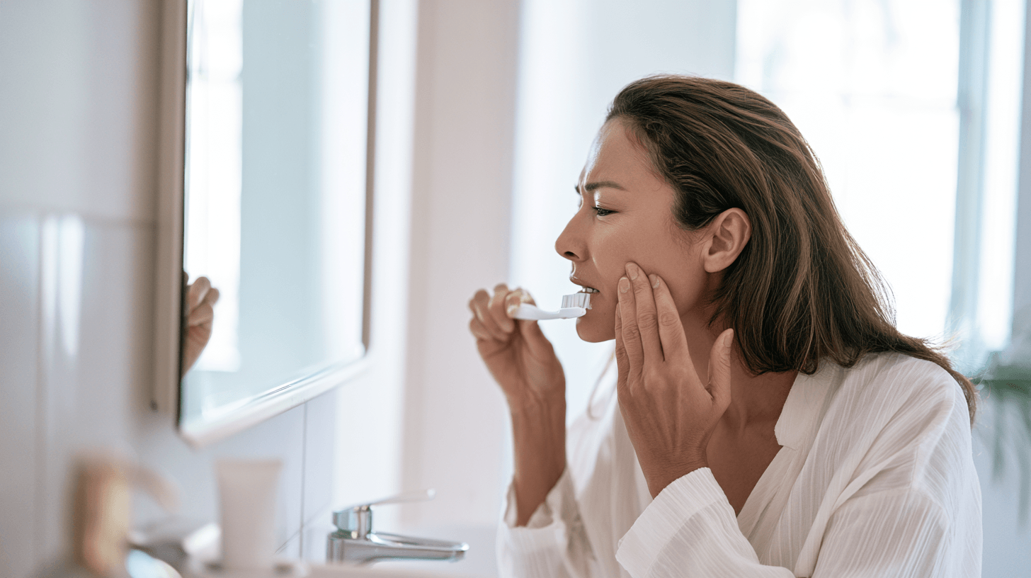
Trigeminal neuralgia develops when the trigeminal pain pathway becomes abnormally excitable. In many patients, the problem begins near the point where the trigeminal nerve enters the brainstem. A blood vessel may compress the nerve, damage its protective myelin covering and allow ordinary sensory signals to trigger bursts of severe pain [5–7].
However, this is not the only possible cause. Trigeminal neuralgia may also occur because of multiple sclerosis, a tumour, a vascular abnormality or another lesion affecting the nerve or brainstem. In some patients, the clinical pattern is unmistakable, but MRI does not identify a definite cause. This is classified as idiopathic trigeminal neuralgia [1,3,5,6].
You should therefore avoid assuming that all trigeminal neuralgia has the same origin. The correct diagnosis depends on your symptoms, neurological examination and appropriate imaging rather than pain intensity alone.
Neurovascular Compression
Neurovascular compression is the most widely recognised mechanism in classical trigeminal neuralgia. It occurs when an artery or, less commonly, a vein presses against the trigeminal nerve root near the brainstem [5–7].
Repeated pulsation from the vessel may injure the myelin that normally insulates the nerve. Once this insulation becomes disturbed, signals can cross abnormally between nearby nerve fibres. A light facial touch that should produce a normal sensory signal may then initiate an intense burst of pain.
This mechanism can explain why brushing your teeth, touching your cheek or feeling a cool breeze may cause an attack even though there is no visible injury on the face.
The presence of a blood vessel near the trigeminal nerve on MRI does not automatically prove that it is responsible for the pain. Blood vessels may touch cranial nerves in people who have no symptoms. Clinically meaningful compression is more likely when imaging also shows displacement, distortion, indentation or atrophy of the trigeminal nerve and when the finding corresponds with the painful side [3,5,6].
We should therefore distinguish between simple neurovascular contact and neurovascular compression accompanied by morphological changes. That distinction may influence whether microvascular decompression is considered later.
Multiple Sclerosis and Trigeminal Neuralgia
Multiple sclerosis can affect the myelin surrounding nerve pathways within the brainstem. When demyelination involves the trigeminal pathway, it may produce pain resembling classical trigeminal neuralgia [2,3,6].
A younger person with trigeminal neuralgia, pain affecting both sides of the face or additional neurological symptoms requires especially careful evaluation. Possible accompanying symptoms may include changes in vision, limb weakness, altered sensation, balance problems or bladder disturbance.
These findings do not prove that multiple sclerosis is present, but they increase the importance of neurological assessment and suitable MRI examination. The clinician may need to inspect the brainstem and other areas of the brain for demyelinating lesions rather than looking only for a compressing blood vessel.
Some patients with multiple sclerosis may have both a brainstem lesion and neurovascular compression. Their treatment pathway can therefore be more complex than that of a patient with uncomplicated classical trigeminal neuralgia [3,6].
Tumours and Other Structural Lesions
Tumours affecting the cerebellopontine angle, trigeminal nerve, skull base or nearby structures can occasionally produce trigeminal neuralgia or another form of facial pain. Cysts, vascular malformations and other structural abnormalities may also affect the trigeminal pathway [2,3,6].
The Royal College of Surgeons guideline reports that up to approximately 10% of people presenting with trigeminal neuralgia may have an underlying condition detectable through neuroimaging, including a tumour, multiple sclerosis or vascular malformation [2]. This is a guideline estimate and not an individual prediction.
Most people with trigeminal neuralgia do not have a tumour. Nevertheless, imaging remains important because the consequences of missing a secondary cause may be serious.
Persistent facial numbness, hearing changes, facial weakness, balance difficulty, visual symptoms, unexplained weight loss or previous cancer should not be dismissed as ordinary features of classical trigeminal neuralgia.
Idiopathic Trigeminal Neuralgia
In idiopathic trigeminal neuralgia, the patient has a characteristic clinical pain pattern, but imaging and other investigations do not demonstrate a definite cause [1,5,6].
The term “idiopathic” may be frustrating because you may feel that no one has explained why the pain is occurring. It does not mean that your pain is psychological or imaginary. It means that current investigations have not identified a structural or neurological cause that fully accounts for the symptoms.
Treatment may still be effective even when MRI does not show definitive neurovascular compression. The diagnosis and management plan should remain based on the complete clinical picture.
Why Trigeminal Neuralgia Is Often Mistaken for Toothache
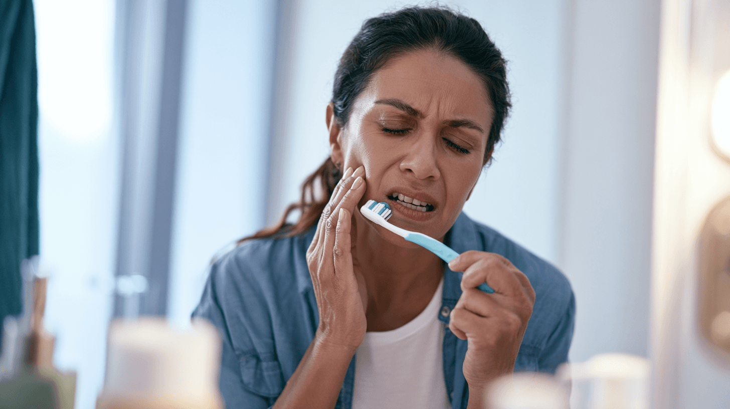
Trigeminal neuralgia frequently affects the maxillary and mandibular divisions of the trigeminal nerve. Because these divisions supply the upper teeth, lower teeth, gums, cheek and jaw, the pain may appear to originate from a particular tooth [2,6,8,18].
A patient may visit a dentist first because the pain is felt inside the mouth. This is understandable. Dental disease is common, and genuine dental problems can produce severe facial pain. The difficulty arises when a healthy tooth is treated because the neurological pattern has not been recognised.
Some patients undergo fillings, root-canal procedures or tooth extractions without lasting relief. The pain may temporarily change because of local anaesthesia, medication or natural fluctuation, only to return in the same or an adjacent part of the face.
A 2025 retrospective study of 104 patients with trigeminal neuralgia documented a substantial history of dental procedures performed before the neurological diagnosis was established. Many of those procedures failed to provide meaningful or sustained relief [8]. Because it was a single-centre retrospective study, the findings should not be treated as a universal rate, but they highlight a preventable clinical problem.
Features That Suggest Trigeminal Neuralgia Rather Than Tooth Disease
Trigeminal neuralgia becomes more likely when the pain arrives as sudden electric shocks, lasts for seconds, ends abruptly and is triggered by touching the face, speaking, chewing or brushing the teeth [1,2,6].
The pain may seem to move between several teeth or affect an entire region of the jaw. A person may struggle to identify one consistently painful tooth. Dental examination and radiographs may fail to reveal pathology that adequately explains the severity and pattern of the pain.
Previous root-canal treatment or tooth extraction that does not alter the characteristic attacks should prompt diagnostic reconsideration. Continuing to treat additional teeth without a clear dental finding may cause irreversible harm without addressing the nerve disorder.
Features That Support a Dental Cause
Dental pain may be more localised to one tooth and may be accompanied by decay, fracture, gum swelling, infection or an abnormal dental radiograph. Pain may be provoked by biting pressure, percussion, sweets or hot and cold substances, depending on the underlying problem.
Dental pain may throb or persist for much longer than a typical trigeminal neuralgia paroxysm. However, overlap can occur, and some patients may have both dental disease and trigeminal neuralgia.
We should therefore not tell patients to avoid dentists. The safer approach is to confirm whether objective dental pathology corresponds with the painful site before carrying out irreversible treatment.
Before Agreeing to an Irreversible Dental Procedure
Before undergoing extraction or another irreversible procedure for unexplained shock-like facial pain, ask whether the dental abnormality clearly explains the attacks. You may also ask whether the pain pattern has been assessed for trigeminal neuralgia and whether neurological evaluation is appropriate.
A dentist, neurologist, oral-medicine specialist or facial-pain clinician may need to work together when the diagnosis remains uncertain. The purpose is not to delay necessary dental care but to prevent a healthy tooth from being treated as the source of neuropathic pain [2,8,9].
Trigeminal Neuralgia Versus Other Causes of Facial Pain

Not every sharp or severe facial pain is trigeminal neuralgia. Diagnosis requires comparison with other dental, neurological, headache-related and musculoskeletal conditions [1,2,6,9].
The distinction matters because treatments that work for one disorder may fail or even create harm in another.
Dental Disease
Dental infection, pulp inflammation, a cracked tooth, periodontal disease and impacted teeth can cause pain in the same regions affected by trigeminal neuralgia.
Dental pain is usually supported by an abnormal clinical or radiographic finding. The pain may worsen with biting, tapping the tooth, temperature exposure or pressure over the affected area.
Trigeminal neuralgia, by contrast, typically produces brief attacks triggered by harmless stimulation. Nevertheless, history alone may not always be enough, and a proper dental examination may be necessary.
Temporomandibular Disorders
Temporomandibular disorders commonly cause pain around the jaw joint, temple, cheek or ear. The discomfort may worsen with jaw movement and may be accompanied by clicking, restricted mouth opening or tenderness of the chewing muscles.
This pain is generally more aching or mechanical than the sudden electric-shock attacks of trigeminal neuralgia. A patient with temporomandibular disorder may experience pain while chewing, but the mechanism and duration differ from the triggerable paroxysms of TN.
Postherpetic Neuralgia
Postherpetic neuralgia may occur after herpes zoster, commonly called shingles, affects a trigeminal nerve division. The patient often has a history of a painful blistering rash on one side of the face, forehead or around the eye.
The later pain may be burning, persistent and associated with extreme skin sensitivity. Brief stabbing pains can also occur, but the history of shingles and the continuous neuropathic component help distinguish it from classical trigeminal neuralgia.
Pain involving the eye region after shingles requires careful ophthalmological assessment because the eye may also be affected.
Persistent Idiopathic Facial Pain
Persistent idiopathic facial pain usually produces a continuous or near-continuous aching, burning or poorly localised discomfort. It may not follow the anatomical boundaries of one trigeminal nerve division.
Unlike classical trigeminal neuralgia, the pain is not dominated by repeated electric-shock paroxysms triggered by light touch. Diagnosis usually requires exclusion of dental, neurological and other facial-pain disorders.
The condition is real and can be disabling. Calling it “idiopathic” does not mean that the patient is imagining the symptoms.
Glossopharyngeal Neuralgia
Glossopharyngeal neuralgia produces brief, severe stabbing pain in areas such as the throat, base of the tongue, tonsillar region or deep ear. Swallowing, coughing, yawning or speaking may trigger an attack.
It resembles trigeminal neuralgia because both conditions produce short, triggerable neuralgic pains. The anatomical location of the pain and the provoking activities help distinguish them.
Cluster Headache and Other Headache Disorders
Cluster headache causes severe pain around one eye and may be accompanied by tearing, eye redness, nasal congestion, eyelid swelling or restlessness. Attacks generally last longer than trigeminal neuralgia attacks and follow a different pattern.
Some migraine disorders may also cause facial pain. A headache specialist may be needed when pain is associated with nausea, light sensitivity, prolonged attacks or prominent autonomic symptoms.
Sinus-Related Facial Pain
True sinus-related facial pain is usually accompanied by evidence of nasal or sinus disease, such as congestion, purulent discharge, reduced smell, fever or inflammatory findings on examination or imaging.
Facial pressure alone does not confirm sinusitis. Repeated antibiotic treatment without objective evidence of infection may delay recognition of a neurological or headache-related disorder.
Warning indicators That Require Faster Specialist Assessment
Classical trigeminal neuralgia usually does not cause persistent facial numbness, weakness or other neurological deficits. When these symptoms are present, the clinician must consider a secondary cause or a different diagnosis [1–3,6].
You should seek faster medical assessment when facial pain is accompanied by numbness that persists between attacks, reduced corneal sensation, facial weakness, hearing changes, balance problems, visual disturbance or other neurological symptoms.
Pain affecting both sides of the face, onset at a young age, a history of multiple sclerosis, previous cancer, unexplained fever or weight loss also increases the need for detailed investigation [2,3].
A change in an established pain pattern matters as well. A person previously experiencing brief triggered shocks who develops continuous severe pain, progressive numbness or weakness should be reassessed rather than simply increasing medicine without review.
Inability to eat or drink is another important warning sign. Severe attacks may cause patients to avoid food and water, leading to dehydration, weakness, weight loss and difficulty taking prescribed medication.
Psychological deterioration must also be recognised. If you begin feeling hopeless, unable to cope or at risk of harming yourself, urgent mental-health and emergency support is necessary. Trigeminal neuralgia is a physical neurological disorder, but its emotional consequences may be profound [15,16].
How Trigeminal Neuralgia Is Diagnosed
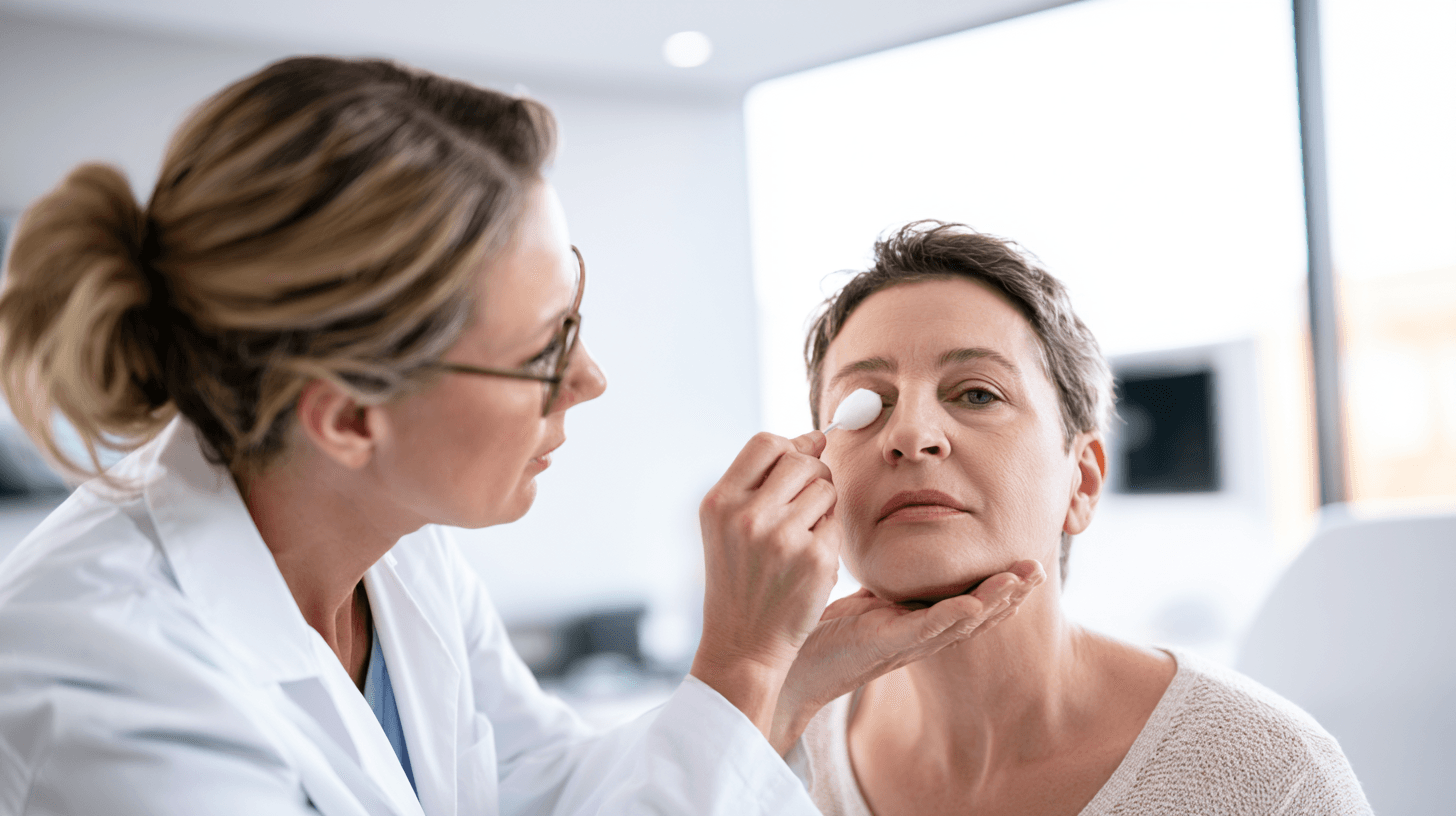
There is no single blood test that confirms trigeminal neuralgia. Diagnosis begins with a detailed clinical history and neurological examination. MRI is then used to investigate possible neurovascular compression and exclude secondary causes [1–3,5,6].
A good diagnosis brings together four elements: the pain pattern, the anatomical distribution, the examination findings and the imaging results.
Detailed Description of the Pain
Your clinician will usually ask where the pain begins, where it spreads, how long each attack lasts and what triggers it. The words you use to describe the pain are important, but they are only part of the assessment.
The clinician may ask whether the pain feels electric, stabbing, shooting, burning, aching or throbbing. He or she will also need to know whether you are completely pain-free between attacks or whether a continuous background pain remains.
Attack frequency should be documented. Some patients experience a few attacks a day, while others have repeated bursts over a short period. It is also useful to describe periods of remission and whether the attacks have become more frequent, more prolonged or easier to trigger.
Mapping the Pain Distribution
The painful region should be mapped according to the ophthalmic, maxillary and mandibular divisions of the trigeminal nerve.
You may feel pain in one small trigger zone, one nerve division or several connected facial regions. The pain usually remains on one side, although bilateral symptoms can occur, particularly in some secondary conditions.
The clinician should also clarify whether pain extends outside the trigeminal distribution. Pain that spreads unpredictably into the neck, shoulder or other non-trigeminal areas may suggest another diagnosis or more than one pain disorder.
Neurological Examination
The neurological examination may include facial sensation, corneal reflexes, facial movement, hearing, eye movement, coordination and examination of the limbs where appropriate.
In classical trigeminal neuralgia, routine examination may be normal. Persistent sensory loss, weakness or other abnormalities increase concern for secondary trigeminal neuralgia or another neurological condition [2,3,6].
A normal examination does not remove the need for MRI, but it helps define the urgency and probable diagnostic category.
Dental and Oral Examination
A dental or oral examination may be appropriate when the pain is felt in the teeth, gums or jaw. The dentist should look for decay, pulp disease, periodontal infection, fracture, impacted teeth and other pathology that may explain the symptoms.
When dental findings are absent or do not correspond with the pain pattern, the diagnosis should be reconsidered before irreversible treatment is performed.
A patient with confirmed trigeminal neuralgia still requires normal preventive dental care. Avoiding all dental visits because brushing or treatment may trigger pain can allow genuine oral disease to develop.
Response to Medicine
A favourable response to carbamazepine or oxcarbazepine may support the clinical impression, but medication response alone should not be used as a diagnostic test [2,3,6].
Some patients with other neuropathic pain disorders may also improve with these medicines. Conversely, a patient with genuine trigeminal neuralgia may respond incompletely, lose benefit over time or be unable to tolerate the medicine.
The diagnosis should therefore not be accepted or rejected solely according to treatment response.
Why MRI Is Important in Trigeminal Neuralgia
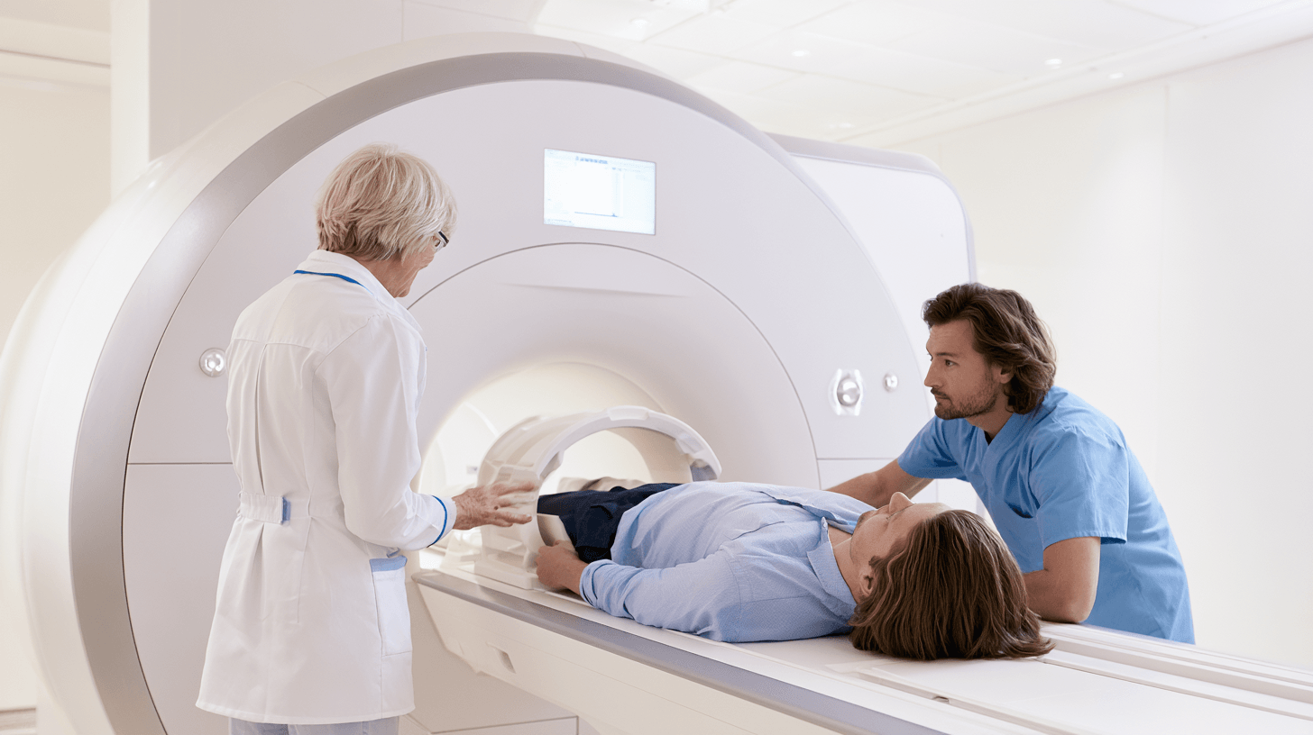
MRI is recommended as part of the diagnostic work-up because clinical symptoms alone cannot reliably exclude secondary trigeminal neuralgia [2,3].
The scan has two main purposes. The first is to evaluate the trigeminal nerve and look for clinically meaningful neurovascular compression. The second is to examine the brain and surrounding structures for multiple sclerosis, tumours, cysts, vascular abnormalities and other causes.
A routine brain MRI may not always provide the detailed views required to assess the trigeminal nerve properly. High-resolution sequences through the posterior fossa are often needed.
MRI Sequences Used to Assess the Trigeminal Nerve
Specialist guidelines describe the value of high-resolution three-dimensional T2-weighted sequences for visualising the trigeminal nerve and surrounding cerebrospinal fluid spaces [2,3,6].
Three-dimensional time-of-flight magnetic resonance angiography may help identify nearby arteries and evaluate their relationship with the nerve. Additional brain sequences are used to look for demyelinating lesions, tumours and other structural abnormalities.
Contrast may be required when the clinical history or initial imaging raises concern for a tumour, inflammation or another secondary process. The exact protocol should be selected by the radiologist and referring specialist.
Neurovascular Contact Is Not the Same as Compression
MRI reports sometimes state that a blood vessel “contacts” the trigeminal nerve. This statement should not automatically be interpreted as confirmation of the cause.
Simple contact can occur without symptoms. Greater diagnostic importance is generally given to compression associated with morphological change, such as displacement, indentation, distortion or atrophy of the nerve [3,5,6].
The location of the vessel also matters. Compression close to the root-entry zone near the brainstem is particularly relevant in classical trigeminal neuralgia.
A specialist should compare the imaging side with the side of the pain. A vessel touching the left trigeminal nerve is less likely to explain attacks occurring exclusively on the right.
Arterial and Venous Compression
An artery is more commonly identified as the offending vessel in classical trigeminal neuralgia, although veins and multiple vessels may also compress the nerve [5,7,13].
The type of vessel may influence surgical planning and the probability of a favourable outcome after microvascular decompression. A 2024 systematic review and meta-analysis found that arterial compression was associated with more favourable outcomes than venous or multiple-vessel compression, although study methods and definitions varied [13].
This does not mean that a patient with venous compression cannot benefit from surgery. It means that imaging details should be considered during individual risk-benefit counselling.
A Normal MRI Does Not Make the Pain Unreal
Some patients have a classic clinical pattern but no definite neurovascular compression or secondary lesion on MRI. They may be diagnosed with idiopathic trigeminal neuralgia after appropriate evaluation [1,5,6].
MRI technology, image quality, sequence selection and interpretation can influence what is detected. When symptoms strongly suggest TN but the report is unclear, review of the actual images by an experienced neuroradiologist, neurologist or neurosurgeon may be valuable.
A normal scan should not be used to dismiss characteristic neuropathic pain. At the same time, it should prevent clinicians from claiming that a structural cause has been demonstrated when it has not.
What a Trigeminal Neuralgia MRI Report Should Clarify
A useful MRI report should explain whether neurovascular contact or definite compression is present, which vessel is involved and whether the trigeminal nerve shows displacement, indentation, distortion or atrophy [3,5,6,13].
It should identify the side and location of the finding and indicate whether it corresponds with the patient’s symptoms. Where possible, the report should distinguish arterial, venous and multiple-vessel involvement.
The report should also mention whether there is evidence of demyelination, a cerebellopontine-angle lesion, tumour, cyst, vascular malformation or another abnormality affecting the trigeminal pathway.
Patients should preserve both the written report and the original MRI images. A second specialist may need to inspect the images rather than relying only on the report summary.
Questions You Can Ask About Your MRI
You may ask whether the scan used a high-resolution trigeminal nerve protocol, whether the vessel merely touches the nerve or causes morphological compression and whether the finding corresponds with the painful side.
You may also ask whether the remainder of the brain was evaluated for multiple sclerosis, tumours and other secondary causes.
These questions do not require you to interpret the scan yourself. They help ensure that the clinical team has addressed the information necessary for treatment planning.
Which Specialist Treats Trigeminal Neuralgia?
A neurologist often leads the initial diagnostic and medical treatment process. He or she can assess the pain pattern, exclude other neurological disorders, select medicine and monitor response and adverse effects.
A neurosurgeon becomes particularly important when medicine does not provide adequate control, causes unacceptable adverse effects or imaging demonstrates neurovascular compression that may be suitable for a procedure.
A dentist, oral-medicine specialist or maxillofacial clinician may help exclude dental and oral causes. A pain physician may assist with complex medication regimens, selected procedures and multidisciplinary pain management.
Psychological support may be valuable when fear of attacks, sleep disturbance, depression or social withdrawal is becoming severe. This does not imply that the pain is psychological. It recognises that prolonged neurological pain affects the entire person [10,15].
A qualified Ayurvedic physician may participate in an integrative plan, but Ayurvedic treatment should not replace neurological investigation, MRI or neurosurgical assessment when these are indicated.
We prefer a coordinated approach because no single specialist always answers every part of the problem. Once the diagnosis and probable cause are clear, you can compare medical treatment, procedural options and responsible Ayurvedic support with greater confidence.
Trigeminal Neuralgia Treatment: What Are the Main Goals?
Trigeminal neuralgia treatment should do more than reduce the intensity of an electric-shock attack. The wider objective is to help you eat, drink, speak, sleep, work, maintain oral hygiene and interact with other people without constantly anticipating the next episode. A treatment may appear successful on a numerical pain scale but still be inadequate if you remain unable to chew, brush your teeth or leave home because of fear. For this reason, clinicians should assess both pain reduction and restoration of daily function [2,3,6,10].
When we evaluate progress, we should consider the number of attacks, their duration, trigger sensitivity, background facial pain, medicine requirements and adverse effects. A patient who experiences fewer attacks but develops severe dizziness, poor balance, confusion or excessive drowsiness may not have achieved an acceptable outcome. Treatment decisions must balance pain control with safety, independence and overall quality of life rather than focusing exclusively on whether the medicine reduces pain [2,3,10].
Another important goal is to identify the underlying form of trigeminal neuralgia. A person with classical trigeminal neuralgia caused by neurovascular compression may have a different long-term pathway from someone whose pain is related to multiple sclerosis, a tumour or an unidentified cause. Medical treatment may provide adequate control for one patient, while another may require early neurosurgical discussion because medicine is ineffective or intolerable. The diagnosis, MRI findings and individual treatment response should therefore guide the plan together [2,3,5,6].
A complete trigeminal neuralgia treatment plan should also include a clear escalation strategy. You should know what to do if attacks suddenly become more frequent, if you cannot eat or drink, if prescribed medicine stops working or if new neurological symptoms appear. Waiting until the pain becomes unmanageable can lead to dehydration, weight loss, emergency admission and severe psychological distress. A planned pathway is safer than repeatedly responding to each crisis without reviewing the overall diagnosis and treatment strategy [2,3,15].
First-Line Medicines for Trigeminal Neuralgia

Carbamazepine and oxcarbazepine are widely recognised as the principal first-line medicines for trigeminal neuralgia [2,3,6]. These medicines reduce abnormal electrical activity within nerve pathways and may substantially decrease the frequency and severity of shock-like attacks. They are different from ordinary painkillers because trigeminal neuralgia is a neuropathic pain disorder rather than a typical inflammatory, dental or musculoskeletal pain condition.
The medicine that is most suitable for you will depend on your age, general health, other medicines, kidney and liver function, previous adverse reactions and the pattern of your facial pain. Some patients obtain good control with one medicine, while others require a different option or a carefully supervised combination. No patient should copy another person’s dose or alter treatment independently because the safe and effective amount varies considerably between individuals.
Medicine can control the symptoms without necessarily removing the underlying cause. If a blood vessel is compressing the trigeminal nerve, an anticonvulsant may reduce abnormal nerve firing but does not physically separate the vessel from the nerve. This distinction becomes important when a patient develops recurrent pain despite increasing medication or experiences adverse effects that interfere with daily life.
Carbamazepine for Trigeminal Neuralgia
Carbamazepine has long been considered a standard first-line treatment for trigeminal neuralgia and remains one of the most established medical options [2,3,6,18]. Many patients experience a meaningful reduction in paroxysmal attacks when the medicine is introduced and adjusted appropriately. A favourable response can strengthen the clinical impression, although response to carbamazepine alone is not sufficient to confirm the diagnosis.
Treatment is generally started and adjusted gradually under medical supervision. The prescribing clinician considers pain control, tolerability and any relevant laboratory findings before deciding whether the dose should be changed. Increasing the amount too quickly may worsen dizziness, drowsiness, nausea, unsteadiness or cognitive slowing, while remaining on an inadequate amount may leave the pain poorly controlled.
Carbamazepine can interact with several medicines and may influence liver enzymes, blood-cell counts and electrolyte balance in some patients. Depending on your medical history and local clinical guidance, the doctor may arrange blood tests before treatment and during follow-up. A new rash, fever, mouth ulcers, marked weakness, unusual bruising, severe dizziness, jaundice or another significant reaction requires prompt medical assessment rather than self-adjustment of the medicine.
Some patients remain stable on carbamazepine for a prolonged period, while others gradually experience breakthrough attacks or increasing adverse effects. A recurrence does not always mean that the disease has suddenly become severe. It may result from changes in the underlying condition, medicine absorption, interactions, missed doses, reduced tolerance or an incomplete original diagnosis. The correct response is a structured review rather than repeated unsupervised dose escalation.
Oxcarbazepine for Trigeminal Neuralgia
Oxcarbazepine is another recognised first-line option and is commonly considered when carbamazepine is ineffective, poorly tolerated or unsuitable [2,3,6]. It acts through a related mechanism and may reduce abnormal nerve firing responsible for the electric-shock attacks. Some patients find it easier to tolerate, but this does not mean that it is automatically safer or more effective for everyone.
Like carbamazepine, oxcarbazepine may cause dizziness, sleepiness, unsteadiness, nausea and disturbances in blood sodium. Low sodium may present with fatigue, headache, confusion, weakness, worsening balance or, in severe cases, more serious neurological symptoms. Monitoring may therefore be required, particularly in older adults, people taking other medicines that influence sodium or those with relevant medical conditions.
A patient should not abruptly switch between carbamazepine and oxcarbazepine without a clinician-directed plan. Although they are related medicines, their dosing, interactions and individual responses are not identical. The transition may require gradual adjustment to avoid uncontrolled pain, excessive sedation or other complications.
The choice between carbamazepine and oxcarbazepine should be based on the whole person rather than on a general statement that one is universally superior. We need to consider the patient’s pain pattern, previous response, medical conditions, other prescriptions, laboratory results, occupation and ability to tolerate potential cognitive or balance effects.
Why Ordinary Painkillers May Not Control Trigeminal Neuralgia
Patients often try paracetamol, anti-inflammatory medicines or other common analgesics before trigeminal neuralgia is diagnosed. These medicines may be useful for many dental, muscular or inflammatory conditions, but they often provide limited relief for the brief electrical attacks of trigeminal neuralgia because the underlying mechanism is abnormal nerve signalling [2,6].
The Royal College of Surgeons guideline does not support routine opioid treatment for trigeminal neuralgia [2]. Opioids may expose a patient to sedation, constipation, dependence and other harms without reliably controlling the triggerable paroxysms. Their limited usefulness also explains why repeated emergency treatment with conventional painkillers may fail when the diagnosis has not yet been recognised.
A lack of response to an ordinary painkiller does not prove trigeminal neuralgia. It simply indicates that the pain may not be behaving like common inflammatory or tissue-injury pain. The diagnosis must still be based on the characteristic history, neurological assessment and appropriate imaging.
Monitoring Trigeminal Neuralgia Medication
Medication monitoring should begin with a clear baseline. Before treatment, the clinician should document the number of daily attacks, pain distribution, trigger sensitivity, background pain, ability to eat and speak and any pre-existing symptoms such as dizziness or poor balance. Without a baseline, it becomes difficult to determine whether a later problem is caused by the disease, the medicine or another health condition.
During treatment, you should record both benefits and adverse effects. A medicine diary can include the time of each dose, breakthrough attacks, missed doses, drowsiness, dizziness, nausea, skin changes, balance difficulty and any new symptoms. This information gives the doctor a more reliable basis for adjusting treatment than a general statement that the medicine is either “working” or “not working.”
We should also review the complete medicine list, including over-the-counter products, herbal preparations and supplements. Carbamazepine in particular has clinically important interactions, and combining treatments without disclosure can change medicine levels or increase adverse effects. Ayurvedic treatment should therefore be coordinated with neurological care rather than added secretly or used as a reason to discontinue prescribed medicine abruptly.
The goal is the lowest treatment burden that provides acceptable and sustained control. This does not necessarily mean the lowest numerical dose, nor does it mean increasing medicine indefinitely. It means finding a safe balance while preserving a clear pathway to specialist reassessment when medical treatment no longer achieves its purpose.
What to Do During a Severe Trigeminal Neuralgia Attack

A severe trigeminal neuralgia flare may involve dozens or even hundreds of brief attacks over a short period. The patient may become unable to eat, drink, speak or swallow medicine because each movement provokes another electric shock. Although individual attacks are brief, the repeated pattern can create an extended crisis that requires urgent medical support.
During a flare, you may temporarily use softer, nutritionally adequate food and liquids at a temperature that does not trigger the painful area. The face can be protected from direct cold air, and unnecessary pressure over a known trigger zone should be reduced. These practical measures may lower avoidable stimulation, but they should not replace medical assessment when the attacks are escalating.
It is important not to repeatedly touch the face to test whether the trigger remains active. Patients sometimes provoke multiple attacks while trying to identify the exact boundary of the painful area. The priority during a severe episode is hydration, safe nutrition, continuation of prescribed treatment where possible and timely contact with the treating clinician.
When a Trigeminal Neuralgia Crisis Requires Urgent Care
Urgent assessment is appropriate when pain prevents you from drinking enough fluid, eating adequate food or taking prescribed medicine. Signs such as reduced urination, marked weakness, dry mouth, dizziness on standing or rapid weight loss may indicate dehydration or nutritional compromise. Older adults and patients with other medical conditions may deteriorate more rapidly.
Emergency evaluation is also necessary if the facial pain is accompanied by new numbness, weakness, confusion, visual symptoms, hearing changes, loss of balance or another neurological deficit. These findings are not typical features of uncomplicated classical trigeminal neuralgia and may indicate a secondary cause, medicine toxicity or a separate neurological event.
Severe psychological distress must be treated with the same seriousness as physical deterioration. A patient who feels unable to continue, expresses suicidal thoughts or appears at risk of self-harm requires immediate emergency and mental-health support. We should never dismiss such distress as an exaggerated response to pain because trigeminal neuralgia can profoundly affect sleep, nutrition, independence and emotional resilience [15,16].
Hospital-Based Rescue Treatment
Specialist guidelines describe several hospital-administered options that may be considered during a severe trigeminal neuralgia crisis, particularly when oral treatment cannot be taken or is not providing control [2,3]. These treatments may include monitored intravenous medicines selected by an experienced medical team. Their use depends on the patient’s cardiac status, other medical conditions, current medicines and the resources of the treating facility.
These interventions are not home remedies and should never be attempted without appropriate monitoring. Some can affect heart rhythm, blood pressure, consciousness or coordination and therefore require clinical supervision. The objective is to interrupt the acute crisis safely while the medical team reviews the longer-term treatment plan.
An emergency intervention should not become a substitute for definitive reassessment. If a patient repeatedly requires rescue care, we need to ask whether the diagnosis is correct, whether the first-line medicine remains suitable, whether MRI findings have been adequately reviewed and whether neurosurgical consultation is now appropriate.
Second-Line and Add-On Medicines for Trigeminal Neuralgia

Not every patient obtains adequate or sustainable relief from first-line treatment. Some experience incomplete control, while others cannot tolerate the adverse effects of carbamazepine or oxcarbazepine. In such cases, a neurologist may consider another medicine, an add-on treatment or a combination selected according to the individual clinical situation [2,3,6].
Second-line treatment does not mean that every option has equal supporting evidence. Some medicines are widely used in clinical practice despite more limited trigeminal-neuralgia-specific trial data. The physician should explain why a particular option is being selected, what improvement is expected, which adverse effects require monitoring and when the treatment should be judged unsuccessful.
Lamotrigine
Lamotrigine may be considered as an additional or alternative medicine in selected patients, particularly when first-line treatment is insufficient or poorly tolerated [2,3,6]. It reduces abnormal electrical activity within nerve pathways, but treatment generally requires slow and careful introduction.
Rapid escalation may increase the risk of serious skin reactions. A patient who develops a new rash, blistering, fever, facial swelling, mouth ulcers or systemic illness while taking lamotrigine requires urgent medical evaluation. Because of the need for gradual adjustment, lamotrigine may not provide immediate relief during an acute crisis.
The clinician must also review interactions with other medicines. When lamotrigine is combined with anticonvulsants, the appropriate introduction and maintenance plan may change. It should therefore not be added independently to an existing trigeminal neuralgia regimen.
Baclofen
Baclofen may be used alone or with another medicine in selected cases [2,3,6]. It acts on inhibitory pathways within the nervous system and may reduce neuralgic attacks in some patients. It can be particularly relevant when muscle-related symptoms or multiple sclerosis coexist, although treatment must remain individualised.
Possible adverse effects include drowsiness, weakness, dizziness and reduced coordination. These effects may be especially important for older patients or anyone already taking medicines that cause sedation. Baclofen should not be stopped suddenly after regular use because abrupt withdrawal can produce significant symptoms.
A patient should understand that adding baclofen may increase the total sedative burden when it is combined with carbamazepine, oxcarbazepine, pregabalin or other centrally acting medicines. The physician must therefore assess pain relief and function together rather than simply adding one treatment after another.
Gabapentin and Pregabalin
Gabapentin and pregabalin are commonly used for several neuropathic pain disorders and may be considered in trigeminal neuralgia when first-line treatment is unsuitable, incompletely effective or associated with unacceptable adverse effects [2,3,6]. They may be more helpful for some patients who also experience continuous burning or aching pain between the shock-like attacks.
The evidence supporting these medicines specifically for classical trigeminal neuralgia is less established than that supporting carbamazepine and oxcarbazepine. They should therefore not be presented as equivalent first-line substitutes in every case. Their role depends on the clinical subtype, background pain, medical history and previous treatment experience.
Drowsiness, dizziness, leg swelling, blurred vision, impaired concentration and weight gain may occur. Dose adjustment may be required when kidney function is reduced. Their effect should be assessed against measurable outcomes such as attack frequency, eating ability, sleep and daily function rather than continued indefinitely without clear benefit.
Combination Treatment
Some patients require more than one medicine to control trigeminal neuralgia. Combination treatment may allow the clinician to target different pain components or avoid pushing one medicine to a poorly tolerated level. However, each additional medicine increases the possibility of sedation, dizziness, falls, cognitive impairment and drug interactions.
A combination should have a defined purpose. The clinician should document which symptom each medicine is intended to treat, how long the trial will continue and what would justify maintaining or stopping it. Without this structure, a patient may gradually accumulate several medicines while remaining uncertain which one is helping.
When we review a complex regimen, it is useful to examine the treatment sequentially. We should identify the medicine that provided the clearest benefit, the one associated with the greatest adverse effects and whether any treatment was continued merely because no formal review occurred.
Botulinum Toxin Type A
Botulinum toxin type A has been studied as a possible treatment for selected patients with trigeminal neuralgia, particularly those with inadequate response or intolerance to conventional medicines [11]. It is administered through injections placed according to the painful distribution by a suitably trained clinician.
A 2024 systematic review with meta-analyses reported potential benefit, but the included studies differed in injection technique, dosage, treatment sites, follow-up duration and methodological quality [11]. The evidence is therefore promising but does not establish botulinum toxin as a universal first-line therapy.
Possible adverse effects include temporary facial weakness, asymmetry, swelling, bruising or changes in local sensation, depending on the injection area. The procedure should be considered within a specialist treatment pathway rather than promoted as a simple cosmetic-style injection or guaranteed solution.
Why Trigeminal Neuralgia Medicine May Stop Working
A patient may obtain good relief initially and later notice that attacks are returning. This may occur because the underlying condition has progressed, the pain pattern has changed, medicine levels are affected by interactions or adherence has become inconsistent. It may also indicate that the original diagnosis requires reconsideration.
Natural fluctuation can complicate interpretation because trigeminal neuralgia may enter periods of remission and recurrence. Improvement after starting a medicine may partly coincide with a natural remission, while later recurrence may not necessarily mean that the drug has completely failed. A pain diary helps us distinguish temporary variation from a sustained decline in control.
Increasing medicine repeatedly without reassessing the diagnosis can expose a patient to adverse effects without resolving the problem. If pain becomes continuous, spreads beyond the previous distribution or is accompanied by numbness or weakness, the clinician should consider whether another facial-pain disorder or secondary cause is present.
Loss of medical control is also a reason to review MRI findings and discuss procedural options. Surgical consultation does not commit you to surgery. It gives you an opportunity to understand whether an anatomical treatment is appropriate before severe pain and medicine intolerance remove the time needed for thoughtful decision-making.
When Should Surgery Be Considered for Trigeminal Neuralgia?
Surgery or another interventional procedure should be considered when appropriately selected medicines do not provide adequate control, when effective doses cause unacceptable adverse effects or when the overall disease burden remains severe despite medical management [2,3,6,12–14]. The decision is based on more than the number of attacks. Eating ability, hydration, sleep, work, balance, cognitive function and emotional health should also influence the timing of referral.
A patient with classical trigeminal neuralgia and clear neurovascular compression may be a potential candidate for microvascular decompression. Someone who is older, medically frail, unwilling to undergo open surgery or without suitable compression may be counselled about stereotactic radiosurgery or a percutaneous procedure. The correct choice depends on the diagnostic subtype, MRI findings, surgical fitness, previous procedures and the patient’s priorities.
Neurosurgical discussion should not be delayed until every possible medicine has been tried at intolerable levels. Prolonged exposure to uncontrolled pain may lead to weight loss, social isolation, depression and loss of independence. At the same time, surgery should not be recommended simply because an MRI report mentions vascular contact. The clinical diagnosis and imaging must support one another [3,5,6,9].
A 2025 prospective observational study reported more medication-free pain freedom at five years among patients who underwent microvascular decompression than among those managed medically [12]. However, the patients were not randomly assigned, and the treatment groups differed in clinically relevant ways. These results support informed discussion but do not prove that microvascular decompression is superior or suitable for every patient.
The next step is to compare the available procedures carefully. Microvascular decompression, stereotactic radiosurgery, radiofrequency treatment, balloon compression and glycerol rhizolysis differ in how they act, how quickly relief may occur, how durable the benefit may be and the likelihood of facial numbness or other complications. A patient should understand these differences before deciding which pathway best matches his or her diagnosis, health and personal risk tolerance.
Trigeminal Neuralgia Procedures Compared: How the Options Differ

Trigeminal neuralgia surgery is not a single treatment. Microvascular decompression attempts to remove pressure from the trigeminal nerve, whereas stereotactic radiosurgery and percutaneous procedures reduce pain by altering the way the nerve transmits signals. These approaches differ in invasiveness, speed of pain relief, durability, recovery time and the possibility of facial numbness [2,12–14].
No procedure is automatically best for every patient. A younger and medically fit person with classical trigeminal neuralgia and clear neurovascular compression may value long-term pain freedom while preserving facial sensation. An older person with serious medical conditions may prefer a less invasive intervention, even when the probability of recurrence or numbness is higher. We therefore need to match the treatment to the diagnosis, MRI findings, medical fitness, pain pattern and the patient’s personal priorities.
The decision should not be based only on an advertised “success rate.” Studies may define success differently. One study may count any reduction in pain as success, while another may require complete pain freedom without medication. Some reports measure the result after six months, whereas others follow patients for five or ten years. You should always ask how success was defined, how long patients were followed and how many required another treatment.
Microvascular Decompression
Microvascular decompression, commonly abbreviated as MVD, is designed primarily for classical trigeminal neuralgia caused by a blood vessel compressing the trigeminal nerve near the brainstem. Through an opening behind the ear, the neurosurgeon identifies the offending vessel and separates it from the nerve. Unlike ablative procedures, MVD does not intentionally damage trigeminal nerve fibres [2,12–14].
The major advantage of microvascular decompression is the possibility of durable pain relief while preserving normal facial sensation. This can be particularly important for a patient who is young, medically fit and has convincing arterial compression with displacement, indentation or atrophy of the trigeminal nerve. The presence of a vessel merely touching the nerve, however, is not sufficient by itself to justify surgery. The clinical pain pattern and imaging findings must correspond [3,5,6,13].
A five-year prospective observational study reported medication-free pain freedom in 59% of patients who underwent microvascular decompression compared with 19% of those managed medically [12]. These findings are clinically important, but the patients were not randomly allocated to treatment. The surgical and medical groups differed in several respects, so the study should support shared decision-making rather than be used to claim that MVD is superior for every patient.
Microvascular decompression is also the most invasive of the commonly used trigeminal neuralgia procedures. Potential complications include hearing impairment, facial sensory change, cerebrospinal-fluid leakage, infection, balance disturbance, vascular injury and other complications associated with cranial surgery [2,12–14]. Serious complications are uncommon in experienced centres, but they cannot be described as impossible. The surgeon’s experience, the patient’s health and the anatomical complexity of the compression all influence risk.
A patient considering MVD should ask whether the MRI demonstrates simple contact or definite compression, whether the presumed offending vessel is arterial or venous, whether there are multiple vessels and whether the finding corresponds with the painful side. He or she should also ask about the centre’s personal outcome data, complication rates, hearing-monitoring practices and management plan if no convincing compression is found during surgery.
Stereotactic Radiosurgery
Stereotactic radiosurgery delivers a highly focused dose of radiation to a selected part of the trigeminal nerve root. Gamma Knife is the best-known system, although other stereotactic technologies may be used. No surgical incision is made, and the procedure is often considered for patients who are not suitable for open cranial surgery or who prefer a less invasive option [2,14,18].
Pain relief after stereotactic radiosurgery is generally not immediate. The biological effect develops gradually, which means the patient may need to continue medical treatment while waiting for a response. This is an important distinction for someone who is unable to eat or drink because of uncontrolled attacks. A procedure with delayed benefit may not be the most appropriate sole response to an acute nutritional or hydration crisis.
Facial numbness, tingling or altered sensation may develop after radiosurgery, sometimes several months after the procedure. Pain can also recur after an initially successful response, and repeat radiosurgery may increase the cumulative likelihood of sensory complications. We should therefore discuss not only the first treatment but also what options would remain if the pain returned [2,14].
Stereotactic radiosurgery may be reasonable when a patient is medically frail, does not have a suitable neurovascular conflict for MVD, has undergone previous procedures or does not wish to undergo open surgery. Its non-incisional nature should not be confused with an absence of risk. Radiation injury, delayed numbness and recurrence remain relevant considerations.
Radiofrequency Thermocoagulation
Radiofrequency thermocoagulation is a percutaneous procedure in which a needle is advanced through the cheek towards the trigeminal ganglion. Controlled heat is used to alter selected nerve fibres responsible for transmitting pain. The procedure can provide relatively rapid relief and may be useful when immediate control is important or when open surgery is unsuitable [2,14].
The principal trade-off is that the procedure intentionally creates a controlled nerve injury. Facial numbness is therefore not merely an accidental complication; some degree of sensory change may accompany the therapeutic effect. The physician attempts to reduce pain while preserving useful facial sensation, but the final response cannot be predicted with complete precision.
Possible complications include bothersome numbness, unpleasant tingling, weakness of the chewing muscles, corneal sensory loss and dysesthesia. A rare but particularly difficult complication is anaesthesia dolorosa, in which a numb facial area remains painfully burning or aching. A patient should understand the difference between ordinary numbness and painful numbness before consenting to an ablative procedure [2,14].
Special caution is required when pain involves the ophthalmic division around the forehead or eye. Reducing corneal sensation may interfere with the protective blink response and increase the risk of corneal injury. Baseline facial and corneal sensation should therefore be documented, and appropriate ophthalmological follow-up may be necessary when V1 is involved.
Percutaneous Balloon Compression
Percutaneous balloon compression involves passing a catheter through the cheek to the region of the trigeminal ganglion. A small balloon is temporarily inflated to compress selected nerve fibres and interrupt abnormal pain transmission. It is less invasive than microvascular decompression and may be considered for older patients, medically complex patients or those requiring a shorter procedural pathway [2,14].
Pain relief may occur quickly, but facial numbness is common because the treatment works by modifying the nerve. Temporary weakness of the chewing muscles can also occur because the mandibular division of the trigeminal nerve contains motor fibres. Patients should be advised about difficulty chewing after the procedure and should know how postoperative nutrition will be managed if jaw weakness develops.
Balloon compression does not remove a compressing blood vessel and therefore does not correct the anatomical mechanism of classical trigeminal neuralgia in the way that MVD attempts to do. It can nevertheless be a valuable pain-control procedure for appropriately selected patients. Recurrence remains possible, and the treating team should explain whether repeat compression or another intervention would be considered later.
Glycerol Rhizolysis
Glycerol rhizolysis is another percutaneous procedure. Glycerol is introduced around the trigeminal nerve within the trigeminal cistern, producing a controlled chemical effect on pain-transmitting fibres. It can offer meaningful relief without open cranial surgery, but the distribution and degree of the nerve effect may be less predictable than patients assume [2,14].
Facial numbness, altered sensation and recurrence are important considerations. Some patients obtain prolonged relief, while others experience a shorter response and require repeat treatment. The procedure should therefore be viewed as part of a long-term management strategy rather than as an isolated intervention with a guaranteed permanent result.
Glycerol rhizolysis may be selected according to the painful trigeminal divisions, previous procedures, medical fitness and local expertise. The most appropriate procedure is often the one performed regularly and transparently by an experienced team, rather than the treatment with the most impressive headline percentage.
What Many Trigeminal Neuralgia Treatment Pages Do Not Explain
Patients are often given information about how a procedure is performed but receive less guidance about how to compare its real-life consequences. Pain relief is essential, but normal facial sensation, eye protection, eating ability, medication burden, recurrence planning and psychological recovery also matter. These issues should be discussed before treatment rather than after an unexpected difficulty develops.
Pain Freedom With Numbness Is Not the Same as Complete Recovery
A procedure may stop electric-shock attacks while leaving facial numbness, tingling or reduced corneal sensation. Some patients consider mild numbness an acceptable exchange for relief from incapacitating pain, while others find it disturbing. We cannot assume that every person will value the same outcome.
Before an ablative procedure, facial sensation should be mapped and documented in all three trigeminal divisions. When V1 is involved, corneal sensation deserves particular attention. This baseline allows the patient and clinician to determine whether later sensory changes are new, pre-existing or progressing.
The consent discussion should include the possibility of dysesthesia, in which altered sensation becomes unpleasant, and anaesthesia dolorosa, in which a numb area is itself painful. These complications are uncommon, but they are difficult to treat. A patient should not hear about them for the first time after the procedure.
Electric-Shock Pain and Continuous Background Pain Must Be Measured Separately
Many patients have two pain components: brief electric-shock attacks and a continuous aching, burning or throbbing pain between attacks. These components should be documented separately because they may not respond equally to medicine or surgery [1,2,6].
A procedure may substantially reduce the paroxysmal shocks while leaving some background pain. If the patient expected every facial sensation to disappear, he or she may consider the treatment a failure even when the principal neuralgic attacks have improved. Clear counselling protects the patient from an unrealistic expectation and helps the clinician select the correct outcome measures.
Continuous pain also raises the need to reconsider the diagnosis. Persistent idiopathic facial pain, postherpetic neuralgia, painful trigeminal neuropathy, dental disease and other conditions may coexist with or resemble trigeminal neuralgia. A destructive procedure directed at the trigeminal nerve can worsen some neuropathic pain states if the original diagnosis is uncertain [1,6,9].
The First Procedure Can Influence Future Choices
Treatment planning should include more than the immediate intervention. A patient should ask what will happen if pain returns after six months, three years or ten years. Repeat radiosurgery, another percutaneous procedure, microvascular decompression or continued medication may remain possible, but the risks and expected outcomes can change after previous nerve injury.
An ablative procedure may produce sensory changes that complicate later assessment. Previous surgery may create adhesions or alter operative anatomy. Repeat radiation contributes additional exposure and may increase sensory risk. For these reasons, the first intervention should be selected with a reasonable long-term sequence in mind rather than solely because it is available quickly.
We should document why the chosen procedure is preferred, what outcome would be considered successful and which treatment would be considered next if the pain recurs. This approach converts a one-time procedure into a continuing care plan.
Medication Should Not Be Stopped Immediately Without a Plan
Some patients stop carbamazepine or oxcarbazepine as soon as a procedure is completed because they expect immediate permanent relief. This may be particularly problematic after stereotactic radiosurgery, where improvement can be delayed. Abrupt medication changes can allow severe attacks to return before the procedural effect develops.
The prescribing clinician should provide a written continuation and tapering plan. Medication reduction is generally considered only after pain control has become stable, and the pace should reflect the treatment used, previous dose, duration of therapy and individual medical risk. An Ayurvedic physician should not independently discontinue anticonvulsant treatment prescribed by a neurologist.
A patient who remains pain-free after gradual medication reduction has achieved a different outcome from one who is pain-free only while taking a high and poorly tolerated dose. Both results may be clinically useful, but they should be documented separately.
The Actual MRI Images Are More Important Than the Report Alone
Patients frequently bring only the written MRI report to a neurosurgical consultation. The report may mention “vascular contact” without describing whether the trigeminal nerve is displaced, indented or atrophic. It may also fail to specify whether the vessel is arterial, venous or located on the same side as the pain.
You should preserve the original imaging files and make them available for specialist review. An experienced neuroradiologist or neurosurgeon may identify details that were not included in a general report. Conversely, specialist review may determine that a reported contact is unlikely to explain the symptoms.
A second imaging opinion is particularly valuable when the symptoms are atypical, the scan did not use a high-resolution trigeminal protocol or an invasive procedure is being considered. Seeking clarification is not unnecessary delay; it is part of preventing treatment based on an incidental anatomical finding.
Treatment Success Should Include Daily Function
A complete outcome assessment should record attack frequency, attack intensity, background pain, trigger sensitivity, medication use and adverse effects. It should also evaluate whether the patient can eat, speak, brush the teeth, sleep, work and leave home without overwhelming fear [2,10,12].
A patient may report a 50% reduction in attack frequency but remain unable to chew solid food. Another may still experience occasional mild shocks but return to normal meals, work and sleep. The second patient may consider the treatment more successful despite not being completely pain-free.
We should establish these outcomes before treatment and reassess them at defined intervals. Without a baseline, improvement may be exaggerated during a good week or underestimated during a temporary flare.
Trigeminal neuralgia can alter how a patient eats long before a clinician notices measurable weight loss. The person may chew only on one side, avoid textured foods, drink less water or skip meals because the movement of the lips and jaw triggers pain. Over time, this may lead to dehydration, protein deficiency, constipation, weakness and reduced tolerance of medication.
Nutritional assessment should include weight change, hydration, meal frequency, food texture and the patient’s ability to swallow medicines. During a severe flare, softer and adequately nourishing meals may be necessary, but a prolonged low-variety diet should not be accepted without review. The aim is to protect nutrition while definitive pain control is being pursued.
Oral hygiene can also deteriorate because touching the gums or teeth provokes an attack. Avoiding brushing may then produce genuine dental disease, which further complicates the diagnosis. A gentle oral-care plan, regular dental review and communication between the dentist and neurologist can prevent this cycle.
Another under-recognised problem is chewing-muscle weakness after some percutaneous procedures. If the patient already has poor nutrition before treatment, temporary postoperative chewing difficulty may have a greater effect. Nutritional and dental planning should therefore begin before the procedure rather than after weight loss develops.
Trigeminal Neuralgia, Fear and Mental Health
A trigeminal neuralgia attack may last only seconds, but fear of the next attack can remain throughout the day. Patients begin anticipating pain while eating, speaking, smiling or meeting other people. This anticipatory anxiety can become nearly as restrictive as the physical attack itself.
Research has linked trigeminal neuralgia with anxiety, depression and sleep disturbance [15]. A separate cross-sectional study involving people with trigeminal neuralgia and related neuralgias identified clinically important suicidal ideation in a portion of the recruited sample [16]. These findings do not mean that every patient will develop a mental-health disorder, but they show why psychological screening should be incorporated into routine care.
We should never tell a patient that the pain is “caused by stress” merely because anxiety is present. Trigeminal neuralgia is a neurological pain disorder. Stress, sleep deprivation and fear may increase trigger sensitivity or reduce coping capacity, but they do not invalidate the neurological diagnosis.
Psychological care can help a patient manage anticipatory fear, disrupted sleep, social withdrawal and the uncertainty of recurrence. It should be offered alongside medical, surgical or Ayurvedic treatment rather than used as a substitute for investigating the trigeminal nerve.
Ayurvedic Understanding of Trigeminal Neuralgia

Ayurvedic literature does not use the modern neurological term trigeminal neuralgia. The condition is commonly discussed through a clinical correlation with Anantavata, one of the Shirorogas described in the Suśruta Saṃhitā. This correlation is based on selected similarities in the distribution and severity of craniofacial pain, not on an assumption that the two diagnoses are identical [19].
In the K. L. Bhishagratna English edition used in reference [19], the description of Anantavata appears in Suśruta Saṃhitā, Uttara-tantra, Śiroroga Vijñānīya Adhyāya 25, verse 11. Other Sanskrit editions divide and number the same passage differently, sometimes as verses 9–10 or within a broader 13–15 sequence. The published article should therefore cite the verse numbering from the exact edition being quoted rather than combining numbering from different editions.
Suśruta describes intense pain involving the Manyā and Ghāṭā regions at the back of the neck, extending towards the eye, eyebrow and temple, with features involving the cheek and jaw. The condition is described as arising through the combined disturbance of the three Doṣas [19]. This Tridoṣaja description is important because Anantavata should not be reduced to a simplistic statement that it is only a Vāta disorder.
Vāta remains highly relevant because sudden, severe, mobile, stabbing and shock-like pain is interpreted through Vāta qualities. Pitta and Rakta may require greater consideration when burning, heat sensitivity or inflammatory features predominate, while Kapha or Āvaraṇa-related considerations may arise when heaviness, stiffness or obstructive features accompany the pain. These are Ayurvedic assessment frameworks and should not be presented as substitutes for MRI-based identification of vascular compression, multiple sclerosis or a tumour.
Classical Treatment Reference for Anantavata
The treatment reference appears in Suśruta Saṃhitā, Uttara-tantra, Śiroroga Pratiṣedha Adhyāya 26, verses 36–37 [19]:
“एष एव प्रयोक्तव्यः शिरोरोगे कफात्मके ।
अनन्तवाते कर्तव्यः सूर्यावर्तहरो विधिः ॥३६॥
सिराव्यधश्च कर्तव्योऽनन्तवातप्रशान्तये ।
आहारश्च विधातव्यो वातपित्तविनाशनः ॥३७॥”
Transliteration:
“Eṣa eva prayoktavyaḥ śiroroge kaphātmake,
Anantavāte kartavyaḥ sūryāvartaharo vidhiḥ.
Sirāvyadhaśca kartavyo’nantavātapraśāntaye,
Āhāraśca vidhātavyo vātapittavināśanaḥ.”
The passage indicates the application of the therapeutic approach described for Sūryāvarta in Anantavata, together with Sirāvyadha and a diet intended to pacify Vāta and Pitta [19]. Sirāvyadha is an invasive classical procedure and should not be converted into unsupervised bloodletting. When considered at all, it requires a properly trained Ayurvedic physician, appropriate patient selection, sterile technique, evaluation of haemoglobin and bleeding risk, and compliance with applicable clinical regulations.
The classical reference provides a therapeutic framework, but it does not establish that every modern patient with trigeminal neuralgia requires the same procedure. A patient taking anticoagulants, having anaemia, thrombocytopenia, uncontrolled hypertension, infection or other relevant conditions may face additional risk. Classical treatment must be interpreted through present-day clinical safety rather than reproduced without patient-specific assessment.
Why Anantavata and Trigeminal Neuralgia Should Not Be Treated as Exact Synonyms
The comparison between Anantavata and trigeminal neuralgia is clinically useful but incomplete. Modern trigeminal neuralgia is defined by brief, unilateral, electric-shock-like facial pain within the anatomical distribution of the fifth cranial nerve. The classical description of Anantavata includes posterior neck regions and ocular or jaw manifestations that are not required by modern diagnostic criteria [1,19].
A patient may therefore fulfil the ICHD-3 criteria for trigeminal neuralgia without demonstrating every classical feature of Anantavata. Another patient may resemble Anantavata but actually have cervicogenic pain, temporomandibular disease, glossopharyngeal neuralgia, migraine or another craniofacial disorder.
We should use the Ayurvedic correlation to guide Doṣa, Dūṣya, Srotas, Bala and treatment assessment after establishing the modern diagnosis. We should not use the word Anantavata to bypass neurological examination or MRI.
Ayurvedic Assessment Before Starting Treatment
Ayurvedic treatment for trigeminal neuralgia should begin with Roga and Rogī Parīkṣā rather than a fixed prescription copied for every patient. The physician should evaluate Prakṛti, current Doṣa disturbance, Agni, Āma, Koṣṭha, Bala, Satmya, Satva, age, chronicity and the presence of continuous pain between attacks. Sleep, bowel pattern, appetite, weight loss, anxiety, cold sensitivity, burning and previous treatment response also influence the plan.
The pain map must be documented according to V1, V2 and V3 distribution. We should record attack frequency, duration, triggers, background pain and neurological findings before Ayurvedic treatment begins. The MRI images and report, dental history, current neurological medicines and previous procedures should also be reviewed.
This approach prevents two common errors. The first is treating all facial pain as Anantavata without confirming trigeminal neuralgia. The second is selecting medicine only from the symptom “pain” while ignoring digestion, nutrition, comorbid disease, medicine interactions and the underlying neurological cause.
A frail patient who is dehydrated and unable to eat should not be subjected automatically to intensive Śodhana. A person with active infection, uncontrolled systemic disease or significant neurological warning signs may require stabilisation and specialist referral before any procedure is considered. Individualisation is the foundation of Ayurvedic care, not an optional modification.
Ayurvedic Treatment Principles for Trigeminal Neuralgia
An Ayurvedic treatment plan may include Nidāna Parivarjana, Doṣa-specific Śamana, carefully selected Snehana, appropriate Swedana, Nasya, Kavala, Gaṇḍūṣa and selected internal medicines. The sequence depends on the patient’s strength, digestive capacity, pain stage and associated Doṣas. These measures should not be presented as a universal package.
Nidāna Parivarjana means identifying and reducing factors that repeatedly aggravate the patient. Cold wind, irregular meals, prolonged fasting, sleep deprivation, dehydration, excessive physical or mental strain and repeated exposure of a facial trigger zone may be relevant in an individual case. Avoidance should remain practical because extreme restriction can worsen nutrition, fear and social isolation.
Snehana-oriented measures may be considered when Rūkṣatā and Vāta predominance are evident, but the route, substance, quantity and timing must be individualised. More oil or ghee is not automatically better. A patient with weak digestion, nausea, metabolic disease or poor tolerance requires a different approach from one with strong Agni and marked dryness.
Nasya is frequently discussed for Ūrdhvajatrugata conditions, but it should be performed according to classical indications, contraindications, season, strength and clinical status. The oil, dosage and form of Nasya cannot be selected merely from an internet list. Irritation, aspiration risk, active respiratory illness and patient tolerance need consideration.
Kavala and Gaṇḍūṣa may support local comfort and oral function in selected patients, but forceful jaw movement can trigger pain during an acute flare. The procedure must be adapted to the patient rather than requiring him or her to endure repeated attacks in order to complete a prescribed duration.
Śirodhara and other calming procedures may help sleep, autonomic arousal and anticipatory anxiety in some patients. Their role should be described as supportive unless stronger controlled clinical evidence becomes available. Relaxation does not mechanically separate a blood vessel from the trigeminal nerve.
What Modern Research Says About Ayurveda for Trigeminal Neuralgia
The modern clinical evidence for Ayurvedic treatment of trigeminal neuralgia remains limited. A 2023 case report described a 39-year-old man with MRI-confirmed classical trigeminal neuralgia caused by indentation from the right superior cerebellar artery. He received a three-month multimodal Ayurvedic programme involving oral medicines, Nasya, Kavala and Gaṇḍūṣa [20].
The authors reported improvement in visual analogue pain scores, anxiety and depression measures and multiple quality-of-life domains [20]. The case is useful because the diagnosis was supported by MRI and the outcomes were documented rather than described only as a testimonial. It also demonstrates how an Ayurvedic protocol can assess pain together with psychological and functional outcomes.
How Ayurveda Can Be Integrated Responsibly

Ayurveda may be considered alongside neurological care when the diagnosis has been adequately evaluated and the treatment objectives are clearly defined. These objectives may include reducing attack frequency, lowering trigger sensitivity, improving sleep, preserving nutrition, supporting emotional well-being and reducing medicine burden when the prescribing neurologist considers tapering appropriate.
The first stage should be diagnostic verification. We review the pain pattern, neurological findings, MRI images and report, dental records, medication history and warning signs. A patient with suspected tumour, demyelinating disease, progressive numbness, weakness or inadequate imaging should be referred for the necessary modern investigation before an integrative programme is finalised.
The second stage is baseline measurement. Attack frequency, severity, duration, affected division, background pain, daily medication, eating ability, oral hygiene, sleep and psychological burden should be recorded. Without baseline data, temporary natural remission may be incorrectly credited to treatment.
The third stage is individualised Ayurvedic intervention with continued neurological coordination. The plan should specify which symptoms are being targeted, which medicines or procedures are being used, how safety will be monitored and when the case will be reassessed. Changing several interventions every few days makes it difficult to evaluate effectiveness or adverse reactions.
The fourth stage is objective follow-up. Reviews may compare the patient’s status at two, four, eight and twelve weeks with the documented baseline. A clinically useful response should include more than a general statement that the patient “feels better.” We should record attack reduction, medication change, return to eating and speaking, sleep quality, adverse events and recurrence.
The fifth stage is escalation. If attacks remain uncontrolled, nutrition deteriorates, new neurological symptoms develop or medicine toxicity is suspected, the patient requires reassessment rather than indefinite continuation of the same Ayurvedic plan. Neurosurgical consultation should not be delayed when medical treatment is failing and imaging demonstrates a potentially treatable neurovascular compression.
Safety of Ayurvedic Medicines With Neurological Treatment
All Ayurvedic medicines, supplements and procedures should be disclosed to the neurologist. Interaction data are incomplete for many combinations, and products with sedating effects may increase dizziness, drowsiness or fall risk when used with carbamazepine, oxcarbazepine, gabapentin, pregabalin or baclofen.
If a herbo-mineral formulation or Rasaushadhi is considered, it should not be assumed safe merely because it has a classical name. The physician should verify the manufacturer, preparation standard, batch quality and appropriate testing. Patient-specific monitoring of liver, kidney, blood-cell or electrolyte parameters may be required according to the formulation, duration, concurrent medicines and medical history.
Medicine reduction should occur only through the prescribing clinician. Abruptly stopping carbamazepine, oxcarbazepine or another anticonvulsant because Ayurvedic treatment has begun can result in rapid return of pain and other avoidable complications. Integrative care means coordination, not competition between systems.
We should also document adverse events from Ayurvedic treatment. Nausea, diarrhoea, rash, excessive sedation, worsening pain, nasal irritation or abnormal laboratory findings should not be dismissed as a necessary “healing reaction.” A treatment that causes harm or prevents adequate nutrition must be reconsidered.
When Ayurveda Should Not Delay Neurological or Surgical Care
Ayurvedic treatment should not postpone urgent evaluation when facial pain is accompanied by persistent numbness, weakness, hearing loss, visual disturbance, bilateral symptoms, fever, unexplained weight loss, previous malignancy or another neurological deficit. These findings may indicate secondary trigeminal neuralgia or a different disease requiring prompt investigation [2,3].
A patient who cannot eat, drink or take prescribed medicine because of repeated attacks may need hospital-based rescue management. He or she should not be advised to continue home treatment while becoming dehydrated or malnourished.
When suitable MRI demonstrates significant neurovascular compression and properly supervised medicines have failed or caused unacceptable adverse effects, neurosurgical consultation is appropriate. Seeking a surgical opinion does not force the patient to undergo surgery. It allows the person to compare MVD, radiosurgery, percutaneous procedures and continued integrative care before another severe crisis develops.
Ayurveda has a meaningful potential role in symptom control, functional recovery, nutrition, sleep, psychological support and individualised long-term care. Its credibility is strongest when we explain what it may support, measure the response objectively and remain equally clear.
Trigeminal Neuralgia Diet and Daily Living: Protecting Nutrition Without Unnecessary Restrictions

A trigeminal neuralgia diet cannot remove a compressing blood vessel, reverse demyelination or treat a tumour affecting the trigeminal nerve. Diet can still play an important supportive role because eating, chewing, swallowing and exposure to food temperature may trigger severe attacks. When pain repeatedly interrupts meals, the patient may gradually reduce food and fluid intake, lose weight and become physically weaker without immediately recognising the extent of the problem [2,17,18].
The most useful dietary objective is therefore not to follow an unnecessarily restrictive list. It is to maintain adequate calories, protein, hydration and medicine tolerance while reducing avoidable stimulation of the painful region. You may need temporary changes in food temperature, texture, meal size and chewing pattern during an active flare, but these adaptations should be reviewed once the pain is controlled. A long-term liquid or highly restricted diet can create nutritional deficiencies and should not be treated as a permanent solution.
What to Eat During a Severe Trigeminal Neuralgia Flare
During a severe flare, soft and nutritionally dense meals may be easier to consume than dry, hard, crunchy or highly chewy foods. Well-cooked grains, soft rice preparations, porridge, soups, mashed vegetables, soft lentils, yoghurt when tolerated, paneer, eggs where acceptable and other easy-to-chew protein sources may help maintain nutrition. The appropriate selection will depend on the patient’s country, dietary preferences, digestion, metabolic conditions and whether temperature or chewing is the stronger trigger.
Small meals may be easier than one large meal because prolonged chewing can repeatedly stimulate a trigger zone. However, eating too little at every meal can gradually produce a significant calorie and protein deficit. Family members and caregivers should observe whether the patient is actually completing meals rather than simply placing food on the plate and avoiding it because of fear.
Some people begin chewing exclusively on the unaffected side. This may be useful temporarily, but prolonged one-sided chewing can strain the temporomandibular joint and chewing muscles. Once pain control improves, normal balanced chewing should be restored gradually under medical and dental guidance.
Food Temperature May Matter More Than the Food Name
For many patients, the temperature of food or drink is more important than whether the food is considered healthy or unhealthy. Very cold water, ice cream, hot tea, hot soup or steam rising towards the face may trigger an attack when temperature-sensitive areas are involved. Lukewarm or neutral-temperature food may therefore be more tolerable during an active phase [17,18].
This does not mean that every person with trigeminal neuralgia must permanently avoid all hot or cold foods. You should identify whether temperature genuinely provokes your attacks through careful observation. Unnecessary restrictions can make the diet monotonous, reduce appetite and increase anxiety around eating without providing additional pain relief.
Texture also matters. Dry toast, raw carrots, nuts, hard snacks and chewy meat may demand more jaw movement than soft cooked foods. The aim is not to label these foods as causes of trigeminal neuralgia but to adapt their form temporarily when chewing acts as a trigger.
Hydration Should Be Monitored, Not Assumed
A patient may appear to drink normally while actually taking only small sips because movement of the lips or contact with the cup triggers pain. Reduced fluid intake can contribute to dizziness, weakness, constipation, poor concentration and difficulty tolerating medication. This becomes particularly important when anticonvulsant medicines are already causing drowsiness or unsteadiness.
Daily fluid intake should be assessed according to the person’s age, climate, kidney function, heart condition and medical advice. A straw may help some patients but trigger others because suction activates facial or oral muscles. Drinking from a wide cup, using lukewarm water or taking smaller but more frequent amounts may be easier, depending on the location of the trigger zone.
If you cannot maintain adequate hydration, are urinating very little or feel faint when standing, home dietary measures are no longer sufficient. Urgent medical assessment is needed because repeated facial-pain attacks can create a genuine hydration and nutritional crisis [2,18].
Ayurvedic Diet Principles for Trigeminal Neuralgia
The Suśruta Saṃhitā advises Vāta-Pitta-pacifying dietary consideration in the management of Anantavata [19]. This classical principle may guide individualisation, but it should not be converted into one rigid diet for every patient. Trigeminal neuralgia may present with sharp, cold-sensitive and highly mobile Vāta-dominant features in one person, while another may report burning, heat sensitivity, irritability, poor sleep or other Pitta-associated characteristics.
When suitable for the patient, an Ayurvedic physician may favour freshly prepared, adequately nourishing, softer and moderately warm food rather than prolonged fasting, excessively dry meals or food that repeatedly aggravates the person’s known triggers. The physician must also consider Agni, bowel function, appetite, body weight, Prakṛti, climate, diabetes, kidney disease, liver disease and concurrent medicines before recommending a specific diet.
The term Vāta-Pitta-pacifying should not be used to justify severe restriction. A person who is already losing weight requires nutritional restoration, not a prolonged elimination diet. Similarly, a patient with diabetes, renal impairment or another metabolic condition should not be advised to consume large quantities of ghee, sweet food or fruit without considering the wider medical context.
No individual food should be promoted as a proven cure for trigeminal neuralgia. Turmeric, ginger, garlic, ghee, herbal teas and other commonly discussed foods may suit some patients and aggravate others. Their use should be based on individual tolerance and the complete medical picture rather than online claims that a particular ingredient can regenerate the trigeminal nerve or remove vascular compression.
Oral Hygiene and Dental Care During Trigeminal Neuralgia
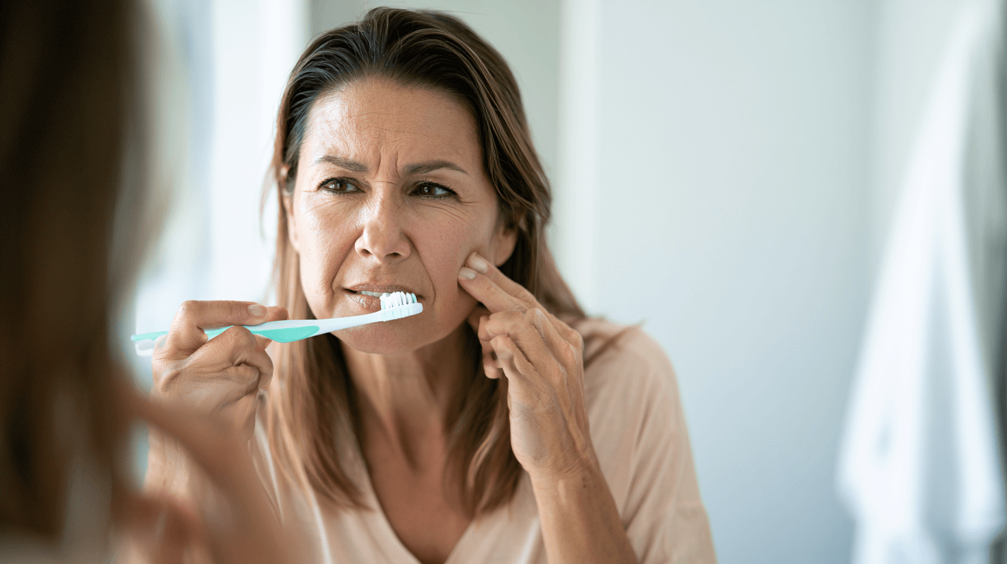
Brushing the teeth is a common trigger, and some patients begin avoiding oral hygiene because even gentle contact produces electric-shock pain. This may provide short-term relief from triggering but can eventually lead to plaque accumulation, gum inflammation, dental infection and genuine tooth pain. Once dental disease develops, it becomes even more difficult to determine whether the patient is experiencing trigeminal neuralgia, dental pain or both.
A soft toothbrush, gentle technique and tolerable water temperature may help during a flare. The patient should avoid repeated forceful cleaning over a known trigger zone, but complete abandonment of oral hygiene is not a safe strategy. A dentist familiar with facial-pain disorders can help design a temporary oral-care plan until neurological control improves.
Dental appointments should not be cancelled indefinitely. The dentist should be informed about the diagnosis, affected side, triggers, current medicine and previous unnecessary dental procedures. Preventive examination remains important, but extraction, root-canal treatment or another irreversible procedure should be performed only when objective dental findings adequately explain the symptoms [2,8,9].
A temporary reduction in pain after local anaesthesia does not automatically prove that a tooth caused the disorder. Local anaesthetic can interrupt nerve transmission from a wider area and may temporarily influence neuropathic pain. The complete dental examination, radiographs, clinical pattern and neurological assessment must still be considered together.
Sleep, Stress and Anticipatory Fear
Sleep disturbance can increase fatigue, reduce emotional resilience and make medication-related cognitive effects more difficult to tolerate. Some patients remain awake because they fear turning onto the painful side or triggering an attack through facial contact with the pillow. Others wake repeatedly because of background pain or anxiety about the following day [15].
A regular sleep schedule, comfortable pillow position and reduction of direct pressure over the trigger zone may help. Strong sedative remedies should not be added casually because carbamazepine, oxcarbazepine, baclofen, pregabalin and several other treatments can already cause drowsiness or impaired coordination. The physician should review the combined sedative burden before recommending additional sleep medicines or calming herbal preparations.
Stress does not create neurovascular compression, and patients should not be told that the condition exists because they are emotionally weak. Nevertheless, stress and anticipatory fear may heighten vigilance, disturb sleep and make ordinary activities feel dangerous. Psychological support can help a patient regain confidence in eating, speaking, travelling and interacting socially while medical or surgical treatment addresses the neurological disorder [10,15].
Breathing practices, meditation, counselling, guided relaxation and gentle restorative routines may support sleep and emotional regulation. These methods should be presented as adjunctive care rather than proof that trigeminal neuralgia is psychological or that relaxation can replace neurological treatment.
Work, Driving and Daily Safety
Trigeminal neuralgia itself may interrupt concentration, speaking and work performance. The medicines used to control it may also cause drowsiness, dizziness, blurred vision, slowed reactions or poor balance. A person who drives, operates machinery, works at height or performs safety-sensitive duties should discuss these risks with the prescribing clinician.
You should not assume that you are safe to drive merely because pain has improved. Treatment-related sedation can be most prominent during the first days after starting or increasing a medicine. Local driving regulations and medical advice should be followed, particularly when attacks remain unpredictable or vision, balance or alertness is affected.
Employers may be able to provide temporary adjustments such as flexible speaking responsibilities, protection from direct air conditioning, access to softer food, scheduled medication breaks or remote work during a severe flare. The patient does not necessarily need to disclose every medical detail, but a practical workplace plan can prevent repeated absence and reduce fear of an attack occurring in public.
Travel Planning for a Patient With Trigeminal Neuralgia
Travel can expose the face to cold wind, air conditioning, irregular meals, sleep disruption and missed medication. Before travelling, the patient should carry an adequate supply of prescribed medicine, a copy of the prescription, the MRI report, relevant medical summaries and the treating clinician’s contact details. Medicines should remain in their labelled packaging, particularly during international travel.
A written crisis plan is useful. It should explain what the patient normally takes, which symptoms require urgent assessment and where emergency care can be obtained at the destination. The person should not rely on obtaining the same medicine immediately in another country because brand names, availability and prescribing rules vary.
Air travel is not automatically prohibited, but direct cold airflow towards the face may trigger attacks in susceptible patients. A soft covering, adjustable air vent and access to suitable food and fluids may reduce avoidable provocation. If attacks are uncontrolled or the patient cannot eat and drink reliably, stabilisation should take priority over non-essential travel.
A 30-Day Trigeminal Neuralgia Action Plan

A structured 30-day plan helps transform an uncertain and frightening situation into measurable steps. It does not promise complete pain relief within one month. Its purpose is to clarify the diagnosis, protect nutrition, establish a safe treatment baseline and identify whether medical, surgical or integrative escalation is needed [2,3,6,10].
Days 1–7: Document the Actual Pain Pattern
During the first week, record every major attack or representative attack cluster. Note the side, facial division, duration, quality, trigger, background pain and effect on eating, speaking, sleep and oral hygiene. Record all medicines, missed doses, benefits and adverse effects. This creates a more reliable clinical history than trying to recall several weeks of fluctuating pain during one consultation.
Collect previous MRI scans, radiology reports, dental X-rays, extraction records, root-canal details, neurological notes and laboratory results. The original MRI images are particularly important because a written report may not clearly distinguish simple vascular contact from compression associated with nerve displacement or atrophy.
The patient should also record body weight, approximate fluid intake and the number of meals completed. These simple observations may reveal nutritional deterioration that has not yet appeared in routine medical records.
Days 8–14: Confirm the Diagnosis and Review Red Flags
The second week should focus on diagnostic clarification. A neurologist or appropriate facial-pain specialist should review whether the symptoms fulfil recognised trigeminal neuralgia criteria and whether another condition remains possible. Facial sensation, motor function, hearing, vision and other neurological findings should be examined where clinically appropriate [1–3,6].
If MRI has not been performed, the clinician should determine the appropriate imaging protocol. When a previous scan was routine, technically limited or inconsistent with the symptoms, specialist review or repeat high-resolution imaging may be necessary.
Dental disease should be evaluated when clinically plausible, but irreversible procedures should not be undertaken simply because the pain is felt in the teeth. By the end of this stage, the patient should understand whether the working diagnosis is classical, secondary or idiopathic trigeminal neuralgia, or whether further evaluation is required.
Days 15–21: Compare Treatment Pathways
During the third week, the patient and clinician should review whether current medication is providing meaningful control. The discussion should include attack reduction, ability to eat and speak, cognitive effects, balance, laboratory monitoring and medicine interactions. A medicine should not be considered successful merely because the pain is lower if the patient is unable to function safely.
When medical treatment is inadequate or poorly tolerated, neurosurgical consultation should be discussed. The patient can compare microvascular decompression, stereotactic radiosurgery and percutaneous procedures without committing immediately to an operation. Understanding the options before a crisis allows a more thoughtful decision [2,12–14].
Ayurvedic care may be introduced or reviewed during this stage when the diagnosis and safety requirements are clear. The Ayurvedic physician should define the intended goals, disclose the evidence level and coordinate with neurological treatment. Prescribed anticonvulsants should not be withdrawn independently.
Days 22–30: Measure Progress and Establish an Escalation Plan
During the final week, the clinician should compare the patient’s current state with the baseline recorded during the first seven days. Attack frequency, pain intensity, continuous background pain, food intake, hydration, sleep, medication burden and functional ability should all be reviewed.
The patient should receive a clear plan explaining what treatment will continue, when the next review will occur and which findings require earlier contact. Failure to improve is clinically useful information. It may indicate that the diagnosis should be reconsidered, the medicine should be adjusted, imaging should be reviewed or a procedural opinion should be prioritised.
The plan should also state what will happen if the patient becomes unable to eat or drink, develops neurological deficits or experiences severe psychological distress. A written escalation pathway prevents the family from having to make complex decisions during an uncontrolled pain crisis.
How to Measure Whether Trigeminal Neuralgia Treatment Is Working
Treatment success should be measured with the same discipline whether the patient receives neurological medicine, surgery, an interventional procedure or Ayurvedic care. A general statement such as “the pain is better” is not enough to determine whether improvement is clinically meaningful, sustainable or associated with unacceptable adverse effects [2,10,12,20].
Measure Attack Burden, Not Only Pain Intensity
Attack burden includes the number of attacks, duration of attack clusters, trigger sensitivity and recovery time. A patient whose pain remains intense but decreases from fifty attacks a day to two attacks a week has experienced a major change that a simple zero-to-ten pain score may not reflect.
The daily record should distinguish spontaneous attacks from triggered attacks. It should also identify whether the patient can touch previously sensitive areas, chew more normally or tolerate brushing. These changes may show improvement before complete pain freedom occurs.
Continuous background pain should be recorded separately. The electric shocks may improve while burning or aching persists, and the two pain components may require different treatment decisions [1,2,6].
Measure Functional Recovery
Important functional outcomes include the ability to eat a normal meal, drink sufficient fluid, brush the teeth, wash the face, speak without interruption, sleep adequately, work and participate socially. These milestones often matter more to the patient than a modest numerical change in pain intensity.
One useful approach is to identify three activities that the patient cannot perform at baseline. Progress is then assessed by whether those specific activities become possible. For example, the goals may be eating on both sides, completing oral hygiene and holding a ten-minute conversation without repeated attacks.
Family observations may also be valuable. A patient who has lived with severe pain for months may underestimate improvement or deterioration, while a family member notices that meals are being completed, social interaction has returned or sedation has increased.
Measure Medication Burden and Adverse Effects
Pain relief achieved through intolerable sedation, confusion or imbalance should not be recorded as complete treatment success. The medicine name, dose, daily frequency and adverse effects should be documented alongside pain outcomes.
Medication-free pain freedom is different from pain control requiring continued treatment. Both may be worthwhile, but they should not be reported as the same outcome. This distinction is especially important when surgical or Ayurvedic treatment results are presented publicly.
A reduction in prescribed medication should be counted only when it has been directed or approved by the prescribing clinician. A patient stopping medicine independently is not evidence that the treatment safely reduced medication dependence.
Distinguish Natural Remission From Treatment Effect
Trigeminal neuralgia may naturally alternate between active periods and remission. Pain may improve shortly after a new treatment by coincidence, especially when several interventions are started at the same time. This is one reason controlled studies are required before a treatment can be declared generally effective.
In individual practice, the likelihood of a meaningful treatment response becomes stronger when improvement is temporally consistent, measurable across several outcomes and maintained during follow-up. Repeated improvement after treatment and deterioration when treatment is withdrawn may also provide clinically useful information, although it does not replace controlled evidence.
A short symptom-free period should not automatically be described as a permanent cure. Sustained follow-up is necessary, particularly when the patient previously experienced long natural remissions.
Know When to Stop an Ineffective Plan
Any treatment should have defined review points. Continuing the same plan indefinitely despite uncontrolled attacks, worsening nutrition or progressive neurological symptoms exposes the patient to harm and delays more appropriate care.
An intervention should be reassessed when there is no meaningful improvement within the agreed trial period, when adverse effects exceed benefit or when the pain pattern changes. Ayurvedic treatment is not exempt from this standard. A patient should not be told that worsening pain for several weeks is automatically a cleansing response or evidence that the therapy is working.
The treatment plan may be modified, but repeated changes without objective documentation make it impossible to identify what is helping. Good care requires enough stability to evaluate an intervention and enough flexibility to stop it when the evidence from that patient is unfavourable.
Who May Benefit From Integrative Ayurvedic Care?
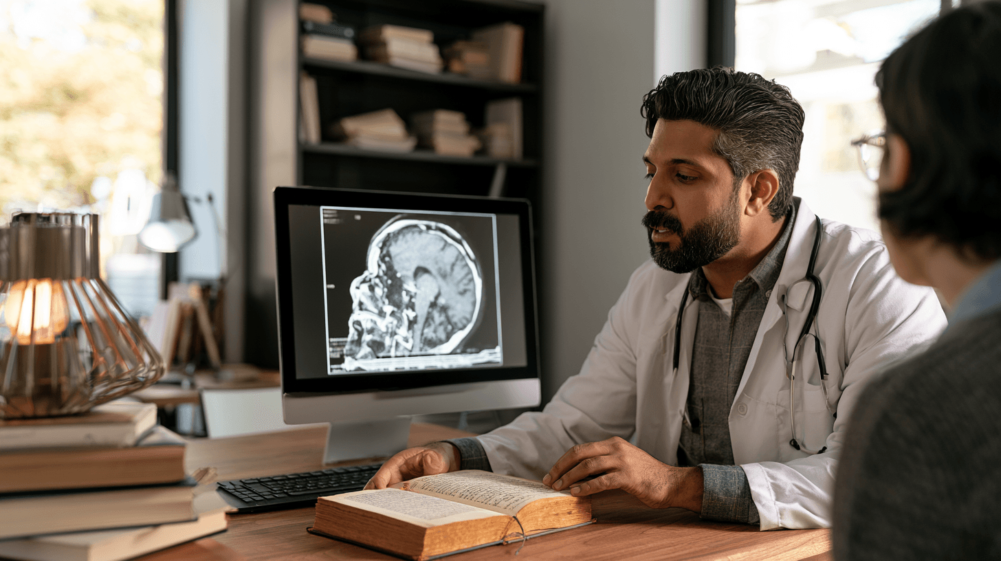
Integrative Ayurvedic care may be considered by patients with a confirmed or carefully evaluated diagnosis who want additional support for attack reduction, sleep, nutrition, emotional resilience and functional recovery. It may also be relevant for people who obtain partial benefit from neurological medicine but experience residual symptoms or treatment-related difficulties.
Patients seeking Ayurveda should understand the current evidence. Classical descriptions provide a traditional rationale, while the available modern trigeminal neuralgia evidence includes limited case-level documentation rather than large controlled trials [19,20]. This does not make every Ayurvedic intervention ineffective, but it means that results should be presented honestly and measured carefully.
The strongest integrative approach does not ask the patient to choose between neurology and Ayurveda. It uses neurological diagnosis, MRI and emergency care where necessary while individualising Ayurvedic support according to the patient’s constitution, digestion, strength, symptoms, concurrent medicines and treatment goals.
Patients Who Require Medical Stabilisation or Referral First
A patient with new facial numbness, weakness, visual symptoms, hearing changes, bilateral pain, fever, unexplained weight loss, previous cancer or other neurological findings should undergo appropriate medical evaluation before beginning a routine integrative programme [2,3].
The same applies to someone who cannot maintain hydration or nutrition, has a serious medicine reaction or is experiencing suicidal thoughts. In these situations, immediate safety and diagnostic clarification take priority over outpatient Ayurvedic treatment.
A tumour, multiple sclerosis lesion or significant neurovascular compression must not be managed as though it were only a Doṣic imbalance. Ayurvedic assessment may be added after the condition has been properly identified, but it should not obscure the anatomical or neurological diagnosis.
Preparing for an Integrative Trigeminal Neuralgia Consultation
Before consultation, the patient should organise the MRI report and original images, neurological notes, dental records, current medicine list, previous treatments and relevant laboratory results. A seven-day facial-pain diary should document attack frequency, distribution, triggers, background pain and functional limitations.
The patient should also report all herbs, supplements, oils, powders and over-the-counter medicines. Concealing Ayurvedic treatment from the neurologist or hiding neurological medicine from the Ayurvedic physician creates avoidable interaction and safety risks.
During consultation, we should agree on measurable goals. These may include fewer daily attacks, improved food intake, restoration of oral hygiene, better sleep or reduced medicine-related impairment. The goal should not be a vague promise that the nerve will be permanently cured without confirming how improvement will be measured.
What If Trigeminal Neuralgia Treatment Is Not Working?
When trigeminal neuralgia treatment is not producing adequate relief, the safest response is not automatically to increase the medicine, add several new remedies or proceed directly to surgery. The first step is to determine why the treatment appears to be failing. The diagnosis may still be correct, but the medicine may be inadequate, poorly tolerated or affected by inconsistent use and drug interactions. In other cases, the pain pattern may have changed, a continuous neuropathic component may have emerged or the original diagnosis may need reconsideration [2,3,6,9].
A structured review should compare the present condition with the baseline. We need to know whether the electric-shock attacks are becoming more frequent, whether background pain is increasing, whether new facial numbness has appeared and whether the patient can still eat, speak and maintain oral hygiene. The clinician should also review every prescription, supplement and Ayurvedic medicine because treatment interactions or cumulative sedation may affect both safety and perceived effectiveness.
Treatment failure is not a personal failure. A patient should not be blamed for having “resistant pain,” nor should he or she be told to tolerate uncontrolled attacks indefinitely. Persistent symptoms provide useful clinical information and may indicate that the diagnosis, imaging or treatment pathway requires modification.
When Medicine Worked Initially but the Pain Returned
Trigeminal neuralgia may fluctuate naturally, with active periods followed by partial or complete remission. A medicine may appear highly effective during a remission and less effective when the disorder becomes active again. This does not automatically mean that the medicine has become completely useless, but it does require reassessment of the attack pattern and treatment burden [1,2,6].
The clinician should verify whether doses have been missed, whether a new medicine has altered drug metabolism or whether the patient has reduced treatment because of drowsiness or dizziness. Sometimes the person reports that treatment “stopped working” when the real problem is that the effective dose was no longer tolerable. In this situation, simply increasing the medicine may worsen balance, cognition and daily function without restoring satisfactory pain control.
When attacks repeatedly break through an appropriately supervised first-line regimen, specialist review becomes important. The neurologist may consider a different medicine, combination therapy, updated imaging or neurosurgical consultation. If MRI demonstrates clinically meaningful neurovascular compression, the patient should be allowed to compare anatomical treatment with continued medical management before another severe crisis develops [2,3,12–14].
When Electric-Shock Attacks Improve but Continuous Pain Remains
Some treatments control the brief paroxysms more effectively than the continuous aching, burning or throbbing component. A patient may therefore report fewer shocks but remain troubled by persistent facial discomfort. This does not necessarily mean that treatment has failed completely, but the two pain components should be evaluated separately [1,2,6].
Continuous pain may be part of trigeminal neuralgia with concomitant continuous pain, but it may also indicate painful trigeminal neuropathy, postherpetic neuralgia, persistent idiopathic facial pain, temporomandibular disease or another overlapping condition. The clinician should reconsider the history, sensory findings, previous dental procedures and any nerve injury before assuming that additional destruction of trigeminal fibres will solve the problem.
This distinction is particularly important before an ablative procedure. A treatment that reduces electric-shock attacks by intentionally altering the nerve may not eliminate existing continuous neuropathic pain and, in uncommon cases, may leave troublesome sensory symptoms. The expected effect on each pain component should be discussed during informed consent [2,14].
When MRI Does Not Show Neurovascular Compression
A normal or inconclusive MRI does not exclude trigeminal neuralgia. A patient whose pain meets recognised clinical criteria may be diagnosed with idiopathic trigeminal neuralgia when no definite compression or secondary lesion is identified [1,5,6]. The pain remains genuine even when imaging cannot demonstrate a structural explanation.
Before accepting that the scan is definitively normal, the clinical team should confirm whether an appropriate high-resolution trigeminal nerve protocol was used. A routine brain MRI may be adequate for identifying a large tumour or major demyelinating lesion but may not provide sufficient detail to assess the relationship between small vessels and the trigeminal nerve.
When symptoms are strongly characteristic and the report is unclear, review of the original images by an experienced neuroradiologist, neurologist or neurosurgeon may be valuable. However, we should not keep repeating imaging merely to obtain a desired finding. If no anatomical cause is demonstrated after appropriate assessment, the treatment discussion should reflect that uncertainty rather than claiming that a compressing vessel has been identified.
When MRI Shows Vascular Contact but the Symptoms Are Atypical
Vascular contact is common enough that it may be found incidentally. If a patient has continuous poorly localised facial pain without characteristic electric-shock attacks or triggers, a report mentioning vascular contact should not be treated as proof of classical trigeminal neuralgia [3,5,6,9].
The specialist should determine whether the vessel causes displacement, indentation, distortion or atrophy of the nerve and whether the finding is located on the same side as the pain. The clinician should also assess whether the symptoms follow one or more trigeminal divisions and satisfy recognised diagnostic criteria.
This is one of the situations where a second opinion can prevent unnecessary surgery. An operation based on incidental contact may fail to relieve the pain if the underlying disorder was not classical trigeminal neuralgia. Diagnostic confidence should come from agreement between the pain history, examination and imaging rather than from one radiology phrase.
When Surgery Relieved the Pain but It Returned
Recurrence after surgery or another procedure does not necessarily mean that the original treatment was worthless. A patient may have experienced meaningful pain freedom for several years before attacks returned. The duration and quality of relief should be considered when evaluating the outcome [12–14].
Recurrent pain requires a fresh assessment rather than an automatic repetition of the same procedure. The clinician should clarify whether the returning pain has the same distribution, quality and triggers as the original attacks. New continuous pain, painful numbness or sensory loss may indicate a different mechanism and may require a different treatment strategy.
Updated imaging may be appropriate after previous microvascular decompression, particularly when recurrent neurovascular compression, prosthetic material displacement or another structural explanation is suspected. After radiosurgery or a percutaneous procedure, the existing level of facial and corneal sensation should be documented before considering another ablative intervention.
The next option may include renewed medical treatment, repeat surgery, stereotactic radiosurgery or a percutaneous procedure, depending on the previous treatment, current anatomy, medical fitness and sensory status. The decision should be made by a team experienced in recurrent trigeminal neuralgia rather than treated as an ordinary first presentation.
When Numbness or Burning Develops After a Procedure
Mild facial numbness may be an expected consequence of some percutaneous treatments and can also occur after radiosurgery or, less commonly, microvascular decompression. The patient should nevertheless report new or progressive sensory changes because facial numbness is not a single uniform outcome [2,14].
Reduced sensation around the forehead and eye requires particular attention. If corneal sensation is impaired, the person may not recognise dryness, irritation or injury. Eye redness, pain, visual change or persistent foreign-body sensation warrants prompt ophthalmological assessment.
Burning pain within an area of numbness is different from ordinary sensory reduction. Dysesthesia and anaesthesia dolorosa are difficult complications in which altered or absent sensation becomes unpleasant or painful. Repeating a destructive procedure without careful evaluation may worsen the problem.
Ayurvedic local therapies should also be used cautiously over an area with impaired sensation. Excessively hot applications, strong massage or irritating oils may cause injury that the patient does not feel normally. The physician must assess sensory function before selecting any local procedure.
When Should You Seek a Second Opinion?
A second opinion is not a rejection of the first doctor. It is a reasonable step when the diagnosis is uncertain, an irreversible procedure is proposed or treatment has failed despite prolonged care. Trigeminal neuralgia involves neurology, neuroradiology, neurosurgery, dentistry and pain medicine, so a complex case may benefit from more than one perspective [2,6,9,10].
You should bring the complete record rather than only a short written summary. The reviewing specialist needs the original MRI images, report, previous operation notes, dental records, medicine history and pain diary. Without this information, the second opinion may repeat the same assumptions instead of genuinely reassessing the case.
Before Tooth Extraction or Root-Canal Treatment
A second opinion is particularly valuable when shock-like pain is being attributed to a tooth despite limited objective dental evidence. The dentist should be able to explain which abnormal finding is present and how it corresponds with the painful site and pattern.
If several teeth have already been treated without sustained benefit, the possibility of trigeminal neuralgia or another neuropathic facial-pain disorder should be reconsidered. Continuing to treat additional teeth simply because the pain is felt in the jaw may lead to irreversible loss without addressing the neurological problem [2,8,9].
This does not mean that every patient with trigeminal neuralgia has healthy teeth. Dental pathology and neuralgic pain may coexist. The second opinion should therefore distinguish between appropriate dental care and procedures being used as diagnostic experiments.
Before Microvascular Decompression
Before microvascular decompression, the patient should understand whether the diagnosis is classical trigeminal neuralgia and whether MRI demonstrates meaningful neurovascular compression. A second neurosurgical or neuroradiological opinion may be helpful when the scan shows only contact, the symptoms are atypical or the proposed surgery is based primarily on the report rather than image review.
The patient should ask how the centre defines successful treatment, how frequently the procedure is performed and how complications are recorded. He or she should also ask what the surgeon expects if the compression is venous, involves several vessels or is not clearly identified during the operation [12,13].
A second opinion is especially reasonable after previous surgery, in the presence of continuous pain or when the patient has significant medical risks. The objective is not to search indefinitely for a doctor who guarantees success. It is to make sure that the proposed operation is based on a well-supported diagnosis and transparent discussion of benefits and risks.
Before a Repeat Ablative Procedure
A repeat radiofrequency procedure, balloon compression, glycerol rhizolysis or radiosurgery may provide additional relief, but cumulative sensory injury becomes an important concern. The clinician should map current facial and corneal sensation and clarify whether the returning pain remains typical trigeminal neuralgia [2,14].
A patient with substantial existing numbness, painful altered sensation or corneal impairment requires particularly cautious decision-making. Another destructive procedure may reduce attacks, but it may also increase sensory complications. A specialist experienced in recurrent and post-procedural pain should review the complete history before treatment is repeated.
Before Replacing Neurological Care With Ayurveda
A second opinion is appropriate when a patient has been advised to discontinue neurological medicine, avoid MRI or reject neurosurgical assessment solely because Ayurvedic treatment is being offered. Classical Ayurvedic care does not require denial of modern diagnosis. A responsible integrative physician should understand the role of imaging, emergency treatment and referral.
Ayurvedic treatment should be able to state its objectives clearly. The plan may aim to reduce attack burden, improve sleep, support nutrition and enhance function, but it should not claim to remove neurovascular compression without objective evidence. The physician should also explain which classical references guide the intervention and what modern clinical evidence is available [19,20].
How to Choose a Trigeminal Neuralgia Treatment Centre
A good trigeminal neuralgia centre should provide diagnostic clarity before promoting a particular procedure. The team should review the pain history, neurological examination and original imaging rather than relying only on the referral label. It should also recognise when the patient has a different facial-pain disorder or requires dental, ophthalmological or mental-health support [2,6,9,10].
You should ask whether the centre has access to neurologists, neuroradiologists and neurosurgeons experienced in trigeminal neuralgia. For integrative treatment, the Ayurvedic physician should be willing to coordinate with these specialists and document both benefits and adverse effects.
Ask How Success Is Defined
A centre that reports a high success rate should explain what that percentage means. Complete medication-free pain freedom is different from partial pain reduction while continuing medication. Immediate postoperative relief is different from sustained relief after five or ten years.
The patient should also ask how recurrence, numbness, hearing impairment, corneal complications and repeat procedures are included in the outcome data. Reporting only the percentage of patients who initially improved can create an incomplete picture [12–14].
An honest centre may provide a range rather than one absolute promise because outcomes depend on diagnostic subtype, imaging findings, previous treatment and follow-up duration. Transparency is more valuable than an impressive percentage without context.
Ask Whether the Original MRI Images Will Be Reviewed
A treatment centre should not make an invasive decision solely from a written MRI report. The original images should be assessed to determine the side, location and severity of neurovascular conflict and whether morphological nerve changes are present [3,5,6,13].
When the images are technically inadequate, the team should explain why repeat or specialised imaging is necessary. Repeating a scan should have a clinical purpose, not merely increase the number of investigations.
Ask About Long-Term Follow-Up
Trigeminal neuralgia may recur after an initially successful treatment. A centre should therefore explain how patients are followed, who manages medication after the procedure and what happens if pain returns.
Follow-up should include pain pattern, medication use, facial sensation, eating ability and psychological well-being. A patient discharged immediately after a procedure without a recurrence plan may struggle to obtain appropriate care when symptoms return months or years later.
Ask How Different Specialists Communicate
Disconnected treatment can create contradictory advice. A neurologist may increase medication while another clinician starts sedating herbal products, or a dentist may plan extraction without knowing that a neurosurgical review is underway.
The centre should have a process for sharing relevant information and defining responsibility. The patient should know who is managing anticonvulsants, who is monitoring laboratory tests, who will assess new neurological symptoms and who will decide whether surgery is appropriate.
A Structured Integrative Trigeminal Neuralgia Pathway

A credible integrative pathway should begin with the diagnosis rather than with a standard medicine package.
This approach allows us to distinguish a patient who may be suitable for supportive Ayurvedic care from someone who requires urgent neurological investigation or neurosurgical evaluation. It also helps prevent a common error in which every facial pain is treated as Anantavata without confirming whether the modern diagnostic criteria for trigeminal neuralgia are actually present.
Diagnostic Review Before Ayurvedic Treatment
The review should include the original MRI images and report, neurological records, dental history, previous procedures, current medicines and a seven-day pain diary. We should document whether pain is located in V1, V2 or V3, whether attacks are triggerable and whether continuous pain, numbness or weakness is present.
Red flags must be screened before a formulation is selected. Progressive sensory loss, bilateral symptoms, hearing or visual changes, previous cancer, unexplained systemic symptoms or inadequate imaging may require referral for further investigation [2,3].
This diagnostic review does not replace the neurologist. It helps the integrative physician understand where Ayurveda can be used safely and where modern specialist management remains essential.
Ayurvedic Assessment and Individualisation
After modern diagnostic information has been reviewed, the physician can perform Roga and Rogī Parīkṣā. This includes Prakṛti, Vikṛti, Doṣa involvement, Agni, Āma, Koṣṭha, Bala, Satva, Satmya, age, chronicity, sleep, appetite and bowel pattern.
Anantavata may provide a classical correlation, but the treatment should reflect the individual presentation rather than the disease name alone. The Suśruta Saṃhitā describes a Tridoṣaja craniofacial condition and advises therapeutic principles associated with Sūryāvarta management, Sirāvyadha and Vāta-Pitta-pacifying diet [19]. Any classical procedure must be interpreted according to present-day safety, contraindications and practitioner competence.
The Ayurvedic plan may include selected internal medicines, Nasya, Kavala, Gaṇḍūṣa, Snehana-oriented measures or supportive Shiro therapies when clinically suitable. The exact combination should depend on the patient’s strength, digestion, trigger pattern, comorbidities and concurrent neurological treatment.
Establishing Measurable Treatment Goals
Before treatment begins, we should agree on outcomes that can be verified. These may include reducing the number of daily attacks, extending pain-free intervals, improving food intake, restoring oral hygiene, improving sleep or reducing medicine-related functional impairment.
A goal such as “cure the nerve” is too vague to measure. By contrast, a reduction from thirty daily attacks to five, the ability to complete meals or restoration of uninterrupted sleep can be documented and compared over time.
Medication reduction should not be used as an outcome unless it has been supervised by the prescribing clinician. A patient stopping treatment independently cannot be presented as evidence that Ayurveda safely replaced neurological medicine.
Reviewing Progress at Defined Intervals
Follow-up should occur at clinically appropriate intervals, with comparison against the original baseline. The physician should record attack frequency, background pain, trigger sensitivity, nutrition, sleep, medicine use and adverse events.
Improvement should be sustained before it is described as remission. Trigeminal neuralgia may fluctuate naturally, and a few pain-free days cannot establish permanent recovery. Longer follow-up is especially important when the patient has experienced spontaneous remissions in the past.
When there is no meaningful improvement, the plan should be reconsidered. Ayurveda should not be continued indefinitely on the assumption that every patient requires more time. Diagnostic reassessment, medicine review, repeat imaging or neurosurgical consultation may become necessary.
Coordinating With Neurology and Neurosurgery
The neurologist should remain responsible for anticonvulsant management and relevant laboratory monitoring. The Ayurvedic physician should communicate any new medicine, significant adverse effect or change in symptoms that could influence neurological treatment.
When MRI demonstrates meaningful neurovascular compression and medical treatment is failing, the patient should be encouraged to obtain a neurosurgical opinion. An integrative consultation can help the person understand and prepare for the decision, but it should not delay a potentially appropriate anatomical treatment.
Ayurvedic care may also support the patient before or after a procedure through individualised nutrition, sleep support, stress management and recovery planning. Any postoperative intervention should respect surgical restrictions, wound healing and neurological advice.
What Results Can a Patient Realistically Expect?
The outcome of trigeminal neuralgia treatment varies widely. Some patients respond rapidly to first-line medicine, while others require combination treatment, surgery or repeated procedures. An Ayurvedic intervention may support symptom reduction and function in an individual case, but the present modern evidence does not allow a guaranteed response or universal timeline [2,3,12–14,20].
A realistic treatment discussion should distinguish immediate relief, short-term control and sustained remission. A patient who improves for one week has achieved a different outcome from someone who remains pain-free without medicine for several years.
Early Improvement
Early improvement may appear as fewer attacks, lower trigger sensitivity or the ability to eat and speak with less fear. Sleep and appetite may improve as the attack burden decreases. These changes are valuable even when complete pain freedom has not yet been achieved.
However, early improvement should be interpreted cautiously because the disorder can enter spontaneous remission. The response becomes more convincing when it is sustained, repeatedly documented and associated with functional recovery.
Partial Response
A partial response may still provide meaningful benefit. A patient who continues to experience occasional attacks but can eat, work and sleep normally may prefer the current plan to a more invasive treatment. Another person may consider the same residual pain unacceptable.
Treatment decisions should therefore include the patient’s priorities. We should not define success only from the doctor’s perspective or assume that every patient will accept the same trade-off between pain relief, medication and numbness.
Remission and Long-Term Follow-Up
Remission means that pain has substantially reduced or disappeared for a period, but it does not guarantee that the disorder will never return. Long-term follow-up should record whether the patient remains pain-free, whether medicine is still required and whether new sensory symptoms have developed.
When public case outcomes are presented, the duration of follow-up should always be stated. A testimonial recorded after ten pain-free days cannot be compared with a study reporting medication-free pain freedom after five years.
How Future Ayurvedic Research Should Be Improved
Future studies should confirm the trigeminal neuralgia subtype using recognised diagnostic criteria and suitable MRI. Researchers should separately record electric-shock attacks and continuous background pain rather than combining them into one score.
The intervention should be described clearly, including the classical rationale, formulation source, dosage, procedures, duration and concurrent neurological treatment. Without this information, clinicians cannot understand which component may have contributed to the result.
Outcomes should include attack frequency, pain intensity, trigger sensitivity, eating, oral hygiene, sleep, medicine use, adverse events and quality of life. Follow-up should continue long enough to distinguish treatment-related improvement from a brief natural remission.
Controlled comparative studies would provide stronger evidence than isolated case reports. Until such research is available, clinical claims should remain proportionate to the evidence.
Making an Informed Trigeminal Neuralgia Treatment Decision
An informed decision begins by confirming that the pain is actually trigeminal neuralgia. The patient should understand whether the condition is classical, secondary or idiopathic, whether continuous pain is present and whether MRI demonstrates meaningful neurovascular compression [1,3,5,6].
The next step is to compare the expected benefits and limitations of each treatment. Medicine may control attacks but cause adverse effects. Microvascular decompression may provide durable medication-free relief in a suitable patient but requires cranial surgery. Radiosurgery and percutaneous procedures are less invasive but may carry different risks of delayed benefit, recurrence and facial numbness [12–14].
Ayurveda may offer individualised supportive care for pain burden, sleep, nutrition, emotional resilience and functional recovery. Its role is strongest when the intervention is based on classical assessment, coordinated with neurological treatment and evaluated through measurable outcomes [19,20].
You do not need to choose between hope and evidence. A patient can remain hopeful while asking precise questions, requesting appropriate imaging, comparing treatment pathways and refusing unsupported guarantees. We can provide better care when the patient, family and clinical team make the decision together rather than allowing fear or marketing claims to determine the next step.
Can Ayurveda Cure Trigeminal Neuralgia?
Trigeminal neuralgia is a neurological pain disorder, not cancer. The research discussed below therefore concerns trigeminal neuralgia, trigeminal neuropathic pain and related mechanisms, not anticancer activity.
Ayurveda may help selected patients achieve substantial pain reduction, longer pain-free intervals, restoration of eating and speaking, improved sleep, lower trigger sensitivity and sustained remission.Modern diagnostic criteria recognise classical, idiopathic and secondary trigeminal neuralgia, and each category requires a different treatment pathway [1–3]. ICHD-3
When I use the term “Ayurvedic cure” clinically, it should describe a documented outcome such as sustained absence of electric-shock attacks, restoration of normal daily activities, stable neurological findings, supervised reduction of conventional medicine where appropriate and adequate long-term follow-up. A few pain-free days should be described as early improvement or remission rather than permanent cure.
Modern human evidence for Ayurveda remains limited. A 2023 MRI-confirmed case report documented improvement in pain, anxiety, depression and quality of life after three months of multimodal Ayurvedic treatment. A separate 2025 report described improvement in one patient with painful post-traumatic trigeminal neuropathy after a longer multimodal Panchakarma and medicine programme. Both are clinically encouraging, but neither was a controlled trial, and neither proves that one Avaleha alone can cure every patient since every patient is different. PubMed
Ayurveda may help carefully selected patients achieve sustained control or remission of trigeminal neuralgia through an individualised programme that addresses Vāta-Pitta imbalance, neurological pain burden, sleep, nutrition, digestion and functional recovery. MRI, neurological assessment and neurosurgical referral remain necessary when a structural or secondary cause is suspected.
Best Classical-Inspired Avaleha Name

Anantavāta–Majjā–Medhya Rasāyana Avaleha
Sanskrit: अनन्तवात–मज्जा–मेध्य रसायन अवलेह
This name communicates four aspects of the formulation:
Anantavāta identifies the principal Ayurvedic clinical correlation described under Śiroroga.
Majjā reflects the tissue-nourishing and deeper neurological support rationale. Majjā Dhātu should not be presented as an exact anatomical synonym for the trigeminal nerve.
Medhya represents support for sleep, cognition, emotional stability and the wider neurological burden of chronic pain.
Rasāyana indicates a restorative, nourishing and long-term functional-recovery approach rather than a formulation designed only for short-term analgesia.
This exact name and combination do not appear as one verbatim classical formulation in a single Saṃhitā. It should therefore be described as a physician-designed, classical-inspired proprietary Avaleha, not as an unchanged classical yoga.
Classical Texts Inspiring the Formulation
| Classical text | Chapter details | Principle used in this Avaleha |
|---|---|---|
| Suśruta Saṃhitā | Uttara-tantra, Śiroroga Vijñānīya Adhyāya 25 | Classical description of Anantavāta and its craniofacial distribution |
| Suśruta Saṃhitā | Uttara-tantra, Śiroroga Pratiṣedha Adhyāya 26 | Treatment principles for Anantavāta, including the Sūryāvarta approach and Vāta-Pitta-pacifying diet |
| Caraka Saṃhitā | Cikitsā-sthāna 28, Vātavyādhi Cikitsā | Snehana, Bṛṃhaṇa, Nasya, milk, ghee, Daśamūla and treatment according to Vāta, Āvaraṇa and patient strength |
| Caraka Saṃhitā | Cikitsā-sthāna 1, Rasāyana Adhyāya | Rasāyana, tissue support, strength, functional restoration and resistance to disease |
| Aṣṭāṅga Hṛdaya | Uttara-sthāna 23–24, Śiroroga Vijñāna and Pratiṣedha | Diagnosis and treatment principles for diseases of the head |
| Bhaiṣajya Ratnāvalī | Vātavyādhi Cikitsā Prakaraṇa 26 and Śiroroga Cikitsā Prakaraṇa 65 | Formulation strategy for Vāta disorders and diseases of the head; chapter numbering should be checked against the edition used |
| Śārṅgadhara Saṃhitā | Madhyama-khaṇḍa 8, Avaleha Kalpanā | Avaleha preparation, sweetening base, Prakṣepa addition, Pāka and Siddhi Lakṣaṇa |
The chapter locations are supported by published digital editions and classical-text resources, although verse numbering may differ between Sanskrit editions and translations [19,52–55]. Easy Ayurveda Hospital
Anantavāta–Majjā–Medhya Rasāyana Avaleha: Exact 30-Day Formula
The prescribed dose is 15 g twice daily, which equals 30 g per day. A complete 30-day course therefore requires:
30 g × 30 days = 900 g finished Avaleha
The quantities below are raw-material starting weights. The final batch is standardised to exactly 900 g after controlled reduction and addition of honey and the physician-supervised mineral microblend.
This formulation is designed for an adult patient only after modern diagnosis, MRI review, medicine reconciliation and safety screening. It must not be prepared or taken as a general home remedy.
Coarse Herbs for the Decoction
Use authenticated, clean, air-dried herbs in coarse Yavakūṭa form.
| Ayurvedic ingredient | Botanical name and part used | Principal formulation role | Weight |
|---|---|---|---|
| Bilva | Aegle marmelos, root bark | Daśamūla, Vāta-Kapha regulation | 6 g |
| Agnimantha | Premna integrifolia syn. P. serratifolia, root | Daśamūla, Vāta regulation | 6 g |
| Śyonāka | Oroxylum indicum, root bark | Daśamūla, Śotha and Vāta support | 6 g |
| Pāṭalā | Stereospermum suaveolens, root bark | Daśamūla, Vāta-Pitta support | 6 g |
| Gambhārī | Gmelina arborea, root | Daśamūla, nourishing Vāta support | 6 g |
| Śālaparṇī | Desmodium gangeticum, root or whole plant | Bṛṃhaṇa and Vāta support | 6 g |
| Pṛśniparṇī | Uraria picta, root or whole plant | Bṛṃhaṇa and Vāta support | 6 g |
| Bṛhatī | Solanum indicum, root | Vāta-Kapha support | 6 g |
| Kaṇṭakārī | Solanum xanthocarpum, root | Vāta-Kapha support | 6 g |
| Gokṣura | Tribulus terrestris, root | Daśamūla, tissue and urinary support | 6 g |
| Balā | Sida cordifolia, root | Balya, Bṛṃhaṇa and Vāta support | 30 g |
| Guḍūcī | Tinospora cordifolia, stem | Rasāyana and inflammatory-modulation rationale | 30 g |
| Rāsnā | Pluchea lanceolata, root or pharmacopoeial whole plant | Vāta-Śūla and inflammatory-pain support | 25 g |
| Nirgundī | Vitex negundo, leaf | Śotha and pain-support rationale | 20 g |
| Eraṇḍa Mūla | Ricinus communis, root | Classical Vāta and Śūla support | 15 g |
| Yaṣṭimadhu | Glycyrrhiza glabra, root | Tissue-soothing and formulation harmonisation | 10 g |
| Śuṇṭhī | Zingiber officinale, dried rhizome | Dīpana, Āma management and neuroinflammatory rationale | 10 g |
| Śatāvarī | Asparagus racemosus, root | Bṛṃhaṇa, Rasāyana and Pitta balance | 20 g |
| Total coarse decoction drugs | 220 g |
Use 3,520 mL purified water, equivalent to 16 times the total coarse-drug weight, and reduce it to 880 mL, equivalent to one-quarter of the starting water.
Fine Powders Added During Avaleha Preparation
Use authenticated ingredients in fine powder, preferably around 80-mesh where technically suitable.
| Ayurvedic ingredient | Botanical name and part used | Principal formulation role | Weight |
|---|---|---|---|
| Aśvagandhā | Withania somnifera, root | Balya, Rasāyana, sleep and neuropathic-pain support | 60 g |
| Maṇḍūkaparṇī | Centella asiatica, whole plant | Direct preclinical trigeminal-pain rationale | 40 g |
| Brāhmī | Bacopa monnieri, whole plant | Medhya, neuroprotective and cognitive support | 30 g |
| Śaṅkhapuṣpī | Convolvulus pluricaulis, whole plant | Medhya, sleep and emotional support | 25 g |
| Jaṭāmāṃsī | Nardostachys jatamansi, rhizome | Sleep, anxiety and CNS-calming support | 10 g |
| Āmalakī | Phyllanthus emblica, fruit pericarp | Rasāyana and Pitta support | 20 g |
| Harītakī | Terminalia chebula, fruit pericarp | Vāta regulation, bowel and Rasāyana support | 10 g |
| Vaṃśalocana | Bambusa bambos, pharmacopoeial siliceous concretion | Classical supporting and binding ingredient | 10 g |
| Elā | Elettaria cardamomum, seed | Aroma, digestion and formulation acceptability | 5 g |
| Tvak | Cinnamomum verum, bark | Dīpana, palatability and formulation support | 5 g |
| Mustā | Cyperus rotundus, rhizome | Digestion and Pitta-Kapha balance | 5 g |
| Total fine powders | 220 g |
Avaleha Base and Pharmacy-Only Mineral Microblend
| Ingredient | Form and requirement | Weight |
|---|---|---|
| Purified jaggery | Clean, filtered and free from extraneous matter | 360 g |
| Cow ghee | Pharmacopoeial or food-grade clarified butter | 60 g |
| Honey | Authenticated; add only after cooling below 40°C | 90 g |
| Abhraka Bhasma | Sahasrapuṭī, batch-tested | 3.75 g |
| Muktā Piṣṭi | Authenticated and batch-tested | 3.75 g |
| Suvarṇa Bhasma | Authenticated, batch-tested and physician-supervised | 0.50 g |
| Total mineral microblend | 8 g | |
| Final finished Avaleha | Adjusted by controlled cooking | 900 g |
At the prescribed daily dose, the mineral quantities are approximately:
| Mineral | Per 15 g dose | Per day |
|---|---|---|
| Abhraka Bhasma | 62.5 mg | 125 mg |
| Muktā Piṣṭi | 62.5 mg | 125 mg |
| Suvarṇa Bhasma | 8.3 mg | 16.7 mg |
The mineral blend is not mandatory merely because the patient has severe pain. It should be included only when the physician has established a clear indication and has batch-specific quality documentation. If the bhasma quality cannot be demonstrated, the mineral blend should be omitted.
For a mineral-free 900 g batch, cook the herbal base to 810 g, cool it and add 90 g honey. For the mineral-containing version, cook the herbal base to 802 g, cool it and add 8 g mineral blend plus 90 g honey.
The strongest Ayurvedic formula is not the formulation containing the greatest possible number of herbs and minerals. It is the formulation in which every ingredient has a coherent classical purpose, an acceptable safety profile, a rational dosage and a measurable treatment objective.
I have deliberately not added mercurial or arsenical Rasa preparations, lead-containing bhasmas, Vatsanābha, or Heeraka Bhasma to this standard public formulation.
Patient-Friendly Preparation Method

This preparation method is written so a patient can understand how the medicine is made, but the Avaleha should be manufactured only by a qualified Ayurvedic pharmacy or trained professional using calibrated equipment and documented raw materials.
Preparing the Decoction
Clean and authenticate all coarse decoction herbs. Grind or cut them into coarse Yavakūṭa form rather than fine powder.
Place all 220 g of coarse herbs in a stainless-steel vessel and add 3,520 mL purified water. Allow them to soak for approximately 8–12 hours. Soaking improves uniform wetting and extraction.
Heat the soaked mixture gently in an open vessel. Maintain a slow simmer rather than vigorous boiling. Continue until the liquid is reduced to approximately 880 mL.
Filter the decoction through clean, multilayer muslin while it is still warm. Press gently to recover the liquid but avoid forcing fine sediment through the cloth. Keep approximately 100 mL of the filtered decoction aside temporarily in case consistency adjustment is required during cooking. It must be used before the final cooking endpoint, not after honey has been added.
Preparing the Jaggery Syrup
Add 360 g purified jaggery to the filtered decoction. Warm gently until the jaggery dissolves completely.
Filter the liquid again to remove sand, fibres or other impurities from the jaggery.
Continue cooking over low heat until a soft one-thread or Tantu Pāka stage develops. The syrup should form a light thread between the thumb and finger after cooling a small test portion. The hot syrup itself should never be touched directly.
Adding Ghee and Herbal Powders
Add 60 g cow ghee gradually while stirring continuously. The ghee should blend uniformly without forming a free oily layer.
Mix all 220 g fine herbal powders separately before addition. Add the mixed powder in small portions while stirring continuously. Slow addition prevents lump formation and ensures uniform distribution.
Continue cooking over low heat. Scrape the sides and bottom of the vessel regularly to prevent scorching.
Use a pre-weighed or tared stainless-steel vessel. For the mineral-containing version, continue cooking until the warm herbal base has a net weight of 802 g. For the mineral-free version, continue to 810 g.
If the mixture becomes too thick before reaching the required weight, a small amount of the reserved filtered decoction may be added while the preparation is still cooking. Water should never be added after the Avaleha has cooled or after honey has been incorporated.
Adding Honey and the Mineral Microblend
Remove the vessel from heat and allow the herbal base to cool to 40°C or below. Honey should not be cooked or added to a very hot preparation.
Triturate the 8 g mineral microblend thoroughly with a small portion of the 90 g honey until a smooth, uniform paste is formed. There should be no visible mineral lumps.
Add this mineral-honey paste to the cooled Avaleha. Add the remaining honey and mix thoroughly until the batch is uniform.
Verify the finished net weight as 900 g. Divide it into two sterilised, completely dry, wide-mouth amber containers of 450 g each.
The classical Avaleha method, including preparation of the sweet base, addition of Prakṣepa powders and assessment of Siddhi Lakṣaṇa, is described in Śārṅgadhara Saṃhitā, Madhyama-khaṇḍa 8 and in later pharmaceutics reviews [54]. WJPR
Signs That the Avaleha Is Properly Prepared
A properly prepared Avaleha should be a smooth, glossy and uniform semisolid paste. It should not contain dry powder lumps, a separated ghee layer or visible free water.
A small cooled quantity should form a soft thread between the fingers and retain a light finger impression. It should have the characteristic aroma and taste of the ingredients without a burnt odour.
The final product should not be excessively dry, rubbery or liquid. An undercooked product may ferment, while an excessively cooked product may become difficult to measure and consume.
Unless water activity, Brix, microbial limits and stability have been professionally validated, the container should be refrigerated after opening. A completely dry spoon must be used. The medicine should be discarded if mould, gas formation, fermentation, sour odour or abnormal water separation develops.
Dosage and 30-Day Course
The adult dose is:
15 g after breakfast and 15 g after the evening meal for 30 days
The total daily dose is 30 g, and the complete course consumes 900 g.
Use a calibrated gram scale or a validated measuring scoop. A household tablespoon is not sufficiently accurate because the density of Avaleha varies.
The usual Anupāna may be 50–100 mL lukewarm water. Milk may be considered only when it suits the patient’s digestion, metabolic status and overall treatment plan.
Treatment should be reviewed around days 7, 14 and 30. At each review, I would record electric-shock attack frequency, background pain, trigger sensitivity, eating, speaking, sleep, oral hygiene, neurological medicine, sedation, balance and adverse effects.
Carbamazepine, oxcarbazepine, baclofen, gabapentin, pregabalin or another neurological medicine must not be stopped, reduced or replaced independently. Any reduction should be supervised by the prescribing neurologist.
How Each Ingredient May Support Trigeminal Neuralgia
The exact Avaleha described above has not been tested in a clinical trial. Modern evidence supports only selected ingredients, and much of that evidence is preclinical or indirect. The evidence should therefore be presented according to its actual strength rather than implying that every ingredient has been proven to cure trigeminal neuralgia.
Direct and Indirect Modern Evidence
| Ingredient | Research data relevant to the formulation | Important limitation |
|---|---|---|
| Maṇḍūkaparṇī—Centella asiatica | A standardised extract, ECa 233, reduced pain hypersensitivity in an experimental trigeminal neuropathic-pain model and influenced P2X3 and related trigeminal pain signalling [35]. PubMed Central (PMC) | This was an animal study using a standardised extract, not a human trial and not ordinary whole-plant powder. |
| Aśvagandhā—Withania somnifera | Root extract reduced hyperalgesia and inflammatory mediators in rat models of postoperative and neuropathic pain [36]. PubMed | Preclinical evidence; it does not establish human trigeminal-neuralgia efficacy. |
| Guḍūcī—Tinospora cordifolia | Extracts reduced behavioural hypersensitivity and biochemical markers in rat models of paclitaxel-associated and diabetic neuropathic pain . PubMed | These were peripheral-neuropathy animal studies, not trigeminal-neuralgia trials. Liver-safety concerns require screening. |
| Śuṇṭhī—Zingiber officinale | A rhizome extract reduced neuropathic-pain behaviour and neuroinflammatory signalling in mice. PubMed | Preclinical extract evidence; the effect cannot be transferred directly to the dose of crude powder in an Avaleha. |
| Balā—Sida cordifolia | A rat neuropathic-pain study reported reduced behavioural hypersensitivity involving KIF17–NR2B signalling . PubMed | Animal evidence only. Pharmacopoeial identity and cardiovascular suitability are important. |
| Nirgundī—Vitex negundo | A small clinical study in Gridhrasī/sciatica reported improvement with Nirgundi Ghana Vati, with or without Matra Basti . PubMed Central (PMC) | This was not a trigeminal-neuralgia trial and involved a different pain disorder. |
| Rāsnā—Pluchea lanceolata | Reviews describe anti-inflammatory and analgesic constituents supporting its traditional use in painful Vāta disorders . PubMed | No direct controlled human trigeminal-neuralgia trial was identified. |
| Yaṣṭimadhu—Glycyrrhiza glabra | Glycyrrhizin and related constituents have anti-inflammatory and neuroprotective mechanisms relevant to pain research. PubMed | The review concluded that a clinical analgesic role remains unestablished. Glycyrrhizin can cause hypertension, fluid retention and hypokalaemia. |
| Brāhmī—Bacopa monnieri | Human and preclinical literature supports cognitive, antioxidant and neuroprotective effects, which may be useful when chronic pain, sleep disturbance and medicine burden impair function [45]. PubMed Central (PMC) | It has not been proven to stop trigeminal-neuralgia paroxysms in humans. |
| Jaṭāmāṃsī—Nardostachys jatamansi | Animal research demonstrates CNS-depressant and calming activity that may support sleep and anticipatory distress [44]. PubMed Central (PMC) | This is not direct trigeminal-pain evidence and may add to sedation from neurological medicines. |
| Daśamūla | Daśamūla has a strong classical role in Vāta disorders, Śotha and pain-support formulations and is also used within the broader Caraka Vātavyādhi treatment framework . Wisdom Library | No controlled human trial of this specific Daśamūla dose in trigeminal neuralgia was identified. |
| Eraṇḍa Mūla | Included for classical Vāta and Śūla rationale within the broader Vātavyādhi framework. | No direct human trigeminal-neuralgia evidence was identified. |
| Śatāvarī | Included for Bṛṃhaṇa, Pitta balance and tissue-support rationale in patients weakened by chronic pain and restricted eating. | No direct human trigeminal-neuralgia evidence was identified. |
| Śaṅkhapuṣpī | Included as Medhya support for sleep, mental strain and functional recovery. | No direct human trigeminal-neuralgia trial was identified. |
| Āmalakī and Harītakī | Included for Rasāyana, digestion, bowel support and Vāta-Pitta balance. | Their inclusion is classical and supportive; it is not evidence of direct trigeminal-nerve decompression. |
| Mustā, Elā and Tvak | Included primarily to improve digestion, formulation tolerance, aroma and acceptability. | These are supporting ingredients, not independently proven trigeminal-neuralgia treatments. |
| Vaṃśalocana | Included as a traditional supporting and binding component in a nourishing Avaleha. | No direct modern trigeminal-neuralgia evidence was identified. |
| Abhraka Bhasma, Muktā Piṣṭi and Suvarṇa Bhasma | Included only through a conservative classical Rasāyana and physician-formulation rationale. | No direct controlled human evidence establishes efficacy in trigeminal neuralgia. Quality and contaminant testing are mandatory. |
What the Human Ayurveda Evidence Actually Shows
The 2023 case report involved one patient with MRI-confirmed classical trigeminal neuralgia and used a multimodal programme rather than this Avaleha alone. Reported improvements included pain and quality-of-life outcomes, but the design cannot distinguish treatment effect from natural remission or identify which component produced the response [20]. PubMed
The 2025 post-traumatic trigeminal neuropathy case reported a reduction in numerical pain score from six to one after Panchakarma and seven months of medicines. That diagnosis differs from ordinary classical trigeminal neuralgia, and the finding remains a single-patient observation . PubMed
The strongest direct ingredient evidence is currently the experimental Centella asiatica trigeminal-pain model. Ashwagandha, Guduchi, ginger and Bala have preclinical neuropathic-pain evidence, while Nirgundi provides indirect human pain evidence from sciatica. For the remaining ingredients, the justification is mainly classical, pharmacological or supportive rather than direct clinical proof.
Safety, Screening and Monitoring
This is a clinically intensive polyherbal Avaleha. It should not be used as a standard formula for every patient with facial pain. Before prescribing, the physician should confirm whether the patient has classical, idiopathic or secondary trigeminal neuralgia and should identify neurological red flags, significant neurovascular compression, multiple sclerosis, tumour, postherpetic neuropathy or post-traumatic nerve injury.
Patients for Whom This Formula Should Not Be Used as Written
The formula should not be used unchanged during pregnancy, breastfeeding or childhood.
It is unsuitable in its present sweet Avaleha form for uncontrolled diabetes, severe insulin resistance or situations in which 15 g of combined jaggery and honey per day would be inappropriate. Such patients require a separately designed dosage form rather than casually reducing the sweetening base, because preservation and Avaleha consistency would change.
Significant liver disease, autoimmune hepatitis, advanced chronic kidney disease, uncontrolled hypertension, arrhythmia, hypokalaemia, active medicine rash, severe polypharmacy or unexplained jaundice require modification or avoidance.
Ashwagandha has been associated with rare but clinically important liver injury, particularly in people with pre-existing advanced liver disease. Tinospora has also been linked to acute and autoimmune-like liver injury in susceptible patients. Their presence in a formula should therefore trigger screening rather than the assumption that traditional use guarantees universal safety . NCBI
Yaṣṭimadhu may elevate blood pressure, promote fluid retention and lower potassium when glycyrrhizin exposure is significant. Balā requires caution in patients with uncontrolled hypertension, arrhythmia or stimulant sensitivity. Jaṭāmāṃsī, Brāhmī and Aśvagandhā may add to dizziness or sedation when combined with carbamazepine, oxcarbazepine, baclofen, gabapentin, pregabalin or sleep medicines.
Baseline and Follow-Up Assessment
A reasonable baseline assessment may include:
| Assessment | Reason |
|---|---|
| Complete blood count | Baseline health and concurrent medicine monitoring |
| Liver-function panel | Ashwagandha, Guduchi, polyherbal treatment and existing medicine safety |
| Serum creatinine and eGFR | Renal status and concurrent neurological medicine safety |
| Serum sodium and potassium | Oxcarbazepine/carbamazepine, Yaṣṭimadhu and weakness or confusion |
| Fasting glucose and HbA1c | Suitability of the jaggery-honey Avaleha base |
| Blood pressure and pulse | Yaṣṭimadhu, Balā and general treatment safety |
| Thyroid tests where indicated | Existing thyroid disease or thyroid medicine use |
| Complete medicine and supplement reconciliation | Interaction, sedation and duplication risk |
Repeat testing should be individualised according to baseline abnormalities, duration, symptoms and concurrent neurological treatment.
Treatment should be stopped and urgent medical assessment obtained for severe rash or blistering, jaundice, dark urine, marked confusion, severe weakness, repeated falls, progressive facial numbness, facial weakness, visual disturbance, hearing change or inability to eat and drink.
Quality-Control Standards for the Mineral Blend
The bhasmas must come from a licensed, GMP-compliant source with batch-level documentation. At minimum, the manufacturing record and certificate of analysis should address identity, raw-material traceability, processing, microbiological quality and contaminant limits.
Where available, quality assessment should include elemental assay, phase or crystalline characterisation, and particle/morphology analysis appropriate to the preparation. Contaminant testing should include lead, mercury, arsenic and cadmium, together with pesticide residues, microbial contamination and aflatoxins where applicable.
WHO guidance emphasises authenticated raw materials, controlled processing, documentation, packaging and quality-control testing throughout herbal-medicine manufacture . World Health Organization
The FDA has also warned that certain unapproved Ayurvedic products have been associated with heavy-metal poisoning. This does not mean every classical preparation is unsafe, but it makes batch-specific quality evidence essential, particularly for international patients [48].
You can add the following section immediately after the Avaleha formulation and dosage section:
Important Warning: Do Not Buy a Ready-Made Trigeminal Neuralgia Avaleha From the Market

A patient with trigeminal neuralgia should not buy a ready-made Avaleha from the market merely because the label mentions nerve pain, Vāta disorder, facial pain, Anantavāta or neuralgia. Trigeminal neuralgia is not one uniform disease, and Ayurveda does not support treating every patient with an identical formulation. Classical Ayurvedic treatment is based on Roga and Rogī Parīkṣā, including Doṣa, Prakṛti, Vikṛti, Agni, Āma, Koṣṭha, Bala, Satva, Satmya, age, disease duration, stage, associated disorders and the patient’s ability to tolerate treatment [19,52].
The Anantavāta–Majjā–Medhya Rasāyana Avaleha described in this article is a physician-designed, classical-inspired formulation. It should never be purchased as an apparently similar commercial product or prepared independently at home. The exact ingredients, quantities, preparation stage, raw-material quality, decoction reduction, Avaleha Pāka, mineral quality, dosage and duration must be determined and supervised by a qualified Ayurvedic physician.
A medicine that is highly appropriate for one patient may be ineffective, poorly tolerated or unsafe for another patient with the same modern diagnosis. This is one of the principal reasons why a commercially available “trigeminal neuralgia Ayurvedic medicine” may fail even when some of its ingredients appear similar.
Why a Market-Bought Avaleha May Not Work
The Patient May Not Actually Have Classical Trigeminal Neuralgia
The first and most important reason for treatment failure is incorrect diagnosis. Electric-shock facial pain may represent classical trigeminal neuralgia, idiopathic trigeminal neuralgia, secondary trigeminal neuralgia, postherpetic neuralgia, painful post-traumatic trigeminal neuropathy, persistent idiopathic facial pain, dental disease, temporomandibular disorder, glossopharyngeal neuralgia or another craniofacial pain disorder [1–3,6].
A commercial Avaleha cannot determine which disorder the patient has. If a person with post-traumatic painful numbness takes a formulation designed primarily around a classical Anantavāta-like presentation, the expected response may be completely different. Similarly, a tumour, multiple sclerosis lesion or meaningful neurovascular compression cannot be safely managed merely by purchasing a bottle labelled for nerve pain.
When I select treatment, I first review the pain phenotype, MRI findings, neurological examination, dental history and previous procedures. Ayurveda is then individualised according to the confirmed or most likely diagnosis.
Classical, Secondary and Idiopathic Trigeminal Neuralgia Are Not the Same
A patient with classical trigeminal neuralgia caused by neurovascular compression has a different underlying situation from a patient whose MRI is normal or from someone whose facial pain is secondary to multiple sclerosis or another structural condition [1–3].
The Ayurvedic medicine therefore cannot be selected by the name “trigeminal neuralgia” alone. A commercial preparation does not know whether the patient has arterial compression, venous compression, demyelination, tumour-related involvement, postherpetic damage or no demonstrable structural cause.
This is particularly important when a patient has progressive numbness, bilateral pain, visual changes, hearing disturbance or another neurological abnormality. These findings may require neurological or neurosurgical investigation before an Ayurvedic programme is prioritised.
Age Changes the Treatment Requirement
A 25-year-old patient and a 75-year-old patient should not automatically receive the same Avaleha in the same composition, strength and dose. Age influences Bala, Agni, tissue reserve, kidney and liver function, medicine metabolism, fall risk, nutritional requirements and sensitivity to sedating treatment.
An older adult may already be taking medicines for blood pressure, diabetes, heart disease, sleep, urinary problems or other chronic conditions. A formulation containing several Medhya, calming or Vāta-modulating ingredients may increase dizziness or daytime sedation when combined with neurological medicine.
A younger patient with bilateral symptoms, on the other hand, may require more intensive investigation for multiple sclerosis or another secondary cause before treatment is finalised [2,3].
The physician therefore treats the person who has trigeminal neuralgia, not merely the disease name.
Disease Duration Changes the Ayurvedic Strategy
A patient experiencing trigeminal neuralgia for three weeks is not clinically identical to someone who has lived with recurrent attacks for ten years. Chronic disease may be associated with greater nutritional depletion, psychological fear, altered sleep, repeated medicine exposure, dental procedures, sensory changes and loss of normal function.
Long-standing disease may also present with stronger Dhātu Kṣaya, Vāta predominance, altered Agni, constipation, weakness and poor sleep. Another patient with prolonged illness may instead develop burning continuous pain, post-procedural sensory loss or a mixed Vāta-Pitta presentation.
The herbal proportion, Bṛṃhaṇa requirement, Medhya support, dosage form and duration may therefore need modification. A fixed commercial product cannot assess how chronicity has changed the individual disease expression.
The Stage of the Disease Matters
The same medicine may not be appropriate during an acute attack, early stabilisation, functional rehabilitation and long-term remission.
During an acute crisis, the priority may be maintaining hydration, nutrition and neurological safety. A patient who cannot swallow, drink or take prescribed medicine may require urgent medical treatment rather than a strong oral Avaleha.
During the stabilisation phase, treatment may focus more on reducing attack burden and trigger sensitivity. During recovery, nutrition, sleep, Agni, Bala and restoration of normal chewing and speaking may become more important. During remission, treatment should usually become simpler rather than more intensive.
A commercial product cannot recognise which stage the patient is currently experiencing.
The Doṣa Pattern Is Different in Each Patient
Although Anantavāta is described within the classical Shiroroga framework and has important Vāta-Pitta considerations, not every patient presents identically [19].
One patient may have predominantly sudden, stabbing, cold-sensitive pain with dryness, constipation, poor sleep and low body weight, suggesting strong Vāta features. Another may report burning continuous pain, heat sensitivity, irritability and disturbed sleep, requiring greater Pitta consideration. A third patient may show mixed Tridoṣaja features.
Giving the exact same formulation to all three patients may reduce its effectiveness and may occasionally aggravate symptoms. The physician must decide which herbs require emphasis, reduction, substitution or omission.
Prakṛti Influences Medicine Selection
Two patients with the same neurological diagnosis may have entirely different Prakṛti. A Vāta-dominant patient with low body weight and poor sleep may require greater nourishment, while a Kapha-Pitta patient with obesity, diabetes and sluggish digestion may not tolerate an excessively heavy or sweet Avaleha.
Prakṛti does not replace the modern diagnosis, but it helps determine dosage form, ingredient balance, Anupāna, diet and treatment tolerance. A ready-made market product cannot individualise these factors.
Agni Determines Whether the Avaleha Can Be Digested Properly
A strong Avaleha is useful only when the patient can digest and assimilate it appropriately. Poor Agni, nausea, bloating, reflux, heaviness, diarrhoea or severe constipation may require modification before a rich Rasāyana-style preparation is prescribed.
An Avaleha containing ghee, jaggery, honey and several Bṛṃhaṇa herbs may be appropriate for one depleted patient but feel excessively heavy to another person with Mandāgni, metabolic disease or marked Āma.
If the medicine is not digested properly, simply increasing the dose is unlikely to improve the result. The physician may need to correct Agni, change the dosage form or modify the formula first.
The Presence of Āma Changes the Treatment Sequence
Ayurvedic treatment differs when significant Āma is present. A heavily nourishing formulation may not be the first choice when the patient has poor appetite, coated tongue, heaviness, sluggish digestion and other findings interpreted within an Āma framework.
In contrast, prolonged Langhana or highly drying treatment may aggravate a weak, underweight and Vāta-dominant patient.
A commercial Avaleha cannot assess whether the patient requires Dīpana-Pācana first, simultaneous treatment or immediate Bṛṃhaṇa and Rasāyana support.
Bala and Nutritional Status Matter
A person who has lost 8 kg because chewing triggers pain requires a different strategy from a patient whose body weight, muscle strength and food intake remain normal. Long-standing trigeminal neuralgia can cause patients to survive on liquids or soft carbohydrate-rich foods because eating is frightening.
A weak and nutritionally depleted patient may require carefully planned Bṛṃhaṇa, protein and calorie restoration. Another patient with obesity and insulin resistance may require a different nutritional strategy.
The same Avaleha dose cannot be prescribed purely from the disease label without considering Bala and nutritional reserve.
Diabetes Can Make a Standard Avaleha Unsuitable
Traditional Avaleha preparations commonly contain jaggery, sugar or honey. The formulation described here also uses a sweet base to achieve appropriate Avaleha consistency and delivery.
A patient with uncontrolled diabetes, severe insulin resistance or major metabolic syndrome may therefore require a modified dosage form. Simply buying a commercial Avaleha can expose such a patient to an inappropriate carbohydrate load.
The physician must decide whether an Avaleha is suitable at all, whether the base requires modification or whether another pharmaceutical form is preferable.
Liver Disease Changes Herb and Mineral Selection
Several herbs and neurological medicines are metabolically relevant to the liver. Patients with fatty liver, hepatitis, cirrhosis, previous abnormal liver tests or active liver injury require greater caution.
Ashwagandha and Tinospora, for example, have documented rare liver-injury reports in susceptible patients [49,50]. This does not mean they are unsafe for everyone, but it does mean that a commercial combination should not be consumed blindly by someone with significant liver disease.
The physician may modify the formulation, reduce specific ingredients, obtain baseline liver tests or choose an alternative medicine depending on the patient.
Kidney Disease Changes Safety and Dosage
Reduced kidney function can influence the handling of neurological medicines and changes the overall safety context for a complex Ayurvedic formulation. Electrolytes, hydration and the use of herbo-mineral ingredients may require closer monitoring.
A patient with chronic kidney disease should not buy an Avaleha containing unknown minerals or proprietary ingredients without full disclosure. The physician needs to know the eGFR, creatinine, electrolyte status, concurrent medicines and disease stage before prescribing.
Hypertension and Heart Disease May Require Modification
Ingredients such as Yaṣṭimadhu require caution in patients with hypertension, fluid retention, heart failure or low potassium. Balā also requires thoughtful use in patients with cardiovascular sensitivity.
Commercial products rarely adjust their composition according to blood pressure, heart rhythm, anticoagulant treatment or cardiovascular disease. A formulation appropriate for a healthy adult may therefore be unsuitable for a patient with several cardiovascular conditions.
Neurological Medicines Can Interact With the Overall Formula
Many patients with trigeminal neuralgia already take carbamazepine, oxcarbazepine, baclofen, gabapentin, pregabalin or another centrally acting medicine [2,3,6].
Aśvagandhā, Jaṭāmāṃsī, Brāhmī and other calming herbs may contribute to the overall sedative burden in some patients. A person who is already dizzy or unsteady may become more impaired if an additional complex formulation is added without supervision.
A ready-made product cannot know the patient’s neurological dose, serum sodium, work requirements, driving responsibilities or previous adverse effects.
Other Chronic Disorders Change the Treatment Plan
The patient may have rheumatoid arthritis, thyroid disease, autoimmune disorders, epilepsy, migraine, asthma, inflammatory bowel disease, chronic liver disease, kidney disease, heart disease, hypertension, cancer, multiple sclerosis or another neurological disorder.
Each condition can influence medicine choice, monitoring and expected response. A formulation designed around uncomplicated trigeminal neuralgia may require substantial modification in a medically complex patient.
The greater the number of chronic disorders and concurrent medicines, the greater the importance of individual supervision.
Previous Dental Procedures Can Change the Pain Mechanism
A patient may initially have classical trigeminal neuralgia but later undergo several extractions, implants, root canals or nerve injections. Persistent numbness or burning after these procedures can indicate an additional painful trigeminal neuropathy [1,8,9].
The current condition may therefore differ from the original diagnosis. A commercial nerve-pain Avaleha cannot identify that the pain mechanism has changed.
I first reconstruct the history and separate the original electric shocks from the later continuous burning or numbness before deciding whether the same Ayurvedic strategy remains appropriate.
Previous Neurosurgery or Nerve Procedures Change Treatment Safety
Microvascular decompression, Gamma Knife radiosurgery, radiofrequency thermocoagulation, balloon compression and glycerol rhizolysis can all change the clinical situation [12–14].
A patient may have altered facial sensation, painful numbness, reduced corneal sensitivity or chewing weakness after a procedure. Strong local massage, fomentation, Nasya or another therapy may then require modification or temporary avoidance.
A market product or generic treatment package cannot evaluate these postoperative factors.
Continuous Pain and Electric-Shock Pain Require Different Assessment
Some patients have only brief electric shocks. Others have both attacks and continuous background pain. Another patient may primarily have burning numbness following nerve injury.
These patterns should not be treated identically. When the electric shocks improve but continuous neuropathy remains, simply increasing the same Avaleha may not solve the problem. The physician must decide whether the remaining symptom is part of trigeminal neuralgia, a second neuropathic disorder or a complication of previous treatment.
V1, V2 and V3 Involvement Changes Clinical Priorities
Pain affecting V1 around the forehead and eye creates different safety concerns from isolated V3 lower-jaw pain. Corneal sensation and eye protection become particularly important when V1 is involved.
V3 disease may interfere severely with chewing and oral intake. V2 involvement is often mistaken for upper dental disease.
The distribution therefore affects not only diagnosis but also dietary advice, local procedures and monitoring.
Psychological Burden Influences Treatment Response
Chronic severe facial pain may produce anxiety, sleep disruption, anticipatory fear, social withdrawal and depression [15,16]. A patient may continue avoiding chewing or touching the face even after the neuralgic attacks begin improving.
The medicine alone may therefore not restore complete function. Psychological support, gradual rehabilitation and sleep management may be required alongside neurological and Ayurvedic treatment.
A generic Avaleha cannot determine whether the principal remaining disability is active nerve pain or severe fear of triggering another attack.
Poor Sleep Can Prevent Meaningful Recovery
Sleep deprivation can worsen fatigue, cognitive function, emotional resilience and tolerance of neurological medicines. A patient who remains awake because of pain or anticipatory anxiety may require additional sleep-focused management.
The same Avaleha should not simply be increased because daytime exhaustion persists. The physician needs to determine whether the symptom is caused by pain, medicine sedation, insomnia, sleep apnoea or another problem.
Constipation and Bowel Pattern Affect Vāta Management
Long-term anticonvulsant therapy, restricted food intake, dehydration and inactivity may contribute to constipation. From an Ayurvedic perspective, bowel disturbance may also influence the clinical expression of Vāta.
A patient with severe constipation and Rūkṣatā may need the treatment plan adjusted accordingly. Another person with loose stools and poor Agni requires a different approach. A market formulation cannot recognise this distinction.
Climate and Season May Change the Patient’s Trigger Pattern
Cold wind is a recognised trigger for many patients with trigeminal neuralgia [1,2,17,18]. A person living in a cold, dry climate may require different lifestyle and Vāta-supportive measures from someone living in a hot, humid environment.
Ayurveda also considers Kāla and Deśa in treatment planning. A formula and Anupāna that suit one climate may require modification in another, particularly when the patient’s digestion and Pitta features change with the season.
Body Weight and Constitution Affect Dose Tolerance
A frail patient weighing 45 kg and a strong patient weighing 90 kg cannot be assumed to tolerate every ingredient and procedure identically. Although Ayurvedic dosage is not calculated purely by body weight, Bala, age, digestive capacity and general health influence the practical dose.
The prescribed 15 g twice daily in this article is therefore a physician-selected adult target dose, not a universal over-the-counter instruction. The doctor may reduce, titrate or change the dose according to the individual patient.
Pregnancy and Breastfeeding Require a Different Formulation
The full formulation described in this article should not be used unchanged during pregnancy or breastfeeding. The safety of individual herbs, mineral ingredients and procedures must be assessed specifically for the mother, fetus or breastfeeding infant.
A commercial product marketed as natural cannot be presumed pregnancy-safe. The physician should coordinate care with the obstetric or relevant medical team.
The Patient May Be Taking an Incorrect Product Despite a Similar Name
Two products sold under a similar Ayurvedic name can differ substantially in ingredients, ratios, raw-material identity, extraction, processing, preservatives and mineral content.
A product labelled Anantavāta Avaleha, nerve tonic or Medhya Rasāyana is not automatically equivalent to the formulation described here. Even classical formulations can vary across manufacturers and editions.
Patients should never substitute another commercial product merely because the name sounds similar.
Raw-Material Identity Can Be Wrong or Inconsistent
Ayurvedic herbs must be botanically authenticated. Substitution, adulteration, incorrect plant part, poor storage and geographical variation can alter the final preparation.
Balā, Brāhmī, Śaṅkhapuṣpī, Rāsnā and several other Ayurvedic drugs have regional substitutes or commercially confused species. Using an incorrect botanical material can alter both efficacy and safety.
WHO quality standards emphasise botanical authentication, raw-material control and documentation throughout herbal-medicine manufacturing [46,47].
Weak or Old Herbs Can Reduce Potency
A formula containing the correct herb names may still be ineffective when the raw drugs are old, poorly stored, contaminated with moisture, repeatedly exposed to heat or obtained after active constituents have deteriorated.
The colour, smell, storage history and supplier quality matter. A commercial product may disclose the ingredient but not tell the patient whether the raw plant material was fresh and appropriately processed.
Incorrect Plant Parts Can Change the Medicine
The root, stem, leaf, fruit and bark of one species do not necessarily contain identical constituents or carry the same traditional indication. For example, the formulation may specify Balā root, Guḍūcī stem or Nirgundī leaf.
Using whatever part is cheapest or easiest to obtain can change the pharmacological and Ayurvedic character of the formulation.
Decoction Preparation Can Be Incorrect
The extraction stage is critical. Coarse herbs require appropriate water quantity, soaking, heat, reduction and filtration. If the decoction is boiled too rapidly, under-reduced or excessively concentrated, extraction and final consistency change.
The formulation described here starts with a defined herb-to-water ratio and controlled reduction. A patient cannot assume that every commercial Avaleha has been prepared with the same extraction discipline.
Incorrect Avaleha Pāka Can Reduce Stability and Effectiveness
An undercooked Avaleha may contain excessive moisture and can ferment or spoil. An overcooked Avaleha may become excessively hard, degrade heat-sensitive constituents and make accurate dosing difficult.
The classical Avaleha preparation process uses Pāka and Siddhi Lakṣaṇa to judge the correct stage [54]. This is one reason the medicine should be prepared by an experienced pharmacy rather than in a domestic kitchen.
Heating Honey Can Alter the Intended Preparation
In this formulation, honey is added after the base has cooled rather than boiled vigorously with the Avaleha. Incorrect commercial or home preparation can expose honey and other heat-sensitive ingredients to unnecessary prolonged heat.
The exact sequence of ingredients affects texture, stability and traditional pharmaceutical rationale. A list of ingredients alone does not guarantee an equivalent product.
Mineral Quality Can Completely Change the Safety Profile
Suvarṇa Bhasma, Abhraka Bhasma and Muktā Piṣṭi should never be purchased merely because a label mentions the correct name. The source, Śodhana and Māraṇa process, batch quality and contaminant testing are critical.
Poorly manufactured or adulterated mineral preparations can expose patients to unwanted heavy metals or incorrect elemental forms. International regulators have documented heavy-metal poisoning associated with some unapproved Ayurvedic products [48].
If reliable batch-specific evidence cannot be obtained, the mineral blend should be omitted rather than replaced with an uncertain market product.
Storage Conditions Can Damage an Otherwise Correct Avaleha
Heat, moisture, sunlight, repeated opening, wet spoons and contaminated containers can promote deterioration. A product manufactured correctly can become unsuitable when storage is poor.
The patient should receive clear storage instructions and should not consume an Avaleha showing mould, fermentation, gas formation, abnormal odour or water separation.
The Market Dose May Not Be Appropriate for That Patient
The dosage printed on a commercial bottle is designed for broad consumer use and cannot account for the patient’s neurological medicine, age, Agni, Bala, diabetes, liver disease, kidney disease or stage of trigeminal neuralgia.
One patient may require a smaller initial dose, another may tolerate the full target dose and a third may need a completely different pharmaceutical form.
The 15 g twice-daily schedule described here should be used only after physician evaluation. It should not be copied automatically to another commercial product.
Never Prepare This Avaleha Without an Ayurvedic Doctor’s Supervision
The formulation should never be prepared independently by the patient, family member, herbal shop or untrained medicine maker. It contains multiple botanicals with defined plant parts, a controlled decoction process, a precise Avaleha Pāka and potentially physician-supervised mineral ingredients. Errors in identification, weighing, extraction, temperature, concentration or mineral sourcing can alter both effectiveness and safety.
I would expect a qualified Ayurvedic physician to approve the patient-specific formula before manufacturing and a competent pharmacy to document the complete batch. The physician should determine whether any ingredient should be increased, decreased, substituted or removed according to the patient’s diagnosis, age, Prakṛti, Doṣa, Agni, Bala, disease duration, neurological medication and comorbid disorders.
The treatment should also include planned follow-up. A patient should not consume the entire 30-day supply without review when new symptoms, intolerance or neurological deterioration appears.
Warning Signs That Mean the Patient Should Stop Self-Treatment
A patient should not continue a market-bought or self-prepared Avaleha while developing progressive facial numbness, weakness, visual disturbance, hearing change, severe imbalance or bilateral neurological symptoms. These findings require medical evaluation because secondary disease or a treatment complication may be present [2,3].
Severe rash, blistering, jaundice, dark urine, marked confusion, repeated falls, persistent vomiting, significant diarrhoea or suspected electrolyte disturbance also require prompt assessment.
If the patient becomes unable to eat, drink or swallow prescribed neurological medicine, outpatient Ayurvedic treatment is no longer the immediate priority. Hydration, nutrition and acute pain control may require urgent medical support.
Why Doctor-Supervised Customisation Gives the Best Chance of Response
The objective of customised Ayurvedic treatment is not to make the formula unnecessarily complicated. It is to remove ingredients that do not suit the patient, emphasise the therapeutic principles that match the present condition and coordinate the Avaleha with neurological treatment.
I may modify the formula because of age, disease duration, V1 versus V2–V3 involvement, continuous pain, postherpetic history, previous surgery, diabetes, hypertension, liver disease, kidney disease, constipation, poor sleep, low body weight or medication intolerance. Another Ayurvedic physician may make a different rational modification after examining the same patient.
This is consistent with the core Ayurvedic principle that treatment is selected according to both Roga and Rogī, not solely according to the disease name [19,52].
A ready-made market formulation cannot perform this reasoning. It cannot read the MRI, examine sensory loss, identify a secondary cause, measure Agni, assess Bala, review interactions or recognise when the patient should be referred for neurosurgery.
For these reasons, a patient should never purchase a generic “trigeminal neuralgia Avaleha” and expect it to reproduce the result of a physician-supervised customised formulation. The herbs are only one part of treatment. Correct diagnosis, authentic raw materials, precise preparation, appropriate dosage, compatible diet, neurological coordination, follow-up and patient-specific modification determine whether the formulation has a reasonable chance of helping .
Which Tests Are Actually Useful in Trigeminal Neuralgia?
Trigeminal neuralgia diagnosis is primarily based on the pattern of facial pain, not on a blood test or a single MRI finding. The clinician must determine whether the attacks are brief, severe, electric-shock-like, usually unilateral and triggered by harmless stimulation within one or more divisions of the trigeminal nerve. Neurological examination and appropriate imaging are then used to classify the condition and investigate possible secondary causes [1–3,5,6].
Patients are sometimes advised to undergo multiple laboratory panels without a clear explanation of how the results will change treatment. Blood tests can be valuable for medication safety, general health assessment and investigation of a specific suspected disease, but they do not confirm classical trigeminal neuralgia. No routine blood result can demonstrate that a vessel is compressing the trigeminal nerve or prove that the pain fulfils recognised neurological criteria.
The most useful investigation pathway is therefore selective. We should begin with the clinical history, neurological examination and MRI for trigeminal neuralgia. Dental imaging, blood tests or other investigations should be added when the history, examination, medicine being prescribed or suspected secondary condition provides a clear reason.
Can a Blood Test Diagnose Trigeminal Neuralgia?
There is no standard blood test that confirms trigeminal neuralgia. A normal complete blood count, inflammatory marker or metabolic panel does not exclude the disorder, while an abnormal result does not automatically explain the facial pain. The diagnosis remains dependent on clinical characteristics and appropriate neurological evaluation [1–3,6].
Blood tests may be useful when the clinician suspects infection, inflammation, autoimmune disease, nutritional deficiency or another systemic condition contributing to facial pain. The selection should be guided by the actual symptoms. Ordering a large number of unrelated tests can produce incidental abnormalities that increase anxiety without clarifying the cause of the attacks.
Antibody positivity alone should also be interpreted cautiously. Many adults have antibodies showing previous exposure to common viruses, but this does not establish that the virus is causing current trigeminal neuralgia. The clinician must distinguish previous infection, active disease, post-infectious neuropathy and an unrelated laboratory finding before linking a positive test to the facial pain.
Blood Tests for Medication Safety
Blood testing may be required before or during treatment with carbamazepine, oxcarbazepine or other neurological medicines. The exact monitoring plan depends on the medicine, age, medical history, other prescriptions and local clinical guidelines. The physician may consider blood-cell counts, liver function, kidney function and electrolyte levels when clinically appropriate [2,3,6].
Monitoring is particularly important when a patient develops unusual fatigue, confusion, worsening balance, jaundice, fever, mouth ulcers, easy bruising or another concerning symptom. These findings should not be assumed to be ordinary consequences of trigeminal neuralgia. They may indicate a medicine-related adverse effect or a separate medical problem requiring prompt assessment.
The patient should keep copies of baseline and follow-up laboratory reports. When neurological and Ayurvedic treatments are used together, both clinicians should know which tests have been performed and whether any values are changing. This prevents duplicated investigations and helps identify whether a symptom began before or after a particular treatment was introduced.
MRI, MRA and High-Resolution Trigeminal Imaging
MRI is the principal imaging investigation for trigeminal neuralgia because it can examine the brain, brainstem, trigeminal nerve and surrounding structures. It may identify neurovascular compression, demyelinating lesions, tumours, cysts, vascular abnormalities or other secondary causes [2,3,5,6].
Magnetic resonance angiography can help visualise arteries and their relationship with the trigeminal nerve. High-resolution three-dimensional sequences are particularly valuable when the question is whether a vessel simply touches the nerve or causes displacement, indentation, distortion or atrophy. These morphological details are more clinically meaningful than the word “contact” alone [3,5,6,13].
The written report should be interpreted together with the original images and the patient’s pain pattern. A technically excellent MRI cannot compensate for an incorrect clinical diagnosis, while a characteristic pain history should not be dismissed solely because a routine scan failed to show a definite vascular conflict.
When CT or Dental Imaging May Be Helpful
CT scanning is not generally the best investigation for examining the trigeminal nerve root or detecting subtle neurovascular compression. It may nevertheless be useful when MRI cannot be performed or when the clinician is investigating bony structures, sinus disease, skull-base abnormalities or other specific conditions.
Dental X-rays and cone-beam CT may help identify decay, fractures, periapical disease, impacted teeth, jaw pathology and other dental causes. These investigations are valuable when the pain is felt in the teeth or jaw and a genuine dental disorder remains possible. They do not replace MRI when the clinical question concerns the trigeminal nerve near the brainstem.
A normal dental scan does not prove trigeminal neuralgia, but it may reduce the likelihood that one specific tooth is responsible. Similarly, an incidental dental abnormality should not automatically be treated as the cause if it does not correspond with the location, duration and trigger pattern of the pain.
When Repeat MRI May Be Justified
Repeat imaging is not required simply because pain continues. It may be reasonable when the first scan did not include an appropriate trigeminal protocol, image quality was inadequate, symptoms have changed or new neurological findings have appeared. Repeat MRI may also be considered when a patient develops recurrent pain after surgery or when the original imaging is no longer sufficient for a new procedural decision [2,3,6].
A person who develops persistent numbness, weakness, hearing changes, balance disturbance, visual symptoms or bilateral pain after an earlier normal scan should be reassessed. The new symptoms may alter the diagnostic question and justify further investigation.
Repeat imaging should have a defined purpose. The clinician should explain what new information is being sought and how the result could change treatment. Repeating scans without a clinical question may increase cost and anxiety without improving care.
Trigeminal Neuralgia After Shingles or Other Nerve Injury
Not every trigeminal pain disorder is classical trigeminal neuralgia. Herpes zoster, commonly called shingles, can affect one or more trigeminal nerve divisions and cause acute pain, blistering and later persistent neuropathic symptoms. When pain follows shingles, the diagnosis may be painful trigeminal neuropathy attributed to herpes zoster or postherpetic neuralgia rather than classical neurovascular-compression trigeminal neuralgia [1].
The distinction matters because the pain pattern, underlying mechanism and treatment expectations may differ. Classical trigeminal neuralgia is usually dominated by brief, electric-shock-like attacks triggered by harmless stimulation. Postherpetic pain is often more continuous, burning, sensitive to light touch and associated with a previous rash in the same region, although stabbing exacerbations may also occur.
A patient may occasionally have more than one pain mechanism. He or she may have a history of shingles and later develop paroxysmal attacks resembling trigeminal neuralgia. The clinician must review the timeline, sensory findings, imaging and rash distribution rather than assuming that every later facial pain is caused by persistent viral activity.
Facial Pain After a Shingles Rash
The history of the rash provides valuable diagnostic information. Shingles usually affects a defined sensory distribution on one side and may be accompanied by pain, burning, tingling or extreme skin sensitivity. After the skin lesions heal, some patients continue to experience persistent neuropathic pain in the same area.
Continuous burning pain after shingles should not automatically be treated as classical trigeminal neuralgia merely because the trigeminal nerve is involved. The patient may require a different medication strategy and a different discussion about procedures. Destructive trigeminal interventions should be approached cautiously when the dominant problem is painful numbness or continuous neuropathy rather than typical triggerable paroxysms.
Laboratory antibody tests are usually less informative than the clinical history of a characteristic rash and the relationship between the rash and pain. A positive antibody result obtained months or years later cannot by itself prove that current facial pain is caused by active shingles.
Shingles Involving the Forehead or Eye Region
Shingles affecting the ophthalmic division may involve the forehead, eyelid, nose or area around the eye. Eye-related symptoms require prompt medical and ophthalmological assessment because corneal or other ocular involvement may threaten vision.
A patient with facial pain, a recent blistering rash near the eye, eye redness, light sensitivity, reduced vision or persistent watering should not wait for routine Ayurvedic treatment. Immediate evaluation takes priority. The patient’s pain may later require long-term neuropathic management, but the first responsibility is to identify and protect the eye.
Local oils, strong fomentation, irritating herbal applications or unsupervised Nasya should not be used over an active facial rash or near an affected eye. Once the acute condition has been medically assessed and the skin has healed, an Ayurvedic physician may consider supportive treatment according to the remaining symptoms and the patient’s overall status.
Facial Pain After Dental or Surgical Nerve Injury
Painful trigeminal neuropathy can also occur after dental injections, extraction, implant placement, jaw surgery, facial trauma or another procedure that affects a peripheral trigeminal branch. The patient may report numbness, tingling, burning or electric sensations in the lip, chin, tongue, gum or cheek.
The temporal relationship is important. If numbness and pain began immediately after a procedure, direct nerve injury should be considered. This is different from a patient who had electric-shock attacks before dental treatment and underwent an unnecessary extraction because the pain was mistaken for tooth disease.
Repeated invasive treatment may worsen an already injured nerve. Before another procedure is proposed, the clinician should determine whether any remaining dental pathology exists and whether the dominant problem is neuropathic pain. The presence of numbness within the painful area is especially important because it may indicate injury rather than uncomplicated classical trigeminal neuralgia [1,8,9].
Ayurvedic Care for Postherpetic or Post-Traumatic Facial Pain
Ayurveda may be used supportively for sleep, nutrition, emotional distress and individual pain features after appropriate neurological, dental or ophthalmological assessment. However, a patient with nerve injury or postherpetic neuralgia should not automatically receive the same treatment as someone with classical Anantavata-like paroxysmal pain.
The Ayurvedic physician should assess whether the presentation is dominated by Rūkṣatā, burning, altered sensation, local tissue injury, sleep disturbance or weakness. Local treatment must be gentle when sensation is reduced because the patient may not recognise excessive heat, pressure or irritation normally.
Claims of complete nerve regeneration should be avoided unless supported by objective neurological evidence. A meaningful clinical goal may be reduced pain, improved sleep, better touch tolerance and restored daily function. These outcomes should be measured separately from any claim about structural nerve healing.
Choosing Trigeminal Neuralgia Treatment According to the Patient Profile
Trigeminal neuralgia treatment becomes more accurate when it is matched to a defined patient profile. Two patients may describe severe electric-shock facial pain but require different pathways because their MRI findings, age, continuous pain, sensory examination, medicine tolerance and underlying cause are not the same.
The following clinical profiles are not rigid categories. They illustrate how diagnosis, anatomy, function and patient preference should influence treatment rather than applying one procedure or one Ayurvedic formulation to everyone.
Typical Classical Trigeminal Neuralgia With Clear Compression
A patient with brief, unilateral, triggerable attacks, a normal routine neurological examination and MRI evidence of neurovascular compression with morphological nerve changes has a relatively strong classical trigeminal neuralgia profile [1,3,5,6].
First-line medication may provide effective control, and some patients remain satisfied with medical treatment. When medicine becomes ineffective or causes unacceptable adverse effects, microvascular decompression deserves serious discussion because it attempts to address the anatomical conflict without intentionally damaging the trigeminal nerve [2,12–14].
Ayurvedic care may be added to support sleep, digestion, nutrition, anxiety and overall function. However, the patient should understand that improvement with Ayurveda does not prove that the compressing vessel has moved away from the nerve. If medical control is deteriorating, neurosurgical consultation should not be postponed indefinitely.
Older or Medically Frail Patient
An older or medically frail patient may have typical trigeminal neuralgia but face greater risk from open cranial surgery. The decision should consider cardiovascular health, respiratory status, cognitive function, fall risk, nutrition and the adverse effects of current medication.
Stereotactic radiosurgery or a percutaneous procedure may offer a less invasive alternative, but the trade-offs must be explained. Delayed relief, facial numbness, chewing weakness, corneal sensory changes and recurrence may have a major effect on an older person’s independence [2,14].
Ayurvedic treatment in this profile should be conservative and strength-preserving. Aggressive purification, prolonged fasting or an excessively restrictive diet may worsen frailty. The physician should prioritise Bala, hydration, nutrition, bowel regularity, sleep and safe coordination with neurological medicine.
Patient With Continuous Pain and Sensory Change
A patient whose main complaint is continuous burning, aching or painful numbness requires more diagnostic caution than someone with purely paroxysmal attacks. He or she may have trigeminal neuralgia with concomitant continuous pain, postherpetic neuralgia, painful trigeminal neuropathy, persistent idiopathic facial pain or more than one condition [1,6,9].
Before a destructive procedure is performed, the clinician should document facial and corneal sensation and clarify whether the treatment is expected to improve the continuous component. A procedure that reduces electric-shock attacks may not eliminate background burning, and further nerve injury may create additional sensory difficulty.
Ayurvedic treatment should also distinguish the pain components. The physician may target sleep, local sensitivity, nourishment and functional restoration, but should not describe all continuous pain as classical Anantavata. Diagnostic uncertainty should remain visible in the treatment plan.
Patient With Multiple Sclerosis or Another Secondary Cause
When trigeminal neuralgia is associated with multiple sclerosis, tumour or another structural condition, management must address both the facial pain and the underlying disease. The neurological or oncological pathway cannot be replaced by a symptom-focused treatment programme [2,3,6].
Medication and procedures may still provide relief, but expected outcomes may differ from classical trigeminal neuralgia caused solely by vascular compression. The patient should receive advice specific to the secondary condition rather than general surgical percentages derived from another population.
Ayurveda may be used for supportive care after the underlying diagnosis is established. The physician should coordinate with the treating specialist, disclose all formulations and avoid claims that a Doṣa-based treatment alone has corrected a demyelinating lesion or tumour without objective evidence.
Patient With Partial Medication Response Seeking Ayurveda
Some patients obtain meaningful but incomplete relief from carbamazepine, oxcarbazepine or another medicine. They may continue to experience breakthrough attacks, poor sleep, digestive disturbance, fear of eating or medication-related fatigue. This profile may be appropriate for carefully coordinated integrative care when the diagnosis has already been evaluated.
The Ayurvedic plan should begin with measurable goals. We may aim to reduce breakthrough attacks, improve food intake, support sleep or lower the overall functional burden. Neurological medicine should remain unchanged unless the prescribing clinician decides that a gradual adjustment is safe.
A positive response should be documented through attack frequency, trigger sensitivity, daily function and medication use. If Ayurveda provides no meaningful improvement within an agreed period, the treatment should be reassessed rather than continued indefinitely.
How to Use a Trigeminal Neuralgia Pain Diary Clinically
A pain diary should do more than prove that the patient has pain. Its purpose is to reveal patterns that influence diagnosis and treatment. When completed properly, it can show whether attacks remain brief and triggerable, whether continuous pain is increasing and whether medication improves daily function or merely causes sedation [2,10].
The diary should remain simple enough to complete during a difficult week. A patient who is asked to record too many details after every individual attack may stop using it, particularly when attacks occur dozens of times a day. During severe clusters, representative entries and an estimated daily count may be more realistic.
Record the Electric-Shock Attacks Separately
The patient should record the approximate number of paroxysmal attacks, their usual duration, affected side and most common triggers. When attacks occur in clusters, it is useful to note how long the cluster lasted and whether complete pain-free intervals occurred.
The diary should identify whether the distribution remains stable. Pain that originally affected the right lower jaw but later develops persistent numbness around the eye deserves reassessment. A changing anatomical pattern can be more clinically important than a small increase in the pain score.
Record Continuous Pain Separately
Background aching, burning, throbbing or painful numbness should not be combined with the electric-shock attacks into one daily score. The continuous component may respond differently to medicine, surgery or Ayurvedic treatment.
A patient may report fewer shocks while the continuous pain remains unchanged. This represents a partial and clinically meaningful response, but not complete control. Recording the two components separately prevents the result from being exaggerated or misunderstood.
Record Function and Avoidance Behaviour
The diary should note whether the patient completed meals, maintained hydration, brushed the teeth, spoke normally and slept adequately. Avoidance behaviour should also be recorded because the person may report fewer triggered attacks simply because he or she stopped eating, speaking or touching the face.
This is a crucial distinction. Fewer attacks achieved through severe restriction are not equivalent to neurological improvement. A successful treatment should allow the patient to resume ordinary activities with less pain and fear.
Record Adverse Effects Alongside Pain Relief
Dizziness, drowsiness, poor balance, blurred vision, confusion, nausea and skin changes should be recorded next to the medicine schedule. This helps the clinician determine whether treatment benefit is being achieved at an acceptable cost.
The patient should also note Ayurvedic medicines, procedures and any symptoms that follow them. If nasal irritation repeatedly occurs after Nasya or excessive sedation begins after a new formulation, the timing may help identify the cause.
Use Weekly Comparisons Rather Than One Good or Bad Day
Trigeminal neuralgia can fluctuate sharply from one day to the next. Judging treatment from the best or worst day may produce a misleading conclusion. Weekly averages and functional trends provide a more reliable picture.
At each review, we should compare the current week with the original baseline. A reduction in attack frequency, improved meals, better sleep and lower trigger sensitivity together provide stronger evidence of progress than one isolated pain-free day.
Avoiding Both Overtreatment and Undertreatment
Trigeminal neuralgia can be harmed by treatment that is too aggressive and by treatment that is delayed excessively. Overtreatment includes unnecessary tooth extraction, repeated nerve destruction without diagnostic reassessment, excessive medication and unsupported combinations of therapies. Undertreatment includes leaving a patient unable to eat, delaying MRI despite warning signs and postponing neurosurgical discussion despite treatment failure.
A patient-centred pathway must protect against both extremes. The aim is not to perform the greatest number of interventions, nor is it to avoid all procedures. The aim is to select the least harmful treatment capable of achieving meaningful and sustained control.
Signs of Overtreatment
Overtreatment may be present when several teeth have been treated without objective pathology, multiple medicines have been added without defined goals or repeated procedures are proposed without reassessing the diagnosis. It may also occur when a patient is receiving numerous Ayurvedic products and procedures simultaneously, making it impossible to identify which intervention is helping or causing harm.
Each treatment should have a clear indication, expected outcome and review point. If no meaningful benefit is observed, continuing indefinitely may expose the patient to unnecessary cost, adverse effects and delayed access to more appropriate care.
Signs of Undertreatment
Undertreatment may be present when the patient remains unable to eat, drink, speak or sleep but is repeatedly told to continue the same plan. It may also occur when medication side effects are accepted as unavoidable even though the person is falling, confused or unable to function independently.
A patient with suitable neurovascular compression should not be denied information about surgery merely because medicine produced partial relief. Similarly, a patient with new neurological deficits should not remain in routine outpatient care without further investigation.
Ayurvedic treatment should not be used to normalise prolonged suffering. If attacks remain uncontrolled or nutrition is deteriorating, the physician must reconsider the plan and facilitate appropriate neurological or emergency care.
Understand Trigeminal Neuralgia Treatment Ethically
Patients with trigeminal neuralgia are highly vulnerable to absolute promises because the pain can make almost any treatment appear worth trying. Ethical communication should provide hope without removing uncertainty. We should explain what the diagnosis means, what each treatment attempts to achieve and which outcomes cannot be guaranteed.
A responsible physician does not need to minimise the potential of treatment. He or she can describe meaningful improvement, long-term pain freedom and functional recovery while still acknowledging recurrence, adverse effects and evidence limitations.
Use Precise Outcome Language
“Pain reduced by 70% over twelve weeks” is more informative than “the patient was cured.” “Medication-free pain freedom at five-year follow-up” is more precise than “permanent success.” The duration, concurrent treatment and method of measurement should always be stated [12].
Ayurvedic outcomes should follow the same standard. If a patient experienced fewer attacks while continuing neurological medicine, that combined context must remain visible. A case report should not be converted into proof that every patient will obtain the same result [20].
Explain What the Treatment Cannot Confirm
Symptom improvement does not prove that neurovascular compression has disappeared. A normal blood test does not exclude trigeminal neuralgia, and a positive antibody result does not automatically establish the cause of facial pain. A medicine response supports but does not confirm the diagnosis.
Making these distinctions protects the patient from false interpretations. It also increases trust because the clinician is demonstrating that evidence is being used to answer the correct question.
Avoid Fear-Based Conversion
Patients should not be pressured into treatment by being told that the nerve will become permanently damaged within a fixed time unless evidence for that individual supports the claim. They should also not be frightened away from surgery through exaggerated descriptions or pushed towards surgery by presenting one outcome percentage without its risks and limitations.
The most convincing clinical communication is problem solving. We identify the patient’s immediate danger, explain the options and show how progress will be measured. The decision then becomes a shared process rather than a response to fear
The Next Decision Should Solve the Patient’s Main Problem
A patient may have several concerns, but one problem usually requires priority. For one person, the immediate danger is dehydration because every swallow triggers pain. For another, it is severe medication toxicity. A third may need diagnostic clarification before another tooth is extracted, while a fourth needs a neurosurgical opinion for medically refractory classical trigeminal neuralgia.
Treatment becomes more effective when the next step is chosen according to this priority rather than attempting to solve every issue at once. We should ask what is causing the greatest immediate harm, what information is missing and which intervention could change the clinical course most meaningfully.
Ayurvedic care can then be positioned appropriately. It may provide individualised support for pain burden, nutrition, sleep, digestion and recovery, but it should remain connected to the patient’s neurological diagnosis and the main unresolved problem.
Trigeminal neuralgia management is strongest when every step has a purpose. The investigation should answer a clinical question, the medicine should target a defined symptom, the procedure should match the likely mechanism and the Ayurvedic plan should include measurable outcomes. This approach gives the patient not merely more treatment, but better-directed treatment.
Ayurvedic Medicines Used in Trigeminal Neuralgia

Ayurvedic medicines used in trigeminal neuralgia are selected according to the nature of the facial pain, the stage and duration of the disease, the patient’s strength, digestive capacity, constitution, associated disorders and ongoing neurological treatment. Two patients may have the same modern diagnosis but require substantially different Ayurvedic treatment because their pain characteristics and overall health are not identical.
In my clinical approach, I first distinguish brief electric-shock attacks from continuous burning, aching, numbness or post-procedural neuropathic pain. I also review the affected trigeminal division, MRI findings, trigger pattern, disease duration, previous dental procedures, current neurological medicines, sleep, bowel function, appetite and nutritional condition. This assessment determines which classes of Ayurvedic medicines are appropriate and which ones may be unnecessary or unsuitable.
The classical description of Anantavāta in the Suśruta Saṃhitā, Uttara-tantra, Śiroroga Vijñānīya Adhyāya 25 provides an important Ayurvedic correlation for severe craniofacial pain. The therapeutic principles are described in Suśruta Saṃhitā, Uttara-tantra, Śiroroga Pratiṣedha Adhyāya 26, where treatment comparable to Sūryāvarta management, Sirāvyadha and Vāta-Pitta-pacifying diet are discussed [19]. Broader principles of Vāta treatment, including Snehana, Bṛṃhaṇa, Nasya and treatment according to the patient’s Bala and associated Doṣas, are described in Caraka Saṃhitā, Cikitsā-sthāna 28, Vātavyādhi Cikitsā .
Vāta-Śāmaka Medicines for Electric-Shock Facial Pain
Sudden, sharp, stabbing and electric-shock-like facial pain has a strong relationship with aggravated Vāta because Vāta is associated with rapid movement, instability, dryness, cold sensitivity and severe piercing pain. Vāta-Śāmaka medicines are therefore considered when attacks are brief, highly triggerable and aggravated by cold wind, irregular meals, sleep deprivation or physical and mental exhaustion.
The purpose of this class is to reduce excessive neural irritability, stabilise the pain pattern and improve tolerance to ordinary facial activities. A successful response may appear as fewer electric-shock attacks, longer pain-free intervals, reduced sensitivity to touch and improved ability to eat, speak or brush the teeth.
Vāta-Śāmaka treatment is not identical for every patient. A thin, constipated and undernourished patient with poor sleep may require a nourishing and stabilising approach. A patient with obesity, Mandāgni, metabolic disease or significant Āma may require a lighter and more carefully balanced treatment. The physician must therefore assess the patient’s entire condition rather than prescribing solely from the symptom of pain.
Snehana and Bṛṃhaṇa Medicines for Weakness and Nutritional Depletion
Patients with severe trigeminal neuralgia may gradually reduce food and fluid intake because chewing, swallowing or touching the lips triggers pain. Some begin surviving on liquids or very soft carbohydrate-rich foods and develop weight loss, dryness, weakness, constipation and disturbed sleep. In such cases, pain control alone is not sufficient; the treatment must also restore Bala and nutritional stability.
Snehana and Bṛṃhaṇa medicines are selected when the patient shows signs of depletion, dryness and reduced tissue reserve. Their purpose is to nourish the patient, support recovery, improve sleep and help the body tolerate neurological and Ayurvedic treatment more effectively.
Bṛṃhaṇa does not mean giving excessively heavy, oily or sweet medicines to everyone. The patient’s Agni, blood glucose, liver function, body weight and metabolic condition must be considered. A rich Avaleha may suit an underweight patient with adequate digestion, while the same dosage form may be inappropriate for someone with uncontrolled diabetes, severe obesity or marked digestive heaviness.
This class becomes especially relevant after the acute pain has started stabilising. It supports the patient’s return to normal eating and function, but it should not delay urgent medical care when the person is dehydrated, unable to swallow or neurologically unstable.
Medhya Medicines for Sleep, Concentration and Emotional Stability
Trigeminal neuralgia affects more than the trigeminal nerve. Repeated unpredictable attacks may disturb sleep, concentration, confidence and emotional stability. A patient may remain constantly alert to the possibility of pain and may avoid speaking, eating or touching the face even after the attack frequency begins to decline.
Medhya medicines are selected to support cognitive function, sleep, emotional resilience and the patient’s ability to recover from the wider burden of chronic pain. Their use does not suggest that trigeminal neuralgia is psychological. The neurological pain is real, but its prolonged impact can affect Satva, sleep and daily functioning.
Modern research on selected Medhya herbs has reported neuroprotective, antioxidant and cognition-supporting effects. Experimental work on Centella asiatica has also demonstrated modulation of trigeminal neuropathic-pain pathways, although this evidence is preclinical and cannot establish a human cure [35]. Research on Bacopa monnieri supports its neuroprotective and cognitive rationale, while studies on Nardostachys jatamansi describe central calming effects.
When Medhya medicines are used with carbamazepine, oxcarbazepine, baclofen, gabapentin or pregabalin, sedation must be monitored carefully. The objective is better-quality sleep and clearer daytime function, not excessive drowsiness, confusion or poor balance.
Rasāyana Medicines for Long-Standing and Recurrent Disease
Rasāyana medicines are considered when trigeminal neuralgia is chronic, recurrent or associated with reduced strength, poor recovery and prolonged functional restriction. The Rasāyana principles described in Caraka Saṃhitā, Cikitsā-sthāna 1 emphasise restoration of strength, functional capacity and resistance to disease .
In trigeminal neuralgia, Rasāyana treatment may be used to support sustained recovery, nutritional restoration, sleep, neurological function and resilience after repeated pain episodes. It is especially relevant when the patient has experienced several years of disease, prolonged medication use, weight loss or repeated relapses.
Rasāyana does not mean that the patient must remain dependent on a tonic indefinitely. Each medicine should have a defined purpose and review period. As the patient becomes stable, the treatment should usually become simpler.
Majjā-supportive language may also be used within the Ayurvedic framework, but Majjā Dhātu should not be described as an exact anatomical synonym for the trigeminal nerve. Modern imaging and neurological examination remain necessary for identifying neurovascular compression, demyelination, tumours and other structural conditions.
Vedanāsthāpana Medicines for Pain Modulation
Vedanāsthāpana medicines are selected to reduce pain burden and improve the patient’s ability to function. Their role is determined by the type of pain rather than by pain intensity alone.
Brief electric-shock attacks, continuous burning pain, painful numbness and secondary jaw-muscle pain have different clinical mechanisms. A patient with typical paroxysmal trigeminal neuralgia may require a different combination from someone with postherpetic neuralgia or painful neuropathy after a dental procedure.
Experimental research has reported antihyperalgesic and neuropathic-pain effects from several Ayurvedic plants. Ashwagandha reduced pain hypersensitivity and inflammatory mediators in preclinical neuropathic-pain models. Guḍūcī showed effects in experimental diabetic and chemotherapy-associated neuropathic pain. Ginger and Balā have also demonstrated modulation of neuroinflammatory and neuropathic-pain pathways in animal research.
These findings support biological plausibility, but they do not prove that any one herb or Avaleha cures human trigeminal neuralgia. Clinical benefit should be measured through attack frequency, trigger sensitivity, food intake, sleep and restoration of normal activities.
Śothahara Medicines for Inflammatory and Neuroinflammatory Features
Śothahara medicines may be selected when the clinical presentation includes tenderness, burning, inflammatory irritation or an associated inflammatory component. Their purpose is to reduce inflammatory burden and support pain control where such features are genuinely present.
Trigeminal neuralgia itself should not be described simply as an inflammatory disease. Classical trigeminal neuralgia is commonly associated with abnormal nerve excitability and may involve focal demyelination caused by neurovascular compression. Postherpetic neuropathy, dental infection, temporomandibular inflammation and painful nerve injury involve different processes.
The physician must therefore determine what is actually producing the inflammatory feature. Facial swelling, fever, dental abscess, active shingles or persistent eye redness requires direct medical, dental or ophthalmological assessment. Śothahara medicines should not replace treatment of confirmed infection or another urgent structural condition.
Research on Rāsnā, Nirgundī, Guḍūcī, Ashwagandhā and ginger provides anti-inflammatory and analgesic rationale, although direct controlled human evidence in trigeminal neuralgia remains limited.
Dīpana and Pācana Medicines When Digestion Is Weak
An Avaleha containing nourishing herbs, ghee, jaggery or honey may be poorly tolerated when the patient has weak digestion, nausea, bloating, heaviness, coated tongue or reduced appetite. Dīpana and Pācana medicines are selected when Agni requires support or clinically significant Āma is present.
Their purpose is to improve digestion, medicine tolerance and assimilation. They are not added merely to make the formulation taste pungent or appear more powerful.
The strength of Dīpana-Pācana treatment must be balanced carefully. Excessively hot or drying medicines may aggravate oral burning, reflux, Pitta symptoms or Vāta-related depletion. A weak and underweight patient should not receive prolonged Langhana or aggressive Pācana that further reduces Bala.
In some patients, correction of Agni may precede a stronger Rasāyana or Bṛṃhaṇa programme. In others, digestive and nourishing support may need to be given together. This sequence is decided through individual examination.
Anulomana Medicines for Constipation and Disturbed Apāna Vāta
Constipation is common in patients who are dehydrated, eating poorly, physically inactive or taking several neurological medicines. From an Ayurvedic perspective, disturbed Apāna Vāta may complicate the management of a wider Vāta disorder.
Anulomana medicines are selected to restore comfortable and regular bowel function without causing excessive purgation or fluid loss. Improved bowel function may support appetite, sleep, medicine tolerance and overall Vāta regulation.
A dehydrated, frail or underweight patient should not receive strong purgative treatment. A person with chronic diarrhoea, inflammatory bowel disease, kidney disease or another gastrointestinal condition requires a different approach. The bowel plan must be selected according to the patient rather than added automatically to every prescription.
Pitta-Śāmaka Medicines for Burning and Heat-Sensitive Pain
Some patients experience continuous burning, heat sensitivity, oral irritation, disturbed sleep or worsening from hot substances. These features may require greater Pitta consideration alongside Vāta management. The classical treatment context of Anantavāta also includes a Vāta-Pitta-pacifying dietary approach [19].
Burning facial pain must first be classified properly. It may represent trigeminal neuralgia with concomitant continuous pain, postherpetic neuralgia, painful post-traumatic trigeminal neuropathy or dysesthesia following an ablative procedure. These conditions should not be treated identically merely because the patient uses the word burning.
Pitta-Śāmaka medicines are used to reduce the burning and heat-sensitive component while preserving digestion and nutrition. The treatment should not become excessively cooling, restrictive or weakening.
Persistent eye redness, visual disturbance, active facial rash or oral ulceration requires appropriate examination. A Pitta interpretation should complement, not replace, the identification of ophthalmological, dental, infectious or neurological disease.
Nidrājanana and Manas-Supportive Medicines for Sleep and Fear of Attacks
Many patients remain afraid of eating, speaking, washing the face or sleeping on one side because these activities previously triggered severe pain. Anticipatory fear may continue even after the neurological attacks begin to improve.
Nidrājanana and Manas-supportive medicines are selected when sleep disturbance, anxiety, emotional exhaustion or hypervigilance has become a major part of the illness. Their role is to help the patient regain normal sleep and confidence while neurological or surgical treatment addresses the underlying pain disorder.
These medicines require caution when the patient is already receiving sedating neurological drugs. Excessive daytime sleepiness, morning confusion, impaired concentration and falls indicate that the combined treatment burden may be too high.
A successful response means more refreshing sleep, improved daytime alertness and gradual return to normal activities. Psychological counselling and graded functional rehabilitation may also be necessary because medicine alone may not reverse years of avoidance behaviour [15,16].
Ūrdhvajatrugata Medicines and Nasya Preparations
Nasya and other Ūrdhvajatrugata treatments may be considered because trigeminal neuralgia affects the head and face. Their use, however, depends on the patient’s stage, nasal health, respiratory condition, trigger sensitivity, previous surgery and neurological status.
A nourishing Nasya may suit one Vāta-dominant patient, while another patient with marked Pitta, nasal irritation, active infection or recent sinus or cranial surgery may require postponement or a different preparation.
Nasya should not repeatedly provoke severe attacks. If nasal stimulation produces intense electric shocks, irritation, breathing difficulty or worsening symptoms, the treatment must be reassessed.
Kavala and Gaṇḍūṣa may also be used selectively, particularly when oral and jaw function require support. They must be modified when jaw movement triggers pain, swallowing is difficult or oral sensation has been reduced by a dental or trigeminal procedure.
Herbo-Mineral Medicines in Selected Patients
Herbo-mineral medicines and Bhasmas may be considered in selected chronic or depleted patients when the physician identifies a specific Rasāyana or therapeutic rationale. They are not mandatory because the pain is severe, and adding more minerals does not automatically make a formulation stronger.
Age, pregnancy, liver and kidney function, blood pressure, concurrent medicines, disease duration and the patient’s ability to undergo laboratory monitoring all influence whether a herbo-mineral preparation is appropriate.
Suvarṇa Bhasma, Abhraka Bhasma and Muktā Piṣṭi do not have direct controlled human evidence proving that they cure trigeminal neuralgia or remove neurovascular compression. When used, their role should be described as a physician-supervised classical and Rasāyana strategy rather than as established modern proof of disease eradication.
The product must come from a licensed and reliable source with batch-specific identity, processing and contaminant documentation. If quality cannot be verified, the mineral preparation should not be used. Mercurial, arsenical, lead-containing or toxic-plant medicines should never be added merely to make the prescription appear more powerful.
The Dosage Form Is Selected According to the Patient
Ayurvedic medicine is not determined by ingredients alone. The dosage form, timing, Anupāna and method of administration influence whether the treatment can be used safely and consistently.
An Avaleha may suit an undernourished adult who can digest a nourishing semisolid preparation. The same form may be unsuitable for uncontrolled diabetes, marked Mandāgni or a patient who cannot move the jaw and tongue without triggering pain.
A decoction may be difficult for someone with nausea or poor appetite. A large tablet burden may be impractical when swallowing provokes attacks. Nasya or oral procedures may be unsuitable during an acute crisis.
I select the dosage form according to Agni, Bala, metabolic health, disease stage and the patient’s practical ability to take the medicine. A theoretically suitable medicine cannot work consistently when the patient cannot swallow, digest or tolerate it.
Why More Than One Medicine Class May Be Required
Trigeminal neuralgia may affect pain, sleep, nutrition, digestion, bowel function, emotional stability and daily activity at the same time. The treatment may therefore combine more than one therapeutic class.
One class may target brief electric-shock attacks, another may address nutritional depletion and a third may support sleep or digestion. The combination should remain focused, with a defined purpose for each component.
The treatment also changes according to stage. Acute care may prioritise safety and attack reduction. Stabilisation may focus on trigger control and medicine tolerance. Recovery may require Bṛṃhaṇa, Medhya and functional rehabilitation. During sustained remission, the number of medicines should usually decrease.
How I Assess Whether the Ayurvedic Medicines Are Working
I evaluate the response through measurable clinical changes. These include the number of electric-shock attacks, duration of attack clusters, continuous facial pain, trigger sensitivity, eating, drinking, speaking, oral hygiene, sleep, neurological medicine use and adverse effects.
Improvement in digestion, sleep or strength is valuable, but it should not be described as complete control of trigeminal neuralgia when the electric-shock attacks remain unchanged. Similarly, fewer attacks achieved only because the patient has stopped eating or speaking do not represent full recovery.
The available modern human evidence includes an MRI-confirmed case report in which multimodal Ayurvedic treatment was associated with improvement in pain, psychological measures and quality of life [20]. This supports carefully monitored integrative treatment but does not establish that one medicine class or formulation will work for every patient.
When treatment does not produce measurable improvement, I reassess the diagnosis, disease stage, Agni, medicine tolerance, MRI findings and current neurological care. The correct response may be modification, simplification, neurological review or neurosurgical consultation rather than simply adding stronger medicines.
Ayurvedic medicines provide the greatest value when each class is selected for a specific clinical purpose, adapted to the patient and reviewed through objective outcomes. The goal is to reduce pain, restore normal function and maintain safety through a coordinated neurological and Ayurvedic treatment plan.
Ayurvedic Management When Carbamazepine Is Already Being Taken
Many patients seek Ayurvedic treatment for trigeminal neuralgia after carbamazepine has already reduced the pain only partially, stopped working as effectively as before or produced dizziness, drowsiness, poor balance, nausea or difficulty concentrating. Others obtain good control from carbamazepine but remain worried about long-term medicine dependence and want to understand whether Ayurveda can be introduced safely.
Carbamazepine is an established first-line medicine for trigeminal neuralgia. Ayurvedic treatment does not require the patient to stop it before consultation or before beginning a customised Ayurvedic programme. I do not advise a patient to discontinue, reduce or alter carbamazepine independently. The neurologist or prescribing clinician should remain responsible for its dose, monitoring and any future tapering [2,3,6]. PubMed
The immediate objective of integrative treatment is not to replace carbamazepine overnight. It is to understand how much benefit the medicine is providing, identify its adverse effects, classify the remaining facial pain correctly and introduce Ayurveda in a way that can be measured safely. A patient may later become suitable for supervised dose reduction, but this decision should follow sustained clinical improvement rather than precede it.
Carbamazepine Does Not Prevent Ayurvedic Treatment
A patient who is already taking carbamazepine may still be considered for Ayurvedic care after the diagnosis, MRI findings, current dose, treatment response and general health have been reviewed. In many cases, keeping the neurological medicine unchanged during the initial Ayurvedic treatment period provides a stable clinical baseline.
If carbamazepine is reduced at the same time that several Ayurvedic medicines and procedures are introduced, later improvement or deterioration becomes difficult to interpret. Returning attacks may result from medicine reduction rather than failure of Ayurveda, while improvement may be incorrectly attributed to Ayurveda even though the carbamazepine dose was increased simultaneously.
I therefore prefer a coordinated sequence whenever the patient is medically stable. The prescribed carbamazepine continues under the neurologist, the customised Ayurvedic programme is introduced carefully, and the response is measured through attack frequency, trigger sensitivity, continuous pain, food intake, sleep, speaking ability, oral hygiene and adverse effects.
This approach protects the patient from abrupt recurrence and provides more credible evidence about whether the integrative treatment is adding meaningful value.
The First Question Is What Carbamazepine Is Actually Controlling
Carbamazepine may reduce the brief, electric-shock-like paroxysms of trigeminal neuralgia more effectively than continuous burning, aching or painful numbness. A patient may therefore say that the medicine is no longer working even though the electrical attacks have decreased substantially and a different continuous pain component remains [2,3,6].
When I review the response, I separate the pain into distinct patterns. I ask how many electric-shock attacks occur each day, how long the attack clusters last, whether touch or chewing still triggers them and whether the patient is completely pain-free between attacks. I record continuous burning, aching, tingling or numbness separately.
This distinction is essential because a patient may have trigeminal neuralgia with concomitant continuous pain, postherpetic neuralgia, painful post-traumatic trigeminal neuropathy, temporomandibular pain or sensory symptoms following a previous nerve procedure. Increasing carbamazepine or adding a general market-bought nerve tonic may not correct a pain mechanism that has never been classified properly.
A partial response can provide useful diagnostic information, but response to carbamazepine alone does not prove the diagnosis. The pain history, neurological examination and MRI findings must remain part of the assessment [1–3,6].
What I Review Before Adding Ayurvedic Medicines
Before designing an Ayurvedic programme, I review the exact generic medicine, tablet strength, immediate-release or extended-release formulation, daily schedule, duration of treatment, recent dose changes and whether any doses are being missed. A patient may miss treatment because swallowing, touching the lips or opening the mouth triggers pain, while another may reduce the dose secretly because drowsiness is interfering with work or driving.
I also review every other prescription, over-the-counter medicine, vitamin, supplement and Ayurvedic product. Carbamazepine has clinically important interactions, and other medicines may increase or decrease its exposure or alter the effect of accompanying treatment. Hormonal contraception, anticoagulants, antidepressants, antifungal medicines, antibiotics and other neurological medicines may require particular attention, depending on the individual regimen. The official prescribing information advises patients to disclose all prescription medicines, non-prescription products, vitamins and herbal supplements and not to start or stop another treatment without discussing it with the healthcare provider. DailyMed
The modern diagnostic information is equally important. I review whether the patient has classical, idiopathic or secondary trigeminal neuralgia, whether MRI demonstrates meaningful neurovascular compression and whether multiple sclerosis, tumour, postherpetic neuropathy or dental nerve injury has been considered. Ayurveda should be integrated around the actual diagnosis rather than around the general symptom of facial pain.
Carbamazepine Adverse Effects May Be Mistaken for Disease Progression
Dizziness, drowsiness, nausea, poor coordination and unsteadiness are recognised carbamazepine adverse effects. A patient may believe that the neurological disease is worsening when the principal problem is treatment toxicity, interaction with another medicine, dehydration or cumulative sedation [2,3,6].
Confusion, weakness, headache and worsening balance may also occur when sodium becomes low. These symptoms should not be interpreted only as Vāta aggravation, general weakness or a need for a stronger Rasāyana. Serum sodium and the complete medical regimen may need review.
The patient should report whether symptoms appeared after carbamazepine was initiated, increased or changed to another formulation. The relationship between dose timing and dizziness or sleepiness can also be clinically useful. A person who becomes markedly unsteady one or two hours after every dose has a different problem from someone whose balance remains poor throughout the day because of another neurological condition.
Ayurvedic treatment should not conceal or normalise medicine toxicity. If the patient is confused, falling, unable to work safely or experiencing significant laboratory abnormalities, the neurological regimen requires medical review. Ayurveda may continue to support nutrition, sleep and functional recovery where appropriate, but it should not be used to make serious adverse effects appear acceptable.
Serious Symptoms Require Immediate Medical Review
A new rash, blistering or peeling skin, mouth ulcers, facial swelling, fever, persistent sore throat, unusual bruising, bleeding, jaundice, dark urine or severe weakness requires prompt medical assessment. Carbamazepine can rarely cause serious cutaneous, haematological, hepatic or multiorgan reactions, particularly during the earlier treatment period, although delayed reactions can also occur. DailyMed
If carbamazepine has only recently been started or is being restarted after a significant gap, the neurologist may also review ancestry-related risk for serious skin reactions and determine whether HLA testing is relevant. A negative genetic result does not replace clinical vigilance, and a patient should still report a new rash or systemic illness immediately. DailyMed
Such symptoms should never be described as detoxification, cleansing, disease elimination or an expected Ayurvedic healing response. Introducing several Ayurvedic medicines at the same time as carbamazepine has recently been started can make the cause of a rash or systemic reaction harder to identify. This is another reason for supervised and sequential treatment.
Baseline Tests Help Separate Medicine Effects From Ayurvedic Treatment Effects
The required tests depend on the patient’s age, treatment duration, symptoms, medical history and concurrent medicines. A reasonable safety assessment may include a complete blood count, liver-function panel, serum sodium and potassium, kidney function, blood glucose where a sweet Avaleha is being considered, and blood pressure.
Carbamazepine blood levels are not required routinely for every patient with trigeminal neuralgia. They may be considered when adherence is uncertain, toxicity is suspected, several interacting medicines are being used or an unexpected change in control has occurred. The test should answer a clinical question rather than be ordered automatically.
Baseline results become especially useful when Ayurveda is being added. If liver enzymes, sodium or blood counts later change, the clinical team can compare them with the values recorded before the new formulation began. Without this baseline, one practitioner may blame carbamazepine while another attributes the change to an Ayurvedic medicine, and the actual cause may remain uncertain.
Ayurvedic Medicines Are Selected Around the Carbamazepine Response
The Ayurvedic formulation should be customised according to what carbamazepine is controlling and which problems remain. A patient whose electric-shock attacks have improved but who remains undernourished, constipated and unable to sleep requires a different plan from someone whose attacks remain uncontrolled despite an adequately supervised dose.
Vāta-Śāmaka and Vedanāsthāpana medicines may be selected when paroxysmal pain and trigger sensitivity remain prominent. Bṛṃhaṇa and Rasāyana support may be considered when the patient has lost weight, strength and nutritional reserve. Medhya and sleep-supportive medicines may be used when chronic pain has disturbed concentration, sleep and emotional resilience. Dīpana, Pācana or Anulomana support may be necessary when digestion and bowel function prevent the patient from tolerating a nourishing formulation.
These classes should not all be added automatically. The purpose of each component must be clear, and its sedating, digestive, metabolic and organ-related effects must be considered alongside carbamazepine.
A patient already experiencing severe drowsiness or imbalance should not receive an unnecessarily sedating combination of Medhya and Nidrājanana medicines. The objective is to improve sleep and neurological stability without worsening daytime alertness, coordination or fall risk.
Liver Safety Requires Particular Attention
Carbamazepine is metabolised in the liver and can be associated with clinically important hepatic reactions in a small number of patients. The complete Ayurvedic formulation should therefore be selected carefully in anyone with fatty liver, hepatitis, cirrhosis, previous medicine-related liver injury or abnormal liver tests. DailyMed
Ashwagandhā and Guḍūcī are not automatically prohibited, but published safety literature has documented rare liver injury associated with products containing these herbs in susceptible individuals. Their use requires botanical authentication, appropriate dose, product quality, patient selection and monitoring rather than the assumption that every Rasāyana herb is universally liver-protective.
A patient with jaundice, dark urine, persistent nausea, loss of appetite or unusual fatigue should not simply add a liver-supportive Ayurvedic product and continue the same treatment. The cause must be evaluated medically, and all recently introduced medicines should be reviewed.
Market-Bought Ayurvedic Products Are Especially Risky With Carbamazepine
A ready-made Avaleha or “nerve tonic” cannot review the patient’s carbamazepine dose, liver function, sodium level, other medicines, MRI findings or degree of sedation. The label may contain ingredients with calming, stimulating, metabolic or herbo-mineral effects that are unsuitable for that particular patient.
Two products with similar names may differ in ingredients, proportions, extraction methods, mineral content, preservatives and quality. A patient may unknowingly combine several sedating herbs with carbamazepine or consume an unidentified herbo-mineral preparation without appropriate liver, kidney or contaminant assessment.
This is why I do not advise a patient taking carbamazepine to purchase a general trigeminal neuralgia Avaleha from the market. The formulation must be designed around the neurological medicine, current symptoms, age, Agni, Bala, Prakṛti, disease duration, comorbid disorders and laboratory findings.
The patient should also never prepare a complex Avaleha independently. Incorrect plant identity, wrong proportions, poor decoction extraction, excessive cooking, inappropriate honey heating or uncertain mineral quality can alter both effectiveness and safety.
Neurological Medicine Should Remain Stable During the Initial Assessment
Keeping carbamazepine stable during the early Ayurvedic assessment allows the clinical team to determine whether the integrative programme is producing additional benefit. This is especially important when the patient has already found a dose that provides partial control.
During follow-up, I record the number of daily attacks, painful distribution, background pain, trigger tolerance, food intake, oral hygiene, sleep, work capacity, dizziness and balance. A reduction in attacks accompanied by return to normal meals and improved sleep provides stronger evidence of meaningful progress than a general statement that the patient feels better.
If several major treatments are changed simultaneously, this clinical clarity is lost. The patient may improve, but no one can determine whether the change came from carbamazepine adjustment, Ayurveda, natural remission or reduced trigger exposure.
Carbamazepine Reduction Is a Later Decision, Not the First Promise
A patient may reasonably hope that successful integrative treatment will reduce the need for carbamazepine. This can be included as a possible long-term objective, but it should not be guaranteed before the individual response is known.
The neurologist may consider gradual reduction when electric-shock attacks have remained controlled, normal activities have returned, no new neurological signs are present and the improvement has been sustained through an appropriate observation period. Any taper should be individualised, and usually only one major treatment change should be made at a time.
The patient should not reduce the dose because the face has been pain-free for a few days. Trigeminal neuralgia may enter natural remission, and premature reduction can lead to severe recurrence. Official prescribing information instructs patients not to stop carbamazepine or change its dose without guidance from the healthcare provider. DailyMed
If attacks return during supervised reduction, the clinician should determine whether they represent recurrence of the original electric-shock pain, temporary breakthrough symptoms or another facial-pain condition. The decision may be to pause the taper, restore a previous dose, reassess the diagnosis or discuss another treatment pathway.
Ayurveda Should Not Delay a Necessary Neurosurgical Opinion
Some patients remain on carbamazepine because they are afraid of surgery, even though effective doses have become poorly tolerated or no longer control the attacks. Ayurvedic treatment may support the person, but it should not prevent an appropriate neurosurgical consultation.
Guidelines recommend offering surgery when pain is not adequately controlled medically or when medical treatment is poorly tolerated. Microvascular decompression is an important consideration for appropriately selected patients with classical trigeminal neuralgia and corresponding neurovascular compression [2,3,6]. PubMed
A neurosurgical opinion does not commit the patient to an operation. It allows him or her to understand whether an anatomical procedure is suitable and how it compares with continued medicine, radiosurgery or a percutaneous treatment.
Ayurveda may continue to support nutrition, sleep, digestion, strength and recovery while the patient considers these options, provided the treatment remains medically compatible.
What a Patient Can Realistically Expect From Combined Care
The first signs of meaningful improvement may include fewer electric-shock attacks, longer pain-free intervals, reduced sensitivity to touch or chewing, improved sleep and better ability to complete meals. A patient may also become less fearful of brushing the teeth, speaking or moving through cold air.
Improvement should be measured while normal activities are returning. Fewer attacks because the patient has stopped eating, speaking and touching the face do not represent complete recovery. The objective is reduced pain together with greater functional freedom.
Ayurvedic treatment may also help address constipation, nutritional loss, disturbed sleep, digestive weakness and emotional exhaustion that carbamazepine alone does not necessarily resolve. These supportive benefits are clinically valuable, but they should not be described as proof that neurovascular compression has disappeared.
The available modern evidence for Ayurveda in trigeminal neuralgia remains limited to preliminary case-level documentation, including one MRI-confirmed patient who improved during multimodal Ayurvedic care [20]. This supports a carefully monitored integrative approach but does not guarantee that every patient will become permanently pain-free.
How I Manage a Patient Taking Carbamazepine
At Panaceayur, I first review the neurological diagnosis, MRI images and report, exact carbamazepine prescription, treatment duration, medicine response, adverse effects, dental history and previous procedures. The patient is asked to provide a complete list of all medicines and supplements together with relevant blood reports.
I then separate electric-shock attacks from continuous pain, numbness and treatment-related symptoms. The Ayurvedic assessment examines Prakṛti, Vikṛti, Doṣa, Agni, Āma, Koṣṭha, Bala, Satva, nutritional condition, sleep, bowel function, disease duration and associated chronic disorders.
The formulation is prepared according to this combined assessment. Carbamazepine continues under the prescribing clinician while the Ayurvedic response is evaluated through measurable outcomes. If sustained improvement later makes dose reduction reasonable, the neurologist remains responsible for the taper.
The important question is therefore not whether carbamazepine and Ayurveda can ever be used together. The important question is whether the combination has been designed, monitored and adjusted for that individual patient. A supervised integrative plan offers a safer and more clinically meaningful pathway than either abruptly stopping carbamazepine or adding a market-bought Ayurvedic medicine without reviewing the complete case.
Ayurvedic Treatment After Failed Dental Procedures
Persistent facial pain after a root canal, tooth extraction, dental implant, local anaesthetic injection or another dental procedure does not always mean that the dentist performed the procedure incorrectly. In some patients, the original pain was neurological and was mistakenly attributed to a tooth. In others, the dental procedure may have treated genuine dental disease, but a separate trigeminal pain disorder remains. A smaller group may develop a painful trigeminal neuropathy after direct irritation or injury to a peripheral nerve during treatment [1,2,8,9].
When a patient tells me that several dental procedures have failed, I do not immediately prescribe a stronger pain formulation. I first reconstruct the sequence of events. I need to know whether the electric-shock attacks existed before the dental treatment, whether numbness or burning began immediately afterwards, whether any objective dental disease was found and whether the quality of the pain changed following the procedure. This timeline often determines whether the present condition is trigeminal neuralgia, painful post-traumatic trigeminal neuropathy, persistent idiopathic facial pain, residual dental disease or a combination of more than one disorder.
Ayurvedic treatment can be considered after this distinction has been made, but it should not be used to avoid neurological, dental or oral-medicine reassessment. The treatment must correspond with the current pain mechanism rather than remaining fixed to the assumption that every post-dental facial pain is Anantavāta.
When the Original Tooth Was Not the Source of Pain
Trigeminal neuralgia affecting the maxillary or mandibular division may be felt intensely in the upper teeth, lower teeth, gums or jaw. Because the patient can often point to one painful tooth, the disorder may initially be treated as pulp inflammation, infection or a cracked tooth. Root-canal treatment or extraction may then be performed even though the actual pain originates from abnormal activity within the trigeminal pathway [1,2,6].
This possibility becomes more likely when the pain existed before the dental procedure, arrived as sudden electric shocks, lasted for seconds and was triggered by touching the face, speaking, chewing, shaving or brushing the teeth. If the same characteristic attacks continue after the tooth has been treated or removed, the neurological diagnosis should be reconsidered before another tooth is selected for treatment.
A retrospective study of 104 patients with trigeminal neuralgia documented that many had undergone dental procedures before the correct neurological diagnosis was established, and several procedures did not provide lasting relief [8]. Specialist guidance similarly warns against using irreversible dental treatment as a diagnostic experiment when objective dental pathology does not adequately explain the pain [2].
A patient should therefore not agree to another extraction merely because the previous procedure failed to stop the attacks. A neurologist, oral-medicine specialist or facial-pain clinician may need to review the complete pain pattern, dental imaging and MRI before further irreversible treatment is considered.
When the Dental Procedure Has Added a New Nerve-Injury Pain
Pain beginning after extraction, implant placement, mandibular injection, wisdom-tooth surgery or another procedure may represent painful post-traumatic trigeminal neuropathy. In this condition, the patient commonly reports continuous burning, tingling, numbness, painful cold, hypersensitivity or unpleasant altered sensation within the territory of the affected nerve [1].
The timing is clinically important. New numbness of the lower lip, chin, tongue, gum or cheek beginning immediately after a procedure is different from classical trigeminal neuralgia that existed before treatment. Some patients may have both conditions: the original electric-shock attacks remain, while a new continuous neuropathic pain appears after the dental intervention.
When I assess such a patient, I map the electric-shock attacks separately from the numb or burning region. The two symptoms may overlap anatomically but arise through different mechanisms. Treating them as one undifferentiated pain can result in further procedures that reduce neither component adequately.
Another extraction, implant revision or destructive trigeminal procedure should not be performed automatically. The treating team must determine whether correctable dental pathology remains or whether additional intervention could aggravate an already injured nerve [8,9].
When Persistent Idiopathic Facial Pain Is More Likely
Persistent idiopathic facial pain usually presents as continuous or near-continuous aching, pressure, heaviness, burning or poorly localised discomfort. It may not follow the precise boundaries of one trigeminal division and may continue for several hours or most of the day. The pain is genuine, but dental and neurological investigations do not identify a condition that fully explains it [1,9].
This condition may begin after dental treatment even when no objective nerve injury is demonstrated. Because the pain remains near the treated tooth or jaw, the patient may continue searching for another dental cause. Repeated procedures can then create additional tissue injury without resolving the underlying persistent facial-pain disorder.
A few stabbing sensations within continuous pain do not automatically establish trigeminal neuralgia. The clinician must determine whether stereotyped, brief and triggerable electric-shock attacks remain the dominant feature. If the pain is mainly persistent and poorly localised, the expected response to carbamazepine, microvascular decompression or an ablative trigeminal procedure may be different from that of classical trigeminal neuralgia [1,6,9].
Ayurvedic treatment in this situation should focus on the present pain pattern, sleep, nutrition, emotional burden and functional restriction. It should not be promoted as a treatment that removes a vascular compression unless such compression has been demonstrated and clinically correlated.
Genuine Dental Complications Must Still Be Treated
The presence of trigeminal neuralgia or neuropathic facial pain does not mean that every dental symptom should be ignored. Infection, retained root fragments, implant complications, fracture, periodontal disease and poorly fitting prostheses can produce genuine pain and require appropriate dental care.
Facial swelling, fever, pus, gum inflammation, localised biting tenderness, abnormal radiographs or a clear mechanical implant problem should be evaluated directly. Ayurveda may support general recovery, but it should not replace drainage, treatment of infection, correction of a mechanical problem or another necessary dental intervention.
When both neurological and dental disease are present, each condition should be treated according to its own evidence. Dental treatment may resolve the local infection while electric-shock attacks continue because the trigeminal neuralgia remains active. That outcome should not be interpreted as proof that the dental treatment failed technically.
Ayurvedic Assessment After Unsuccessful Dental Treatment
Before prescribing Ayurveda, I review the original pain, the dental diagnosis, the procedure performed and the exact change that followed it. I ask whether the patient now has electric shocks, continuous burning, numbness, oral sensitivity, muscular jaw pain or more than one of these symptoms.
The modern clinical assessment is combined with Roga and Rogī Parīkṣā. Prakṛti, Vikṛti, Doṣa involvement, Agni, Āma, Koṣṭha, Bala, Satva, Satmya, age, disease duration, nutritional condition, sleep, bowel function and medicine tolerance influence the Ayurvedic plan. MRI findings, sensory examination, dental records and current neurological medicines remain equally important.
The classical correlation with Anantavāta may be relevant when severe craniofacial pain involves the cheek, jaw, temple, eye or related regions. Anantavāta is described in the Suśruta Saṃhitā, Uttara-tantra, Śiroroga Vijñānīya Adhyāya 25, while treatment principles are described in Śiroroga Pratiṣedha Adhyāya 26 [19]. However, painful post-traumatic neuropathy and persistent idiopathic facial pain should not automatically be declared exact equivalents of Anantavāta.
Vāta-Śāmaka Treatment for Electric and Triggerable Pain
Vāta-Śāmaka medicines may be selected when the patient continues to experience sudden, sharp, stabbing or electric-shock-like attacks after the dental procedure. Cold wind, light touch, chewing, speaking and oral movement may remain important triggers.
The purpose of this treatment class is to reduce instability and excessive neurological reactivity, improve pain-free intervals and help the patient resume eating, speaking and oral hygiene. A weak and undernourished patient may require a more nourishing Vāta-Śāmaka strategy, while a person with Mandāgni, obesity or significant Āma may require a lighter approach.
A persistent electric-shock pattern after dental treatment should also prompt neurological evaluation. Ayurveda may be integrated, but another tooth should not be treated merely because the pain continues to be perceived in the dental region.
Pitta-Śāmaka and Śothahara Treatment for Burning and Irritation
Burning, heat sensitivity, oral irritation and continuous painful hypersensitivity may require greater Pitta and Śotha consideration. These features are often more prominent in painful post-traumatic trigeminal neuropathy, postherpetic neuralgia or irritated oral tissues than in purely paroxysmal classical trigeminal neuralgia.
The physician must first exclude infection, active inflammation, ulceration and unhealed surgical tissue. Persistent swelling, pus, fever or worsening wound pain requires dental or medical treatment rather than exclusive Ayurvedic management.
Once urgent local pathology has been addressed, Pitta-Śāmaka and carefully selected Śothahara medicines may be used to reduce burning, irritation and continuous pain. The treatment should not become excessively cooling or nutritionally restrictive, particularly when the patient has already lost weight because of painful chewing.
Bṛṃhaṇa and Rasāyana Treatment After Repeated Procedures
Patients who undergo several dental procedures may become physically and emotionally depleted. Restricted eating, repeated antibiotics, poor sleep, fear of further pain and continued neurological medication can reduce Bala and digestive stability.
Bṛṃhaṇa and Rasāyana classes may be considered when the patient is weak, underweight, dry, constipated or recovering slowly. Their role is to restore strength, nutrition, sleep and functional capacity rather than merely add another analgesic.
The Rasāyana approach should remain compatible with diabetes, liver disease, kidney disease, obesity and Agni. A heavy sweet Avaleha may benefit one depleted patient but may be unsuitable for another person with uncontrolled blood glucose or marked digestive heaviness.
Rasāyana treatment should also have a defined purpose and review period. It should not be continued indefinitely when no measurable improvement occurs.
Medhya and Manas-Supportive Treatment for Fear of Further Dental Care
A patient who has undergone painful or unsuccessful dental procedures may become afraid of every sensation in the mouth. He or she may avoid dentists, brushing, chewing and even touching the treated region. This fear is understandable because ordinary contact may previously have caused severe pain.
Medhya and Manas-supportive medicines may be selected when poor sleep, anxiety, hypervigilance and loss of confidence have become major parts of the condition. Their role is to support recovery, not to imply that the pain is imagined.
Psychological or behavioural rehabilitation may also be required. A patient can remain afraid to chew even after the active neuralgic attacks have improved. Gradual restoration of oral hygiene and normal food texture should occur only when the neurological and dental condition is stable.
Local Ayurvedic Procedures Require Greater Caution
Nasya, Kavala, Gaṇḍūṣa, Snehana and selected local therapies may be considered in suitable patients, but recent dental surgery, oral ulceration, implant complications, numbness and active trigger zones change their safety.
A patient with reduced oral or facial sensation may not recognise excessive heat, pressure or irritation. Strong fomentation, vigorous massage and irritating oils can injure tissue without producing the expected warning pain. Local treatment should therefore be mild, supervised and adapted to the sensory findings.
Kavala or Gaṇḍūṣa may be poorly tolerated when jaw movement or liquid contact provokes electric shocks. A fixed volume or duration should not be imposed on every patient. In some cases, the procedure may need to be postponed until pain control and wound healing improve.
Nasya should not be introduced immediately after sinus, maxillofacial or cranial procedures without reviewing healing status and the instructions of the treating surgeon.
Neurological Medicines Should Not Be Stopped Because Dental Treatment Failed
Many patients begin carbamazepine, oxcarbazepine, gabapentin, pregabalin or another neurological medicine after unsuccessful dental treatment. These medicines should not be stopped independently when Ayurveda begins.
I first record which pain component the neurological medicine is controlling. Carbamazepine may reduce the electric-shock attacks while continuous burning from nerve injury remains. Stopping it abruptly may allow the paroxysms to return without improving the neuropathic component.
Ayurvedic treatment should be introduced in coordination with the prescribing clinician. If sustained improvement later supports medicine reduction, the taper should remain medically supervised and documented [2,3,6].
Why a Ready-Made Market Medicine Is Unlikely to Solve the Problem
Pain persisting after dental procedures may represent several different diagnoses. A market-bought facial-pain Avaleha cannot determine whether the patient has classical trigeminal neuralgia, nerve injury, persistent idiopathic facial pain, dental infection or muscular jaw pain.
It also cannot examine numbness, review the MRI, assess wound healing, identify medicine interactions or modify treatment according to age, Agni, Bala, diabetes, liver disease, kidney function and previous procedures.
For these reasons, the patient should not purchase a generic “trigeminal neuralgia medicine” or prepare a complex formulation without a qualified Ayurvedic physician. The exact treatment must be based on the current post-procedural condition, not only on the original disease label.
What Modern Ayurvedic Evidence Shows
Modern evidence for Ayurveda after dental or trigeminal nerve injury remains limited. A 2025 case report described one patient with painful post-traumatic trigeminal neuropathy who received a multimodal Panchakarma and Ayurvedic medicine programme. The authors reported improvement in pain over a prolonged treatment period [51].
This report is clinically relevant because the diagnosis involved post-traumatic trigeminal neuropathy rather than uncomplicated classical trigeminal neuralgia. However, it remains a single-patient report without a control group. It cannot establish universal effectiveness, identify which component produced the response or prove complete nerve regeneration.
A separate 2023 case report described improvement in one MRI-confirmed classical trigeminal neuralgia patient receiving multimodal Ayurveda [20]. Together, these reports support further clinical research and carefully monitored individual treatment, but they do not justify a guaranteed cure claim.
How I Measure Improvement After Failed Dental Procedures
The response should be measured according to the current pain phenotype. For electric-shock pain, I record attack frequency, trigger sensitivity and pain-free intervals. For post-traumatic neuropathy, I separately record continuous burning, numbness, touch intolerance and oral function.
Eating, hydration, speech, oral hygiene, sleep and medicine burden should also be reviewed. A patient who reports lower pain but remains unable to chew or brush has not achieved complete functional recovery.
Sensory safety is especially important. Worsening numbness, progressive oral injury, corneal symptoms or new facial weakness requires reassessment rather than simply increasing Ayurvedic medicine.
When Further Dental or Neurological Referral Is Necessary
Persistent swelling, fever, pus, implant mobility, local tissue breakdown or abnormal dental imaging requires dental or maxillofacial management. Progressive numbness, facial weakness, bilateral pain, hearing change, visual disturbance or other neurological symptoms require neurological assessment [2,3].
A patient with repeated electric-shock attacks and meaningful neurovascular compression may require a neurosurgical opinion, particularly when medical treatment is ineffective or poorly tolerated. Ayurveda should not delay this discussion.
Severe nutritional decline, inability to drink or suicidal thinking requires urgent medical and mental-health support. Routine outpatient Ayurvedic treatment is not sufficient during such deterioration [15,16].
How I Manage These Cases
I first review the original dental complaint, every procedure performed, pre-procedure and post-procedure pain, sensory changes, current neurological medicines and available MRI and dental images. The purpose is to determine what the dental treatment addressed and which pain mechanism remains.
The Ayurvedic assessment then identifies the appropriate therapeutic classes according to the patient’s Doṣa pattern, Agni, Bala, nutritional condition, sleep, bowel function and chronic disorders. Treatment may focus on Vāta-Śamana, pain modulation, burning and inflammatory features, Bṛṃhaṇa, Rasāyana, Medhya support or functional rehabilitation according to the individual case.
The patient’s neurological medicine remains under the prescribing clinician. The Ayurvedic response is reviewed through measurable changes rather than a general promise. When the evidence suggests that another dental procedure, neurological investigation or neurosurgical opinion is required, that referral becomes part of the treatment plan.
Failed dental procedures should not lead automatically to more dental procedures or to an indiscriminate Ayurvedic medicine package. The safest approach is to identify whether the present pain arises from trigeminal neuralgia, nerve injury, persistent facial pain or residual dental disease and then direct each treatment towards the mechanism it can reasonably address.
Ayurveda After Microvascular Decompression, Gamma Knife or Radiofrequency
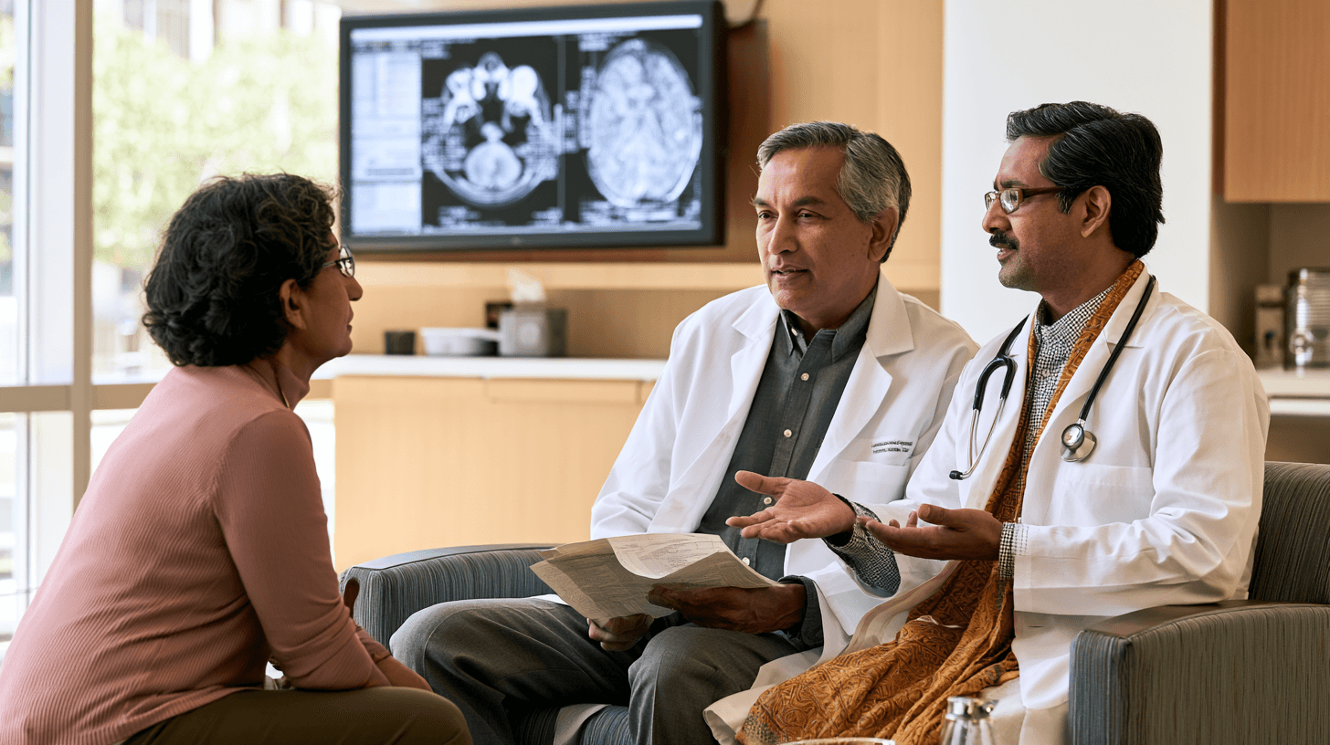
Ayurvedic treatment may be introduced after microvascular decompression, Gamma Knife radiosurgery or radiofrequency treatment, but the recovery plan cannot be identical after all three procedures. Microvascular decompression is an open neurosurgical operation that attempts to separate an offending blood vessel from the trigeminal nerve. Gamma Knife delivers focused radiation and usually produces its therapeutic effect gradually. Radiofrequency thermocoagulation intentionally creates a controlled lesion in selected trigeminal fibres and may provide more rapid pain relief, but facial numbness and other sensory changes are more closely connected with its mechanism [2,3,12–14]. UEMS Neuroboard
When I evaluate a patient after any trigeminal neuralgia procedure, I first identify what has improved and what remains. The electric-shock attacks, continuous facial pain, numbness, burning, chewing ability, corneal sensation, sleep and neurological medicine requirements are recorded separately. A patient may no longer experience the original shocks but may remain troubled by postoperative headache, facial numbness, jaw weakness or fear of touching the face. These problems should not be combined into one general statement that the procedure either succeeded or failed.
Ayurveda may support appetite, digestion, bowel function, sleep, strength, emotional recovery and return to normal daily activities. It may also be considered for residual pain and trigger sensitivity after the surgical team has excluded a complication. It should not replace postoperative neurological care, wound assessment, ophthalmological protection or neurosurgical review when symptoms recur or change.
The Procedure Report Must Be Reviewed Before Ayurvedic Treatment
Before starting post-procedural Ayurveda, I review the exact procedure, date, treated side, affected trigeminal divisions, operative or radiosurgical report, current medicines and the patient’s immediate response. The terms microvascular decompression, Gamma Knife and radiofrequency do not describe interchangeable interventions, and their potential complications and recovery timelines differ significantly.
After microvascular decompression, the operative note may identify an arterial, venous or multiple-vessel compression and explain how the nerve was decompressed. After Gamma Knife, the treatment report may specify the radiation target and dose. After radiofrequency treatment, the record may describe the trigeminal divisions targeted and the sensory response produced during the procedure.
The pre-procedure pain pattern must also remain visible. A patient who originally had brief triggerable shocks requires a different review from someone whose principal symptom was continuous burning or painful numbness. When the initial diagnosis was incomplete, a technically successful procedure may not resolve every facial-pain component.
Ayurvedic Care Immediately After Microvascular Decompression
Microvascular decompression involves cranial surgery and requires an initial recovery period led by the neurosurgeon. The first priorities are neurological observation, wound healing, pain control, hydration, nutrition, hearing and balance assessment and safe continuation or adjustment of prescribed medicines. Ayurveda should not interfere with these priorities.
I do not begin facial massage, head massage, Shirodhara, strong Swedana, Nasya or oil application around the head and incision immediately after microvascular decompression. The absence of visible external swelling does not mean that deeper surgical healing is complete. Local Ayurvedic procedures should begin only after the neurosurgeon has confirmed that the incision has healed adequately and there is no concern regarding infection, cerebrospinal-fluid leakage or another postoperative complication.
A patient should seek prompt surgical assessment for fever, wound redness, increasing swelling, discharge, clear fluid leakage, a severe or worsening headache, new facial weakness, confusion, seizure, sudden hearing deterioration, marked balance disturbance or another new neurological symptom. Such findings should never be described only as Vāta aggravation, postoperative cleansing or a temporary Ayurvedic healing response.
During the early recovery period, carefully selected internal Ayurvedic medicines may be considered for appetite, digestion, constipation, sleep and general strength when the neurosurgeon is aware of the complete formulation. The prescription must be compatible with analgesics, anticonvulsants, antibiotics, anti-nausea medicines and any other postoperative treatment. A medically complex patient may require laboratory review before a polyherbal or herbo-mineral formulation is introduced.
Neurological Medicines After Microvascular Decompression
Some patients become free from electric-shock pain immediately after microvascular decompression and assume that carbamazepine or oxcarbazepine can be stopped on the same day. Medication reduction should still follow a plan from the neurologist or neurosurgeon. The safe timing depends on the preoperative dose, duration of treatment, immediate pain response, other medicines and the treating centre’s postoperative protocol.
Medication-free pain freedom is an important long-term outcome, but it should be reached through supervised reduction. Abrupt withdrawal can produce renewed attacks and make it difficult to determine whether the pain represents persistent trigeminal neuralgia, temporary postoperative irritation or premature medicine reduction.
The five-year prospective evidence indicates that appropriately selected patients undergoing microvascular decompression may achieve substantial long-term medication-free pain control, although postoperative sensory change, hearing impairment and recurrence remain possible [12]. The findings support careful patient selection and long-term follow-up rather than a guarantee that every patient will remain permanently pain-free. PubMed Central (PMC)
When Pain Continues After Microvascular Decompression
Pain persisting after microvascular decompression should be classified before another treatment is prescribed. The patient may have persistent electric-shock attacks, continuous background pain that existed before surgery, postoperative incisional pain, muscular discomfort or a different facial-pain disorder that was not addressed by decompression.
I ask whether the current pain has the same side, trigeminal distribution, duration and triggers as the preoperative attacks. Persistent seconds-long shocks triggered by chewing or light touch have a different meaning from continuous aching around the surgical area or burning within a numb region.
The operative report and MRI should be reviewed when the expected improvement has not occurred. The absence of complete relief does not automatically mean that a stronger Ayurvedic medicine is required. The nerve may require further neurological assessment, the original diagnosis may need reconsideration or a continuous neuropathic component may require a different treatment strategy.
Ayurveda may be used supportively once the surgical team has excluded an acute complication. The goals may include reducing residual pain burden, improving sleep, restoring food intake, supporting bowel function and helping the patient regain normal activities. Ayurveda should not be claimed to repeat, repair or reverse the surgical decompression without objective evidence.
Ayurvedic Rehabilitation After Successful Microvascular Decompression
Even after the electric-shock attacks stop, a patient may remain afraid to eat, brush the teeth, wash the face or sleep on the previously painful side. Months or years of repeated attacks can establish powerful avoidance patterns. Pain relief alone may therefore not restore complete function immediately.
During this stage, I use a gradual rehabilitation plan. Food texture is normalised progressively, oral hygiene is restored, speaking duration increases and unnecessary facial avoidance is reduced. The patient should not be forced to provoke the old trigger zone, but ordinary activities should return as confidence and neurological stability improve.
Bṛṃhaṇa and Rasāyana-oriented medicines may be considered when the patient has lost weight, strength or sleep during the preoperative illness. Medhya and Manas-supportive treatment may assist emotional recovery and fear of recurrence. Dīpana, Pācana or Anulomana support may be used when surgery, reduced activity and medicines have disturbed appetite or bowel function.
The formulation must be selected according to Agni, Bala, age, diabetes, liver and kidney function and the complete postoperative medicine list. Recovery after successful surgery is not a reason to prescribe every available Rasāyana. The treatment should become simpler as normal strength and function return.
Ayurvedic Care After Gamma Knife Radiosurgery
Gamma Knife radiosurgery does not require an open cranial incision, but the lack of a wound does not mean that its effect is immediate. Focused radiation gradually alters pain transmission in the trigeminal nerve, and meaningful relief may take time to develop. During this interval, patients commonly continue their neurological medicines according to the treating clinician’s instructions [2,3,14]. UEMS Neuroboard
A patient should not declare Gamma Knife unsuccessful because attacks continue during the early post-treatment period. The pain diary should record whether attack frequency, trigger sensitivity and pain-free intervals are beginning to change. At the same time, the person should not wait indefinitely with uncontrolled pain, dehydration or nutritional decline merely because a delayed response is expected.
Ayurveda during this waiting period may support sleep, digestion, nutrition, constipation and emotional stability. The treatment may also help the patient maintain normal food and fluid intake while the radiosurgical effect develops. Neurological medicine should remain stable whenever clinically possible so that the response can be interpreted clearly.
A large new Ayurvedic treatment package introduced together with neurological dose reduction can create confusion. If pain worsens, the patient may not know whether Gamma Knife has failed, carbamazepine was reduced too early or an additional treatment has altered the clinical picture.
Delayed Facial Numbness After Gamma Knife
Facial numbness, tingling or altered sensation may appear after Gamma Knife, sometimes after the pain has already started improving. The patient should report when the sensory change began, where it is located and whether it is ordinary numbness, unpleasant tingling, continuous burning or painful numbness.
When the ophthalmic division is affected, corneal safety becomes particularly important. Eye redness, persistent watering, dryness, visual change, light sensitivity or a foreign-body sensation requires ophthalmological assessment. Reduced corneal sensation may prevent the patient from recognising irritation or injury normally.
I avoid aggressive facial massage, strong Swedana and excessively hot oils when sensation has become reduced. The patient may not recognise excessive temperature or pressure. Local treatment should be mild, carefully observed and discontinued if redness, burning, swelling or worsening sensory symptoms appear.
Gamma Knife-related numbness should not be treated as proof that the nerve requires stronger stimulation. The radiosurgical team or neurologist should first determine whether the sensory change is within the expected range or represents a clinically significant complication.
When Pain Returns After Gamma Knife
Recurrence after a period of relief should be distinguished from continuous post-radiosurgical sensory pain. Familiar brief electric shocks triggered by touch or chewing suggest return of the neuralgic paroxysms. Continuous burning within a numb area may represent a different neuropathic consequence.
The distinction matters before repeat radiosurgery or another ablative treatment is considered. Cumulative sensory injury becomes increasingly relevant after repeated procedures. Existing corneal sensation, facial numbness and painful dysesthesia should be documented carefully [2,14].
Ayurveda may support the patient during renewed medical or procedural assessment, but it should not delay review of recurrent attacks. A repeat procedure, microvascular decompression, percutaneous treatment or continued medical care may be considered according to the diagnosis, imaging, previous response and current sensory status.
Ayurvedic Care After Radiofrequency Thermocoagulation
Radiofrequency thermocoagulation works by creating a controlled thermal lesion in selected trigeminal fibres. Pain relief may occur rapidly, but some degree of facial sensory change is closely connected with the procedure’s mechanism. Numbness, altered sensation, chewing-muscle weakness, dysesthesia and corneal sensory problems are important considerations, particularly when V1 or V3 fibres are treated [2,14]. PubMed Central (PMC)
When I evaluate a patient after radiofrequency treatment, I do not ask only whether the electric shocks stopped. I map facial sensation across V1, V2 and V3 and document numbness, burning, tingling, oral injury, eye symptoms and chewing ability. The post-procedural sensory map becomes the foundation for safe Ayurvedic treatment.
A patient may feel that the procedure was successful because the face no longer produces intense pain, but the same reduced sensation can prevent recognition of excessive heat, skin injury, oral biting or corneal irritation. Sensory protection is therefore part of recovery rather than a secondary issue.
Heat and Massage Require Special Caution After Radiofrequency
Strong facial Swedana, hot compresses, heated oils and vigorous massage should not be used over an area with reduced sensation. The patient may sustain a burn or pressure injury without experiencing normal warning pain. A treatment that feels mild to the practitioner may be excessive for tissue that the patient cannot assess accurately.
I also avoid using strong local stimulation with the explanation that it will awaken or regenerate the nerve. Painful numbness and dysesthesia can worsen when an already injured sensory pathway is repeatedly irritated. Local Ayurvedic treatment should remain gentle and should be stopped when it increases burning, tingling or unpleasant sensitivity.
The skin should be inspected regularly for redness, blisters, scratches and ulceration. A patient who experiences itching or crawling sensations may rub the area repeatedly and create wounds without recognising the degree of injury.
Protecting the Eye After Radiofrequency
Radiofrequency treatment involving the ophthalmic division can reduce corneal sensation. The eye may become dry or injured without producing the usual degree of discomfort. The patient should follow the procedural team’s eye-protection advice and obtain ophthalmological review when symptoms appear.
Nasya, facial oil application, steam and herbal preparations around the eye should not be introduced casually when corneal sensation is reduced. Persistent eye redness, blurred vision, discharge, watering or foreign-body sensation requires direct examination rather than adjustment of the Ayurvedic medicine alone.
The absence of eye pain is not reassuring when sensation has been altered. Corneal protection must be guided by eye findings, not by whether the patient feels discomfort.
Chewing Weakness and Nutrition After Radiofrequency
When the mandibular division is treated, temporary weakness or fatigue of the chewing muscles may occur. The patient may struggle with harder food even though the electric-shock attacks have improved. This difficulty should not be attributed only to fear or poor effort.
A temporary softer diet may be necessary, but it should remain nutritionally complete. Protein, calories, hydration and food variety must be maintained while chewing function recovers. Prolonged dependence on sweet liquids or nutritionally weak soft food can create another health problem.
Ayurvedic dietary care may support Agni, bowel function and Bala, but the plan should be adapted to mechanical chewing ability. Kavala and Gaṇḍūṣa may be inappropriate when the jaw is weak, fatigued or easily triggered. Forceful oral procedures should wait until the patient can move the jaw safely.
Painful Numbness After Radiofrequency
Painful numbness is different from ordinary sensory reduction. The patient may describe burning, pressure, aching or unpleasant crawling in an area that also feels numb. This pattern should not be labelled automatically as recurrence of classical trigeminal neuralgia.
Another destructive procedure may not improve the continuous neuropathic component and can increase sensory loss. The treating neurologist, pain specialist or neurosurgeon should determine whether the patient has recurrent paroxysms, post-procedural trigeminal neuropathy, dysesthesia or anaesthesia dolorosa before further intervention is selected.
Ayurvedic care may support sleep, emotional burden, nutrition and general function, but local treatment must remain conservative. A claim that intense Snehana, Swedana or stimulation will regenerate all injured fibres is not supported by current clinical evidence.
Neurological Medicine After Gamma Knife or Radiofrequency
Carbamazepine, oxcarbazepine or another neurological medicine should not be stopped independently after Gamma Knife or radiofrequency treatment. The timing of reduction differs because Gamma Knife relief may be delayed, while radiofrequency relief may occur more rapidly but still requires observation.
The neurologist or procedural specialist should provide the tapering plan. The patient should record the return of familiar electric-shock attacks separately from continuous numbness, burning or procedural discomfort. This prevents the medicine from being increased automatically for a sensory complication that may not respond in the same way as classical trigeminal neuralgia.
Medication-free pain freedom is a useful outcome only when it is sustained and reached through supervised reduction. A few pain-free days after a procedure are encouraging but do not establish permanent cure.
Ayurvedic Medicine Selection After a Trigeminal Procedure
The Ayurvedic prescription is selected according to the patient’s present condition rather than the pre-procedure diagnosis alone. A person recovering from open surgery may initially require support for appetite, bowel function, sleep and strength. A patient waiting for Gamma Knife benefit may require help maintaining nutrition and emotional stability without increasing sedation. A person with numbness after radiofrequency requires greater sensory protection and a cautious approach to local treatment.
Vāta-Śāmaka and Vedanāsthāpana medicines may be considered when familiar electric-shock attacks or trigger sensitivity remain. Pitta-Śāmaka treatment may be relevant when continuous burning and heat sensitivity are prominent. Bṛṃhaṇa and Rasāyana support may be selected after prolonged weight loss, restricted food intake or surgical depletion. Medhya and Nidrā-supportive medicines may help sleep and fear of recurrence, but the combined sedative burden must be reviewed.
These therapeutic classes are not combined mechanically. The formulation is adjusted according to age, Agni, Bala, diabetes, blood pressure, liver and kidney function, sensory findings and current neurological medicines.
Herbo-mineral preparations are not automatically required because surgery or radiosurgery has failed. Any Bhasma or Rasaushadhi requires a specific indication, authenticated preparation, batch-level quality documentation and patient-specific safety monitoring.
When Ayurvedic Treatment Should Be Postponed
Routine Ayurvedic treatment should be postponed when the patient has fever, an unhealed or infected surgical wound, suspected cerebrospinal-fluid leakage, severe or worsening postoperative headache, new weakness, confusion, seizure, sudden hearing loss or significant balance disturbance after microvascular decompression.
It should also be postponed when Gamma Knife or radiofrequency is followed by progressive numbness, severe painful dysesthesia, visual change, eye redness, corneal symptoms, marked chewing weakness or inability to maintain nutrition. These findings require assessment by the procedural team, neurologist or ophthalmologist before a supportive programme is expanded.
A patient who cannot drink, eat or take prescribed medicine requires urgent medical stabilisation. Severe psychological deterioration or suicidal thinking also requires immediate mental-health and emergency support.
Referral and temporary postponement do not mean that Ayurveda has no role. They ensure that a complication is not concealed by newly introduced medicines or interpreted exclusively through Doṣa.
What Ayurveda Can Realistically Achieve After These Procedures
Ayurveda may assist the patient in recovering appetite, digestion, bowel regularity, sleep, strength, emotional stability and normal daily function. It may also be monitored for its effect on residual attacks, background pain and trigger sensitivity after the procedural team has excluded a complication.
The classical Anantavāta framework and broader Vātavyādhi principles provide a basis for individualising post-procedural care according to Vāta, Pitta, Agni, Bala and the stage of recovery [19]. However, no controlled clinical study has established that Ayurveda specifically reverses the effects of Gamma Knife, restores every fibre altered by radiofrequency or guarantees that pain will never recur after microvascular decompression.
The available modern Ayurvedic evidence consists mainly of limited case-level documentation in trigeminal neuralgia and painful trigeminal neuropathy rather than controlled studies of post-MVD, post-Gamma Knife or post-radiofrequency care [20]. The treatment goals should therefore remain clinically measurable and proportionate to the evidence.
How I Manage Post-Procedural Trigeminal Neuralgia at Panaceayur
Before preparing an Ayurvedic plan, I review the procedure report, pre-procedure MRI, painful side, trigeminal divisions, immediate pain response, current neurological medicines and all new sensory or neurological symptoms. Patients are asked to provide the operative or radiosurgical record, discharge summary, current medicine list and a pain-and-sensation diary.
The diary separates electric-shock attacks from continuous pain, numbness, burning and muscular discomfort. It also records eating, drinking, speaking, oral hygiene, sleep, balance, hearing and eye symptoms. This distinction helps determine whether the patient has residual neuralgia, delayed procedural response, recurrence, postoperative recovery symptoms or a sensory complication.
The Ayurvedic assessment then examines Prakṛti, Vikṛti, Doṣa, Agni, Āma, Koṣṭha, Bala, Satva, nutritional status, bowel function, disease duration and associated chronic disorders. Treatment is coordinated with the neurologist or procedural specialist, and prescribed neurological medicine remains under their supervision.
The objective is not to repeat the procedure through Ayurveda or to make an unsupported promise of permanent cure. It is to help the patient recover safely, reduce the remaining burden where possible and restore eating, speaking, sleep, strength and confidence without overlooking a complication that requires modern specialist care.
What Modern Research Says About Ayurvedic Herbs for Neuropathic Pain

Modern research on Ayurvedic herbs for neuropathic pain is increasingly identifying biological pathways that may help explain their traditional use in Vāta-dominant pain, burning neuropathy, sleep disturbance and neurological recovery. The available studies suggest that selected plants may influence pain sensitisation, inflammatory cytokines, oxidative stress, microglial activity, purinergic receptors, sodium-channel-related signalling and other mechanisms involved in abnormal nerve excitability.
The evidence, however, is not equally strong for every herb or every pain disorder. Most studies have been conducted in laboratory cells or animal models of nerve injury, diabetic neuropathy, chemotherapy-induced neuropathy or trigeminal neuropathic pain. Direct human evidence in trigeminal neuralgia remains limited mainly to individual case reports involving multimodal Ayurvedic treatment rather than trials of one herb or one Avaleha.
When I interpret this research clinically, I do not assume that a herb showing an effect in a rat model will produce the same result in every patient. I examine whether the experimental pain mechanism resembles the patient’s condition, whether the study used a standardised extract or crude powder, and whether the proposed ingredient is safe with carbamazepine, oxcarbazepine or another neurological medicine. This approach allows modern research to strengthen Ayurvedic selection without turning preliminary findings into an unsupported cure claim.
Maṇḍūkaparṇī and Direct Experimental Trigeminal-Pain Research
Among the Ayurvedic plants studied for neuropathic pain, Maṇḍūkaparṇī, identified botanically as Centella asiatica, has one of the most directly relevant experimental studies for trigeminal neuropathic pain. A 2024 study used a standardised extract known as ECa 233 in an animal model created through chronic constriction injury of the infraorbital nerve, a sensory branch of the trigeminal nerve .
The study reported a reduction in mechanical pain hypersensitivity and changes in trigeminal pain-signalling pathways, including modulation of P2X3-related activity within the trigeminal ganglion and markers of neuronal activation. P2X3 receptors participate in the transmission and amplification of pain signals, making this pathway relevant to trigeminal hypersensitivity . PubMed Central (PMC)
This finding is important because the model directly involved the trigeminal system rather than a nerve in the leg or spinal region. It provides a plausible modern basis for considering Maṇḍūkaparṇī within an individualised formulation when trigeminal nerve hypersensitivity, continuous neuropathic pain or post-traumatic sensory irritation is present.
The study does not prove that ordinary Maṇḍūkaparṇī powder, a household preparation or an Avaleha will produce the same effect in a human patient. ECa 233 was a chemically standardised extract with a defined triterpenoid composition, while crude herb preparations may differ in concentration, absorption and bioavailability. The research supports the therapeutic rationale, but it does not establish a human dose or guarantee permanent pain freedom.
Aśvagandhā and Reduction of Pain Hypersensitivity
Aśvagandhā, or Withania somnifera, has been investigated in experimental models of postoperative and neuropathic pain. In a rat study involving spared nerve injury and surgical-incision pain, Aśvagandhā root extract reduced mechanical hypersensitivity and pain-related behaviour. The researchers also reported effects on inflammatory and immune-signalling pathways associated with the development of chronic pain. PubMed
These findings provide a biological basis for the traditional use of Aśvagandhā as a Balya and Rasāyana medicine in patients with chronic pain, disturbed sleep, weakness and reduced neurological resilience. In trigeminal neuralgia, it may be considered when the patient is physically depleted, has lost weight, sleeps poorly or remains exhausted by repeated attacks.
Aśvagandhā should not be described as a rapid substitute for carbamazepine. The experimental study did not involve human trigeminal neuralgia, and the extract and doses used in animals cannot be transferred directly to an Avaleha prescription. Its most rational role may involve supporting pain modulation, sleep, Bala and long-term recovery as one component of a wider treatment plan.
Safety also requires attention. Aśvagandhā has been associated with uncommon but clinically significant liver injury in susceptible individuals. A patient with pre-existing liver disease, unexplained jaundice or previous herb-related liver injury requires careful selection and monitoring [49].
Guḍūcī and Experimental Neuropathic-Pain Models
Guḍūcī, or Tinospora cordifolia, has been studied in more than one experimental form of neuropathic pain. A 2012 study evaluated it in diabetic neuropathy, while a 2024 study investigated its effects in paclitaxel-induced neuropathic pain. The studies reported reductions in pain-related behaviour and changes in biochemical pathways associated with oxidative stress, inflammation and nerve dysfunction. PubMed
The use of different neuropathy models is valuable because it suggests that Guḍūcī may act on broader mechanisms involved in nerve hypersensitivity rather than on one isolated cause. This supports its Ayurvedic role as a Rasāyana and Śothahara medicine where chronic inflammation, metabolic disease or treatment-related neuropathy contributes to the patient’s burden.
The models differ significantly from classical trigeminal neuralgia caused by neurovascular compression. Diabetic and chemotherapy-associated neuropathies usually involve a different distribution and underlying nerve injury. Guḍūcī research therefore provides indirect mechanistic support rather than proof that it can stop triggerable trigeminal paroxysms or reposition a blood vessel compressing the nerve.
Guḍūcī also requires responsible botanical identification and safety assessment. Published reports have linked some Tinospora products with acute or autoimmune-like liver injury, particularly when species identity, susceptibility or pre-existing disease is relevant . It should not be added automatically to every chronic-pain formula merely because it is considered a Rasāyana.
Śuṇṭhī and Neuroinflammatory Pain Pathways
Śuṇṭhī, or dried Zingiber officinale rhizome, has been investigated in a mouse model of peripheral neuropathic pain. A standardised ginger extract reduced mechanical and thermal allodynia and influenced several pathways associated with neuroinflammation. The study reported reduced activation of NF-κB-related signalling and lower release of inflammatory mediators including interleukin-1 beta, tumour necrosis factor-alpha and interleukin-6. PubMed
This research helps explain why Śuṇṭhī may provide more than digestive support in a neuropathic-pain formulation. Neuroinflammation can contribute to the persistence of pain after nerve injury, and modulation of inflammatory signalling may reduce the sensitised state in which ordinary touch becomes painful.
Śuṇṭhī also supports Dīpana and Pācana, which can be useful when a patient cannot tolerate a nourishing Avaleha because of Mandāgni, nausea or heaviness. Its inclusion should still reflect the patient’s Pitta status, oral sensitivity, reflux and nutritional condition. A strong pungent component may aggravate burning or gastric symptoms in an unsuitable patient.
The experimental study used a concentrated extract in mice rather than ordinary dry ginger powder in humans. Its results cannot determine the clinical dose required for trigeminal neuralgia, and the study did not demonstrate any change in neurovascular compression.
Balā and NMDA-Receptor-Related Neuropathic Signalling
Balā, or Sida cordifolia, has also been investigated in an animal model of neuropathic pain. A 2024 study reported that Balā root extract and one of its fractions reduced behavioural hypersensitivity in rats with chronic constriction nerve injury. The proposed mechanism involved modulation of KIF17–NR2B signalling associated with trafficking and activity of NMDA receptors . PubMed
NMDA-receptor activity is relevant to central sensitisation, a state in which pain pathways become increasingly responsive after prolonged or repeated nerve input. This gives Balā a modern mechanistic rationale alongside its classical Balya, Bṛṃhaṇa and Vāta-Śāmaka applications.
In a patient with long-standing trigeminal neuralgia, Balā may be considered when severe pain is accompanied by weakness, low body weight, disturbed sleep and loss of strength. Its role should be adjusted in people with hypertension, cardiac rhythm concerns or sensitivity to stimulating constituents.
The study remains preclinical. It does not establish that Balā reverses human trigeminal nerve damage or that every preparation sold as Balā contains the correct species, plant part and active profile. Botanical authentication is particularly important because regional substitution and species confusion can affect both efficacy and safety.
Nirgundī and Indirect Human Pain Evidence
Nirgundī, or Vitex negundo, has some human clinical evidence in a different nerve-related pain condition. A clinical study enrolled 119 patients with Gridhrasī, clinically compared with sciatica, and 102 completed treatment. Nirgundī Ghana Vaṭi was used alone in one group and with Mātrā Basti in another. Both groups showed improvement, with greater relief reported in the combined-treatment group. PubMed
This is relevant because it provides human evidence that an Ayurvedic plant-based intervention may influence pain, neurological symptoms and function. It also reflects an important Ayurvedic principle: the combined treatment strategy appeared more useful than the oral extract alone.
The study did not involve trigeminal neuralgia, was not a large blinded placebo-controlled trial and evaluated sciatica rather than cranial nerve pain. Its findings cannot be transferred directly to a patient with neurovascular compression of the trigeminal nerve.
Nirgundī may nevertheless be selected for its Vedanāsthāpana and Śothahara rationale when pain and inflammatory sensitivity are present. Its inclusion should be based on the individual pain phenotype rather than on an assumption that any medicine helping sciatica will necessarily cure trigeminal neuralgia.
Rāsnā and the Anti-Inflammatory–Analgesic Rationale
Rāsnā, commonly identified as Pluchea lanceolata, has a long classical association with Vāta disorders and painful conditions. A modern pharmacological review identified several constituents and experimental effects relevant to inflammation and pain, supporting its traditional use as part of Vāta-Śūla treatment. PubMed
The evidence for Rāsnā is predominantly pharmacological and preclinical. No controlled human trial has established Rāsnā as an effective standalone treatment for trigeminal neuralgia. Its value lies in the broader formulation strategy, particularly when the patient has pain, stiffness, inflammatory sensitivity or additional Vāta-related musculoskeletal symptoms.
This distinction matters because classical use and modern mechanistic plausibility are not identical to condition-specific proof. Rāsnā may be rationally included in an individualised formula without claiming that it has independently demonstrated medication-free remission in trigeminal neuralgia.
Brāhmī and Jaṭāmāṃsī Address the Wider Neurological Burden
Brāhmī, or Bacopa monnieri, has been studied more extensively for cognitive and neuroprotective effects than for direct analgesia. Modern reviews describe antioxidant, neurotransmitter-related and cognitive effects that may support attention, memory and emotional regulation . PubMed
These effects may be clinically relevant in a patient whose concentration, confidence and mental stamina have deteriorated because of persistent pain, poor sleep or sedating neurological medicine. Brāhmī should therefore be understood primarily as Medhya and supportive treatment rather than as a proven direct treatment for electric-shock attacks.
Jaṭāmāṃsī, or Nardostachys jatamansi, has demonstrated central calming and sleep-related effects in experimental animals. One study found that Jaṭāmāṃsī rhizome powder shortened the onset of sleep and prolonged sleep duration in animals without significantly impairing muscle coordination in the experimental setting . PubMed
In trigeminal neuralgia, Jaṭāmāṃsī may help selected patients whose recovery is limited by insomnia, hypervigilance and fear of the next attack. It should not be combined indiscriminately with carbamazepine, baclofen, gabapentin, pregabalin or sleep medicine because cumulative sedation may impair balance, driving and daytime function.
These Medhya and Nidrā-supportive herbs can improve the patient’s capacity to recover, but better sleep should not be interpreted as proof that neurovascular compression has resolved.
Yaṣṭimadhu Has Mechanistic Promise but Important Limitations
Yaṣṭimadhu, or Glycyrrhiza glabra, contains glycyrrhizin, flavonoids and other compounds with anti-inflammatory, antioxidant and neuroprotective activity. A modern review considered whether liquorice-derived compounds could contribute to pain treatment and identified several plausible mechanisms, including modulation of inflammatory signalling . PubMed
The same review concluded that the clinical analgesic role of liquorice-derived compounds had not yet been established. Yaṣṭimadhu should therefore be regarded as a supportive, soothing and formulation-balancing medicine rather than a clinically proven treatment for trigeminal neuralgia.
Its safety profile is particularly important. Glycyrrhizin exposure can contribute to hypertension, fluid retention and low potassium in susceptible patients. A person with uncontrolled blood pressure, heart failure, kidney disease, hypokalaemia or medicines affecting fluid and electrolyte balance may require avoidance, dose restriction or close monitoring.
Human Evidence for Ayurveda in Trigeminal Pain Remains Preliminary
Direct human evidence for Ayurvedic treatment of trigeminal neuralgia remains limited. A 2023 case report described one patient with MRI-confirmed classical trigeminal neuralgia who received a three-month multimodal Ayurvedic programme involving oral medicines and procedures. The authors reported improvements in pain, psychological measures and quality of life [20]. PubMed Central (PMC)
A 2025 case report described one patient with painful post-traumatic trigeminal neuropathy treated through a prolonged programme involving Panchakarma and Ayurvedic medicines. The authors reported improvement in pain and associated symptoms [51]. Post-traumatic trigeminal neuropathy, however, differs from classical trigeminal neuralgia caused by neurovascular compression. PubMed
These reports are valuable because they show that Ayurvedic treatment can be documented through modern diagnosis, pain scores, psychological assessment and functional outcomes. They also support further clinical investigation of integrated treatment.
A case report cannot establish general effectiveness. There is no control group, the condition may fluctuate naturally, several interventions were used together and the individual contribution of each herb or procedure cannot be determined. The results cannot provide a universal cure rate or prove that the same treatment will work for another patient.
Standardised Extract Research Cannot Be Converted Directly Into an Avaleha Dose
Many positive experimental studies used concentrated or chemically standardised extracts. These extracts may contain a defined percentage of selected constituents and may be produced through water, alcohol, carbon-dioxide extraction or another laboratory process. A traditional powder, decoction or Avaleha can have a different chemical profile and absorption pattern.
Animal doses expressed in milligrams per kilogram also cannot be converted directly into a human prescription by simple multiplication. Differences in metabolism, body-surface area, preparation, route of administration and biological availability must be considered.
The combined action of several herbs in an Avaleha may differ from the action of each extract when studied independently. Synergy is possible, but it should not be assumed without evidence. Combining every herb with a positive laboratory study may increase treatment complexity and interaction risk without producing a stronger clinical result.
When I design a formulation, I use experimental evidence to support the selection of a therapeutic class and mechanism. The final quantities are determined according to Ayurvedic pharmacology, classical compatibility, patient-specific factors, dosage form, safety and practical clinical experience not by copying animal doses.
Modern Research Supports Individualised Integration, Not Self-Medication
Modern research gives a reasonable biological foundation for using selected Ayurvedic herbs in neuropathic-pain care. Maṇḍūkaparṇī has direct experimental relevance to trigeminal pain signalling, while Aśvagandhā, Guḍūcī, Śuṇṭhī and Balā have shown effects in other neuropathic-pain models. Nirgundī provides indirect human evidence from sciatica, and Medhya herbs may support sleep, cognition and emotional recovery.
The research does not show that a market-bought herbal product can diagnose the pain subtype, replace MRI, remove neurovascular compression, reverse a multiple sclerosis plaque, eliminate a tumour or guarantee permanent cure. It also does not establish that every herb should be combined into the same formulation.
A patient with brief triggerable electric shocks requires a different formulation strategy from someone with continuous postherpetic burning or painful numbness after dental nerve injury. Age, disease duration, Agni, Bala, diabetes, liver and kidney function, blood pressure, current neurological medicines and previous procedures can all alter the correct selection.
In my clinical approach, modern evidence is used to strengthen rather than replace Ayurvedic individualisation. I select herbs according to the patient’s pain mechanism and Ayurvedic presentation, introduce major treatment changes carefully and measure the result through attack frequency, trigger tolerance, food intake, sleep, oral hygiene, medicine burden and daily function.
The present evidence supports Ayurveda as a scientifically plausible and potentially valuable component of integrated neuropathic-pain management. Its credibility is greatest when the formulation is customised, the neurological diagnosis remains visible, adverse effects are monitored and clinical outcomes are described accurately rather than through an unsupported universal promise.
What Published Evidence Exists for Ayurveda in Trigeminal Neuralgia?
Published evidence for Ayurveda in trigeminal neuralgia does exist, but its present strength must be understood correctly. The direct human literature consists predominantly of individual case reports and case studies in which oral Ayurvedic medicines were combined with procedures such as Nasya, Gandūṣa, Kavala, Śirodhārā, Śirovasti, Mātrā Basti, Agnikarma or Viddhakarma. These reports describe clinically meaningful improvement in selected patients, but they do not provide the same certainty as a randomised controlled trial, a comparative cohort study or a systematic review of high-quality clinical trials.
A review of the accessible published literature up to August 8, 2026 did not identify a randomised controlled human trial evaluating a defined Ayurvedic formulation or standardised Ayurvedic protocol specifically for trigeminal neuralgia. The available evidence therefore supports clinical plausibility, carefully monitored integrative treatment and the need for larger studies. It does not establish a universal cure rate, prove that every patient can discontinue carbamazepine or demonstrate that Ayurveda physically removes neurovascular compression.
This distinction is important for patients. A published case report is stronger than an undocumented testimonial because it usually describes the diagnosis, treatment and measured outcome. However, one patient’s response cannot predict how frequently the same treatment will work in a wider population.
The 2023 MRI-Confirmed Classical Trigeminal Neuralgia Case
The most detailed direct human report was published in the Journal of Ayurveda and Integrative Medicine in 2023. It involved a 39-year-old man with two years of right-sided facial pain affecting the upper lip, cheek and chin. His attacks occurred every one to two hours, lasted approximately one to two minutes and were triggered by speaking, chewing, smiling, washing the face and brushing the teeth [20].
MRI showed indentation of the right trigeminal nerve by a vascular loop of the right superior cerebellar artery, supporting a diagnosis of classical trigeminal neuralgia. The patient had previously taken carbamazepine but continued to experience acute exacerbations. The Ayurvedic programme lasted three months and included Daśamūla Kvātha, Aśvagandhā Cūrṇa, Ojasvinī Cūrṇa, Ekāṅgavīra Rasa and staged procedures involving Kukkuta Aṇḍa Svedana, Kṣīrabala Taila Nasya, Kavalagraha and Gandūṣa [20]. PubMed Central (PMC)
The reported visual analogue pain score decreased from 9 at baseline to 2 after 90 days. Acute attacks that had originally occurred every one to two hours were no longer reported by day 90, although some dull background pain remained. Anxiety scores decreased from 14 to 6, depression scores decreased from 12 to 4, and the authors reported substantial improvement across quality-of-life domains [20]. PubMed Central (PMC)
One of the most valuable findings was that repeat MRI after treatment continued to show the same vascular indentation. This indicates that clinical symptoms improved even though the anatomical neurovascular relationship remained visible. The report therefore supports possible pain modulation and functional improvement, but it does not support a claim that Ayurveda removed the compressing vascular loop [20]. PubMed Central (PMC)
The report followed a single patient, used several interventions simultaneously and did not include a control group, blinding or comparison with continued neurological care. Trigeminal neuralgia may also enter natural remission. It is therefore impossible to determine how much improvement came from the oral medicines, individual procedures, natural fluctuation or their combined effect.
The 2024 Acute Type 1 Trigeminal Neuralgia Case
A second PubMed-indexed case report was published in the Journal of Pharmacy and Bioallied Sciences in late 2024. It described a 41-year-old woman with approximately 2.3 years of intermittent right lower-jaw pain, tingling and touch-triggered symptoms. She had previously taken carbamazepine, reportedly with inadequate sustained benefit and treatment-related nausea and fatigue. PubMed Central (PMC)
MRI did not demonstrate neurovascular compression, and the authors described the condition as Type 1 trigeminal neuralgia. The patient received a 15-day programme combining Śamana and Śodhana treatment. The programme included oral medicines, Śirodhārā, Nasya and Trisnehadhāraṇa. Follow-up was recorded on days 7, 15 and 30. PubMed Central (PMC)
The authors reported progressive reduction in electric-shock pain, numbness, attack frequency and fear of recurrence. By day 30, the recorded scores for pain, numbness, attacks and subjective fear had reached zero. This is an encouraging observation, particularly because function and fear were assessed alongside pain. PubMed Central (PMC)
The evidence remains limited by the single-patient design, short follow-up, absence of a comparator and use of a multimodal protocol. A symptom-free state at 30 days cannot establish permanent remission or predict whether attacks will recur after several months or years. The report supports further study of a structured Ayurvedic approach but cannot provide a population-level success rate.
Older Case Reports Show Both Improvement and Treatment Failure
Older literature contains several Ayurvedic case reports, but their diagnostic and reporting quality varies. This variation is clinically useful because the publications do not all show the same outcome.
A 2015 report by Mamidi and Gupta described a 55-year-old man with severe left-sided trigeminal neuralgia associated with vascular-loop compression. He had previously used carbamazepine and was considering radiofrequency ablation. Ayurvedic treatment included Nasya, Kṣīra Dhūma, Ghṛtapāna, Gandūṣa, Karṇapūraṇa and local therapies. JPSI Journal
The patient experienced temporary relief during some procedures, particularly Karṇapūraṇa, but sustained improvement was not achieved. His McGill Pain Questionnaire score increased from 53 to 58, and he ultimately chose radiofrequency ablation. The authors concluded that the interventions helped some acute episodes but did not provide durable control in that patient. JPSI Journal
This negative report is important. It demonstrates that published Ayurvedic evidence does not consist only of successful cases and that Ayurveda may not provide sufficient control for every patient. It also shows why comorbid disease, inability to perform indicated procedures, withdrawal of neurological treatment, disease severity and patient selection can influence the outcome.
A 2017 report described a 64-year-old woman treated in relation to Anantavāta. The authors reported symptom relief and reduced recurrence following an Ayurvedic programme that included Mātrā Basti and Śirovasti . The abstract does not provide enough comparative or long-term data to determine the magnitude or durability of the effect reliably. iJam
Published Case Studies From 2019 to 2021
A 2019 case study from the International Journal of AYUSH Case Reports described a 34-year-old woman with two years of left-sided maxillary and mandibular jabbing pain aggravated by face washing, cold wind and particular times of day. The treatment included Rāsnāsaptaka Kvātha, Ābhā Guggulu and Pippalīmūla Cūrṇa. The authors reported marked symptom relief after one month and no pain episode during face washing after three months. IJA Care
Another 2019 case study used Śirodhārā, Gandūṣa, Nasya and oral medicines in a 37-year-old woman. The authors reported significant improvement after a treatment sequence extending over approximately 45 days . The publication remains a single case without a control group or independent outcome assessment. IJA Care
A 2021 case report described a 46-year-old woman who had not obtained satisfactory relief from conventional medication. She received a Vātahara and Bṛṃhaṇa programme involving Karpāsāsthyādi Taila Nasya, Kṣīrabala Taila Śiropicu and Karṇapūraṇa, Mūrcchita Tila Taila Gandūṣa and oral medicines. Pain and temporomandibular stiffness were assessed using pain scales, and the authors reported satisfactory improvement . JAIMS
These reports show that several Ayurvedic institutions have independently documented improvement in individual patients. However, the protocols differed substantially, the diagnostic work-up was not equally detailed in every case and long-term recurrence was often not reported. They therefore suggest possible benefit but cannot identify one standard treatment protocol.
The 2024 and 2025 Case-Report Literature
A 2024 AYUSHDHARA publication described the successful Ayurvedic management of one patient with trigeminal neuralgia. The intervention involved an individualised combination of medicines and procedures, and the authors reported improvement without surgical intervention . As with other single-patient reports, the result should be interpreted as clinical documentation rather than proof that surgery is unnecessary for all patients with neurovascular compression. Ayushdhara
A 2025 case report described a 43-year-old man with shock-like pain affecting the left head, cheek and mandibular region. MRI was reported as suggestive of trigeminal neuralgia. Treatment continued for four months and included Ekāṅgavīra Rasa, Mahāvātavidhvaṃsana Rasa, Aśvagandhā Cūrṇa, Laśuna Kṣīrapāka, Daśamūla Kvātha with Ghṛta and Nasya. The authors reported successful clinical management without surgery. JAIMS
The conclusion that no surgery was required is valid only for that reported patient during the observed period. It cannot be generalised to a patient with progressive neurological deficits, disabling medication intolerance or strong MRI evidence of clinically meaningful neurovascular compression.
Another 2025 report evaluated Ayurvedic parasurgical procedures in a 40-year-old woman with right-sided triggerable facial pain who had taken carbamazepine for four years. Oral medicines, Daśamūla steam inhalation, Viddhakarma and indirect Agnikarma around selected Marma regions were administered for approximately two months. The authors reported significant relief from recurrent pain and improved quality of life . Aam Journal
This report broadens the published literature beyond oral medicines and Panchakarma, but it remains a single-patient observation. It cannot establish the independent contribution of Viddhakarma, Agnikarma, the oral medicines or the concurrent treatment history.
The 2026 Nasya-Based Case Report
A July 2026 publication described a 74-year-old woman with longstanding severe, shock-like pain on the left side of the face. The Ayurvedic protocol focused on Nasya with oral medicines and was administered for eight consecutive days . Ayushdhara
The authors reported marked reductions in pain severity, frequency and duration, together with improvement in jaw movement and facial-muscle relaxation. This recent case adds to the observation that Nasya-based multimodal treatment may be clinically useful in selected patients. Ayushdhara
The treatment period was short, and the publication does not establish long-term recurrence-free control. An eight-day response should therefore be described as short-term clinical improvement rather than permanent cure.
Evidence From Painful Post-Traumatic Trigeminal Neuropathy
A 2025 PubMed-indexed case report described Ayurvedic management of painful post-traumatic trigeminal neuropathy rather than classical trigeminal neuralgia. The patient was a 45-year-old man with eight months of right-sided facial pain that had not responded adequately to conventional medication. PubMed
The programme included Virecana, Kṣīra-bāṣpa Svedana, Nasya, Śirodhārā, Mātrā Basti and seven months of oral medicines. The authors reported a reduction in the numerical pain score from 6 to 1, improved sleep and emotional well-being, better quality of life and reduced dependence on conventional medicine . PubMed
This report is relevant to patients whose facial pain began after dental treatment, trauma or another injury. However, painful post-traumatic trigeminal neuropathy is not identical to classical trigeminal neuralgia caused by neurovascular compression. The result cannot be transferred automatically to every patient with electric-shock facial pain.
What the Published Evidence Shows Collectively
Taken together, the published reports suggest that individualised Ayurvedic treatment may be associated with reduction in attack frequency, pain intensity and trigger sensitivity in selected patients. Some reports also describe improvement in eating, jaw movement, sleep, anxiety, depression and overall quality of life. PubMed Central (PMC)
The literature also suggests that treatment has usually been multimodal. Oral medicines were commonly combined with Nasya, Gandūṣa, Kavala, Śirodhārā, Basti, local treatment or parasurgical procedures. The evidence therefore does not show that one herb, one Avaleha, one Bhasma or one Panchakarma procedure independently produced the observed outcomes.
The case reports provide clinical signals that justify further investigation. They show that Ayurvedic treatment can be documented through modern diagnosis, pain scales, functional outcomes and quality-of-life measures rather than through testimonials alone.
What the Published Evidence Does Not Establish
The available publications do not establish a reliable percentage of patients who will respond. Because most reports involve only one patient, there is no meaningful denominator showing how many people were treated, how many improved, how many failed treatment and how many experienced adverse effects.
The evidence does not establish that Ayurveda removes neurovascular compression. In the best-documented MRI-confirmed case, the patient’s symptoms improved while repeat MRI continued to show the same vascular indentation [20]. PubMed Central (PMC)
The evidence does not prove that carbamazepine or oxcarbazepine can be stopped safely when Ayurveda begins. Some reports involved patients with previous or concurrent conventional treatment, and medication changes were not standardised across publications.
The evidence also does not establish that one protocol is superior. The published programmes used different diagnoses, medicines, procedures, treatment durations and outcome measures. This heterogeneity prevents direct comparison and makes it inappropriate to describe one market-bought formulation as the scientifically proven Ayurvedic treatment for trigeminal neuralgia.
Why the Certainty of Evidence Remains Low
Case reports are highly vulnerable to natural remission, placebo and contextual effects, selective publication and observer bias. Trigeminal neuralgia may fluctuate naturally, so improvement beginning after treatment does not prove automatically that the intervention caused it.
Most publications did not include control groups, randomisation, blinding or independent assessment. Treatment frequently involved several medicines and procedures simultaneously, making it impossible to identify which component was effective.
Follow-up was often short, and recurrence was not reported consistently. A patient who is pain-free after eight days or one month has achieved an encouraging result, but this cannot be compared with medication-free pain freedom maintained for several years.
Diagnostic quality also varied. Some reports included MRI-confirmed classical trigeminal neuralgia, while others relied mainly on symptoms or older classifications. Classical trigeminal neuralgia, idiopathic trigeminal neuralgia, post-traumatic neuropathy and persistent facial pain should not be combined when treatment effectiveness is evaluated.
How I Apply This Evidence in Clinical Practice
I use the published evidence as support for a carefully monitored Ayurvedic treatment trial in appropriately selected patients, not as justification for guaranteeing the same result to everyone. The first step remains confirmation of the pain phenotype, neurological examination, MRI review, medicine reconciliation and exclusion of secondary causes.
A treatment baseline should record electric-shock attack frequency, continuous pain, trigger sensitivity, eating, speaking, oral hygiene, sleep, neurological medicine and adverse effects. The response should then be reviewed at defined intervals rather than judged through a general impression that the patient feels better.
Ayurveda may be particularly valuable when the patient requires support for residual attack burden, nutrition, digestion, bowel function, sleep, emotional exhaustion and gradual return to normal activity. Its role may be primary, adjunctive or rehabilitative depending on the diagnosis, disease stage and existing neurological treatment.
A patient with progressive numbness, weakness, visual or hearing symptoms, inability to eat or strong MRI evidence of surgically relevant compression should not be retained indefinitely in an outpatient Ayurvedic programme. Appropriate neurological or neurosurgical referral remains part of responsible integrative care.
The Most Accurate Evidence-Based Conclusion
Published human evidence shows that multimodal Ayurvedic treatment has been associated with clinically important improvement in several individual cases of trigeminal neuralgia and related trigeminal neuropathic pain. The strongest reports document reductions in pain attacks and improvements in function, psychological health and quality of life. PubMed Central (PMC)
The evidence remains low-certainty because it is based mainly on uncontrolled single-patient reports. It supports personalised treatment, careful monitoring and larger clinical studies, but it does not yet establish a universal cure, a proven standard Avaleha or a replacement for MRI, neurological medicine and appropriately selected surgery.
For the patient, this means that Ayurveda offers a credible and clinically promising integrative pathway, but the result depends on correct diagnosis, individualisation, treatment quality, concurrent neurological care and objective follow-up. The most convincing claim is not that every trigeminal neuralgia patient will be cured. It is that selected patients may achieve substantial pain reduction, functional recovery and sustained remission under a properly supervised and measured Ayurvedic programme.
Frequently Asked Questions About Trigeminal Neuralgia
What is trigeminal neuralgia?
Trigeminal neuralgia is a neurological disorder causing sudden, severe, usually one-sided facial pain. Attacks often feel like electric shocks and may be triggered by touching the face, chewing, speaking, washing or brushing the teeth
What are the main symptoms of trigeminal neuralgia?
The main symptom is brief, recurrent, electric-shock-like pain affecting the cheek, jaw, teeth, lips, forehead or eye region. Some patients also experience continuous aching, burning or throbbing between attacks
How long does a trigeminal neuralgia attack last?
An individual attack usually lasts from a fraction of a second to about two minutes. Several attacks may occur close together, making the painful episode feel much longer
Can trigeminal neuralgia feel like toothache?
Yes. Trigeminal neuralgia affecting the maxillary or mandibular divisions may feel like severe pain in the teeth, gums or jaw. Dental treatment should be performed only when objective dental disease is confirmed
Does every patient need an MRI?
MRI is generally recommended to look for neurovascular compression and exclude multiple sclerosis, tumours and other secondary causes. High-resolution trigeminal nerve imaging may provide more detail than a routine brain MRI
Does a vascular loop on MRI confirm trigeminal neuralgia?
No. Blood vessels may touch the trigeminal nerve without causing symptoms. Compression is more clinically significant when it occurs on the painful side and causes nerve displacement, indentation, distortion or atrophy
Can trigeminal neuralgia occur with a normal MRI?
Yes. A patient may have typical trigeminal neuralgia symptoms even when MRI shows no definite compression or secondary lesion. This may be classified as idiopathic trigeminal neuralgia after appropriate evaluation
Which medicine is commonly used for trigeminal neuralgia?
Carbamazepine and oxcarbazepine are the principal first-line medicines. Other treatments may be considered when these medicines provide inadequate relief or cause unacceptable adverse effects
Can Ayurveda be taken with carbamazepine?
Ayurveda may be introduced alongside carbamazepine under coordinated medical supervision. Carbamazepine should not be stopped or reduced independently, and all Ayurvedic ingredients must be reviewed for sedation, organ safety and possible interactions
What is the difference between microvascular decompression and Gamma Knife?
Microvascular decompression physically separates a compressing vessel from the trigeminal nerve through surgery. Gamma Knife uses focused radiation to alter pain transmission
Can trigeminal neuralgia return after surgery?
Yes. Some patients achieve long-lasting medication-free pain freedom, while others experience recurrence after months or years. Returning pain should be reassessed before repeating medicine or another procedure
Does diet cure trigeminal neuralgia?
No specific diet has been proven to cure trigeminal neuralgia or remove neurovascular compression. Softer foods and comfortable temperatures may reduce chewing and thermal triggers while protecting nutrition and hydration
Reference
[1] International Diagnostic Criteria
Headache Classification Committee of the International Headache Society. (2018). The International Classification of Headache Disorders, 3rd edition. Cephalalgia, 38(1), 1–211.
Article link:
https://pubmed.ncbi.nlm.nih.gov/29368949/
Direct trigeminal neuralgia diagnostic criteria:
https://ichd-3.org/13-painful-cranial-neuropathies-and-other-facial-pains/13-1-trigeminal-neuralgia/
Used for: The internationally recognised diagnostic definition of trigeminal neuralgia, including recurrent unilateral facial-pain paroxysms, severe electric-shock-like, shooting, stabbing or sharp pain, attack duration from a fraction of a second to two minutes, innocuous triggers and recognised clinical subtypes. This should be the primary reference for the symptom self-check and formal diagnostic criteria.
[2] Royal College of Surgeons Clinical Guideline
Faculty of Dental Surgery, Royal College of Surgeons of England. (2021). Guidelines for the management of trigeminal neuralgia. Royal College of Surgeons of England.
Link:
https://www.rcseng.ac.uk/-/media/files/rcs/fds/guidelines/trigemina-neuralgia-guidelines_2021_v4.pdf
Used for: Clinical diagnosis, warning signs, specialist referral, MRI investigation, first-line and adjunctive medicines, management of acute pain crises, surgical procedures, multidisciplinary care, patient-reported outcome measures and supportive care. It is also important for explaining dental misdiagnosis and the need to avoid unnecessary irreversible dental procedures.
[3] European Academy of Neurology Guideline
Bendtsen, L., Zakrzewska, J. M., Abbott, J., Braschinsky, M., Di Stefano, G., Donnet, A., Eide, P. K., Leal, P. R. L., Maarbjerg, S., May, A., Nurmikko, T., Obermann, M., Jensen, T. S., & Cruccu, G. (2019). European Academy of Neurology guideline on trigeminal neuralgia. European Journal of Neurology, 26(6), 831–849.
Link:
https://pubmed.ncbi.nlm.nih.gov/30860637/
Used for: The role of MRI in identifying secondary causes, differentiation among classical, idiopathic and secondary trigeminal neuralgia, carbamazepine and oxcarbazepine as first-line medicines, other pharmacological options, management of acute exacerbations and criteria for considering surgery.
[4] Global Incidence and Prevalence
Jeong, Y. D., Jo, Y., Son, Y., Kim, D. K., Kim, T. H., Cho, J., Kim, S., Kang, J., Smith, L., Woo, H. G., & Yon, D. K. (2026). Global incidence and prevalence of trigeminal neuralgia, 1945–2024: A systematic review and meta-regression analysis. Journal of Clinical Neurology, 22(1), 102–112.
Link:
https://pmc.ncbi.nlm.nih.gov/articles/PMC12802065/
Used for: The epidemiology section, including pooled estimates of incidence, annual prevalence and lifetime prevalence. It should also support an explanation of the substantial heterogeneity among studies, differences in diagnostic methods and the difficulty of applying one pooled estimate to every country or healthcare system.
[5] Classification and Diagnostic Grading
Cruccu, G., Finnerup, N. B., Jensen, T. S., Scholz, J., Sindou, M., Svensson, P., Treede, R.-D., Zakrzewska, J. M., & Nurmikko, T. (2016). Trigeminal neuralgia: New classification and diagnostic grading for practice and research. Neurology, 87(2), 220–228.
Link:
https://pmc.ncbi.nlm.nih.gov/articles/PMC4940067/
Used for: Classification of classical, secondary and idiopathic trigeminal neuralgia; the importance of clinical pain characteristics; trigger zones; sensory examination; neurovascular compression; and the distinction between simple vascular contact and compression associated with morphological changes in the trigeminal nerve.
[6] Practical Diagnosis and Management Guide
Lambru, G., Zakrzewska, J. M., & Matharu, M. (2021). Trigeminal neuralgia: A practical guide. Practical Neurology, 21(5), 392–402.
Link:
https://pmc.ncbi.nlm.nih.gov/articles/PMC8461413/
Used for: Patient history, clinical examination, differential diagnosis, MRI evaluation, first-line treatment, alternative medicines, surgical decision-making and practical management of classical and secondary trigeminal neuralgia. It is particularly useful for translating specialist recommendations into understandable patient-facing language.
[7] Modern Review of Pathophysiology and Treatment
Bendtsen, L., Zakrzewska, J. M., Heinskou, T. B., Hodaie, M., Leal, P. R. L., Nurmikko, T., Obermann, M., Cruccu, G., & Maarbjerg, S. (2020). Advances in diagnosis, classification, pathophysiology, and management of trigeminal neuralgia. The Lancet Neurology, 19(9), 784–796.
Link:
https://pubmed.ncbi.nlm.nih.gov/32822636/
Used for: Neurovascular compression, demyelination, abnormal trigeminal nerve activity, diagnostic classification, MRI interpretation, pharmacological treatment and modern surgical management. It should support the technical but patient-friendly explanation of why otherwise harmless stimulation can produce severe facial pain.
[8] Dental Misdiagnosis and Unnecessary Extraction
Hassan, A. F., Al Sheakh, A., Badran, S. A., Mahmood, A. A., & Alhamdani, F. Y. (2025). Misdiagnosis-driven dental extractions in patients with trigeminal neuralgia: A retrospective study. Journal of Multidisciplinary Healthcare, 18, 1999–2005.
Link:
https://pmc.ncbi.nlm.nih.gov/articles/PMC11995997/
Used for: The section explaining how trigeminal neuralgia may be mistaken for tooth pain and how diagnostic errors can lead to unnecessary dental extractions. The study reviewed 104 patients at one neurosurgical centre and found that many dental procedures did not relieve the neuralgic pain. It should be described as a retrospective, single-centre study based partly on clinical records and patient-reported dental history.
[9] Diagnostic Re-Evaluation Before Neurosurgery
Slettebø, H. (2021). Is this really trigeminal neuralgia? Diagnostic re-evaluation of patients referred for neurosurgery. Scandinavian Journal of Pain, 21(4), 788–793.
Link:
https://pubmed.ncbi.nlm.nih.gov/34333890/
Used for: The importance of carefully reassessing an uncertain trigeminal neuralgia diagnosis before neurosurgical intervention. It supports discussion of differential diagnosis, overdiagnosis, inappropriate surgical referral and the danger of assuming that every MRI report mentioning vascular contact confirms classical trigeminal neuralgia.
[10] Multidisciplinary Management and Long-Term Support
Heinskou, T. B., Maarbjerg, S., Wolfram, F., Rochat, P., Brennum, J., Olesen, J., & Bendtsen, L. (2019). Favourable prognosis of trigeminal neuralgia when enrolled in a multidisciplinary management program: A two-year prospective real-life study. The Journal of Headache and Pain, 20, Article 23.
Link:
https://pmc.ncbi.nlm.nih.gov/articles/PMC6734423/
Used for: Structured multidisciplinary management involving medicine optimisation, patient education, nursing support, physiotherapy, psychological support and timely neurosurgical referral. It also supports measuring the overall burden of trigeminal neuralgia rather than relying only on one pain-intensity score. Because it was an observational study, it cannot establish which individual component produced the reported improvement.
[11] Botulinum Toxin Type A
Hu, X., Xia, Y., Li, J., Wang, X., Liu, H., Hu, J., Bi, J., Wu, J., Wang, T., Lin, Z., & Xiong, N. (2024). Efficacy and safety of botulinum toxin type A in the treatment of trigeminal neuralgia: An update on systematic review with meta-analyses. The Clinical Journal of Pain, 40(6), 383–392.
Link:
https://pubmed.ncbi.nlm.nih.gov/38385501/
Used for: Discussion of botulinum toxin type A as a possible adjunctive option for selected patients, particularly when conventional medicines provide inadequate relief or are poorly tolerated. The article should acknowledge variation in injection techniques, dosage, study quality and follow-up duration. Botulinum toxin should not be presented as a universal or established first-line treatment.
[12] Five-Year Medical Management Versus Microvascular Decompression
Worm, J., Heinskou, T. B., Rochat, P., Springborg, J. B., Smilkov, E. A., Bendtsen, L., Schytz, H. W., & Maarbjerg, S. (2025). Five-year prospective outcomes of medical management and microvascular decompression in trigeminal neuralgia. Journal of Neurology, 272(10), Article 701.
Link:
https://pmc.ncbi.nlm.nih.gov/articles/PMC12532645/
Used for: Five-year comparison of medically managed patients and patients who underwent microvascular decompression, including medication-free pain outcomes, medicine-related adverse effects and postoperative complications. The article must explain that treatment was not randomly allocated and that differences between the patient groups may have influenced the results.
[13] Factors Affecting Microvascular Decompression Outcomes
Gomes-da Silva de Rosenzweig, P., Pastrana-Brandes, S., Merikansky-Gerson, S., Victoria-Garcia, L. O., Curtius-Caruso, M. S., & Carrillo-Ruiz, J. D. (2024). Factors associated with outcomes following microvascular decompression for the treatment of primary trigeminal neuralgia in adults: A systematic review and meta-analysis. Journal of Dental Anesthesia and Pain Medicine, 24(4), 227–243.
Link:
https://pmc.ncbi.nlm.nih.gov/articles/PMC11304043/
Used for: Patient selection for microvascular decompression, the relationship between arterial or venous compression and surgical outcome, the presence of multiple offending vessels, pain-assessment methods and the importance of detailed preoperative MRI interpretation.
[14] Comparison of Surgical Procedures
Rapisarda, A., Battistelli, M., Izzo, A., D’Ercole, M., D’Alessandris, Q. G., Polli, F. M., Santi, S., Martinelli, R., & Montano, N. (2023). Outcome comparison of drug-resistant trigeminal neuralgia surgical treatments—An umbrella review of meta-analyses and systematic reviews. Brain Sciences, 13(4), Article 530.
Link:
https://pmc.ncbi.nlm.nih.gov/articles/PMC10137121/
Used for: Comparison of microvascular decompression, stereotactic radiosurgery and percutaneous procedures. It supports discussion of initial pain relief, recurrence, durability and sensory complications. The article should acknowledge that the underlying reviews were heterogeneous and that the certainty of comparative evidence was often limited.
[15] Anxiety, Depression and Sleep Disturbance
Martinelli, R., Vannuccini, S., Burattini, B., D’Alessandris, Q. G., D’Ercole, M., Izzo, A., Chieffo, D. P. R., Doglietto, F., & Montano, N. (2025). Psychological assessment in patients affected by trigeminal neuralgia: A systematic review. Neurosurgical Review, 48(1), Article 414.
Link:
https://pmc.ncbi.nlm.nih.gov/articles/PMC12069416/
Used for: The psychological burden of trigeminal neuralgia, including anxiety, depression and sleep disturbance, and the need to incorporate psychological assessment and support into multidisciplinary care. The review included 11 relevant studies and also examined changes in psychological outcomes after surgical treatment.
[16] Suicidal Ideation and Mental-Health Safety
Fishbein, N. S., Bakhshaie, J., & Greenberg, J. (2025). Suicidal ideation and self-injury in trigeminal neuralgia. Journal of Pain Research, 18, 2003–2010.
Link:
https://pmc.ncbi.nlm.nih.gov/articles/PMC12002323/
Used for: The mental-health warning box, compassionate discussion of suicidal thoughts and the recommendation for urgent psychological or emergency support when necessary. This was a cross-sectional survey of 229 adults with trigeminal neuralgia and related neuralgias. Its findings should not be presented as the population-wide prevalence of suicidal ideation among all people with confirmed classical trigeminal neuralgia.
[17] United States Patient Information Source
National Institute of Neurological Disorders and Stroke. (2026, March 13). Trigeminal neuralgia.
Link:
https://www.ninds.nih.gov/health-information/disorders/trigeminal-neuralgia
Used for: Patient-friendly explanation of trigeminal nerve anatomy, symptoms, triggers, causes, diagnosis, available treatments and the daily impact of recurrent facial pain. This United States government source is particularly useful for accessible wording intended for international patients.
[18] NHS Patient Information and Treatment Pathway
National Health Service. (2023, January 27). Trigeminal neuralgia.
Main page:
https://www.nhs.uk/conditions/trigeminal-neuralgia/
Diagnosis page:
https://www.nhs.uk/conditions/trigeminal-neuralgia/diagnosis/
Treatment page:
https://www.nhs.uk/conditions/trigeminal-neuralgia/treatment/
Used for: Patient-facing explanations of pain attacks, triggers, periods of remission, dental assessment, MRI, carbamazepine, alternative medicines, percutaneous procedures, stereotactic radiosurgery and microvascular decompression. It is useful for simplifying complex medical information, but the specialist guidelines in references [2] and [3] should remain the principal clinical authorities.
[19] Classical Ayurvedic Reference for Anantavata
Suśruta. (1916). An English translation of the Sushruta Samhita: Vol. 3, Uttara-tantra (K. L. Bhishagratna, Trans.). S. L. Bhaduri.
Link:
https://archive.org/details/dli.bengal.10689.8551
Used for: The classical Ayurvedic description of Anantavata in the Suśruta Saṃhitā, Uttara-tantra, Śiroroga Vijñānīya Adhyāya 25, particularly verses commonly numbered 13–15. These verses describe severe craniofacial pain involving regions such as the neck, eye, eyebrow, temple, cheek and jaw. It should also be used for the treatment principles described in Uttara-tantra, Śiroroga Pratiṣedha Adhyāya 26, particularly verses commonly numbered 36–37, including treatment comparable to Sūryāvarta management and Vāta-Pitta-pacifying considerations. Verse numbering should be checked against the exact Sanskrit edition quoted in the final article.
[20] Modern Ayurvedic Case Report
De Silva, U. M. G. D., Mangal, G., Attanayake, A. M. H. S., Upadhyay, A., & Vedpathak, S. M. (2023). Effects of Ayurveda interventions on acute pain and quality of life of a trigeminal neuralgia patient: A case report. Journal of Ayurveda and Integrative Medicine, 14(4), Article 100743.
Link:
https://pmc.ncbi.nlm.nih.gov/articles/PMC10382661/
Used for: Preliminary clinical documentation of Ayurvedic intervention in one MRI-confirmed patient with classical trigeminal neuralgia. The report describes a three-month combination of oral and procedural Ayurvedic interventions and reported changes in pain, psychological measures and quality of life. Because it is a single-patient case report without a control group, it cannot establish general effectiveness, treatment superiority, permanent cure or population-level safety.