



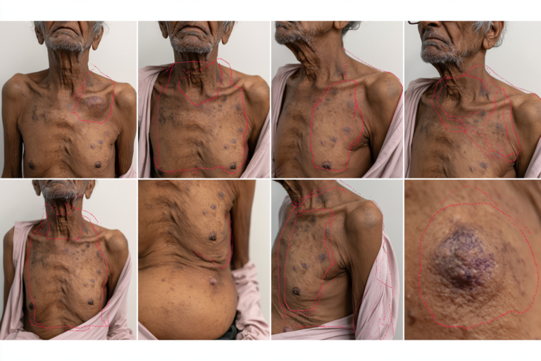
- Published Case Study Snapshot
- How Metastatic Esophageal Cancer Develops and Spreads
- Where Does Metastatic Esophageal Cancer Commonly Spread?
- Symptoms of Metastatic Esophageal Cancer
- Warning Signs That Need Urgent Medical Attention in Metastatic Esophageal Cancer
- How Metastatic Esophageal Cancer Is Diagnosed
- Biomarker Testing in Metastatic Esophageal Cancer
- Treatment for Metastatic Esophageal Cancer
- Radiation Therapy for Metastatic Esophageal Cancer
- Esophageal Stenting for Severe Dysphagia
- Dysphagia and Nutrition in Metastatic Esophageal Cancer
- Metastatic Esophageal Cancer Survival and Prognosis
- The Ayurveda Curative Model for Metastatic Esophageal Cancer
- Ayurvedic Understanding of Advanced Metastatic Esophageal Cancer
- Rasayana Therapy in the Ayurveda Curative Model for Metastatic Esophageal Cancer
- Kushmanda Amalaki Bala Rasayana Avaleha (Medicine)
- Complete 30-Day Kushmanda Amalaki Bala Rasayana Avaleha Formula for Metastatic Esophageal Cancer
- Published Integrative Ayurveda Case Study in Esophageal Squamous Cell Carcinoma
- Frequently Asked Questions
- Reference List
Metastatic esophageal cancer is cancer that began in the esophagus and has spread to a distant organ or nonregional lymph node. The esophagus is the muscular tube that carries food and liquids from the throat to the stomach. When cancer cells leave the primary esophageal tumor, travel through lymphatic channels or the bloodstream, and establish tumors elsewhere in the body, the disease is classified as metastatic or stage IVB esophageal cancer [1–3].
This diagnosis does not mean that every patient has the same disease course. Treatment and prognosis depend on the tumor type, location, biomarker profile, organs involved, number of metastatic sites, nutritional condition, general fitness, and response to the first treatment. Modern systemic therapies can slow disease progression, reduce tumor burden, relieve swallowing difficulty, and help some carefully selected patients achieve a prolonged response, although metastatic disease remains medically serious [1,2].
Published Case Study Snapshot
Case at a Glance
| Published case | Integrated Ayurveda Care in Esophageal Squamous Cell Carcinoma |
|---|---|
| Patient condition | 40-year-old woman with advanced mid-thoracic esophageal squamous cell carcinoma, severe dysphagia, weight loss and feeding-pipe dependence |
| Baseline status | Marked esophageal narrowing; body weight 51 kg; distant stage IVB metastasis was not documented |
Treatment and Reported Outcome
| Treatment approach | Chemotherapy, reported radiotherapy exposure, Catharanthus roseus Sheeta Kashaya, Yashtimadhu and dietary support |
|---|---|
| Reported outcome | Improved swallowing and appetite, feeding pipe removed, weight increased from 51 kg to 57 kg, later gastroscopy showed no visible growth |
| Long-term finding | Follow-up CT reportedly showed no residual esophageal thickening, with no clinical complaints at approximately 18 months |
Why This Case Matters
| Decision value | Demonstrates coordinated oncology and supervised Ayurveda with repeated scans, endoscopy, blood tests and nutritional monitoring |
|---|---|
| First 30-day goals | Easier swallowing, improved appetite and hydration, weight stabilization, better strength and treatment tolerance |
Note: Case reports describe individual clinical observations. Outcomes may differ according to cancer stage, biomarkers, previous treatment, nutrition, organ function and treatment response.
Esophageal Squamous Cell Carcinoma
Esophageal squamous cell carcinoma develops from the thin, flat squamous cells lining the inside of the esophagus. It can arise anywhere along the esophageal tube but is particularly common in the upper and middle portions. Globally, it remains the most frequently diagnosed histological type of esophageal cancer, although its prevalence varies considerably between regions [1,2].
Major risk factors include tobacco use, heavy alcohol consumption, long-standing nutritional deficiencies, repeated exposure to very hot beverages, and certain chronic esophageal conditions. These risk factors do not determine how an individual patient will respond to treatment, but they may help explain how the disease developed.
Metastatic esophageal squamous cell carcinoma may spread to distant lymph nodes, the lungs, liver, bones, brain, or several organs simultaneously. Treatment usually relies on systemic therapy because medication can circulate throughout the body and reach both the primary tumor and distant cancer deposits. Depending on the patient’s condition and PD-L1 biomarker results, treatment may include platinum-based chemotherapy, fluoropyrimidine chemotherapy, immunotherapy, or selected combinations [1,2].
Esophageal Adenocarcinoma
Esophageal adenocarcinoma begins in gland-forming cells, most commonly in the lower esophagus near the gastroesophageal junction, where the esophagus meets the stomach. It is especially common in many Western countries and is strongly associated with chronic gastroesophageal reflux disease, Barrett’s esophagus, obesity, and smoking [1,2].
Because adenocarcinoma often develops close to the stomach, precise classification may require careful review of the endoscopy, biopsy, imaging, and tumor location. Some tumors are classified as esophageal adenocarcinoma, while others are categorized as gastroesophageal junction cancer. This distinction can influence staging, clinical-trial eligibility, and treatment planning.
Metastatic esophageal adenocarcinoma commonly spreads to the liver, lungs, distant lymph nodes, peritoneum, bone, or multiple sites. Biomarker testing is particularly important because some adenocarcinomas express HER2 or show other molecular features that can affect treatment selection. PD-L1, HER2, mismatch-repair proteins, microsatellite instability, and selected genomic alterations may therefore be assessed before or during treatment planning [2].
Stage IVA vs Stage IVB Esophageal Cancer
Stage IVA and stage IVB are both advanced stages, but they do not mean the same thing. Stage IVA esophageal cancer is generally very advanced within the chest or regional lymphatic system but has not produced confirmed distant metastasis. It may involve extensive local invasion, multiple regional lymph nodes, or nearby structures, depending on the tumor’s exact TNM classification [3].
Stage IVB esophageal cancer means that distant metastatic disease is present. In TNM terminology, this is recorded as M1 disease. Examples include spread to the liver, lungs, bones, brain, peritoneum, or lymph nodes classified as nonregional for the original tumor location [3].
The distinction matters because stage IVA disease may sometimes be approached with intensive combined treatment intended to control the tumor within the esophagus and surrounding region. Stage IVB disease usually requires systemic treatment as the main strategy because cancer cells are present beyond the original anatomical area. Radiation therapy, esophageal stenting, nutritional support, or other local procedures may still be used to relieve dysphagia, bleeding, pain, obstruction, or symptoms caused by a particular metastatic site [1,2].
A stage IVB diagnosis should therefore be interpreted together with the complete clinical picture rather than as an isolated label. A patient with one limited metastatic site, good functional status, preserved nutrition, and a treatment-responsive biomarker may have different options and outcomes from someone with multiple-organ metastases, severe weight loss, and rapidly declining strength. Accurate staging, histological confirmation, biomarker testing, nutritional assessment, and multidisciplinary review are essential before an individualized treatment plan is finalized [1–3].
How Metastatic Esophageal Cancer Develops and Spreads
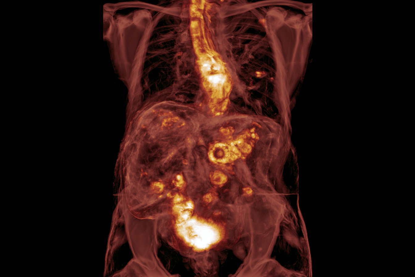
Metastatic esophageal cancer develops when malignant cells move beyond the original tumor in the esophagus and establish cancer deposits in distant tissues. This does not usually happen in a single step. Cancer cells must invade nearby tissue, enter lymphatic channels or blood vessels, survive while circulating through the body, leave those vessels, and begin growing in another location [2–4].
The esophagus has an extensive lymphatic network and lies close to important structures within the chest. These anatomical features can allow an esophageal tumor to involve regional lymph nodes or nearby organs before it becomes very large. However, local invasion, lymph-node involvement, and distant metastasis are separate parts of staging and should not be treated as interchangeable terms [2,3].
Lymphatic Spread of Metastatic Esophageal Cancer
The lymphatic system is one of the main routes through which esophageal cancer spreads. Lymphatic vessels collect fluid from tissues and carry it through lymph nodes, where immune cells normally help identify infections and abnormal cells. Cancer cells can enter these vessels and travel to lymph nodes connected to the part of the esophagus where the primary tumor developed.
The esophagus has lymphatic channels that run lengthwise along much of the organ. As a result, cancer cells may reach lymph nodes above or below the primary tumor rather than spreading only to the nearest node. A tumor in the upper, middle, or lower esophagus can therefore produce different patterns of lymph-node involvement [2,3].
Regional lymph nodes are included in the N component of the TNM staging system. Their significance depends not only on whether cancer is present, but also on the number and anatomical location of the affected nodes. Regional lymph-node disease can occur without distant metastasis and may still be classified as locally advanced rather than stage IVB cancer [3].
A lymph node becomes evidence of distant metastasis when it is classified as nonregional for the location of the primary esophageal tumor. This distinction is important because an enlarged lymph node on a scan is not automatically metastatic, and not every cancer-containing lymph node represents M1 disease. Imaging findings may sometimes require confirmation through endoscopic ultrasound-guided sampling, image-guided biopsy, or multidisciplinary radiological review.
Lymphatic spread can also indicate that the tumor has acquired the ability to move beyond its original tissue boundaries. However, lymph-node involvement does not predict the same outcome for every patient. Tumor biology, histological type, treatment response, general health, and the presence or absence of distant organ metastases remain important.
Bloodstream Spread of Metastatic Esophageal Cancer
Esophageal cancer can also spread through the bloodstream. This process begins when malignant cells invade small blood vessels within or around the tumor. The cells may then circulate through the venous system and become trapped in distant organs, where some may form new tumors.
The liver is a common site of bloodstream spread because blood from much of the digestive system passes through the portal circulation before returning to the heart. The lungs are another frequent metastatic site because circulating cancer cells may pass through pulmonary blood vessels. Bone and brain metastases are less common than liver or lung involvement but can occur, particularly as the disease becomes more advanced [4].
Not every cancer cell that enters the bloodstream forms a metastasis. Most circulating tumor cells are unable to survive, avoid immune destruction, leave the blood vessel, or adapt to the environment of another organ. Metastatic disease develops only when some cells successfully complete these steps and begin sustained growth.
The pattern of bloodstream spread may differ between esophageal squamous cell carcinoma and esophageal adenocarcinoma. Population studies suggest that adenocarcinoma is more frequently associated with liver metastasis, while the overall distribution of distant sites can vary according to tumor histology, primary location, and disease burden [2,4].
Bloodstream metastases may be discovered during initial staging or may appear later after treatment. This is why CT, PET-CT, and clinically appropriate follow-up imaging are important even when the primary tumor or regional lymph nodes appear to be responding. New symptoms such as persistent bone pain, neurological changes, breathlessness, abdominal swelling, or jaundice may also prompt investigation for distant disease.
Local Invasion and Distant Metastasis
Local invasion occurs when the primary esophageal tumor grows through the layers of the esophageal wall and enters nearby tissues. Depending on its location and depth, an advanced tumor may involve the pleura, pericardium, diaphragm, airway, aorta, vertebral structures, or other neighbouring tissues within the neck, chest, or upper abdomen [2,3].
Local invasion can cause serious symptoms even when distant metastasis has not occurred. Extension into surrounding structures may worsen swallowing difficulty, produce chest or back pain, cause bleeding, affect the airway, or contribute to the formation of a tracheoesophageal fistula. These complications may require urgent endoscopic, surgical, radiation, respiratory, or nutritional management.
Distant metastasis means that cancer has spread to an organ or lymph-node region outside the accepted regional field of the primary tumor. Under the TNM system, confirmed distant spread is classified as M1 disease and places esophageal cancer in stage IVB [3].
Some patients have one or a small number of metastatic deposits, sometimes described as oligometastatic disease, while others have cancer involving several organs. The number, size, location, and biological activity of these metastases influence treatment planning. A limited metastatic pattern in a medically fit patient may occasionally lead to consideration of local treatment alongside systemic therapy, whereas widespread disease is generally managed primarily with systemic treatment and symptom-directed care.
The primary tumor and distant metastases arise from the same cancer, but they may not remain biologically identical. Genetic changes can accumulate as the disease progresses, and biomarker expression may differ between the original tumor and a later metastatic lesion. When a new biopsy is safe and likely to influence treatment, testing a metastatic site may help confirm the diagnosis or identify a treatment-relevant molecular feature.
Understanding how metastatic esophageal cancer spreads helps explain why treatment cannot focus only on the visible tumor inside the esophagus. Systemic therapy is usually central in stage IVB disease because it can reach cancer cells in multiple locations. Local treatments such as radiation, stenting, surgery, or ablation may still be used selectively to relieve obstruction, bleeding, pain, or symptoms arising from a particular metastatic site [1–4].
Where Does Metastatic Esophageal Cancer Commonly Spread?
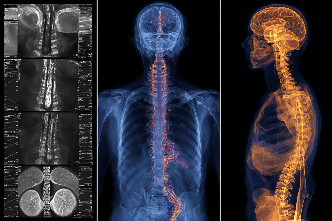
Metastatic esophageal cancer most commonly spreads to the liver, lungs, distant lymph nodes, and bones. Brain metastases are less frequent but can occur, particularly in advanced disease. Some patients have one dominant metastatic site, while others develop cancer in several organs at the same time [3,4].
The pattern of spread is not identical in every patient. It can vary according to whether the tumor is esophageal squamous cell carcinoma or adenocarcinoma, where the primary tumor is located, how aggressive its biology is, and how long the disease has been present. Imaging such as contrast-enhanced CT and PET-CT helps determine the number, size, and location of metastatic deposits before treatment is planned [1,2].
Esophageal Cancer Spread to the Liver
The liver is one of the most common sites of metastatic esophageal cancer, particularly in patients with esophageal adenocarcinoma. Cancer cells can enter blood vessels around the primary tumor and travel through the circulation until they become established in liver tissue [4].
Small liver metastases may cause no symptoms and may be discovered only during staging scans. As liver involvement increases, a patient may develop discomfort or heaviness in the right upper abdomen, reduced appetite, nausea, fatigue, abdominal swelling, or unexplained worsening of general health.
Jaundice can occur if metastatic disease interferes with bile drainage or significantly affects liver function. It may cause yellowing of the skin or eyes, dark urine, pale stools, and itching. These symptoms require prompt medical assessment because jaundice may also result from bile duct obstruction, medication toxicity, infection, or another condition unrelated to the metastases.
Liver-function blood tests can remain normal when metastatic deposits are small. Abnormal liver enzymes therefore help assess organ function but cannot reliably confirm or exclude liver metastases. CT, PET-CT, MRI, and occasionally image-guided biopsy may be needed to establish the diagnosis.
Treatment usually depends on systemic therapy because liver metastasis confirms disease beyond the original esophageal region. Local treatment to an individual liver lesion may occasionally be considered in carefully selected oligometastatic cases, but it is not appropriate for every patient.
Esophageal Cancer Spread to the Lungs
The lungs are another frequent site of distant spread. Cancer cells may reach pulmonary tissue through the bloodstream or involve structures within the chest through lymphatic spread and direct extension [4].
Small lung metastases may not produce symptoms. When symptoms occur, they can include a persistent cough, shortness of breath, chest discomfort, reduced exercise tolerance, or recurrent respiratory infections. These symptoms are not specific to metastatic cancer and may also arise from aspiration, pneumonia, pleural fluid, anemia, treatment toxicity, or pre-existing lung disease.
Fluid can sometimes accumulate around the lungs, producing a pleural effusion. A large effusion may compress the lung and cause increasing breathlessness. Examination, chest imaging, and analysis of pleural fluid may be required to determine whether the fluid is malignant, infectious, inflammatory, or related to another cause.
A lung nodule seen on a scan is not automatically an esophageal cancer metastasis. Benign nodules, primary lung cancer, infection, and inflammatory disease can have a similar appearance. Comparison with earlier scans, PET-CT findings, growth over time, or biopsy may be necessary when the result could change treatment.
Esophageal Cancer Spread to Distant Lymph Nodes
Esophageal cancer frequently spreads through lymphatic channels. Regional lymph-node involvement forms part of the N category in TNM staging, whereas cancer in a nonregional lymph node is classified as distant metastatic disease [3].
This distinction depends on the location of the primary tumor and the anatomical position of the affected lymph node. A node that is considered regional for one esophageal tumor may not be classified the same way for a tumor arising in another part of the esophagus. Staging should therefore be based on accepted anatomical criteria rather than simply describing every affected lymph node as distant spread.
Distant lymph nodes may be found above the collarbone, deeper within the chest, or in the abdomen, depending on the route of spread. Many affected nodes cause no symptoms. Enlarged nodes near the airway or major vessels may occasionally contribute to cough, hoarseness, swelling, discomfort, or breathing difficulty.
CT and PET-CT can identify suspicious lymph nodes, but size and metabolic activity do not provide absolute proof of malignancy. Endoscopic ultrasound-guided biopsy, bronchoscopy-guided sampling, or image-guided needle biopsy may be recommended when confirmation is likely to influence treatment.
Esophageal Cancer Spread to Bone
Bone metastases occur less often than liver or lung metastases but can cause substantial pain and loss of mobility. The spine, ribs, pelvis, and long bones may be affected [4].
Persistent, localized bone pain is a common warning symptom. The pain may be worse at night, occur without activity, or gradually become more intense. Bone metastases can weaken the affected area and increase the risk of a pathological fracture, sometimes after only minor strain.
Metastatic involvement of the spine requires particular attention. Severe back pain accompanied by leg weakness, numbness, altered sensation, difficulty walking, or loss of bladder or bowel control may indicate spinal cord compression. This is an oncological emergency that requires immediate hospital assessment.
Bone metastases can also disturb calcium regulation. A high blood calcium level may cause thirst, constipation, nausea, confusion, weakness, excessive urination, or drowsiness. Blood tests, bone imaging, CT, MRI, PET-CT, or a bone scan may be used according to the clinical situation.
Treatment may include systemic anticancer therapy, pain medication, targeted radiation, and medicines intended to reduce skeletal complications. Surgery may occasionally be required to stabilize a weakened bone or relieve spinal cord compression.
Esophageal Cancer Spread to the Brain
Brain metastases from esophageal cancer are uncommon compared with liver, lung, or bone metastases, but they are clinically important because they can cause sudden neurological deterioration [4].
Possible symptoms include new or progressively worsening headaches, nausea, vomiting, seizures, weakness on one side of the body, altered balance, difficulty speaking, personality changes, confusion, or visual disturbance. The symptoms depend on the number, size, and location of the metastatic lesions.
A new neurological symptom in a patient with esophageal cancer should not automatically be attributed to treatment fatigue or general weakness. It may result from brain metastasis, stroke, infection, electrolyte imbalance, medication effects, or another urgent condition.
Contrast-enhanced MRI is generally more sensitive than CT for detecting small brain metastases. Treatment may include corticosteroids to reduce swelling, stereotactic radiosurgery, whole-brain radiation, surgery for selected lesions, systemic therapy, or a combination of these approaches. Management should involve medical oncology, radiation oncology, neurosurgery, and supportive-care teams where appropriate.
Multiple Organ Metastases
Some patients have metastatic esophageal cancer in more than one organ at diagnosis. Others initially have a single metastatic site and later develop additional lesions as the disease progresses. Multiple-organ metastases generally indicate a greater overall tumor burden and can make treatment, nutrition, symptom control, and organ-function monitoring more complex [4].
The seriousness of multiple-organ disease depends on more than the number of affected sites. A few small, treatment-responsive deposits may behave differently from extensive liver involvement, widespread bone disease, or metastases that impair the lungs, brain, or other essential organs.
Treatment is usually centered on systemic therapy because cancer cells are present in several anatomical locations. Local treatments may still be important when one site is causing disproportionate symptoms, such as radiation for painful bone metastases, drainage of a pleural effusion, or treatment of a symptomatic brain lesion.
Regular assessment should include swallowing ability, body weight, performance status, blood counts, liver and kidney function, treatment tolerance, and interval imaging. Symptom improvement alone cannot confirm that metastatic deposits are shrinking. Objective response should be assessed through appropriate scans and standardized oncology criteria [2,13].
Symptoms of Metastatic Esophageal Cancer
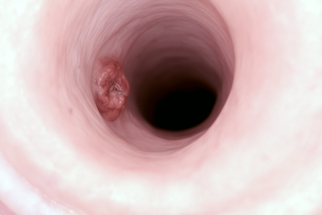
Symptoms of metastatic esophageal cancer may come from the primary tumor narrowing the esophagus, the effects of cancer on nutrition and strength, or tumors that have spread to distant organs. Progressive difficulty swallowing is the most characteristic symptom, but unexplained weight loss, painful swallowing, regurgitation, chest discomfort, fatigue, and symptoms related to liver, lung, bone, brain, or lymph-node metastases may also occur [1,2,4,11].
Symptoms vary considerably between patients. Some people develop severe swallowing problems while the distant metastases remain relatively silent. Others have symptoms from an affected organ before the esophageal tumor causes complete obstruction. Symptoms alone cannot confirm the location or extent of metastatic disease, so new or worsening problems require clinical assessment and appropriate imaging.
Progressive Difficulty Swallowing
Progressive difficulty swallowing, medically known as dysphagia, is one of the most common symptoms of metastatic esophageal cancer. It usually develops when the tumor narrows the inner passage of the esophagus or interferes with its normal muscular movement [1,2].
Dysphagia often begins with solid foods such as bread, rice, meat, or raw vegetables. A patient may feel that food is sticking behind the breastbone, moving slowly, or requiring repeated swallowing and extra fluids to pass. As the narrowing progresses, softer foods may also become difficult, followed by thick liquids and, in severe cases, water or saliva.
Patients may adapt without realizing how serious the problem has become. They may take smaller bites, chew for longer, avoid eating with other people, drink frequently during meals, or gradually change to soups and liquids. These adaptations can temporarily conceal the progression of esophageal obstruction while nutritional intake continues to decline.
Swallowing ability should therefore be assessed according to the types and quantities of food a patient can manage, not simply by asking whether eating is still possible. Rapidly worsening dysphagia may indicate increasing obstruction and can lead to dehydration, malnutrition, aspiration, or inability to take oral medicines. Coughing, choking, vomiting after eating, and the sensation that food remains stuck are recognized signs of clinically important swallowing dysfunction.
Painful Swallowing
Painful swallowing is called odynophagia. The discomfort may be felt in the throat, behind the breastbone, or occasionally in the upper abdomen as food passes through the esophagus. It may feel like burning, pressure, scraping, or sharp pain.
Odynophagia may be caused by the primary tumor, ulceration of the esophageal lining, inflammation, infection, reflux, or irritation caused by treatment. Chemotherapy and radiation therapy can also produce mucositis, which may make swallowing painful even when the tumor itself is responding [1,2].
Painful swallowing can reduce food and fluid intake before complete mechanical obstruction develops. Patients may begin avoiding protein-rich or calorie-dense foods because they are uncomfortable to swallow, increasing the risk of weight loss and weakness. Persistent odynophagia should therefore be evaluated rather than managed only by switching to liquids.
The cause matters because tumor narrowing, mucositis, fungal infection, reflux, and radiation-related inflammation require different forms of treatment. Pain during swallowing and inability to swallow are recognized complications that can increase the risk of further nutritional and respiratory problems.
Unintentional Weight Loss and Muscle Wasting
Unintentional weight loss in metastatic esophageal cancer may result from both reduced food intake and cancer-related metabolic changes. Dysphagia limits how much a patient can eat, while pain, nausea, early fullness, altered taste, depression, treatment toxicity, and loss of appetite may reduce intake further [11].
Advanced cancer can also cause cachexia, a complex wasting syndrome involving progressive loss of skeletal muscle and often body fat. Cachexia is not simply starvation and may not be fully reversed by increasing calories alone. Inflammation and altered metabolism can continue to weaken the body even when a patient is trying to eat [11].
Muscle loss may appear as thinner arms and legs, reduced grip strength, difficulty rising from a chair, slower walking, or increasing dependence on others. A person may lose substantial muscle even when body weight changes only modestly, particularly if fluid retention or abdominal swelling masks the loss.
Weight and nutritional intake should be monitored from the time of diagnosis rather than after severe wasting has developed. Early support may include texture-modified meals, calorie-dense foods, protein supplementation, treatment of nausea and pain, dietitian involvement, and consideration of enteral feeding when oral intake becomes inadequate. Cancer cachexia is associated with loss of muscle and fat and is especially common in advanced cancer.
Regurgitation, Vomiting and Aspiration
Regurgitation occurs when swallowed food, liquid, or saliva returns into the mouth because it cannot pass normally through the narrowed esophagus. Unlike ordinary vomiting, regurgitation may happen without strong abdominal contractions or prolonged nausea.
Food may return shortly after eating or several hours later, particularly when a patient lies down. Retained material can cause an unpleasant taste, bad breath, excessive salivation, disturbed sleep, and fear of eating. Repeated regurgitation also increases the risk that food or liquid will enter the airway.
Aspiration occurs when saliva, food, fluid, or regurgitated material enters the lungs instead of the stomach. It may cause coughing during meals, choking, a wet or gurgling voice, breathlessness, fever, chest congestion, or repeated pneumonia. Some weakened patients may aspirate without a dramatic cough.
Vomiting can also occur because of severe obstruction, treatment toxicity, infection, medication effects, liver involvement, altered electrolytes, or another gastrointestinal problem. Persistent vomiting should not automatically be attributed to the esophageal tumor.
A patient who repeatedly coughs after swallowing, cannot manage saliva, or develops breathing difficulty after eating needs prompt assessment. These findings may indicate severe obstruction, aspiration, or an abnormal connection between the esophagus and airway. Swallowing problems associated with cancer commonly include choking, vomiting after eating, food sticking, painful swallowing, and unintentional weight loss.
Chest and Upper Abdominal Pain
Metastatic esophageal cancer may cause discomfort behind the breastbone, between the shoulder blades, or in the upper abdomen. The pain may arise from the primary tumor, ulceration, inflammation, local invasion, esophageal spasm, or pressure on nearby structures [1,2].
Some patients experience pain mainly during swallowing, while others have a persistent ache unrelated to meals. Pain extending into the back can occur when an advanced tumor involves deeper tissues, although back pain is common and does not by itself prove local invasion.
Upper abdominal pain may also result from a tumor near the gastroesophageal junction, liver metastases, enlarged abdominal lymph nodes, or other digestive conditions. The location and intensity of pain cannot reliably identify its cause without examination and imaging.
New pain should be described according to where it occurs, what triggers it, whether it affects sleep or eating, and whether it is accompanied by vomiting, bleeding, breathlessness, weakness, or neurological symptoms. Adequate pain control is important because uncontrolled discomfort can further reduce nutrition, mobility, sleep, and treatment tolerance.
Fatigue and Progressive Weakness
Fatigue associated with metastatic esophageal cancer is more than ordinary tiredness. It may persist despite rest and make routine activities such as bathing, dressing, walking, preparing food, or attending appointments increasingly difficult.
Several problems may contribute at the same time. Reduced calorie and protein intake, muscle loss, dehydration, anemia, pain, poor sleep, infection, emotional distress, organ dysfunction, and the systemic effects of cancer can all cause weakness. Chemotherapy, immunotherapy, radiation, and pain medicines may add further fatigue [1,11].
A sudden increase in weakness should not be assumed to represent inevitable cancer progression. Treatable causes may include dehydration, anemia, infection, low sodium, high calcium, medication effects, bleeding, or inadequate nutrition.
Functional change is often as important as the patient’s description of tiredness. Needing more help with daily activities, spending longer in bed or a chair, walking less, or struggling to stand may indicate declining performance status and should be reported to the treatment team.
Symptoms According to the Site of Metastasis
Symptoms of metastatic esophageal cancer can change according to the organ involved. Liver metastases may cause right upper abdominal discomfort, poor appetite, nausea, abdominal swelling, jaundice, dark urine, pale stools, or itching. Small liver deposits may produce no noticeable symptoms and may be detected only on imaging [4].
Lung metastases may cause persistent cough, shortness of breath, chest discomfort, reduced exercise tolerance, or fluid around the lungs. Similar symptoms can also arise from aspiration pneumonia, infection, anemia, treatment toxicity, or pre-existing heart and lung disease.
Bone metastases may produce persistent localized pain, pain that becomes worse at night, reduced mobility, or a fracture after minor stress. Spread to the spine can cause back pain, limb weakness, numbness, difficulty walking, or changes in bladder and bowel control.
Brain metastases may cause persistent headache, nausea, vomiting, seizures, confusion, altered behavior, poor balance, visual disturbance, speech difficulty, or weakness affecting one side of the body. These symptoms can develop gradually or appear suddenly.
Distant lymph-node metastases often cause no symptoms. Depending on their location, enlarged nodes may contribute to neck swelling, hoarseness, cough, abdominal discomfort, or pressure on nearby blood vessels and airways.
Many of these symptoms have possible causes other than cancer progression. Their appearance should lead to medical evaluation rather than an assumption that a particular organ is involved. Imaging, blood tests, examination, and sometimes biopsy are needed to distinguish metastatic disease from treatment effects, infection, nutritional complications, or unrelated illness [2,4].
Warning Signs That Need Urgent Medical Attention in Metastatic Esophageal Cancer
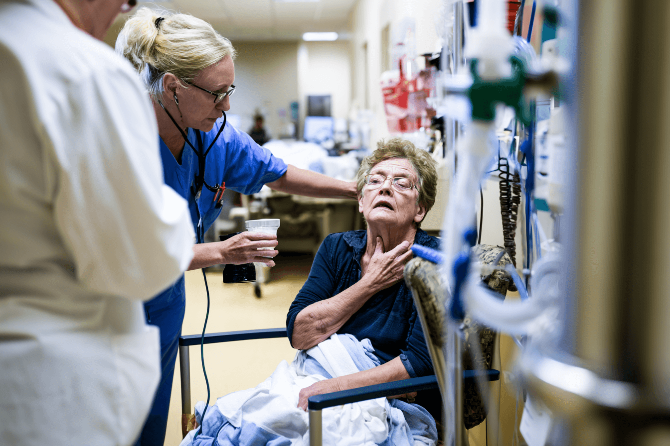
Metastatic esophageal cancer can cause urgent complications when the esophagus becomes severely obstructed, swallowed material enters the airway, the tumor bleeds, or the patient can no longer maintain hydration and nutrition. Families should pay particular attention to sudden changes, rapidly worsening symptoms, and any loss of the ability to swallow liquids or saliva.
A patient should not wait for a routine oncology appointment when breathing is affected, significant bleeding is suspected, consciousness changes, or fluids cannot be swallowed. Early assessment may prevent aspiration pneumonia, severe dehydration, major blood loss, kidney injury, and further nutritional decline [1,10,11].
Inability to Swallow Liquids or Saliva
A patient who can no longer swallow water, prescribed medicines, or saliva may have severe or nearly complete esophageal obstruction. Warning signs include constant drooling, repeated spitting, food returning immediately after swallowing, a persistent sensation of blockage, or saliva collecting in the mouth because it cannot pass through the esophagus.
Difficulty with solid food alone may sometimes be managed temporarily with an appropriately modified diet while the patient is assessed. However, difficulty swallowing liquids represents more advanced dysphagia and requires prompt medical attention. Complete obstruction may require endoscopic assessment, esophageal stenting, feeding access, intravenous fluids, radiation, or another intervention depending on the patient’s condition [1,10].
Food, tablets, nutritional drinks, avaleha, or other oral preparations should not be forced when a patient cannot swallow safely. Trying repeatedly to push material through an obstructed esophagus may increase regurgitation and aspiration risk. Cancer-related dysphagia can also make it impossible to take essential medicines and can quickly result in dehydration and weakness.
Choking, Coughing or Breathing Difficulty During Swallowing
Coughing or choking while eating or drinking may indicate that food, fluid, or saliva is entering the airway. Other concerning changes include a wet or gurgling voice after swallowing, chest congestion, repeated throat clearing, breathlessness after meals, or recurrent chest infections.
Aspiration can occur when the normal swallowing mechanism fails or when regurgitated material enters the lungs. It may cause inflammation, infection, breathing difficulty, and reduced oxygen levels. A weakened patient may aspirate without producing a strong cough, so unexplained fever, new chest congestion, or increasing breathlessness also requires attention [10].
Persistent coughing immediately after every sip may occasionally raise concern for an abnormal connection between the esophagus and airway, known as a tracheoesophageal fistula. This is a serious complication that requires urgent specialist assessment. A patient who is struggling to breathe, becoming unusually drowsy, or turning blue around the lips should receive emergency medical care. The American Cancer Society advises emergency assessment for swallowing problems accompanied by breathing difficulty, worsening fever, weakness, or dizziness.
Vomiting Blood or Passing Black Tarry Stools
Esophageal tumors can sometimes bleed. The blood may appear bright red in vomit, dark brown like coffee grounds, or mixed with regurgitated material. Blood that has passed through the digestive tract may make the stools black, sticky, and tar-like.
Even a small amount of visible blood should be reported promptly because bleeding may increase unexpectedly. Heavy vomiting of blood, repeated black stools, fainting, marked dizziness, rapid heartbeat, pale or clammy skin, confusion, or severe weakness may indicate substantial blood loss and require emergency treatment.
Bleeding may also be worsened by a low platelet count, anticoagulant medicines, tumor ulceration, infection, or treatment-related injury. Patients should not attempt to control suspected internal bleeding with home remedies or oral herbal medicines while delaying assessment. Esophageal cancer is among the conditions that can cause blood in vomit or black tarry stool.
Persistent Vomiting or Regurgitation
Repeated vomiting or regurgitation can occur because of worsening esophageal obstruction, treatment toxicity, infection, medication effects, liver or brain involvement, or an unrelated gastrointestinal condition. The cause cannot be determined safely from symptoms alone.
Urgent assessment is needed when the patient cannot keep fluids down, vomits repeatedly after every attempt to eat, develops increasing abdominal or chest pain, or becomes progressively weak. Vomiting accompanied by severe headache, confusion, reduced alertness, or neurological change requires particularly rapid evaluation.
Uncontrolled vomiting can cause dehydration, electrolyte imbalance, weight loss, kidney dysfunction, and interruption of cancer treatment. Treatment may involve intravenous fluids, anti-nausea medicines, blood tests, imaging, nutritional support, or a procedure to relieve obstruction [1,11].
Signs of Severe Dehydration
Patients with metastatic esophageal cancer can become dehydrated rapidly when dysphagia, vomiting, fever, diarrhea, or poor appetite reduces fluid intake. Early signs may include thirst, dry mouth, dark urine, reduced urination, headache, and increasing fatigue.
More serious dehydration may cause dizziness when standing, fainting, confusion, muscle cramps, rapid heartbeat, rapid breathing, or profound weakness. Older patients and people receiving chemotherapy may deteriorate quickly because their kidney function, electrolyte balance, or general reserve may already be reduced.
A patient who cannot swallow enough fluid, urinates very little, becomes confused, or feels too weak to stand needs urgent assessment. Intravenous fluids may be required, but the underlying cause of reduced intake must also be treated. Nutritional guidelines recommend early recognition and active management of reduced intake, malnutrition, and dehydration in people with cancer [11].
Fever, Chest Congestion or Suspected Aspiration Pneumonia
A new fever in a patient with metastatic esophageal cancer may indicate aspiration pneumonia, another infection, or treatment-related immune suppression. Fever accompanied by cough, chest congestion, chills, shortness of breath, confusion, or rapid deterioration requires prompt medical evaluation.
Patients receiving chemotherapy may have a reduced white blood cell count and may be unable to fight infection normally. A fever that appears mild can therefore become serious quickly. The oncology team should provide an individual temperature threshold and instructions for seeking emergency care during treatment.
Aspiration pneumonia should be considered when fever or breathing symptoms follow choking, regurgitation, vomiting, or difficulty swallowing. Treatment may require chest imaging, oxygen assessment, antibiotics, swallowing evaluation, and temporary restriction of unsafe oral intake. Swallowing impairment increases the possibility of food or liquid entering the lungs, which can lead to infection and breathing problems.
Sudden Severe Weakness, Dizziness or Confusion
Progressive fatigue is common in advanced cancer, but sudden or marked deterioration should not automatically be attributed to the disease. Severe weakness, dizziness, fainting, confusion, or inability to get out of bed may result from bleeding, dehydration, infection, anemia, electrolyte abnormalities, low blood pressure, high calcium, low blood sugar, medication effects, or organ dysfunction.
These changes are especially concerning when they occur together with vomiting, black stools, fever, reduced urine output, breathlessness, or inability to swallow. Prompt blood tests and clinical assessment may identify a treatable complication.
A patient who loses consciousness, becomes newly confused, cannot remain awake, develops severe breathing difficulty, or cannot stand safely should receive emergency care rather than being transported to a routine clinic appointment.
When to Contact the Oncology Team and When to Seek Emergency Care
The oncology team should be contacted promptly when swallowing becomes noticeably worse, tablets can no longer be swallowed, food repeatedly sticks, oral intake declines, vomiting increases, or weight falls rapidly. These changes may require an earlier scan, endoscopy, swallowing assessment, dietitian review, stenting, feeding support, or adjustment of cancer treatment [1,10,11].
Emergency care is appropriate when the patient cannot manage saliva, has serious breathing difficulty, repeatedly chokes, vomits blood, passes black tarry stools, faints, develops marked confusion, or cannot keep fluids down and is becoming weak or dizzy. While help is being arranged, the patient should remain upright, and oral food or medicine should not be forced when swallowing is unsafe.
Recognizing these warning signs early does not mean that every symptom represents rapid cancer progression. Infection, treatment toxicity, dehydration, anemia, and other reversible complications can cause similar changes. The safest approach is timely medical assessment so that the cause can be identified and treated before the patient’s condition worsens.
How Metastatic Esophageal Cancer Is Diagnosed
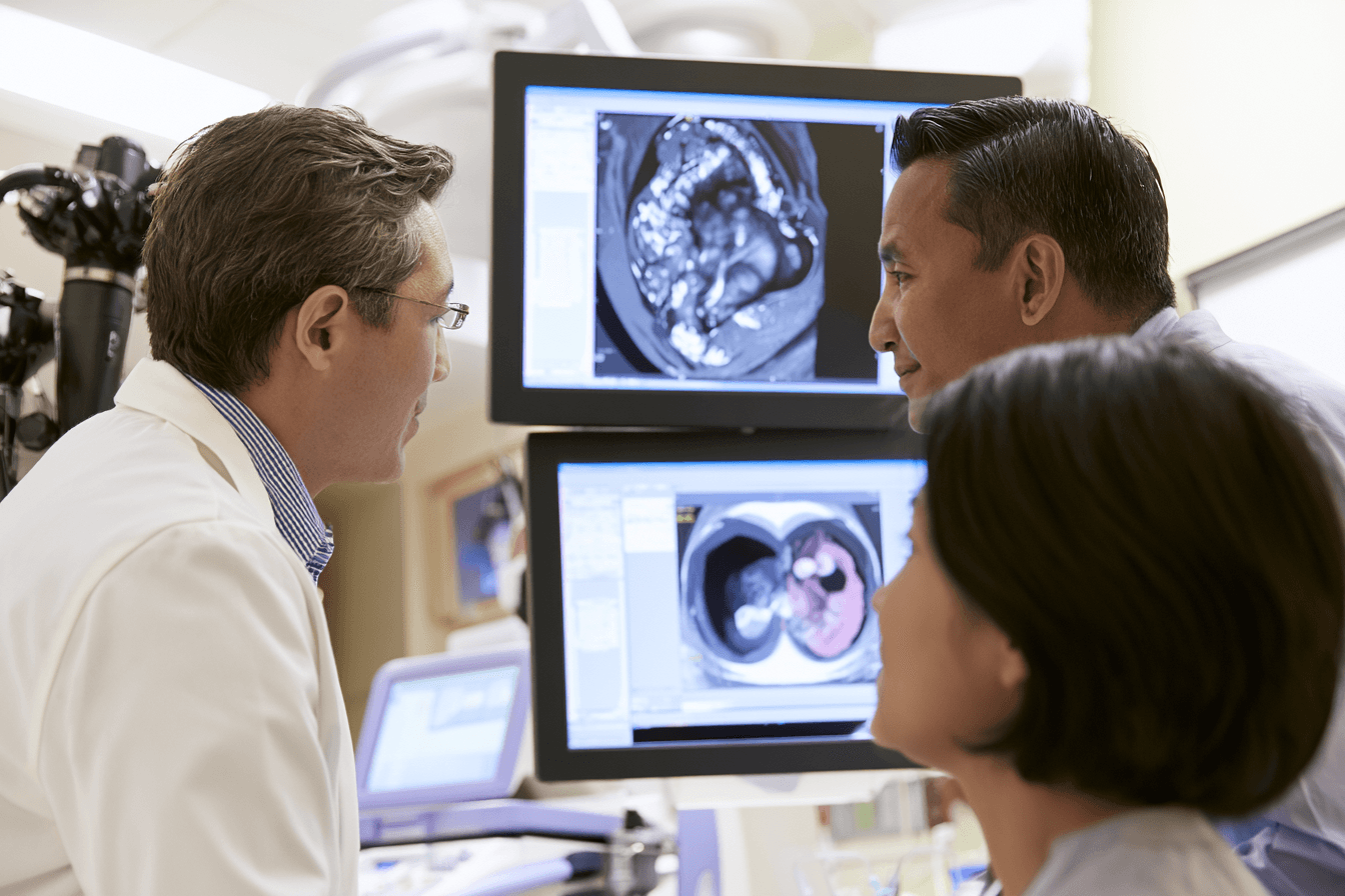
Metastatic esophageal cancer is diagnosed by confirming that a tumor began in the esophagus, identifying its histological type, and determining whether it has spread beyond the regional lymph nodes to distant organs. No single test provides every answer. Diagnosis usually combines upper gastrointestinal endoscopy, tissue biopsy, pathology review, CT imaging, PET-CT, blood tests, and, in selected cases, biopsy of a suspected metastatic lesion [1–3].
The diagnostic process should establish the tumor location, whether it is squamous cell carcinoma or adenocarcinoma, the TNM stage, the organs involved, the patient’s nutritional condition, and whether molecular biomarkers could guide treatment. These findings allow the multidisciplinary cancer team to distinguish locally advanced stage IVA disease from metastatic stage IVB disease and develop an individualized treatment plan.
Upper GI Endoscopy and Biopsy
Upper gastrointestinal endoscopy is the main procedure used to examine a suspected esophageal tumor. During the test, a thin flexible tube containing a camera is passed through the mouth and into the esophagus. This allows the specialist to identify narrowing, ulceration, bleeding, abnormal tissue, and the approximate length and location of the tumor [1,2].
Several tissue samples are usually taken from the suspicious area during the same procedure. This step is essential because imaging alone cannot confirm that an abnormality is cancer. A biopsy enables a pathologist to examine the cells under a microscope and establish the diagnosis.
The endoscopy report may describe where the tumor begins, how much of the esophageal passage remains open, whether the endoscope can pass through the narrowing, and whether the tumor extends toward the stomach or gastroesophageal junction. These details are important when planning nutrition, stenting, radiation, surgery, or systemic treatment.
Endoscopy must be performed carefully when the esophageal passage is severely narrowed. A patient with complete obstruction, major bleeding, unstable breathing, or suspected fistula may require urgent stabilization before a routine diagnostic procedure can be completed.
Histopathology and Tumor Type
Histopathology is the microscopic examination of the biopsy specimen. It confirms whether malignant cells are present and identifies the type of esophageal cancer. The two principal forms are esophageal squamous cell carcinoma and esophageal adenocarcinoma [1,2].
Squamous cell carcinoma develops from the flat cells lining the esophagus. Adenocarcinoma develops from gland-forming cells and most often occurs in the lower esophagus or near the gastroesophageal junction. The distinction is clinically important because the two tumor types have different risk factors, patterns of spread, biomarker profiles, and treatment pathways.
The pathology report may also describe how closely the tumor resembles normal tissue, known as tumor differentiation or grade. Poorly differentiated tumors appear more abnormal under the microscope, although grade alone does not determine how a patient will respond to treatment.
Additional staining may be required when the biopsy is small, poorly differentiated, or located near the junction between the esophagus and stomach. Pathology review can help determine whether the tumor is a primary esophageal cancer, a gastroesophageal-junction cancer, or a metastasis from another organ.
Biopsy tissue should also be preserved for biomarker testing whenever possible. PD-L1, HER2, mismatch-repair proteins, microsatellite instability, and selected molecular alterations can influence systemic treatment decisions in advanced disease [2].
CT Scan
A contrast-enhanced CT scan of the chest, abdomen, and often the pelvis is a central part of metastatic esophageal cancer staging. It provides detailed anatomical images of the primary tumor, regional lymph nodes, nearby structures, liver, lungs, adrenal glands, bones, and other potential metastatic sites [1–3].
CT can show whether the tumor appears to extend beyond the esophageal wall or involve nearby organs. It can also identify enlarged lymph nodes, liver lesions, lung nodules, pleural fluid, abdominal disease, and other findings that may affect stage and treatment.
Not every abnormality seen on CT is cancer. Enlarged lymph nodes may be inflammatory, lung nodules may be benign, and liver lesions may represent cysts or other noncancerous conditions. The radiologist interprets these findings according to their size, shape, location, enhancement pattern, and relationship to earlier imaging.
Intravenous contrast improves the visibility of blood vessels, organs, and many metastatic deposits. Kidney function and previous contrast reactions should be reviewed before contrast is administered. Alternative imaging may be required when contrast cannot be used safely.
CT also provides a baseline against which later scans can be compared. Changes in the measurable tumor deposits help the oncology team assess whether treatment is producing a partial response, stable disease, or progression [13].
PET CT
PET-CT combines anatomical imaging with information about tissue metabolism. Before the scan, a small amount of radioactive glucose tracer is administered. Many cancers use glucose actively and may therefore appear as areas of increased tracer uptake.
PET-CT can help identify metastatic deposits that are difficult to recognize on CT alone. It may reveal active disease in distant lymph nodes, bones, the liver, lungs, or other locations and can therefore change the final stage or treatment plan [1,2].
However, increased uptake does not prove that cancer is present. Infection, inflammation, healing tissue, and some benign conditions can also appear active on PET-CT. Conversely, very small lesions, some low-activity tumors, and deposits near organs with naturally high tracer uptake may be missed.
PET-CT is most useful when its findings are interpreted together with endoscopy, pathology, contrast-enhanced CT, symptoms, and other investigations. A suspicious PET-CT finding may require MRI, targeted CT, follow-up imaging, or biopsy when confirmation would alter treatment.
Blood glucose should be appropriately controlled before the scan because high glucose levels can reduce image quality. Recent surgery, infection, radiation, or inflammation should also be considered when the results are interpreted.
Biopsy of Metastatic Lesions
A biopsy of every suspected metastatic lesion is not routinely required when the imaging pattern is clear and the primary esophageal cancer has already been confirmed. However, tissue sampling may be recommended when the metastatic finding is unusual, uncertain, or likely to change treatment [2].
For example, a solitary lung nodule could represent an esophageal metastasis, a new primary lung cancer, infection, or a benign lesion. Similarly, an isolated liver lesion or distant lymph node may require confirmation if the result would change the stage from potentially localized disease to stage IVB cancer.
A metastatic-site biopsy may also provide additional tissue for biomarker testing when the original endoscopic sample is too small or no longer available. In some patients, the molecular characteristics of a metastatic deposit may differ from those of the original tumor.
Biopsies can be obtained through image-guided needle procedures, endoscopic ultrasound, bronchoscopy, or another method depending on the location of the lesion. The expected benefit must be weighed against risks such as bleeding, infection, pain, pneumothorax, or injury to nearby structures.
A biopsy should therefore be performed when the information is clinically useful, not simply because an abnormality is visible on a scan.
Blood Tests and Nutritional Assessment
Blood tests cannot diagnose metastatic esophageal cancer by themselves, but they provide essential information about the patient’s condition and ability to tolerate treatment. A complete blood count can identify anemia, infection risk, and low platelet levels. Liver-function tests may show whether liver metastases, bile obstruction, medication toxicity, or another condition is affecting hepatic function.
Kidney-function tests are important before contrast imaging and before many chemotherapy treatments. Electrolytes, calcium, albumin, glucose, and other biochemical measurements can identify dehydration, malnutrition, organ dysfunction, or metabolic complications.
Tumor-marker blood tests are not sufficiently accurate to confirm or exclude esophageal cancer. They may occasionally be followed in selected patients, but endoscopy, tissue pathology, and imaging remain the foundation of diagnosis.
Nutritional assessment should begin at the time of diagnosis rather than after severe weight loss develops. The clinical team should record recent weight change, current body weight, food and fluid intake, swallowing ability, muscle loss, dehydration, and symptoms that interfere with eating [11].
A patient may appear to have an acceptable body weight while still losing substantial muscle. Rapid weight loss, reduced grip strength, weakness, and declining mobility can indicate serious nutritional deterioration. Early dietitian involvement, texture modification, oral supplements, swallowing assessment, esophageal stenting, or feeding-tube support may be required.
The final diagnosis of metastatic esophageal cancer should therefore include more than the name and stage of the tumor. It should describe the histological type, biomarker profile, sites of spread, organ function, swallowing ability, nutritional status, and functional capacity. Together, these findings determine which treatments are medically appropriate and how safely they can be delivered [1–3,11].
Biomarker Testing in Metastatic Esophageal Cancer

Biomarker testing in metastatic esophageal cancer examines specific proteins, genes, and molecular features within the tumor that may influence treatment. These results can help determine whether a patient may benefit from immunotherapy, HER2-targeted treatment, a tumor-agnostic medicine, or a biomarker-selected clinical trial.
Testing should be requested as early as possible after the biopsy confirms the diagnosis, ideally before the first systemic treatment is finalized. The required biomarkers depend partly on whether the cancer is esophageal squamous cell carcinoma or esophageal adenocarcinoma. PD-L1 and mismatch-repair or microsatellite-instability testing can be relevant in both major tumor types, while HER2 testing is primarily used for adenocarcinoma [2,5–9]. Current guidance also supports broader molecular profiling when the result could identify an actionable alteration or trial option.
A biomarker result does not predict the future with certainty. A positive result may increase the likelihood of benefit from a particular treatment, but it does not guarantee that the cancer will respond. Similarly, a negative result does not mean that treatment is impossible. Histology, disease burden, symptoms, nutrition, organ function, performance status, previous treatment, and local drug approvals must all be considered.
PD-L1 Testing in Metastatic Esophageal Cancer
PD-L1 is a protein that can help cancer cells reduce the activity of immune cells attacking the tumor. Immune checkpoint inhibitors block the PD-1 or PD-L1 pathway and may restore part of the immune response against cancer.
PD-L1 testing is performed on tumor tissue using immunohistochemistry. The result may be reported using a combined positive score, known as CPS, or a tumor proportion score, known as TPS. CPS considers PD-L1 staining in tumor cells and certain immune cells, while TPS measures the percentage of viable tumor cells showing PD-L1 expression.
The scoring system and treatment threshold are not universal. They depend on the histological type, the immunotherapy medicine, the laboratory assay, the treatment combination, and the regulatory guidance followed in the patient’s country. A report stating only that PD-L1 is “positive” or “negative” may therefore be insufficient. The oncology team needs the actual score, testing method, and assay used [2,7,8].
Higher PD-L1 expression is often associated with a greater average benefit from some immunotherapy regimens, particularly in advanced esophageal squamous cell carcinoma. However, PD-L1 is an imperfect biomarker. Some patients with high expression do not respond, while selected patients with low expression may still benefit from an approved chemoimmunotherapy combination. PD-L1 should therefore guide treatment rather than serve as the only factor determining eligibility.
PD-L1 expression may also vary within different areas of the same tumor. A small biopsy may not represent every cancer deposit, and expression can change after treatment. Repeat testing may be considered when the original sample is inadequate, the pathology report lacks a valid score, or a new biopsy is being performed after disease progression.
HER2 Testing in Metastatic Esophageal Cancer
HER2 is a growth-promoting protein produced by the ERBB2 gene. Some esophageal adenocarcinomas contain excessive HER2 protein or additional copies of the ERBB2 gene. When this occurs, HER2-directed medicines may become part of the treatment plan.
HER2 testing is routinely relevant to advanced or metastatic esophageal adenocarcinoma and gastroesophageal-junction adenocarcinoma. It is not normally used to guide standard treatment for esophageal squamous cell carcinoma [5].
The first test is usually HER2 immunohistochemistry. The result is scored as 0, 1+, 2+, or 3+. A score of 3+ indicates strong HER2 protein expression. A score of 2+ is considered equivocal and normally requires in situ hybridization to determine whether the HER2 gene is amplified. Scores of 0 or 1+ are generally classified as HER2-negative for currently established first-line HER2-directed treatment pathways [5].
Accurate interpretation requires criteria designed specifically for gastroesophageal adenocarcinoma. Breast-cancer HER2 scoring rules should not simply be applied to an esophageal adenocarcinoma because the staining pattern and tumor heterogeneity can differ. Gastroesophageal tumors may contain strongly HER2-positive areas beside areas with little or no expression, making adequate tissue sampling important [5].
HER2 status can directly change the first-line treatment. On August 25, 2026, the United States Food and Drug Administration approved zanidatamab-based combinations for eligible patients with HER2-positive unresectable locally advanced or metastatic gastric, gastroesophageal-junction, or esophageal adenocarcinoma. Eligibility depends on HER2 immunohistochemistry and, for equivocal cases, in situ hybridization performed with an approved test [9].
A previous negative result may occasionally require review when the original sample was very small, poorly preserved, or contained insufficient viable tumor. Testing an available metastatic lesion may also be considered when the result could alter treatment. Retesting is not automatically necessary for every patient and should be guided by the pathologist and oncology team.
MSI and Mismatch Repair Testing
Mismatch-repair proteins correct certain errors that occur when cells copy their DNA. The principal proteins assessed are MLH1, PMS2, MSH2, and MSH6. If one or more are absent, the tumor may be described as mismatch-repair deficient, or dMMR.
A related condition is microsatellite instability-high, abbreviated MSI-H. MSI testing examines repetitive DNA regions that become unstable when the mismatch-repair system is not functioning normally. dMMR and MSI-H usually identify the same biological problem, although occasional results may be indeterminate or discordant.
Testing can be performed through immunohistochemistry for the four mismatch-repair proteins, polymerase chain reaction, or a validated next-generation sequencing panel. When one method produces an unclear or unexpected result, confirmation with another method may be appropriate.
Although dMMR or MSI-H is relatively uncommon in esophageal cancer, identifying it can have major therapeutic importance. These tumors may be particularly sensitive to immune checkpoint inhibition, sometimes even when PD-L1 expression is not high. This is why mismatch-repair or MSI testing should not be replaced by PD-L1 testing alone [2,6,8]. Updated ASCO guidance recommends assessment of dMMR or MSI-H in advanced gastroesophageal adenocarcinoma and esophageal squamous cell carcinoma.
An abnormal mismatch-repair pattern may occasionally raise the possibility of an inherited cancer syndrome such as Lynch syndrome. It does not automatically prove that the condition is hereditary. The patient’s age, family history, tumor type, pattern of protein loss, and additional genetic investigations help determine whether referral for genetic counseling is appropriate.
Next Generation Sequencing and Molecular Profiling
Next-generation sequencing examines many cancer-related genes at the same time. Depending on the panel, it may identify mutations, amplifications, deletions, gene fusions, microsatellite instability, tumor mutational burden, or other molecular alterations.
Broad molecular profiling is not a replacement for established tests such as PD-L1 immunohistochemistry, HER2 testing, or mismatch-repair assessment. Instead, it can complement these tests by identifying rare alterations that may provide access to an approved tumor-agnostic treatment or a clinical trial [6].
Not every mutation found by sequencing can be treated. Some results describe the tumor’s biology without offering an established therapy. Others may be relevant only in an experimental trial, while a small number may identify a clinically actionable target. Results should therefore be interpreted by an oncologist, molecular pathologist, or molecular tumor board rather than being treated as a list of medicines that can automatically be prescribed.
Testing may be performed on tissue from the primary esophageal tumor or a metastatic lesion. The selected sample should contain enough viable cancer cells and should be processed by an accredited laboratory. DNA sequencing may be combined with RNA sequencing because certain gene fusions are more reliably detected at the RNA level. ESMO guidance recommends considering broad sequencing when it has a reasonable possibility of identifying an actionable alteration, improving access to a clinical trial, or clarifying an unusual tumor profile [6].
A blood-based liquid biopsy can sometimes detect circulating tumor DNA when tissue is unavailable or a new tissue biopsy would be unsafe. However, a negative liquid-biopsy result does not reliably exclude a molecular alteration because some tumors release little DNA into the bloodstream. Liquid biopsy also cannot replace tissue immunohistochemistry when protein expression, such as PD-L1 or HER2, must be assessed.
Why Biomarkers Can Change Treatment
Biomarkers can divide metastatic esophageal cancer into clinically different groups even when the tumors began in the same organ. Two patients with stage IVB esophageal adenocarcinoma may require different first-line treatments if one has a HER2-positive tumor and the other has HER2-negative, dMMR disease. A patient with esophageal squamous cell carcinoma and high PD-L1 expression may also have a different expected benefit from immunotherapy than a patient whose tumor has low expression [2,5,7–9].
Biomarker results may influence whether treatment includes chemotherapy alone, chemotherapy with immunotherapy, a HER2-directed medicine, or a molecularly selected clinical trial. They may also prevent the use of a targeted medicine when the required biological target is absent.
Testing should therefore be completed early enough to guide treatment, but it should not delay urgent care for severe dysphagia, bleeding, dehydration, aspiration, or malnutrition. Esophageal stenting, feeding support, intravenous fluids, radiation, or another symptom-relieving intervention may be needed while molecular results are being processed.
Patients should ask whether the pathology report contains the exact tumor type, PD-L1 score and assay, HER2 score where relevant, mismatch-repair or MSI result, and confirmation that enough tissue remains for additional testing. When the cancer progresses, the oncology team may reconsider molecular testing if the original sample was inadequate, new treatments have become available, or a repeat biopsy reveals that the tumor’s biology has changed.
Biomarker testing improves treatment precision, but the results must always be interpreted within the complete clinical picture. The best treatment is not selected by one laboratory value alone. It is chosen by combining the biomarker profile with the patient’s symptoms, disease distribution, nutritional status, functional condition, previous therapy, treatment goals, and access to approved medicines or clinical trials [2,5–9].
Treatment for Metastatic Esophageal Cancer

Treatment for metastatic esophageal cancer is usually centered on systemic therapy because cancer cells are present beyond the original esophageal tumor. The main goals are to reduce tumor burden, slow disease progression, relieve swallowing difficulty and pain, preserve strength, and extend survival while maintaining an acceptable quality of life. A minority of patients may experience a prolonged response, particularly when the tumor has a treatment-sensitive biomarker [1,2,7–9].
The treatment plan should not be selected from the stage alone. It depends on whether the cancer is squamous cell carcinoma or adenocarcinoma, the PD-L1 score, HER2 status, mismatch-repair or microsatellite-instability status, molecular findings, previous treatment, sites of metastasis, organ function, nutritional condition, and performance status. Severe dysphagia, bleeding, aspiration, dehydration, or malnutrition may require urgent treatment alongside systemic cancer therapy rather than waiting for chemotherapy or immunotherapy to produce a response [1,2].
Treatment for Metastatic Esophageal Squamous Cell Carcinoma
First-line treatment for metastatic esophageal squamous cell carcinoma commonly combines a platinum-based chemotherapy regimen with a fluoropyrimidine and an immune checkpoint inhibitor. Current guidance gives particular importance to PD-L1 expression because patients with higher PD-L1 levels are generally more likely to benefit from adding immunotherapy [2,7].
For eligible patients, treatment options may include nivolumab with fluoropyrimidine and platinum chemotherapy, pembrolizumab with platinum and fluoropyrimidine chemotherapy, or nivolumab with ipilimumab. Current United States prescribing information requires qualifying PD-L1 expression for these first-line immunotherapy approaches, although the scoring method, threshold, approved medicine, and availability vary between countries [7]. Five-year follow-up from CheckMate 648 continued to show an overall survival benefit with nivolumab plus chemotherapy and with nivolumab plus ipilimumab compared with chemotherapy alone [7].
The choice between chemoimmunotherapy and a chemotherapy-free nivolumab plus ipilimumab regimen requires careful clinical judgment. A patient with rapidly worsening dysphagia, extensive liver or lung metastases, bleeding, or other symptoms may need a treatment capable of reducing tumor burden relatively quickly. A patient who cannot tolerate chemotherapy may have different options, but immunotherapy alone or dual immunotherapy is not suitable for everyone.
Patients who are not candidates for immunotherapy may receive platinum-based doublet chemotherapy alone or a modified regimen based on their health and organ function. Frailty, severe malnutrition, uncontrolled infection, major liver or kidney dysfunction, and poor performance status can substantially reduce treatment tolerance. In such cases, reducing the treatment intensity may be safer than giving a standard full-dose regimen that causes prolonged hospitalization or further functional decline.
Treatment for Metastatic Esophageal Adenocarcinoma
Treatment for metastatic esophageal adenocarcinoma is strongly influenced by HER2, PD-L1, and mismatch-repair or microsatellite-instability results. These biomarkers should ideally be available before the first systemic regimen is finalized because HER2-positive, HER2-negative, and MSI-high tumors may require different treatment strategies [2,5,8,9].
For HER2-negative adenocarcinoma with proficient mismatch repair and qualifying PD-L1 expression, first-line treatment commonly combines nivolumab or pembrolizumab with fluoropyrimidine and platinum chemotherapy. The five-year CheckMate 649 follow-up continued to support nivolumab plus chemotherapy in eligible patients with advanced gastric, gastroesophageal-junction, or esophageal adenocarcinoma [8]. The expected immunotherapy benefit is generally greater with higher PD-L1 expression, while chemotherapy alone may remain appropriate when PD-L1 is negative, immunotherapy is contraindicated, or the expected risks outweigh the likely benefit.
Adenocarcinomas that are mismatch-repair deficient or microsatellite instability-high may be particularly sensitive to immune checkpoint inhibition. Because this biomarker can influence both first-line and later treatment decisions, it should not be replaced by PD-L1 testing alone. The oncology team may consider an immunotherapy-focused approach according to the patient’s disease burden, symptoms, other biomarkers, current approvals, and treatment guidelines.
HER2-positive esophageal adenocarcinoma requires a HER2-directed strategy. The exact combination depends on the HER2 score, whether gene amplification has been confirmed, PD-L1 expression, previous HER2 treatment, and the medicines approved in the patient’s country. The HER2-targeted options changed significantly in 2026 and should therefore be reviewed using current regulatory guidance rather than relying on an older treatment plan [9].
Chemotherapy
Chemotherapy remains an important part of metastatic esophageal cancer treatment, even when immunotherapy or targeted therapy is added. It can attack cancer cells in the esophagus, lymph nodes, and distant metastatic sites at the same time. Tumor shrinkage may improve swallowing, pain, bleeding, and other symptoms, although the degree and duration of response cannot be predicted with certainty [1,2].
A common chemotherapy backbone combines a fluoropyrimidine, such as intravenous fluorouracil or oral capecitabine, with a platinum medicine such as oxaliplatin or cisplatin. FOLFOX and CAPOX are frequently used fluoropyrimidine and oxaliplatin combinations. Some squamous cell carcinoma regimens use platinum with fluorouracil or a taxane, depending on regional practice, previous treatment, and patient fitness.
The ability to swallow must be considered when choosing between oral and intravenous medicines. Capecitabine may be unsuitable when tablets cannot pass safely through the esophagus, while intravenous fluorouracil may require a central venous access device and an infusion pump. The practical burden of treatment matters because frequent hospital visits can be difficult for a patient with severe weakness or limited mobility.
Oxaliplatin can cause peripheral neuropathy and sensitivity to cold, while cisplatin can affect kidney function, hearing, hydration, and electrolyte balance. Fluoropyrimidines may cause diarrhea, mouth inflammation, low blood counts, hand-foot syndrome, or, rarely, cardiac complications. Treatment should therefore be supported by regular blood tests, assessment of nutrition and hydration, and dose adjustment when toxicity becomes clinically significant.
Chemotherapy is normally given in cycles followed by recovery periods. Imaging is performed after an appropriate treatment interval to determine whether the measurable tumors are shrinking, remaining stable, or progressing. Continuing an ineffective regimen merely because symptoms have temporarily improved can delay a more appropriate second-line treatment.
Immunotherapy
Immunotherapy helps the immune system recognize and attack cancer cells by blocking inhibitory pathways such as PD-1 and PD-L1. Nivolumab, pembrolizumab, tislelizumab, and other checkpoint inhibitors may be used in selected advanced esophageal cancers, depending on histology, PD-L1 expression, prior treatment, national approvals, and available clinical evidence [2,7–9].
Immunotherapy does not work in every patient. PD-L1 expression helps estimate the probability of benefit, but it is not a perfect predictor. A tumor with high PD-L1 expression may fail to respond, while an occasional tumor with lower expression may still respond when treatment is given within an approved combination. The actual PD-L1 score and scoring system should therefore be reviewed rather than relying on a report that only says positive or negative.
Checkpoint inhibitors have a different toxicity profile from conventional chemotherapy. They can cause the immune system to inflame healthy organs, leading to pneumonitis, colitis, hepatitis, thyroid dysfunction, adrenal or pituitary problems, kidney inflammation, skin reactions, and other immune-mediated complications.
New shortness of breath, persistent diarrhea, severe abdominal pain, jaundice, unusual weakness, confusion, marked headache, reduced urine output, or a rapidly spreading rash should be reported promptly. Immune-related adverse effects may develop during treatment or several weeks after the last dose. Early treatment can prevent a mild reaction from becoming serious.
Patients with an organ transplant, active autoimmune disease, previous severe immune toxicity, or certain chronic infections require individualized assessment. Immunotherapy should not be assumed to be safer than chemotherapy simply because it does not produce the same pattern of hair loss, nausea, or bone-marrow suppression.
HER2 Targeted Therapy
HER2-targeted therapy is relevant to metastatic esophageal adenocarcinoma when validated testing confirms HER2 overexpression or gene amplification. HER2 testing should use gastroesophageal scoring criteria because HER2 staining in these tumors can be uneven and differs from the pattern commonly seen in breast cancer [5].
On August 25, 2026, the United States Food and Drug Administration approved zanidatamab with fluoropyrimidine and platinum chemotherapy plus tislelizumab as first-line treatment for HER2-positive unresectable locally advanced or metastatic gastric, gastroesophageal-junction, or esophageal adenocarcinoma with HER2 IHC 3+ or IHC 2+ and ISH-positive results. Zanidatamab with fluoropyrimidine and platinum chemotherapy, without tislelizumab, was also approved for tumors with HER2 IHC 3+ expression [9].
These approvals do not mean that the same combination is immediately available or preferred in every country. Trastuzumab-based regimens may continue to be used under other national guidelines, according to the patient’s biomarker profile and local regulatory status. The pathology result, companion diagnostic, cardiac function, and potential treatment toxicities must be reviewed before HER2-directed therapy begins.
HER2 expression may differ between separate areas of the tumor and may be lost after previous HER2-targeted treatment. When the cancer progresses, repeat biopsy or reassessment of HER2 may be useful if the result could determine whether another HER2-directed medicine is appropriate.
Other Biomarker Directed Treatments
A small proportion of metastatic esophageal cancers contain uncommon molecular alterations that may permit a tumor-agnostic treatment. These medicines are selected according to the molecular abnormality rather than the organ where the cancer began.
Examples include treatments for MSI-high or mismatch-repair-deficient tumors, NTRK gene fusions, high tumor mutational burden, and selected BRAF V600E alterations. These findings are uncommon in esophageal cancer, and the presence of a mutation does not automatically mean that a targeted medicine will work. The exact alteration, testing method, previous treatment, regulatory indication, and availability of satisfactory standard options must all be reviewed [6].
A genomic report may contain several variants that have no established clinical treatment. Some alterations are only relevant to a clinical trial, while others are harmless background findings. Results should therefore be interpreted by an experienced oncologist, molecular pathologist, or molecular tumor board rather than matched to medicines through an automated report alone.
CLDN18.2 testing may be relevant when a tumor is classified and treated as gastric or gastroesophageal-junction adenocarcinoma. Its current role should not automatically be extrapolated to every true esophageal primary because regulatory indications and supporting evidence differ by anatomical classification.
Treatment After Cancer Progression
When metastatic esophageal cancer progresses, the treatment plan should be reassessed rather than automatically repeating the first regimen. The oncology team should review which medicines were previously used, how long the cancer remained controlled, which side effects remain, whether performance status has changed, and whether new biopsy or molecular testing could identify another option [1,2].
For esophageal squamous cell carcinoma, subsequent treatment may include a taxane such as paclitaxel or docetaxel, irinotecan, or immunotherapy when the patient has not previously received an appropriate checkpoint inhibitor. Nivolumab has demonstrated benefit after fluoropyrimidine and platinum chemotherapy, but its role is different when the cancer has already progressed during first-line immunotherapy [1].
For adenocarcinoma, later treatment may include taxane-based or irinotecan-based chemotherapy, FOLFIRI, or a biomarker-directed medicine. Ramucirumab with paclitaxel is established for advanced gastric and gastroesophageal-junction adenocarcinoma and may be considered when the tumor is classified and treated within that pathway. It should not be applied automatically to every esophageal adenocarcinoma without reviewing the tumor location, pathology, previous treatment, and relevant guideline.
A previously identified HER2-positive tumor may qualify for another HER2-directed treatment if HER2 expression remains present and the medicine is approved for that clinical setting. Similarly, new molecular profiling may identify a rare tissue-agnostic option, but repeat testing should be ordered only when the result has a realistic possibility of changing management.
The expected benefit of another treatment line must be balanced against its burden. When severe weakness, organ dysfunction, uncontrolled infection, or progressive cachexia makes further anticancer therapy unlikely to help, active symptom management, nutritional care, pain control, and palliative care may provide greater clinical benefit than another toxic regimen.
Clinical Trials
Clinical trials are an important treatment option for metastatic esophageal cancer and should be considered at diagnosis as well as after progression. They are not limited to patients who have exhausted every approved treatment. A suitable trial may provide access to a new immunotherapy combination, HER2-directed agent, antibody-drug conjugate, molecularly targeted medicine, or treatment strategy designed for a specific metastatic pattern [1,2].
Trial eligibility may depend on tumor histology, PD-L1 expression, HER2 status, molecular alterations, previous treatment, measurable disease, organ function, and performance status. Testing and referral should begin early because a patient may become ineligible if the cancer progresses rapidly or nutritional and functional status deteriorate.
Participation does not guarantee that the experimental treatment will be effective, and some trials include randomization to an established standard treatment. The consent process should explain the treatment arms, expected visits, additional biopsies or scans, possible side effects, travel requirements, and which expenses are covered.
A clinical trial should remain part of an integrated plan that addresses swallowing, hydration, nutrition, pain, emotional health, and family support. Experimental treatment does not remove the need for timely stenting, radiation, feeding support, or palliative care when these measures are clinically necessary.
Radiation Therapy for Metastatic Esophageal Cancer
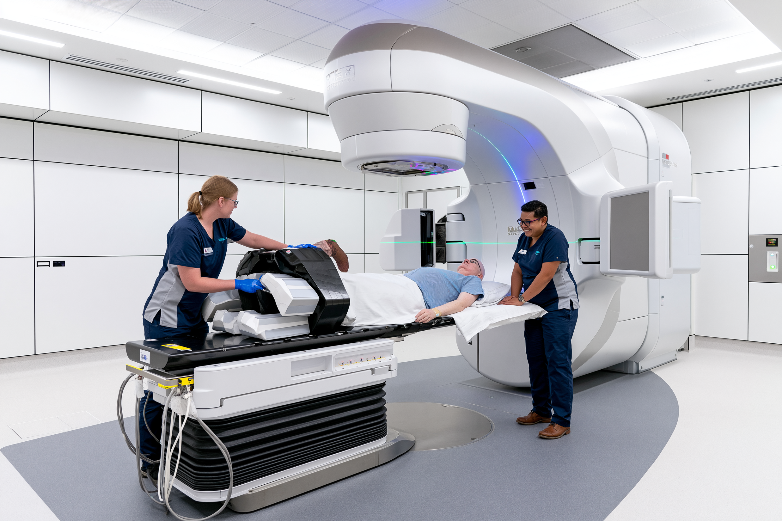
Radiation therapy for metastatic esophageal cancer uses precisely directed high-energy beams to treat a specific tumor or metastatic site. In stage IVB disease, radiation is usually used to relieve symptoms rather than treat every cancer deposit in the body. It may improve difficulty swallowing, control tumor bleeding, reduce pain, or treat a symptomatic bone or brain metastasis, while chemotherapy, immunotherapy, or targeted therapy addresses disease at multiple sites [1,2].
Receiving palliative radiation does not mean that active cancer treatment has stopped. Palliative describes the treatment goal: reducing a symptom or preventing a local complication. Radiation may therefore be given before, during, or between systemic-treatment cycles when its expected benefit outweighs the risks.
Treatment normally begins with a planning CT scan. The radiation team maps the tumor, determines the treatment position, and limits exposure to nearby structures such as the lungs, heart, spinal cord, stomach, and healthy esophagus. Some patients receive a short course over several visits, while others require a longer schedule according to the location being treated, previous radiation, performance status, expected benefit, and treatment goals.
External-beam radiation does not make the patient radioactive. Family members can remain nearby after each session. Intraluminal brachytherapy, which delivers radiation from inside the esophagus for a limited period, is a different technique and is available only in selected specialist centers [1].
Radiation for Difficulty Swallowing in Metastatic Esophageal Cancer
Radiation can relieve dysphagia when the primary esophageal tumor is narrowing the passage and the patient is not undergoing surgery. By damaging cancer cells, it may gradually reduce tumor bulk and allow food or liquids to pass more easily. The National Cancer Institute includes external-beam radiation and intraluminal brachytherapy among the palliative treatment options for stage IV esophageal cancer [1].
Improvement is not immediate. Radiation usually requires time to affect the tumor, and swallowing may temporarily become more painful because treatment can inflame the esophageal lining. A patient who cannot swallow water or saliva, repeatedly aspirates, or has nearly complete obstruction may need an esophageal stent, feeding access, intravenous hydration, or another urgent intervention rather than waiting for radiation to work.
Radiation may be particularly useful when the patient still has a reasonably open esophageal passage and can maintain safe oral or enteral nutrition during treatment. A randomized trial in advanced esophageal cancer found that a short course of radiotherapy alone could provide palliative dysphagia relief and was better tolerated than adding concurrent cisplatin and fluorouracil, which increased significant acute toxicity without producing a clear survival advantage.
An esophageal stent and radiation serve different practical purposes. A stent can reopen the esophagus rapidly, whereas radiation aims to reduce tumor-related obstruction more gradually. Radiation should not automatically be added after every stent. A randomized study found that routine external-beam radiation after stent placement did not significantly reduce recurrent dysphagia or improve survival, although fewer bleeding events were observed in the radiation group. Treatment should therefore be individualized rather than routinely combining both procedures.
Possible side effects include fatigue, nausea, reduced appetite, painful swallowing, acid reflux, chest discomfort, and temporary worsening of dysphagia. The risk depends on the treated area, radiation dose, number of sessions, and whether chemotherapy is given at the same time. Nutritional planning should begin before treatment, especially when the patient is already losing weight or relying mainly on liquids.
Soft, moist, pureed, or liquid foods may be required temporarily. Adequate protein, calories, and hydration remain important even when radiation is expected to improve swallowing later. A dietitian and swallowing specialist can help determine whether oral intake remains safe or whether temporary tube feeding is needed.
Radiation for Bleeding or Pain
An esophageal tumor may bleed because its surface is ulcerated, fragile, or invading nearby tissue. Radiation can reduce tumor activity and damage the abnormal blood vessels supplying the cancer, helping to control persistent or recurrent bleeding in selected patients [1,2].
Radiation is not an immediate substitute for emergency bleeding management. Vomiting a substantial amount of blood, passing black tarry stools, fainting, developing a rapid heartbeat, or becoming pale and confused requires urgent hospital care. Initial treatment may include intravenous fluids, blood transfusion, correction of abnormal clotting, endoscopy, interventional radiology, or another procedure before radiation can provide longer-term local control.
Radiation may also relieve pain arising from the primary esophageal tumor, enlarged lymph nodes, or invasion of nearby chest structures. It can be directed at the area causing pain without treating the entire body. Pain relief may develop gradually, so prescribed analgesics should usually continue while the response is assessed.
Chest radiation can sometimes produce temporary inflammation of the esophagus, lungs, skin, or nearby soft tissues. A dry cough, breathlessness, worsening chest pain, fever, or inability to swallow should be reported promptly because these symptoms may represent treatment inflammation, aspiration, infection, tumor progression, or another complication.
Previous radiation to the same region requires particular caution. Reirradiation may be possible in selected patients, but the radiation oncologist must review the earlier treatment plan and cumulative exposure to the esophagus, airway, lungs, heart, and spinal cord. The risk of ulceration, perforation, fistula, or injury to nearby organs may be higher when the tissues have already received substantial radiation.
Radiation for Bone or Brain Metastases
Radiation therapy can provide effective local treatment when metastatic esophageal cancer has spread to a painful or structurally important bone. It may reduce pain, limit further local growth, and help preserve movement. Treatment may be delivered in one session or over a short series of sessions, depending on the affected bone, fracture risk, previous radiation, neurological symptoms, and expected survival. ASTRO identifies external-beam radiation as an effective treatment for symptomatic bone metastases.
A weakened weight-bearing bone may require orthopedic stabilization before or after radiation. Radiation can treat the tumor within the bone, but it cannot immediately restore the mechanical strength of a severely damaged hip, femur, or vertebra. New inability to bear weight or sudden severe pain may indicate an impending or completed fracture and requires urgent imaging.
Spinal metastases need rapid assessment when back pain is accompanied by leg weakness, numbness, difficulty walking, or loss of bladder or bowel control. These symptoms may indicate spinal cord compression. Emergency MRI, corticosteroid treatment, surgical review, and urgent radiation may be required according to the location and stability of the spine.
Brain metastases are treated according to their number, size, position, symptoms, and effect on surrounding brain tissue. Stereotactic radiosurgery directs a highly focused radiation dose at one or a limited number of lesions. Whole-brain radiation treats a wider area and may be considered when numerous or diffuse metastases are present. Surgery may be appropriate for a large accessible lesion causing pressure or when tissue confirmation is needed.
Guidelines recommend local treatment for symptomatic brain metastases regardless of which systemic therapy is being used. Stereotactic radiosurgery, whole-brain radiation, surgery, or a combination may be selected through multidisciplinary review. Large tumors producing significant pressure are more likely to require surgical consideration, while focused radiation may be suitable for selected smaller lesions.
Brain radiation can cause fatigue, scalp irritation, temporary hair loss, nausea, headache, or swelling around the treated lesion. Whole-brain radiation may also affect memory and cognitive function. Corticosteroids may be prescribed when swelling causes neurological symptoms, but they should be taken only under medical supervision and reduced according to the treating team’s instructions.
Radiation does not replace systemic treatment for cancer elsewhere in the body. Its value lies in controlling a specific area that is causing symptoms, threatening organ function, or creating an immediate risk. The decision should balance the probability of meaningful relief against travel demands, treatment toxicity, nutritional status, functional condition, previous radiation, and the patient’s overall treatment goals [1,2].
Esophageal Stenting for Severe Dysphagia
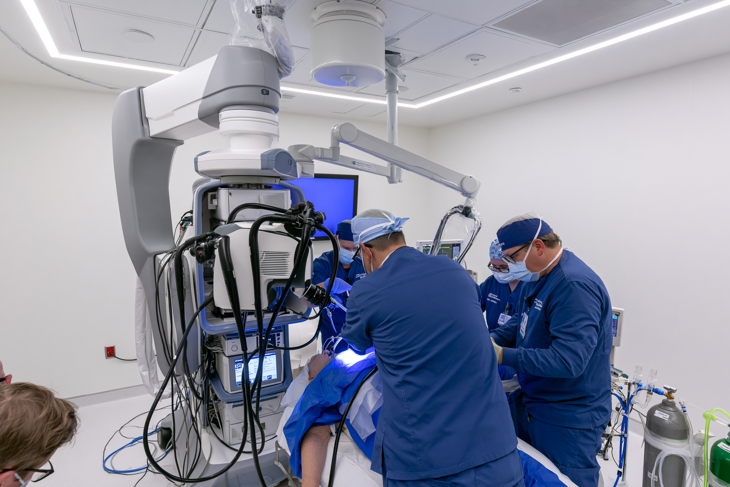
Esophageal stenting for severe dysphagia is a palliative procedure used to reopen an esophagus that has been narrowed by cancer. A flexible tube, usually a self-expanding metal stent, is positioned across the obstructed area during endoscopy or with imaging guidance. Once released, the stent expands against the tumor and creates a passage through which food and liquids may move more easily [10].
The main purpose of an esophageal stent is to improve swallowing quickly. It does not remove the primary tumor, treat distant metastases, or replace chemotherapy, immunotherapy, radiation, or nutritional care. Its value lies in relieving obstruction when a patient cannot maintain adequate oral intake or when waiting for systemic treatment or radiation to reduce the tumor would be unsafe.
When Esophageal Stenting for Severe Dysphagia May Be Needed
Esophageal stenting may be considered when progressive tumor narrowing prevents a patient from swallowing normal food, liquids, medicines, or saliva. It is particularly relevant when dysphagia is causing rapid weight loss, dehydration, repeated regurgitation, distress during meals, or an immediate risk of losing oral intake [10].
A stent may also be used when the expected time for radiation or systemic treatment to improve swallowing is too long for the patient’s present condition. Chemotherapy and radiotherapy can reduce tumor-related obstruction in some patients, but the response is not immediate and cannot be guaranteed. A stent can provide more rapid mechanical relief because it physically holds the narrowed passage open.
Before recommending the procedure, the clinical team evaluates the position and length of the obstruction, how much of the esophageal passage remains open, the condition of the airway, the patient’s performance status, and the expected benefits of restoring oral intake. Endoscopy, CT imaging, and sometimes a contrast-swallow study help determine whether stenting is technically possible and clinically appropriate.
Stenting may be especially useful when a patient has metastatic disease, a short expected interval before systemic treatment begins, or severe dysphagia that makes eating and drinking increasingly difficult. It may also be considered when the patient is not medically suitable for major surgery or when the purpose of treatment is rapid symptom relief.
A malignant connection between the esophagus and airway, known as a tracheoesophageal fistula, may sometimes be sealed with a covered stent. This is a specialized indication because the airway may also be narrowed or vulnerable to compression. Gastroenterology, thoracic surgery, respiratory medicine, and interventional teams may need to plan the procedure together. In selected cases, an airway stent may be required before or alongside the esophageal stent [10].
Esophageal stenting is not automatically the best option for every patient with dysphagia. Tumors very close to the upper esophageal sphincter can make stent placement uncomfortable and may increase the sensation of a foreign body, pain, coughing, or aspiration. A stent crossing the gastroesophageal junction can increase acid reflux because it holds the lower esophageal passage open.
The procedure may also be unsuitable when swallowing difficulty is caused mainly by impaired muscular coordination rather than a fixed mechanical obstruction. Similarly, a patient with widespread aspiration, severe mucositis, uncontrolled vomiting, or profound weakness may remain unable to eat safely even after the esophageal lumen has been reopened.
A feeding tube may sometimes be more appropriate when the main goal is reliable nutrition rather than restoration of swallowing. The choice between stenting, radiation, feeding access, or a combination should be based on the patient’s anatomy, symptoms, nutritional condition, expected treatment course, and personal priorities [10,11].
Benefits and Limitations of Esophageal Stenting
The principal benefit of esophageal stenting is relatively rapid improvement in malignant dysphagia. A patient who was limited to liquids may sometimes progress to soft or semisolid foods after successful placement. Improved swallowing can reduce distress during meals, make hydration easier, and allow some medicines to be taken orally.
Restoring the passage can also help the patient participate more comfortably in family meals and reduce anxiety associated with food becoming stuck. These quality-of-life benefits may be important even when the stent does not substantially change the overall course of metastatic esophageal cancer.
A stent can also reduce leakage through a malignant fistula when a covered device successfully seals the abnormal opening. This may decrease the passage of food or liquid into the airway, although aspiration can continue for other reasons and must be monitored carefully [10].
The procedure is commonly performed with sedation rather than major surgery. However, it still carries risks. Chest discomfort or pain is common during the early period as the stent expands. Some patients experience nausea, increased salivation, reflux, coughing, or a persistent sensation of pressure behind the breastbone.
Stent migration occurs when the device moves away from its intended position. This may happen after the tumor shrinks, particularly during chemotherapy or radiation, or when the stent crosses the gastroesophageal junction. A migrated stent may no longer relieve the obstruction and can require repositioning, removal, or replacement.
Dysphagia may return if tumor tissue grows through or around the stent, food becomes impacted inside it, or the device changes position. Recurrent swallowing difficulty should be reported promptly rather than managed by repeatedly forcing food or large quantities of liquid through the stent.
More serious complications include bleeding, perforation of the esophageal wall, fistula formation, airway compression, aspiration, and severe pain. These events are less common but can be life-threatening. New breathing difficulty, vomiting blood, black stools, fever, severe chest pain, inability to swallow saliva, or sudden worsening of dysphagia requires urgent medical assessment.
Eating habits usually need to change after stent placement. Food should be soft, moist, and thoroughly chewed. Small mouthfuls and slow eating can reduce the risk of blockage. Dry, sticky, fibrous, or poorly chewed foods may become trapped within the stent. The patient should follow the individualized instructions provided by the endoscopy and dietetic teams.
Improved swallowing does not necessarily mean that nutritional recovery will occur automatically. A patient may continue to eat too little because of poor appetite, treatment-related nausea, fatigue, altered taste, pain, or cancer cachexia. Weight, hydration, protein intake, muscle strength, and functional status therefore require continued monitoring after the procedure [11].
A stent may also affect future treatment planning. Radiation delivered across a recently placed stent can increase tissue irritation in some patients, while tumor shrinkage during systemic treatment may contribute to migration. The oncology and endoscopy teams should coordinate the timing of stenting, radiation, chemotherapy, and immunotherapy.
Esophageal stenting for severe dysphagia should therefore be viewed as a targeted symptom-relieving intervention rather than a complete treatment for metastatic esophageal cancer. It can restore a passage for food and fluids, but its benefits must be balanced against the risks of migration, reflux, pain, recurrent obstruction, bleeding, perforation, and aspiration. Careful patient selection and continued nutritional and oncological monitoring are essential [10,11].
Dysphagia and Nutrition in Metastatic Esophageal Cancer

Dysphagia and nutrition in metastatic esophageal cancer require early, active management because reduced food intake can quickly lead to dehydration, weight loss, muscle wasting, weaker treatment tolerance, and declining independence. Nutrition is not an optional part of supportive care. It is a core component of the treatment plan alongside systemic therapy, radiation, stenting, symptom control, and palliative care [10,11].
A patient who cannot swallow water or saliva, repeatedly chokes, regurgitates most meals, or develops breathing difficulty while eating needs urgent medical assessment. Food, tablets, nutritional drinks, avaleha, or other oral preparations should not be forced when swallowing is unsafe. The immediate priority is to protect the airway, restore hydration, and establish a safe route for nutrition [10,11].
Why Swallowing Becomes Difficult in Metastatic Esophageal Cancer
Swallowing usually becomes difficult because the primary tumor narrows the inner passage of the esophagus. Food may initially stick only when the patient eats bread, meat, rice, or other solid foods. As the obstruction progresses, soft foods, thick liquids, water, and eventually saliva may become difficult to swallow.
The tumor can also interfere with the coordinated muscular movements that normally carry food toward the stomach. Ulceration, inflammation, pain, reflux, infection, treatment-related mucositis, or external compression from enlarged lymph nodes may worsen the problem even when the esophageal passage is not completely blocked.
Dysphagia can continue or temporarily worsen during treatment. Radiation may inflame the esophageal lining, while chemotherapy can contribute to mucositis, nausea, altered taste, fatigue, and reduced appetite. A patient may therefore eat less because swallowing is physically difficult, painful, exhausting, or associated with fear of choking.
The severity of dysphagia should be assessed according to what the patient can actually swallow. Being able to take a few sips or several spoonfuls does not necessarily mean that hydration and nutrition are adequate. The clinical team should ask about meal duration, coughing, regurgitation, food sticking, pain, recent weight loss, urine output, and the amount consumed during an average day.
When mechanical obstruction is the main cause, an esophageal stent may provide rapid relief in selected patients. Radiation or systemic treatment may gradually reduce tumor-related narrowing, while a feeding tube may be required when safe oral intake cannot meet nutritional needs. These options are not interchangeable, and the most appropriate approach depends on the degree of obstruction, aspiration risk, treatment plan, expected response, and patient preference [10,11].
Soft, Pureed and Liquid Diets for Metastatic Esophageal Cancer
Food texture should match the patient’s current swallowing ability rather than the diet they could manage several weeks earlier. A person who can swallow soft food safely may not need to rely entirely on liquids, while someone who coughs with thin fluids may require a formal swallowing assessment before the consistency is changed.
Soft foods require little chewing and remain moist enough to pass through the esophagus more easily. Suitable choices may include scrambled eggs, oatmeal, soft pasta with sauce, mashed potatoes, well-cooked vegetables, cottage cheese, Greek yogurt, soft fish, tender minced meat with gravy, and smooth lentil preparations. Dry bread, tough meat, raw vegetables, popcorn, nuts, and sticky foods may be difficult to pass through a narrowed esophagus.
Pureed foods should be smooth, moist, and free from lumps, skins, seeds, or fibrous pieces. Soups, cooked vegetables, beans, eggs, fish, chicken, or other foods can be blended with broth, milk, yogurt, gravy, olive oil, or another suitable liquid. Blending food only with water can make the portion larger while reducing its calorie and protein concentration.
A liquid diet may include milk, lactose-free milk, fortified plant beverages, smooth soups, oral nutritional supplements, drinkable yogurt, custard, and carefully prepared smoothies. A drink that appears nutritious may still provide too little protein or energy, particularly when it consists mainly of fruit, tea, clear broth, or juice.
Thin liquids are not automatically safer than thicker drinks. Some patients aspirate water and other thin fluids more easily because they move quickly through the throat. Thickened liquids should be used only when recommended after an appropriate swallowing assessment because unnecessary thickening can reduce fluid intake and make dehydration more likely.
Small, frequent meals are often easier than three large meals. The patient should sit upright, take small mouthfuls, chew thoroughly, and eat slowly without being rushed. Remaining upright after meals may reduce regurgitation and reflux. Straws, large tablets, and mixed textures such as thin soup containing solid pieces may be unsafe for some patients, so advice should be individualized.
A sudden change in swallowing should not be managed only by further blending food at home. Rapidly worsening dysphagia may indicate increasing obstruction, food impaction, stent migration, treatment-related inflammation, or fistula formation and should be assessed promptly [10,11].
Preventing Dehydration
Dehydration can develop quickly when swallowing is difficult because fluids provide little energy and may be neglected while the patient concentrates on eating. Regurgitation, vomiting, diarrhea, fever, infection, and treatment toxicity can increase fluid losses further.
Early signs include a dry mouth, dark urine, reduced urination, headache, thirst, dizziness, constipation, and increasing fatigue. More advanced dehydration may cause rapid heartbeat, low blood pressure, confusion, fainting, kidney dysfunction, or severe weakness.
Fluid intake should be spread throughout the day in small amounts that the patient can swallow safely. Milk, oral nutritional supplements, smooth soups, yogurt drinks, ice cream, custard, and other fluid-rich foods can contribute to both hydration and nutrition. Clear fluids may be useful for hydration but should not replace calorie-dense drinks when the patient is losing weight.
There is no single fluid target that is appropriate for every patient. Requirements change with body size, fever, vomiting, kidney function, heart function, medication use, and treatment. The oncology or dietetic team should provide an individualized goal, particularly when the patient has fluid retention, pleural effusion, ascites, heart disease, or impaired kidney function.
Intravenous fluids may be required when the patient cannot swallow enough, repeatedly vomits, becomes dizzy, urinates very little, or develops abnormal blood tests. Intravenous hydration can correct an immediate deficit, but it does not solve the underlying obstruction or provide complete nutritional support.
Families can monitor the approximate amount consumed, urine frequency, body weight, vomiting, and ability to take prescribed medicines. A patient who cannot swallow water, has very little urine, becomes confused, or feels too weak to stand needs urgent assessment rather than continued attempts to manage dehydration through oral fluids alone [11].
Protein and Calorie Support
Patients with metastatic esophageal cancer often need food that provides more nutrition in a smaller volume. A large plate of low-calorie food may be impossible to finish, while a smaller serving enriched with protein and energy may be more practical.
Protein supports muscle, immune function, tissue repair, and recovery from treatment. Useful soft or liquid sources may include eggs, Greek yogurt, cottage cheese, milk, smooth nut butter when safe to swallow, pureed beans, lentils, tofu, soft fish, minced poultry, protein powders, and prescribed oral nutritional supplements.
Calories can be increased without greatly enlarging the meal by adding olive oil, butter, cream, powdered milk, yogurt, cheese, avocado, smooth nut butter, or calorie-dense nutritional products when clinically appropriate. The choice should take account of diabetes, reflux, diarrhea, lactose intolerance, kidney disease, liver dysfunction, and personal dietary preferences.
ESPEN guidance generally estimates energy requirements at approximately 25 to 30 kilocalories per kilogram of body weight each day when individual measurement is unavailable. Protein intake is commonly recommended above 1 gram per kilogram daily and, when possible, up to approximately 1.5 grams per kilogram [11]. For a patient weighing 70 kilograms, or about 154 pounds, this would correspond to an estimated 1,750 to 2,100 calories and roughly 70 to 105 grams of protein daily. These figures are clinical starting points rather than fixed prescriptions.
A patient may be unable to reach the full target immediately. The initial aim may be to stop further decline, improve hydration, and gradually increase intake through fortified foods and supplements. Oral nutritional supplements are often better tolerated between meals because taking them immediately before food can reduce appetite.
Dietary restrictions should not be unnecessarily severe. Avoiding sugar, dairy products, gluten, oils, or all processed nutritional products without a specific medical reason can make it harder to meet calorie and protein requirements. Food safety, swallowing safety, tolerance, and adequate intake are usually more urgent priorities during active treatment.
A severely malnourished patient should not suddenly receive a very large nutritional load without medical supervision. Rapid feeding after prolonged poor intake can produce dangerous electrolyte changes known as refeeding syndrome. Nutrition may need to be introduced gradually with monitoring of phosphate, potassium, magnesium, glucose, fluid balance, and organ function [11].
Feeding Tube Support
A feeding tube may be needed when the digestive system can still absorb nutrition but food cannot pass safely through the esophagus. It can deliver a complete liquid formula containing protein, calories, fluid, vitamins, and minerals without requiring the patient to swallow each meal.
Short-term feeding may be delivered through a tube passed through the nose into the stomach or small intestine. Longer-term access may involve a gastrostomy tube placed into the stomach or a jejunostomy tube placed into the small intestine. The route depends on the site of obstruction, aspiration risk, anatomy, previous surgery, anticipated treatment, and expected duration of support.
A feeding tube does not mean that cancer treatment has failed. It may help stabilize body weight, correct dehydration, improve medication delivery, and allow chemotherapy or radiation to continue more safely. Some patients can still take small amounts orally for comfort when swallowing has been assessed as safe.
Stenting and tube feeding serve different goals. A stent aims to restore passage through the esophagus and may allow oral eating. A feeding tube bypasses the swallowing problem and provides a more reliable nutritional route. A stent may be preferred when rapid restoration of oral intake is realistic, while tube feeding may be safer when aspiration, severe pain, fistula, treatment-related inflammation, or complex obstruction makes swallowing unsafe [10,11].
Tube feeding does not completely eliminate aspiration risk because saliva, refluxed stomach contents, or regurgitated material can still enter the airway. The patient’s position, feeding rate, tube location, gastric emptying, and respiratory symptoms require continued assessment.
When the gastrointestinal tract cannot be used safely or cannot absorb enough nutrition, parenteral nutrition may be considered. This delivers nutrients into a vein and carries risks such as bloodstream infection, blood-glucose disturbance, fluid imbalance, and liver complications. It is generally reserved for carefully selected situations in which oral and enteral nutrition are not feasible or adequate [11].
Cancer Cachexia and Muscle Loss
Cancer cachexia is a complex syndrome involving ongoing loss of skeletal muscle, with or without loss of body fat. It is common in advanced cancer and can develop through a combination of reduced food intake, inflammation, altered metabolism, treatment effects, and reduced physical activity [11].
Cachexia is not simply ordinary hunger or inadequate willpower. Encouraging a patient to eat more may help when intake is low, but food alone may not completely reverse the metabolic changes driving muscle loss. Blaming the patient for not eating can increase distress without improving nutrition.
Early signs may include loose clothing, thinner arms or legs, reduced grip strength, slower walking, difficulty climbing stairs, or needing help to rise from a chair. Body weight alone may underestimate the problem because edema, ascites, or pleural fluid can conceal the loss of muscle and fat.
Management should address every treatable factor reducing intake. This may include relieving dysphagia, controlling pain and nausea, treating constipation or infection, managing mouth soreness, improving reflux, addressing depression or anxiety, and reviewing medicines that suppress appetite or cause sedation.
Nutrition should be combined with appropriate physical activity whenever the patient is medically able. Gentle walking, supervised resistance exercise, and regular movement may help preserve function more effectively than prolonged bed rest. Exercise must be adapted when bone metastases, severe anemia, breathlessness, neurological weakness, or fracture risk is present.
The most useful outcome is not always weight gain. Stabilizing weight, slowing muscle loss, improving strength, reducing hospital admissions, maintaining treatment tolerance, and preserving independence may be meaningful achievements. Regular assessment by the oncology team and a cancer dietitian allows nutrition to be adjusted as swallowing ability, disease burden, treatment, and functional status change [11].
Metastatic Esophageal Cancer Survival and Prognosis

Metastatic esophageal cancer survival varies widely and cannot be predicted accurately from the stage alone. Stage IVB disease is serious because cancer has spread to a distant organ or nonregional lymph node, but prognosis is also influenced by tumor type, biomarker status, metastatic burden, general fitness, nutritional condition, organ function, and response to treatment [2,7,8,11,12].
Survival figures are most useful for understanding outcomes across large patient populations. They should not be interpreted as a fixed life expectancy for an individual patient or as a deadline after diagnosis.
Stage IVB Esophageal Cancer Survival
The closest widely reported population estimate for stage IVB esophageal cancer comes from the distant-stage category used by the United States Surveillance, Epidemiology, and End Results program. Based on patients diagnosed between 2016 and 2022, the five-year relative survival rate for distant-stage esophageal cancer was 5.3 percent [12].
Distant-stage disease in the SEER system broadly represents cancer that has metastasized, but it is not identical to the AJCC stage IVB classification. The two systems group patients differently, so the SEER percentage should be presented as a population estimate rather than an exact stage IVB survival rate.
Five-year relative survival compares people with esophageal cancer with people of the same age and sex in the general population. It does not mean that every patient who survives five years is cancer-free, that everyone else lives for the same limited period, or that only 5.3 percent of patients can benefit from treatment.
Registry statistics also reflect patients treated during earlier years. They may not fully capture the effect of newer immunotherapy combinations, HER2-directed treatments, improved molecular testing, modern supportive care, and earlier nutritional intervention. Current treatment outcomes may therefore differ from older population averages, especially in carefully selected patients with treatment-responsive disease [7,8,12].
Factors That Influence Metastatic Esophageal Cancer Survival
Tumor histology is important because esophageal squamous cell carcinoma and esophageal adenocarcinoma have different biological features, biomarker patterns, and treatment pathways. Prognosis may also differ according to whether the tumor expresses PD-L1, overexpresses HER2, or has mismatch-repair deficiency or high microsatellite instability [2,7,8].
The extent of metastatic disease also matters. A patient with one limited metastatic site may have a different outlook from someone with extensive liver involvement, multiple bone lesions, malignant fluid around the lungs, or cancer affecting several organs. The location of the metastases is important when it interferes with liver function, breathing, mobility, neurological function, or another essential body process.
Performance status describes how independently a patient can carry out daily activities. A person who remains mobile, performs self-care, and spends much of the day out of bed is generally more likely to tolerate systemic treatment than someone who is largely confined to a bed or chair. Performance status can change during treatment, so it should be assessed repeatedly rather than recorded only at diagnosis [2].
Nutrition has a direct influence on treatment tolerance and functional reserve. Severe dysphagia, rapid weight loss, dehydration, and skeletal muscle loss can increase weakness, treatment complications, and interruptions in therapy. Early management of swallowing problems, calorie and protein intake, hydration, and cancer cachexia may help a patient remain eligible for effective treatment, although nutrition alone cannot control metastatic cancer [11].
Liver, kidney, heart, lung, and bone-marrow function also affect which medicines can be given and at what dose. Severe organ dysfunction may limit chemotherapy or targeted therapy, while uncontrolled infection, bleeding, aspiration, or electrolyte disturbance may require stabilization before treatment can continue.
The response to first-line treatment is one of the strongest practical indicators of the subsequent disease course. A substantial and sustained reduction in measurable tumors is generally more favorable than early progression. Response must be evaluated through appropriate imaging and clinical assessment rather than inferred only from improved appetite, reduced pain, or easier swallowing.
Why Population Survival Statistics Cannot Predict Individual Survival
Population survival statistics combine thousands of patients with different ages, histological types, biomarkers, metastatic sites, accompanying illnesses, treatment regimens, and levels of physical fitness. An average calculated from such a diverse group cannot determine what will happen to one particular patient [12].
A newly diagnosed patient with preserved organ function, stable weight, limited metastatic disease, and an actionable biomarker is not clinically identical to a patient with extensive multi-organ metastases, severe cachexia, aspiration pneumonia, and declining performance status. Both may be described as having stage IVB disease, but their treatment options and likely outcomes may be substantially different.
Survival estimates also cannot account fully for future events. A patient may respond exceptionally well, develop unexpected toxicity, acquire a new complication, or become eligible for a clinical trial. Tumor biology can also change over time, and resistance may develop after an initially successful response.
For these reasons, prognosis is best discussed as a range of possible outcomes rather than a precise number of months. The oncology team can provide a more individualized estimate after reviewing the pathology, biomarker results, imaging, treatment response, nutritional status, organ function, and overall pattern of illness.
Can Some Patients Respond for Longer Than Expected?
Some patients with metastatic esophageal cancer experience disease control that lasts considerably longer than the population average. Long-term follow-up from the CheckMate 648 trial showed that the overall survival benefit of nivolumab with chemotherapy or nivolumab with ipilimumab continued at five years in patients with advanced esophageal squamous cell carcinoma [7].
Five-year follow-up from CheckMate 649 also demonstrated sustained benefit from nivolumab plus chemotherapy in advanced HER2-negative gastroesophageal adenocarcinoma. Among patients with a PD-L1 combined positive score of at least 5, the five-year overall survival rate was 16 percent with nivolumab plus chemotherapy and 6 percent with chemotherapy alone [8]. This trial included gastric, gastroesophageal-junction, and esophageal adenocarcinomas, so these percentages should not be presented as esophageal-only survival rates.
These results show that long-term survival is possible for a minority of appropriately selected patients, but they do not guarantee the same outcome for every person receiving immunotherapy. Biomarker expression, tumor biology, general health, treatment tolerance, and the depth and duration of response all influence whether prolonged control occurs.
A long-lasting response is also not automatically the same as cure. Even when scans show a complete response, microscopic cancer cells may remain and recurrence can occur. Continued clinical review, imaging, laboratory monitoring, nutritional assessment, and management of delayed treatment effects remain necessary.
The most reliable way to understand an individual prognosis is to reassess it over time. Early staging provides the starting point, but the patient’s actual response to treatment, stability of metastatic sites, ability to maintain nutrition, and preservation of daily function provide more meaningful information as care progresses.
The Ayurveda Curative Model for Metastatic Esophageal Cancer

The Ayurveda Curative Model for metastatic esophageal cancer is a structured integrative approach that combines modern cancer diagnosis and treatment with individualized Ayurvedic assessment, nutritional support, symptom management, and continuous monitoring. It aims to address the tumor, swallowing difficulty, digestive capacity, progressive weakness, treatment tolerance, and overall functional decline as connected clinical problems rather than treating each symptom in isolation.
The word curative in this model describes the intention to pursue the best possible disease control and restoration of health through coordinated care. It should not be interpreted as proof that Ayurveda alone can cure stage IVB esophageal cancer. Chemotherapy, immunotherapy, targeted therapy, radiation, stenting, and other medically indicated interventions remain essential when appropriate. Any claim of tumor response must be confirmed through standardized imaging and oncology assessment [1,2,13,16].
What the Ayurveda Curative Model Means for Metastatic Esophageal Cancer
The model begins by identifying every factor that may influence treatment response. This includes the histological type of esophageal cancer, biomarker profile, location of metastases, degree of esophageal obstruction, nutritional status, organ function, performance status, and current oncology treatment.
Ayurvedic assessment is then added to understand how the illness is affecting digestion, food tolerance, strength, sleep, bowel function, mental well-being, and the patient’s ability to recover between treatments. These findings may guide supportive dietary measures, carefully selected formulations, and symptom-focused interventions.
The model does not separate tumor management from nutritional and functional recovery. A patient whose swallowing, hydration, body weight, and physical strength are deteriorating may be unable to receive effective oncology treatment even when a suitable medicine is available. Supporting these areas can therefore be clinically important, although supportive improvement must not be confused with direct tumor regression [11,16].
Modern Diagnosis Remains the Foundation of the Model
Modern diagnosis remains the foundation of the Ayurveda Curative Model because metastatic esophageal cancer cannot be accurately assessed through symptoms, pulse examination, tongue appearance, or Ayurvedic terminology alone. The diagnosis must be established through endoscopy, biopsy, histopathology, TNM staging, CT or PET-CT, and appropriate biomarker testing [1,2].
The treatment team needs to know whether the tumor is squamous cell carcinoma or adenocarcinoma, whether distant metastases are present, and whether PD-L1, HER2, mismatch-repair deficiency, microsatellite instability, or another molecular feature can guide treatment. These findings determine which systemic therapies may be appropriate.
Ayurvedic concepts may contribute to understanding the patient’s functional condition, but they cannot identify the molecular subtype of the tumor or measure metastatic disease. Modern staging and pathology should therefore be completed before an individualized integrative plan is finalized.
Ayurvedic Clinical Assessment
Ayurvedic clinical assessment examines the patient’s constitution, current doshic disturbance, digestive capacity, bowel pattern, sleep, appetite, tissue depletion, strength, and tolerance of food and medicine. In advanced esophageal cancer, the assessment should also consider dysphagia, painful swallowing, regurgitation, aspiration risk, weight loss, fatigue, and treatment-related adverse effects.
Prakriti describes the patient’s underlying constitutional tendency, while Vikriti describes the present imbalance. These observations may help individualize diet and supportive care, but they should not be used to predict tumor stage or replace oncology investigations.
The assessment must be repeated as the clinical condition changes. A formulation tolerated before chemotherapy may become unsuitable during severe mucositis, vomiting, liver dysfunction, kidney impairment, or worsening esophageal obstruction. Treatment should therefore remain responsive to the patient’s current condition rather than follow a fixed prescription.
Agni and Digestive Capacity
Agni is the Ayurvedic term for the body’s capacity to digest, transform, and assimilate food. In metastatic esophageal cancer, digestive capacity may decline because of reduced intake, treatment-related nausea, altered taste, inflammation, constipation, diarrhea, pain, anxiety, or prolonged physical weakness [19].
A patient may feel hungry but remain unable to swallow enough food. Another may have an open esophageal passage but experience nausea, early fullness, or poor digestion after only a small meal. These are different clinical problems and should not be managed in the same way.
Assessment of Agni should include appetite, meal tolerance, abdominal discomfort, bowel movements, nausea, bloating, and the amount of nourishment the patient can realistically consume. Strong digestive herbs should not be prescribed automatically because pungent or irritating ingredients may worsen reflux, mucositis, painful swallowing, or treatment-related gastrointestinal symptoms.
The objective is to support comfortable digestion without increasing irritation or interfering with prescribed cancer treatment. Food consistency, meal volume, timing, and nutritional density may be as important as the selection of Ayurvedic medicines.
Annavaha Srotas and Swallowing
Annavaha Srotas refers to the Ayurvedic channels concerned with the intake and initial processing of food. In advanced esophageal cancer, this concept may be used to describe the functional consequences of obstructed swallowing, regurgitation, painful intake, and reduced nourishment [19].
It should not be treated as a substitute for the anatomical diagnosis of esophageal narrowing. The degree of obstruction must be assessed through endoscopy and imaging. Severe dysphagia may require stenting, radiation, feeding-tube support, or another medical intervention [1,2].
Ayurvedic oral formulations are appropriate only when the patient can swallow safely. Avaleha, tablets, powders, oils, or decoctions should not be forced through a severely narrowed esophagus. Coughing after swallowing, repeated regurgitation, inability to manage saliva, or suspected aspiration requires urgent reassessment.
The safest integrative approach is to match every oral preparation to the patient’s current swallowing capacity. When oral intake is unsafe, the immediate priority is airway protection, hydration, and reliable nutritional access.
Dhatu Kshaya, Bala and Progressive Weakness
Dhatu Kshaya describes progressive depletion of body tissues, while Bala refers to physical strength, resilience, and the ability to withstand disease and treatment. These concepts are relevant when a patient develops rapid weight loss, muscle wasting, reduced mobility, fatigue, and declining independence [19].
In modern clinical terms, these changes may reflect malnutrition, cancer cachexia, anemia, inflammation, dehydration, organ dysfunction, or treatment toxicity. Ayurvedic interpretation should complement rather than obscure these measurable causes.
A patient who loses muscle may become less able to tolerate chemotherapy, recover after hospitalization, or perform basic daily activities. Supportive care should therefore focus on adequate protein and calories, symptom control, hydration, safe movement, and correction of reversible medical problems [11].
Bala should be assessed functionally. Useful indicators include the ability to walk, rise from a chair, perform self-care, remain out of bed, and complete ordinary daily activities. Improvement in Bala may be meaningful, but it does not by itself prove that the cancer has responded.
Ojas and Overall Functional Reserve
Ojas is traditionally associated with vitality, stability, resistance, and the integrated strength of the body. In the Ayurveda Curative Model, it can be used as a broad clinical concept for the patient’s overall functional reserve rather than as a laboratory measurement [19].
A person with preserved appetite, stable weight, reasonable sleep, clear mental function, and adequate mobility may have greater reserve for treatment than someone with severe cachexia, repeated infections, dehydration, and prolonged bed rest. These differences influence how safely intensive treatment can be delivered.
Ojas should not be described as an immune marker or used to claim that a formulation can directly strengthen antitumor immunity. Immune function in cancer is complex, and immunotherapy response cannot be predicted through Ayurvedic assessment alone.
The practical aim is to preserve the patient’s ability to eat, move, sleep, think clearly, and tolerate treatment. These outcomes can be followed clinically and discussed alongside performance status, nutritional measurements, blood tests, and treatment toxicity.
Supporting Nutrition and Treatment Tolerance
Nutrition is central to the Ayurveda Curative Model for metastatic esophageal cancer because dysphagia and cancer cachexia can rapidly weaken the patient. The diet must be adapted to the degree of swallowing difficulty and should provide sufficient calories, protein, fluids, and micronutrients in the smallest safely tolerated volume [11].
Soft, moist, pureed, or liquid foods may be required. Meals should be individualized according to reflux, nausea, bowel function, diabetes, kidney function, liver function, and cultural preferences. Restrictive diets should be avoided when they make it harder to meet nutritional requirements.
Ayurvedic dietary principles may help organize meal timing, digestibility, warmth, texture, and individual tolerance. However, they should not prevent the use of clinically necessary oral nutritional supplements, fortified foods, esophageal stenting, or feeding-tube support.
Improved nutritional status may help reduce treatment interruptions, support recovery, and preserve mobility. It should still be understood as supportive care rather than proof of direct anticancer activity.
Integrating Ayurveda With Oncology Treatment
Ayurveda should be integrated with oncology through shared information and coordinated decision-making. The oncology team should know every herb, formulation, supplement, bhasma, and over-the-counter product being used. The Ayurvedic clinician should know the exact chemotherapy, immunotherapy, targeted therapy, radiation schedule, laboratory results, and treatment-related toxicities [16,17].
The timing and composition of Ayurvedic treatment may need to change during active oncology care. A patient receiving chemotherapy may develop low blood counts, diarrhea, mucositis, kidney dysfunction, or liver abnormalities. A patient receiving immunotherapy may develop immune-mediated inflammation affecting the lungs, liver, bowel, thyroid, kidneys, or other organs.
New symptoms should not automatically be interpreted as doshic imbalance or detoxification. They may represent cancer progression, infection, drug toxicity, aspiration, electrolyte disturbance, or another urgent medical problem.
The safest model avoids competition between treatment systems. Oncology addresses the tumor through evidence-based systemic and local treatment, while Ayurveda may contribute to individualized supportive care when its use is clinically appropriate and carefully monitored.
Herb Drug Interaction Screening
Herb-drug interaction screening is essential because natural products can alter drug metabolism, transport, bleeding risk, blood pressure, blood sugar, sedation, and liver or kidney function. Some herbs may increase or reduce exposure to anticancer medicines, while others may intensify treatment toxicity [17].
Every ingredient should be reviewed individually rather than assuming that a classical formulation is automatically safe. The dose, preparation method, duration, product quality, and combination with other medicines all affect risk.
Particular caution is required with concentrated extracts, piperine-containing products, anticoagulant or antiplatelet medicines, medicines metabolized through major liver enzymes, and formulations containing mineral ingredients. Liver function, kidney function, blood counts, and clinical symptoms should be monitored according to the patient’s treatment.
During immunotherapy, unexplained hepatitis, diarrhea, breathing difficulty, rash, endocrine disturbance, or kidney injury requires oncology evaluation. Adding or stopping herbs without coordinated review may make it harder to identify the cause of toxicity.
Objective Monitoring of Treatment Response
Objective monitoring is required to determine whether metastatic esophageal cancer is responding. Improvement in appetite, pain, swallowing, energy, or sleep is valuable, but these changes can occur after stenting, hydration, nutritional support, pain relief, or treatment of an infection without any reduction in tumor burden.
Tumor response should be assessed through comparative CT, MRI, PET-CT when appropriate, endoscopic findings in selected cases, and standardized oncology criteria such as RECIST [13]. The same measurable lesions should be compared across scans whenever possible.
Monitoring should also include body weight, food intake, dysphagia severity, performance status, blood counts, liver and kidney function, treatment toxicity, hospital admissions, and the ability to carry out daily activities [1,2,11].
A responsible Ayurveda Curative Model records both positive and negative outcomes. Stable disease, partial response, progression, improved nutrition, worsening obstruction, and treatment-related complications should all be documented accurately. The model is clinically meaningful only when supportive improvements are distinguished clearly from objective tumor response.
Ayurvedic Understanding of Advanced Metastatic Esophageal Cancer

The Ayurvedic understanding of advanced metastatic esophageal cancer considers the tumor together with impaired swallowing, reduced food intake, weakened digestion, tissue depletion, and loss of physical strength. Classical concepts such as Granthi, Arbuda, Annavaha Srotas, Agnimandya, and Dhatu Kshaya can help structure this assessment.
These concepts should be applied with precision. Classical descriptions provide a framework for understanding the patient’s condition, but they do not independently identify esophageal squamous cell carcinoma, adenocarcinoma, biomarker status, or distant metastasis. Histopathology, TNM staging, biomarker testing, and imaging remain essential for determining the exact nature and extent of the disease [3,19,20].
Arbuda and Granthi Concepts
The Suśruta Saṃhitā describes Granthi as a rounded, elevated, knot-like swelling produced when disturbed Doshas affect tissues such as Māṃsa, Rakta, and Meda.
Sanskrit
वातादयो मांसमसृक् च दुष्टाः सन्दूष्य मेदश्च कफानुविद्धम् ।
वृत्तोन्नतं विग्रथितं तु शोफं कुर्वन्त्यतो ग्रन्थिरिति प्रदिष्टः ॥३॥
Transliteration
Vātādayo māṃsam asṛk ca duṣṭāḥ sandūṣya medaś ca kaphānuviddham।
Vṛttonnataṃ vigrathitaṃ tu śophaṃ kurvanty ato granthir iti pradiṣṭaḥ॥
Simple English translation
When aggravated Vata and the other Doshas disturb muscle tissue, blood, and fat associated with Kapha, they produce a rounded, raised, knot-like swelling. This is described as Granthi.
Classical source: Suśruta Saṃhitā, Nidānasthāna, Chapter 11, Granthyapacyarbudagalagaṇḍa Nidāna, verse 3 [20].
Granthi is therefore a broad classical description of a localized knot-like swelling. It should not automatically be translated as either a benign tumor or a malignant tumor. Modern pathology is required to determine the actual cellular nature of any mass.
The same chapter describes Arbuda as a larger, stable, deeply established swelling that grows gradually and does not readily suppurate.
Sanskrit
गात्रप्रदेशे क्वचिदेव दोषाः सम्मूर्च्छिता मांसमभिप्रदूष्य ।
वृत्तं स्थिरं मन्दरुजं महान्तमनल्पमूलं चिरवृद्ध्यपाकम् ॥१३॥
कुर्वन्ति मांसोपचयं तु शोफं तमर्बुदं शास्त्रविदो वदन्ति ।
वातेन पित्तेन कफेन चापि रक्तेन मांसेन च मेदसा च ॥१४॥
Transliteration
Gātrapradeśe kvacideva doṣāḥ sammūrcchitā māṃsam abhipradūṣya।
Vṛttaṃ sthiraṃ mandarुjaṃ mahāntam analpamūlaṃ ciravṛddhyapākam॥
Kurvanti māṃsopacayaṃ tu śophaṃ tam arbudaṃ śāstravido vadanti।
Vātena pittena kaphena cāpi raktena māṃsena ca medasā ca॥
Simple English translation
When disturbed Doshas affect muscle tissue in a part of the body, they may produce a large, rounded, stable, mildly painful, deeply rooted swelling that grows slowly and does not readily suppurate. Classical authorities describe this tissue-increasing swelling as Arbuda.
Classical source: Suśruta Saṃhitā, Nidānasthāna, Chapter 11, Granthyapacyarbudagalagaṇḍa Nidāna, verses 13–14 [20].
Arbuda provides a more relevant classical comparison for an established tumor because the description emphasizes firmness, stability, depth, persistent growth, and non-suppuration. However, Arbuda is not an exact synonym for metastatic esophageal cancer. The verse does not distinguish squamous cell carcinoma from adenocarcinoma, identify molecular biomarkers, or establish distant spread.
In the Ayurveda Curative Model, the confirmed esophageal tumor may be interpreted through Arbuda principles while its actual diagnosis remains based on endoscopy, biopsy, histopathology, and imaging. This allows classical reasoning to contribute without replacing the precision of modern oncology.
Annavaha Srotas Obstruction in Metastatic Esophageal Cancer
Annavaha Srotas refers to the classical system involved in receiving, carrying, and beginning the processing of food. Charaka identifies its principal roots and describes characteristic disturbances affecting appetite and digestion.
Sanskrit
अन्नवहानां स्रोतसामामाशयो मूलं वामं च पार्श्वं, प्रदुष्टानां तु खल्वेषामिदं विशेषविज्ञानं भवति; तद्यथा—अनन्नाभिलषणमरोचकविपाकौ छर्दिं च दृष्ट्वाऽन्नवहान्यस्य स्रोतांसि प्रदुष्टानीति विद्यात् ॥८॥
Transliteration
Annavahānāṃ srotasām āmāśayo mūlaṃ vāmaṃ ca pārśvam।
Praduṣṭānāṃ tu khalv eṣām idaṃ viśeṣavijñānaṃ bhavati; tad yathā—anannābhilaṣaṇam, arocaka-vipākau, chardiṃ ca dṛṣṭvā annavahāny asya srotāṃsi praduṣṭānīti vidyāt॥
Simple English translation
The stomach and the left side are described as principal roots of Annavaha Srotas. Loss of desire for food, poor appetite, impaired digestion, and vomiting indicate disturbance of these food-carrying channels.
Classical source: Caraka Saṃhitā, Vimānasthāna, Chapter 5, Srotovimāna, verse 8 [19].
This passage does not specifically describe esophageal cancer or define the esophagus as the complete Annavaha Srotas. Its direct signs focus mainly on appetite, digestion, and vomiting. In metastatic esophageal cancer, however, tumor-related dysphagia interferes with the passage of food and eventually disturbs the entire process of nourishment.
A patient may retain a normal appetite but remain unable to swallow adequate food because the esophageal passage is narrowed. Another patient may have both mechanical obstruction and reduced appetite caused by chemotherapy, inflammation, nausea, pain, or advanced disease. These problems must be separated during assessment.
Annavaha Srotas obstruction in this setting should therefore be understood as a functional Ayurvedic interpretation of impaired food passage and nourishment. It does not replace endoscopy or imaging. Severe dysphagia, aspiration, complete obstruction, or inability to swallow liquids may require stenting, radiation, feeding-tube support, or another urgent intervention before oral Ayurvedic medicines can be considered.
Agnimandya and Progressive Nutritional Decline
Agni represents the capacity to digest, transform, and utilize nourishment. Charaka connects balanced Agni with strength, health, tissue development, Ojas, vitality, and life itself.
Sanskrit
आयुर्वर्णो बलं स्वास्थ्यमुत्साहोपचयौ प्रभा ।
ओजस्तेजोऽग्नयः प्राणाश्चोक्ता देहाग्निहेतुकाः ॥३॥
शान्तेऽग्नौ म्रियते युक्ते चिरं जीवत्यनामयः ।
रोगी स्याद्विकृते मूलमग्निस्तस्मान्निरुच्यते ॥४॥
Transliteration
Āyur varṇo balaṃ svāsthyam utsāhopacayau prabhā।
Ojas tejo’gnayaḥ prāṇāś cokta dehāgnihetukāḥ॥
Śānte’gnau mriyate, yukte ciraṃ jīvaty anāmayaḥ।
Rogī syād vikṛte, mūlam agnis tasmān nirucyate॥
Simple English translation
Life span, complexion, strength, health, enthusiasm, nourishment, radiance, Ojas, Tejas, the different forms of Agni, and vital functions depend on the body’s digestive and metabolic fire. When Agni is extinguished, life ends; when it is balanced, a person may live healthily, and when it is disturbed, illness develops. Agni is therefore described as a foundation of health.
Classical source: Caraka Saṃhitā, Cikitsāsthāna, Chapter 15, Grahaṇīdoṣa Cikitsā, verses 3–4 [19].
In advanced metastatic esophageal cancer, Agni may become disturbed through reduced intake, nausea, vomiting, mucositis, altered taste, constipation, diarrhea, emotional distress, liver dysfunction, infection, or the effects of treatment. The result may be poor meal tolerance, impaired digestion, weakness, and progressive nutritional decline.
Mechanical obstruction must not be mistaken for Agnimandya. A patient who is hungry but cannot swallow because of tumor narrowing does not primarily need stronger digestive stimulation. The obstruction must be evaluated and relieved through appropriate oncology or endoscopic care.
Similarly, highly pungent Dīpana or Pācana medicines should not be prescribed automatically. They may aggravate painful swallowing, reflux, mucosal irritation, diarrhea, or treatment-related inflammation. Supporting Agni in this setting means helping the patient tolerate nourishment safely, digest it comfortably, and maintain adequate nutritional intake without increasing irritation or interacting with cancer treatment.
Dhatu Kshaya in Advanced Cancer
Dhatu Kshaya means depletion of the body’s supporting tissues and functional reserves. In advanced esophageal cancer, it may appear as progressive weight loss, reduced muscle mass, weakness, poor recovery, fatigue, declining mobility, and reduced ability to tolerate treatment.
The preceding Agni verses directly connect balanced digestion and metabolism with Bala, Upacaya, and Ojas. Bala refers to strength and resilience, while Upacaya refers to healthy nourishment and tissue development. When food intake becomes inadequate and Agni is disturbed for a prolonged period, the patient may progressively lose these protective reserves [19].
Dhatu Kshaya is not identical to modern cancer cachexia, but the two concepts may overlap clinically. Cancer cachexia involves ongoing muscle loss driven by reduced food intake, systemic inflammation, altered metabolism, inactivity, and the biological effects of advanced disease. Increasing calories alone may not fully reverse it [11].
Assessment should therefore include recent weight loss, muscle strength, walking ability, food intake, swallowing capacity, hydration, bowel function, and the amount of assistance required for daily activities. Fluid retention can conceal muscle loss, so body weight should not be considered in isolation.
The Ayurveda Curative Model approaches Dhatu Kshaya by addressing the causes that can still be modified. These may include esophageal obstruction, painful swallowing, inadequate protein intake, dehydration, nausea, vomiting, constipation, anemia, infection, sleep disturbance, and prolonged inactivity. Ayurvedic nourishment or Rasayana measures may be considered only when the patient can swallow safely and the ingredients are compatible with oncology treatment.
Improved appetite, weight, Bala, or mobility represents meaningful functional recovery. It should not be reported as tumor shrinkage unless comparative imaging confirms an objective response.
Why Classical Ayurvedic Concepts Should Not Replace TNM Staging
Classical Ayurvedic concepts and modern TNM staging answer different clinical questions. Arbuda and Granthi describe patterns of abnormal swelling, while Annavaha Srotas, Agni, and Dhatu Kshaya help assess food intake, digestion, nourishment, and progressive weakness. None of these concepts can determine the microscopic tumor type or confirm distant metastasis.
TNM staging identifies how deeply the primary esophageal tumor has invaded, how many regional lymph nodes are involved, and whether cancer has spread to a distant organ or nonregional lymph node. Confirmed distant spread is classified as M1 disease and places esophageal cancer in stage IVB [3].
An Ayurvedic examination cannot establish whether a liver lesion is metastatic, whether a lymph node is regional or distant, or whether the tumor expresses PD-L1 or HER2. These findings require pathology, imaging, and validated laboratory testing.
The most clinically responsible approach is therefore to combine both systems without confusing their roles. Modern oncology establishes tumor histology, stage, biomarkers, metastatic distribution, and objective treatment response. Ayurveda contributes an individualized understanding of Dosha, Agni, Annavaha Srotas, Dhatu Kshaya, Bala, food tolerance, bowel function, sleep, and overall recovery.
When applied in this way, classical Ayurvedic principles add depth to patient assessment while TNM staging continues to provide the objective foundation for treating metastatic esophageal cancer [3,19,20].
Rasayana Therapy in the Ayurveda Curative Model for Metastatic Esophageal Cancer
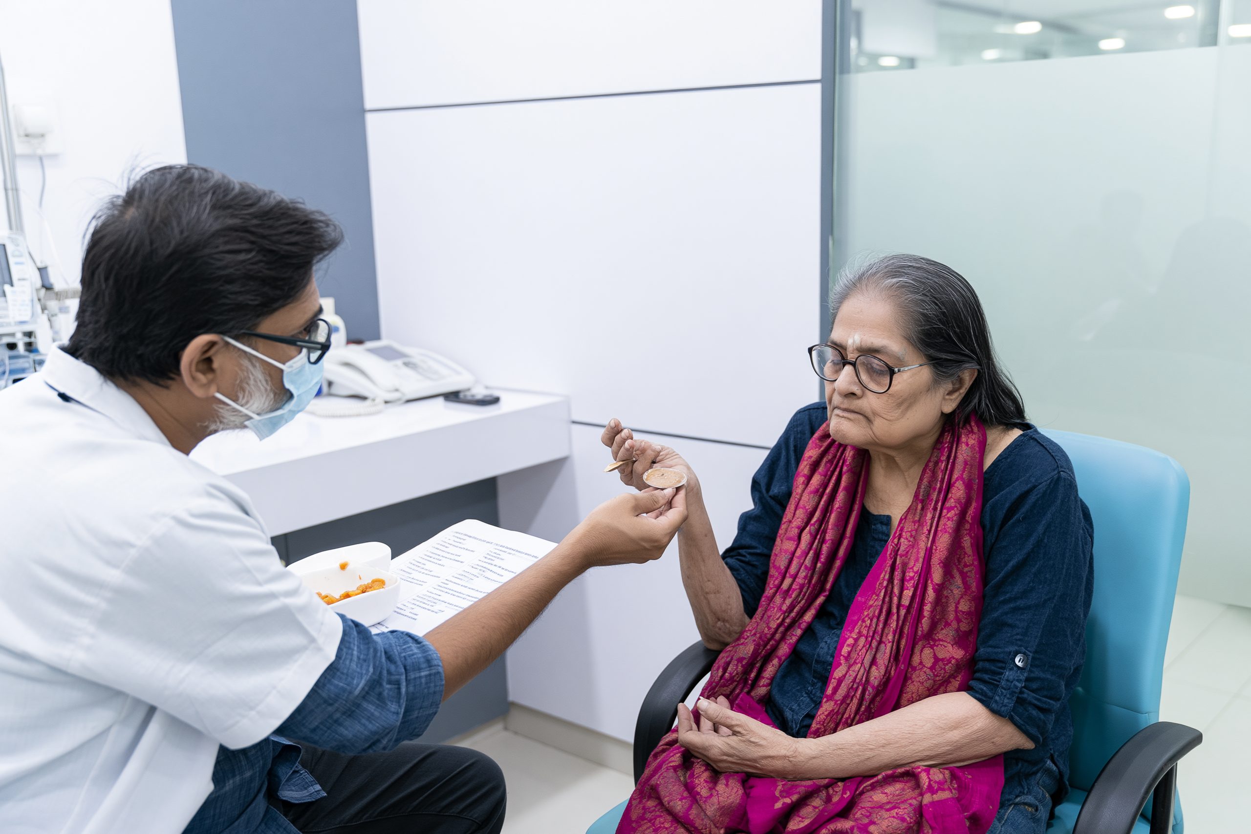
Rasayana therapy in the Ayurveda Curative Model for metastatic esophageal cancer is intended to support nourishment, strength, digestive tolerance, recovery, and overall functional reserve. It is not limited to giving a general tonic. The selection of Rasayana depends on swallowing ability, Agni, nutritional depletion, bowel function, treatment-related symptoms, liver and kidney function, and the medicines already being used [19].
In advanced esophageal cancer, Rasayana should be introduced only after urgent problems such as severe obstruction, aspiration, dehydration, uncontrolled vomiting, bleeding, infection, or profound malnutrition have been addressed. A formulation cannot provide meaningful support when the patient is unable to swallow or retain it safely.
Role of Rasayana in Advanced Cancer Care
Charaka describes Rasayana as a means of attaining healthy longevity, memory, intellectual clarity, freedom from disease, youthful function, bodily strength, sensory capacity, complexion, voice, and vitality.
Sanskrit
दीर्घमायुः स्मृतिं मेधामारोग्यं तरुणं वयः ।
प्रभावर्णस्वरौदार्यं देहेन्द्रियबलं परम् ॥७॥
वाक्सिद्धिं प्रणतिं कान्तिं लभते ना रसायनात् ।
लाभोपायो हि शस्तानां रसादीनां रसायनम् ॥८॥
Transliteration
Dīrgham āyuḥ smṛtiṃ medhām ārogyaṃ taruṇaṃ vayaḥ।
Prabhā-varṇa-svaraudāryaṃ dehendriya-balaṃ param॥
Vāk-siddhiṃ praṇatiṃ kāntiṃ labhate nā rasāyanāt।
Lābhopāyo hi śastānāṃ rasādīnāṃ rasāyanam॥
Simple English translation
Through Rasayana, a person seeks healthy longevity, memory, intelligence, freedom from illness, youthful function, radiance, healthy complexion and voice, and optimum strength of the body and senses. Rasayana is the means of promoting excellence in Rasa and the tissues nourished from it.
Classical source: Charaka Samhita, Chikitsa Sthana, Chapter 1, Rasayana Adhyaya, Abhaya-Amalakiya Pada, verses 7–8 [19].
This description shows that Rasayana is concerned with the quality of nourishment and the functional condition of the whole person. The verse does not describe esophageal cancer or claim that Rasayana alone eliminates a malignant tumor. Within advanced cancer care, its principles are applied to preserve strength, support recovery, improve food tolerance, and reduce progressive depletion.
Rasayana selection should follow assessment of Agni and the condition of Annavaha Srotas. A patient with painful swallowing, severe reflux, mucositis, or repeated regurgitation may not tolerate pungent powders, large tablets, concentrated decoctions, or irritating formulations. A softer preparation may be considered only when semisolid intake is clinically safe.
The immediate purpose may differ between patients. One person may require support for appetite and digestion, while another needs help maintaining strength during chemotherapy. A patient with profound tissue depletion may require a nourishing approach, whereas someone with nausea, diarrhea, impaired liver function, or active mucositis may first need a simpler and lighter plan.
Supporting Bala and Nutritional Recovery
Bala refers to the patient’s physical strength, endurance, stability, and ability to perform normal activities. In metastatic esophageal cancer, declining Bala may appear as difficulty walking, reduced grip strength, prolonged bed rest, dependence on family members, poor recovery after treatment, and inability to complete planned therapy.
Rasayana therapy should support, rather than replace, the measures required to correct this decline. Adequate calories, protein, fluids, electrolyte correction, pain control, treatment of nausea, relief of esophageal obstruction, and safe nutritional access remain fundamental. Esophageal stenting or tube feeding may be necessary when the patient cannot obtain sufficient nourishment by mouth.
Ayurvedic assessment can help determine the texture, quantity, timing, and digestive suitability of food. Small portions of smooth, moist, nutrient-dense food may be better tolerated than large meals. The formulation should not occupy so much stomach capacity that it reduces the intake of essential food, fluids, prescribed medicines, or oral nutritional supplements.
Nutritional recovery should be assessed through more than body weight. Improvement may be reflected in greater walking capacity, increased time out of bed, better meal tolerance, reduced treatment interruption, improved hydration, and preservation of muscle strength. Fluid retention can increase body weight while muscle loss continues, so weight gain alone is not sufficient evidence of recovery.
Rasayana may be continued only when it is being swallowed, digested, and tolerated without worsening reflux, pain, vomiting, diarrhea, or aspiration. Any increase in appetite, energy, or Bala is clinically valuable, but it should be recorded as functional improvement rather than described as tumor regression.
Rasayana During Chemotherapy and Immunotherapy
Rasayana during chemotherapy or immunotherapy must be individualized according to the exact anticancer medicines, treatment schedule, blood counts, organ function, and current adverse effects. A formulation that appears appropriate before treatment may become unsuitable when the patient develops mucositis, diarrhea, neutropenia, thrombocytopenia, liver inflammation, kidney impairment, or severe vomiting.
Every ingredient should be reviewed for possible interaction with chemotherapy, immunotherapy, anticoagulants, pain medicines, antiemetics, antibiotics, and other prescribed drugs. Herbal ingredients can affect liver enzymes, drug transporters, platelet function, blood glucose, blood pressure, sedation, and the absorption or elimination of medicines [17].
The phrase “immune boosting” should be used cautiously during immunotherapy. Immune checkpoint inhibitors work by modifying specific immune-control pathways, and excessive or poorly understood immune stimulation is not automatically beneficial. New diarrhea, jaundice, breathlessness, rash, severe fatigue, endocrine disturbance, or reduced urine output may represent immune-related toxicity and requires oncology assessment rather than interpretation as purification or a healing reaction [16,17].
The safest approach is to introduce only clinically justified formulations, document every ingredient and dose, and avoid making several changes at the same time. This makes it easier to identify the cause if an adverse effect or laboratory abnormality develops.
Rasayana may support appetite, food tolerance, strength, sleep, bowel regularity, and recovery between treatment cycles. These outcomes should be monitored alongside complete blood counts, liver and kidney tests, weight, performance status, hospital admissions, treatment delays, and the patient’s ability to carry out daily activities.
Tumor response must remain a separate outcome. It should be determined through comparative imaging and standardized oncology criteria rather than inferred from increased energy, improved swallowing, reduced pain, or better appetite [13,16]. This distinction allows Rasayana therapy to be integrated responsibly while preserving an accurate assessment of metastatic esophageal cancer.
Kushmanda Amalaki Bala Rasayana Avaleha (Medicine)

Kushmanda Amalaki Bala Rasayana Avaleha for metastatic esophageal cancer is a clinician-designed semisolid formulation inspired by classical Kushmanda Avaleha and Rasayana principles. The name reflects three priorities: Kushmanda as the nourishing avaleha base, Amalaki as a classical Rasayana ingredient, and preservation of Bala, meaning the patient’s functional strength and capacity to tolerate illness and treatment [19,21].
This formulation is not reproduced word for word from a single classical text. Its pharmaceutical foundation comes from the Kushmanda Avaleha tradition described in the Sharangadhara Samhita, while its broader clinical purpose is guided by the Rasayana principles explained in the Charaka Samhita [19,21]. The customized additions must therefore be described as inspired by these texts rather than presented as an unchanged classical prescription.
Within the Ayurveda Curative Model, the avaleha is intended to support food tolerance, nutrition, digestive comfort, strength, and recovery. It should be used only when the patient can swallow semisolid food safely. It cannot mechanically reopen a severely obstructed esophagus, seal a tracheoesophageal fistula, or replace chemotherapy, immunotherapy, targeted therapy, radiation, stenting, or feeding support.
Why Kushmanda Avaleha Was Selected as the Base
Kushmanda, commonly identified as ash gourd or winter melon and botanically known as Benincasa hispida, has a long history of dietary and medicinal use. Its high moisture content and soft pulp make it suitable for preparation as a smooth avaleha base. Modern reviews describe its nutritional constituents and experimental antioxidant, gastroprotective, and anti-inflammatory properties, although these findings do not establish clinical anticancer effectiveness in metastatic esophageal cancer [30].
A Kushmanda-based avaleha may be more practical than large tablets or coarse powders for a patient who can safely manage smooth semisolid food. The concentrated form also allows several finely powdered ingredients to be incorporated into a relatively small volume. This may be useful when appetite is poor and the patient cannot tolerate bulky preparations.
The selection of Kushmanda does not mean that it can reverse a physical esophageal obstruction. A patient who cannot swallow liquids or saliva, repeatedly aspirates, or regurgitates nearly everything requires urgent medical assessment. In such cases, stenting, radiation, intravenous hydration, or feeding access may be needed before any oral avaleha is considered.
Kushmanda was also selected because the classical formulation tradition combines a nourishing food-like base with digestive and aromatic ingredients. This creates a more balanced pharmaceutical approach than simply mixing multiple concentrated herbal powders. However, sweetness, fat content, viscosity, and serving size must be adjusted carefully in patients with diabetes, reflux, nausea, impaired digestion, or severe cachexia.
Classical Inspiration for the Formulation
Kushmanda Amalaki Bala Rasayana Avaleha is inspired primarily by the Charaka Samhita and the Sharangadhara Samhita. The Charaka Samhita provides the Rasayana framework of nourishment, strength, tissue quality, and functional preservation. The Sharangadhara Samhita provides the pharmaceutical principles of Avaleha Kalpana and a classical Kushmanda Avaleha tradition [19,21].
The classical foundation does not mean that every ingredient in the customized formulation appears together in the same verse. Amalaki and other selected herbs are added according to the intended clinical objectives, contemporary ingredient research, digestive tolerance, and the patient’s individual condition.
This distinction is important for transparency. A classical formula should be named as classical only when its ingredients, proportions, and preparation follow the cited text. When additional herbs are introduced or quantities are substantially modified, the finished preparation becomes an adapted or clinician-designed formulation inspired by classical principles.
The formulation should therefore be presented as a structured adaptation rather than as a newly discovered ancient cancer medicine. Its value must be assessed through tolerability, nutritional intake, swallowing safety, performance status, laboratory monitoring, and objective oncology outcomes.
Charaka Samhita Rasayana Principles
The Charaka Samhita explains Rasayana as a method for promoting healthy longevity, nourishment, functional strength, sensory capacity, vitality, and excellence of the tissues.
Sanskrit
दीर्घमायुः स्मृतिं मेधामारोग्यं तरुणं वयः ।
प्रभावर्णस्वरौदार्यं देहेन्द्रियबलं परम् ॥७॥
वाक्सिद्धिं प्रणतिं कान्तिं लभते ना रसायनात् ।
लाभोपायो हि शस्तानां रसादीनां रसायनम् ॥८॥
Transliteration
Dīrgham āyuḥ smṛtiṃ medhām ārogyaṃ taruṇaṃ vayaḥ।
Prabhāvarṇasvaraudāryaṃ dehendriyabalaṃ param॥
Vāksiddhiṃ praṇatiṃ kāntiṃ labhate nā rasāyanāt।
Lābhopāyo hi śastānāṃ rasādīnāṃ rasāyanam॥
Simple English translation
Rasayana is described as a means of promoting healthy longevity, memory, intelligence, freedom from illness, youthful function, radiance, healthy complexion and voice, and optimum strength of the body and senses. It supports excellence in Rasa and the tissues nourished from it.
Classical source: Charaka Samhita, Chikitsa Sthana, Chapter 1, Rasayana Adhyaya, Abhaya-Amalakiya Pada, verses 7–8 [19].
These verses establish the classical purpose of Rasayana, but they do not describe metastatic esophageal cancer or state that an avaleha can eradicate distant metastases. Their relevance lies in guiding care toward nourishment, Bala, tissue support, and preservation of function during a prolonged and debilitating illness.
In a patient with metastatic esophageal cancer, these principles may be applied by supporting safe nutrition, digestion, sleep, bowel function, mobility, and recovery between treatments. Rasayana therapy becomes clinically meaningful when it helps the patient maintain food intake, strength, independence, and treatment tolerance without increasing toxicity or interfering with oncology medicines.
Sharangadhara Samhita Avaleha Kalpana
Avaleha Kalpana is the Ayurvedic pharmaceutical method used to prepare semisolid herbal formulations. The process generally involves preparing a decoction, juice, or pulp, concentrating it with a sweetening medium, and incorporating finely powdered ingredients at the appropriate stage. Ghee, oil, or honey may be added according to the classical formula and required processing method [21].
The Sharangadhara Samhita, Madhyama Khanda, Chapter 8, explains Avaleha Kalpana and includes the classical Kushmanda Avaleha in verses 22–28 [21]. This textual location provides the principal pharmaceutical inspiration for the present formulation.
Correct preparation depends on controlling heat, concentration, consistency, ingredient sequence, and moisture. Excessive heating can damage aromatic ingredients and honey, while inadequate cooking can leave excessive water and reduce stability. Finely powdered ingredients should be distributed evenly so that each prescribed dose contains a reasonably consistent composition.
A modern pharmaceutico-analytical evaluation of classical Kushmanda Avaleha examined manufacturing stages, organoleptic characteristics, and physicochemical parameters. This supports the need for standardized preparation and quality control but does not demonstrate efficacy against metastatic esophageal cancer [24].
The finished product should therefore be prepared under hygienic conditions with authenticated raw materials, documented batch quantities, controlled heating, and appropriate storage. Its texture should remain smooth and uniform, without coarse particles that could make swallowing more difficult.
Kushmanda Avaleha Classical Tradition
The classical Kushmanda Avaleha tradition uses Kushmanda as the principal base and transforms it into a concentrated, palatable semisolid preparation through Avaleha Kalpana [21]. The traditional formula belongs to the wider Ayurvedic approach of combining nourishment with selected digestive and aromatic ingredients rather than relying on a single isolated compound.
The proposed Kushmanda Amalaki Bala Rasayana Avaleha retains this classical pharmaceutical logic but expands the formulation for the clinical priorities of advanced illness. Amalaki is included within the Rasayana framework, while other ingredients are selected for digestive support, nutritional decline, treatment-related discomfort, and the biological pathways explored in preliminary research.
Evidence must remain ingredient-specific and proportionate. Research involving curcumin, berberine, piperlongumine, or other isolated compounds cannot be presented as proof that the corresponding whole herb or finished avaleha will produce the same concentration or anticancer effect in a patient. A compound used in a laboratory may have a different concentration, absorption, metabolism, and biological effect from the whole herb used in clinical Ayurveda.
The formulation should therefore be evaluated as a complete clinical preparation rather than as the sum of laboratory claims attached to individual ingredients. Improvements in appetite, digestion, weight stability, strength, or treatment tolerance are valuable supportive outcomes, but tumor response must be determined separately through comparative imaging and standardized oncology assessment.
Complete 30-Day Kushmanda Amalaki Bala Rasayana Avaleha Formula for Metastatic Esophageal Cancer
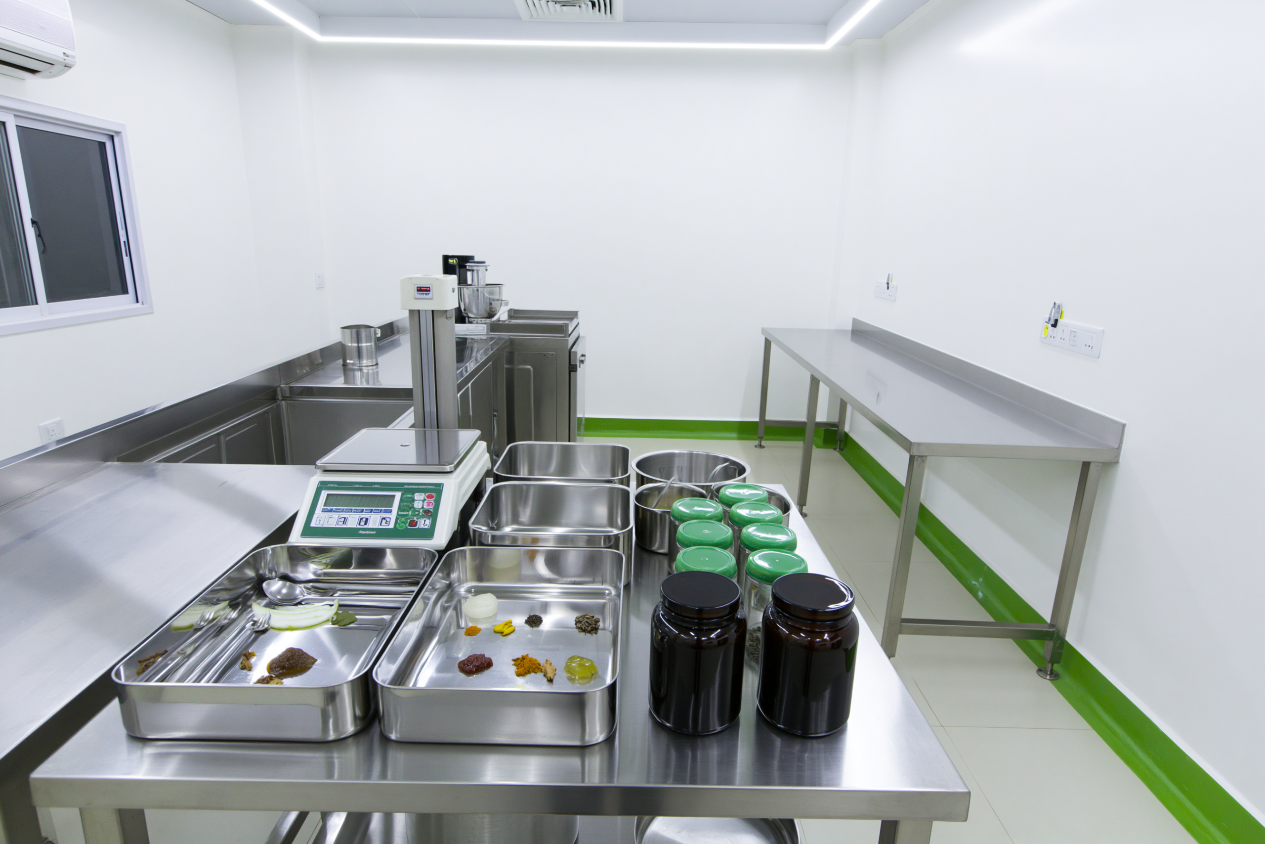
No clinical trial has established a single “best” or “strongest” avaleha for metastatic esophageal cancer. The formulation below is therefore presented as an evidence-informed, clinician-designed supportive preparation within the Ayurveda Curative Model. It follows the pharmaceutical framework of Kushmanda Avaleha described in the Sharangadhara Samhita, Madhyama Khanda, Chapter 8, verses 22–28, while adding selected herbs according to swallowing tolerance, nutritional decline, treatment-related symptoms, and preliminary research [21,24].
The formula produces exactly 900 grams, providing 60 doses of 15 grams. It is not a verbatim classical formula and has not been clinically tested as a complete formulation in metastatic esophageal cancer. It should be compounded by a licensed Ayurvedic pharmacy and used only after confirming that the patient can swallow a smooth semisolid safely.
Why This Avaleha Uses a Focused Formula
A clinically strong formulation is not necessarily the one containing the greatest number of herbs. Excessive ingredients can make the avaleha bitter, irritating, difficult to swallow, harder to standardize, and more likely to interact with chemotherapy, immunotherapy, anticoagulants, or other medicines.
This formula retains the nourishing Kushmanda and Amalaki base while using limited quantities of Kalmegha, Haridra, Daruharidra, Pippali, Shunthi, Yashtimadhu, and Shatavari. The classical aromatic ingredients are retained in small amounts to improve palatability and digestion. The word Bala in the formulation name refers to the objective of preserving strength and functional reserve; it does not mean that Sida cordifolia has been added.
Exact 900-Gram Kushmanda Amalaki Bala Rasayana Avaleha Formula
The quantities below refer to the ingredients present in the finished 900-gram batch. Because the water content of fresh fruit varies, Kushmanda and Amalaki are standardized by the weight of their finished reduced concentrates rather than by an unreliable fixed weight of raw fruit.
| Ingredient | Botanical or pharmaceutical identity | Part used | Exact quantity |
| Kushmanda pulp concentrate | Benincasa hispida | Reduced fruit pulp | 230 g |
| Amalaki pulp concentrate | Phyllanthus emblica | Reduced fruit pulp | 50 g |
| Sharkara | Purified sugar candy | Avaleha base | 350 g |
| Go-ghrita | Purified cow ghee | Lipid base | 60 g |
| Madhu | Tested natural honey | Avaleha base | 90 g |
| Kalmegha | Andrographis paniculata | Fine aerial-part powder | 22 g |
| Haridra | Curcuma longa | Fine rhizome powder | 16 g |
| Daruharidra | Berberis aristata | Fine stem powder | 10 g |
| Yashtimadhu | Glycyrrhiza glabra | Fine root powder | 10 g |
| Shatavari | Asparagus racemosus | Fine root powder | 16 g |
| Amalaki Churna | Phyllanthus emblica | Fine fruit powder | 16 g |
| Shunthi | Zingiber officinale | Fine dried rhizome powder | 8 g |
| Pippali | Piper longum | Fine fruit powder | 8 g |
| Maricha | Piper nigrum | Fine fruit powder | 3 g |
| Dhanyaka | Coriandrum sativum | Fine fruit powder | 3 g |
| Jeeraka | Cuminum cyminum | Fine fruit powder | 3 g |
| Ela | Elettaria cardamomum | Fine seed powder | 2 g |
| Tvak | Cinnamomum verum | Fine bark powder | 2 g |
| Tejapatra | Cinnamomum tamala | Fine leaf powder | 1 g |
| Final batch weight | 900 g |
The classical Kushmanda Avaleha described in Sharangadhara Samhita contains Kushmanda, Sharkara, Ghrita, Madhu, Pippali, Shunthi, Maricha, Dhanyaka, Jeeraka, Ela, Tvak, and Tejapatra. A modern pharmaceutical evaluation prepared the formula through controlled pulp processing, sugar-syrup formation, addition of finely sieved powders, and incorporation of honey after cooling. The present formula retains that pharmaceutical structure but adds Amalaki, Kalmegha, Haridra, Daruharidra, Yashtimadhu, and Shatavari as a deliberate clinical adaptation [21,24]. (Asian Journal of Research in Chemistry)
Why Bhasma and Rasaushadhi Are Not Added to the Fixed Batch
Heeraka Bhasma, Suvarna Bhasma, Rajata Bhasma, Abhraka Bhasma, Tamra Bhasma, Lauha Bhasma, Mukta preparations, Rasasindura, and other mineral or metallic medicines should not be incorporated into this fixed 900-gram avaleha.
There is no clinically validated universal mineral combination or dose for metastatic esophageal cancer. Premixing several mineral medicines also prevents individual withdrawal or dose adjustment if liver, kidney, neurological, hematological, or gastrointestinal toxicity develops. Some inadequately controlled Ayurvedic products have been found to contain potentially toxic quantities of lead, mercury, or arsenic, making batch-specific analytical testing essential. (NCCIH)
When a qualified Rasashastra clinician considers a mineral medicine appropriate, it should be prescribed and dispensed separately, with its own name, manufacturer, batch number, daily dose, duration, and certificate of analysis. The certificate should include validated testing for lead, mercury, arsenic, cadmium, microbial contamination, pesticides, and relevant pharmaceutical parameters. CBC, liver function, kidney function, electrolytes, and treatment-related symptoms should be monitored so that the individual medicine can be stopped immediately if necessary.
Adding multiple bhasmas does not make this avaleha scientifically stronger. For a patient with metastatic esophageal cancer, safety, reproducibility, swallowing tolerance, interaction control, and objective monitoring are more important than the number of ingredients.
Patient-Friendly Preparation Method
This easy-to-understand method is intended for preparation in a qualified Ayurvedic pharmacy rather than an ordinary home kitchen.
First, fresh Kushmanda should be washed, peeled, deseeded, steamed until soft, and passed through a fine sieve. The smooth pulp should be reduced slowly until exactly 230 grams of Kushmanda concentrate remains. Amalaki should be deseeded, softened, finely pulped, sieved, and reduced separately until exactly 50 grams of Amalaki concentrate remains. The concentrates should not contain coarse fibers, seeds, skin, or hard particles that could aggravate dysphagia.
Next, every dry herb should be authenticated, dried appropriately, powdered separately, and passed through an 80-mesh sieve. The powders should then be blended until the 120-gram Prakshepa mixture is completely uniform. Coarse powder should not be used because it can make the avaleha gritty and more difficult to swallow.
The 350 grams of Sharkara should be dissolved in the smallest practical quantity of the strained fruit liquid. It should be heated at a controlled temperature until a one-thread syrup is obtained. The modern pharmaceutical study of Kushmanda Avaleha used controlled heating of approximately 85°C to 95°C during syrup preparation [24]. (Asian Journal of Research in Chemistry)
The 60 grams of Go-ghrita should be warmed in a separate stainless-steel vessel. Kushmanda and Amalaki concentrates should be added and cooked gently until they form a smooth, homogeneous mass without visible free water. The sugar syrup should then be incorporated gradually while the mixture is stirred continuously.
Heating should continue until the combined fruit, sugar, and ghee base reaches 690 grams. The mixture should then be allowed to cool to approximately 60°C to 68°C, after which the complete 120-gram herbal powder blend should be added slowly with continuous mixing. The weight at this stage should be 810 grams.
When the preparation has cooled to 35°C to 40°C, the 90 grams of honey should be added and mixed thoroughly. Honey should not be added during active boiling or reheated after incorporation. The finished batch should weigh exactly 900 grams. The pharmaceutical evaluation of classical Kushmanda Avaleha similarly incorporated the herbal powders after partial cooling and added honey at approximately 35°C [24]. (Asian Journal of Research in Chemistry)
The finished avaleha should be divided into two sterile, dry, airtight jars of 450 grams each. Each jar should carry the preparation date, batch number, ingredients, prescribed dose, storage instructions, and the patient’s name. Unless the product has undergone validated stability and microbial testing, it should be refrigerated and used only for the intended 30-day period. A dry spoon must be used each time. Fermentation, gas formation, mold, watery separation, or an altered odor requires disposal of the batch.
Recommended Dosage for 30 Days
The intended dose is 15 grams twice daily, preferably after breakfast and after the evening meal, providing a total of 30 grams daily.
The calculation is:
15 grams × 2 doses × 30 days = 900 grams
Each dose should be measured by weight with a calibrated 15-gram scoop or digital scale. Household tablespoons are not reliable because the weight changes with the avaleha’s density and moisture content.
The patient should remain seated upright and take the dose slowly in two or three small portions. A small quantity of lukewarm water may be used only when thin liquids have already been confirmed as safe to swallow. The avaleha should not be forced when coughing, choking, regurgitation, food impaction, or difficulty swallowing saliva is present.
This preparation should not be administered through a feeding tube because its viscosity, fibers, ghee, and suspended powders may block the tube, and tube compatibility has not been established. Separating the avaleha from conventional medicines by one or two hours also does not eliminate metabolic herb–drug interactions. The complete formula must still be reviewed by the treating oncologist or oncology pharmacist [17].
What Research Shows About Each Ingredient
| Ingredient | Research relevant to metastatic esophageal cancer | Evidence interpretation |
| Kalmegha | A prospective, single-arm phase II study enrolled patients with metastatic or locally advanced esophageal squamous cell carcinoma. Thirty patients received treatment and ten completed four months of standardized Andrographis paniculata granules. Those completing treatment had better reported survival and quality-of-life outcomes than those who did not complete it [25]. | This is preliminary human evidence, not proof of cure. There was no randomized untreated control group, and comparing completers with non-completers creates substantial selection bias. The study used standardized concentrated granules at 130 mg/kg daily, prohibited concurrent chemotherapy and other herbs during the initial treatment period, and does not validate the much smaller whole-herb quantity used in this polyherbal avaleha. (Wiley Online Library) |
| Haridra | Curcumin inhibited STAT3 signaling, reduced growth, and induced apoptosis in esophageal squamous cancer cells and primary tumor-derived mouse xenografts [26]. | This was cell and animal research. It supports investigation of curcumin-related mechanisms but does not prove that Haridra powder in an avaleha shrinks metastatic cancer in patients. (Springer) |
| Pippali | Piperlongumine produced growth-inhibitory effects in esophageal squamous cell carcinoma cell and animal models, including activation of a reactive-oxygen-related pyroptosis pathway [27]. | Piperlongumine is an isolated chemical constituent. Its laboratory concentration and biological effect cannot be treated as equivalent to Pippali fruit powder taken orally. (ScienceDirect) |
| Daruharidra | Berberine inhibited growth of esophageal squamous and adenocarcinoma cell lines. In the more responsive squamous cell line, it produced G2/M cell-cycle arrest, increased apoptosis, and affected Akt, mTOR, and AMPK signaling [28]. | The experiment was performed in cultured cells. It does not establish clinical effectiveness, oral bioavailability, or a therapeutic dose of Daruharidra in metastatic esophageal cancer. (WJGNet) |
| Yashtimadhu | Human studies in head-and-neck cancer have examined Glycyrrhiza glabra preparations for reducing radiation-associated oral mucositis [29]. | This supports a possible supportive-care role for mucosal discomfort, not a tumor-directed effect in esophageal cancer. Licorice can increase blood pressure and lower potassium, so it requires particular caution in patients with hypertension, edema, heart disease, kidney impairment, or hypokalemia. (PubMed) |
| Kushmanda | Reviews describe the nutritional composition and experimental antioxidant, anti-inflammatory, and gastroprotective properties of Benincasa hispida [30]. | Kushmanda provides the classical food-like avaleha base. No controlled clinical trial has shown that Kushmanda treats metastatic esophageal cancer. (PubMed Central (PMC)) |
| Amalaki | Reviews of Phyllanthus emblica describe experimental effects on oxidative stress, proliferation, apoptosis, invasion, and other cancer-related pathways [31]. | The evidence is primarily laboratory and animal based. There is no clinical evidence that Amalaki alone or in this avaleha produces tumor regression in metastatic esophageal cancer. (PubMed Central (PMC)) |
| Shatavari | Pharmacological reviews describe antioxidant, gastrointestinal, anti-inflammatory, and adaptogenic properties of Asparagus racemosus [32]. | Shatavari is included for the formulation’s nourishing and supportive rationale. Direct clinical evidence in esophageal cancer is absent. (PubMed Central (PMC)) |
| Shunthi | A systematic review and meta-analysis of 18 articles found that ginger may reduce acute chemotherapy-related vomiting in some settings, although it did not consistently reduce overall nausea or delayed vomiting [33]. | Shunthi may have a supportive antiemetic role but should not replace prescribed anti-nausea medicines. It has no established tumor-shrinking effect in esophageal cancer. (PubMed) |
| Maricha | Piperine can inhibit intestinal P-glycoprotein and hepatic or intestinal CYP3A4, potentially changing the blood concentration of medicines using these pathways [34]. | Maricha is retained only in a small classical quantity. “Bioavailability enhancement” may increase toxicity as well as exposure, so it should not be promoted as automatically beneficial during chemotherapy or targeted treatment. (PubMed) |
| Dhanyaka, Jeeraka, Ela, Tvak and Tejapatra | These aromatic ingredients belong to the classical Kushmanda Avaleha pharmaceutical tradition [21,24]. | They support taste, aroma, digestive tolerance, and formula acceptability. Direct evidence that they treat metastatic esophageal cancer is not available. (Asian Journal of Research in Chemistry) |
| Sharkara, Ghrita and Madhu | These ingredients create the semisolid avaleha matrix, improve palatability, distribute the powdered ingredients, and contribute to pharmaceutical stability [21,24]. | They are dosage-form components, not proven anticancer agents. Sharkara and Madhu make the formulation unsuitable for some patients with uncontrolled diabetes or severe glucose intolerance. (Asian Journal of Research in Chemistry) |
Essential Safety Screening Before Dispensing
The avaleha should not be given when there is complete esophageal obstruction, inability to swallow liquids or saliva, recurrent aspiration, suspected tracheoesophageal fistula, severe mucositis, repeated vomiting, active gastrointestinal bleeding, or dependence on tube feeding.
It also requires reconsideration in uncontrolled diabetes, significant liver or kidney dysfunction, severe thrombocytopenia, anticoagulant use, uncontrolled hypertension, edema, heart failure, or low serum potassium. Yashtimadhu can affect blood pressure and potassium, while Pippali and Maricha may alter drug metabolism. Haridra and Shunthi require additional review when bleeding risk is elevated.
The treating team should know the complete formulation before chemotherapy, immunotherapy, radiation, or targeted therapy begins. Cancer-related or treatment-related liver inflammation, diarrhea, rash, breathlessness, endocrine disturbance, falling blood counts, or kidney dysfunction should not be interpreted as detoxification or a healing reaction. The preparation should be stopped and medically reviewed when a clinically significant adverse effect occurs [16–18].
Improved appetite, swallowing comfort, bowel function, energy, or body weight can be recorded as supportive outcomes. Reduction of the primary tumor or metastatic lesions must be established separately through comparative imaging and standardized oncology response assessment.
Published Integrative Ayurveda Case Study in Esophageal Squamous Cell Carcinoma
A published case report in the International Journal of Ayurvedic Medicine described an integrated oncology approach in a 40-year-old woman with biopsy-confirmed squamous cell carcinoma of the mid-thoracic esophagus. The case was managed through initial conventional cancer treatment followed by a supervised Ayurvedic maintenance protocol using Catharanthus roseus flower Sheeta Kashaya, Yashtimadhu, dietary modification, and serial radiological monitoring. (iJAM)
Patient Condition at Diagnosis
The patient was diagnosed on 28 August 2024 after upper gastrointestinal endoscopy identified an esophageal growth. The baseline PET-CT performed on 29 August showed intensely FDG-avid circumferential soft-tissue thickening in the mid-thoracic esophagus. The lesion produced substantial narrowing of the esophageal lumen and dilation of the esophagus above the obstructed area. (iJAM)
Additional imaging described the wall thickening above and below the carina, indentation of the left main bronchus, and loss of the normal fat plane between the tumor and the aorta. The reported angle of contact with the aorta was approximately 55 degrees, indicating locally advanced anatomical involvement. (iJAM)
The patient presented with persistent difficulty swallowing, reduced appetite, weight loss, constipation, chemotherapy-associated hair loss, and dependence on a food pipe for nutritional support. Clinical examination described pallor, mild emaciation, and slight swelling near the esophageal notch, without prominent cervical lymphadenopathy. Her baseline body weight was 51 kilograms. (iJAM)
The Ayurvedic assessment described Pitta-predominant Vata Prakriti, reduced digestive capacity or Avara Agnibala, and relatively preserved Shareera Bala. Her recorded blood pressure was 100/70 mm Hg, oxygen saturation was 96 percent, and respiratory rate was 19 breaths per minute. The condition was interpreted through the Ayurvedic concept of Arbuda. (iJAM)
Cancer Treatment Received Before Ayurveda
The patient received five weekly cycles of paclitaxel and carboplatin during September 2024. Surgery was advised on 24 September, but the patient was not prepared to undergo the operation and had arranged another specialist consultation for February 2025. The paper also refers to radiotherapy and describes uptake on a later PET-CT as probably post-radiotherapy reactive change, although the exact radiation schedule was not reported. (iJAM)
She attended the Ayurvedic hospital in Hyderabad on 2 October 2024, after completing the initial chemotherapy schedule, and began the integrated maintenance protocol. (iJAM)
Ayurvedic Treatment Protocol
The principal Ayurvedic intervention was a Sheeta Kashaya prepared from fresh flowers of Catharanthus roseus, commonly known as periwinkle. According to the published case, three fresh flowers were placed in approximately 20 millilitres of drinking water and kept overnight. The flowers were removed the following morning, and the prepared liquid was administered on an empty stomach before sunrise. The flower preparation was continued regularly for approximately eight months. (iJAM)
Yashtimadhu, botanically identified as Glycyrrhiza glabra, was prescribed as a 500-milligram tablet twice daily before food with water. The authors used Yashtimadhu as an accompanying supportive medicine during the treatment and follow-up period. (iJAM)
The dietary plan included daily pomegranate juice with black pepper and one cucumber. The patient was encouraged to consume leafy vegetables, Amalaki powder, black pepper, and buttermilk. Refined sugar, refined flour or maida, and non-vegetarian foods were excluded from the reported dietary protocol. (iJAM)
Early Clinical Response
At the follow-up on 5 November 2024, the PET-CT showed mildly diffuse FDG-avid thickening of the mid-thoracic esophageal wall. The report interpreted this as likely post-radiotherapy reactive uptake. Clinically, the patient had gained approximately one kilogram, her appetite had improved, and her general condition was described as slightly better. The food pipe was subsequently removed. (iJAM)
The November CT comparison also showed a mild reduction in circumferential esophageal wall thickening. The residual thickening measured approximately 7 millimetres, abutted the left main bronchus, and maintained clearer fat planes with the surrounding structures. (iJAM)
Three-Month Assessment
A contrast-enhanced CT performed before the 8 January 2025 review showed mild irregular circumferential thickening of the mid-thoracic esophagus. The findings were considered static compared with the November PET-CT. A few fibrous strands were also described in the lower lobe of the left lung. The case report states that no additional conventional treatment was being administered during this interval. (iJAM)
The reported hematological monitoring showed a hemoglobin level of 12.2 g/dL and a white blood cell count of 5,590 cells/mm³ in January 2025. In February, the hemoglobin was 11 g/dL and the white blood cell count was 5,603 cells/mm³. The authors reported normalization after the earlier myelosuppressive phase and did not identify a major hematological adverse effect during maintenance. (iJAM)
Barium Swallow and February CT Findings
A barium swallow performed on 1 February 2025 showed mild, short-segment luminal narrowing with mucosal irregularity in the middle third of the esophagus. Importantly, contrast passed freely through the distal esophagus and into the stomach. Surgery was not advised at that review, and the patient was given general vitamin support. (iJAM)
A CT thorax reviewed at Christian Medical College, Vellore, on 20 February 2025 was compared with the August and November scans. The report described mild interval reduction in the circumferential esophageal wall thickening, with the residual lesion remaining stable and the surrounding fat planes maintained. (iJAM)
May PET-CT and Gastroscopy Results
A repeat whole-body PET-CT performed on 26 May 2025 showed a further mild interval decrease in the circumferential thickening of the mid-thoracic esophagus. A few subcentimetric bilateral deep cervical and retroperitoneal lymph nodes were noted, together with a non-obstructive left renal calculus. (iJAM)
Gastroscopy performed on 27 May reported normal esophageal mucosa, with no obvious ulcer or growth. The Z-line and gastroesophageal junction were noted at 36 centimetres, and the report concluded that no visible esophageal abnormality was detected during the procedure. (iJAM)
By the May follow-up, the patient’s body weight had increased from 51 to 57 kilograms. The case report also documented improved appetite, hair regrowth, improved general health, and better functional condition. The authors reported no clinically significant adverse effects from the Ayurvedic maintenance regimen during this period. (iJAM)
Long-Term Follow-Up
At the August 2025 review, the patient continued to report overall improvement, and no adverse effect was documented. The same treatment was continued. (iJAM)
A CT thorax performed at Christian Medical College on 8 December 2025 reportedly showed no residual esophageal thickening and no new lung lesion. The authors interpreted this as absence of radiological relapse during the reported follow-up period. Catharanthus roseus Sheeta Kashaya was then discontinued, while Yashtimadhu 500 milligrams twice daily was continued with water. (iJAM)
At the follow-up visit on 2 March 2026, the patient reported no clinical complaints. The complete observation period extended for approximately 18 months from diagnosis and included repeated PET-CT, CT thorax, barium swallow, gastroscopy, clinical assessment, body-weight monitoring, and hematological testing. (iJAM)
Reported Outcome
The authors described the outcome as disease stabilization followed by major radiological and endoscopic improvement, preservation of quality of life, weight gain, recovery of appetite, hair regrowth, free passage of contrast through the esophagus, normal gastroscopy, and absence of recurrent esophageal thickening on the December 2025 CT. They reported that high-risk surgery was avoided during the documented follow-up. (iJAM)
The report attributed the integrated outcome to the combined treatment course, which included paclitaxel and carboplatin chemotherapy, evidence of radiotherapy exposure, the Catharanthus roseus flower preparation, Yashtimadhu, dietary support, and close radiological monitoring. The paper did not document a baseline TNM stage or confirmed distant stage IVB metastasis. The authors identified the single-patient design as the principal limitation and called for controlled studies with larger patient groups.
Published Reference
Mutnuri, V. L., and Murali Krishna, C. (2026). Case report of esophagus squamous cell carcinoma treated with novel drug preparation method of Catharanthus roseus (Periwinkle): An integrated oncological approach. International Journal of Ayurvedic Medicine, 17(1), 311–314. Read the complete published case report. (iJAM)
Frequently Asked Questions
Is metastatic esophageal cancer the same as stage four esophageal cancer?
Metastatic esophageal cancer is classified as stage IVB because the cancer has spread to a distant organ or nonregional lymph node. Stage IVA is also advanced, but it generally involves extensive local or regional disease without confirmed distant metastasis.
Where does metastatic esophageal cancer commonly spread?
Metastatic esophageal cancer most commonly spreads to the liver, lungs, distant lymph nodes, and bones. Brain metastases are less common but may occur. The pattern of spread depends on the tumor type, location, biology, and stage at diagnosis.
Can metastatic esophageal cancer be treated?
Yes. Treatment may include chemotherapy, immunotherapy, HER2-targeted therapy, other biomarker-directed medicines, radiation, esophageal stenting, nutritional support, and palliative care. Treatment is individualized according to tumor histology, biomarkers, metastatic sites, organ function, swallowing ability, and overall health.
What is the best treatment for metastatic esophageal cancer?
There is no single best treatment for every patient. The most appropriate treatment depends on whether the tumor is squamous cell carcinoma or adenocarcinoma and whether it expresses PD-L1, HER2, mismatch-repair deficiency, microsatellite instability, or another actionable molecular feature.
Can immunotherapy work in metastatic esophageal cancer?
Immunotherapy can produce meaningful and sometimes prolonged responses in selected patients. The likelihood of benefit depends on tumor type, PD-L1 expression, mismatch-repair status, previous treatment, and the specific immunotherapy regimen. It does not work equally well for every patient.
Why is HER2 testing important in esophageal cancer?
HER2 testing is important in metastatic esophageal adenocarcinoma because a HER2-positive result may make the patient eligible for targeted treatment. HER2 testing is not normally used to guide standard treatment for esophageal squamous cell carcinoma.
Can metastatic esophageal cancer go into remission?
Some patients achieve a partial or complete response with modern treatment. However, remission is not always permanent, and complete disappearance of measurable tumors does not necessarily mean that every cancer cell has been eliminated. Continued monitoring remains essential.
How long can someone live with metastatic esophageal cancer?
Survival varies considerably. Tumor type, biomarker status, metastatic burden, nutrition, performance status, organ function, and treatment response all influence prognosis. Population survival statistics cannot accurately predict how long an individual patient will live.
What can be done when a patient cannot swallow?
Severe swallowing difficulty may require an esophageal stent, radiation, feeding-tube support, intravenous hydration, or another urgent intervention. Food, tablets, herbal powders, nutritional drinks, or avaleha should not be forced when the patient cannot swallow liquids or saliva safely.
What is the Ayurveda Curative Model for metastatic esophageal cancer?
The Ayurveda Curative Model combines modern diagnosis and oncology treatment with individualized Ayurvedic assessment, nutritional support, symptom management, interaction screening, and objective monitoring. Its purpose is to pursue maximum possible disease control and functional recovery without confusing supportive improvement with proven tumor response.
What is Kushmanda Amalaki Bala Rasayana Avaleha?
Kushmanda Amalaki Bala Rasayana Avaleha is an adapted semisolid formulation inspired by classical Kushmanda Avaleha and Rasayana principles. It is intended to support nutrition, digestion, strength, and treatment tolerance when the patient can swallow semisolid food safely.
Can Ayurvedic medicines shrink metastatic esophageal cancer?
Panaceayur reports that selected patients treated through its individualized Ayurveda Curative Model have shown objective tumor reduction during follow-up. Depending on the patient’s constitution, organ function, swallowing capacity, cancer treatment, and laboratory findings, the model may include carefully selected herbs and individualized Rasashastra medicines such as Heerak Bhasma.
How is treatment response monitored?
Treatment response is monitored through comparative CT, MRI, PET-CT when appropriate, blood tests, nutritional assessment, swallowing ability, and performance status. Improved appetite, energy, digestion, weight, or pain is valuable but does not alone prove that the tumor or metastases have reduced.
Reference List
1. PDQ Adult Treatment Editorial Board. (2025, March 21). Esophageal cancer treatment (PDQ®): Health professional version. National Cancer Institute.
https://www.cancer.gov/types/esophageal/hp/esophageal-treatment-pdq
2. Obermannová, R., Alsina, M., Cervantes, A., et al. (2022). Oesophageal cancer: ESMO Clinical Practice Guideline for diagnosis, treatment and follow-up. Annals of Oncology, 33(10), 992–1004.
https://www.annalsofoncology.org/article/S0923-7534(22)01850-6/fulltext
3. Rice, T. W., Patil, D. T., & Blackstone, E. H. (2017). 8th edition AJCC/UICC staging of cancers of the esophagus and esophagogastric junction: Application to clinical practice. Annals of Cardiothoracic Surgery, 6(2), 119–130.
https://pmc.ncbi.nlm.nih.gov/articles/PMC5387145/
4. Ai, D., Zhu, H., Ren, W., et al. (2017). Patterns of distant organ metastases in esophageal cancer: A population-based study. Journal of Thoracic Disease, 9(9), 3023–3030.
https://jtd.amegroups.org/article/view/15473/html
5. Bartley, A. N., Washington, M. K., Colasacco, C., et al. (2017). HER2 testing and clinical decision making in gastroesophageal adenocarcinoma: Guideline from the College of American Pathologists, American Society for Clinical Pathology, and American Society of Clinical Oncology. Journal of Clinical Oncology, 35(4), 446–464.
https://pubmed.ncbi.nlm.nih.gov/28129524/
6. Mosele, M. F., Westphalen, C. B., Stenzinger, A., et al. (2024). Recommendations for the use of next-generation sequencing for patients with advanced cancer in 2024: A report from the ESMO Precision Medicine Working Group. Annals of Oncology, 35(7), 588–606.
https://www.annalsofoncology.org/article/S0923-7534(24)00111-X/fulltext
7. Kato, K., Ajani, J. A., Doki, Y., et al. (2026). Nivolumab plus chemotherapy or ipilimumab versus chemotherapy as first-line treatment for advanced esophageal squamous cell carcinoma: Five-year follow-up results from CheckMate 648. Annals of Oncology. Advance online publication.
https://pubmed.ncbi.nlm.nih.gov/42575473/
8. Janjigian, Y. Y., Shitara, K., Ajani, J. A., et al. (2026). Nivolumab plus chemotherapy as first-line treatment for advanced gastric, gastroesophageal junction, and esophageal adenocarcinoma: Five-year follow-up results from CheckMate 649. Annals of Oncology, 37(6), 787–797.
https://www.annalsofoncology.org/article/S0923-7534(26)00059-1/fulltext
9. U.S. Food and Drug Administration. (2026, August 25). FDA approves zanidatamab-hrii and tislelizumab-jsgr for HER2-positive gastric, gastroesophageal junction, or esophageal adenocarcinoma.
https://www.fda.gov/drugs/resources-information-approved-drugs/fda-approves-zanidatamab-hrii-and-tislelizumab-jsgr-her2-positive-gastric-gastroesophageal-junction
10. Spaander, M. C. W., van der Bogt, R. D., Baron, T. H., et al. (2021). Esophageal stenting for benign and malignant disease: European Society of Gastrointestinal Endoscopy guideline, update 2021. Endoscopy, 53(7), 751–762.
https://pubmed.ncbi.nlm.nih.gov/33930932/
11. Muscaritoli, M., Arends, J., Bachmann, P., et al. (2021). ESPEN practical guideline: Clinical nutrition in cancer. Clinical Nutrition, 40(5), 2898–2913.
https://pubmed.ncbi.nlm.nih.gov/33946039/
12. Surveillance, Epidemiology, and End Results Program. (n.d.). Esophageal cancer: Cancer stat facts. National Cancer Institute.
https://seer.cancer.gov/statfacts/html/esoph.html
13. Eisenhauer, E. A., Therasse, P., Bogaerts, J., et al. (2009). New response evaluation criteria in solid tumours: Revised RECIST guideline, version 1.1. European Journal of Cancer, 45(2), 228–247.
https://pubmed.ncbi.nlm.nih.gov/19097774/
14. Liu, Q., Chen, J., Lin, Y., et al. (2024). Systemic therapy with or without local intervention for oligometastatic oesophageal squamous cell carcinoma: ESO-Shanghai 13, an open-label, randomised, phase 2 trial. The Lancet Gastroenterology & Hepatology, 9(1), 45–55.
https://pubmed.ncbi.nlm.nih.gov/37980921/
15. Sanders, J. J., Temin, S., Ghoshal, A., et al. (2024). Palliative care for patients with cancer: ASCO guideline update. Journal of Clinical Oncology, 42(19), 2336–2357.
https://pubmed.ncbi.nlm.nih.gov/38748941/
16. National Center for Complementary and Integrative Health. (n.d.). Cancer and complementary health approaches: What you need to know.
https://www.nccih.nih.gov/health/cancer-and-complementary-health-approaches-what-you-need-to-know
17. Yeung, K. S., Gubili, J., & Mao, J. J. (2018). Herb–drug interactions in cancer care. Oncology, 32(10), 516–520.
https://pubmed.ncbi.nlm.nih.gov/30334243/
18. National Center for Complementary and Integrative Health. (n.d.). Ayurvedic medicine: In depth.
https://www.nccih.nih.gov/health/ayurvedic-medicine-in-depth
19. Agniveśa. (n.d.). Caraka Saṃhitā: Vimānasthāna 5, Srotovimāna; Cikitsāsthāna 1, Rasāyana Adhyāya; and Cikitsāsthāna 15, Grahaṇīdoṣa Cikitsā. National Institute of Indian Medical Heritage, Central Council for Research in Ayurvedic Sciences.
https://niimh.nic.in/ebooks/ecaraka/
20. Suśruta. (n.d.). Suśruta Saṃhitā: Nidānasthāna 11, Granthi-Apaci-Arbuda-Galagaṇḍa Nidāna; and Cikitsāsthāna 18, Granthi-Apaci-Arbuda-Galagaṇḍa Cikitsā. National Institute of Indian Medical Heritage, Central Council for Research in Ayurvedic Sciences.
https://niimh.nic.in/ebooks/esushruta/?mod=read
21. Śārṅgadhara. (2013). Śārṅgadhara Saṃhitā (B. Tripathi, Ed.; Madhyama Khaṇḍa, Chapter 8, Avaleha Kalpanā, Kūṣmāṇḍa Avaleha, verses 22–28). Chaukhambha Surbharati Prakashan.
https://archive.org/details/dLhu_sharangadhara-samhita-of-sharangadhara-acharya-containing-anjananidana-of-agnive
22. World Health Organization. (2011). Quality control methods for herbal materials.
https://iris.who.int/items/acf98bbd-8408-449b-a3d4-e6ce38bb217f
23. Pharmacopoeia Commission for Indian Medicine & Homoeopathy. (n.d.). Pharmacopoeial standards and monographs for Ayurveda. Ministry of Ayush, Government of India.
https://www.portal.pcimh.gov.in/
24. Bishnoi, A., Verma, P., Ramnani, R., Sharma, R. P., & Srivastava, A. (2026). Pharmaceutico-analytical evaluation of Kushmanda Avaleha prepared as per Sharangadhara Samhita. Asian Journal of Research in Chemistry, 19(1), 13–18.
https://ajrconline.org/HTML_Papers/Asian%20Journal%20of%20Research%20in%20Chemistry__PID__2026-19-1-4.html
25. Chiu, P. W. Y., Yue, G. G. L., Cheung, M. K., et al. (2023). The effect of Andrographis paniculata water extract on palliative management of metastatic esophageal squamous cell carcinoma: A phase II clinical trial. Phytotherapy Research, 37(8), 3438–3452.
https://pubmed.ncbi.nlm.nih.gov/37042309/
26. Liu, Y., Wang, X., Zeng, S., et al. (2018). The natural polyphenol curcumin induces apoptosis by suppressing STAT3 signaling in esophageal squamous cell carcinoma. Journal of Experimental & Clinical Cancer Research, 37, Article 303.
https://pmc.ncbi.nlm.nih.gov/articles/PMC6280482/
27. Cui, Y., Chen, X. B., Liu, Y., Wang, Q., Tang, J., & Chen, M. J. (2024). Piperlongumine inhibits esophageal squamous cell carcinoma in vitro and in vivo by triggering NRF2/ROS/TXNIP/NLRP3-dependent pyroptosis. Chemico-Biological Interactions, 390, Article 110875.
https://pubmed.ncbi.nlm.nih.gov/38242274/
28. Jiang, S. X., Qi, B., Yao, W. J., et al. (2017). Berberine displays antitumor activity in esophageal cancer cells in vitro. World Journal of Gastroenterology, 23(14), 2511–2518.
https://pmc.ncbi.nlm.nih.gov/articles/PMC5394514/
29. Najafi, S., Koujan, S. E., Manifar, S., et al. (2017). Preventive effect of Glycyrrhiza glabra extract on oral mucositis in patients under head and neck radiotherapy: A randomized clinical trial. Journal of Dentistry, 14(5), 267–274.
https://pmc.ncbi.nlm.nih.gov/articles/PMC5748454/
30. Islam, M. T., Quispe, C., El-Kersh, D. M., et al. (2021). A literature-based update on Benincasa hispida (Thunb.) Cogn.: Traditional uses, nutraceutical, and phytopharmacological profiles. Oxidative Medicine and Cellular Longevity, 2021, Article 6349041.
https://pmc.ncbi.nlm.nih.gov/articles/PMC8683187/
31. Zhao, T., Sun, Q., Marques, M., & Witcher, M. (2015). Anticancer properties of Phyllanthus emblica (Indian gooseberry). Oxidative Medicine and Cellular Longevity, 2015, Article 950890.
https://pmc.ncbi.nlm.nih.gov/articles/PMC4477227/
32. Alok, S., Jain, S. K., Verma, A., Kumar, M., Mahor, A., & Sabharwal, M. (2013). Plant profile, phytochemistry and pharmacology of Asparagus racemosus (Shatavari): A review. Asian Pacific Journal of Tropical Disease, 3(3), 242–251.
https://pmc.ncbi.nlm.nih.gov/articles/PMC4027291/
33. Crichton, M., Marshall, S., Marx, W., McCarthy, A. L., & Isenring, E. (2019). Efficacy of ginger (Zingiber officinale) in ameliorating chemotherapy-induced nausea and vomiting and chemotherapy-related outcomes: A systematic review update and meta-analysis. Journal of the Academy of Nutrition and Dietetics, 119(12), 2055–2068.
https://www.jandonline.org/article/S2212-2672(18)31522-3/abstract
34. Bhardwaj, R. K., Glaeser, H., Becquemont, L., Klotz, U., Gupta, S. K., & Fromm, M. F. (2002). Piperine, a major constituent of black pepper, inhibits human P-glycoprotein and CYP3A4. Journal of Pharmacology and Experimental Therapeutics, 302(2), 645–650.
https://pubmed.ncbi.nlm.nih.gov/12130727/