



- Iron Overload in Thalassemia at a Glance
- Why Iron Overload in Thalassemia Develops
- Which Organs Are Most Vulnerable to Iron Overload in Thalassemia?
- Ferritin in Iron Overload in Thalassemia
- Liver MRI for Iron Overload in Thalassemia
- Cardiac MRI T2* in Iron Overload in Thalassemia
- How to Read Ferritin Liver MRI and Cardiac MRI Together in Iron Overload in Thalassemia
- When Chelation Is Started for Iron Overload in Thalassemia
- Iron Chelation Medicines for Iron Overload in Thalassemia
- How Doctors Choose Chelation for Iron Overload in Thalassemia
- Combination Chelation for Severe Iron Overload in Thalassemia
- What Successful Chelation Should Achieve in Iron Overload in Thalassemia
- How Long Does It Take to Reduce Iron Overload in Thalassemia?
- Why Missing Chelation Doses Matters in Iron Overload in Thalassemia
- Published Clinical Evidence for Better Control of Iron Overload in Thalassemia
- What Can Be Measured in the First Thirty Days of Iron Overload in Thalassemia Care
- Ayurveda Centred Organ Protection in Iron Overload in Thalassemia
- Ayurveda Centred Recovery Map for Iron Overload in Thalassemia
- Can Iron Containing Ayurvedic Medicines Be Used in Iron Overload in Thalassemia?
- Herbo Mineral Medicine Safety During Chelation for Iron Overload in Thalassemia
- Organ Protection Checklist for Iron Overload in Thalassemia
- When Iron Overload in Thalassemia Needs Urgent Medical Review
- Frequently Asked Questions
- Reference
Iron Overload in Thalassemia at a Glance
Iron overload in thalassemia develops when more iron enters the body than it can safely store or remove. In transfusion dependent thalassemia, repeated red blood cell transfusions are the main source. In non transfusion dependent thalassemia, reduced hepcidin activity can increase intestinal iron absorption even when transfusions are infrequent. Because the body has no effective pathway for eliminating large quantities of iron, the excess gradually accumulates in tissues [1,2].
The liver is usually the largest storage site, but potentially toxic iron can also enter the heart, pancreas, pituitary gland, thyroid, parathyroid glands and reproductive organs. Progressive iron exposure may contribute to liver fibrosis, cardiac rhythm disturbances, weakened heart function, diabetes, delayed puberty, hormonal disorders, impaired fertility and reduced bone strength. Early iron control is therefore an essential part of long term organ protection [1,3].
What Each Iron Test Reveals
Ferritin, liver MRI and cardiac MRI measure different aspects of iron overload in thalassemia. They are most useful when interpreted together with the transfusion history, chelation record, kidney function, liver function and signs of organ involvement.
| Clinical question | Most useful assessment | What it reveals |
|---|---|---|
| Is the iron burden rising or falling? | Serial serum ferritin | The direction of iron control over time |
| How much iron is stored in the liver? | Liver MRI with liver iron concentration | Liver iron and an estimate of total body iron burden |
| Has iron entered the heart muscle? | Cardiac MRI T2* | Myocardial iron and cardiac risk |
| Is chelation keeping pace with transfusions? | Ferritin trend, liver MRI, cardiac MRI and transfusion history | The balance between iron entering and leaving the body |
| Are the organs remaining protected? | Cardiac, liver, endocrine, kidney and bone assessments | Early functional changes that may occur before symptoms |
Serum ferritin is useful because it is affordable and can be repeated frequently. A pattern collected over several months is more meaningful than one isolated result. Rising ferritin may reflect increasing iron, but infection, inflammation, hepatitis and liver injury can also raise it. A single ferritin value cannot show exactly how much iron is present in the liver or whether iron has entered the heart [1,2].
Liver MRI estimates liver iron concentration and provides a more direct assessment of stored iron. Cardiac MRI T2* measures iron within the heart muscle. These two MRI results may not move in the same direction because the liver and heart accumulate and release iron at different rates. A patient may therefore have improving liver iron while clinically important cardiac iron remains present [1,2]. MRI based monitoring is considered central to modern assessment because ferritin alone cannot reliably define organ iron.
Why Iron Overload in Thalassemia May Be Silent
Iron overload in thalassemia can progress without pain or obvious physical symptoms. Liver enzymes may remain near the reference range despite substantial liver iron, and routine echocardiography may appear normal while cardiac MRI already shows myocardial iron. Symptoms such as fatigue may also be attributed to anemia even when endocrine, liver or cardiac effects are developing.
Regular monitoring allows treatment to be adjusted before permanent organ dysfunction becomes established. Chelation therapy binds excess iron and promotes its removal from the body. Its effectiveness depends on the correct medicine, adequate dose, regular intake, transfusional iron input and continuing safety assessment. Kidney, liver and blood monitoring remain important because chelator requirements and tolerance can change as the iron burden falls [1,3].
Ayurveda Centred Organ Protection in Iron Overload in Thalassemia
Ayurvedic care places the patient’s nutritional state, digestion, strength, recovery and treatment tolerance at the centre of long term management. Agni means digestive and metabolic function. Bala means functional strength and physical capacity. Ojas describes systemic resilience and the ability to maintain stability during chronic illness. Rasayana is an individualized restorative approach used to support nourishment, tissue recovery and sustained vitality. Its classical foundation is described in the Charaka Samhita, Chikitsa Sthana, Chapter 1, Rasayana Adhyaya.
In practical integrative care, chelation provides direct removal of excess iron, while Ayurveda supports appetite, digestion, sleep, bowel regularity, nutritional assimilation, functional strength and tolerance of prolonged treatment. The Ayurvedic plan is selected after reviewing ferritin trends, liver iron concentration, cardiac T2*, transfusion frequency, chelator use, liver tests and kidney function.
This distinction is particularly important because low hemoglobin in thalassemia does not automatically indicate iron deficiency. A patient may have anemia while carrying substantial iron in the liver or heart. Iron containing preparations, including Loha or Mandura formulations, require confirmed iron deficiency and coordinated clinical monitoring. Individualized, non iron Rasayana care can then be planned around the patient’s organ findings, constitution, digestive capacity and current medical treatment.
Why Iron Overload in Thalassemia Develops

Iron overload in thalassemia develops when iron enters the body faster than it can be removed. Regular blood transfusions are the main cause in transfusion dependent thalassemia, while increased intestinal iron absorption can cause progressive accumulation in non transfusion dependent thalassemia. Both pathways may eventually expose the liver, heart, pancreas and hormone producing glands to toxic forms of iron [1–3].
Repeated Blood Transfusions Cause Iron Overload in Thalassemia
Red blood cell transfusions provide the healthy haemoglobin required to control severe anaemia, suppress excessive bone marrow activity and support normal growth and organ function. However, every transfusion also delivers iron contained within the haemoglobin of the donor red blood cells.
Each millilitre of transfused packed red cells contains approximately 1 mg of iron. A typical adult red cell unit may therefore add about 200 to 250 mg of iron. When transfusions are repeated every few weeks, the amount entering the body greatly exceeds normal daily iron losses. After one year of regular transfusions, several grams of additional iron may have accumulated unless chelation removes it [1–3].
The transfusion itself is essential treatment and should not be reduced merely to limit iron exposure. Inadequate transfusion can worsen anaemia, increase bone marrow expansion, impair growth and place additional strain on the heart. Iron accumulation is controlled through planned chelation and monitoring rather than by withholding clinically required blood.
The Body Cannot Remove Large Amounts of Excess Iron
The body carefully regulates how much iron enters through the intestine, but it has no active system for removing a large excess. Small quantities are normally lost through shedding of skin and intestinal cells, menstrual blood and minor bleeding. These natural losses are far too limited to balance the iron introduced through regular transfusions.
Once transfusional iron has entered the body, it remains stored unless it is removed by an iron chelator. Therapeutic phlebotomy, which removes blood to lower iron in hereditary haemochromatosis, is generally unsuitable for patients who remain anaemic or dependent on transfusions. Chelation is therefore the principal method used to bind excess iron and allow its excretion through urine or stool [1–3].
This progressive retention explains why a patient may feel well during the early years of transfusion therapy while iron is quietly accumulating. Symptoms usually appear only after tissues have been exposed for a prolonged period. Ferritin and MRI monitoring must therefore begin before visible organ dysfunction develops.
Increased Iron Absorption in Non Transfusion Dependent Thalassemia
Iron overload can also develop in patients who receive few or no regular transfusions. In non transfusion dependent thalassemia, chronic ineffective red blood cell production sends a biological signal that the body needs more iron, even when iron stores are already elevated.
The expanding bone marrow releases erythroferrone, a hormone that suppresses hepcidin. Hepcidin normally limits iron absorption by controlling ferroportin, the protein that moves iron from intestinal cells into the circulation. When hepcidin remains abnormally low, the intestine continues absorbing dietary iron and stored iron is released more freely into the blood [2,7].
This process usually causes iron accumulation more slowly than regular transfusions, but it continues over many years. The liver may develop substantial iron deposition even when serum ferritin appears only moderately elevated. Ferritin values in non transfusion dependent thalassemia must therefore be interpreted differently from those in regularly transfused patients. Liver MRI can reveal clinically important iron that ferritin alone may underestimate [2,7].
How Iron Overload in Thalassemia Becomes Toxic
Under normal conditions, circulating iron is attached to transferrin, a transport protein that carries it safely through the bloodstream. When iron accumulation exceeds transferrin’s binding capacity, transferrin saturation rises and non transferrin bound iron begins to appear.
A particularly reactive portion of this iron is called labile plasma iron. It can enter cells without the normal protective controls and promote the formation of reactive oxygen species. These unstable molecules can damage cell membranes, proteins, mitochondria and genetic material. Continued exposure may gradually impair the function of the liver, heart muscle, pancreas, pituitary gland, thyroid, parathyroid glands and reproductive system [1–3].
The toxicity of iron depends not only on the total amount stored but also on where it is located, how long the tissue has been exposed and whether reactive iron remains continuously present. Two patients with similar ferritin values may therefore have different levels of liver or cardiac involvement. This is why ferritin, liver MRI, cardiac MRI and organ function tests must be interpreted together.
Why the Liver and Heart Accumulate Iron Differently
The liver is the principal storage organ and generally begins accumulating transfusional iron before the heart. Liver iron concentration often reflects the wider body iron burden, but it cannot independently confirm whether iron has entered the heart muscle.
Cardiac iron may remain absent for several years and then begin accumulating more rapidly. The heart also removes stored iron more slowly than the liver during treatment. A patient may therefore show falling ferritin and improving liver iron while cardiac MRI T2* remains abnormal.
This difference explains why a normal liver result cannot replace cardiac MRI and why an improving ferritin level cannot prove that the heart is protected. Cardiac T2* directly measures the magnetic effect of iron within the myocardium and may identify risk before symptoms, arrhythmia or reduced pumping function appears [1–4].
Ayurvedic Assessment of Progressive Iron Accumulation
Ayurvedic management begins with a clear distinction between anaemia and iron deficiency. Thalassemia causes low haemoglobin because normal globin chain production is impaired. Low haemoglobin does not automatically mean that the body lacks iron. A patient may remain anaemic while carrying excessive iron in the liver, heart and endocrine organs.
Ferritin, liver iron concentration, cardiac T2*, transfusion history, liver function and kidney function therefore guide the safe Ayurvedic treatment plan. Loha, Mandura and other iron containing preparations are not selected merely because the haemoglobin is low. Their use requires a separate diagnosis of true iron deficiency and coordinated monitoring.
The Ayurveda centred plan focuses on maintaining Agni, Bala and Ojas while reducing the functional burden created by chronic disease and repeated treatment. Digestion, appetite, nutritional assimilation, bowel function, sleep, physical strength and tolerance of chelation are assessed together with the objective iron measurements. Rasayana is then individualized according to the patient’s constitution, organ findings, metabolic capacity and concurrent medicines.
Chelation directly removes excess iron, while individualized Ayurvedic care strengthens the broader clinical foundation required for long term organ protection. The two approaches are coordinated through measurable outcomes rather than symptoms alone, with ferritin trends, liver MRI, cardiac MRI and organ function determining whether iron control is progressing safely.
Which Organs Are Most Vulnerable to Iron Overload in Thalassemia?

Iron overload in thalassemia can affect several organs long before clear symptoms appear. The liver usually stores the largest amount of excess iron, while the heart, pancreas, pituitary gland, thyroid, parathyroid glands and reproductive system are particularly vulnerable to reactive iron entering their cells. The kidneys and bones may also become affected through a combination of iron toxicity, chronic anaemia, endocrine disturbance, transfusion related complications and chelation exposure [1–4].
The pattern is not identical in every patient. One person may develop high liver iron without cardiac involvement, while another may have myocardial iron despite a moderate ferritin value. Organ protection therefore requires more than a single blood test. Ferritin trends, liver iron concentration, cardiac MRI T2*, transfusion history, chelation adherence and organ function tests must be interpreted together [1,2].
Liver Damage from Iron Overload in Thalassemia
The liver is the principal storage site for excess iron and usually becomes overloaded before the heart. Iron initially accumulates within liver macrophages and hepatocytes. When storage capacity is exceeded, reactive iron promotes oxidative stress, inflammation and gradual formation of fibrous tissue [1,2].
Persistent liver iron may contribute to elevated liver enzymes, fibrosis, cirrhosis and declining liver function. The risk becomes greater when iron overload occurs together with chronic viral hepatitis, fatty liver disease, alcohol exposure or another cause of liver injury. Long standing liver disease can also increase the risk of hepatocellular carcinoma [1,2].
Normal liver enzymes do not exclude significant iron accumulation. A patient can have a high liver iron concentration while alanine aminotransferase and aspartate aminotransferase remain close to the laboratory reference range. Liver MRI is therefore more useful than routine liver enzymes for measuring stored hepatic iron, while blood tests assess whether the liver is showing biochemical evidence of injury.
From an Ayurvedic perspective, liver protection is closely connected with the condition of Agni, meaning digestive and metabolic function. Appetite, digestion, bowel regularity, nutritional assimilation and tolerance of prescribed medicines are assessed together with liver MRI, bilirubin, liver enzymes and fibrosis evaluation. Ayurvedic formulations are selected only after reviewing hepatic safety because a preparation that is unsuitable for the current liver condition may increase the treatment burden.
Heart Damage from Iron Overload
Iron can enter heart muscle cells after the body’s safer storage and transport systems become overwhelmed. Myocardial iron generates oxidative injury within cell membranes and mitochondria, affecting the heart’s electrical activity, relaxation and pumping function. Progressive involvement may lead to cardiomyopathy, rhythm disturbances and heart failure [1,2,6,10].
Cardiac iron may remain silent for years. A patient can feel well and have a normal routine echocardiogram while cardiac MRI T2* already demonstrates myocardial iron. Echocardiography evaluates heart structure and function, whereas cardiac MRI T2* assesses iron within the myocardium. These tests provide different information and neither should be used as a complete substitute for the other.
The heart also releases stored iron more slowly than the liver during chelation. Ferritin and liver iron concentration may improve before cardiac T2* returns to a safer range. Chelation should therefore not be reduced solely because liver iron has fallen when myocardial iron remains present [1,2].
Ayurvedic cardiac support is individualized around exercise tolerance, sleep, appetite, anxiety, palpitations, fatigue and overall Bala, meaning functional strength and physical capacity. These observations help assess how the patient is functioning, but cardiac T2*, electrocardiography and echocardiography remain necessary for measuring myocardial iron, rhythm and pumping function.
Pancreatic Iron and Diabetes Risk
The pancreas is vulnerable because its cells can take up reactive non transferrin bound iron. Iron deposition may gradually impair insulin production and contribute to insulin resistance, abnormal glucose tolerance and diabetes [1,2,7].
Pancreatic dysfunction can develop before fasting glucose becomes consistently abnormal. Periodic glucose assessment may therefore include fasting glucose and an oral glucose tolerance test according to the patient’s age and risk. Haemoglobin A1c can be difficult to interpret in regularly transfused patients because transfused red cells and altered red cell survival may affect the result.
Ayurvedic assessment examines appetite, thirst, digestion, body weight, energy, sleep and urinary changes alongside glucose testing. Dietary care should protect protein and calorie intake rather than impose severe restrictions that weaken the patient. Concentrated herbs or supplements marketed for blood sugar control require careful review because they may interact with prescribed medicines or place additional stress on the liver and kidneys.
Pituitary Iron and Growth
The pituitary gland regulates growth, puberty, reproductive hormones, thyroid activity and adrenal function. Iron deposition within the anterior pituitary can contribute to reduced growth hormone secretion, delayed puberty and hypogonadotropic hypogonadism. These changes may begin before the patient or family notices a clear physical difference [1,2].
Children and adolescents require regular measurement of height, growth velocity, pubertal development and relevant hormone levels. Adults may require evaluation for menstrual irregularity, reduced libido, erectile dysfunction, infertility or symptoms of hormonal deficiency. Delayed recognition can allow some endocrine damage to become difficult to reverse.
Ayurvedic care follows growth, body weight, appetite, digestion, sleep, muscle development, energy and emotional wellbeing as part of the patient’s longitudinal record. Rasayana means an individualized restorative approach intended to support nourishment, recovery and long term resilience. It is planned according to the patient’s age, digestive capacity, organ findings and current medical treatment rather than selected from the diagnosis alone.
Thyroid and Parathyroid Damage
Iron overload may affect the thyroid gland and contribute to hypothyroidism. Symptoms can include fatigue, cold intolerance, constipation, dry skin, slowing of physical activity and changes in body weight, although early disease may produce few symptoms. Thyroid stimulating hormone and free thyroxine are therefore monitored even when the patient appears clinically stable [1,2].
The parathyroid glands regulate calcium and phosphate balance. Damage may lead to hypoparathyroidism, low calcium, muscle cramps, tingling, spasms or abnormal heart rhythm. Calcium, phosphate, vitamin D and parathyroid hormone testing helps distinguish parathyroid dysfunction from nutritional deficiency or bone disease.
These symptoms can overlap with anaemia, poor nutrition and general fatigue. Ayurvedic symptom assessment is therefore linked with hormone testing rather than used alone to identify the cause. Diet, digestive function and individualized Rasayana support can strengthen recovery, while confirmed thyroid or parathyroid hormone deficiency requires appropriate medical management.
Reproductive and Fertility Effects
Pituitary and gonadal involvement may affect puberty, menstrual cycles, testosterone production, sexual function and fertility. Women may experience delayed puberty, irregular or absent menstruation and difficulty conceiving. Men may develop reduced testosterone, low libido, erectile dysfunction or impaired sperm production [1,2,7].
Fertility potential varies considerably. Early and consistent iron control improves the possibility of preserving endocrine and reproductive function. Hormone assessment, reproductive counselling and fertility planning should begin before pregnancy is being actively considered, particularly when cardiac or liver iron is present.
Ayurvedic reproductive support begins with nutrition, Agni, sleep, stress, body weight and general strength. Any formulation used for fertility must be reviewed against ferritin, liver MRI, cardiac MRI, hormone results, chelation therapy and pregnancy plans. Iron containing tonics are not prescribed merely because the patient has low haemoglobin.
Bone Weakness and Osteoporosis
Bone disease in thalassemia is usually multifactorial. Iron related endocrine dysfunction, delayed puberty, reduced sex hormones, bone marrow expansion, low vitamin D, reduced physical activity, nutritional insufficiency and chronic illness may all contribute. Some patients develop reduced bone mineral density, back pain, fractures or osteoporosis despite apparently adequate transfusion treatment [1,7].
Bone protection includes assessment of growth, calcium, phosphate, vitamin D, parathyroid function, sex hormones and bone mineral density when indicated. Weight bearing activity may be useful when it is safe for the patient’s cardiac status, anaemia level and fracture risk.
Ayurveda describes Brimhana as nourishing and tissue building care. In thalassemia, Brimhana focuses on adequate nutrition, digestive tolerance, muscle maintenance, healthy body weight and gradual improvement in physical capacity. It does not mean increasing food indiscriminately or using mineral preparations without laboratory review.
Kidney Vulnerability During Long Term Treatment
Kidney changes in thalassemia may result from chronic anaemia, iron related oxidative stress, tubular dysfunction, other medical conditions or adverse effects of chelation. Deferasirox requires particular attention to creatinine, estimated glomerular filtration rate, urine protein and signs of tubular injury. Dehydration, vomiting, diarrhoea or fever may increase renal risk during treatment [1–4].
A normal creatinine result does not always exclude early tubular dysfunction. Urinalysis, urine protein and additional tubular markers may be required when there is persistent thirst, frequent urination, electrolyte disturbance, weakness or an unexpected change in kidney function.
Ayurvedic medicines are selected only after reviewing renal function, hydration, urine findings and the chelator being used. Herbo mineral formulations require authenticated ingredients, appropriate processing, contaminant testing and individualized dosing. Products with uncertain composition should be avoided because chronic kidney exposure can make an already complex clinical situation more difficult to assess.
Ayurveda Centred Organ Protection
Ayurveda centred organ protection combines the patient’s constitution, digestion, nutrition, sleep, bowel function, physical strength and treatment tolerance with objective monitoring of iron burden. Ojas refers to systemic resilience and the capacity to maintain stability during chronic illness. Ojas support depends on adequate nourishment, restorative sleep, stable digestion, emotional balance and treatment continuity rather than on a single medicine.
Chelation directly removes excess iron, while individualized Ayurvedic care supports Agni, Bala, Ojas, nutrition and tolerance of long term treatment. The plan is reviewed whenever ferritin, liver iron concentration, cardiac T2*, kidney function, liver function or endocrine findings change.
The safest approach does not wait for symptoms. Regular MRI, organ function testing, consistent chelation and individualized supportive care can identify vulnerability before advanced dysfunction develops. This allows treatment to protect the liver, heart, endocrine glands, kidneys and bones while preserving strength and quality of life over the long term [1–4].
Ferritin in Iron Overload in Thalassemia
Serum ferritin is the most frequently repeated blood test used to monitor iron overload in thalassemia. It provides an indirect estimate of stored iron and helps show whether the iron burden is rising, stable or falling during chelation. Ferritin is valuable for long term follow up, but one result cannot accurately measure liver iron or determine whether iron has entered the heart [1–4].
What Serum Ferritin Measures
Ferritin is a protein that stores iron inside cells. A small amount circulates in the blood and can be measured through a routine laboratory test. In regularly transfused patients, ferritin generally increases as iron accumulates and decreases when chelation removes more iron than transfusions add.
The relationship is not exact. Serum ferritin reflects stored iron, inflammation, liver cell injury and several metabolic influences at the same time. It should therefore be treated as a monitoring marker rather than a direct measurement of total body iron.
Liver MRI measures liver iron concentration more accurately, while cardiac MRI T2* evaluates iron within the heart muscle. Ferritin cannot replace either MRI examination [1–4].
Why the Ferritin Trend Matters More Than One Result
A single ferritin value can change because of infection, inflammation, fever, liver injury or recent illness. A series of results collected under similar clinical conditions provides a more dependable picture.
Ferritin is generally measured every one to three months in transfusion dependent thalassemia. The treating team should compare the current result with values from the previous six to twelve months rather than reacting to one isolated increase or decrease [1,2].
A steady rise may mean that transfusional iron input is greater than the amount being removed by chelation. It may also reflect missed doses, an inadequate chelator dose, intolerance, increasing transfusion requirements or inflammation. A consistent fall usually suggests improving iron balance, but liver MRI and cardiac MRI are still needed to confirm that stored organ iron is declining.
The rate of change is also important. A very rapid fall may indicate successful intensive chelation, but it may also increase the possibility of excessive chelator exposure when body iron has become low. Dose adjustment should therefore consider ferritin, liver iron concentration, cardiac T2*, transfusion rate, kidney function and liver function together [1–4].
Ferritin Levels in Iron Overload in Thalassemia
Ferritin values should not be interpreted as rigid boundaries. The duration of elevation, transfusion history, age, chelation adherence and MRI findings determine the clinical importance of the result.
| Ferritin pattern | Practical interpretation |
|---|---|
| Below approximately 1,000 ng/mL | Often associated with better long term iron control when liver and cardiac MRI findings are also satisfactory |
| Approximately 1,000 to 2,500 ng/mL | Requires assessment of the trend, transfusion burden, chelation adherence and MRI findings |
| Persistently above 2,500 ng/mL | Associated with a greater risk of cardiac, liver and endocrine complications |
| Above approximately 4,000 ng/mL | Ferritin becomes less reliable for estimating the actual liver iron concentration |
| Sudden unexpected increase | Infection, inflammation, hepatitis or liver injury should be considered |
| Rapid decline or unexpectedly low result | Liver iron, transfusion exposure and the risk of excessive chelation should be reviewed |
A long term ferritin range of approximately 500 to 1,000 ng/mL is often used as a maintenance goal in well monitored transfusion dependent thalassemia. This range is meaningful only when liver iron concentration, cardiac T2*, transfusion needs and chelator safety are also satisfactory [1,2].
A patient with ferritin below 1,000 ng/mL may still have residual liver or cardiac iron. Similarly, a result above 2,500 ng/mL does not reveal which organ is affected or whether permanent damage has occurred. MRI and organ function testing provide that additional information.
Long term observational evidence has shown that sustained elevations in ferritin and liver iron concentration are associated with poorer survival in transfusion dependent beta thalassemia. The clinical goal is therefore not simply to produce one lower ferritin result, but to maintain controlled iron exposure over many years [5].
Why Ferritin Can Rise Without More Stored Iron
Ferritin is an acute phase reactant, which means that it may increase when the body is responding to inflammation. Fever, infection, autoimmune inflammation, recent surgery and tissue injury can temporarily raise the result.
Liver disease has a particularly strong effect because the liver stores a large amount of ferritin. Hepatitis, fatty liver disease, alcohol related injury or active liver inflammation may release ferritin into the blood. In these situations, a higher ferritin result may not represent an equivalent increase in stored iron.
When ferritin rises unexpectedly, the result should be reviewed with symptoms, C reactive protein, liver enzymes, bilirubin, hepatitis status and recent clinical events. Repeating the test after the acute condition has settled may provide a clearer estimate of the underlying trend.
An elevated ferritin should not automatically lead to a higher chelator dose. Increasing treatment without checking MRI findings, kidney function and liver function may expose the patient to unnecessary toxicity.
Why Ferritin and MRI May Disagree
Ferritin and liver iron concentration do not always rise or fall together. Ferritin may decrease before a substantial reduction in liver iron becomes visible, or it may remain elevated because of inflammation even when liver iron has improved.
The disagreement can become greater at very high ferritin levels. Liver MRI is particularly important when ferritin remains above approximately 4,000 ng/mL, changes unexpectedly or does not match the transfusion and chelation history [1,2].
Ferritin correlates even less reliably with cardiac iron. A patient may have a moderate or declining ferritin level while cardiac MRI T2* shows clinically important myocardial iron. The liver and heart absorb and release iron at different rates, and myocardial iron usually clears more slowly during treatment.
A satisfactory ferritin result therefore cannot confirm that the heart is protected. Cardiac MRI T2* remains necessary according to the patient’s age, transfusion history, previous results and clinical risk.
Ferritin in Non Transfusion Dependent Thalassemia
Ferritin requires different interpretation in non transfusion dependent thalassemia. Increased intestinal absorption can produce substantial liver iron even when ferritin appears only moderately elevated.
In these patients, ferritin may underestimate liver iron concentration. A value that appears less concerning than those commonly seen in transfusion dependent thalassemia may still be associated with clinically important hepatic iron. Liver MRI becomes especially valuable when ferritin is steadily increasing, liver enzymes are abnormal or the patient has received intermittent transfusions [1,2].
The ferritin thresholds used to begin, adjust or stop chelation should not be transferred directly from transfusion dependent thalassemia to non transfusion dependent disease. The thalassemia type and liver MRI findings must be considered before treatment decisions are made.
How Ferritin Guides Ayurveda Centred Care
Ayurvedic management begins by separating low haemoglobin from true iron deficiency. A patient with thalassemia may have severe anaemia while ferritin, liver iron concentration and cardiac iron are elevated. Iron containing medicines should not be prescribed solely because haemoglobin is low.
Ferritin trends are reviewed with liver MRI, cardiac MRI, transfusion frequency, kidney function, liver function and the current chelator. Loha and Mandura preparations require laboratory confirmed iron deficiency and a clear clinical indication. They are not routine medicines for thalassemia with iron overload.
Ayurvedic care focuses on Agni, meaning digestive and metabolic function, and Bala, meaning functional strength and physical capacity. Appetite, digestion, bowel regularity, weight, sleep, fatigue and tolerance of chelation are followed alongside objective iron measurements.
Rasayana refers to an individualized restorative approach intended to support nourishment, resilience and long term recovery. In a patient with iron overload, Rasayana selection should avoid unnecessary iron exposure and should be compatible with liver function, kidney function and prescribed chelation. Ferritin is useful for following the direction of treatment, while MRI and organ assessments determine whether the liver, heart and other vulnerable tissues are being protected.
Liver MRI for Iron Overload in Thalassemia
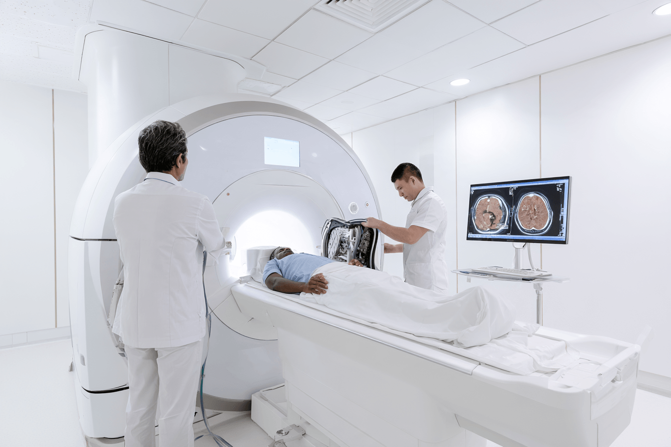
Liver MRI is one of the most important tests for measuring iron overload in thalassemia because it provides a numerical estimate of iron stored inside the liver. In Ayurveda centred integrative care, this measurement creates an objective foundation for protecting liver function, choosing compatible formulations and monitoring whether chelation is removing iron effectively. Ferritin remains useful for frequent follow up, but liver MRI provides a more direct assessment of the stored iron burden [1,2,4].
The result is reported as liver iron concentration, commonly abbreviated as LIC. It is usually expressed as milligrams of iron per gram of dry liver tissue, written as mg Fe/g dry weight. The result should be interpreted with ferritin trends, transfusion exposure, chelation adherence, liver tests and cardiac MRI findings.
What Liver MRI Measures in Iron Overload in Thalassemia
Iron stored within the liver changes the behaviour of magnetic signals during MRI. Specially calibrated sequences measure how rapidly those signals weaken and convert that information into an estimated liver iron concentration.
The examination is non invasive and usually does not require an injection of contrast material. The patient lies inside the MRI scanner while several images are collected during short periods of breath holding. Younger children or patients who cannot remain still may require additional preparation or sedation according to the imaging centre’s protocol.
A routine abdominal MRI is not automatically sufficient for iron measurement. The request should clearly specify quantitative liver iron concentration by a validated MRI method. A report that only states “iron deposition is present” does not provide enough information for monitoring chelation or comparing change over time [1,2,4].
Why Liver Iron Concentration Is More Reliable Than Ferritin
Ferritin is influenced by iron stores, but it can also rise during infection, inflammation, hepatitis, fatty liver disease and liver cell injury. Liver MRI measures the effect of iron within the tissue and is therefore less affected by these short term inflammatory changes.
The liver contains a large proportion of the body’s stored iron. Liver iron concentration is consequently considered the most practical non invasive estimate of total body iron burden in regularly transfused patients. Serial LIC results can show whether the patient is accumulating iron, maintaining iron balance or entering negative iron balance during chelation [1,2].
Ferritin and LIC can sometimes move in different directions. Ferritin may fall while a considerable amount of iron remains in the liver, or it may stay elevated because of inflammation after liver iron has improved. Treatment decisions should not be based on either result in isolation.
Liver MRI also cannot determine whether iron has entered the heart. A patient can have improving LIC while myocardial iron remains present because the heart generally releases stored iron more slowly than the liver. Cardiac MRI T2* is required for that separate assessment.
Liver Iron Concentration Values Explained
The following ranges provide a practical framework for interpreting liver MRI in transfusion dependent thalassemia. They are not rigid treatment boundaries because age, transfusion requirements, chelation, cardiac iron and the rate of change must also be considered [1,2,4].
| Liver iron concentration | Common clinical interpretation |
|---|---|
| Up to approximately 1.8 mg Fe/g dry weight | Conventional normal range |
| Above 1.8 to below 3 mg/g | Increased iron with a relatively low stored burden |
| 3 to 7 mg/g | Mild to moderate iron accumulation requiring continued monitoring |
| Above 7 to 15 mg/g | Clinically important high liver iron burden |
| Above 15 mg/g | Severe iron overload associated with greater risk when sustained |
A result below 7 mg/g does not prove that the heart or endocrine organs are free from iron. It means that the liver iron burden is within a lower risk range compared with higher LIC values. Cardiac MRI and organ function tests remain necessary.
Sustained LIC above 15 mg/g dry weight has been associated with worsening liver fibrosis, abnormal liver function and poorer clinical outcomes. A very high result requires review of the prescribed chelator, actual treatment adherence, transfusion exposure, renal and hepatic safety, and the possibility of combination chelation [1,2,5].
A falling LIC is generally favourable, but the rate of decline also matters. When liver iron approaches a low range, an intensive chelator dose that was appropriate during severe overload may become excessive. Kidney function, liver function, ferritin and LIC should therefore be reviewed before continuing the same treatment intensity.
MRI Methods Used to Measure Liver Iron
Several MRI techniques can quantify liver iron, including R2, R2 star and T2 star based methods. These terms describe how the MRI signal changes in the presence of tissue iron.
With increasing iron, T2 and T2 star values become shorter, while R2 and R2 star values become higher. The machine does not simply detect darkness within the liver. The measured signal must be converted into LIC using a method specific calibration.
Validated R2 methods and appropriately calibrated R2 star or T2 star methods can provide reliable results. The important requirement is that the imaging protocol, scanner settings, analysis software and calibration have been validated for quantitative iron assessment [1,2,4].
A T2 star number from the liver should not be interpreted using cardiac T2 star thresholds. Liver and cardiac measurements use different calibration systems and answer different clinical questions. The final liver report should ideally provide a numerical LIC in mg Fe/g dry weight.
When Liver MRI Should Be Repeated
The interval between liver MRI examinations depends on the current LIC and the speed at which it is changing. Stable LIC between approximately 3 and 7 mg/g dry weight may be reassessed every one to two years. LIC above 7 mg/g is generally reassessed yearly or sooner when clinically necessary [1,2].
When LIC is below 3 mg/g or falling rapidly during intensive chelation, MRI may be repeated every six to twelve months. More frequent assessment helps prevent excessive chelation after the stored iron burden has become low.
An earlier repeat may also be needed when ferritin and MRI results disagree, ferritin rises persistently, chelation has been interrupted, transfusion requirements have increased or a major treatment change has been made.
The repeat interval should be short enough to detect meaningful change but long enough for the expected treatment response to become measurable. Repeating MRI too soon may show little change and may not alter management.
Why the Same MRI Method and Centre Matter
Different MRI methods may produce results that are not directly interchangeable. Scanner strength, image sequence, software, calibration and analysis technique can all affect the reported LIC.
Whenever possible, follow up examinations should use the same validated method and imaging centre. This improves the ability to determine whether the iron burden has genuinely changed rather than appearing different because another technique was used [1,2,4].
When a different centre or method must be used, the previous report should be provided to the radiologist and hematologist. The units should also be checked carefully. A result reported in mg Fe/g dry weight should not be directly compared with a result expressed in another unit without appropriate conversion.
The MRI report should mention the quantitative method, numerical LIC, measurement unit and comparison with the previous study. Descriptive terms such as mild, moderate or severe are helpful, but the numerical value is required for accurate longitudinal monitoring.
What Liver MRI Cannot Show
Liver iron concentration measures stored iron, but it does not independently measure liver inflammation, fibrosis or functional capacity. A patient may have high LIC without major fibrosis, while another patient may have fibrosis caused by a combination of iron, viral hepatitis, metabolic fatty liver disease, alcohol exposure or another liver disorder.
Liver protection therefore also requires alanine aminotransferase, aspartate aminotransferase, bilirubin, albumin and other clinically appropriate liver tests. Hepatitis screening and fibrosis assessment may be needed according to the patient’s history and imaging findings.
Transient elastography or another validated fibrosis assessment may provide additional information, but it does not replace quantitative liver iron MRI. Similarly, normal liver enzymes do not exclude substantial iron accumulation.
Liver MRI does not measure myocardial iron. Cardiac MRI T2* remains necessary when the patient’s age, transfusion history and clinical protocol indicate cardiac monitoring.
Ayurveda Centred Liver Protection After Liver MRI
The LIC result allows Ayurvedic treatment to be planned around the patient’s actual liver burden rather than haemoglobin or ferritin alone. Appetite, digestion, bowel function, body weight, sleep, fatigue and tolerance of chelation are assessed together with LIC, liver enzymes, kidney function and cardiac T2*.
A patient with low haemoglobin and high LIC should not automatically receive an iron containing formulation. Loha, Mandura and other iron based medicines require a separate diagnosis of confirmed iron deficiency. Thalassemia related anaemia and iron deficiency are not the same condition.
Rasayana care is individualized to support digestion, nutritional assimilation, functional strength and long term resilience. The formulation must remain compatible with the patient’s chelator, liver condition and kidney function. Products with uncertain composition, unverified mineral content or inadequate contaminant testing can make liver and renal monitoring more difficult.
Chelation directly removes accumulated iron. Ayurveda supports the wider clinical needs that influence long term recovery, including nutrition, treatment tolerance, digestive stability, sleep and physical strength. Progress is measured through repeat LIC, ferritin trends, liver function, cardiac assessment and changes in the patient’s functional condition rather than symptoms alone.
Cardiac MRI T2* in Iron Overload in Thalassemia
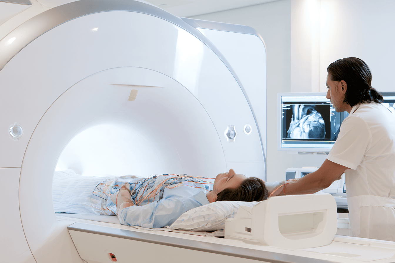
Cardiac MRI T2* is the most important non invasive test for detecting iron inside the heart muscle in iron overload in thalassemia. It can identify myocardial iron before breathlessness, palpitations, heart failure or reduced pumping function becomes apparent. In Ayurveda centred care, the result provides an objective basis for protecting cardiac function while supporting Bala, or functional strength, treatment tolerance, nutrition, sleep and long term resilience [1,2,4].
Ferritin and liver MRI remain essential, but neither can confirm whether the heart contains excess iron. Cardiac MRI T2* answers this separate question and helps determine the intensity of chelation, the frequency of cardiac monitoring and the need for specialist cardiology assessment.
What Cardiac MRI T2* Measures
Iron deposited inside the myocardium disturbs the magnetic signal produced during MRI. Cardiac T2* measures how quickly this signal fades. The result is expressed in milliseconds.
A higher T2* value indicates less myocardial iron, while a lower value indicates a greater cardiac iron burden. The relationship is therefore inverse. As myocardial iron increases, cardiac T2* becomes shorter [1,2,6].
The measurement is commonly taken from the interventricular septum, the muscular wall between the main pumping chambers. A validated 1.5 tesla MRI protocol is generally used for established clinical thresholds. Values produced by different magnetic field strengths, software systems or analysis techniques may not be directly interchangeable.
Cardiac iron assessment usually does not require an injection of contrast material. The examination also includes cine images that assess chamber size, ventricular movement and pumping function. The report should state the cardiac T2* value, scanner strength, ventricular function and comparison with previous examinations whenever available.
Why Ferritin Cannot Predict Cardiac Iron
Ferritin provides useful information about the overall direction of iron burden, but it does not measure iron inside the myocardium. Ferritin can also increase because of infection, inflammation, hepatitis or liver injury, while myocardial iron may change independently.
A patient may have a moderate or falling ferritin level while cardiac T2* remains abnormal. This is more likely when substantial iron exposure occurred in previous years or when liver iron has responded to chelation faster than heart iron.
Ferritin should therefore guide regular monitoring but should not be used to declare the heart free from iron. A satisfactory ferritin trend becomes clinically reassuring only when it is supported by an appropriate cardiac T2* result, liver iron concentration and cardiac function assessment [1,2,4].
Ayurvedic assessment may identify reduced exercise tolerance, disturbed sleep, anxiety, fatigue or a decline in Bala. These findings are clinically important, but they cannot determine whether myocardial iron is present. Cardiac MRI provides the objective measurement needed to distinguish iron deposition from anaemia, deconditioning, endocrine dysfunction or another cause of reduced strength.
Why Liver Iron Cannot Reliably Predict Cardiac Iron
The liver and heart absorb, store and release iron at different rates. The liver usually accumulates iron first and contains the largest proportion of total body iron. Myocardial iron may remain absent during the earlier stages and later increase when circulating reactive iron remains persistently elevated.
During effective chelation, liver iron often decreases more quickly than cardiac iron. A patient may therefore reach an acceptable liver iron concentration while myocardial iron is still present. The reverse pattern can also occur, particularly when recent treatment has improved cardiac iron but liver stores remain high.
Liver MRI and cardiac MRI should be viewed as complementary measurements. Liver MRI estimates the principal body iron store, while cardiac T2* measures iron within the heart muscle. One result should not be used as a substitute for the other [1,2,4].
Cardiac MRI T2* Values Explained
The following ranges are commonly used for cardiac MRI performed with a validated 1.5 tesla method. The precise interpretation also considers the previous T2* result, ventricular function, symptoms, chelation history and quality of the MRI measurement [1,2,6].
| Cardiac T2* value | Clinical interpretation |
|---|---|
| Above 20 milliseconds | No significant myocardial iron by the conventional threshold |
| 10 to 20 milliseconds | Mild to moderate myocardial iron |
| 6 to below 10 milliseconds | Severe myocardial iron with increased cardiac risk |
| Below 6 milliseconds | Very severe myocardial iron with a particularly high risk of cardiac complications |
A value above 20 milliseconds is reassuring for current myocardial iron, but it does not mean that chelation or future cardiac monitoring can be stopped. The patient may still have high liver iron, continuing transfusion exposure or a history of previous cardiac iron.
A value between 10 and 20 milliseconds confirms myocardial iron and requires review of treatment adherence, chelation intensity and the direction of change. A result that has improved from 12 to 16 milliseconds indicates cardiac iron removal, even though the value has not yet reached the conventional normal range.
A value below 10 milliseconds represents severe myocardial iron and requires specialist directed chelation with close cardiac surveillance. A value below 6 milliseconds indicates very severe loading and a particularly high risk of heart failure or rhythm disturbance. Cardiac symptoms or impaired ventricular function increase the urgency further [1,2,6,10].
The landmark cardiac T2* outcome study demonstrated that values below 10 milliseconds strongly predicted subsequent heart failure and arrhythmia in patients with thalassemia major. The greatest risk occurred in those with the lowest measurements, supporting early treatment before pumping function declines [6].
Can Cardiac Iron Exist With a Normal Echocardiogram
Cardiac iron can be present even when echocardiography shows normal pumping function. Cardiac T2* measures myocardial iron, while echocardiography evaluates heart structure, blood flow, relaxation and contraction. They answer different clinical questions.
Iron may accumulate inside myocardial cells before the damage becomes sufficient to reduce the ejection fraction. A normal echocardiogram therefore does not exclude early or clinically important cardiac iron.
Echocardiography remains necessary because cardiac problems in thalassemia are not caused only by iron. Chronic anaemia, high cardiac output, pulmonary hypertension, valvular disease, endocrine disorders and age related cardiovascular conditions may also affect the heart. Cardiac protection requires the combined interpretation of T2*, electrocardiography, echocardiography, symptoms and clinical examination [1,4,10].
Ayurvedic follow up adds practical information about exercise capacity, recovery after activity, sleep, appetite, chest discomfort, awareness of heartbeat and general Bala. These observations help measure daily function, while cardiac MRI, electrocardiography and echocardiography determine the underlying cardiac status.
When Cardiac MRI T2* Should Be Repeated
The interval between cardiac MRI examinations depends mainly on the T2* result, its direction of change and the patient’s clinical condition. Stable values above 20 milliseconds may be reassessed every one to two years according to the treating centre’s protocol.
Values between 10 and 20 milliseconds are generally monitored at least annually. A value below 10 milliseconds may require reassessment approximately every six months because treatment intensity and cardiac risk can change more rapidly [1,2].
Earlier imaging may be needed when cardiac symptoms develop, ventricular function changes, chelation has been interrupted or a major chelation adjustment has been made. The same validated imaging centre and analysis method should be used whenever possible because small differences between techniques can make comparison difficult.
A change in T2* should be interpreted with the absolute value and the quality of the measurement. For example, an increase from 7 to 9 milliseconds represents improvement, but severe myocardial iron remains present. An increase from 18 to 22 milliseconds indicates movement into the conventional range without removing the need for continued iron control.
Why Cardiac Iron May Take Longer to Clear
Myocardial iron generally clears more slowly than liver iron. The liver is highly responsive to changes in chelation and may show a measurable reduction in stored iron before a comparable change appears in the heart.
Removing substantial cardiac iron can require prolonged, uninterrupted treatment. The time needed depends on the starting T2* value, chelator selection, treatment intensity, transfusion burden, adherence and ventricular function. Improvement should be measured through serial cardiac T2* results rather than ferritin alone [1,2,4].
Chelation may also reduce exposure to reactive circulating iron before all stored myocardial iron has been removed. Consistent daily treatment therefore provides ongoing protection even when the MRI value changes gradually.
Chelation should not be reduced simply because ferritin or liver iron has improved while cardiac T2* remains abnormal. The cardiac result must remain central to decisions about myocardial iron treatment.
Ayurveda Centred Cardiac Protection After Cardiac MRI
Cardiac MRI allows Ayurvedic care to be adapted to the patient’s actual level of myocardial risk. A patient with a T2* above 20 milliseconds may receive a different activity, nutrition and Rasayana plan from a patient with severe cardiac iron, reduced exercise tolerance or impaired ventricular function.
Rasayana is an individualized restorative approach used to support nourishment, recovery and systemic resilience. In cardiac iron overload, the formulation is selected after reviewing cardiac T2*, heart function, liver iron concentration, kidney function, chelation therapy and the patient’s digestive capacity.
The Ayurvedic assessment follows appetite, digestion, body weight, bowel regularity, sleep, emotional stress, physical capacity and recovery after activity. Stable Agni, meaning digestive and metabolic function, supports nutrition and medicine tolerance. Preserving Bala helps the patient maintain daily function without exceeding the level of activity considered safe for the current cardiac condition.
Formulations containing stimulants, uncertain mineral ingredients or substances that may affect heart rhythm require careful evaluation. Herbo mineral preparations should have authenticated ingredients, appropriate classical processing, contaminant testing and compatibility with the patient’s liver, kidneys and prescribed medicines.
Chelation removes myocardial iron, while Ayurveda centred care supports the nutritional, digestive and functional foundation needed for sustained treatment. Progress is measured through cardiac T2*, ventricular function, rhythm assessment, ferritin, liver iron concentration and changes in the patient’s physical capacity. This combined approach protects the heart before symptoms become advanced and maintains long term care around measurable organ outcomes [1,2,4,10].
How to Read Ferritin Liver MRI and Cardiac MRI Together in Iron Overload in Thalassemia

Iron overload in thalassemia cannot be assessed accurately through ferritin, liver MRI or cardiac MRI alone. Ferritin shows the direction in which the iron burden may be moving, liver MRI measures the main stored iron burden, and cardiac MRI T2* identifies iron within the heart muscle. These results must be interpreted together with transfusion frequency, chelation adherence, kidney function, liver function and previous measurements [1–4].
The three tests may not improve or worsen at the same time. Ferritin can change within weeks, liver iron usually changes over months, and myocardial iron may require a longer period of consistent chelation before cardiac MRI shows substantial improvement. A favourable result in one test does not confirm that every organ is protected.
Why One Test Cannot Define Iron Overload in Thalassemia
Serum ferritin is suitable for frequent monitoring because it is inexpensive and widely available. A sustained rise may indicate that transfusional iron input is greater than iron removal. A sustained fall generally suggests improving iron balance. Ferritin is nevertheless affected by infection, inflammation, hepatitis, fatty liver disease and liver cell injury, so it remains an indirect marker [1,2].
Liver MRI provides a numerical liver iron concentration and is the most practical non invasive measurement of the main body iron store. A high LIC confirms substantial stored iron even when ferritin is moderate or falling. A lower LIC indicates improved liver iron control, but it cannot confirm that myocardial iron has cleared [1,2,4].
Cardiac MRI T2* directly assesses myocardial iron. Unlike LIC, the T2* value improves by becoming higher. A result above 20 milliseconds is conventionally considered free from significant myocardial iron, while progressively lower values indicate increasing cardiac iron and greater cardiac risk [1,2,6].
The different direction of the cardiac value can be confusing. Falling ferritin and falling LIC usually indicate improvement, whereas a falling cardiac T2* indicates worsening myocardial iron. A rising cardiac T2* generally indicates that iron is being removed from the heart.
What the Combined Results Mean
The relationship between the three measurements reveals whether iron is mainly stored in the liver, has entered the heart, is responding to treatment or is being overestimated by ferritin.
| Combined test pattern | Clinical interpretation |
|---|---|
| High ferritin, high LIC and cardiac T2* above 20 ms | Substantial liver and total body iron without significant myocardial iron by the conventional threshold |
| High ferritin, high LIC and cardiac T2* below 20 ms | Liver and myocardial iron are both present |
| Falling ferritin, high LIC and cardiac T2* above 20 ms | The ferritin trend is improving, but substantial stored liver iron remains |
| Controlled ferritin, controlled LIC and cardiac T2* below 20 ms | Residual myocardial iron remains despite improved liver and overall iron control |
| High ferritin, low LIC and cardiac T2* above 20 ms | Inflammation, infection or liver injury may be contributing to the ferritin elevation |
| Rising ferritin with stable LIC and stable cardiac T2* | Transfusion exposure, inflammation, missed chelation and the timing of repeat MRI require review |
| Falling LIC with improving cardiac T2* | Chelation is removing iron from both the liver and heart |
| Low LIC with rapidly falling ferritin | The possibility of excessive chelator exposure should be assessed |
These patterns describe the most likely interpretation rather than an automatic treatment decision. Previous results, the interval between examinations, transfusion burden and the reliability of each measurement remain important.
High Ferritin With High Liver Iron and Normal Cardiac T2*
High ferritin and high LIC indicate that transfusional iron has accumulated significantly within the liver and wider body stores. A cardiac T2* above 20 milliseconds means that clinically significant myocardial iron has not been detected by the conventional threshold at that examination [1,2].
This pattern does not mean that the heart will remain protected without effective treatment. Persistently high liver iron increases the pool of stored and potentially reactive iron from which extrahepatic organs may eventually load. Chelation should continue to reduce liver iron while regular cardiac MRI confirms that myocardial iron remains controlled.
Cardiac function should still be assessed according to the patient’s age and clinical status. Thalassemia related heart problems may arise from anaemia, high cardiac output, pulmonary hypertension, endocrine disease or other cardiovascular conditions even when cardiac iron is absent.
High Ferritin High Liver Iron and Low Cardiac T2*
This pattern confirms substantial iron accumulation in both the liver and heart. The level of cardiac risk depends strongly on the T2* value, symptoms, ventricular function and direction of change.
A cardiac T2* between 10 and 20 milliseconds indicates myocardial iron. A value below 10 milliseconds represents severe cardiac iron, while a value below 6 milliseconds identifies very severe loading and a particularly high risk of cardiac complications [1,2,6].
Ferritin and LIC remain important, but the cardiac result becomes central to the treatment plan. Chelation intensity, missed doses, tolerability and the possible need for combination therapy require specialist review. Electrocardiography and echocardiography are used alongside cardiac MRI to assess rhythm and cardiac function.
Symptoms such as palpitations, fainting, chest discomfort, new breathlessness, reduced exercise tolerance or leg swelling require prompt clinical assessment. Treatment should not wait for the ejection fraction to decline because myocardial iron can be present while routine echocardiography remains normal.
Falling Ferritin With Persistently High Liver Iron
Ferritin may begin falling before the stored liver iron burden has reached a lower range. This pattern may indicate that the patient has entered negative iron balance, but it does not prove that sufficient iron has already been removed from the liver.
The LIC value determines how much hepatic iron remains. Chelation should not be reduced solely because ferritin has improved when liver MRI continues to show clinically important overload. The next decision depends on the degree of LIC elevation, its change from the previous MRI, transfusion requirements and chelator safety [1,2,4].
Ferritin can also fall after recovery from infection or inflammation. A rapid decrease may therefore partly reflect resolution of an acute condition rather than removal of a large quantity of stored iron.
Controlled Liver Iron With Persistent Cardiac Iron
Liver iron usually responds to chelation faster than myocardial iron. A patient may reach a satisfactory LIC and ferritin range while cardiac T2* remains below 20 milliseconds.
This pattern requires particular care because reducing chelation according to ferritin or LIC alone may leave the heart insufficiently protected. Cardiac directed treatment may need to continue until serial T2* measurements show sustained improvement [1,2,4].
At the same time, low liver iron reduces the body’s buffering capacity and may increase the risk of excessive chelator exposure in other tissues. The regimen must therefore provide adequate myocardial iron removal without producing renal, hepatic, auditory, ocular or other toxicity. This balance requires specialist adjustment rather than a simple increase or decrease based on one result.
An increase in cardiac T2* from 8 to 12 milliseconds represents meaningful improvement, but myocardial iron remains present. Similarly, an increase from 16 to 21 milliseconds indicates entry into the conventional normal range, although ongoing chelation and surveillance remain necessary because transfusions continue to add iron.
High Ferritin With Low Liver and Cardiac Iron
A high ferritin result with a low LIC and cardiac T2* above 20 milliseconds suggests that ferritin may be elevated for a reason other than excessive stored iron. Infection, systemic inflammation, hepatitis, fatty liver disease and liver cell injury should be considered [1–3].
The result should be compared with C reactive protein, liver enzymes, bilirubin, clinical symptoms and recent illness. Repeating ferritin after an acute inflammatory condition has resolved may clarify the underlying trend.
Increasing chelation only because ferritin is elevated can expose a patient with relatively low iron stores to overchelation. Kidney function, liver function, urine findings and the current chelator dose should be reviewed before treatment is intensified.
Rising Ferritin With Stable MRI Results
Ferritin may rise between MRI examinations even when the latest LIC and cardiac T2* remain stable. The first step is to determine whether the increase is persistent and whether an acute inflammatory or hepatic cause is present.
The transfusion record should be reviewed because a higher number of transfusions or larger transfused volume increases iron input. Chelation adherence, gastrointestinal intolerance, treatment interruptions and changes in body weight may also alter iron balance.
Stable MRI results are reassuring only for the period in which they were measured. If ferritin continues rising, chelation has been interrupted or transfusion exposure has increased substantially, repeat MRI may be required earlier than originally planned.
How Changes Should Be Compared Over Time
The most informative comparison uses serial measurements rather than isolated values. Ferritin should be assessed as a trend over several months. LIC should be compared with the previous examination performed through the same validated method whenever possible. Cardiac T2* should be compared using the same scanner strength, analysis technique and experienced centre [1,2,4].
The direction and size of change both matter. A small difference may reflect normal measurement variability, while a consistent change across sequential examinations is more likely to represent a true treatment response.
| Test | Change suggesting improvement | Change requiring review |
|---|---|---|
| Ferritin | Sustained downward trend without inflammation | Persistent rise or unexpectedly rapid fall |
| Liver MRI LIC | Progressive reduction in mg Fe/g dry weight | Rising LIC or failure to improve during adequate treatment |
| Cardiac MRI T2* | Increasing value in milliseconds | Falling value, particularly below 20 ms |
| Organ function | Stable or improving results | New cardiac, liver, endocrine or renal abnormalities |
A patient whose ferritin, LIC and cardiac T2* are all improving is demonstrating a broad response to chelation. A patient with improvement in only one measurement requires a more detailed assessment of where iron remains and whether the current regimen is providing continuous protection.
How the Combined Results Guide Chelation
Chelation decisions are based on iron distribution as well as total iron burden. High LIC with normal cardiac T2* usually places greater emphasis on reducing body and liver iron. A low cardiac T2* requires treatment capable of providing sustained myocardial protection. When both LIC and cardiac iron are severe, an intensive or combined regimen may be considered under specialist supervision [1–4].
The patient’s ability to follow the regimen is equally important. A powerful chelation plan that is frequently missed may provide less protection than a tolerable regimen taken consistently. Nausea, diarrhoea, infusion burden, work schedules, emotional fatigue and cost may all affect adherence.
Safety monitoring remains necessary as iron decreases. The chelator dose that was appropriate during severe overload may become excessive after LIC and ferritin enter a lower range. Creatinine, estimated glomerular filtration rate, urine protein, liver tests, blood counts and chelator specific monitoring help maintain a safe balance.
Ayurveda Centred Interpretation of Ferritin and MRI Results
Ayurveda centred care uses the combined test pattern to design organ compatible treatment rather than treating low haemoglobin as evidence of iron deficiency. A patient can have anaemia, high LIC and myocardial iron at the same time. Loha, Mandura and other iron containing formulations therefore require separately confirmed iron deficiency and a clear clinical indication.
When LIC is high, formulation selection gives particular attention to liver compatibility, digestion, appetite and medicine tolerance. When cardiac T2* is low, physical capacity, sleep, palpitations, recovery after activity and cardiac medicine interactions require closer assessment. When LIC is low during intensive chelation, kidney and liver monitoring helps avoid adding unnecessary treatment burden.
Agni, meaning digestive and metabolic function, influences nutritional assimilation and tolerance of long term medicines. Bala, meaning functional strength, is followed through daily activity, exercise tolerance and recovery. Rasayana care supports nourishment and systemic resilience, but it is individualized according to ferritin, LIC, cardiac T2*, kidney function, liver function and the prescribed chelator.
Chelation removes accumulated iron, while Ayurveda centred care supports nutrition, digestion, strength, sleep and treatment continuity. The effectiveness of the combined plan is measured through improving ferritin trends, falling LIC, rising cardiac T2*, stable organ function and better functional capacity rather than haemoglobin or symptoms alone [1–4].
When Chelation Is Started for Iron Overload in Thalassemia

Chelation for iron overload in thalassemia is started before symptoms or permanent organ dysfunction develops. In transfusion dependent thalassemia, treatment commonly begins after approximately 10 to 20 red blood cell transfusions, when serum ferritin repeatedly approaches or exceeds 1,000 ng/mL, or when quantitative liver MRI confirms clinically important iron accumulation. These are practical decision points rather than automatic cutoffs based on one result [1–4].
An Ayurveda centred plan begins with the same objective assessment. The transfusion record, ferritin trend, liver iron concentration, kidney function, liver function, nutrition and medicine tolerance are reviewed together. Ayurvedic support can strengthen digestion, nutritional status and treatment adherence, but it should not delay chelation when excess iron has reached a clinically important level.
Starting Chelation After Repeated Transfusions
Every transfusion introduces additional iron that the body cannot actively excrete. After approximately 10 to 20 transfusions, the accumulated amount may become sufficient to justify chelation, especially when regular transfusions are expected to continue [1–3].
The decision should consider the actual transfused volume rather than relying only on the number of hospital visits. Children of different body weights may receive different quantities of red blood cells at each transfusion. A detailed transfusion record showing dates, volumes and body weight provides a more accurate picture of iron exposure.
Clinically necessary transfusions should not be reduced or delayed simply to limit iron accumulation. Inadequate transfusion can worsen anaemia, increase bone marrow expansion, impair growth and place additional strain on the heart. The safer approach is to maintain appropriate transfusion therapy while introducing chelation at the correct time.
The need for chelation may arise earlier when transfusion exposure is intensive, ferritin is rising rapidly or liver MRI already shows significant iron. It may arise later when transfusions are less frequent and objective iron measurements remain low. The treatment plan is therefore based on accumulated exposure and measured iron burden rather than age alone.
Why Ferritin Around 1000 Requires Context
A ferritin level around 1,000 ng/mL is commonly used as a signal to consider starting chelation in transfusion dependent thalassemia. The value should usually be confirmed as part of a sustained trend because infection, inflammation, fever, hepatitis and liver injury can temporarily raise ferritin [1,2].
For a patient receiving regular transfusions, repeated ferritin results near or above 1,000 ng/mL generally indicate that iron stores are increasing. Waiting until ferritin becomes extremely high can allow unnecessary iron accumulation before treatment begins. Chelation is most protective when introduced before iron has entered vulnerable organs.
One elevated result during an acute illness should not automatically lead to long term chelation. The clinician should review previous ferritin values, recent infection, liver tests, transfusion frequency and any available MRI results. Repeating ferritin after the acute condition has settled may clarify whether the elevation represents stored iron or an inflammatory response.
Ferritin below 1,000 ng/mL does not always exclude clinically important iron. The result may underestimate the burden in non transfusion dependent thalassemia, and it may not reflect iron within the heart. When ferritin does not match the transfusion history or clinical picture, liver MRI provides a more direct assessment of stored iron.
Role of Liver MRI Before Chelation
Quantitative liver MRI can help confirm whether chelation is needed when ferritin is uncertain, when transfusion exposure has been irregular or when inflammation makes ferritin difficult to interpret. Liver iron concentration provides a numerical estimate of the principal body iron store [1,2,4].
An LIC around or above 3 mg Fe/g dry weight indicates that iron has accumulated above the conventional normal range. The significance depends on whether transfusions are continuing, how rapidly LIC is rising and whether other organs are affected. A higher or progressively increasing LIC strengthens the need for chelation.
Liver MRI is valuable, but treatment should not always be postponed until MRI becomes available. In a child receiving regular transfusions with a clear rise in ferritin and substantial transfusion exposure, the clinical evidence may already be sufficient to begin treatment. Delaying chelation for many months while waiting for imaging may permit further iron accumulation.
MRI can be more difficult in very young children who cannot remain still or follow breath holding instructions. The hematology team may use transfusion exposure, ferritin trend and available laboratory results until a reliable quantitative MRI can be completed.
Cardiac MRI is not usually the primary test used to decide when routine chelation should first begin in a young regularly transfused child. It becomes increasingly important with age, longer transfusion exposure, high iron burden or previous inadequate chelation. Any evidence of myocardial iron requires a more urgent and cardiac focused treatment strategy.
Why Chelation Starts Before Organ Damage
Iron related injury can begin before the patient feels unwell. The liver may accumulate a substantial iron burden while liver enzymes remain near the reference range. Myocardial iron may also develop before echocardiography shows reduced pumping function [1–4].
Chelation binds excess iron and allows it to leave the body through urine or stool, depending on the medicine used. Starting treatment before symptoms appear reduces prolonged exposure to reactive iron and helps protect the liver, heart, pancreas, pituitary gland and other endocrine organs.
The aim at the beginning of treatment is not to remove every iron store immediately. The first goal is to prevent further accumulation and reduce exposure to toxic circulating iron. As treatment continues, the regimen may create negative iron balance, meaning that more iron is removed than is added through transfusions.
Starting too early, before meaningful iron accumulation has occurred, can also be harmful. Chelators may cause toxicity when the dose is excessive relative to the available iron burden. The clinician must therefore balance early organ protection with the risk of overchelation.
This balance explains why chelation is not started from haemoglobin alone. Low haemoglobin in thalassemia results from impaired globin production and does not prove iron deficiency. A patient can remain severely anaemic while excess iron is accumulating in the liver and other organs.
Baseline Assessment Before Starting Chelation
Before chelation begins, the clinical team reviews the complete blood count, ferritin trend, transfusion history, body weight, kidney function, liver function and urine findings. Liver MRI and cardiac assessment are included according to age, transfusion exposure and clinical availability [1–4].
The selected chelator determines additional baseline testing. Deferiprone requires careful assessment of the neutrophil count because of the risk of neutropenia and agranulocytosis. Deferasirox requires renal, urine and liver assessment. Deferoxamine may require baseline hearing, vision, growth and skeletal evaluation, particularly when prolonged treatment is planned.
The patient and family should understand how the medicine is taken, which adverse effects require urgent attention and which laboratory tests must be repeated. A treatment plan that is not practical for daily life may lead to missed doses and continuing iron accumulation.
Digestive tolerance is also important. Nausea, abdominal discomfort, altered bowel habits or difficulty swallowing can reduce adherence. These concerns should be addressed early rather than waiting for the patient to discontinue treatment without informing the clinical team.
Starting Chelation in Non Transfusion Dependent Thalassemia
Iron overload develops differently in non transfusion dependent thalassemia. Increased intestinal iron absorption can cause progressive liver iron accumulation even when transfusions are infrequent. Ferritin may remain lower than expected for the actual liver iron burden [2].
The thresholds used for regularly transfused patients should not be applied automatically. Liver iron concentration is particularly important when deciding whether chelation is needed in non transfusion dependent thalassemia. Age, ferritin trend, LIC, liver health, previous transfusions and the expected duration of treatment are considered together.
A patient with a moderately raised ferritin may still have clinically important liver iron. Conversely, one increased ferritin result does not prove that chelation is necessary. Quantitative liver MRI provides the clearest basis for treatment when the ferritin pattern is uncertain.
Once treatment begins, the stopping threshold is as important as the starting threshold. Non transfusion dependent patients may not continue receiving regular iron through transfusions, so chelation must be reduced or stopped when LIC and ferritin reach a sufficiently low range. Continuing the same intensity after iron stores have fallen increases the risk of overchelation.
Ayurveda Centred Preparation Before Chelation
Ayurveda centred care prepares the patient for long term treatment by assessing Agni, appetite, bowel function, nutritional intake, sleep, body weight, Bala and tolerance of existing medicines. These factors influence whether chelation can be taken consistently and whether the patient can maintain adequate strength during ongoing transfusion therapy.
The Ayurvedic prescription is planned after reviewing ferritin, LIC, cardiac findings, kidney function, liver function and the selected chelator. Non iron Rasayana support may be considered to strengthen nutrition, digestive stability and systemic resilience without adding to the existing iron burden.
Loha, Mandura and other iron containing preparations are not selected merely because haemoglobin is low. Their use requires confirmed iron deficiency through appropriate investigations and a clear clinical indication. In a patient with elevated ferritin or LIC, unnecessary iron exposure may complicate iron control.
Herbal and herbo mineral formulations should be compatible with the liver, kidneys and prescribed chelator. Their ingredients, processing, dose and laboratory quality should be clearly documented. Changes in appetite, bowel function, sleep, weight and strength can be followed clinically, while ferritin, liver MRI, cardiac MRI and organ function tests determine whether iron overload is being controlled safely.
Chelation remains the treatment that directly removes excess iron. Ayurveda supports the patient’s ability to tolerate, continue and recover during long term treatment. Beginning both parts of care at the appropriate stage creates a stronger foundation for preventing liver, cardiac and endocrine complications.
Iron Chelation Medicines for Iron Overload in Thalassemia

Iron chelation medicines remove excess iron that accumulates after repeated blood transfusions or increased intestinal absorption. Three principal chelators are used for iron overload in thalassemia: deferasirox, deferiprone and deferoxamine. Each medicine differs in its route of administration, duration of action, pattern of iron removal, organ response and safety monitoring. The most appropriate chelator is selected according to ferritin trends, liver iron concentration, cardiac MRI T2*, transfusion burden, age, kidney function, liver function and the patient’s ability to continue treatment consistently [1–4].
Ayurveda centred integrative care supports digestion, nutrition, functional strength and treatment tolerance while the prescribed chelator performs the direct work of binding and removing excess iron. This distinction is important because no Ayurvedic herb, diet or general supplement should be treated as a substitute for clinically indicated chelation.
How Chelation Medicines Remove Iron
Only a small proportion of stored iron is available for removal at any moment. Iron is gradually released from circulating reactive iron pools, damaged red blood cells and stored ferritin within tissues. Chelators bind this available iron and form a complex that can be eliminated through urine, bile or stool, depending on the medicine used [1,2].
Chelation has two related purposes. The first is to reduce exposure to reactive iron that can damage cell membranes, mitochondria and genetic material. The second is to remove stored iron from the liver, heart and other organs over time.
Reactive circulating iron may decrease within hours when adequate chelator is present, but the removal of stored organ iron is much slower. Liver iron generally requires months of treatment to decline substantially, while myocardial iron may take months or years to clear. Regular exposure is therefore more protective than irregular treatment taken only when ferritin rises [1,2].
The prescribed dose must change as the iron burden changes. A dose that is appropriate during severe overload may become excessive after ferritin and liver iron concentration fall. Continuing high intensity treatment without reassessment can increase the risk of renal, hepatic, auditory, visual or other chelator related toxicity.
Deferasirox for Iron Overload in Thalassemia
Deferasirox is an oral chelator commonly used for transfusional iron overload. It is available in different formulations, including film coated tablets, dispersible tablets and granules in some countries. These formulations are not identical in absorption, so their doses should not be exchanged without medical adjustment.
Deferasirox binds iron and promotes its elimination mainly through bile and stool. Its relatively long action allows once daily administration in many treatment plans. This convenience can improve treatment continuity for patients who find infusions or several daily doses difficult.
The medicine can reduce ferritin and liver iron concentration when its intensity is matched to transfusional iron input and taken consistently. It may also improve myocardial iron over time, although severe cardiac loading may require a more intensive or combined strategy [1,2].
Kidney monitoring is essential during deferasirox treatment. Serum creatinine, estimated glomerular filtration rate, urine protein and signs of renal tubular dysfunction may be assessed before and during therapy. A rising creatinine level, new proteinuria, electrolyte disturbance or reduced urine output requires clinical review.
Liver tests are also monitored because deferasirox may contribute to hepatic injury in susceptible patients. Alanine aminotransferase, aspartate aminotransferase and bilirubin are interpreted with the patient’s existing liver iron, hepatitis status and other medicines.
Abdominal discomfort, nausea, diarrhoea and skin rash can interfere with adherence. Severe abdominal pain, gastrointestinal bleeding, jaundice, marked weakness or changes in urine output require prompt assessment. Dehydration caused by vomiting, diarrhoea or fever can increase renal risk, particularly in children and patients with existing kidney vulnerability [1,2,4].
Ayurvedic support during deferasirox treatment focuses on Agni, meaning digestive and metabolic function. Appetite, nausea, bowel regularity, hydration, weight and food tolerance are reviewed so that digestive symptoms do not lead to repeated missed doses. Any Ayurvedic formulation must be assessed for renal and hepatic compatibility before it is used alongside deferasirox.
Deferiprone for Cardiac and Systemic Iron
Deferiprone is an oral chelator with the ability to enter cells and remove iron from several tissues. It has an established role in patients with myocardial iron and may be considered when cardiac T2* is abnormal, when another chelator has not provided sufficient control or when a combination regimen is required [1,2].
Deferiprone is commonly taken in divided doses, although dosing frequency and formulation vary by country. The iron complex is eliminated mainly through urine, which may become reddish brown after a dose. This colour change can reflect iron excretion and is not necessarily evidence of blood in the urine, although unexplained urinary symptoms still require assessment.
The most important safety concern is neutropenia and agranulocytosis. Neutrophils are white blood cells that protect against bacterial and fungal infections. Agranulocytosis is a severe reduction in these cells and can become life threatening if infection develops.
A complete blood count with absolute neutrophil count is therefore required before treatment and regularly during therapy according to the local prescribing protocol. Fever, sore throat, mouth ulcers, chills or another sign of infection requires urgent medical evaluation and an immediate blood count. The patient should follow the treatment interruption instructions provided by the hematology team rather than waiting for the next scheduled appointment [1,2,4].
Liver enzymes are monitored because deferiprone may cause elevations in some patients. Joint pain, nausea, abdominal symptoms and reduced zinc levels may also occur. The decision to continue, interrupt or adjust treatment depends on the severity of the finding and the patient’s iron burden.
Deferiprone should not be assumed to be the best medicine for every patient with cardiac iron. The cardiac T2* value, ventricular function, liver iron, blood count history, treatment adherence and previous chelator response must be considered together. Severe myocardial iron may require combination therapy rather than deferiprone alone.
Ayurvedic care alongside deferiprone supports appetite, digestion, sleep, nutritional adequacy and Bala, meaning functional strength and physical capacity. Herbs or herbo mineral medicines should not be started during fever or unexplained infection without reviewing the blood count, because symptoms must not be masked when neutropenia is possible.
Deferoxamine for Long Term and Intensive Chelation
Deferoxamine is the longest established iron chelator used in thalassemia. It is not absorbed effectively through the digestive tract and must therefore be given by subcutaneous, intravenous or, less commonly, intramuscular administration.
For routine long term treatment, deferoxamine is commonly delivered through a slow subcutaneous infusion over several hours on multiple days each week. Intensive intravenous administration may be used in hospital when severe cardiac iron, cardiac dysfunction or another urgent clinical situation requires continuous chelator exposure [1,2].
Deferoxamine removes iron through urine and bile. When administered at an adequate dose and frequency, it can produce negative iron balance and reduce organ complications. Its principal limitation is the practical burden of prolonged infusions, pumps, needles and infusion site discomfort.
Adherence strongly determines its effectiveness. A medically appropriate dose provides little protection when the infusion is repeatedly shortened or omitted. Treatment planning should therefore address school, work, sleep, travel, discomfort and emotional fatigue rather than interpreting every poor response as biological failure.
Deferoxamine can cause redness, swelling, itching or discomfort at the infusion site. Excessive dosing relative to the remaining iron burden may affect hearing and vision. Periodic audiometry and ophthalmological assessment are therefore important, particularly during prolonged treatment or when iron stores have fallen.
Children require monitoring of growth and skeletal development because excessive exposure at a low iron burden may affect growth plates and bones. Kidney and liver function are also followed. Fever, severe abdominal symptoms or systemic illness during deferoxamine treatment requires medical assessment because certain infections may become more serious in the presence of iron loaded deferoxamine complexes [1,2,4].
Ayurvedic support can improve the practical tolerance of infusion based treatment by addressing sleep, appetite, bowel function, skin condition around infusion sites and emotional fatigue. Oil application or local herbal products should not be placed directly over an active infusion site unless the treating team confirms that they are suitable, because contamination and local irritation must be avoided.
Comparing the Three Chelators
| Chelator | Route | Main clinical strengths | Essential monitoring |
|---|---|---|---|
| Deferasirox | Oral | Convenient daily treatment and effective liver and body iron control | Kidney function, urine protein, liver tests and gastrointestinal tolerance |
| Deferiprone | Oral | Important option for myocardial iron and combination therapy | Complete blood count, neutrophils, liver tests and infection symptoms |
| Deferoxamine | Subcutaneous or intravenous | Long clinical experience and intensive treatment capability | Hearing, vision, growth, bones, kidney function and infusion sites |
No comparison should be made from convenience alone. An oral medicine may be easier to take, but it may not be appropriate when kidney function is deteriorating or when previous toxicity has occurred. An infusion may be burdensome, but it can provide intensive and controllable chelator exposure in a high risk cardiac situation.
The most effective regimen is the one that matches the location and severity of iron, remains safe for the patient’s organs and can be followed with sufficient regularity. Long term outcomes depend on actual chelator exposure rather than the prescription written in the medical record [1–4].
Why One Chelator Is Not Best for Every Patient
Chelator selection begins with the current iron pattern. High liver iron with a cardiac T2* above 20 milliseconds may require a different strategy from severe myocardial iron with a cardiac T2* below 10 milliseconds. A patient with controlled liver iron but persistent cardiac iron also requires a different balance from a patient whose ferritin and LIC are both rapidly falling.
Kidney disease may limit the use or intensity of deferasirox. Recurrent neutropenia may make deferiprone unsuitable. Hearing, visual or growth concerns may affect deferoxamine treatment. Pregnancy plans, age, swallowing ability, transfusion intensity and access to laboratory monitoring also influence the choice.
Previous response matters. A chelator that produces falling ferritin but little improvement in LIC may require adjustment. A regimen that improves liver iron while cardiac T2* worsens requires a more cardiac focused review. A medicine that works biologically but causes repeated intolerable symptoms may fail because the patient cannot take it consistently.
Treatment should therefore be individualized rather than changed according to ferritin alone. Serial ferritin, LIC, cardiac T2*, organ function and chelator specific safety tests show whether the selected medicine is providing adequate protection.
Ayurveda Centred Support During Chelation
Ayurveda centred care begins with the measurable iron burden and the safety requirements of the prescribed chelator. It does not select an iron containing tonic simply because haemoglobin is low. Loha, Mandura and other iron containing preparations require a separate diagnosis of true iron deficiency and should not be routinely used when ferritin or liver iron is elevated.
The Ayurvedic assessment follows Agni, appetite, bowel regularity, body weight, sleep, hydration, physical capacity and emotional tolerance of long term treatment. These factors directly affect whether the patient can continue chelation without frequent interruptions.
Rasayana is an individualized restorative approach used to support nourishment, recovery and systemic resilience. During chelation, non iron Rasayana care may support nutritional assimilation, Bala and treatment tolerance, provided that the formulation is compatible with kidney function, liver function, blood counts and the selected chelator.
Herbo mineral preparations require authenticated ingredients, correct classical processing, contaminant testing and documented composition. A product with uncertain metal content can complicate ferritin interpretation and increase hepatic or renal risk. Concurrent supplements should also be reviewed because concentrated botanical extracts may alter liver enzymes, kidney function or medicine metabolism.
Chelation directly removes excess iron. Ayurveda strengthens the digestive, nutritional and functional foundation required to continue that treatment. Progress is measured through ferritin trends, falling liver iron concentration, rising cardiac T2*, stable kidney and liver results, healthy blood counts and sustained improvement in the patient’s daily strength and treatment tolerance.
How Doctors Choose Chelation for Iron Overload in Thalassemia

Choosing chelation for iron overload in thalassemia requires more than comparing three medicines. The decision depends on where iron is stored, how quickly it is accumulating, whether the heart is involved, how well the kidneys and liver are functioning, and whether the patient can follow the prescribed regimen consistently. Ferritin, liver iron concentration, cardiac MRI T2*, transfusion exposure and chelator safety results must be interpreted together [1–4].
No chelator is universally suitable for every patient. Deferasirox, deferiprone and deferoxamine can all contribute to iron removal, but their routes, tissue effects, safety concerns and monitoring requirements differ. The most appropriate regimen is the one that provides sufficient iron removal for the current organ burden while remaining safe and practical enough for uninterrupted long term use.
Ayurveda centred care supports this decision by assessing Agni, nutrition, physical strength, sleep, bowel function and treatment tolerance alongside the medical findings. The Ayurvedic plan must remain compatible with the selected chelator and the patient’s liver, kidneys, blood count and cardiac condition.
Iron Distribution Determines the Main Chelation Goal
The location of stored iron is one of the most important factors in chelator selection. High liver iron concentration with a cardiac T2* above 20 milliseconds indicates that the principal measurable burden remains within the liver and general body stores. The immediate treatment goal is usually to reduce total body iron while preventing future cardiac loading [1,2].
A cardiac T2* below 20 milliseconds confirms myocardial iron and changes the clinical priority. The regimen must provide sustained protection against reactive iron while gradually removing iron from the heart. A value below 10 milliseconds indicates severe myocardial iron and requires more intensive specialist management. A value below 6 milliseconds represents very severe cardiac loading, particularly when ventricular function is impaired or cardiac symptoms are present [1,2,4].
Deferiprone has an established role in strategies directed towards myocardial iron, while deferoxamine and deferasirox can also improve cardiac iron when used at an appropriate intensity. Severe or worsening myocardial iron may require combination chelation rather than simple substitution of one medicine for another. The final decision depends on cardiac T2*, ventricular function, liver iron, previous treatment response and the patient’s ability to maintain the regimen [1–4].
A patient with controlled liver iron but persistent cardiac iron requires a different strategy from a patient with high liver iron and no myocardial deposition. Reducing chelation because ferritin or liver iron has improved can leave the heart insufficiently protected when cardiac T2* remains abnormal.
The Severity of Iron Overload Guides Treatment Intensity
Chelator selection and dose depend on the size of the existing iron burden and the amount of iron entering through continuing transfusions. A patient with mildly increased liver iron requires a different treatment intensity from someone with an LIC above 15 mg Fe/g dry weight and a rapidly rising ferritin trend.
Transfusion records help estimate continuing iron input. The number of units, transfused volume, body weight and frequency of transfusion are more informative than the number of hospital visits alone. When transfusional iron input remains high, a low intensity regimen may be unable to create neutral or negative iron balance even when every prescribed dose is taken [1–3].
Neutral iron balance means that chelation removes approximately the same amount of iron that transfusions add. The stored burden remains relatively stable. Negative iron balance means that chelation removes more iron than is entering, allowing ferritin and liver iron concentration to decline over time.
A steadily rising ferritin and LIC may indicate insufficient chelator exposure, missed treatment, increased transfusion requirements or poor absorption. Before changing medicines, the clinical team should confirm adherence, formulation, dose in relation to body weight, recent transfusion burden and the reliability of the laboratory and MRI results.
Kidney Function Influences Chelator Choice
Kidney function is particularly important when deferasirox is being considered or continued. Baseline assessment commonly includes serum creatinine, estimated glomerular filtration rate, urine protein and other tests when renal tubular dysfunction is suspected. These measurements are repeated regularly because renal changes can appear after treatment begins or after the dose is increased [1,2,4].
A patient with pre existing kidney impairment, persistent proteinuria, electrolyte loss or previous deferasirox related renal injury may require a different regimen or closer monitoring. Vomiting, diarrhoea, fever and dehydration can further increase renal vulnerability. A written sick day plan helps the patient know when medical review or temporary treatment interruption may be necessary.
Kidney safety also affects the Ayurvedic prescription. Herbal and herbo mineral preparations should be reviewed for nephrotoxic potential, mineral content, contamination and interaction with prescribed medicines. Unlabelled products or preparations with uncertain composition make it more difficult to determine whether a change in kidney function is caused by the chelator, the underlying disease or another medicine.
Hydration, appetite, urine changes and bowel function are followed as part of Ayurveda centred care, but symptoms alone cannot exclude renal injury. Creatinine, estimated glomerular filtration rate, urinalysis and urine protein remain necessary for objective monitoring.
Liver Function Affects Treatment Selection
The liver is both the main iron storage organ and an important site for medicine metabolism. Chelator selection therefore considers liver iron concentration, alanine aminotransferase, aspartate aminotransferase, bilirubin, albumin, hepatitis status and other evidence of liver disease [1–4].
Abnormal liver enzymes may result from iron toxicity, viral hepatitis, fatty liver disease, another medicine or the chelator itself. A rise after starting or increasing treatment must be interpreted against the previous liver pattern rather than attributed automatically to one cause.
Severe liver iron may require effective and sustained chelation, but active liver dysfunction can limit the dose or choice of medicine. The clinical team must balance the need to remove iron against the possibility of increasing hepatic injury.
Ayurvedic liver support begins with Agni, meaning digestive and metabolic function. Appetite, digestion, bowel regularity, weight and medicine tolerance are assessed alongside objective liver results. A formulation is not considered liver protective merely because it contains a traditionally used herb. Its ingredients, dose, extraction method, mineral content and compatibility with the chelator must be reviewed for the individual patient.
Blood Counts Matter When Deferiprone Is Considered
Deferiprone can cause neutropenia and, rarely, agranulocytosis. The absolute neutrophil count must therefore be assessed before treatment and monitored regularly according to the prescribing protocol [1,2,4].
A history of recurrent neutropenia, unexplained low white cell counts or difficulty completing regular blood monitoring may influence whether deferiprone is suitable. This does not mean that every patient with a previously low count is permanently excluded. The cause, severity, duration and recurrence pattern require specialist interpretation.
Fever, sore throat, mouth ulcers, chills or another sign of infection during deferiprone treatment requires urgent medical assessment and an immediate blood count. These symptoms should not be managed only with home remedies while neutropenia remains possible.
Ayurvedic medicines used for fever, throat discomfort or immunity support should not delay the required blood test. The immediate clinical priority is to identify or exclude severe neutrophil reduction. Ayurveda centred care can support recovery and nourishment after the urgent cause has been clarified.
Age Growth and Reproductive Plans Affect Chelation
Age influences formulation, monitoring and the ability to follow treatment. Younger children may have difficulty swallowing tablets, tolerating dispersible preparations or remaining attached to a deferoxamine infusion pump. Growth, body weight, bone development, hearing and vision require continued review during long term therapy [1,2].
Chelator doses must be reconsidered as a child gains weight or as transfusion requirements change. A dose that was appropriate several months earlier may become insufficient after substantial growth. The opposite can occur when iron burden falls but treatment intensity remains unchanged.
Adolescents may understand the medical importance of chelation but still find daily treatment difficult because of school, social activity, treatment fatigue or concern about appearing different. A regimen that fits daily life is more likely to provide continuous protection than one that is repeatedly omitted.
Pregnancy planning also changes the risk assessment. Chelator use during conception, pregnancy and breastfeeding requires specialist guidance because the safety of individual medicines differs and maternal cardiac iron may make prolonged interruption unsafe. Patients should not stop chelation independently when planning pregnancy. Cardiac T2*, liver iron, fertility status and maternal risk should be reviewed before conception so that a coordinated plan can be prepared [1,2,4].
Previous Treatment Response Guides the Next Decision
A chelator should be judged by objective response, safety and actual use. Ferritin trends, liver iron concentration and cardiac T2* reveal whether treatment is controlling the relevant iron compartment.
Falling ferritin with falling LIC indicates improvement in body and liver iron. A rising cardiac T2* indicates that myocardial iron is decreasing. These changes should be interpreted over sufficient time because organ iron does not disappear within a few weeks.
An inadequate response does not always mean that the chelator is biologically ineffective. Missed doses, incorrect administration, gastrointestinal intolerance, increased transfusion exposure, weight gain, inflammation and inconsistent MRI methods may all create an apparent treatment failure.
Before changing therapy, the clinician should determine whether the medicine was taken at the prescribed intensity and whether adverse effects made regular treatment unrealistic. Changing from one chelator to another without addressing the reason for missed treatment may reproduce the same problem.
Previous toxicity remains equally important. A patient who developed significant renal injury, recurrent neutropenia, severe gastrointestinal intolerance, auditory changes or another serious adverse effect may require a different medicine or a modified regimen. Rechallenge decisions should be made by a specialist with appropriate monitoring.
Adherence Can Determine Which Chelator Works Best
Theoretical effectiveness has limited value when the patient cannot take the medicine regularly. Route, taste, tablet size, frequency, infusion duration, adverse effects, work commitments, school attendance, travel and emotional fatigue all affect actual chelator exposure [1–4].
An oral chelator may be more practical than repeated subcutaneous infusions, but oral treatment is not automatically safer or more effective. A patient with kidney vulnerability may not tolerate the preferred oral option. Another patient may tolerate deferoxamine well but struggle with infusion scheduling. The decision must balance organ need with daily feasibility.
Adherence should be discussed without blame. A patient may omit treatment because of nausea, diarrhoea, abdominal pain, infusion site discomfort or fear of toxicity. Identifying the actual barrier allows the team to adjust formulation, administration timing, symptom support or treatment method.
Ayurveda centred care can improve continuity by addressing appetite, nausea, bowel disturbance, sleep, fatigue and nutritional weakness. These measures support the patient’s ability to continue chelation, but they should not conceal a serious adverse effect that requires medical investigation.
Low Iron Burden Requires Protection From Excessive Chelation
Chelation intensity must be reduced when iron stores become low. The same dose that was necessary during severe overload can produce excessive exposure after LIC and ferritin have fallen substantially [1–4].
Overchelation can contribute to renal dysfunction, liver abnormalities, hearing or visual problems, growth disturbance and other medicine specific toxicities. Risk is influenced by the chelator, dose, age, body weight and remaining iron burden.
A falling ferritin should not trigger an automatic treatment interruption because ferritin may be temporarily low or may not reflect residual cardiac iron. Liver MRI, cardiac T2*, transfusion exposure and safety tests determine whether the dose should be reduced, changed or continued.
A patient with low liver iron but persistent myocardial iron presents a particularly complex balance. Treatment must continue to remove cardiac iron without creating excessive exposure in other tissues. This situation requires specialist adjustment and closer monitoring rather than a simple decision based on ferritin.
Ayurveda Centred Integration With the Selected Chelator
Ayurveda centred care begins after the current iron pattern and chelation requirements are clearly defined. The prescription considers liver iron concentration, cardiac T2*, kidney function, liver function, blood counts, transfusion frequency, digestion and physical capacity.
Low haemoglobin does not justify routine use of Loha, Mandura or another iron containing formulation. Iron deficiency must be confirmed separately because a patient with thalassemia can remain anaemic while carrying substantial liver and cardiac iron.
Rasayana is an individualized restorative approach used to support nourishment, recovery and long term resilience. During chelation, non iron Rasayana care may support appetite, digestion, sleep, healthy body weight and Bala, meaning functional strength. The formulation must remain compatible with the patient’s chelator and organ status.
Changes in appetite, bowel function, sleep, fatigue and treatment tolerance provide useful clinical information, but chelator effectiveness is measured objectively. Falling LIC, rising cardiac T2*, controlled ferritin, stable kidney and liver function and preserved organ performance show whether the complete plan is protecting the patient safely.
The final chelator decision is therefore not based on convenience, ferritin or one MRI result alone. It is built from the location of iron, continuing transfusion burden, organ safety, previous response and the patient’s ability to maintain treatment. Ayurveda strengthens the nutritional and functional foundation required for long term adherence while chelation performs the direct removal of excess iron.
Combination Chelation for Severe Iron Overload in Thalassemia

Combination chelation for severe iron overload in thalassemia uses two iron chelators within one coordinated treatment plan. It may be considered when one chelator does not provide adequate control, when cardiac MRI shows severe myocardial iron, when liver iron remains very high despite consistent treatment, or when continuing transfusions add iron faster than a single medicine can remove it [1–4].
Combination chelation is not required for every patient with an elevated ferritin result. The decision depends on ferritin trends, liver iron concentration, cardiac MRI T2*, transfusion burden, previous treatment response, medicine tolerance and organ function. In Ayurveda centred integrative care, chelation performs the direct removal of excess iron, while individualized care supports digestion, nutrition, physical strength and the ability to continue intensive treatment safely.
When One Chelator May Not Be Enough
A single chelator may be insufficient when iron measurements continue to worsen despite an appropriate prescribed dose. Ferritin may remain persistently elevated, liver iron concentration may fail to decline, or cardiac T2* may become progressively lower. These patterns suggest that iron entering the body is greater than the amount being removed [1–4].
An inadequate response does not automatically mean that the chelator has failed. Missed doses, gastrointestinal intolerance, infusion difficulties, weight gain, increased transfusion requirements, incorrect administration and temporary treatment interruption can all reduce effective chelator exposure. Inflammation or liver injury may also raise ferritin without a corresponding increase in stored iron.
Before adding a second chelator, the clinical team reviews whether the first medicine has been taken consistently, whether its intensity matches the patient’s body weight and transfusion burden, and whether kidney, liver or blood abnormalities prevent further adjustment. Liver MRI and cardiac MRI are particularly important because ferritin alone cannot determine which organ requires more intensive treatment.
Combination chelation becomes more appropriate when objective iron measurements remain unacceptable despite confirmed adherence and the maximum safe or tolerated intensity of one chelator. It may also be introduced earlier when myocardial iron creates an immediate threat to cardiac function.
Combination Chelation for Severe Cardiac Iron in Thalassemia
Cardiac iron is one of the most important reasons for using combination chelation. A cardiac T2* below 20 milliseconds confirms myocardial iron. A value below 10 milliseconds indicates severe cardiac loading, while a value below 6 milliseconds represents very severe myocardial iron and a particularly high risk of heart failure or rhythm disturbance [1,2,6,10].
The urgency increases when a low cardiac T2* occurs with reduced ventricular function, palpitations, fainting, breathlessness, swelling or an abnormal heart rhythm. These patients require coordinated care from a thalassemia specialist and cardiologist. Hospital based treatment may be necessary when cardiac dysfunction is present.
Combination chelation aims to maintain more continuous protection against reactive circulating iron while gradually removing stored myocardial iron. Deferiprone and deferoxamine have the longest established evidence as a combination for cardiac siderosis. Controlled clinical research has shown greater improvement in myocardial T2* and cardiac function with combined deferiprone and deferoxamine than with deferoxamine alone in selected patients with cardiac iron [11].
The heart usually releases stored iron more slowly than the liver. Ferritin and liver iron concentration may therefore improve before cardiac T2* reaches a safer range. Combination therapy is not reduced solely because ferritin has fallen when significant myocardial iron remains present.
A cardiac T2* increase from 7 to 10 milliseconds represents improvement, but severe cardiac iron is still present. An increase from 14 to 18 milliseconds also shows iron removal, although the result remains below the conventional normal threshold. The absolute value and its direction of change must be considered together.
Combination Chelation for Persistently High Liver Iron
Combination chelation may also be considered when liver iron concentration remains very high despite consistent monotherapy. An LIC above 15 mg Fe/g dry weight represents severe hepatic iron accumulation, particularly when the result remains elevated across repeated examinations [1–4].
Persistent high LIC increases the risk of liver inflammation, fibrosis and progressive dysfunction. It also indicates a large total body iron burden that may eventually increase exposure of the heart, pancreas and endocrine glands.
Before intensifying treatment, the clinician reviews the previous LIC, ferritin trend, transfused red cell volume, body weight, chelator dose and treatment adherence. The same validated MRI method is preferably used for follow up because changing scanners or analysis systems can create an apparent difference that does not reflect a true biological change.
A high LIC does not automatically require two chelators. Some patients respond adequately when the original chelator is optimized and treatment interruptions are corrected. Combination treatment becomes more relevant when LIC continues rising, fails to decline after an adequate treatment period, or cannot be controlled with one medicine because of dose limiting toxicity.
The goal is to create negative iron balance, meaning that more iron is removed than is added through transfusions. As LIC declines, treatment intensity must be reviewed to prevent excessive chelation.
Deferiprone With Deferoxamine
Deferiprone with deferoxamine is the most established combination for severe myocardial iron. Deferiprone is taken orally and can enter cells containing iron, while deferoxamine is administered through subcutaneous or intravenous infusion. Their different routes and patterns of iron binding can provide broader chelator exposure than either medicine alone [1,2,4,11].
This combination may be used when cardiac T2* is severely reduced, cardiac function is deteriorating or previous monotherapy has not removed myocardial iron adequately. In patients with symptomatic iron related cardiac dysfunction, prolonged or continuous deferoxamine exposure may be combined with deferiprone under close specialist supervision.
The main practical challenge is treatment burden. The patient must manage oral doses, infusion equipment, regular blood counts and additional safety monitoring. Nausea, joint symptoms, infusion site discomfort and treatment fatigue can reduce adherence if they are not addressed early.
Deferiprone requires regular absolute neutrophil count monitoring because it can cause neutropenia or agranulocytosis. Fever, sore throat, mouth ulcers or chills require urgent medical assessment and an immediate blood count. Deferoxamine requires monitoring of hearing, vision, growth, skeletal health and infusion sites according to age and treatment duration [1,2,4].
Deferiprone With Deferasirox
Deferiprone with deferasirox provides an entirely oral combination and may be considered when intensive iron removal is required but deferoxamine infusions are impractical or poorly tolerated. Clinical studies have reported improvement in ferritin, liver iron and cardiac T2* in selected patients, although the evidence base is smaller and less established than that for deferiprone with deferoxamine [1–4].
An all oral regimen can reduce infusion burden, but it does not reduce the need for close monitoring. Deferiprone retains its risk of severe neutrophil reduction, while deferasirox requires careful assessment of kidney function, urine protein and liver tests.
The combination may be unsuitable when the patient has recurrent neutropenia, significant renal impairment, persistent proteinuria, active liver injury or previous serious intolerance to either medicine. Regulatory approval and product licence conditions also differ between countries, so specialist oversight remains necessary.
Digestive effects can become clinically important because both medicines may cause nausea, abdominal discomfort or altered bowel habits. Persistent symptoms can lead to missed treatment and reduce the expected benefit of an oral combination.
Deferasirox With Deferoxamine
Deferasirox with deferoxamine may be used in selected patients with a very high total body or liver iron burden when one medicine has not created adequate negative iron balance. The oral and infused chelators provide different periods of exposure and routes of iron elimination [1–4].
This combination has less extensive evidence than deferiprone with deferoxamine for severe myocardial iron. Its use is generally based on the distribution of iron, previous chelator response, organ safety and the patient’s ability to manage the regimen.
Kidney and liver monitoring are particularly important because deferasirox can affect renal and hepatic function. Deferoxamine related auditory, visual and skeletal monitoring also continues. Combining the medicines does not remove the individual safety requirements of either chelator.
Measuring the Response to Combination Chelation
The response to combination chelation is measured through serial ferritin, liver MRI LIC, cardiac MRI T2*, transfusion records and organ function. Symptoms alone cannot confirm that stored iron is declining.
Ferritin may begin changing before MRI shows a substantial difference. A sustained downward ferritin trend can indicate improving iron balance, but infection, inflammation and liver injury must still be considered. Liver iron concentration usually requires several months to show a meaningful treatment response.
Cardiac improvement is demonstrated by a rising T2* value. Myocardial iron often clears slowly, particularly when the starting value is very low. Ventricular function, electrocardiography, rhythm monitoring and symptoms are assessed alongside cardiac MRI when severe cardiac iron is present [1,2,6,10].
A favourable response includes falling LIC, rising cardiac T2*, controlled ferritin and stable kidney, liver and blood results. Improvement in only one measurement requires continued assessment because liver and cardiac iron may respond at different rates.
Transfusion exposure must also be included in the interpretation. Stable LIC during a period of high transfusional iron input may represent successful neutral iron balance, while the same stable result in a patient with lower iron input may indicate that chelation intensity remains inadequate.
Safety During Combination Chelation
Using two chelators increases treatment complexity and may expose the patient to the adverse effects of both medicines. Monitoring is based on the specific combination rather than on ferritin alone [1–4].
A regimen containing deferiprone requires repeated complete blood counts with absolute neutrophil counts. A regimen containing deferasirox requires kidney function, urine protein, electrolytes and liver tests. Deferoxamine requires hearing, vision, growth, bone and infusion site assessment according to the patient’s age and duration of treatment.
New fever, sore throat, reduced urine output, severe vomiting, persistent diarrhoea, jaundice, hearing changes, visual symptoms or severe weakness require prompt clinical review. New breathlessness, chest pain, palpitations, fainting or swelling require urgent cardiac assessment.
Treatment intensity must be reduced when iron stores approach a low range. Continuing an intensive combination after LIC and ferritin have fallen can cause excessive chelation and organ toxicity. The presence of residual cardiac iron can make dose reduction more complex, because myocardial treatment may still be necessary when liver iron is already controlled.
Ayurveda Centred Support During Combination Chelation
Ayurveda centred care supports the patient through the nutritional, digestive and functional demands of intensive chelation. Agni means digestive and metabolic function. Appetite, nausea, bowel regularity, hydration and nutritional assimilation are followed because disturbances in these areas can lead to missed medicine and loss of physical strength.
Bala means functional strength and physical capacity. Daily activity, exercise tolerance, muscle strength, sleep and recovery after exertion help show how the patient is coping with treatment. These observations complement cardiac MRI, liver MRI and laboratory monitoring but do not replace them.
Ojas refers to systemic resilience during chronic illness. Adequate nutrition, restorative sleep, emotional stability and consistent treatment contribute to maintaining Ojas. Rasayana is an individualized restorative approach used to support nourishment and long term recovery. During combination chelation, Rasayana selection must remain compatible with the blood count, liver, kidneys, heart and prescribed medicines.
Low haemoglobin does not justify routine use of Loha, Mandura or another iron containing preparation. A patient may remain anaemic while carrying severe liver and cardiac iron. Iron containing treatment requires separately confirmed iron deficiency and coordinated monitoring.
Herbo mineral preparations require authenticated ingredients, appropriate classical processing, contaminant testing and a clearly documented composition. Products with uncertain iron or heavy metal content can increase organ burden and make changes in liver or kidney function more difficult to interpret.
Chelation directly removes excess iron. Ayurveda supports digestion, nutrition, sleep, strength and treatment tolerance so that the patient can continue the required regimen. Progress remains anchored to falling liver iron concentration, rising cardiac T2*, controlled ferritin, stable organ function and improved functional capacity.
When Combination Chelation Can Be Reduced
Combination chelation is reassessed when the targeted iron burden has improved. The decision considers ferritin trends, LIC, cardiac T2*, continuing transfusion requirements and treatment toxicity [1–4].
A patient whose LIC has reached a low range but whose cardiac T2* remains abnormal may still require cardiac directed treatment. A patient whose liver and cardiac iron are both controlled may be able to return to maintenance monotherapy if one chelator can balance continuing transfusional iron input.
Reduction is gradual and guided by measurable results. Abruptly stopping intensive chelation while severe cardiac iron remains present can allow reactive iron exposure to return. Continuing the full regimen after iron stores have become low can create overchelation.
The long term goal is not permanent use of the greatest number of medicines. It is the lowest safe treatment intensity that maintains controlled ferritin, acceptable liver iron, a cardiac T2* above the risk range and stable organ function.
What Successful Chelation Should Achieve in Iron Overload in Thalassemia

Successful chelation in iron overload in thalassemia should do more than produce a temporary fall in serum ferritin. It should reduce exposure to toxic circulating iron, prevent further accumulation, remove excess iron already stored in the liver and heart, preserve organ function and remain safe enough for consistent long term use. In Ayurveda centred care, these measurable goals are combined with support for digestion, nutrition, physical strength and treatment tolerance [1–4].
The expected result depends on the starting iron burden. A patient beginning chelation before major accumulation may need to maintain iron balance, while someone with high liver iron or myocardial iron requires negative iron balance. Treatment intensity must later be reduced when iron stores become controlled because continued intensive chelation can cause toxicity.
Reducing Toxic Circulating Iron
One of the earliest goals of chelation is to reduce non transferrin bound iron and labile plasma iron. These reactive forms appear when the normal iron transport protein transferrin becomes saturated. They can enter cells without normal biological control and contribute to oxidative damage in the liver, heart, pancreas and endocrine glands [1–3].
Chelators bind available iron and reduce the amount that can participate in damaging chemical reactions. This protective effect may begin before ferritin, liver iron concentration or cardiac MRI shows a major change.
Reactive iron can return when the chelator is no longer present in the blood or tissues. A regimen that provides appropriate and regular chelator exposure is therefore more protective than irregular treatment followed by occasional intensive dosing. Missing treatment repeatedly may allow toxic circulating iron to reappear even when the most recent ferritin result appears stable [1,2].
Ayurvedic care supports this first goal by improving the practical conditions required for uninterrupted treatment. Appetite, nausea, bowel function, sleep, hydration and medicine tolerance are addressed so that digestive discomfort does not repeatedly interfere with the prescribed chelator.
Achieving Neutral Iron Balance
Neutral iron balance means that the amount of iron removed through chelation is approximately equal to the amount added through ongoing transfusions. When neutral balance is maintained, the total stored iron burden should remain relatively stable rather than continuing to rise.
This may be an appropriate goal when liver iron concentration and cardiac T2* are already within acceptable ranges. The purpose is to prevent new accumulation while avoiding unnecessary chelator exposure.
Stable ferritin alone does not prove that neutral iron balance has been achieved. Ferritin can remain unchanged because of inflammation, liver injury or changes in the distribution of stored iron. Serial liver MRI provides a more direct assessment of whether the main body iron store is stable [1,2].
Transfusion intensity strongly influences the chelator requirement. A patient receiving more red blood cells each month receives more iron and may require greater chelator exposure to maintain the same balance. Changes in body weight, transfusion frequency and transfused volume should therefore be reviewed whenever ferritin or LIC begins to rise.
Ayurveda centred monitoring follows nutrition, body weight and digestive capacity alongside transfusion records. A significant change in weight can alter the effective dose of both chelation and supportive formulations, making regular reassessment necessary.
Creating Negative Iron Balance
Negative iron balance means that chelation removes more iron than transfusions add. This is required when ferritin and LIC are rising, when liver iron is already high or when cardiac MRI shows myocardial iron [1–4].
A falling LIC provides strong evidence that the total stored burden is decreasing. Ferritin may also decline, but the two measurements do not always change at the same rate. Liver iron can improve without a clear ferritin reduction during the earlier months of treatment, particularly when ferritin is very high or affected by inflammation.
Cardiac response is measured differently. Successful removal of myocardial iron causes cardiac T2* to increase. A change from 8 to 12 milliseconds represents improvement, although cardiac iron remains present. Treatment remains necessary until the cardiac burden is controlled and the result is stable.
Negative iron balance is not created simply by prescribing a higher dose. The patient must receive adequate treatment frequency and duration, and the medicine must be taken consistently. Continuing transfusion exposure, absorption, body weight, organ function and missed treatment all influence the result.
| Chelation goal | Expected iron pattern | Main measurements |
|---|---|---|
| Reduce toxic circulating iron | Lower exposure to reactive iron during treatment | Regular chelator coverage and clinical stability |
| Maintain neutral iron balance | Iron burden remains stable | Ferritin trend and stable LIC |
| Create negative iron balance | Stored iron progressively declines | Falling LIC, controlled ferritin and rising cardiac T2* |
| Preserve organ function | No new iron related dysfunction | Cardiac, liver, endocrine, renal and bone monitoring |
| Maintain treatment safety | Iron is removed without excessive toxicity | Blood count, kidney, liver, urine, hearing and vision monitoring |
Protecting Organs Before Symptoms Appear
Successful chelation protects the liver, heart, pancreas, pituitary gland, thyroid, parathyroid glands and reproductive system before obvious symptoms develop. Waiting for jaundice, heart failure, diabetes, delayed puberty or severe hormonal disturbance means that iron exposure has already continued for too long.
Organ protection can begin before all stored iron has been removed. Reducing reactive iron may improve cellular function and decrease ongoing injury even while MRI still shows iron deposits. In some patients, cardiac function improves before cardiac MRI demonstrates a large reduction in myocardial iron [1,2].
The treatment goal is therefore not limited to reaching a particular ferritin number. Liver enzymes, cardiac function, rhythm, glucose regulation, growth, puberty, thyroid function, calcium balance, fertility, kidney function and bone health also show whether long term organ protection is being achieved.
Ayurvedic assessment adds practical measures of recovery, including appetite, sleep, bowel regularity, exercise tolerance, body weight and daily physical capacity. These findings help show how the patient is functioning, while laboratory tests and MRI determine whether iron related organ risk is objectively improving.
Measuring Whether Chelation Is Working
No single result can confirm successful chelation. The response is measured through a pattern of findings collected over time.
Ferritin is useful for frequent monitoring and should generally show a controlled or downward long term trend. LIC should remain stable during maintenance treatment or fall when negative iron balance is required. Cardiac T2* should remain above the conventional risk range or progressively increase when myocardial iron is being removed [1–4].
The transfusion record must be included in the interpretation. Stable LIC in a patient receiving a high transfusional iron load may indicate that chelation is successfully preventing further accumulation. The same stable result in a patient with severe existing overload and lower transfusion exposure may indicate that iron removal remains insufficient.
Adherence must also be assessed honestly. A medicine cannot be judged ineffective when it has not been taken with the prescribed frequency. Treatment barriers such as nausea, diarrhoea, infusion discomfort, work schedules, school, cost and emotional fatigue should be identified before the regimen is changed.
Successful treatment usually produces gradual, sustained improvement rather than a dramatic result within a few weeks. Ferritin may change first, liver iron generally requires several months to decline meaningfully, and severe cardiac iron may require prolonged uninterrupted treatment.
Avoiding Excessive Chelation
Successful chelation must remove iron without causing damage from excessive medicine exposure. The required dose decreases as the stored iron burden falls. A regimen that was appropriate for severe overload may become too intensive after ferritin and LIC enter a lower range [1–4].
Excessive chelation may contribute to kidney dysfunction, liver abnormalities, hearing or visual problems, growth disturbance and other medicine specific adverse effects. Risk is influenced by the chelator, dose, body weight, treatment frequency and remaining iron burden.
A low ferritin result should not automatically lead to complete treatment withdrawal. The patient may still receive regular transfusions, and residual cardiac iron may remain after liver iron has improved. Ferritin, LIC, cardiac T2*, transfusion exposure and safety tests must be reviewed together before reducing treatment.
The most effective long term plan uses the lowest treatment intensity that can maintain controlled iron and protect vulnerable organs. This requires repeated adjustment rather than one fixed dose continued indefinitely.
Ayurveda Centred Support During Successful Chelation
Ayurveda centred care supports the patient’s ability to complete chelation safely and maintain strength during long term treatment. Agni refers to digestive and metabolic function. Stable Agni supports appetite, nutritional assimilation and tolerance of prescribed medicines.
Bala means functional strength and physical capacity. It can be followed through daily activity, muscle strength, recovery after exertion and the ability to continue school, work or normal routines. Ojas refers to systemic resilience during chronic illness and is supported through adequate nourishment, restorative sleep, emotional stability and consistent treatment.
Rasayana is an individualized restorative approach intended to support nourishment, recovery and long term resilience. During chelation, Rasayana care should remain free from unnecessary iron and should be selected according to liver function, kidney function, cardiac findings, blood counts and digestive capacity.
Ayurvedic formulations do not replace prescribed iron chelation. Their role is to support digestion, nutrition, sleep, physical strength and treatment tolerance while chelation removes excess iron. Loha, Mandura and other iron containing preparations require separately confirmed iron deficiency because low haemoglobin in thalassemia does not prove that the body lacks iron.
A successful combined plan is reflected by controlled ferritin, falling liver iron concentration when overload is present, rising cardiac T2* when myocardial iron is being removed, stable organ function and improved treatment tolerance. These outcomes show that iron is being controlled without weakening the patient or creating unnecessary medicine related toxicity.
How Long Does It Take to Reduce Iron Overload in Thalassemia?

The time required to reduce iron overload in thalassemia depends on the amount and location of stored iron, continuing transfusion exposure, the selected chelator, treatment adherence and organ function. Reactive circulating iron may fall soon after adequate chelation begins, but ferritin, liver iron concentration and cardiac MRI T2* usually improve over different time periods. Ayurveda centred care supports digestion, nutrition, strength, sleep and medicine tolerance throughout this gradual process [1–4].
A patient with moderately raised liver iron and no cardiac involvement may improve more quickly than someone with severe liver iron, a cardiac T2* below 10 milliseconds and many years of previous transfusions. A fixed recovery period cannot therefore be predicted from ferritin alone.
What Can Change During the First Thirty Days
The first month is mainly used to establish a safe and practical treatment foundation. Ferritin history, transfusion exposure, liver iron concentration, cardiac T2*, kidney function, liver function and chelation adherence are reviewed together. The patient’s current chelator dose, body weight, administration method and missed doses are also assessed.
Chelation can begin reducing exposure to non transferrin bound iron and labile plasma iron soon after adequate treatment is established. These reactive forms of iron contribute to oxidative cellular injury. Their reduction may improve protection before a major change becomes visible in ferritin or MRI measurements [1,2].
Ferritin may begin moving downward during the first month, but one early change should be interpreted cautiously. Infection, inflammation, hepatitis, recent transfusion and liver injury can alter ferritin independently of stored iron. A clear response is better established through repeated values over several months.
Ayurvedic assessment during this period focuses on Agni, meaning digestive and metabolic function, because nausea, poor appetite, abdominal discomfort and altered bowel habits can interfere with regular chelation. Sleep, hydration, body weight and Bala, meaning functional strength, are also recorded so that future improvement can be measured from a reliable baseline.
Ferritin Changes Over Several Months
Ferritin can respond earlier than MRI because it is measured frequently and reflects changes in iron balance as well as inflammation. A sustained downward trend over three to six months may suggest that chelation is removing more iron than transfusions are adding [1–4].
The speed of decline differs considerably. Ferritin may fall slowly when transfusion requirements remain high, even when chelation is working. It may fall more quickly after an infection resolves or when treatment becomes more consistent. A rapid reduction does not always represent an equally rapid loss of liver or cardiac iron.
The clinical aim is a controlled long term trend rather than the largest possible monthly fall. Excessively intensive chelation can become harmful when body iron approaches a low range. Ferritin must therefore be interpreted with liver iron concentration, cardiac T2*, kidney function, liver function and continuing transfusion exposure.
A stable ferritin may still represent a successful result when the patient receives a substantial transfusional iron load. In this situation, chelation may be preventing further accumulation even before negative iron balance has been achieved.
How Long Liver Iron Takes to Decline
Liver iron concentration usually changes over months rather than weeks. A meaningful reduction may be visible after approximately six to twelve months of consistent chelation, although the exact response depends on the starting LIC, treatment intensity and transfusional iron input [1–4].
A patient with severe liver iron may require several years to reach a lower risk range. The goal is steady reduction without causing kidney, liver or other chelator related toxicity. Very rapid iron removal is not always safer, particularly when the treatment intensity becomes excessive relative to the remaining stores.
Serial liver MRI is more informative than frequent scanning. When LIC is high, repeat imaging is commonly considered after enough time has passed for a measurable biological change. Earlier imaging may be needed when treatment has changed substantially, ferritin and MRI findings disagree or excessive chelation is suspected.
The same validated MRI method should be used whenever possible. A small difference between results from different centres or analysis systems may reflect measurement variation rather than a true change in liver iron.
Why Cardiac Iron Usually Takes Longer to Clear
Myocardial iron generally responds more slowly than liver iron. Cardiac T2* may require many months or several years to return above 20 milliseconds, especially when the starting value is below 10 milliseconds [1,2,6].
Improvement is shown by an increasing cardiac T2* value. A change from 7 to 10 milliseconds represents meaningful iron removal, but severe myocardial iron remains present. A change from 14 to 18 milliseconds also shows progress, although the result is still below the conventional normal threshold.
Liver iron and ferritin may improve well before cardiac iron has cleared. Chelation should therefore not be reduced solely because LIC has entered a satisfactory range when cardiac T2* remains abnormal.
Cardiac function can sometimes improve before the MRI value normalizes because reducing reactive iron may improve cellular function before all stored myocardial iron has been removed. Echocardiography, electrocardiography, symptoms and ventricular function are assessed alongside serial cardiac T2* [1,6,10].
Severe cardiac iron requires uninterrupted treatment and close specialist monitoring. Treatment interruption can allow toxic circulating iron to return even when a previous MRI showed improvement.
Expected Time Course of Iron Reduction
| Monitoring period | Changes that may be assessed |
|---|---|
| First thirty days | Treatment tolerance, adherence, reactive iron protection, kidney and liver safety |
| Three to six months | Ferritin direction, transfusion balance and practical chelation response |
| Six to twelve months | Measurable change in liver iron concentration and clearer ferritin trend |
| Twelve months and beyond | Sustained liver iron reduction, cardiac T2* improvement and organ protection |
| Several years | Clearance of severe liver or myocardial iron and transition to maintenance chelation |
These periods are broad clinical ranges rather than guaranteed deadlines. A patient with continuing high transfusion requirements may need longer treatment than someone receiving fewer transfusions. Kidney or liver toxicity may also require dose adjustment and slow the rate of iron removal.
Why Iron Measurements May Improve at Different Speeds
Ferritin, liver MRI and cardiac MRI measure different parts of the iron burden. Ferritin reflects stored iron indirectly and can change with inflammation. Liver MRI measures the largest body iron store. Cardiac MRI measures myocardial iron, which may accumulate and clear independently.
A patient may therefore show falling ferritin, slowly improving LIC and little early change in cardiac T2*. This does not automatically mean that cardiac treatment is ineffective. The heart may require longer continuous exposure before a measurable change appears.
Another patient may show stable ferritin but improving LIC because inflammation is maintaining the ferritin level. The complete pattern must be reviewed before chelation is changed.
Treatment response should be judged through serial results rather than isolated numbers. The direction of change, time interval, transfusion burden and measurement method all influence interpretation.
Factors That Slow Iron Removal
Iron reduction may be slower when transfusions continue to add a large quantity of iron, chelation doses are missed or treatment is interrupted by gastrointestinal symptoms, infection, renal changes or liver abnormalities.
Weight gain can reduce the effective dose when treatment is not adjusted. A chelator dose that was adequate for a child may become insufficient as body weight and transfusion needs increase. Incorrect administration or switching between formulations without dose adjustment may also reduce effectiveness.
Long standing cardiac iron can require prolonged treatment because myocardial iron leaves the heart more slowly than iron leaves the liver. Severe endocrine or liver complications may also limit how aggressively treatment can be intensified.
Ayurveda centred follow up addresses appetite, digestion, bowel regularity, hydration, sleep and treatment fatigue because these factors influence daily adherence. Persistent adverse effects still require medical assessment rather than symptom suppression alone.
When Slow Improvement Requires Reassessment
A slow response is not necessarily treatment failure. Stable iron during heavy transfusion exposure may show that chelation is preventing further accumulation. However, persistent ferritin elevation, unchanged high LIC or falling cardiac T2* requires a structured review.
The clinician examines the prescribed dose, actual doses taken, body weight, transfusion volume, medicine tolerance and organ safety. Combination chelation may be considered when severe iron remains uncontrolled despite adequate and consistent monotherapy [1–4].
MRI quality also requires review. Results from different scanners or analysis methods may be difficult to compare. A repeat examination using a validated method may clarify whether the iron burden has genuinely remained unchanged.
New breathlessness, palpitations, fainting, swelling, jaundice or reduced urine output requires prompt assessment regardless of the expected treatment timeline. Organ symptoms should not be attributed only to the gradual nature of iron removal.
When Chelation Intensity Should Be Reduced
Iron removal eventually changes from an intensive phase to a maintenance phase. As ferritin and LIC enter a lower range, continuing the original high intensity regimen may expose the patient to excessive chelation [1–4].
Dose reduction depends on liver iron concentration, cardiac T2*, continuing transfusions and medicine specific safety results. A patient with low LIC but persistent cardiac iron requires particularly careful adjustment because myocardial treatment must continue without creating unnecessary renal, hepatic, auditory or visual toxicity.
Chelation is rarely judged complete while regular transfusions continue. The long term goal is usually to maintain enough chelator exposure to balance newly transfused iron after the excessive stores have been removed.
Ayurveda Centred Support During Long Term Iron Reduction
Ayurveda centred care follows the patient through each stage of iron reduction. During intensive treatment, the priority is maintaining Agni, appetite, hydration, sleep, bowel regularity and Bala so that chelation can continue consistently.
Rasayana is an individualized restorative approach used to support nourishment, recovery and long term resilience. In iron overload, Rasayana is selected according to liver iron concentration, cardiac T2*, kidney function, liver function, chelator safety and digestive capacity.
Low haemoglobin does not establish iron deficiency. Loha, Mandura and other iron containing preparations require separate confirmation of true deficiency because a patient can remain anaemic while carrying excessive liver and cardiac iron.
Ayurvedic care may support appetite, body weight, digestive stability, sleep and functional recovery, while chelation directly removes accumulated iron. Progress is measured through sustained ferritin control, falling liver iron concentration, rising cardiac T2*, stable organ function and improved treatment tolerance.
Iron overload in thalassemia is therefore reduced through steady, measurable treatment rather than a rapid fixed course. The safest recovery is one in which stored iron declines, the heart and liver remain protected, chelator toxicity is avoided and the patient retains sufficient strength to continue long term care.
Why Missing Chelation Doses Matters in Iron Overload in Thalassemia

Missing chelation doses can allow iron overload in thalassemia to continue even when the prescribed medicine is clinically appropriate. Chelators protect the body only during the period in which enough medicine is available to bind circulating or stored iron. Repeated interruptions reduce total chelator exposure, allow reactive iron to reappear and make it more difficult to lower ferritin, liver iron concentration and cardiac iron over time [1–4].
The effect of missed treatment may not be immediately visible. A patient can feel well while iron continues accumulating in the liver, heart, pancreas and endocrine glands. Consistent chelation is therefore required for organ protection even when there are no symptoms and the latest ferritin result appears stable.
Continuous Chelator Exposure Protects Against Reactive Iron
Most circulating iron is normally attached to transferrin, which carries it safely through the blood. When transferrin becomes saturated, non transferrin bound iron and labile plasma iron can appear. These reactive forms of iron enter cells more freely and contribute to oxidative damage in the liver, heart, pancreas and hormone producing glands [1–3].
Chelators bind available iron and reduce the amount capable of causing cellular injury. This protective effect depends on sufficient medicine being present for an adequate period. When treatment is repeatedly omitted, reactive iron can return between doses even before ferritin or MRI results begin to worsen.
Ferritin measures the general direction of iron burden, but it does not show the daily reappearance of reactive iron. A patient may therefore have a temporarily stable ferritin result while inconsistent chelation continues to expose vulnerable tissues.
The duration of chelator protection differs between deferasirox, deferiprone and deferoxamine. Their prescribed schedules are designed around their absorption, duration of action and route of iron removal. Altering the schedule without medical guidance can reduce the intended protection.
Why Missed Chelation Worsens Iron Overload in Thalassemia
Regular blood transfusions continue adding iron whether chelation is taken or not. When chelation doses are missed, iron input continues while iron removal decreases. This shifts the patient towards positive iron balance, meaning that more iron enters the body than leaves it.
A few isolated missed doses may not immediately produce a measurable change in liver MRI or cardiac MRI. Repeated gaps over weeks or months can gradually raise ferritin, increase liver iron concentration and allow myocardial iron to develop or worsen [1–4].
The clinical effect depends on the existing burden. A patient with low liver iron and a cardiac T2* above 20 milliseconds may have more reserve than someone with an LIC above 15 mg Fe/g dry weight or a cardiac T2* below 10 milliseconds. In severe cardiac iron overload, repeated interruption can be particularly dangerous because the myocardium requires sustained protection from reactive iron.
Chelation response should therefore be assessed according to actual medicine taken rather than the prescribed dose written in the medical record. A regimen cannot be considered ineffective until adherence, treatment interruptions and tolerability have been reviewed.
Why Patients May Feel Well Despite Missed Treatment
Iron overload often remains silent during its earlier stages. The liver can store a large amount of iron before pain, jaundice or abnormal liver function appears. Cardiac iron may also be present while the patient has no breathlessness, palpitations or reduction in routine echocardiographic function.
This silent progression can make daily chelation feel less urgent than a medicine that relieves an immediate symptom. The patient may not feel different after taking or missing a dose, even though long term organ exposure is changing.
Symptoms are not a reliable guide to iron control. Ferritin trends, liver iron concentration, cardiac T2*, transfusion records and organ function tests provide the objective evidence needed to judge whether treatment is adequate.
A normal day to day condition should be viewed as a result worth protecting rather than proof that chelation is no longer necessary. Preventing future liver, cardiac and endocrine complications is one of the main purposes of treatment.
Common Reasons Chelation Doses Are Missed
Treatment interruption is often related to practical or medical difficulties rather than a lack of concern. Oral chelators may cause nausea, abdominal discomfort, diarrhoea, altered taste or difficulty swallowing. Deferoxamine can create infusion site discomfort, sleep disruption and the burden of managing pumps and needles.
Work, school, travel, family duties and changes in daily routine can interfere with treatment timing. Adolescents and young adults may experience treatment fatigue after years of transfusions, investigations and medicines. Cost, medicine availability and difficulty obtaining regular laboratory monitoring can create additional gaps.
Fear of toxicity may also lead a patient to reduce or stop treatment without informing the clinical team. Kidney changes, liver enzyme elevation, low neutrophil counts, hearing symptoms or gastrointestinal discomfort require assessment, but independent dose reduction can leave severe iron untreated.
The reason for missed treatment must be identified before the regimen is judged unsuccessful. A simpler schedule, another formulation, symptom management or a different chelator may provide better protection than repeatedly insisting on a plan the patient cannot maintain.
Adherence Should Be Assessed Without Blame
Accurate discussion of missed doses is essential for safe treatment. Patients may underreport interruptions when they fear criticism, but this can lead the clinician to increase the dose unnecessarily or add another chelator.
A rising ferritin or unchanged LIC may be interpreted as biological treatment failure when the main issue is inconsistent medicine exposure. Increasing the dose without correcting adherence can produce toxicity on the days when the larger dose is actually taken.
The review should examine how many doses are missed, why they are missed, whether adverse effects are present and whether the schedule fits daily life. Pharmacy refill history, infusion pump records and treatment diaries may provide additional information when available.
Published evidence has consistently associated better chelation adherence with improved ferritin control and lower rates of cardiac, liver and endocrine complications. Treatment continuity also supports better long term quality of life because preventing organ damage reduces the future burden of additional medicines, hospital care and functional limitation [9].
What to Do After a Missed Chelation Dose
Instructions after a missed dose depend on the specific chelator, formulation, dosing schedule and time elapsed. The patient should follow the written advice provided by the hematology team or the approved product instructions.
An extra dose should not be taken automatically to compensate for a missed dose. Doubling treatment can increase gastrointestinal, renal, hepatic or other medicine related toxicity without safely restoring the lost period of protection.
Repeated missed doses require more than a reminder. The cause should be reviewed with the treating team so that administration timing, formulation, dose, symptom control or chelator choice can be adjusted.
Treatment should not be restarted independently after interruption caused by fever, sore throat, reduced urine output, jaundice, severe vomiting, diarrhoea or another significant adverse event. Blood counts, kidney function, liver function or additional assessment may be required before the medicine is resumed.
How Consistent Treatment Is Measured
Consistent chelation should gradually produce a controlled ferritin trend, stable or falling liver iron concentration and a stable or rising cardiac T2* value. The expected pattern depends on whether the goal is neutral iron balance or active removal of existing overload.
Ferritin is useful for frequent follow up, but it can be influenced by inflammation and liver injury. Liver MRI shows whether the main body iron store is declining. Cardiac MRI determines whether myocardial iron is improving, stable or worsening.
Transfusion exposure must be considered at the same time. A patient receiving a high monthly transfusion volume may show stable LIC despite excellent adherence because chelation is balancing a large amount of incoming iron. Another patient with lower transfusion exposure and unchanged severe LIC may require treatment adjustment.
Kidney, liver, blood count, hearing and vision monitoring confirm whether regular treatment remains safe. Successful adherence is not simply taking every dose. It means maintaining the required chelator exposure without ignoring toxicity or continuing an unsuitable regimen.
Ayurveda Centred Support for Chelation Adherence
Ayurveda centred care can strengthen the practical foundation needed for continuous chelation. Agni, meaning digestive and metabolic function, is assessed through appetite, digestion, nausea, abdominal comfort and bowel regularity. Disturbed Agni can make oral medicines difficult to tolerate and may contribute to repeated missed doses.
Bala, meaning functional strength and physical capacity, is followed through daily activity, muscle strength, sleep and recovery after exertion. Poor nutrition, disturbed sleep and treatment fatigue can reduce the patient’s capacity to maintain a demanding chelation schedule.
Ojas refers to systemic resilience during chronic illness. Adequate nourishment, restorative sleep, emotional stability and a manageable treatment routine support Ojas. Rasayana is an individualized restorative approach used to support nourishment, recovery and long term resilience.
Rasayana care during chelation must be selected according to liver function, kidney function, blood counts, cardiac findings and the prescribed chelator. Low haemoglobin does not justify routine use of Loha, Mandura or other iron containing preparations because the patient may already have substantial liver or cardiac iron.
Ayurvedic care may support appetite, digestion, bowel regularity, sleep and treatment tolerance, while chelation directly binds and removes excess iron. Serious medicine related symptoms must still be investigated promptly rather than being managed only through supportive remedies.
The strongest long term result comes from a regimen that the patient can follow consistently, that continues to control ferritin, liver iron and cardiac iron, and that remains safe for the kidneys, liver and blood cells. Protecting adherence is therefore part of protecting every organ vulnerable to iron overload.
Published Clinical Evidence for Better Control of Iron Overload in Thalassemia

Published clinical evidence shows that better control of iron overload in thalassemia is associated with improved survival, lower cardiac risk, reduced organ exposure and better quality of life. The strongest protection comes from controlling iron consistently over time rather than producing one temporarily favourable ferritin result. Serial ferritin, liver iron concentration, cardiac MRI T2*, chelation adherence and organ function must therefore be assessed together [1–6,9].
Long Term Iron Control and Survival
A 2024 Italian cohort study followed 912 patients with transfusion dependent beta thalassemia for up to ten years. The median age was 32 years, and the crude mortality rate was 2.9 percent. The investigators examined average iron measurements during follow up rather than relying only on a single test performed at the beginning of the study [5].
Patients with a period average ferritin above 2,145 ng/mL had a 7.1 fold higher adjusted risk of death from any cause than those whose period average remained at or below this level. Patients with a period average liver iron concentration above 8 mg Fe/g dry weight had a 20.2 fold higher adjusted mortality risk than those with lower average liver iron [5].
Change over time was also important. An increase in ferritin of more than 1,330 ng/mL was associated with a 21.5 fold higher adjusted mortality risk, while an increase in liver iron concentration of more than 1.4 mg Fe/g dry weight was associated with a 27.6 fold higher risk in the study population [5].
These values were statistical thresholds identified within one cohort and are not universal treatment cutoffs for every patient. Their clinical importance lies in the pattern they reveal. Persistent iron exposure and a worsening trend carry greater prognostic significance than one isolated ferritin or MRI result.
The same study found that a lower initial cardiac T2* was associated with increased mortality. This reinforces the need to measure myocardial iron directly rather than assuming that a satisfactory ferritin or liver MRI result confirms cardiac protection [5].
Cardiac MRI Identifies Risk Before Heart Failure
A major cardiac MRI study evaluated 652 patients with thalassemia major from 21 centres in the United Kingdom and analysed 1,442 MRI examinations. Cardiac T2* was substantially more accurate than ferritin or liver iron for predicting heart failure and cardiac rhythm disturbances [6].
A cardiac T2* below 10 milliseconds was associated with a markedly increased risk of heart failure. Among patients whose cardiac T2* was below 6 milliseconds, 47 percent developed heart failure within one year in this historical cohort. Cardiac T2* below 20 milliseconds was also associated with a higher risk of arrhythmia [6].
Cardiac T2* predicted heart failure more accurately than serum ferritin or liver iron measurement. This finding explains why a patient can have an apparently acceptable ferritin result or improving liver iron while clinically important myocardial iron remains present.
The study also demonstrated that cardiac iron can become dangerous before routine symptoms or reduced ejection fraction appear. Early cardiac MRI allows chelation to be intensified while heart function may still be preserved. Contemporary monitoring and treatment have improved since this study was conducted, but the relationship between low cardiac T2* and greater cardiac risk remains clinically important [1,2,6].
Chelation Adherence and Clinical Outcomes
A 2024 systematic review evaluated 20 studies examining chelation adherence, health outcomes and quality of life in patients with thalassemia. Ten of the eleven studies that assessed the relationship between adherence and ferritin reported a significant association or consistent trend between better adherence and lower ferritin levels [9].
Better adherence was also generally associated with fewer iron related cardiac, liver and endocrine complications and improved health related quality of life. The included studies differed in age groups, chelators, methods of measuring adherence and follow up duration, but their findings consistently supported the importance of regular treatment [9].
A chelator can only protect the organs when it is taken with sufficient frequency and duration. A medically appropriate prescription may provide limited benefit when doses are repeatedly missed because of nausea, diarrhoea, infusion discomfort, treatment fatigue, work schedules or difficulty obtaining the medicine.
Improving adherence does not mean ignoring adverse effects. Kidney changes, liver abnormalities, neutropenia, hearing problems, visual symptoms and gastrointestinal intolerance require timely investigation. The treatment plan may need adjustment so that adequate iron removal can continue without creating unnecessary toxicity.
Published Evidence Snapshot
| Published evidence | Patient population | Main finding | Clinical meaning |
|---|---|---|---|
| Ten year Italian cohort [5] | 912 patients with transfusion dependent beta thalassemia | Higher average ferritin and LIC were strongly associated with mortality | Long term iron control is more important than one favourable result |
| Cardiac MRI outcome study [6] | 652 patients and 1,442 MRI examinations | Cardiac T2* predicted heart failure and arrhythmia more accurately than ferritin or liver iron | Direct heart iron measurement identifies risk before advanced dysfunction |
| Chelation adherence review [9] | 20 published studies | Better adherence was generally linked with lower ferritin, fewer complications and better quality of life | A tolerable treatment taken consistently provides stronger organ protection |
These studies do not mean that one ferritin, LIC or cardiac T2* value determines an individual patient’s outcome. Risk is shaped by the duration of iron exposure, direction of change, transfusion requirement, chelation response, age, existing organ damage and treatment continuity.
What Better Iron Control Changes
Better iron control changes the patient’s long term exposure to reactive iron. When chelation matches or exceeds transfusional iron input, non transferrin bound iron decreases, further storage slows and accumulated iron can gradually leave the liver and heart [1–4].
The liver response is demonstrated by a falling liver iron concentration. Cardiac improvement is demonstrated by a rising cardiac T2* value. Ferritin should show a controlled or declining long term trend, but MRI remains necessary because ferritin cannot identify where the remaining iron is stored.
Organ protection is reflected by stable cardiac function, liver function, glucose regulation, thyroid function, calcium balance, growth, puberty, reproductive health, kidney function and bone health. Better laboratory and MRI results are most meaningful when they are accompanied by preserved physical capacity and treatment tolerance.
Treatment success does not require the fastest possible reduction in ferritin. Excessive chelation can damage the kidneys, liver, hearing, vision or other tissues when the dose becomes too high for the remaining iron burden. Effective treatment removes enough iron to protect the organs while maintaining chelator safety.
Applying Published Evidence to an Individual Patient
A study derived threshold is not an individual prognosis. A patient with ferritin above 2,145 ng/mL does not automatically carry the same risk reported in the Italian cohort, and a patient below that value is not automatically protected. The duration of elevation, LIC, cardiac T2*, transfusion history and organ findings determine the personal risk [5].
The strongest favourable pattern is a sustained reduction in ferritin, falling LIC, rising cardiac T2*, stable organ function and a treatment plan that can be followed consistently. Improvement in only one measurement requires further interpretation because ferritin, liver iron and myocardial iron may change at different speeds.
A patient whose ferritin is falling but LIC remains high still has substantial stored iron. A patient with controlled ferritin and LIC but a cardiac T2* below 20 milliseconds still has myocardial iron. A patient with high ferritin but low LIC and normal cardiac T2* may require assessment for inflammation or liver injury before chelation is intensified.
Clinical decisions are therefore based on the full pattern rather than one number. This reduces the risk of undertreating persistent organ iron or exposing a patient with low stores to excessive chelation.
Ayurveda Centred Application of the Evidence
Ayurveda centred care uses ferritin, liver MRI, cardiac MRI and organ function as measurable anchors for individualized treatment. The Ayurvedic assessment adds the patient’s appetite, digestion, nutritional assimilation, body weight, sleep, bowel pattern, physical strength and tolerance of chelation.
Agni means digestive and metabolic function. Stable Agni supports food intake, nutritional assimilation and the ability to tolerate long term medicines. Bala means functional strength and physical capacity. It is assessed through daily activity, muscle strength, exercise tolerance and recovery after exertion.
Ojas describes systemic resilience during chronic illness. Adequate nourishment, restorative sleep, emotional stability and treatment continuity support Ojas. Rasayana is an individualized restorative approach used to strengthen nourishment, recovery and long term resilience.
In iron overload, Rasayana selection is guided by LIC, cardiac T2*, kidney function, liver function, blood counts and the prescribed chelator. A formulation is reviewed whenever these measurements change so that supportive care remains compatible with the patient’s current organ condition.
Low haemoglobin does not establish iron deficiency. Loha, Mandura and other iron containing preparations require separately confirmed deficiency because a patient with thalassemia may remain anaemic while carrying substantial liver and cardiac iron.
Ayurvedic care supports digestion, nutrition, sleep, strength and treatment tolerance, while chelation removes excess iron directly. The combined plan is measured through controlled ferritin, falling liver iron concentration, rising cardiac T2*, stable organ function and sustained improvement in the patient’s functional condition.
What Can Be Measured in the First Thirty Days of Iron Overload in Thalassemia Care

During the first thirty days of iron overload in thalassemia care, the most reliable outcomes are a complete iron baseline, safer chelation, better treatment continuity and measurable improvement in digestion, nutrition, sleep and functional strength. Ferritin may begin to change, but stored liver iron and cardiac iron usually require months or years of consistent treatment before substantial MRI improvement becomes visible [1–4].
Ayurveda centred care begins by assessing the whole clinical pattern rather than using haemoglobin or ferritin alone. Agni, meaning digestive and metabolic function, is evaluated with appetite, food tolerance and bowel regularity. Bala, meaning functional strength, is assessed through daily activity, muscle strength and recovery after exertion. These findings are interpreted with transfusion exposure, ferritin trends, liver iron concentration, cardiac MRI T2*, kidney function, liver function and chelation safety.
Establishing the True Iron Overload Baseline
The first month should establish how much iron has entered the body, where it is stored and whether current chelation is keeping pace with continuing transfusions. A single ferritin result cannot provide this information.
Ferritin values from the previous six to twelve months are reviewed to identify whether the overall trend is rising, stable or falling. The transfusion record should show the number of transfusions, red cell volume, frequency and recent changes in transfusion requirements. This allows the clinical team to compare incoming iron with the intensity of chelation [1–3].
The latest liver iron concentration indicates the main stored iron burden. Cardiac MRI T2* shows whether iron has entered the heart muscle. Kidney function, liver function, urine findings and complete blood count help determine whether the current chelator remains safe.
The patient’s actual medicine use is recorded separately from the prescribed regimen. The number of missed doses, reasons for interruption, formulation used, timing of administration and treatment related symptoms can reveal why iron remains uncontrolled despite an apparently appropriate prescription.
What Ferritin Can Show in Thirty Days
Ferritin may be repeated during the first month when treatment has started, changed or intensified. A downward result can be encouraging, but one early measurement does not prove that substantial stored iron has been removed.
Ferritin can change because of infection, inflammation, recent transfusion, hepatitis or liver injury. The result must therefore be compared with previous values, current symptoms, liver tests and any inflammatory condition [1,2].
A sustained pattern over several months is more informative than the difference between two closely spaced tests. A rapid fall also requires careful interpretation because excessive chelation can develop when treatment intensity becomes high relative to the remaining iron burden.
The first month can confirm whether ferritin is moving in the expected direction, but liver MRI and cardiac MRI remain necessary for determining whether organ iron is actually improving.
Measuring Chelation Safety During the First Month
Early safety monitoring is one of the most important measurable benefits of the first thirty days. The required investigations depend on the chelator, previous organ function and whether treatment has recently begun or changed [1–4].
Deferasirox treatment requires close attention to serum creatinine, estimated glomerular filtration rate, urine protein, renal tubular function and liver tests. Dehydration, vomiting, diarrhoea or fever may increase renal vulnerability and should be reported promptly.
Deferiprone requires regular complete blood counts with absolute neutrophil counts because neutropenia and agranulocytosis can occur. Fever, sore throat, mouth ulcers or chills require urgent medical assessment and an immediate blood count.
Deferoxamine monitoring considers infusion site tolerance, kidney and liver function, and the patient’s hearing, vision, growth and skeletal health according to age and duration of treatment.
The first month can therefore identify an unsafe dose, poor medicine tolerance, early renal or hepatic changes, blood count abnormalities and practical problems that may prevent continuous chelation. Correcting these issues early protects the patient from both iron toxicity and treatment related harm.
Measuring Treatment Adherence and Tolerance
Chelation cannot be evaluated from the written prescription alone. The first month provides enough time to measure how consistently the patient is actually receiving treatment.
A simple daily record can document doses taken, doses missed, infusion duration, nausea, abdominal discomfort, diarrhoea, appetite changes, sleep disruption and other difficulties. This distinguishes biological treatment failure from inadequate chelator exposure.
Ayurvedic assessment is especially useful when digestive symptoms interfere with oral chelation. Agni is followed through hunger, meal tolerance, nausea, bloating, bowel regularity and post meal comfort. Improvements in these areas may help the patient take prescribed chelation more consistently.
Treatment tolerance also includes emotional fatigue, anxiety, difficulty maintaining infusions, disrupted school or work and concern about adverse effects. These factors influence long term organ protection because a theoretically effective treatment cannot control iron when it is repeatedly interrupted.
What Liver MRI Can Show in the First Thirty Days
A liver MRI performed during the first month can establish the liver iron concentration when no recent quantitative result is available. It provides a reliable starting point against which future treatment response can be measured.
Repeating liver MRI every month is not usually useful. Stored liver iron generally changes over months rather than days, and very early differences may reflect measurement variability rather than a true biological response [1,2,4].
A baseline LIC can still change the treatment plan immediately. It can show that ferritin has underestimated the iron burden, confirm severe liver iron or identify relatively low stores that increase the risk of excessive chelation.
The MRI result should be interpreted with liver enzymes, bilirubin, albumin, hepatitis status and fibrosis assessment when indicated. Liver iron concentration measures stored iron but does not independently determine liver inflammation, fibrosis or functional capacity.
What Cardiac MRI Can Show in the First Thirty Days
Cardiac MRI T2* can establish whether myocardial iron is absent, mild, moderate, severe or very severe. This information may immediately alter chelation intensity even though the stored cardiac iron will not disappear within the first month.
A value below 20 milliseconds confirms myocardial iron. A value below 10 milliseconds indicates severe cardiac loading, while a value below 6 milliseconds identifies very severe loading and a high risk of cardiac complications [1,2,6].
Chelation can reduce reactive iron exposure rapidly, and cardiac function may improve within weeks in selected patients receiving intensive continuous treatment. Stored myocardial iron usually clears much more slowly, so a major improvement in cardiac T2* should not be expected after only thirty days [1,2].
The first month is therefore used to establish cardiac risk, review symptoms, assess ventricular function and ensure that chelation provides adequate cardiac protection. Palpitations, fainting, breathlessness, swelling or declining exercise capacity require prompt cardiology assessment regardless of the planned MRI interval.
Ayurveda Centred Measures During the First Thirty Days
Ayurveda centred care records changes that affect the patient’s ability to tolerate long term chelation and maintain physical resilience. These outcomes do not replace ferritin or MRI, but they provide meaningful information about the patient’s daily function.
Appetite, meal completion, nausea, abdominal comfort and bowel regularity show whether Agni is becoming more stable. Body weight and food intake help determine whether the patient is maintaining adequate nutrition during chelation.
Bala is assessed through walking tolerance, ability to complete daily activities, muscle strength and recovery after exertion. Sleep duration, sleep quality and morning freshness provide additional information about restoration and treatment burden.
Ojas refers to systemic resilience during chronic illness. In practical care, it is supported through adequate nourishment, stable sleep, emotional balance and consistent treatment. Changes in these areas can be recorded without claiming that stored iron has been removed.
Rasayana is an individualized restorative approach intended to support nourishment, recovery and long term resilience. During the first month, a non iron Rasayana plan may be adjusted according to digestion, liver function, kidney function, cardiac findings and chelation tolerance.
Loha, Mandura and other iron containing preparations are not selected merely because haemoglobin is low. Iron deficiency must be confirmed separately because a patient with thalassemia can remain anaemic while carrying substantial liver or cardiac iron.
Measurable Outcomes During the First Thirty Days
| First month clinical goal | What can be measured |
|---|---|
| Establish the iron baseline | Ferritin history, transfusion exposure, LIC and cardiac T2* |
| Confirm chelation use | Doses taken, missed doses, infusion duration and treatment interruptions |
| Assess treatment safety | Blood count, kidney function, liver function and urine findings |
| Improve medicine tolerance | Nausea, abdominal discomfort, appetite and bowel regularity |
| Protect nutrition | Body weight, meal intake and identified nutritional deficiencies |
| Measure functional strength | Daily activity, walking tolerance and recovery after exertion |
| Assess restorative function | Sleep quality, fatigue and morning freshness |
| Prepare long term monitoring | Timing of repeat ferritin, liver MRI, cardiac MRI and organ assessment |
These outcomes provide a practical first month record. They show whether treatment is safe, whether the patient can continue it and whether the medical and Ayurvedic plans are working together without creating additional organ burden.
What Should Not Be Promised Within Thirty Days
Thirty days is not usually sufficient to demonstrate major removal of stored liver or cardiac iron. Ferritin may begin to fall, but the result can be influenced by inflammation and recent transfusions. Liver MRI generally requires several months to show a dependable treatment response, while severe myocardial iron may require prolonged uninterrupted chelation [1–4].
The first month can still provide substantial clinical value. It can identify previously unrecognized cardiac or liver risk, correct missed treatment, detect chelator toxicity, improve digestion and nutrition, and create a measurable plan for continuing organ protection.
Individual results depend on the starting iron burden, transfusion requirement, chelator response, adherence, organ function and nutritional condition. The strongest first month outcome is a safe treatment plan that the patient can follow consistently, supported by objective measurements and improved functional tolerance.
Ayurveda Centred Organ Protection in Iron Overload in Thalassemia

Ayurveda centred care for iron overload in thalassemia focuses on protecting digestion, nutrition, physical strength, sleep, organ tolerance and long term treatment continuity. Ferritin, liver iron concentration, cardiac MRI T2*, transfusion exposure and organ function tests provide the measurable clinical foundation. Chelation binds and removes excess iron, while Ayurveda supports the patient’s capacity to tolerate treatment, maintain nourishment and preserve function during prolonged care [1–4].
The Ayurvedic plan is not selected from haemoglobin or ferritin alone. It is individualized according to the patient’s constitution, digestive capacity, body weight, appetite, bowel pattern, sleep, fatigue, physical activity, liver status, kidney function, cardiac findings, endocrine health and current medicines.
Ayurvedic Assessment of Iron Overload in Thalassemia
Thalassemia causes anaemia because normal globin chain production is impaired. This is different from iron deficiency anaemia. A patient may have low haemoglobin while carrying substantial iron in the liver, heart and endocrine organs. Ayurvedic treatment therefore begins by separating the symptoms of anaemia from the actual state of iron storage.
The classical description of Pandu in the Charaka Samhita, Chikitsa Sthana, Chapter 16 provides a useful framework for assessing pallor, fatigue, weakness, reduced appetite and impaired metabolic function [13]. Pandu is not an exact biomedical equivalent of genetic thalassemia, but its clinical principles can help organize symptom assessment and nutritional support.
The physician reviews Prakriti, the individual constitutional pattern, and Vikriti, the present imbalance. These findings are interpreted alongside ferritin trends, liver MRI, cardiac MRI, transfusion history and chelation safety. This prevents treatment from being based only on visible pallor, weakness or a low haemoglobin result.
Agni and Nutritional Protection
Agni means digestive and metabolic function. Stable Agni supports appetite, digestion, nutritional assimilation and tolerance of long term medicines. When Agni is disturbed, the patient may experience poor appetite, nausea, abdominal discomfort, bloating, irregular bowel movements or difficulty completing meals.
These symptoms can weaken the patient and interfere with oral chelation. Repeated nausea or diarrhoea may lead to missed doses, while inadequate food intake can contribute to weight loss, muscle reduction and nutritional deficiencies. Ayurvedic management therefore gives close attention to meal timing, food tolerance, bowel regularity and hydration.
The diet should provide sufficient calories, protein, calcium and other essential nutrients according to age, body weight and organ function. Severe food restriction is usually unsuitable because transfusional iron cannot be removed through diet alone. Restrictive eating may weaken the patient without producing a meaningful reduction in stored liver or cardiac iron [1,2].
Ayurvedic dietary planning is individualized rather than based on a universal list of prohibited foods. The patient’s appetite, digestion, liver condition, glucose status, kidney function and chelator related symptoms determine the most appropriate meals. Supplements are selected according to confirmed deficiency rather than routine use.
Bala and Functional Recovery
Bala means functional strength and physical capacity. In thalassemia, Bala is assessed through walking tolerance, muscle strength, daily activity, school or work participation, recovery after exertion and the ability to maintain normal routines.
Reduced Bala may result from anaemia, cardiac iron, endocrine dysfunction, nutritional deficiency, disturbed sleep, deconditioning or chelator related adverse effects. The cause should be identified before fatigue is attributed only to the thalassemia diagnosis.
Physical activity is adjusted according to haemoglobin, cardiac T2*, ventricular function, bone health and the patient’s symptoms. A person with controlled iron and stable cardiac function may tolerate regular activity, while severe myocardial iron, breathlessness, palpitations or reduced heart function requires a more cautious plan.
Ayurvedic care supports gradual recovery through nutrition, sleep, digestive stability and individualized activity. Improvement is measured through better daily function and exercise tolerance, but cardiac MRI, echocardiography and laboratory monitoring remain necessary when cardiac or endocrine causes are possible.
Ojas and Long Term Resilience
Ojas describes systemic resilience and the capacity to maintain stability during chronic illness. In practical thalassemia care, Ojas is supported through adequate nourishment, restorative sleep, emotional stability, infection prevention and consistent treatment.
Repeated transfusions, frequent investigations and daily chelation can create physical and emotional fatigue. Poor sleep, reduced appetite and anxiety may gradually affect adherence and quality of life. Ayurveda centred care considers these factors part of organ protection because treatment cannot remain effective when the patient becomes unable to follow it consistently.
Supporting Ojas does not depend on one tonic or formulation. It requires coordinated attention to digestion, nutrition, sleep, mental wellbeing, physical capacity and medicine tolerance. Improvements in these areas strengthen the foundation needed for long term chelation and transfusion care.
Rasayana in Iron Overload in Thalassemia
Rasayana is an individualized restorative approach intended to support nourishment, tissue recovery, functional strength and long term resilience. Its classical foundation is described in the Charaka Samhita, Chikitsa Sthana, Chapter 1, Rasayana Adhyaya [12].
Rasayana in iron overload is selected after reviewing liver iron concentration, cardiac T2*, ferritin, kidney function, liver function, blood counts and the prescribed chelator. The formulation must suit the patient’s Agni and current organ condition rather than being selected only because the patient has thalassemia.
A patient with poor appetite and low body weight may require a different Rasayana plan from a patient with fatty liver, diabetes, renal vulnerability or severe cardiac iron. The dose, form and ingredients are adjusted according to digestion, age, constitution, organ findings and treatment tolerance.
Brimhana means nourishing and tissue building care. It may be used when the patient has reduced body weight, poor muscle mass or declining strength. Brimhana should remain compatible with liver, cardiac and glucose findings and should not involve excessive calorie intake or unverified mineral products.
Liver Protection Through Ayurveda Centred Care
The liver is the main storage site for excess iron and an important organ for medicine metabolism. Ayurvedic liver protection begins with the numerical liver iron concentration rather than symptoms alone. Liver enzymes, bilirubin, albumin, hepatitis status and fibrosis assessment provide additional information about hepatic function and injury [1–4].
Appetite, digestion, nausea, stool pattern and food tolerance are reviewed because liver dysfunction and chelation may both affect these areas. Ayurvedic formulations are selected for hepatic compatibility, and treatment is reconsidered when liver enzymes or bilirubin change.
A medicine is not assumed to be liver protective merely because an ingredient has traditional use for liver disorders. The complete formulation, processing method, dose, duration, mineral content and potential interaction with the chelator must be considered.
Chelation remains responsible for direct removal of stored iron. Ayurveda supports digestion, nutrition and functional recovery while serial liver MRI determines whether the hepatic iron burden is actually declining.
Cardiac Protection Through Ayurveda Centred Care
Cardiac protection is guided by cardiac MRI T2*, electrocardiography, echocardiography, symptoms and physical capacity. Ayurveda adds close observation of palpitations, breathlessness, sleep, anxiety, chest discomfort, fatigue and recovery after activity.
These symptoms require clinical interpretation because they may result from myocardial iron, anaemia, arrhythmia, endocrine dysfunction or anxiety. Herbal medicines should not be used to suppress palpitations or breathlessness before serious cardiac causes have been assessed.
The activity and Rasayana plan should reflect the cardiac T2* result. A patient with severe myocardial iron requires greater caution with strenuous exercise, stimulants and formulations that may influence heart rhythm or interact with cardiac medicines.
Improvement in sleep, appetite and Bala can strengthen daily function, but cardiac iron response is measured by a rising T2* value. Ayurveda centred cardiac support remains connected to these objective outcomes rather than symptom improvement alone.
Kidney Endocrine and Bone Protection
Kidney monitoring is essential during chelation, particularly when deferasirox is used. Creatinine, estimated glomerular filtration rate, urine protein and tubular function help identify early renal stress [1–4].
Ayurvedic formulations should be reviewed for nephrotoxic ingredients, uncertain mineral content and interaction with prescribed medicines. Reduced urine output, swelling, persistent vomiting or dehydration requires medical assessment rather than only supportive treatment.
Iron overload may also affect glucose regulation, thyroid function, parathyroid function, puberty, fertility and bone strength. Ayurveda centred care supports nutrition, digestion, healthy body weight, sleep and physical function, while laboratory and hormonal investigations identify the specific endocrine disorder.
Bone care may include Brimhana, adequate protein, calcium, vitamin D assessment and appropriate physical activity. The plan is adjusted when delayed puberty, hypogonadism, low bone density or fracture risk is present.
Safe Ayurvedic Formulation Selection During Chelation
Low haemoglobin does not automatically justify Loha, Mandura or another iron containing preparation. Iron deficiency must be confirmed separately through appropriate investigations because a patient can remain anaemic while ferritin and liver iron concentration are markedly elevated.
Non iron Rasayana formulations are generally considered according to constitution, Agni, nutritional condition and organ findings. Herbo mineral preparations require authenticated ingredients, correct classical processing, documented composition and laboratory testing for contaminants.
The liver, kidneys, blood count and cardiac status should be reviewed before beginning a complex formulation. The same tests are followed during treatment so that any change can be identified promptly and attributed accurately.
The timing and dose of Ayurvedic medicines should be coordinated with chelation and other prescribed treatments. New symptoms should not be managed by repeatedly adding formulations without reviewing the underlying iron burden and organ function.
Measuring Ayurveda Centred Organ Protection
Ayurveda centred progress is measured through both objective and functional outcomes. Ferritin should remain controlled or show an appropriate downward trend. Liver iron concentration should decline when overload is present, and cardiac T2* should increase when myocardial iron is being removed.
Kidney function, liver function, glucose regulation, thyroid results, growth, puberty, fertility and bone health show whether vulnerable organs remain protected. Appetite, digestion, bowel regularity, sleep, body weight, physical capacity and treatment tolerance show how the patient is functioning during care.
A favourable response is present when iron measurements improve, organ function remains stable and the patient gains sufficient strength to continue transfusion and chelation without repeated interruption. This combined model keeps Ayurveda at the centre of daily recovery while ensuring that every decision remains connected to measurable iron control and organ protection.
Ayurveda Centred Recovery Map for Iron Overload in Thalassemia

An Ayurveda centred recovery map for iron overload in thalassemia combines measurable iron control with individualized support for digestion, nutrition, strength, sleep, organ tolerance and long term treatment continuity. Ferritin, liver iron concentration, cardiac MRI T2*, transfusion exposure and organ function tests define the medical risk, while Agni, Bala, Ojas and Rasayana guide the Ayurvedic plan [1–4,12,13].
Chelation remains responsible for binding and removing excess iron. Ayurveda supports the patient’s ability to tolerate treatment, maintain nourishment and preserve function while stored iron is gradually reduced. The recovery map changes whenever MRI findings, ferritin trends, chelation safety or organ function changes.
Clinical Priorities in Iron Overload in Thalassemia
| Clinical priority | Medical monitoring | Ayurveda centred goal | Measurable outcome |
|---|---|---|---|
| Control total iron burden | Ferritin trend, transfusion record and liver MRI | Support regular medicine intake and stable digestion | Controlled ferritin and falling liver iron concentration |
| Protect the heart | Cardiac MRI T2*, ECG and echocardiography | Preserve Bala, sleep and safe physical capacity | Rising cardiac T2* and stable cardiac function |
| Protect the liver | Liver MRI, liver enzymes, bilirubin and fibrosis assessment | Maintain Agni, appetite and bowel regularity | Falling LIC and stable liver function |
| Protect the kidneys | Creatinine, eGFR, urine protein and tubular markers | Maintain hydration and avoid unsuitable formulations | Stable renal function and urine findings |
| Support nutrition | Weight, food intake and deficiency testing | Improve digestion, assimilation and Brimhana | Stable weight, appetite and muscle strength |
| Protect endocrine function | Glucose, thyroid, parathyroid and reproductive hormones | Support metabolism, sleep and healthy body composition | Stable glucose and hormonal function |
| Protect bones | Vitamin D, calcium, phosphate and bone density | Support Brimhana and safe physical activity | Preserved bone density and reduced fracture risk |
| Maintain treatment continuity | Adherence record and adverse effect monitoring | Improve medicine tolerance and daily routine | Fewer missed chelation doses |
| Support long term resilience | Functional review and quality of life assessment | Strengthen Ojas and individualized Rasayana | Better daily activity, sleep and recovery |
Agni as the Foundation of Treatment Tolerance
Agni means digestive and metabolic function. It is assessed through appetite, meal tolerance, nausea, abdominal comfort, bowel regularity and nutritional assimilation.
Chelation may cause nausea, abdominal discomfort, diarrhoea or altered appetite. When these symptoms remain uncontrolled, the patient may begin missing doses and gradually lose iron protection. Ayurveda centred care addresses digestive tolerance while serious adverse effects are investigated through appropriate kidney, liver and blood testing.
Stable Agni supports consistent food intake and makes prolonged treatment easier to tolerate. Improvement is measured through better appetite, regular bowel function, stable weight and fewer interruptions in prescribed chelation.
Bala and Functional Recovery
Bala means functional strength and physical capacity. It is followed through walking tolerance, muscle strength, daily activity, school or work participation and recovery after exertion.
Reduced Bala may result from anaemia, myocardial iron, endocrine dysfunction, poor nutrition, disturbed sleep or chelator related adverse effects. The cause should be identified before weakness is treated as a general symptom.
Ayurvedic care supports Bala through appropriate food, sleep, digestive stability, individualized activity and Rasayana. Exercise intensity is adjusted according to haemoglobin, cardiac T2*, ventricular function, bone health and current symptoms.
Brimhana for Nutrition and Tissue Support
Brimhana means nourishing and tissue building care. It is considered when the patient has low body weight, poor muscle mass, reduced food intake or declining physical strength.
Brimhana does not mean unrestricted calorie intake or routine use of mineral tonics. The plan should remain compatible with liver iron, glucose regulation, cardiac function, kidney status and digestive capacity.
Protein intake, meal tolerance, calcium, vitamin D and other nutritional deficiencies are reviewed individually. Progress is measured through body weight, muscle strength, appetite, activity and laboratory findings rather than weight gain alone.
Ojas and Long Term Resilience
Ojas refers to systemic resilience and the capacity to maintain stability during chronic illness. Repeated transfusions, chelation, hospital visits and continuous monitoring can affect sleep, appetite, emotional wellbeing and treatment adherence.
Ojas is supported through adequate nutrition, restorative sleep, stable digestion, emotional balance and a manageable treatment routine. It is not represented by one laboratory value, but its clinical expression can be followed through energy, recovery, appetite, sleep and the ability to continue necessary treatment.
Persistent fatigue, breathlessness, palpitations, weight loss or reduced daily function should not be attributed only to reduced Ojas. Cardiac iron, anaemia, endocrine dysfunction, infection and treatment toxicity must first be assessed.
Rasayana in the Recovery Map
Rasayana is an individualized restorative approach used to support nourishment, functional recovery and long term resilience. Its classical foundation is described in the Charaka Samhita, Chikitsa Sthana, Chapter 1, Rasayana Adhyaya [12].
Rasayana selection in iron overload in thalassemia is based on ferritin, liver iron concentration, cardiac T2*, kidney function, liver function, blood counts, digestion and the prescribed chelator. A patient with high liver iron requires a different formulation strategy from someone with renal vulnerability, severe cardiac iron or impaired glucose regulation.
Non iron Rasayana care is generally preferred when ferritin or liver iron is elevated. Loha, Mandura and other iron containing preparations require separately confirmed iron deficiency because thalassemia related anaemia does not prove that the body lacks iron.
Liver and Cardiac Recovery
The liver and heart respond to iron removal at different rates. Liver iron often falls before myocardial iron clears. The recovery map must therefore follow both LIC and cardiac T2* rather than assuming that one improving result confirms complete organ protection [1–4].
When LIC is high, Ayurvedic care gives particular attention to appetite, digestion, medicine tolerance and hepatic compatibility. When cardiac T2* is low, activity, sleep, palpitations, breathlessness and recovery after exertion require closer review.
Ayurvedic support does not replace cardiac or liver monitoring. Improvement in appetite or strength may occur while clinically important organ iron remains present. Chelation continues according to objective iron measurements.
Kidney and Treatment Safety
The kidneys require regular monitoring during prolonged chelation, particularly when deferasirox is used. Creatinine, estimated glomerular filtration rate, urine protein and tubular function help identify early toxicity [1–4].
Ayurvedic formulations should be reviewed for mineral content, contaminants, nephrotoxic potential and interaction with prescribed medicines. Uncertain or incompletely labelled preparations make it difficult to determine the cause of changing kidney or liver results.
Hydration, urine pattern, appetite and bowel function are followed clinically, but laboratory monitoring remains necessary. Reduced urine output, swelling, persistent vomiting or severe dehydration requires prompt medical assessment.
Measuring Progress Through the Recovery Map
The recovery map is reviewed through both objective and functional outcomes. Ferritin should remain controlled or show an appropriate downward trend. Liver iron concentration should decline when overload is present, and cardiac T2* should increase when myocardial iron is being removed.
Kidney, liver, endocrine and cardiac function should remain stable. Appetite, bowel regularity, sleep, body weight, muscle strength, exercise tolerance and chelation adherence show whether the patient is coping with long term treatment.
A favourable response is present when stored iron is decreasing, vulnerable organs remain protected and the patient maintains enough strength and nutritional stability to continue transfusion and chelation without repeated interruption.
Can Iron Containing Ayurvedic Medicines Be Used in Iron Overload in Thalassemia?

Iron containing Ayurvedic medicines require careful clinical assessment in iron overload in thalassemia. A low haemoglobin result does not automatically mean that the patient lacks iron. Thalassemia causes anaemia because globin chain production is impaired, while repeated transfusions or increased intestinal absorption can simultaneously produce excessive iron in the liver, heart and endocrine organs [1–4].
Ayurvedic treatment should therefore distinguish between thalassemia related anaemia and laboratory confirmed iron deficiency. Loha, Mandura and other iron containing ingredients may have a place in selected patients with proven deficiency, but they should not be prescribed solely for pallor, weakness, fatigue or a low haemoglobin value.
Why Low Haemoglobin Does Not Always Mean Iron Deficiency
Haemoglobin depends on several factors, including iron availability, globin production, bone marrow activity, vitamin status and red cell survival. In thalassemia, haemoglobin remains low primarily because the body cannot produce normal globin chains efficiently.
Regular transfusions provide additional red cells but also introduce approximately 200 to 250 mg of iron with each adult unit of packed red blood cells. The body cannot actively remove this transfusional iron, so it gradually accumulates unless chelation removes it [1–3].
A patient may therefore have haemoglobin below the reference range while ferritin, liver iron concentration and cardiac iron are elevated. Giving additional iron in this situation does not correct the genetic cause of the anaemia and may add to the existing iron burden.
Symptoms such as fatigue, weakness, breathlessness and reduced exercise tolerance are also not specific to iron deficiency. They may result from anaemia, cardiac iron, endocrine dysfunction, poor nutrition, infection, sleep disturbance or inadequate transfusion support. Laboratory confirmation is necessary before an iron containing medicine is considered.
Loha and Mandura in Iron Overload in Thalassemia
Loha refers to processed iron used within classical Ayurvedic pharmaceutics. Mandura refers to processed iron oxide derived from old iron rust and used after prescribed purification and preparation. These substances may appear alone or as ingredients within compound formulations.
Their use is traditionally determined through the complete clinical assessment rather than the haemoglobin value alone. In a patient with iron overload in thalassemia, this assessment must include modern measurements of stored and circulating iron.
Loha or Mandura should not be selected routinely when ferritin is elevated, liver MRI shows increased LIC or cardiac MRI demonstrates myocardial iron. The presence of anaemia does not cancel the risk created by excess stored iron.
The composition and bioavailability of iron may differ between preparations, but this does not make an iron containing product automatically suitable for a patient with established iron overload. The physician must know the exact ingredients, dose, preparation method and duration before judging whether the formulation is appropriate.
Non iron Ayurvedic treatment can be designed around Agni, Bala, Ojas, nutrition, sleep, bowel function and chelation tolerance without introducing additional iron. This approach allows Ayurveda to remain central while respecting the measurable iron burden.
When True Iron Deficiency Can Occur
Iron deficiency can occasionally coexist with thalassemia, particularly in patients who are not regularly transfused. It may occur because of poor dietary intake, chronic blood loss, heavy menstruation, gastrointestinal bleeding, malabsorption or another medical condition.
It is less common in a regularly transfused patient with established iron overload, but it should not be dismissed when the laboratory pattern strongly supports deficiency. The diagnosis requires more than a low MCV or MCH because these values are already reduced in many forms of thalassemia.
A falling haemoglobin level also does not prove iron deficiency. The change may reflect increased haemolysis, infection, splenic enlargement, bleeding, ineffective transfusion, antibody formation or progression of another illness.
When true deficiency is confirmed, the cause should be investigated and corrected. Iron treatment is then individualized according to the degree of deficiency, transfusion status, ferritin, liver iron concentration and chelation plan. The dose and duration should be limited to the documented need rather than continued indefinitely.
Tests Required Before Iron Containing Ayurvedic Medicines
The complete blood count provides haemoglobin, MCV, MCH, red cell count and red cell distribution width. These values help distinguish patterns, but none can confirm iron deficiency independently in a person with thalassemia.
Serum ferritin reflects iron stores but can rise during infection, inflammation, hepatitis or liver injury. A low ferritin result strongly supports depleted iron stores, while a normal or raised result requires interpretation with the wider clinical picture.
Transferrin saturation estimates how much circulating transferrin is carrying iron. A reduced value may support iron restricted erythropoiesis, but inflammation and chronic disease can also influence the result. Serum iron alone varies throughout the day and should not be used as the only basis for treatment.
C reactive protein or another inflammatory marker can help determine whether inflammation is affecting ferritin interpretation. Liver function tests are important because liver injury can increase ferritin and may alter the safety of both chelation and Ayurvedic formulations.
Liver MRI provides the most useful non invasive estimate of stored hepatic iron. Cardiac MRI T2* determines whether iron has entered the heart muscle. These MRI findings become especially important when a patient has received repeated transfusions or has a previous history of iron overload [1–4].
| Assessment | Why it matters before iron containing medicine |
|---|---|
| Complete blood count | Defines the anaemia pattern but does not confirm iron deficiency alone |
| Serum ferritin | Assesses stored iron and helps identify depleted or excessive stores |
| Transferrin saturation | Assesses circulating iron availability |
| C reactive protein | Helps identify inflammation that may raise ferritin |
| Liver function tests | Evaluates hepatic injury and formulation safety |
| Transfusion history | Estimates previous and continuing transfusional iron exposure |
| Liver MRI LIC | Measures the main stored iron burden |
| Cardiac MRI T2* | Identifies myocardial iron that ferritin cannot exclude |
| Kidney function | Supports safe selection of chelation and Ayurvedic medicines |
A soluble transferrin receptor test or reticulocyte haemoglobin assessment may provide additional information in selected cases. These results require specialist interpretation because increased red cell production in thalassemia can affect some iron markers.
Why MCV and MCH Cannot Confirm Iron Deficiency
MCV measures average red cell size, while MCH measures the average amount of haemoglobin within each red cell. Both values are commonly reduced in thalassemia and iron deficiency.
A low MCV or MCH should therefore not trigger iron treatment automatically. In thalassemia trait, the red cell count may remain relatively high despite marked microcytosis. In iron deficiency, the red cell count is often lower and the red cell distribution width may increase, although these patterns are not absolute.
Haemoglobin electrophoresis, HPLC, ferritin and iron studies help clarify the cause. When both thalassemia and iron deficiency are present, correcting the deficiency may alter some haematological measurements, but it does not remove the underlying genetic haemoglobin disorder.
Risks of Unnecessary Iron Containing Treatment
Unnecessary iron treatment may add to the total body burden, particularly when transfusions are continuing. Additional iron can increase the demand placed on chelation and may prolong the time needed to reduce liver and cardiac iron.
A patient with high LIC or low cardiac T2* already requires careful control of every source of iron exposure. Continuing an iron containing medicine without a documented indication can work against the main goal of organ protection.
The liver and kidneys also require consideration. An Ayurvedic medicine may contain several herbs, minerals and processing agents in addition to iron. The complete formulation should be reviewed for hepatic and renal compatibility, particularly when the patient is receiving deferasirox, deferiprone or deferoxamine.
Poor quality or incompletely labelled products may contain unexpected metals or contaminants. Medicines should therefore come from a reliable source with authenticated ingredients, documented preparation and appropriate laboratory quality testing.
Ayurveda Centred Alternatives Without Additional Iron
Ayurveda can support patients with iron overload without relying on Loha or Mandura. The treatment plan may focus on Agni, Bala, Ojas, Brimhana and Rasayana according to the patient’s digestive capacity, nutritional state and organ findings.
Agni means digestive and metabolic function. Supporting Agni may improve appetite, meal tolerance, bowel regularity and the ability to continue chelation.
Bala means functional strength and physical capacity. It is supported through individualized nutrition, restorative sleep, safe activity and correction of documented deficiencies.
Ojas refers to systemic resilience during chronic illness. It is maintained through stable nourishment, emotional balance, sleep and uninterrupted treatment.
Brimhana means nourishing and tissue building care. In thalassemia, it supports healthy weight, muscle mass and functional recovery without assuming that every weak patient requires iron.
Rasayana is an individualized restorative approach intended to support nourishment, recovery and long term resilience. Its classical foundation is described in the Charaka Samhita, Chikitsa Sthana, Chapter 1, Rasayana Adhyaya [12].
A non iron Rasayana plan is selected after reviewing ferritin, LIC, cardiac T2*, liver function, kidney function, glucose regulation, blood counts and the prescribed chelator. Improvement in appetite or energy is followed clinically, while MRI and laboratory results confirm whether organ protection is being maintained.
Monitoring When Iron Treatment Is Clinically Necessary
When true iron deficiency is confirmed, treatment should be given for a defined clinical purpose with a planned reassessment. Haemoglobin, ferritin, transferrin saturation and symptoms are reviewed after an appropriate interval.
The treatment should be reduced or stopped when the deficiency has been corrected. Continuing iron after the indication has resolved may create unnecessary accumulation, especially when future transfusions remain possible.
A patient receiving chelation requires coordinated management because iron replacement and iron removal have opposing purposes. The hematology and Ayurveda teams should agree on the indication, preparation, dose, duration and monitoring plan before treatment begins.
The safest Ayurveda centred approach does not treat the haemoglobin number in isolation. It identifies whether the patient truly lacks iron, measures where excess iron is stored and selects supportive care that protects the liver, heart, kidneys and endocrine organs while maintaining nutrition and functional strength.
Herbo Mineral Medicine Safety During Chelation for Iron Overload in Thalassemia
Herbo mineral medicine safety requires careful attention during chelation for iron overload in thalassemia. An Ayurvedic formulation may support Agni, nutrition, Bala, Ojas and treatment tolerance, but its ingredients must remain compatible with the patient’s liver, kidneys, heart, blood counts and prescribed chelator. The complete formulation, not only its principal herb, determines whether it is appropriate for long term use [1–4,14,15].
Safety evaluation is especially important because deferasirox, deferiprone and deferoxamine have different renal, hepatic, haematological, auditory and visual monitoring requirements. A new Ayurvedic medicine can complicate the interpretation of an abnormal laboratory result when its composition, dose, source or preparation method is not clearly documented.
Herbo Mineral Medicines in Iron Overload in Thalassemia
Herbo mineral medicines combine plant ingredients with purified and processed mineral substances according to Ayurvedic pharmaceutical procedures. Classical processing may include Shodhana, which means purification and detoxification procedures, and Marana, which means repeated incineration and processing to prepare a fine Bhasma.
A properly prescribed Rasaushadhi is different from an unlabelled product that contains accidental contamination, adulteration or undisclosed metals. The clinical decision depends on the identity of each ingredient, the traditional preparation method, manufacturing quality, batch consistency, dose, duration and the patient’s current organ function.
The presence of a traditionally processed mineral does not make a formulation automatically unsuitable. It also does not make the medicine automatically safe for every patient. A preparation that may be appropriate for one individual can be unsuitable for another person with severe liver iron, impaired kidney function, myocardial iron or chelator related toxicity.
In iron overload in thalassemia, the physician first establishes whether the formulation contains iron, copper, mercury, arsenic, lead or another mineral ingredient. This information is considered with ferritin, liver iron concentration, cardiac T2*, kidney function, liver function and the current chelation regimen.
Why the Complete Ingredient List Matters
Every prescribed ingredient should be documented by its correct botanical or pharmaceutical name. The formulation record should also include the quantity of each ingredient, dose per administration, daily dose, method of preparation and intended duration.
A trade name alone is insufficient when the patient is receiving chelation. Two preparations with similar names may contain different ingredients or concentrations. Proprietary formulations may also contain mineral substances, iron rich ingredients or additional extracts that are not apparent from the product name.
The patient’s hematologist requires this information when kidney function, liver enzymes, blood counts or cardiac symptoms change. Without a complete ingredient list, it becomes difficult to determine whether the change is related to iron overload, the chelator, the Ayurvedic medicine, another prescription medicine or the underlying disease.
Transparent documentation also prevents duplication. A patient may unknowingly receive the same herb or mineral in several formulations, increasing the total dose beyond what was originally intended.
Batch Quality and Laboratory Testing
Herbo mineral medicines used during chelation require consistent manufacturing and appropriate quality testing. Identity testing confirms that the correct raw materials were used. Microbial testing evaluates contamination by bacteria, fungi and other organisms. Pesticide and aflatoxin testing may be relevant according to the ingredients and source.
Elemental analysis is particularly important for a patient with thalassemia. Testing can identify the measured amounts of lead, arsenic, cadmium, mercury and other elements. When a formulation intentionally contains a processed mineral, the report should distinguish the intended ingredient from unintended contamination.
Batch specific testing is more useful than relying only on a general certificate issued for a different production lot. Manufacturing records should allow the medicine to be traced to its source, preparation date and batch number.
Laboratory testing does not replace clinical monitoring. A batch may meet manufacturing specifications but still be unsuitable for a patient with reduced kidney function, active liver injury, severe cardiac iron or a previous adverse reaction.
Liver Safety During Chelation
The liver stores most of the excess iron in thalassemia and also participates in the metabolism of many medicines. Liver iron, hepatitis, fatty liver disease, fibrosis, chelation and other medicines may all influence liver test results [1–4].
Before introducing a complex Ayurvedic formulation, the clinical review includes liver iron concentration, alanine aminotransferase, aspartate aminotransferase, bilirubin, albumin and the patient’s known liver conditions. The same measurements are followed after treatment begins when the formulation or chelator carries hepatic considerations.
An increase in liver enzymes does not automatically prove that the Ayurvedic medicine caused the change. Iron related liver injury, viral infection, metabolic fatty liver disease, alcohol exposure and the prescribed chelator may also contribute. The timing of the change, previous values, symptoms and all medicines taken during the period help identify the most likely cause.
A formulation is not considered hepatically suitable merely because one of its herbs has a traditional indication related to the liver. The complete combination, extraction method, mineral content, dose and treatment duration determine the actual clinical exposure.
Jaundice, dark urine, persistent vomiting, marked loss of appetite, pale stools or increasing upper abdominal discomfort requires prompt medical assessment. Supportive Ayurvedic treatment does not replace investigation of possible hepatic injury.
Kidney Safety During Chelation
Kidney monitoring is essential during iron chelation, particularly when deferasirox is used. Creatinine, estimated glomerular filtration rate, urine protein, electrolytes and markers of tubular function may be required according to the patient’s risk and treatment history [1–4].
A herbo mineral medicine can complicate renal monitoring when its mineral content, concentration or manufacturing quality is uncertain. Dehydration, diarrhoea, vomiting, fever and poor fluid intake can further increase renal vulnerability during chelation.
The Ayurvedic assessment follows thirst, hydration, urine frequency, urine volume, swelling, appetite and bowel function. These observations are clinically useful, but normal symptoms do not exclude early renal dysfunction. Laboratory monitoring remains necessary.
Reduced urine output, swelling, persistent weakness, muscle cramps, severe vomiting or an unexpected increase in creatinine requires immediate review of the chelator, Ayurvedic medicines, hydration and other prescriptions.
Formulations with unclear composition or unnecessary mineral complexity are avoided when kidney function is unstable. A simpler and fully documented non iron formulation may provide safer support for Agni, nutrition and treatment tolerance.
Blood Count Safety With Deferiprone
Deferiprone can cause neutropenia and agranulocytosis. A complete blood count with absolute neutrophil count is therefore monitored according to the prescribing protocol [1–4].
An Ayurvedic medicine used for fever, sore throat, mouth ulcers or infection symptoms must not delay the required blood test. These symptoms may be the first indication of clinically important neutrophil reduction.
Herbs intended to support immunity cannot confirm that the neutrophil count is safe. Fever or throat symptoms during deferiprone treatment require urgent medical assessment even when the patient has previously experienced similar minor infections.
A new Ayurvedic formulation is introduced cautiously when the blood count is unstable. Starting several medicines together can make it difficult to identify the cause of a rash, fever, gastrointestinal symptom or laboratory change.
The treatment record should include the date on which each medicine was started, stopped or adjusted. This creates a clearer timeline if an adverse event occurs.
Cardiac Safety and Severe Myocardial Iron
A patient with cardiac T2* below 20 milliseconds has myocardial iron. Values below 10 milliseconds indicate severe cardiac loading, while values below 6 milliseconds indicate very severe loading and a particularly high risk of cardiac complications [1,2,6].
In this setting, every Ayurvedic formulation is reviewed for possible effects on heart rate, blood pressure, fluid balance and cardiac rhythm. Stimulant ingredients, concentrated extracts and medicines capable of causing significant diarrhoea or vomiting require additional caution.
Electrolyte disturbances caused by vomiting, diarrhoea, kidney dysfunction or other medicines may increase the risk of rhythm abnormalities. Palpitations, fainting, chest discomfort, new breathlessness, leg swelling or a sudden decline in exercise tolerance requires urgent cardiac assessment.
Ayurvedic cardiac support follows sleep, emotional stress, Bala and recovery after activity. These measures support daily function, but they do not replace cardiac MRI T2*, electrocardiography, echocardiography or prescribed cardiac treatment.
Possible Interactions With Chelation Medicines
Herb and medicine interactions can occur through several mechanisms. A formulation may affect gastrointestinal absorption, liver metabolism, kidney elimination, blood pressure, blood glucose, electrolyte balance or the risk of organ toxicity.
The interaction profile is not known for every classical herb or combination. Absence of published evidence does not prove that no interaction exists. The risk is assessed through the known actions of the ingredients, the patient’s organ function and changes observed after treatment begins [14,15].
Separating an Ayurvedic medicine and a chelator by several hours may reduce a direct gastrointestinal interaction in some situations, but timing alone cannot prevent hepatic, renal or haematological interactions. A fixed separation schedule is therefore not treated as a universal safety solution.
The hematologist and Ayurvedic physician should know every medicine, supplement and formulation being taken. This includes over the counter products, nutritional powders and remedies obtained from different practitioners.
A stable medicine schedule is preferable when new treatment begins. Introducing several formulations at the same time makes it difficult to identify which medicine caused benefit, intolerance or an abnormal laboratory result.
Baseline and Follow Up Monitoring
Monitoring is individualized according to the chelator, formulation and existing organ burden. A practical safety framework is shown below.
| Clinical area | Before introducing the formulation | Follow up assessment |
|---|---|---|
| Iron burden | Ferritin trend, LIC and cardiac T2* | Repeat according to thalassemia monitoring plan |
| Liver | Liver enzymes, bilirubin and relevant liver history | Repeat after introduction and when symptoms or values change |
| Kidneys | Creatinine, eGFR, urine protein and electrolytes when indicated | Repeat according to chelator risk and clinical condition |
| Blood counts | Complete blood count and neutrophils when deferiprone is used | Continue required chelator specific monitoring |
| Heart | Cardiac T2*, ECG and echocardiography when indicated | Reassess according to myocardial iron and symptoms |
| Formulation quality | Ingredient list, batch number and quality reports | Confirm consistency with each new batch |
| Clinical tolerance | Appetite, bowel pattern, sleep, weight and symptoms | Record changes after treatment begins |
| Concurrent treatment | Complete prescription and supplement list | Review whenever any medicine is added or removed |
A normal baseline does not remove the need for follow up. Adverse effects can appear after treatment begins, particularly when chelator intensity, hydration, body weight or organ function changes.
Recognising a Possible Adverse Reaction
A possible adverse reaction is assessed through timing, known medicine effects, laboratory changes, response after stopping the suspected medicine and the presence of other possible causes.
New jaundice, dark urine, severe abdominal pain, persistent vomiting, reduced urine output, swelling, marked weakness, rash, mouth ulcers, fever, hearing changes, visual symptoms, palpitations or fainting requires prompt clinical review.
Stopping every medicine without guidance can also be harmful when severe cardiac or liver iron requires uninterrupted chelation. The treating team determines which medicine requires temporary interruption and which treatment remains essential.
The suspected reaction, formulation name, batch number, dose, start date, concurrent medicines and laboratory findings should be documented. Pharmacovigilance reporting supports safer future prescribing and helps identify patterns that may not be visible from one patient alone [14,15].
Ayurveda Centred Pharmacovigilance
Ayurveda centred pharmacovigilance follows benefits and possible harms with equal attention. Improvement in appetite, sleep, bowel regularity, Bala or treatment tolerance is recorded alongside liver tests, kidney tests, blood counts, ferritin and MRI findings.
Agni means digestive and metabolic function. Supporting Agni can improve appetite and medicine tolerance, but persistent nausea, diarrhoea or abdominal pain may also indicate an adverse effect that requires investigation.
Bala means functional strength and physical capacity. A sudden reduction in Bala may reflect anaemia, cardiac iron, infection, endocrine dysfunction, renal injury or medicine intolerance rather than a general constitutional imbalance.
Ojas refers to systemic resilience during chronic illness. It is supported through nutrition, sleep, emotional stability and consistent treatment. Rasayana is an individualized restorative approach intended to support nourishment and long term recovery. Rasayana selection remains connected to the patient’s current organ findings and chelation requirements.
Herbo mineral medicines are not added merely to make a prescription appear stronger. The safest formulation contains only the ingredients required for the patient’s defined clinical goals, with a dose and duration that can be monitored clearly.
Safe Integration With Chelation
Safe integration begins with a complete diagnosis of the iron burden and organ condition. The physician reviews ferritin, LIC, cardiac T2*, kidney function, liver function, blood counts, current chelator and all other medicines before finalizing the Ayurvedic prescription.
Low haemoglobin does not justify routine use of Loha, Mandura or another iron containing ingredient. True iron deficiency requires separate confirmation because thalassemia related anaemia can coexist with severe liver and cardiac iron.
Chelation removes excess iron directly. Ayurveda supports Agni, nutrition, Bala, Ojas, sleep and treatment tolerance. Herbo mineral medicines may be incorporated only when their composition is transparent, their quality is verified and their use is compatible with measurable organ safety.
The strongest integrative plan is one in which ferritin and organ iron improve, chelation continues without unnecessary interruption, liver and kidney function remain stable, and the patient maintains sufficient digestive and physical strength for long term care [1–4,14,15].
Organ Protection Checklist for Iron Overload in Thalassemia

Organ protection in iron overload in thalassemia requires regular assessment of the heart, liver, pancreas, endocrine glands, kidneys, bones and reproductive system. Iron can accumulate silently, so normal daily activity or the absence of symptoms does not confirm that every organ is protected. Ferritin, liver MRI, cardiac MRI T2*, organ function tests and Ayurveda centred functional assessment should be reviewed together [1–4].
Ayurvedic care follows Agni, meaning digestive and metabolic function, Bala, meaning functional strength, and Ojas, meaning systemic resilience during chronic illness. These observations help identify changes in appetite, nutrition, sleep, strength and treatment tolerance, while laboratory tests and imaging determine whether iron related organ injury is developing.
Heart Monitoring in Iron Overload in Thalassemia
The heart requires direct assessment because serum ferritin and liver iron concentration cannot reliably predict myocardial iron. Cardiac MRI T2* measures iron within the heart muscle and may detect cardiac loading before symptoms or reduced pumping function appears [1,2,6].
A cardiac T2* above 20 milliseconds is conventionally associated with no significant myocardial iron. Values between 10 and 20 milliseconds indicate myocardial iron, while values below 10 milliseconds indicate severe cardiac loading. A value below 6 milliseconds is associated with particularly high cardiac risk and requires intensive specialist management [1,2,6].
Echocardiography evaluates chamber size, relaxation, pumping function, valves and pulmonary pressures. Electrocardiography and rhythm monitoring assess electrical abnormalities. These investigations remain important even when cardiac T2* is normal because anaemia, pulmonary hypertension, endocrine disorders and other cardiovascular conditions can affect the heart independently of iron.
Ayurvedic follow up records palpitations, breathlessness, sleep disturbance, exercise tolerance, chest discomfort and recovery after physical activity. A sudden decline in Bala may reflect cardiac iron, arrhythmia, anaemia or endocrine dysfunction and requires objective assessment rather than symptom based treatment alone.
| Cardiac assessment | Clinical purpose |
|---|---|
| Cardiac MRI T2* | Measures myocardial iron |
| Echocardiography | Assesses heart structure and pumping function |
| Electrocardiography | Detects rhythm and conduction abnormalities |
| Rhythm monitoring | Investigates intermittent palpitations or fainting |
| Blood pressure and clinical examination | Identifies haemodynamic and fluid related changes |
| Functional capacity | Measures changes in activity and recovery |
Liver Monitoring and Protection
The liver stores the largest proportion of excess iron and may develop inflammation, fibrosis, cirrhosis or functional impairment after prolonged exposure. Quantitative liver MRI measures liver iron concentration and provides the most practical non invasive estimate of the principal body iron store [1–4].
Alanine aminotransferase, aspartate aminotransferase, bilirubin, albumin and other appropriate liver tests help assess injury and functional capacity. Normal liver enzymes do not exclude a high liver iron concentration. MRI and laboratory results must therefore be interpreted together.
Hepatitis screening remains important because transfusion related or community acquired viral hepatitis can increase liver injury. Metabolic fatty liver disease, alcohol exposure and medicine related toxicity can add to the hepatic burden. Fibrosis assessment may be needed when LIC is high, liver tests remain abnormal or chronic liver disease is suspected.
Ayurveda centred liver care assesses Agni, appetite, nausea, bowel pattern, food tolerance and the hepatic compatibility of every formulation. A medicine is not considered liver protective only because one ingredient has a traditional hepatic indication. The complete composition, dose, processing method and interaction with chelation must be reviewed.
| Liver assessment | Clinical purpose |
|---|---|
| Liver MRI LIC | Quantifies stored hepatic iron |
| ALT and AST | Detects hepatocellular injury |
| Bilirubin | Assesses bilirubin handling and possible liver dysfunction |
| Albumin and coagulation tests | Evaluate liver synthetic function when indicated |
| Hepatitis testing | Identifies concurrent viral liver disease |
| Fibrosis assessment | Evaluates progression towards advanced liver disease |
Pancreas and Glucose Monitoring
Iron deposition within the pancreas can impair insulin production and contribute to insulin resistance, impaired glucose tolerance and diabetes. These changes may develop before fasting glucose becomes consistently abnormal [1,2,7].
Fasting glucose provides a useful screening measurement, but an oral glucose tolerance test may detect earlier abnormalities. Haemoglobin A1c can be difficult to interpret in regularly transfused patients because transfused red cells and altered red cell survival can change the result.
Changes in thirst, urinary frequency, body weight, appetite, vision or fatigue require glucose assessment. These symptoms should not be attributed only to disturbed Agni or general weakness.
Ayurvedic dietary care supports balanced meals, healthy body composition and stable digestion. Severe fasting, excess sugar restriction without nutritional planning or concentrated glucose lowering herbal products may weaken the patient or interact with prescribed treatment. The plan is adjusted according to pancreatic function, liver health and nutritional status.
Thyroid and Parathyroid Monitoring
Iron related endocrine injury may affect the thyroid and parathyroid glands. Hypothyroidism can cause fatigue, constipation, cold intolerance, dry skin, weight changes and reduced physical activity. Early disease may remain clinically silent, so thyroid stimulating hormone and free thyroxine are monitored according to age and risk [1,2].
The parathyroid glands regulate calcium and phosphate metabolism. Hypoparathyroidism may cause low calcium, tingling, muscle cramps, spasms or abnormal heart rhythm. Calcium, phosphate, vitamin D and parathyroid hormone help identify the underlying disturbance.
Ayurvedic assessment follows bowel regularity, temperature tolerance, skin condition, sleep, energy and muscle symptoms. These findings support the clinical evaluation but cannot replace hormone and mineral testing.
Growth Puberty and Reproductive Monitoring
Iron can affect the pituitary gland, gonads and reproductive hormone pathways. Children and adolescents may develop reduced growth velocity, delayed puberty or incomplete sexual maturation. Adults may experience menstrual irregularity, reduced testosterone, low libido, erectile dysfunction or impaired fertility [1,2,7].
Height, weight and growth velocity should be recorded consistently in children. Pubertal development and relevant hormone levels are assessed when maturation is delayed. Adults may require evaluation of gonadal hormones, menstrual function, sexual health and fertility according to symptoms and reproductive plans.
Ayurveda centred care supports nutrition, sleep, healthy weight, Agni and Bala. Rasayana may be individualized to support nourishment and long term resilience, but reproductive symptoms should not be managed without evaluating pituitary function, gonadal hormones and the current iron burden.
Pregnancy planning requires prior assessment of cardiac T2*, liver iron, endocrine function, fertility status and chelation management. Chelation should not be stopped independently when conception is being considered.
Kidney Monitoring During Chelation
Kidney changes may arise from chronic anaemia, iron related oxidative stress, tubular dysfunction, dehydration or chelator toxicity. Renal monitoring is particularly important during deferasirox treatment [1–4].
Serum creatinine and estimated glomerular filtration rate assess overall filtration, while urinalysis and urine protein can reveal additional abnormalities. Electrolytes and tubular markers may be required when the patient has persistent thirst, frequent urination, muscle weakness or unexplained electrolyte disturbance.
A normal creatinine value does not always exclude early tubular injury. Changes from the patient’s previous baseline can be clinically important even when the result remains within the laboratory reference range.
Ayurvedic formulations require review for nephrotoxic ingredients, mineral content and contamination. Reduced urine output, swelling, dehydration, persistent vomiting or severe weakness requires prompt medical assessment rather than management through hydration or herbal remedies alone.
Bone Health Monitoring
Bone disease in thalassemia may result from endocrine dysfunction, delayed puberty, low sex hormones, marrow expansion, nutritional deficiency, reduced physical activity and chronic illness. Osteopenia, osteoporosis, bone pain and fractures may occur even when transfusion and chelation are continuing [1,7].
Calcium, phosphate, vitamin D, parathyroid hormone and reproductive hormones help identify factors affecting bone health. Bone mineral density testing may be required according to age, fracture history, hormone status and clinical risk.
Brimhana means nourishing and tissue building care. In bone protection, Brimhana supports adequate protein, healthy body weight, muscle strength and safe physical activity. It does not mean routine use of mineral preparations or excessive heavy foods without considering Agni, kidney function and metabolic health.
Weight bearing activity may support bone strength when cardiac function, haemoglobin and fracture risk permit. Exercise should be individualized when severe cardiac iron, osteoporosis or previous fractures are present.
Hearing Vision and Growth Monitoring
Deferoxamine can affect hearing and vision, particularly when exposure becomes excessive relative to the remaining iron burden. Periodic audiometry and ophthalmological assessment may therefore be required during long term treatment [1–4].
Children receiving deferoxamine also require monitoring of growth and skeletal development. A decline in growth velocity may reflect endocrine iron, nutritional problems, chronic illness or excessive chelator exposure.
New hearing difficulty, ringing in the ears, visual changes, colour vision disturbance or reduced night vision should be reported promptly. Ayurvedic eye or ear preparations should not be used to delay the required assessment.
Infection and Blood Count Monitoring
Deferiprone can cause neutropenia and agranulocytosis. Complete blood counts with absolute neutrophil counts are therefore required according to the prescribing protocol [1–4].
Fever, sore throat, mouth ulcers or chills during deferiprone treatment require urgent medical assessment and an immediate blood count. These symptoms should not be treated only as a minor respiratory or oral condition while severe neutrophil reduction remains possible.
Ayurvedic support may assist nutrition and recovery after the blood count has been assessed, but immunity supporting formulations cannot confirm that the neutrophil count is safe.
Complete Organ Protection Checklist
| Organ or clinical area | Essential monitoring | Warning findings |
|---|---|---|
| Heart | Cardiac T2*, ECG, echocardiography and rhythm assessment | Low T2*, palpitations, fainting, breathlessness or swelling |
| Liver | LIC, liver enzymes, bilirubin and fibrosis assessment | Rising LIC, jaundice, persistent enzyme elevation or fibrosis |
| Pancreas | Fasting glucose and glucose tolerance testing when indicated | Increased thirst, frequent urination or abnormal glucose |
| Thyroid | TSH and free T4 | Fatigue, cold intolerance, constipation or abnormal results |
| Parathyroid | Calcium, phosphate, vitamin D and PTH | Tingling, cramps, spasms or low calcium |
| Pituitary and gonads | Growth, puberty and reproductive hormones | Delayed growth, menstrual change, low libido or infertility |
| Kidneys | Creatinine, eGFR, urine protein and tubular assessment | Rising creatinine, proteinuria, reduced urine or electrolyte loss |
| Bones | Vitamin D, mineral profile and bone density when indicated | Bone pain, low density or fractures |
| Hearing and vision | Audiometry and eye examination when indicated | Hearing loss, tinnitus or visual change |
| Blood counts | CBC and neutrophil count during deferiprone | Fever, sore throat, mouth ulcers or neutropenia |
Ayurveda Centred Review of Organ Protection
Ayurveda centred organ protection reviews Agni, appetite, bowel regularity, sleep, body weight, Bala and Ojas alongside every relevant laboratory and imaging result. A change in digestion or strength can provide an early clinical signal, but the cause is confirmed through organ specific assessment.
Rasayana is individualized according to the current liver, cardiac, kidney, endocrine and nutritional findings. Non iron formulations are generally preferred when ferritin or liver iron concentration is elevated. Loha, Mandura and other iron containing medicines require separately confirmed iron deficiency.
Chelation directly removes excess iron. Ayurveda supports nutrition, digestion, physical capacity, sleep and treatment continuity. Organ protection is demonstrated by controlled ferritin, falling liver iron concentration, stable or rising cardiac T2*, preserved endocrine and kidney function, healthy bone monitoring and sustained daily strength.
When Iron Overload in Thalassemia Needs Urgent Medical Review

Iron overload in thalassemia requires urgent medical review when cardiac symptoms appear, cardiac MRI T2* enters a high risk range, or chelation is associated with possible blood, liver or kidney toxicity. Iron related complications can progress before routine examination becomes abnormal, so new symptoms should be interpreted with ferritin, liver iron concentration, cardiac MRI, organ function and the current chelation regimen [1–4].
Severe chest discomfort, fainting, marked breathlessness, confusion or rapidly worsening weakness requires emergency assessment. The patient should contact local emergency services rather than wait for a routine thalassemia appointment.
| Finding | Why it matters | Required response |
|---|---|---|
| Cardiac T2* below 10 milliseconds | Severe myocardial iron with increased cardiac risk | Urgent thalassemia and cardiology review |
| Cardiac T2* below 6 milliseconds | Very severe myocardial iron with particularly high cardiac risk | Intensive specialist management without delay |
| Palpitations, fainting or chest discomfort | Possible rhythm disturbance or cardiac dysfunction | Same day urgent cardiac assessment |
| New breathlessness or leg swelling | Possible heart failure, pulmonary hypertension or severe anaemia | Urgent examination and cardiac testing |
| Fever or sore throat during deferiprone treatment | Possible neutropenia or agranulocytosis | Immediate complete blood count with neutrophil count |
| New jaundice or dark urine | Possible liver injury, haemolysis or biliary complication | Urgent liver and haematology assessment |
| Reduced urine output or swelling | Possible kidney dysfunction or chelator toxicity | Same day renal and medication review |
| Persistent vomiting or diarrhoea during chelation | Risk of dehydration and kidney injury | Prompt medical review |
| Rapid unexplained ferritin increase | Possible increased iron input, missed chelation, inflammation or liver injury | Review transfusions, adherence, inflammation and MRI timing |
Cardiac Warning Signs in Iron Overload in Thalassemia
Cardiac iron can interfere with electrical conduction, heart muscle relaxation and pumping function. The patient may initially experience intermittent palpitations, reduced exercise tolerance, unusual fatigue or breathlessness. More advanced involvement may cause fainting, persistent rapid heartbeat, chest discomfort, swelling of the legs or difficulty breathing when lying flat [1,2,6,10].
Palpitations may be caused by anaemia, anxiety, thyroid disease, dehydration or an electrolyte disturbance, but myocardial iron and arrhythmia must be excluded when thalassemia is present. An electrocardiogram may identify a rhythm abnormality, while longer rhythm monitoring may be required when symptoms are intermittent.
Fainting is particularly important because it may indicate a significant rhythm disturbance, reduced cardiac output or another serious cardiovascular problem. New fainting, chest pain or severe breathlessness requires emergency assessment even when the latest echocardiogram was normal.
Echocardiography measures cardiac structure and function, but it cannot exclude myocardial iron. Cardiac MRI T2* may become abnormal before the ejection fraction declines. A normal echocardiogram should therefore not delay specialist review when cardiac T2* is low or concerning symptoms are present [1,2,6].
Urgent Review for a Cardiac T2* Below 10 Milliseconds
A cardiac T2* below 10 milliseconds indicates severe myocardial iron. This result requires prompt review of chelation intensity, actual adherence, previous T2* values, ventricular function and symptoms. The review may also include electrocardiography, echocardiography and rhythm monitoring [1,2,6].
A value below 6 milliseconds represents very severe myocardial iron and a particularly high risk category. The urgency becomes greater when the patient also has breathlessness, palpitations, fainting, swelling or impaired ventricular function.
The direction of change matters. A decrease from 12 to 8 milliseconds indicates worsening cardiac iron even when ferritin is stable. An increase from 6 to 9 milliseconds indicates improvement, but severe myocardial iron remains and intensive treatment usually continues.
Chelation changes in this setting require specialist supervision. The regimen may need greater intensity, improved treatment continuity or combination chelation. Ferritin or liver iron improvement should not be used alone to reduce treatment while cardiac T2* remains severely abnormal.
Breathlessness and Swelling Need Prompt Assessment
Breathlessness in a patient with thalassemia may result from anaemia, myocardial iron, reduced ventricular function, arrhythmia, pulmonary hypertension, infection or fluid accumulation. New symptoms at rest, breathlessness that wakes the patient from sleep, or difficulty lying flat require urgent assessment.
Swelling around the ankles or legs may indicate fluid retention, cardiac dysfunction, liver disease or kidney impairment. A rapid increase in body weight can also reflect fluid accumulation rather than nutritional improvement.
The assessment may include oxygen saturation, haemoglobin, electrocardiography, echocardiography, kidney function, liver function and chest imaging according to the clinical presentation. Severe breathlessness, blue lips, confusion or chest pain requires emergency care.
Ayurvedic assessment of Bala and exercise tolerance can identify an important functional decline, but new cardiopulmonary symptoms require immediate objective evaluation before supportive treatment is adjusted.
Fever or Sore Throat During Deferiprone Treatment
Deferiprone can cause neutropenia and agranulocytosis. Agranulocytosis is a severe reduction in neutrophils, the white blood cells that protect the body from bacterial and fungal infection. Fever, sore throat, chills, mouth ulcers or flu like symptoms during deferiprone treatment therefore require an immediate complete blood count with an absolute neutrophil count [1–4].
The patient should follow the emergency interruption instructions provided by the treating haematologist and the prescribing guidance for the specific deferiprone product. Waiting for the next scheduled blood test can allow a serious infection to progress.
Herbal preparations used for throat discomfort, fever or immune support should not delay the blood count. Ayurvedic supportive care can continue after neutropenia and serious infection have been assessed and the chelation plan has been reviewed.
A normal neutrophil result from a previous week does not exclude a new reduction. The blood count should be repeated whenever warning symptoms develop during deferiprone therapy.
Jaundice and Liver Warning Signs
New or worsening jaundice requires prompt assessment because it may reflect liver injury, haemolysis, gallstones, viral hepatitis, biliary obstruction or medicine related toxicity. Dark urine, pale stools, persistent vomiting, upper abdominal pain, increasing abdominal swelling or marked loss of appetite add to the urgency.
The assessment may include total and direct bilirubin, alanine aminotransferase, aspartate aminotransferase, alkaline phosphatase, gamma glutamyl transferase, albumin, coagulation tests and appropriate imaging.
A raised bilirubin result should not automatically be attributed to iron overload. Thalassemia related haemolysis may predominantly increase indirect bilirubin, while liver or biliary disease can create a different biochemical pattern. The complete result and the patient’s symptoms determine the next investigation.
Ayurvedic formulations should be reviewed whenever liver tests change. The full ingredient list, mineral content, batch information, dose and starting date help determine whether the change is related to iron injury, chelation, another medicine or the Ayurvedic formulation.
Kidney Warning Signs During Chelation
Reduced urine output, new swelling, severe weakness, persistent vomiting or dehydration may indicate kidney stress and require prompt review. Kidney monitoring is especially important during deferasirox treatment, although renal dysfunction can also develop through chronic anaemia, iron related injury, infection or other medicines [1–4].
Serum creatinine and estimated glomerular filtration rate assess filtration, while urine protein, electrolytes and tubular markers may identify additional injury. A clinically important change from the patient’s previous baseline may require action even when the value remains within the laboratory reference range.
Persistent vomiting, diarrhoea or fever can reduce circulating fluid volume and increase renal risk. The chelation team should provide clear instructions covering hydration, laboratory testing and temporary medicine interruption during acute illness.
Ayurvedic medicines and nutritional products should also be reviewed when kidney function changes. Products with uncertain mineral content or incomplete labelling make the cause of renal abnormalities more difficult to determine.
A Rapid Ferritin Rise Requires Structured Review
Ferritin may rise rapidly because transfusional iron input has increased, chelation has been interrupted or treatment intensity is no longer sufficient for the patient’s body weight and transfusion requirement. Infection, systemic inflammation, hepatitis and liver cell injury can also produce a marked increase [1–3].
A rapid ferritin rise does not automatically justify increasing the chelator dose. The review should include recent transfusions, missed doses, fever, inflammatory markers, liver tests, kidney function and the latest LIC and cardiac T2* results.
When ferritin continues rising across repeated measurements, earlier liver MRI may be required to determine whether stored iron has actually increased. Cardiac MRI should also be reviewed according to age, previous cardiac T2*, transfusion history and clinical risk.
An isolated rise during an acute infection may improve after recovery. A sustained upward trend despite consistent chelation requires reassessment of iron input, treatment exposure and possible organ loading.
Ayurveda Centred Care During Urgent Assessment
Ayurveda centred care remains guided by measurable organ risk. A sudden decline in Bala, appetite, sleep or digestive tolerance may provide an early clinical signal, but urgent cardiac, blood, liver or kidney causes must be assessed promptly.
Agni refers to digestive and metabolic function. Persistent vomiting, diarrhoea or complete loss of appetite may disturb Agni, but these symptoms can also indicate chelator toxicity, infection, liver injury or dehydration. Laboratory and clinical assessment identifies the cause before the supportive plan is changed.
Rasayana is an individualized restorative approach used to support nourishment, recovery and long term resilience. During an acute complication, the formulation is reviewed against the patient’s current liver function, kidney function, blood count, cardiac condition and prescribed medicines.
New herbs, mineral preparations or intensive Panchakarma procedures are not introduced during an unstable cardiac, hepatic, renal or haematological episode. Ayurvedic treatment is simplified and coordinated with urgent medical care until the patient is clinically stable.
The safest response to a warning sign is early assessment. Timely review can identify myocardial iron, arrhythmia, neutropenia, liver injury or renal toxicity before the complication progresses, allowing chelation and Ayurveda centred supportive care to continue on a safer clinical foundation [1–4,6,10].
Frequently Asked Questions
What causes iron overload in thalassemia?
Iron overload in thalassemia usually develops from repeated blood transfusions or increased intestinal iron absorption. Excess iron gradually accumulates in the liver, heart, pancreas and hormone producing glands.
Is ferritin enough to diagnose iron overload in thalassemia?
No. Ferritin helps show whether iron burden is rising or falling, but infection, inflammation and liver injury can affect the result. Liver MRI and cardiac MRI are needed to measure organ iron more accurately.
What does liver MRI show in thalassemia?
Liver MRI measures liver iron concentration and provides an estimate of the main stored iron burden. It helps doctors decide whether chelation is working and whether treatment intensity should change.
Why is cardiac MRI important in thalassemia?
Cardiac MRI detects iron inside the heart muscle before symptoms or reduced pumping function may appear. It is essential because ferritin and liver iron cannot reliably predict cardiac iron.
Can heart iron occur with a normal echocardiogram?
Yes. An echocardiogram can remain normal while cardiac MRI already shows myocardial iron. The two tests measure different aspects of heart health.
Can iron overload in thalassemia be reduced?
Yes. Regular iron chelation can lower ferritin, reduce liver iron and gradually remove iron from the heart. Progress depends on the starting iron burden, transfusion frequency, treatment adherence and organ function.
How long does chelation take to reduce iron overload?
Ferritin may begin improving before MRI results change. Liver iron usually takes longer to decline, while severe cardiac iron may require prolonged and uninterrupted treatment.
What are the main chelation medicines for thalassemia?
The principal chelators are deferasirox, deferiprone and deferoxamine. The most suitable medicine depends on liver iron, cardiac iron, kidney function, liver function, age and treatment tolerance.
Can diet remove transfusional iron?
No. Diet cannot remove the large amount of iron introduced through repeated blood transfusions. Balanced nutrition supports the liver, bones, muscles and treatment tolerance, while chelation removes excess iron directly.
Should thalassemia patients take iron supplements?
Iron supplements should be used only when true iron deficiency has been confirmed. Low haemoglobin does not automatically mean low body iron in thalassemia.
Can iron deficiency and iron overload occur together?
Yes. A patient may have thalassemia with excess stored iron and still develop iron deficiency from blood loss, poor intake or another condition. Complete iron studies and transfusion history are required before treatment.
Can iron containing Ayurvedic medicines be used in thalassemia?
Loha, Mandura and other iron containing Ayurvedic medicines require confirmed iron deficiency and careful monitoring. They should not be prescribed only because haemoglobin is low.
Can Ayurveda be combined with chelation therapy?
Yes. Ayurveda centred care can support Agni, nutrition, Bala, Ojas, sleep, bowel regularity and treatment tolerance while chelation removes excess iron. The Ayurvedic plan should be based on liver iron, cardiac iron, kidney function, liver function and the prescribed chelator.
Can Ayurveda replace iron chelation?
Ayurveda supports digestion, nutrition, strength and long term resilience. When clinically significant iron overload is present, chelation remains necessary for direct iron removal and organ protection.
How can patients know whether chelation is working?
Chelation response is assessed through ferritin trends, liver MRI, cardiac MRI and organ function tests. Successful treatment should reduce stored iron while keeping the kidneys, liver, blood counts and overall strength stable.
Reference
- Taher, A. T., Farmakis, D., Porter, J. B., Cappellini, M. D., & Musallam, K. M. (Eds.). (2025). Guidelines for the management of transfusion dependent β thalassaemia (5th ed.). Thalassaemia International Federation.
https://thalassaemia.org.cy/publications/tif-publications/guidelines-for-the-management-of-transfusion-dependent-%CE%B2-thalassaemia-5th-edition-2025/
Brief: Current international guidance covering ferritin, liver iron concentration, cardiac MRI T2 star, chelation, toxicity surveillance, endocrine complications, nutrition, and organ protection.
- Porter, J. B., Wood, J. C., & Coates, T. D. (2025). Iron overload and chelation. In A. T. Taher, D. Farmakis, J. B. Porter, M. D. Cappellini, & K. M. Musallam (Eds.), Guidelines for the management of transfusion dependent β thalassaemia (5th ed., Chapter 3). Thalassaemia International Federation.
https://www.ncbi.nlm.nih.gov/books/NBK614244/
Brief: Detailed guidance on transfusional iron loading, ferritin limitations, liver and cardiac MRI, chelation goals, combination therapy, and overchelation.
- Coates, T. D. (2025). Management of iron overload: Lessons from transfusion dependent hemoglobinopathies. Blood, 145(4), 359–371.
https://pubmed.ncbi.nlm.nih.gov/39293029/
Brief: Reviews iron toxicity, organ specific assessment, reactive iron, treatment intensity, and practical chelation management.
- Shah, F. T., Porter, J. B., Sadasivam, N., Kaya, B., Moon, J. C., Velangi, M., Ako, E., & Pancham, S. (2022). Guidelines for the monitoring and management of iron overload in patients with haemoglobinopathies and rare anaemias. British Journal of Haematology, 196(2), 336–350.
https://onlinelibrary.wiley.com/doi/10.1111/bjh.17839
Brief: Provides structured recommendations for ferritin, MRI monitoring, organ surveillance, and chelator specific safety assessment.
- Musallam, K. M., Barella, S., Origa, R., Ferrero, G. B., Lisi, R., Pasanisi, A., Longo, F., Gianesin, B., & Forni, G. L. (2024). Revisiting iron overload status and change thresholds as predictors of mortality in transfusion dependent β thalassemia: A 10 year cohort study. Annals of Hematology, 103(7), 2283–2297.
https://pubmed.ncbi.nlm.nih.gov/38503936/
Brief: Ten year cohort of 912 patients showing that sustained high ferritin, high liver iron concentration, and worsening iron burden were associated with higher mortality.
- Kirk, P., Roughton, M., Porter, J. B., Walker, J. M., Tanner, M. A., Patel, J., Wu, D., Taylor, J., Westwood, M. A., Anderson, L. J., & Pennell, D. J. (2009). Cardiac T2 star magnetic resonance for prediction of cardiac complications in thalassemia major. Circulation, 120(20), 1961–1968.
https://pubmed.ncbi.nlm.nih.gov/19801505/
Brief: Landmark study showing that cardiac MRI T2 star predicts heart failure and arrhythmia more accurately than ferritin or liver iron.
- Kattamis, A., Kwiatkowski, J. L., & Aydinok, Y. (2022). Thalassaemia. The Lancet, 399(10343), 2310–2324.
https://pubmed.ncbi.nlm.nih.gov/35691301/
Brief: Comprehensive review of thalassemia pathophysiology, ineffective erythropoiesis, transfusion dependence, iron overload, chelation, and multisystem complications.
- Farmakis, D., Porter, J., Taher, A., Cappellini, M. D., Angastiniotis, M., & Eleftheriou, A. (2022). 2021 Thalassaemia International Federation guidelines for the management of transfusion dependent thalassemia. HemaSphere, 6(8), e732.
https://pmc.ncbi.nlm.nih.gov/articles/PMC9345633/
Brief: Practical international guidance on transfusion, chelation, cardiac monitoring, endocrine surveillance, and multidisciplinary care.
- Lee, W. J., Mohd Tahir, N. A., Chun, G. Y., & Li, S. C. (2024). The impact of chelation compliance in health outcome and health related quality of life in thalassaemia patients: A systematic review. Health and Quality of Life Outcomes, 22, Article 14.
https://link.springer.com/article/10.1186/s12955-023-02221-y
Brief: Systematic review showing that better chelation adherence is generally associated with lower ferritin, fewer complications, and improved quality of life.
- Pennell, D. J., Udelson, J. E., Arai, A. E., Bozkurt, B., Cohen, A. R., Galanello, R., Hoffman, T. M., Kiernan, M. S., Lerakis, S., Piga, A., Porter, J. B., Walker, J. M., & Wood, J. (2013). Cardiovascular function and treatment in beta thalassemia major: A consensus statement from the American Heart Association. Circulation, 128(3), 281–308.
https://pubmed.ncbi.nlm.nih.gov/23775258/
Brief: Covers myocardial iron, cardiomyopathy, arrhythmia, cardiac MRI, echocardiography, and cardiovascular management in beta thalassemia major.
- Tanner, M. A., Galanello, R., Dessi, C., Smith, G. C., Westwood, M. A., Agus, A., Roughton, M., Assomull, R., Nair, S. V., Walker, J. M., & Pennell, D. J. (2007). A randomized, placebo controlled, double blind trial of the effect of combined therapy with deferoxamine and deferiprone on myocardial iron in thalassemia major using cardiovascular magnetic resonance. Circulation, 115(14), 1876–1884.
https://pubmed.ncbi.nlm.nih.gov/17372174/
Brief: Demonstrated improved myocardial T2 star and cardiac function with combined deferiprone and deferoxamine in selected patients with cardiac iron.
- Charak Samhita Research, Training and Skill Development Centre. (2020). Rasayana Adhyaya. In Charak Samhita New Edition (Chikitsa Sthana, Chapter 1).
https://www.carakasamhitaonline.com/index.php/Rasayana_Adhyaya
Brief: Classical source for Rasayana principles related to nourishment, strength, longevity, tissue support, and systemic resilience.
- Charak Samhita Research, Training and Skill Development Centre. (2020). Pandu Chikitsa Adhyaya. In Charak Samhita New Edition (Chikitsa Sthana, Chapter 16).
https://www.carakasamhitaonline.com/index.php/Pandu_Chikitsa_Adhyaya
Brief: Classical framework for pallor, weakness, fatigue, reduced appetite, and disturbed metabolic function. Pandu is not an exact biomedical equivalent of genetic thalassemia.
- World Health Organization. (2004). WHO guidelines on safety monitoring of herbal medicines in pharmacovigilance systems.
https://www.who.int/publications/i/item/9241592214
Brief: Provides an international framework for herbal medicine safety monitoring, adverse reaction documentation, product quality assessment, contamination, and interaction reporting.
- Awortwe, C., Makiwane, M., Reuter, H., Muller, C., Louw, J., & Rosenkranz, B. (2018). Critical evaluation of causality assessment of herb drug interactions in patients. British Journal of Clinical Pharmacology, 84(4), 679–693.
https://pmc.ncbi.nlm.nih.gov/articles/PMC5867089/
Brief: Reviews clinical herb drug interactions and supports complete medicine disclosure, interaction assessment, and structured causality evaluation.