



- Which Glioma Molecular Markers Matter Most?
- Why Glioma Diagnosis Combines Histology and Molecular Testing
- How to Read a Glioma Pathology Report
- IDH1 and IDH2 Mutations in Glioma
- 1p/19q Codeletion and Oligodendroglioma
- ATRX and TP53 in Glioma
- CDKN2A/B Deletion and Glioma Grade
- What MGMT Does
- TERT, EGFR and Chromosome +7/−10
- Molecular Markers in Midline and Younger Patients
- DNA-Methylation Profiling in Glioma
- Microscopic Findings in a Glioma Pathology Report
- Why Glioma Molecular Test Results Can Differ
- Diagnostic, Prognostic and Predictive Glioma Markers
- How Molecular Markers Change Glioma Treatment
- Missing, Indeterminate or Conflicting Molecular Results
- When to Get a Second Neuropathology Opinion
- How Molecular Findings Fit Into Complete Glioma Care
- Ayurvedic Support After Molecular Glioma Diagnosis
- Frequently Asked Questions
- References
Which Glioma Molecular Markers Matter Most?
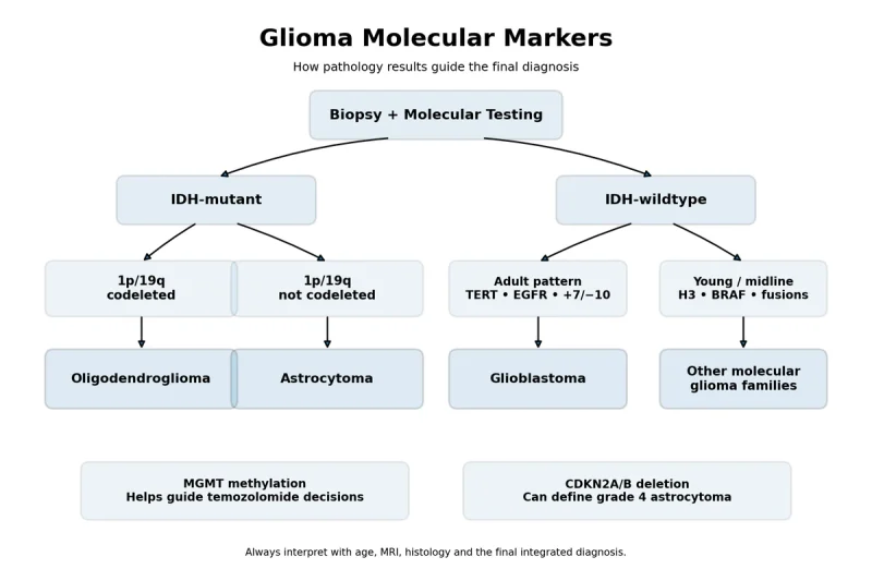
Glioma molecular markers help identify the exact tumour type, determine its grade and assess whether particular treatments may be appropriate. The necessary tests depend on the patient’s age, tumour location and microscopic findings, so the same molecular panel is not suitable for every glioma. [2, 3]
Molecular Markers in Adult Diffuse Gliomas
For adults with diffuse gliomas, the most clinically important findings usually include IDH1 and IDH2 status, 1p/19q codeletion, ATRX, TP53, CDKN2A/B, MGMT promoter methylation, TERT promoter mutation, EGFR amplification and combined chromosome 7 gain with chromosome 10 loss. These findings help distinguish astrocytoma, oligodendroglioma and glioblastoma within the current integrated classification system. [2, 3]
An IDH mutation combined with whole-arm 1p/19q codeletion establishes oligodendroglioma in the appropriate pathological setting. An IDH-mutant tumour without this codeletion may follow an astrocytic pathway, particularly when ATRX loss and TP53 alteration are present. These markers must be interpreted together rather than as isolated laboratory results. [2, 3]
Homozygous CDKN2A/B deletion has a different role. In an IDH-mutant astrocytoma, it can establish CNS WHO grade 4 even when necrosis or microvascular proliferation is not identified under the microscope. This means that molecular testing can sometimes change the final grade beyond what is visible on routine histology alone. [2, 3]
MGMT promoter methylation does not determine the tumour’s name. It is mainly used to estimate the likelihood of benefit from temozolomide in the appropriate clinical setting, particularly in IDH-wildtype glioblastoma. A methylated or unmethylated result should therefore be interpreted alongside the complete diagnosis, patient age, functional status and planned oncology treatment. [2, 7]
Glioma molecular markers provide only one part of the complete clinical picture. Tumour location, CNS WHO grade, extent of surgical removal, residual disease, MRI behaviour, neurological symptoms and the planned oncology treatment must also be reviewed together. For a broader patient guide, read Gliomas: Symptoms, Diagnosis and Ayurvedic Cure for Brain Tumors, covering diagnosis, surgery, radiotherapy, chemotherapy, recurrence, rehabilitation, diet and individualized Ayurvedic support.
Markers in Younger and Midline Glioma Patients
Children, adolescents, younger adults and patients with tumours in midline structures may require additional testing for H3 K27 alterations, H3 G34 mutations, BRAF alterations, FGFR abnormalities, MYB or MYBL1 alterations and selected gene fusions. Applying an adult diffuse-glioma panel alone may miss a biologically distinct paediatric-type or young-adult tumour. [2, 3]
DNA-methylation profiling can be valuable when the tumour’s microscopic appearance and initial molecular findings do not produce a secure diagnosis. It compares the tumour’s molecular pattern with established reference classes and may help resolve unusual, conflicting or rare cases when interpreted together with histology, imaging and clinical information. [3, 4]
How Patients Should Read These Results
The most important line in the pathology report is the final integrated diagnosis, because it combines the microscopic findings, molecular results and CNS WHO grade. A single result such as “IDH-wildtype,” “MGMT methylated” or “TERT-mutant” cannot provide a complete prognosis or treatment recommendation on its own. [2, 3]
Patients should also check whether the report states which testing method was used, whether the tissue was adequate and whether any result was indeterminate, incomplete or limited by a small biopsy. When an important marker is missing or conflicts with the tumour’s appearance or MRI findings, additional testing or specialist neuropathology review may be clinically valuable. [2, 4]
Why Glioma Diagnosis Combines Histology and Molecular Testing

Modern glioma diagnosis combines histology and molecular testing because the appearance of tumour cells alone cannot always identify the tumour’s biological type, expected behaviour or most appropriate treatment pathway. Histology remains essential, but molecular findings now help establish the integrated diagnosis, refine the CNS WHO grade and distinguish tumours that may look similar under the microscope yet behave differently in patients. [1–3]
What Histology Reveals
Histology examines tumour tissue removed during biopsy or surgery. A neuropathologist evaluates the shape and arrangement of the cells, the degree of infiltration into surrounding brain tissue, cellular density, nuclear atypia and evidence of active cell division. The report may also describe necrosis, microvascular proliferation and other features associated with more aggressive behaviour. [1, 2]
These findings remain clinically important because they show how the tumour is growing and whether high-grade characteristics are visible in the sampled tissue. Necrosis and microvascular proliferation, for example, can support a grade 4 diagnosis in the appropriate diffuse glioma. Mitotic activity can also contribute to grading, although no single microscopic feature should be interpreted separately from the complete pathological assessment. [1, 2]
Histology has practical limitations. Gliomas can be heterogeneous, meaning that one region may contain relatively less aggressive tissue while another contains higher-grade disease. A small stereotactic biopsy may therefore sample only one part of the tumour. The pathology findings should be compared with the MRI appearance, the anatomical site, the surgical observations and the amount of representative tumour tissue available for testing. [2, 4]
What Molecular Testing Adds
Molecular testing identifies genetic, chromosomal and epigenetic changes that help determine what type of glioma is present. In adult diffuse gliomas, IDH1 and IDH2 status forms a major diagnostic division. An IDH-mutant tumour with whole-arm 1p/19q codeletion is classified as oligodendroglioma, whereas an IDH-mutant tumour without that codeletion may be classified as astrocytoma when the remaining pathological findings support that lineage. [1–3]
Some molecular alterations can also affect the final grade. Homozygous deletion of CDKN2A/B can establish CNS WHO grade 4 in an IDH-mutant astrocytoma even when necrosis and microvascular proliferation are not seen in the available tissue. This is an important example of why a tumour’s grade can no longer be determined by microscopy alone. [1–3]
Other markers answer different clinical questions. MGMT promoter methylation does not define whether a tumour is an astrocytoma, oligodendroglioma or glioblastoma; its main value is estimating the probability of benefit from alkylating chemotherapy in the appropriate clinical setting. BRAF, H3, FGFR, MYB, MYBL1 and selected gene-fusion findings may be particularly important in paediatric, young-adult, midline or otherwise unusual gliomas. [2, 3]
Molecular testing may involve immunohistochemistry, targeted sequencing, next-generation sequencing, chromosomal analysis, fusion testing or DNA-methylation profiling. The most appropriate method depends on the suspected tumour type, patient age, tissue quantity and results already obtained. A negative immunohistochemical stain may occasionally require confirmatory sequencing when the clinical and histological pattern still suggests a mutation not detected by the initial test. [2–4]
Why Similar-Looking Gliomas Can Receive Different Diagnoses
Two gliomas may both appear diffusely infiltrating and astrocytic under the microscope, yet their molecular profiles may place them in different tumour families. One may be an IDH-mutant astrocytoma, while another may be an IDH-wildtype glioblastoma or a paediatric-type diffuse glioma with an H3 or MAPK-pathway alteration. These distinctions can influence prognosis, surveillance, treatment selection and eligibility for targeted therapy or clinical trials. [1–3]
The reverse situation can also occur: tumours with different microscopic appearances may belong to the same molecularly defined entity. Molecular classification therefore reduces reliance on subjective visual interpretation and helps different treatment centres use a more consistent diagnostic language. It also explains why an initial pathology report may later receive an addendum after molecular testing is completed. [2–4]
Why the Integrated Diagnosis Matters for Patients
The final integrated diagnosis combines the tumour’s microscopic appearance, molecular findings and CNS WHO grade into one clinically meaningful conclusion. This diagnosis should be interpreted together with the patient’s age, tumour location, MRI findings, extent of resection, neurological condition and planned oncology treatment. A single mutation, deletion or methylation result should not replace this complete assessment. [1–4]
For patients receiving surgery, radiotherapy, chemotherapy or targeted treatment, an accurate integrated diagnosis helps the neuro-oncology team select the most appropriate pathway and avoid treating biologically different gliomas as though they were the same disease. It also provides a more reliable foundation for discussing prognosis, follow-up imaging and the value of additional testing when the first specimen is limited or the findings do not fully agree. [2–4]
Any individualized Ayurvedic supportive plan should be developed only after the integrated pathology report and current oncology strategy have been reviewed. The tumour type, grade, treatment schedule, neurological symptoms, digestive tolerance, liver and kidney function and concurrent medicines all influence how supportive care should be planned safely and meaningfully.
How to Read a Glioma Pathology Report

A glioma pathology report can appear complex because it combines tissue appearance, molecular findings, tumour grade and technical laboratory details. The most reliable way to read it is to begin with the integrated diagnosis, then review the grade, molecular results, specimen quality and any comments describing uncertainty or additional testing. The report should be interpreted together with MRI findings, the operative note and the patient’s clinical condition. [1–4]
Table : How to Read a Glioma Pathology Report
| Pathology report term | What it means | What the patient should confirm |
|---|---|---|
| Integrated diagnosis | Final tumour name combining histology and molecular findings | Confirm that this is the latest signed diagnosis rather than a preliminary description |
| Histological diagnosis | Describes how the tumour appears under the microscope | Check whether the description was later refined by molecular testing |
| CNS WHO grade | Indicates biological aggressiveness within that specific tumour type | Ask whether the grade was determined by microscopic findings, molecular findings or both |
| Mutant | A defined genetic alteration was detected | Confirm the exact gene and mutation |
| Wildtype | The tested alteration was not detected within the assay’s scope | Check whether complete sequencing or only a limited test was performed |
| Methylated | Methylation was detected above the laboratory threshold | For MGMT, ask how the result affects the temozolomide discussion |
| Unmethylated | Methylation did not reach the laboratory threshold | Interpret as a lower probability of benefit, not an absolute prediction |
| Amplified | Many additional copies of a gene were detected | Confirm that true high-level amplification was found rather than simple copy-number gain |
| Codeleted | Both specified chromosome regions were lost | For oligodendroglioma, confirm whole-arm 1p and 19q codeletion with an IDH mutation |
| Retained expression | The tested protein remains visible on immunohistochemistry | Ask whether retained staining fully answers the molecular question |
| Loss of expression | The protein is absent from tumour-cell nuclei or cells | Confirm whether the pattern supports a specific molecular alteration |
| Indeterminate or equivocal | The laboratory could not provide a confident positive or negative result | Ask whether another tissue block or testing method could clarify it |
| Not tested | No result is available because the marker was not assessed | Do not interpret this as a negative finding |
| NOS | Required molecular information is unavailable or incomplete | Ask which essential tests are missing |
| NEC | Testing was completed, but the tumour does not fit an established WHO entity | Consider specialist neuropathology review or methylation profiling |
| Addendum pending | Additional molecular results are still being processed | Wait for the final integrated report before relying on the preliminary tumour name |
Integrated Diagnosis
The integrated diagnosis is the most important line in the report because it brings the microscopic and molecular findings together into one final tumour name. Examples include “astrocytoma, IDH-mutant,” “oligodendroglioma, IDH-mutant and 1p/19q-codeleted,” and “glioblastoma, IDH-wildtype.” The CNS WHO grade is usually stated alongside or immediately after this diagnosis. [1–3]
Each part of the diagnosis has a specific purpose. The first term identifies the tumour family, the molecular description confirms its biological category, and the grade indicates its expected clinical behaviour. A report that states only “glioma,” “high-grade glioma” or “malignant glioma” may represent a preliminary description rather than the final integrated diagnosis, particularly when molecular testing is still pending.
Patients may receive an initial surgical pathology report followed by one or more addenda. This usually occurs because histology can be reviewed before sequencing, chromosomal analysis, MGMT testing or DNA-methylation profiling is complete. The most recent signed report should therefore be used for treatment planning.
Histological Diagnosis
The histological diagnosis describes what the tumour looked like under the microscope. It may include terms such as diffuse astrocytic glioma, oligodendroglial morphology, high-grade glioma or glioblastoma. Histology also records cellularity, nuclear atypia, mitotic activity, necrosis and microvascular proliferation. [1, 2]
This part of the report remains clinically valuable, but it may not be the final tumour name. A tumour that appears oligodendroglial under the microscope cannot be classified as oligodendroglioma unless an IDH mutation and whole-arm 1p/19q codeletion are demonstrated. Similarly, an astrocytic appearance does not by itself distinguish an IDH-mutant astrocytoma from an IDH-wildtype diffuse glioma. [1–3]
The pathology report may also describe the specimen as a biopsy, subtotal resection or gross-total resection specimen. A small biopsy provides less tissue than a surgical resection and may not represent every region of a heterogeneous tumour. The neuropathologist may therefore include comments about sampling limitations or recommend correlation with MRI findings.
CNS WHO Grade
The CNS WHO grade describes the biological aggressiveness of the tumour within its specific diagnostic category. Grades are not interpreted identically across every glioma type, so the grade must always be read together with the integrated diagnosis. A grade 4 IDH-mutant astrocytoma and a grade 4 IDH-wildtype glioblastoma are distinct tumour entities with different molecular backgrounds. [1–3]
The grade may be determined by microscopic findings, molecular findings or both. Mitotic activity, necrosis and microvascular proliferation can influence grading. In an IDH-mutant astrocytoma, homozygous CDKN2A/B deletion can establish CNS WHO grade 4 even when necrosis and microvascular proliferation are not identified in the sampled tissue. [1–3]
A pathology grade should not be confused with a clinical stage. Gliomas are generally classified by tumour type and CNS WHO grade rather than by the conventional stage I to stage IV system used for many cancers elsewhere in the body. Prognosis also depends on age, tumour location, extent of resection, neurological function, molecular profile and response to treatment.
Molecular Findings
The molecular section lists the alterations tested and the method used to detect them. Results may be reported as mutant, wildtype, methylated, unmethylated, deleted, retained, amplified, rearranged, positive, negative or indeterminate. Each word has a different meaning and should be interpreted according to the specific marker and testing method. [2–4]
“Mutant” means that a defined genetic alteration was detected. “Wildtype” means that the tested alteration was not detected within the limits of that assay. “Retained expression” on immunohistochemistry means that the relevant protein remains visible in tumour-cell nuclei, whereas “loss of expression” may support an underlying molecular alteration. These terms do not have the same clinical significance for every gene.
For IDH testing, the report may first state whether IDH1 R132H immunohistochemistry was positive or negative. A negative stain does not always complete the assessment, because less common IDH1 or IDH2 mutations require sequencing. Whether further testing is needed depends on the patient’s age, tumour location and histological pattern. [2, 3]
For 1p/19q, the report should establish whole-arm codeletion when oligodendroglioma is being considered. An isolated loss of 1p or 19q does not meet the defining molecular requirement. The report should also specify whether the result was obtained by FISH, chromosomal array, loss-of-heterozygosity analysis, next-generation sequencing or another validated method. [2–4]
MGMT promoter methylation is usually reported separately because it is primarily a treatment-predictive and prognostic biomarker. A methylated result may support greater sensitivity to temozolomide in the appropriate clinical setting, while an unmethylated result indicates a lower probability of benefit rather than an absolute absence of response. Laboratory method, tumour-cell content and assay cut-off can influence interpretation. [2, 4]
The report may also include TERT promoter mutation, EGFR amplification, chromosome 7 gain with chromosome 10 loss, ATRX expression, TP53 alteration, CDKN2A/B deletion, H3 alterations, BRAF changes or gene fusions. These findings should be interpreted as part of a pattern. A single abnormality should not be separated from the tumour’s age group, location, morphology and broader molecular profile.
Specimen Quality and Technical Comments
The report should indicate whether the tissue was adequate for diagnosis and molecular testing. Factors such as extensive necrosis, low tumour-cell content, haemorrhage, cautery artefact, fixation problems or very limited tissue can reduce the reliability of some tests. [2, 4]
An “indeterminate” result means that the laboratory could not confidently classify the finding as positive or negative. This may occur because the signal was near the assay threshold, the DNA quality was poor or the amount of tumour was insufficient. Indeterminate is therefore different from wildtype, negative or unmethylated.
“Not tested” means that the marker was not assessed. It should not be interpreted as a negative result. When an untested marker is essential for classification or treatment planning, the neuro-oncology team may request additional testing on stored tissue or consider repeat sampling when clinically appropriate.
What NOS and NEC Mean
NOS means “not otherwise specified.” This designation is generally used when the molecular information required for a more precise diagnosis is unavailable, incomplete or technically unsuccessful. For example, a diffuse glioma may remain NOS when essential IDH or 1p/19q results cannot be obtained. [1–4]
NEC means “not elsewhere classified.” This term is used when appropriate diagnostic testing has been completed, but the combined findings do not fit an established WHO tumour entity. An NEC diagnosis may reflect an unusual molecular combination or a tumour that falls outside current classification categories. [1, 4]
NOS and NEC should prompt careful review of which tests were performed, whether the sample was adequate and whether specialist neuropathology consultation or DNA-methylation profiling could clarify the diagnosis. These designations do not automatically indicate a worse prognosis; they describe the level of diagnostic classification achieved.
Pathology Comments and Final Clinical Interpretation
The comment section often explains why the pathologist selected the final diagnosis, which findings remain uncertain and whether further testing is recommended. It may also note that the molecular pattern should be correlated with age, tumour site, MRI appearance and clinical history.
Patients should use the integrated diagnosis, CNS WHO grade and final molecular interpretation as the basis for discussions with the neurosurgeon, neuropathologist, radiation oncologist and medical neuro-oncologist. Treatment decisions should not be based on one isolated marker or on an early preliminary report when the complete molecular work-up is still pending.
For individualized Ayurvedic supportive care, the final pathology report should be reviewed together with the oncology plan, neurological symptoms, treatment schedule, digestive capacity, liver and kidney function and concurrent medicines. This allows supportive treatment to be aligned with the confirmed tumour biology and the patient’s current clinical needs rather than being selected from the tumour name alone.
IDH1 and IDH2 Mutations in Glioma
IDH1 and IDH2 mutations in glioma are among the most important findings in modern neuropathology because they help define the tumour family, clarify prognosis and guide treatment planning. IDH status should appear in the integrated diagnosis of an adult diffuse glioma rather than being treated as an isolated laboratory result. [1–3]
What an IDH Mutation Means
IDH1 and IDH2 genes normally produce enzymes involved in cellular energy metabolism. When either gene develops a characteristic mutation, the altered enzyme produces D-2-hydroxyglutarate, an abnormal metabolite that changes DNA and histone methylation and interferes with normal cell maturation. These molecular effects help create the biological environment in which an IDH-mutant glioma develops. [1–3]
IDH1 mutations are more common than IDH2 mutations in diffuse gliomas. The IDH1 R132H alteration is the most frequently identified variant, while other IDH1 substitutions and IDH2 mutations, particularly involving IDH2 R172, are less common. A report stating only that IDH1 R132H immunohistochemistry is negative does not necessarily exclude every possible IDH1 or IDH2 mutation. [2, 3]
In most patients, an IDH mutation is an acquired alteration within the tumour rather than evidence of an inherited cancer syndrome. Its principal clinical importance is therefore the classification and management of the glioma itself. Family members do not ordinarily require genetic testing solely because an IDH mutation was identified in the tumour. [1–3]
An IDH mutation should not be interpreted as another term for a low-grade tumour. IDH-mutant astrocytomas may be classified as CNS WHO grade 2, 3 or 4, depending on their histological and molecular characteristics. Oligodendrogliomas with an IDH mutation and whole-arm 1p/19q codeletion are classified as CNS WHO grade 2 or 3. [1–3]
How IDH Testing Is Performed
Testing commonly begins with immunohistochemistry for the IDH1 R132H mutant protein. This method allows the neuropathologist to see whether tumour cells express the most common mutant form while examining the tissue under a microscope. A clearly positive stain usually establishes the presence of an IDH mutation in the appropriate tumour context. [2, 3]
When the IDH1 R132H stain is negative, the next step depends on the patient’s age, tumour location and microscopic appearance. Sequencing of IDH1 and IDH2 is particularly important when the tumour still appears biologically compatible with an IDH-mutant diffuse glioma, because immunohistochemistry directed at IDH1 R132H will not detect uncommon IDH1 variants or IDH2 mutations. [2, 3]
Sequencing may be performed through a targeted assay, Sanger sequencing or a broader next-generation sequencing panel. A wider panel can simultaneously assess IDH together with TP53, ATRX, TERT, CDKN2A/B, BRAF and other clinically relevant genes, although the exact panel varies between laboratories and tumour types. [2–4]
The wording of the report should be reviewed carefully. “IDH1 R132H negative by immunohistochemistry” describes the result of one protein-based test, whereas “IDH1 and IDH2 wildtype by sequencing” indicates that a broader molecular assessment was completed. The second statement provides stronger evidence that the tumour is truly IDH-wildtype, provided that the specimen contained adequate tumour tissue and the assay covered the relevant mutation sites. [2–4]
Low tumour-cell content, extensive necrosis, poor DNA quality or a very small biopsy can make testing less reliable. An indeterminate or technically unsuccessful result should therefore be distinguished from a confirmed wildtype result. Additional testing on another tissue block may be useful when IDH status is essential for final classification. [2, 4]
How IDH Status Defines the Glioma Type
In adult diffuse gliomas, IDH status creates the first major diagnostic division. When an IDH mutation is present, whole-arm 1p/19q status is assessed to distinguish oligodendroglioma from astrocytoma. An IDH-mutant tumour with whole-arm codeletion of both 1p and 19q is classified as oligodendroglioma. [1–3]
An IDH-mutant diffuse glioma without whole-arm 1p/19q codeletion is generally classified within the astrocytoma pathway when the remaining findings support that diagnosis. Loss of ATRX expression and TP53 alteration commonly support an astrocytic lineage, but the complete molecular pattern must be considered before the integrated diagnosis is finalized. [1–3]
IDH-wildtype means that a reportable IDH1 or IDH2 mutation was not detected by the testing performed. It is not, by itself, a complete tumour diagnosis. In an adult diffuse astrocytic tumour, the neuropathologist may assess necrosis, microvascular proliferation, TERT promoter mutation, EGFR amplification and combined whole-chromosome 7 gain with whole-chromosome 10 loss when determining whether the tumour meets the criteria for glioblastoma, IDH-wildtype. [1–3]
Age remains important when interpreting an IDH-wildtype result. In children, adolescents and younger adults, an IDH-wildtype diffuse glioma may belong to a paediatric-type molecular family involving H3, BRAF, FGFR, MYB, MYBL1 or another alteration. Current diagnostic guidance therefore supports broader molecular evaluation before applying an adult glioblastoma classification to a younger patient whose age, histology or tumour location suggests another entity. [2, 3, 5]
A solitary molecular finding should also be interpreted within the full pathological setting. A TERT promoter mutation alone, particularly in a histologically lower-grade IDH-wildtype tumour, requires correlation with age, imaging, morphology and other molecular findings before it is used to establish an aggressive adult-type diagnosis. [5]
What IDH Status Means for Prognosis
IDH-mutant diffuse gliomas generally have a more favourable clinical course than conventional IDH-wildtype glioblastoma when comparable patient and treatment factors are considered. However, the expected outcome within the IDH-mutant group still varies according to CNS WHO grade, tumour location, residual tumour after surgery, patient age, neurological function, 1p/19q status and additional molecular alterations. [1–3]
An IDH mutation therefore provides valuable prognostic information without replacing the complete clinical assessment. A grade 2 IDH-mutant tumour and a grade 4 IDH-mutant astrocytoma share an important molecular feature but have different levels of biological risk and require different treatment discussions. [1–3]
For IDH-mutant astrocytoma, homozygous CDKN2A/B deletion is especially important because it can establish CNS WHO grade 4 even when necrosis and microvascular proliferation are not identified. For oligodendroglioma, grade is determined through the integrated histological and molecular assessment rather than by IDH status alone. [1–3]
How IDH Status Can Influence Treatment
IDH status helps the neuro-oncology team select among surveillance, radiotherapy, chemotherapy and targeted treatment according to tumour grade, residual disease, symptoms, growth pattern and the patient’s overall clinical condition. It can also determine eligibility for molecularly selected clinical trials. [2, 3]
In the United States, vorasidenib is approved for patients aged 12 years or older with grade 2 astrocytoma or oligodendroglioma carrying a susceptible IDH1 or IDH2 mutation after surgery. In the INDIGO phase III trial, vorasidenib prolonged progression-free survival and delayed the time to the next therapeutic intervention in the selected study population. [9, 10]
This indication does not apply automatically to every patient with an IDH mutation. The tumour grade, previous treatment, residual or recurrent disease, neurological condition and local regulatory approval must be reviewed before an IDH inhibitor is considered. Radiotherapy and chemotherapy remain important treatment components for many IDH-mutant gliomas, particularly when the tumour has higher-risk features. [2, 3, 9]
For integrative care, the IDH result should be reviewed together with the complete pathology report, MRI findings, extent of surgery and oncology plan. An individualized Ayurvedic supportive strategy may then be coordinated with neurological symptoms, treatment-related fatigue, appetite, sleep, digestion, liver and kidney function and concurrent medicines. The IDH mutation alone should not be used to select one fixed formulation for every patient.
1p/19q Codeletion and Oligodendroglioma

The 1p/19q codeletion is a defining molecular marker of oligodendroglioma. It refers to the combined loss of the entire short arm of chromosome 1 and the entire long arm of chromosome 19 within the tumour cells. Under the current integrated classification, a diffuse glioma can be diagnosed as oligodendroglioma only when both an IDH1 or IDH2 mutation and whole-arm 1p/19q codeletion are demonstrated. [1–3]
What 1p/19q Codeletion Means
Chromosomes carry the genetic instructions that regulate normal cell growth and function. Each chromosome has a short arm, labelled “p,” and a long arm, labelled “q.” In a 1p/19q-codeleted glioma, tumour cells have lost the whole short arm of chromosome 1 and the whole long arm of chromosome 19.
This alteration usually develops within the tumour and is not an inherited finding. Its detection does not ordinarily mean that the patient’s children, siblings or other relatives carry the same chromosomal change. Its primary role is to establish the tumour’s molecular identity and assist with prognosis and treatment planning. [1–3]
The word “whole-arm” is important. A focal or partial deletion involving only a small region of 1p or 19q is not equivalent to the characteristic whole-arm codeletion required for oligodendroglioma. A pathology report that states only “1p loss” or “19q loss” should therefore be reviewed to determine whether complete loss of both chromosome arms was confirmed. [2–4]
Isolated loss of one chromosome arm is also insufficient. Both 1p and 19q must show the defining whole-arm loss, and the tumour must carry an IDH mutation before the integrated diagnosis of oligodendroglioma can be made. [1–3]
Why IDH and 1p/19q Must Be Interpreted Together
An IDH mutation establishes that the tumour belongs to the IDH-mutant diffuse glioma group, while 1p/19q status separates the oligodendroglial pathway from the astrocytic pathway. An IDH-mutant tumour with whole-arm 1p/19q codeletion is classified as oligodendroglioma. An IDH-mutant diffuse glioma without this codeletion is generally classified as astrocytoma when the remaining pathological findings support that diagnosis. [1–3]
Microscopic appearance alone cannot establish oligodendroglioma. Some tumours have rounded nuclei, branching blood vessels or other features traditionally associated with oligodendroglial morphology, yet lack the required molecular combination. Under current classification, such a tumour should not receive a final integrated diagnosis of oligodendroglioma solely because it resembles one under the microscope. [1–3]
The opposite principle is also important. When the correct molecular combination is present, the integrated diagnosis may be oligodendroglioma even when every classic microscopic feature is not prominent in the sampled tissue. This is one reason molecular testing has improved diagnostic consistency between pathology laboratories.
A report showing apparent 1p/19q codeletion in an IDH-wildtype tumour deserves careful review. Canonical whole-arm 1p/19q codeletion is characteristically associated with an IDH mutation. A discordant result may reflect a partial deletion, technical limitation, unusual tumour type or incomplete IDH testing and may justify additional molecular analysis or specialist neuropathology review. [2–4]
ATRX and TP53 findings can provide supporting information. Oligodendrogliomas generally retain ATRX expression, whereas ATRX loss and strong evidence of TP53 alteration more commonly support an astrocytic pathway in an IDH-mutant tumour. These are supportive patterns rather than substitutes for direct 1p/19q assessment. [1–3]
How 1p/19q Testing Is Performed
Several laboratory methods can evaluate 1p and 19q. Fluorescence in situ hybridisation, commonly called FISH, uses targeted probes to compare selected chromosomal regions. It is widely available and can provide clinically useful results, but because it examines limited sites, it may not always distinguish whole-arm loss from a smaller partial deletion. [2–4]
Loss-of-heterozygosity analysis, chromosomal microarray, single-nucleotide polymorphism array and next-generation sequencing with validated copy-number analysis can assess broader portions of the chromosome arms. DNA-methylation arrays may also provide a copy-number profile that supports the presence of the characteristic codeletion while helping classify diagnostically difficult tumours. [3, 4]
The report should identify the testing method and state whether whole-arm codeletion was demonstrated. When FISH is used, the neuropathologist should interpret the result together with IDH status, ATRX expression, TP53 findings, histology, patient age and tumour location. Additional testing may be appropriate when the result is unexpected or would substantially change the diagnosis.
A result reported as “1p/19q intact” means that the defining combined whole-arm loss was not detected. It does not necessarily mean that every part of both chromosomes is structurally normal. Other gains, losses or copy-number abnormalities may still be present, but they do not establish oligodendroglioma.
An “indeterminate” result means the laboratory could not confidently classify the tumour as codeleted or intact. Low tumour-cell content, extensive necrosis, poor DNA quality, limited biopsy material or borderline assay signals may contribute to this result. Testing another tissue block or using a broader chromosomal method may provide a clearer answer. [2, 4]
What Codeletion Means for Grade and Prognosis
Oligodendroglioma, IDH-mutant and 1p/19q-codeleted, is classified as CNS WHO grade 2 or grade 3. The grade is determined through the integrated evaluation of microscopic activity and the broader pathological findings. Under the current WHO framework, oligodendroglioma is not assigned CNS WHO grade 4. [1–3]
A grade 2 oligodendroglioma generally shows lower proliferative activity, while a grade 3 tumour demonstrates more active and clinically significant growth. However, the grade should not be interpreted by itself. Age, tumour size, anatomical location, neurological symptoms, residual tumour after surgery, imaging behaviour and molecular features all influence the individual clinical course.
Patients with 1p/19q-codeleted oligodendroglioma generally have a more favourable prognosis than patients with conventional IDH-wildtype glioblastoma. Many patients experience prolonged disease control, particularly when the tumour is surgically accessible and managed through an appropriate long-term treatment plan. The clinical course can still vary substantially, and continued MRI surveillance remains important because even slower-growing oligodendrogliomas can recur or progress over time. [1–3]
The codeletion should therefore be viewed as clinically valuable information rather than as proof that the tumour is harmless. A grade 3 tumour, substantial residual disease, progressive MRI changes or worsening neurological symptoms may require active treatment even when the molecular profile is comparatively favourable.
How 1p/19q Status Influences Treatment
The 1p/19q codeletion helps identify patients who may derive substantial benefit from combined chemotherapy and radiotherapy. Long-term randomized evidence has shown improved survival with radiotherapy plus PCV chemotherapy in appropriately selected patients with codeleted anaplastic oligodendroglial tumours compared with radiotherapy alone. [8]
PCV combines procarbazine, lomustine and vincristine. Whether it is recommended depends on tumour grade, patient age, residual disease, neurological status, blood counts, organ function and expected treatment tolerance. Temozolomide is also used in clinical practice in selected oligodendroglioma settings, although treatment selection should follow the neuro-oncology team’s assessment of the available evidence and the individual patient’s circumstances.
Some patients with completely or substantially resected grade 2 oligodendroglioma may initially enter structured MRI surveillance, while others require earlier systemic treatment or radiotherapy because of residual growth, symptoms or other higher-risk features. Eligible patients with grade 2 IDH-mutant oligodendroglioma may also be considered for IDH-directed treatment according to previous therapy, regulatory approval and the complete clinical context. [9, 10]
The pathology report should therefore guide treatment without becoming the only decision-making factor. The most appropriate plan combines the integrated diagnosis with MRI findings, extent of resection, seizure control, cognitive function, tumour growth rate and the patient’s priorities.
An individualized Ayurvedic supportive plan should likewise be based on the confirmed grade, residual tumour, current oncology treatment, neurological symptoms, digestive tolerance, liver and kidney function and concurrent medicines. The presence of 1p/19q codeletion does not justify one fixed formulation for every patient; it provides a more accurate biological foundation on which coordinated supportive care can be planned.
ATRX and TP53 in Glioma

ATRX and TP53 are important supporting markers in the classification of diffuse gliomas, particularly when an IDH mutation has already been identified. Their combined pattern can strengthen the diagnosis of an IDH-mutant astrocytoma, help distinguish it from oligodendroglioma and indicate whether additional molecular testing is needed. Neither marker should be used alone to name or grade a glioma. [1–3]
What ATRX Loss Means
ATRX is a gene involved in maintaining chromatin structure and protecting telomeres, the specialised ends of chromosomes. When ATRX function is lost, tumour cells may use an alternative mechanism to maintain their telomeres, allowing continued cell division. This biological pathway is frequently seen in IDH-mutant astrocytomas. [1–3]
ATRX is commonly assessed by immunohistochemistry. In normal brain cells and many tumour cells, ATRX protein appears as nuclear staining. When tumour-cell nuclei lose ATRX staining while non-tumour cells retain it as an internal control, the report may describe “loss of nuclear ATRX expression.” This pattern supports an inactivating ATRX alteration in the appropriate pathological setting. [2–4]
ATRX loss is particularly informative when the tumour is IDH-mutant and lacks whole-arm 1p/19q codeletion. That combination supports an astrocytic lineage. By contrast, conventional oligodendrogliomas usually retain ATRX expression and are defined by the combination of an IDH mutation and whole-arm 1p/19q codeletion. [1–3]
Retained ATRX expression does not automatically exclude astrocytoma. Some IDH-mutant astrocytomas retain visible ATRX protein, and immunohistochemistry may not detect every possible ATRX alteration. When the tumour’s morphology and other molecular findings remain uncertain, direct sequencing or broader genomic testing may be needed. [2–4]
ATRX loss is also not unique to adult IDH-mutant astrocytoma. It can occur in other glioma families, including selected paediatric-type and young-adult high-grade tumours. Age, tumour location, H3 status, IDH status and the complete molecular profile must therefore be considered before ATRX is assigned diagnostic significance. [1–3]
What TP53 Alteration Means
TP53 is a tumour-suppressor gene that helps cells respond to DNA damage, control cell division and initiate repair or cell death when damage is severe. Alteration of TP53 can remove these safeguards and allow genetically abnormal cells to survive and proliferate.
TP53 mutations are common in IDH-mutant astrocytomas and frequently occur together with ATRX alterations. This combination can support the astrocytic pathway in an IDH-mutant diffuse glioma, particularly when 1p/19q codeletion is absent. [1–3]
Pathology reports often assess p53 protein expression by immunohistochemistry. A strong and diffuse nuclear staining pattern may suggest a stabilising TP53 mutation, while complete absence of staining in tumour cells may suggest another type of inactivating alteration. A scattered or variable staining pattern may reflect normal protein expression, although interpretation depends on tissue quality, tumour-cell content and the staining pattern within the specimen. [2–4]
P53 immunohistochemistry is a useful surrogate but is not identical to TP53 sequencing. Strong staining does not prove that every tumour cell carries a clinically meaningful TP53 mutation, and a less striking staining pattern does not exclude one. Direct molecular testing may clarify the result when TP53 status is important to the differential diagnosis. [2–4]
TP53 alteration is not specific to glioma and can be found in many tumour types. Even within gliomas, it cannot independently establish astrocytoma, determine CNS WHO grade or predict an individual patient’s outcome. Its value comes from how it fits with IDH status, ATRX expression, 1p/19q status, histology and other molecular findings. [1–3]
How ATRX and TP53 Support Astrocytoma Diagnosis
In an adult diffuse glioma, the combination of an IDH mutation, ATRX loss and a TP53-altered pattern strongly supports an astrocytic molecular lineage. This pattern is especially useful when the microscopic appearance is mixed or when the tumour resembles oligodendroglioma but lacks the defining whole-arm 1p/19q codeletion. [1–3]
The diagnosis of oligodendroglioma still requires direct confirmation of both an IDH mutation and whole-arm 1p/19q codeletion. ATRX retention alone cannot establish oligodendroglioma, and ATRX loss alone cannot replace 1p/19q testing when the differential diagnosis remains unresolved. [1–3]
When ATRX is lost in an IDH-mutant tumour, some diagnostic algorithms allow the neuropathologist to recognise an astrocytic pathway without relying solely on 1p/19q testing. However, the final approach depends on the laboratory method, tissue quality, morphology and the complete report. Direct chromosomal assessment remains valuable when the findings are incomplete, conflicting or clinically important. [2–4]
An unusual pattern deserves further review. Examples include apparent ATRX loss in a tumour reported as 1p/19q-codeleted, an IDH-wildtype tumour with combined ATRX and TP53 abnormalities in a younger patient, or p53 findings that conflict with the broader molecular profile. Such cases may benefit from sequencing, H3 testing, copy-number analysis, RNA-based fusion testing or DNA-methylation profiling, depending on age, location and histology. [2–4]
What These Markers Mean for Grade and Prognosis
ATRX and TP53 primarily support classification rather than directly assigning the CNS WHO grade. An IDH-mutant astrocytoma may be grade 2, 3 or 4 regardless of whether ATRX loss and TP53 alteration are present. Grade is determined through mitotic activity, necrosis, microvascular proliferation, CDKN2A/B homozygous deletion and the complete integrated assessment. [1–3]
The presence of ATRX loss or TP53 alteration should therefore not be interpreted as proof that the tumour is high-grade. Their clinical meaning depends on the established tumour type, grade, extent of surgery, residual disease, MRI behaviour and other molecular risk factors.
These markers can still provide important long-term information because they help ensure that the tumour is placed in the correct biological category. Accurate classification supports more appropriate surveillance, prognosis discussions, treatment selection and eligibility assessment for molecularly defined clinical trials. [2, 3]
For individualized Ayurvedic supportive care, ATRX and TP53 findings should be reviewed as part of the complete integrated diagnosis rather than used to choose treatment independently. The confirmed tumour type, CNS WHO grade, neurological symptoms, oncology schedule, digestive capacity, liver and kidney function and concurrent medicines remain essential when planning safe, coordinated supportive therapy.
CDKN2A/B Deletion and Glioma Grade
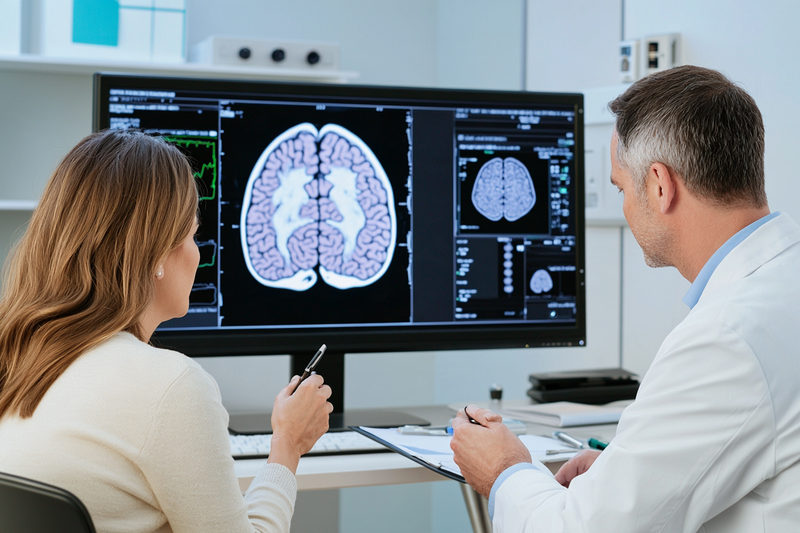
CDKN2A/B deletion is one of the most important molecular findings used to grade an IDH-mutant astrocytoma. When both copies of the CDKN2A and CDKN2B region are lost in the tumour, the finding is called a homozygous deletion. Under the current WHO classification, this alteration is sufficient to classify an IDH-mutant astrocytoma as CNS WHO grade 4, even when necrosis or microvascular proliferation is not identified in the sampled tissue. [1–3]
What CDKN2A and CDKN2B Do
CDKN2A and CDKN2B are neighbouring tumour-suppressor genes located on chromosome 9p21. They produce proteins that help regulate the cell cycle and prevent cells from progressing through division without appropriate control. CDKN2A also participates in pathways involving the RB and p53 tumour-suppressor systems. [2, 3]
When these genes are deleted, tumour cells can lose important restraints on proliferation. The effect becomes particularly significant when both copies of the region are absent, because the protective function of the genes is substantially lost. This biological change is associated with a more aggressive clinical course in IDH-mutant astrocytoma and is therefore incorporated directly into molecular grading. [1–3]
A CDKN2A/B result should always be interpreted within the final tumour diagnosis. Its grade-defining role applies specifically to astrocytoma, IDH-mutant. The same result should not be transferred automatically to every other glioma type because molecular abnormalities can have different significance in different tumour families.
Homozygous and Heterozygous Deletion
A homozygous deletion means that both copies of the relevant CDKN2A/B region have been lost in the tumour cells. This is the alteration that establishes CNS WHO grade 4 in an IDH-mutant astrocytoma under the current classification. [1–3]
A heterozygous or hemizygous deletion means that only one copy has been lost while the other remains detectable. These terms may be used slightly differently depending on the testing method and laboratory, but they generally indicate a single-copy loss rather than complete loss of both copies.
A single-copy deletion is not currently sufficient by itself to assign CNS WHO grade 4. Emerging evidence indicates that some single-copy losses may carry prognostic significance, particularly when they occur with other cell-cycle abnormalities, but their interpretation remains dependent on the complete molecular profile, histology and clinical course. [6]
The report should therefore state clearly whether the result is homozygous deletion, single-copy loss, intact, equivocal or technically unsuccessful. A general phrase such as “CDKN2A/B loss” can be clinically ambiguous unless the copy-number status is specified.
CDKN2A mutation is also different from homozygous deletion. A sequence mutation may disrupt gene function without removing both chromosomal copies. Although selected CDKN2A alterations may be associated with greater biological risk, the established WHO grade-4 criterion is homozygous CDKN2A/B deletion rather than every possible mutation or partial loss. [1–3, 6]
How CDKN2A/B Can Establish Grade 4
An IDH-mutant astrocytoma may be assigned CNS WHO grade 4 through histological or molecular evidence. Histological grade-4 findings include necrosis and microvascular proliferation. Molecular grade 4 can be established by homozygous CDKN2A/B deletion even when those features are absent in the tissue examined. [1–3]
This means that a tumour may appear less aggressive under the microscope but still receive a grade-4 integrated diagnosis after molecular testing. The diagnosis may be written as “astrocytoma, IDH-mutant, CNS WHO grade 4, with CDKN2A/B homozygous deletion.”
The molecular criterion is especially important in a small biopsy. Gliomas are heterogeneous, and a limited specimen may not include the region containing necrosis or vascular proliferation. CDKN2A/B testing can reveal biologically aggressive behaviour that would not be recognised from the sampled histology alone.
The grade-defining rule should not be applied outside the correct diagnostic setting. An IDH-wildtype diffuse glioma is classified using a different combination of histological and molecular criteria. Likewise, oligodendroglioma remains a CNS WHO grade 2 or grade 3 tumour under the current classification; CDKN2A/B deletion does not create a grade-4 oligodendroglioma category. [1–3]
Before CDKN2A/B is used for grading, the tumour should therefore have a secure integrated diagnosis of astrocytoma, IDH-mutant. IDH status, 1p/19q status, ATRX, TP53 and the overall histological pattern help confirm that the tumour belongs to the appropriate molecular family.
How CDKN2A/B Testing Is Performed
CDKN2A/B copy number may be assessed through next-generation sequencing, chromosomal microarray, single-nucleotide polymorphism array, FISH or copy-number analysis generated from a DNA-methylation array. The laboratory should use a validated method capable of distinguishing complete homozygous deletion from partial or single-copy loss. [2–4]
Next-generation sequencing panels can detect mutations and estimate copy-number changes across multiple genes, although the accuracy of copy-number analysis depends on tumour purity, sequencing depth and bioinformatic methods. Chromosomal arrays evaluate broader genomic gains and losses and can show whether deletion extends across the CDKN2A/B region.
FISH uses fluorescent probes to compare the CDKN2A region with a reference chromosome signal. It can be useful when tissue is limited, but interpretation requires careful attention to tumour-cell percentage, nuclear truncation, chromosome copy number and laboratory cut-offs. A borderline result may require confirmation with a broader genomic method.
DNA-methylation profiling may provide both a tumour-class prediction and a copy-number profile. A clear deletion at chromosome 9p21 can support the finding, but the final interpretation should consider classifier confidence, tumour purity and confirmation requirements used by the laboratory. [3, 4]
Low tumour content, extensive necrosis, poor fixation and very small biopsies can make copy-number assessment difficult. When the report describes the result as indeterminate or equivocal, another tissue block or a different validated method may provide a more definitive answer.
What the Result Means for Prognosis
Homozygous CDKN2A/B deletion identifies an IDH-mutant astrocytoma with higher biological risk than an otherwise comparable tumour without the deletion. It is associated with faster progression and shorter survival across multiple clinical series, which is why it was incorporated into WHO molecular grading. [1–3]
The finding does not determine an individual patient’s outcome by itself. Prognosis also depends on age, tumour location, neurological function, extent of resection, residual tumour volume, MRI behaviour, treatment response and additional molecular abnormalities. Two patients with the same grade and CDKN2A/B status may therefore follow different clinical courses.
The absence of homozygous deletion does not automatically make an astrocytoma low-risk. Necrosis, microvascular proliferation, significant mitotic activity and other molecular alterations may still indicate aggressive disease. The grade and prognosis must be based on the entire integrated report.
Current 2026 cIMPACT-NOW recommendations are exploring additional molecular abnormalities that may refine risk within IDH-mutant gliomas. These proposed refinements do not remove the established role of homozygous CDKN2A/B deletion as a grade-4 criterion; they indicate that future grading systems may incorporate a broader set of molecular risk features. [6]
How CDKN2A/B Status Influences Treatment Planning
A grade-4 diagnosis based on homozygous CDKN2A/B deletion can influence the intensity and timing of treatment. The neuro-oncology team may consider the finding together with the extent of surgery, residual disease, symptoms, patient age and functional status when planning radiotherapy, chemotherapy, surveillance and clinical-trial options.
The result does not identify one universally effective drug. Instead, it indicates that the tumour should be managed as a higher-risk IDH-mutant astrocytoma. Treatment decisions may therefore differ from those used for a completely resected grade-2 tumour without molecular features of increased risk.
Patients should confirm whether the report tested both CDKN2A and CDKN2B, whether the deletion was homozygous, which method was used and whether the result directly determined the assigned grade. These questions are especially important when the microscopic description appears lower-grade but the integrated diagnosis states CNS WHO grade 4.
For individualized Ayurvedic supportive care, CDKN2A/B status helps define the seriousness of the tumour and the likely intensity of oncology treatment, but it does not independently select an herbal formulation. Support should be coordinated with the confirmed grade, surgical recovery, radiotherapy or chemotherapy schedule, neurological symptoms, digestive capacity, liver and kidney function and concurrent medicines.
MGMT Promoter Methylation in Glioma
MGMT promoter methylation is an important biomarker in glioma because it helps estimate how likely a tumour is to benefit from alkylating chemotherapy, particularly temozolomide. Unlike IDH mutation or 1p/19q codeletion, MGMT promoter methylation does not determine the tumour’s diagnostic name or CNS WHO grade. Its principal value is predictive and prognostic within the appropriate clinical setting. [2–4, 7]
What MGMT Does

MGMT stands for O6-methylguanine-DNA methyltransferase. The MGMT gene produces a DNA-repair protein that removes certain alkyl groups from damaged DNA. This repair mechanism protects normal cells, but it can also help glioma cells survive chemotherapy-induced DNA damage.
Temozolomide works partly by adding methyl groups to DNA. One clinically important lesion occurs at the O6 position of guanine. When tumour cells produce substantial MGMT protein, they can repair this damage more efficiently, reducing the cytotoxic effect of temozolomide.
Promoter methylation is an epigenetic change rather than a mutation in the DNA sequence itself. When the MGMT promoter region is sufficiently methylated, transcription of the gene is often reduced. The tumour may then produce less MGMT repair protein and retain more temozolomide-induced DNA damage, increasing the probability of treatment response. [2, 7]
The relationship is biologically important but not absolute. MGMT expression is influenced by tumour heterogeneity, assay methodology, methylation density and other DNA-repair pathways. A methylated result therefore increases the probability of benefit rather than guaranteeing response, while an unmethylated result indicates a lower probability rather than complete resistance.
Methylated, Unmethylated and Indeterminate Results
A report stating “MGMT promoter methylated” means that methylation was detected above the laboratory’s validated threshold. In an appropriately classified glioblastoma, this finding is generally associated with greater sensitivity to temozolomide and a more favourable average prognosis than an unmethylated result. [2, 7]
“MGMT promoter unmethylated” means that the measured methylation level did not reach the laboratory’s positive threshold. This result suggests that the tumour may retain greater MGMT-mediated DNA-repair capacity and may be less sensitive to temozolomide. It should not be translated into the categorical statement that temozolomide can never provide benefit.
An “indeterminate,” “equivocal,” “borderline” or “grey-zone” result means that the measured value falls close to the assay cut-off or that technical factors prevent confident classification. The neuro-oncology team may review the numerical result, tumour-cell content, testing method and available tissue before deciding whether repeat testing would add clinical value. [2, 4]
Some laboratories report a percentage, methylation ratio or quantitative score in addition to the final category. These numbers cannot always be compared directly between centres because different assays examine different CpG sites, use different controls and apply different validated thresholds.
A low-level methylated result may therefore be interpreted differently from a strongly methylated result, but there is no single universal numerical cut-off that applies to every laboratory method. The final report should state both the testing method and the laboratory’s interpretation. [2, 4]
MGMT and Temozolomide Response
The clinical importance of MGMT promoter methylation was established through evidence showing that patients with methylated glioblastoma derived greater survival benefit from temozolomide combined with radiotherapy than patients whose tumours were unmethylated. [7]
This does not mean that MGMT status should independently determine all treatment decisions. In younger or medically fit patients with newly diagnosed glioblastoma, temozolomide may still be included in standard multimodal treatment even when the tumour is unmethylated. Age, functional status, extent of resection, neurological condition and patient priorities remain important.
MGMT status may have greater influence when clinicians must choose between treatment strategies, particularly in older or medically frail patients for whom combined therapy may not be suitable. In that setting, a methylated tumour may support a temozolomide-based approach, while an unmethylated result may strengthen consideration of radiotherapy-focused treatment. The final decision belongs within multidisciplinary neuro-oncology planning.
The marker may also contribute to clinical-trial selection. Some studies stratify patients according to MGMT status because methylated and unmethylated glioblastomas can respond differently to temozolomide-containing control regimens.
When MGMT Testing Is Most Useful
MGMT promoter methylation testing is most firmly established in glioblastoma, particularly glioblastoma, IDH-wildtype. Current biomarker guidance supports testing because the result can provide meaningful prognostic information and inform discussions about alkylating chemotherapy. [2, 3]
Its interpretation is less straightforward in IDH-mutant diffuse gliomas. IDH-mutant tumours frequently show widespread epigenetic changes, and MGMT methylation may be common within this biological group. In such cases, IDH status, 1p/19q status, grade and the established treatment evidence for that tumour type may be more important than MGMT alone. [2, 3]
MGMT promoter methylation is not required to diagnose astrocytoma, oligodendroglioma or glioblastoma. It also does not convert one tumour type into another and should not be used to assign CNS WHO grade.
Testing is generally performed on tumour tissue obtained during biopsy or surgical resection. Blood MGMT testing does not replace validated tissue-based assessment for glioma treatment planning.
At recurrence, MGMT results from the original tumour may still be clinically informative, but the tumour may have evolved under treatment pressure. Whether repeat testing is useful depends on the interval, available recurrent tissue, previous therapy and the specific decision being considered.
How MGMT Methylation Is Tested
Laboratories may use methylation-specific PCR, quantitative methylation-specific PCR, pyrosequencing, methylation arrays or other validated molecular methods. Each approach assesses methylation differently and has its own sensitivity, cut-off and technical requirements. [2–4]
Methylation-specific PCR provides a qualitative or semi-quantitative classification but can be sensitive to assay design and DNA quality. Pyrosequencing measures methylation at selected CpG sites and may provide a numerical percentage, although laboratories differ in which sites and thresholds they use.
DNA-methylation arrays can assess MGMT promoter methylation while also helping classify the tumour and generate a broader epigenetic profile. The result still requires interpretation through a validated algorithm and should not be read solely from one isolated probe.
Testing quality depends on the amount of viable tumour present in the sample. Low tumour-cell content, extensive necrosis, haemorrhage, treatment-related change, poor fixation or degraded DNA can affect the result. The pathologist may select a tumour-rich tissue block or macrodissect the specimen to improve assay reliability. [2, 4]
A small biopsy may contain a mixture of tumour and normal brain tissue. Because normal cells can dilute the methylation signal, the reported result should be reviewed alongside the estimated tumour percentage and any technical limitations stated in the report.
Why MGMT Results Can Differ Between Laboratories
MGMT promoter methylation is not measured by one universally standardised assay. Different laboratories may test different promoter regions, CpG sites and methylation thresholds. A sample close to the cut-off may therefore be classified as weakly methylated in one laboratory and borderline or unmethylated in another. [2, 4]
Tumour heterogeneity can also contribute. Different parts of the same glioma may contain different proportions of methylated and unmethylated tumour cells. A small biopsy and a larger resection specimen may therefore produce somewhat different quantitative results.
Treatment can further alter the cellular composition of the tumour. A recurrent specimen obtained after radiotherapy and temozolomide may contain viable tumour, treatment effect, necrosis and inflammatory cells in varying proportions, all of which can complicate molecular interpretation.
When the result will materially change treatment and the report is equivocal, clinicians may request review of the raw value, repeat testing on another tissue block or confirmation using an alternative validated method. A clear methylated or unmethylated result from an adequate specimen generally does not require routine repetition solely because another assay exists.
What MGMT Status Means for Prognosis
In glioblastoma, MGMT promoter methylation is generally associated with longer average survival and improved responsiveness to temozolomide compared with an unmethylated result. This association reflects both treatment sensitivity and underlying tumour biology. [7]
The marker cannot predict an individual survival time. Prognosis also depends on age, functional status, tumour location, extent of resection, residual enhancing disease, neurological deficits, IDH status, treatment completion and response on follow-up MRI.
An unmethylated result should therefore be used to refine expectations and treatment strategy rather than to remove therapeutic options automatically. Some patients with unmethylated tumours still achieve meaningful disease control, while some methylated tumours progress despite treatment.
MGMT status should also be interpreted within the correct tumour type. Its prognostic and predictive strength is best established in glioblastoma and should not be assumed to have identical meaning across every diffuse glioma.
Using MGMT Results in Integrative Care
MGMT promoter methylation helps anticipate the role and likely intensity of temozolomide treatment, which is clinically relevant when planning supportive care. Patients receiving temozolomide may require monitoring of blood counts, liver function, nausea, appetite, bowel function, fatigue and infection risk.
An individualized Ayurvedic supportive plan should be coordinated with the chemotherapy schedule and current laboratory results. Agni, meaning digestive and metabolic capacity, may be assessed alongside appetite, bowel pattern, body weight and treatment tolerance, but it does not replace oncology monitoring or MGMT-based treatment planning.
Herbs, concentrated extracts, mineral preparations and Rasayana therapies should be selected only after reviewing the complete pathology report, prescribed oncology medicines, liver and kidney function and potential interactions. MGMT status can inform the treatment context, but it does not independently determine one Ayurvedic formulation for every glioma patient.
TERT, EGFR and Chromosome +7/−10

TERT promoter mutation, EGFR amplification and combined chromosome 7 gain with chromosome 10 loss are major molecular findings used to classify selected IDH-wildtype diffuse gliomas. In an adult diffuse astrocytic tumour that is IDH-wildtype and H3-wildtype, these abnormalities can support a diagnosis of glioblastoma, IDH-wildtype, CNS WHO grade 4, even when necrosis or microvascular proliferation is not identified in the sampled tissue. Their interpretation must still incorporate patient age, tumour location, microscopic appearance, MRI findings and the complete molecular profile. [1–5]
TERT Promoter Mutation
TERT stands for telomerase reverse transcriptase. Telomerase helps maintain telomeres, the protective structures at the ends of chromosomes. Most mature cells have limited telomerase activity, so their capacity for repeated division is naturally restricted. A TERT promoter mutation can increase TERT expression and allow tumour cells to maintain their telomeres through continued proliferation.
The two most frequently reported TERT promoter mutations are commonly described as C228T and C250T. These alterations occur in many adult IDH-wildtype glioblastomas, but they are not exclusive to this diagnosis. TERT promoter mutations are also frequently present in oligodendroglioma and can occur in selected other central nervous system tumours. The result therefore gains diagnostic meaning only after IDH status, 1p/19q status, histology and tumour location have been established. [1–3]
In an IDH-mutant and 1p/19q-codeleted tumour, a TERT promoter mutation can support the molecular pattern of oligodendroglioma, but it is not required to establish that diagnosis. In an IDH-wildtype diffuse astrocytic tumour, the same TERT alteration may support glioblastoma classification when the remaining clinical and pathological findings are concordant.
Particular care is required when TERT promoter mutation is the only aggressive molecular finding in a histologically lower-grade, IDH-wildtype tumour. Recent cIMPACT-NOW guidance advises against using a solitary TERT promoter mutation as the automatic basis for diagnosing glioblastoma when necrosis, microvascular proliferation, EGFR amplification and the classic +7/−10 copy-number pattern are absent. [5]
In this setting, the neuropathologist may assess whether the tumour is genuinely diffuse, whether MRI shows enhancement or radiological necrosis, whether the biopsy may have undersampled a more aggressive region and whether another tumour type could explain the result. Broader sequencing, copy-number analysis or DNA-methylation profiling may help establish a more secure diagnosis. [3–5]
A TERT promoter mutation should also be distinguished from a general TERT gene alteration or increased telomerase expression. The recognised diagnostic marker is a pathogenic mutation within the promoter region. A report that states only “TERT positive” should be clarified to determine what alteration was actually tested.
EGFR Amplification
EGFR stands for epidermal growth factor receptor, a cell-surface receptor involved in growth and survival signalling. EGFR gene amplification occurs when tumour cells acquire many additional copies of the EGFR gene. This can increase receptor signalling and contribute to aggressive tumour growth.
EGFR amplification is a characteristic molecular feature in a substantial subset of IDH-wildtype glioblastomas. In the correct adult diffuse astrocytic tumour context, it can support a CNS WHO grade 4 diagnosis even when the available tissue lacks necrosis or microvascular proliferation. [1–3]
The word “amplification” has a specific meaning. High-level EGFR amplification is different from a small increase in chromosome 7 copy number, increased EGFR protein staining or a single EGFR sequence variant. A report describing “EGFR gain,” “EGFR overexpression,” “EGFR mutation” or “EGFR amplification” is referring to different biological findings, and these terms should not be treated as interchangeable.
Immunohistochemistry may demonstrate increased EGFR protein expression, but protein staining alone does not reliably establish gene amplification. Amplification is usually assessed through FISH, chromosomal microarray, validated next-generation sequencing copy-number analysis or a copy-number profile obtained from DNA-methylation testing. [2–4]
Some glioblastomas with EGFR amplification also contain EGFRvIII, a rearranged EGFR variant produced by deletion of part of the gene. EGFRvIII may be reported separately because it is biologically distinct from ordinary amplification. Its presence does not change the fundamental requirement to interpret the tumour through the complete integrated diagnosis.
EGFR amplification is not completely specific to conventional adult glioblastoma. It can occasionally occur in other diffuse high-grade gliomas, including selected paediatric-type or young-adult tumours. This distinction is particularly important in patients younger than 40 years, for whom current guidance recommends that EGFR amplification should not serve as the only defining feature of glioblastoma. [5]
In a younger patient, assessment may include H3 alterations, BRAF status, other paediatric-type high-grade glioma markers, broader sequencing and DNA-methylation profiling. This approach helps prevent biologically different tumours from being placed into the same treatment category solely because they share EGFR amplification.
Although EGFR amplification identifies an important biological pathway, it does not automatically select an effective standard EGFR-targeted drug. Blood–brain barrier penetration, tumour heterogeneity, downstream signalling and treatment resistance have limited the effectiveness of many EGFR-directed strategies in glioblastoma. The finding may still support eligibility for selected molecular trials, but treatment decisions should follow evidence for the specific agent and tumour setting. [2, 3]
Chromosome 7 Gain and Chromosome 10 Loss
The +7/−10 signature refers to gain of an entire copy of chromosome 7 together with loss of an entire copy of chromosome 10. It is a broad copy-number pattern affecting many genes at the same time rather than a mutation in one individual gene.
Chromosome 7 contains several genes associated with tumour growth, including EGFR, while chromosome 10 contains tumour-suppressor genes such as PTEN. Gain of chromosome 7 combined with loss of chromosome 10 can therefore increase growth-promoting signals while reducing important controls on cell survival and proliferation.
In an adult diffuse astrocytic glioma that is IDH-wildtype and H3-wildtype, the classic whole-chromosome +7/−10 pattern can support a diagnosis of glioblastoma, IDH-wildtype, CNS WHO grade 4. This may apply even when the tumour appears histologically grade 2 or grade 3 in the tissue examined. [1–3]
The report should distinguish the complete whole-chromosome signature from partial gains or losses. Gain of only part of chromosome 7, loss of only part of chromosome 10 or the presence of only one side of the pattern is not identical to the classic +7/−10 criterion. Such findings may still carry biological significance, but they require interpretation through the broader copy-number profile and pathological setting.
The +7/−10 pattern can be detected through chromosomal microarray, single-nucleotide polymorphism array, validated next-generation sequencing copy-number analysis or DNA-methylation array data. The report may describe it as “whole chromosome 7 gain and whole chromosome 10 loss,” “trisomy 7 with monosomy 10,” or “+7/−10 copy-number signature.” [2–4]
As with EGFR amplification, the pattern is less specific in children and younger adults. Selected H3 G34-mutant diffuse hemispheric gliomas, pleomorphic xanthoastrocytomas and other uncommon tumours may show overlapping copy-number changes. Current guidance therefore recommends that +7/−10 should not be used as the solitary defining feature of glioblastoma in a patient younger than 40 years. [5]
In that setting, the patient’s age, tumour site, histology, IDH and H3 status, BRAF findings, TERT promoter status and methylation class may all contribute to the final interpretation. A high-confidence DNA-methylation match to an established glioblastoma family can provide additional support when copy-number findings alone are not sufficiently specific. [3–5]
When These Markers Support Glioblastoma Diagnosis
Under WHO CNS5, glioblastoma, IDH-wildtype is a diffuse astrocytic glioma that is IDH-wildtype and H3-wildtype and demonstrates at least one recognised grade-4 feature. These features include necrosis, microvascular proliferation, TERT promoter mutation, EGFR gene amplification or the +7/−10 chromosome copy-number signature. [1–3]
This framework allows a tumour without visible necrosis or microvascular proliferation to receive a grade-4 diagnosis when its molecular profile establishes aggressive glioblastoma biology. The pathology report may describe such a case as a “molecularly defined glioblastoma” or state that molecular features establish glioblastoma, IDH-wildtype, CNS WHO grade 4.
The molecular criteria should be applied only after confirming that the tumour is a diffusely infiltrating astrocytic glioma. Circumscribed gliomas, glioneuronal tumours and other central nervous system neoplasms can occasionally share one of these abnormalities but follow different diagnostic pathways.
IDH and H3 status must also be considered. An IDH-mutant astrocytoma follows a separate grading system in which CDKN2A/B homozygous deletion, necrosis and microvascular proliferation are relevant. A diffuse midline glioma with an H3 K27 alteration and a diffuse hemispheric glioma with an H3 G34 mutation are also separate tumour entities, even when additional alterations overlap with glioblastoma.
Recent cIMPACT-NOW guidance refines how the WHO molecular criteria should be applied in diagnostically difficult cases. A histologically low-grade, TERT-only tumour requires expanded assessment, while EGFR amplification or +7/−10 as a solitary finding in a patient younger than 40 years should prompt evaluation for paediatric-type and other molecularly distinct gliomas. These recommendations strengthen diagnostic precision without changing the importance of these markers in an appropriate adult glioblastoma profile. [5]
What These Results Mean for Patients
TERT promoter mutation, EGFR amplification and +7/−10 primarily help classify the tumour and establish its biological grade. They do not provide an exact survival prediction, and they do not replace assessment of tumour location, surgical resection, residual disease, neurological function, MGMT promoter methylation, patient age and treatment response.
When these findings establish glioblastoma, IDH-wildtype, CNS WHO grade 4, they can change the urgency and intensity of treatment planning even when the biopsy initially appeared lower-grade. Surgery, radiotherapy, temozolomide and clinical-trial options are then considered according to the patient’s complete condition, while MGMT status helps refine the discussion about likely temozolomide benefit.
Patients should confirm whether TERT promoter mutation, EGFR amplification and +7/−10 were each directly tested, inferred from a broader molecular assay or reported as absent. They should also check whether the tumour was confirmed as both IDH-wildtype and H3-wildtype, whether the sample contained adequate viable tumour and whether age-specific alternative diagnoses were considered.
An individualized Ayurvedic supportive plan should be coordinated with the confirmed integrated diagnosis and the intensity of planned oncology treatment. These molecular findings may indicate a need for closer monitoring and more intensive multimodal care, but they do not independently determine an Ayurvedic formulation. Supportive planning should incorporate surgical recovery, radiotherapy and chemotherapy schedules, neurological symptoms, blood counts, digestive tolerance, liver and kidney function and all concurrent medicines.
Molecular Markers in Midline and Younger Patients

Molecular testing in midline gliomas and younger patients requires a broader approach than the standard adult diffuse-glioma pathway. A tumour that is IDH-wildtype in a child, adolescent or young adult may belong to a distinct biological family defined by H3, BRAF, FGFR, MYB, MYBL1 or receptor-tyrosine-kinase alterations. Age, anatomical location and growth pattern therefore determine which markers should be tested and how the results should be interpreted. [1–4]
Why Age and Tumour Location Change the Molecular Work-Up
Adult-type diffuse gliomas are commonly separated first by IDH status and then by 1p/19q codeletion. This remains useful in younger adults, but it does not capture the full spectrum of gliomas arising in children, adolescents or midline structures. Many tumours in these groups are IDH-wildtype because they develop through entirely different molecular pathways. [1, 3]
The anatomical site can be as important as the mutation. A diffuse tumour centred in the pons, thalamus or spinal cord raises a different molecular differential diagnosis from a superficial hemispheric tumour causing seizures or a large hemispheric mass in an infant. The same alteration may also carry different diagnostic significance depending on whether the tumour is diffuse, circumscribed, midline or hemispheric.
This distinction is particularly important when a younger patient has an IDH-wildtype tumour with EGFR amplification, TERT promoter mutation or chromosome +7/−10. Current guidance supports broader molecular assessment before such a tumour is classified as conventional adult-type glioblastoma. In patients younger than 40 years, EGFR amplification or +7/−10 should not be used as the sole defining feature without considering paediatric-type diffuse high-grade gliomas and other molecularly distinct entities. [5]
A limited biopsy may make this process more difficult because some tumours show overlapping microscopic features. High cellularity, mitotic activity, necrosis and vascular proliferation can occur in several molecular families. Sequencing, copy-number analysis, RNA-based fusion testing and DNA-methylation profiling may therefore be needed to establish the integrated diagnosis. [2–5]
Diffuse Midline Glioma, H3 K27-Altered
Diffuse midline glioma, H3 K27-altered, is a high-grade infiltrating glioma that arises in a midline structure. Common sites include the pons and other parts of the brainstem, thalamus and spinal cord, although additional midline locations may be involved. It occurs most often in children and adolescents but can also affect adults. [1, 3]
The diagnosis requires more than detection of an H3 alteration. The tumour must show diffuse growth within an anatomically appropriate midline location and demonstrate disruption of the H3 K27 pathway. A tumour carrying an H3 K27 mutation outside this context should not automatically be classified as diffuse midline glioma.
The best-known molecular mechanism is a lysine-to-methionine substitution at position 27 of a histone H3 protein, historically called H3 K27M. The current name “H3 K27-altered” is broader because the same biological pathway can be disrupted through other mechanisms, including EZHIP overexpression and selected EGFR-associated molecular patterns. These tumours commonly show loss of trimethylation at H3 K27, reported as loss of H3K27me3 expression. [1, 3, 4]
Immunohistochemistry may be used to detect the common H3 K27M mutant protein and assess H3K27me3 expression. Sequencing can identify the exact histone-gene alteration, while broader molecular testing can detect alternative mechanisms when the mutant-specific stain is negative but the clinical and pathological pattern remains suggestive.
Loss of H3K27me3 alone should be interpreted carefully because it is a pathway-level finding rather than a completely tumour-specific result. The final diagnosis should incorporate the midline location, diffuse growth, histology and confirmatory molecular information.
Diffuse midline glioma, H3 K27-altered, is assigned CNS WHO grade 4 because of its underlying biology and clinical behaviour. This grade applies even when a small biopsy does not show extensive necrosis or microvascular proliferation. The tumour’s grade is therefore established by its integrated molecular identity rather than by microscopic appearance alone. [1, 3]
The exact tumour location has major implications for treatment. A lesion within the pons, thalamus or spinal cord may not be safely removable in the same way as a superficial hemispheric glioma. Decisions about biopsy, surgery, radiotherapy and clinical-trial eligibility should be made by a specialist paediatric or adult neuro-oncology team according to age, neurological status and procedural risk.
Diffuse Hemispheric Glioma, H3 G34-Mutant
Diffuse hemispheric glioma, H3 G34-mutant, is a distinct high-grade glioma arising in the cerebral hemispheres, most often in adolescents and young adults. It is defined by a mutation affecting glycine 34 of histone H3.3, usually reported as H3 G34R or H3 G34V. [1, 3]
These tumours may involve the frontal, parietal, temporal or occipital lobes and can present with seizures, headaches, focal neurological deficits, cognitive changes or symptoms related to increased intracranial pressure. Their imaging and microscopic appearance can overlap with glioblastoma and other high-grade tumours, making molecular confirmation essential.
H3 G34-mutant gliomas frequently show accompanying TP53 alterations and ATRX loss. These supporting findings can help the neuropathologist recognise the molecular pattern, but neither TP53 nor ATRX alone establishes the diagnosis. Direct demonstration of the relevant H3 G34 mutation is required within the appropriate hemispheric tumour context. [1–3]
Immunohistochemistry may detect common H3 G34R alterations, while sequencing can identify both G34R and G34V variants and clarify cases with inconclusive staining. Broader sequencing is also useful because these tumours may contain additional abnormalities that influence biological interpretation or clinical-trial eligibility.
Diffuse hemispheric glioma, H3 G34-mutant, is assigned CNS WHO grade 4. It should be distinguished from glioblastoma, IDH-wildtype, because the two entities arise through different molecular pathways, commonly affect different age groups and may be managed within different trial or research frameworks. [1, 3]
The diagnosis is especially important in a young patient whose tumour has been provisionally described as “high-grade glioma” or “glioblastoma.” Testing for H3 G34 alterations can convert a general morphological description into a more precise integrated diagnosis and may prompt additional age-appropriate molecular evaluation.
BRAF and MAPK-Pathway Alterations
The MAPK pathway regulates cell growth, differentiation and survival. Alterations affecting BRAF, FGFR and other MAPK-pathway genes occur across several paediatric and young-adult gliomas. These abnormalities can help identify the tumour type and may also support discussion of targeted treatment when standard therapy, recurrence status and available evidence make this appropriate. [1, 3, 4]
BRAF V600E is a specific activating point mutation. It can occur in pleomorphic xanthoastrocytoma, ganglioglioma, selected paediatric-type diffuse gliomas and some high-grade gliomas. Its clinical significance depends on the tumour entity rather than on the mutation alone.
A BRAF fusion is biologically different from BRAF V600E. The KIAA1549::BRAF fusion is commonly associated with pilocytic astrocytoma, particularly in children, although other BRAF rearrangements can occur. A report stating “BRAF-positive” should therefore be clarified to determine whether it refers to V600E, a fusion, amplification or another alteration.
This distinction matters because different BRAF abnormalities may respond differently to targeted drugs. A therapy designed for BRAF V600E should not be assumed to have the same effect in a BRAF-fusion tumour. Treatment selection requires the exact alteration, tumour type, age, prior therapy and evidence for central nervous system activity to be reviewed together.
BRAF V600E may initially be assessed by mutation-specific immunohistochemistry, followed by sequencing when confirmation is required. Fusion detection is often better performed through RNA sequencing, targeted fusion assays or other methods capable of identifying the rearranged gene partners.
FGFR1 alterations, including tyrosine-kinase-domain duplication, can occur in selected paediatric low-grade gliomas. Other FGFR alterations and MAPK-pathway changes may also appear in diffuse or circumscribed tumours. Their presence should be matched with histology, imaging and methylation class because similar pathway activation can occur across more than one diagnostic entity. [1, 3, 4]
The WHO category of diffuse low-grade glioma, MAPK pathway-altered, reflects this biology in selected paediatric-type diffuse tumours. A MAPK alteration alone does not automatically establish the diagnosis; the tumour must also fit the appropriate morphological, anatomical and molecular context, and other established entities should be excluded.
MYB and MYBL1 Alterations
MYB and MYBL1 are transcription-factor genes involved in cellular development and differentiation. Rearrangements or other activating alterations involving these genes occur in selected paediatric-type low-grade diffuse gliomas, particularly tumours affecting children, adolescents and younger adults. [1, 3]
Diffuse astrocytoma, MYB- or MYBL1-altered, commonly arises in the cerebral hemispheres and may present with long-standing seizures. These tumours often have a more indolent clinical course than diffuse high-grade gliomas, although management still depends on tumour location, extent of resection, symptoms and interval growth.
The exact alteration may involve a gene fusion, amplification or structural rearrangement. RNA sequencing can be particularly useful because it identifies expressed fusion transcripts and their partner genes. DNA-based testing may also detect selected rearrangements, but some fusions are more reliably captured through RNA-based methods.
MYB alterations also occur in other tumour types, including angiocentric glioma, where a characteristic MYB::QKI fusion may be present. The same gene family can therefore appear in more than one diagnosis. The tumour’s architecture, anatomical pattern, methylation class and full molecular profile are needed to determine which entity is present. [1, 3, 4]
For patients and families, identifying a MYB- or MYBL1-altered low-grade tumour can prevent it from being grouped incorrectly with more aggressive IDH-wildtype diffuse gliomas. It may also support a surveillance-oriented or surgery-focused strategy when the clinical circumstances permit.
NTRK, ALK, ROS1 and MET Fusions
Infant-type hemispheric glioma is a molecularly distinct tumour that typically arises in the cerebral hemispheres during infancy. These tumours are often driven by gene fusions involving receptor tyrosine kinases such as NTRK1, NTRK2, NTRK3, ALK, ROS1 or MET. [1, 3, 4]
The fusion joins part of one gene to another, creating an abnormal growth signal that remains continuously active. The exact fusion partner can vary, so testing methods need to detect a broad range of possible rearrangements rather than search for only one predefined mutation.
RNA sequencing is often especially valuable because it can identify the expressed fusion and confirm that the rearrangement produces an active transcript. Some DNA-based next-generation sequencing panels can also detect these fusions, but their sensitivity depends on assay design and whether the relevant intronic regions are adequately covered.
These tumours may appear highly cellular or high-grade under the microscope, yet their molecular identity can create specific therapeutic opportunities. Detection of an NTRK, ALK, ROS1 or MET fusion may support consideration of a matched targeted treatment or clinical trial, particularly when complete surgical removal is not possible or the tumour progresses. The choice of agent depends on patient age, the exact fusion, regulatory availability and evidence for activity within the central nervous system.
Although these fusions are most characteristic of infant-type hemispheric glioma, related alterations may occasionally be found in older children or adults. The patient’s age remains highly informative but should not prevent molecular testing when the tumour’s morphology and location suggest a fusion-driven process.
A negative result from a narrow DNA panel does not always exclude a fusion. When the diagnosis remains uncertain or a targetable rearrangement is clinically important, RNA sequencing or another validated fusion assay may provide additional information. [3, 4]
What These Markers Mean for Treatment Planning
The molecular diagnosis can directly change the clinical pathway for a younger or midline glioma patient. H3 K27 or H3 G34 alterations establish distinct grade-4 tumour entities, while BRAF, FGFR, MYB, MYBL1 and receptor-tyrosine-kinase alterations may identify biologically different low-grade, high-grade or potentially targetable tumours.
Treatment planning should integrate the molecular result with tumour location, resectability, neurological symptoms, MRI behaviour, patient age and developmental stage. A marker that supports targeted therapy does not automatically replace surgery, radiotherapy or established systemic treatment. It identifies an additional option that should be assessed within a specialist multidisciplinary team.
A precise diagnosis is also important when reviewing prognosis. The broad label “IDH-wildtype glioma” includes tumours with markedly different biology. A MYB-altered low-grade glioma, an H3 K27-altered diffuse midline glioma and a fusion-driven infant-type hemispheric glioma should not be given the same prognostic interpretation simply because each lacks an IDH mutation.
When tissue is limited or the findings remain conflicting, a specialist neuropathology review may help determine whether additional sequencing, RNA fusion analysis or DNA-methylation profiling is required. The decision to obtain or repeat a biopsy should consider the anatomical site, procedural safety and whether the expected molecular information would change treatment.
For individualized Ayurvedic supportive care, the patient’s age, growth and nutritional needs, tumour location, neurological deficits and oncology schedule require particular attention. Support may be planned around appetite, digestion, sleep, bowel function, strength and treatment tolerance, while all formulations are reviewed for age-appropriate dosing, liver and kidney safety and compatibility with prescribed therapy. Molecular findings guide the medical treatment context but should not be used alone to select one Ayurvedic formulation.
DNA-Methylation Profiling in Glioma

DNA-methylation profiling is an advanced molecular test used to classify gliomas by examining their epigenetic pattern. Epigenetic changes regulate how genes are switched on or off without altering the underlying DNA sequence. Because tumours arising from different cells and developmental pathways develop characteristic methylation patterns, this test can help identify a glioma that remains uncertain after histology, immunohistochemistry and targeted molecular testing. [1, 3, 4]
What DNA-Methylation Profiling Measures
DNA methylation involves the addition of small chemical groups to specific regions of DNA, commonly at sites called CpG sites. These chemical marks influence gene activity and remain sufficiently stable to create a molecular signature reflecting the tumour’s biological origin and developmental pathway.
During testing, DNA is extracted from the biopsy or resection specimen and analysed across a large number of methylation sites. The resulting pattern is compared with reference data from previously classified central nervous system tumours. A computer-based classifier then estimates which established tumour class most closely matches the patient’s specimen. [3, 4]
This process differs from ordinary gene sequencing. Sequencing searches for changes in the DNA code, such as IDH1, IDH2, H3, BRAF or TERT promoter mutations. Methylation profiling examines how DNA is chemically regulated across the genome. The two methods provide complementary information and may be used together when one method alone does not fully resolve the diagnosis. [3, 4]
Methylation profiling is also different from MGMT promoter methylation testing. MGMT testing examines a specific promoter region because of its relationship to temozolomide response. Genome-wide methylation profiling assesses a much broader epigenetic pattern primarily to classify the tumour. A methylation array may also provide information relevant to MGMT status, but the result should be generated and interpreted through a validated laboratory method. [2–4]
When Methylation Profiling Is Most Useful
DNA-methylation profiling is especially valuable when the microscopic appearance and targeted molecular results do not point to one secure diagnosis. This may occur when the biopsy is small, the tumour has unusual features or the results of IDH, H3, BRAF, ATRX, 1p/19q or fusion testing appear discordant. [3, 4]
The test can help distinguish between tumour entities that share similar microscopic appearances. A high-grade tumour in a younger patient, for example, may resemble an adult-type glioblastoma while belonging to a paediatric-type high-grade molecular family. Similarly, a tumour with clear-cell or pleomorphic features may require methylation profiling to separate an oligodendroglioma from another glioma or glioneuronal tumour with overlapping morphology.
Methylation profiling may be particularly useful in paediatric, adolescent and young-adult tumours because many of these entities are defined by developmental and epigenetic biology that cannot always be recognised through routine histology. It may help classify selected diffuse paediatric-type high-grade gliomas, low-grade MAPK-pathway tumours, infant-type hemispheric gliomas and other uncommon central nervous system neoplasms. [1, 3]
Midline tumours may also benefit when H3 K27 immunohistochemistry, sequencing and anatomical findings do not produce a fully concordant diagnosis. Methylation classification can help determine whether the tumour belongs to a recognised diffuse midline glioma family or another molecular entity arising in the same anatomical region.
In adult diffuse gliomas, the test may clarify an IDH-wildtype tumour that lacks the expected combination of histology and molecular findings. It can be helpful when a histologically lower-grade tumour carries only one potentially aggressive alteration, when a younger adult has EGFR amplification or chromosome +7/−10, or when the tumour does not fit securely into an established adult-type category. [3–5]
Methylation profiling is not required for every glioma. A typical adult diffuse glioma with a coherent microscopic appearance and complete defining markers may already have a secure integrated diagnosis. For example, an IDH-mutant tumour with confirmed whole-arm 1p/19q codeletion can usually be classified as oligodendroglioma without requiring methylation profiling.
The test becomes most valuable when it is expected to resolve a meaningful diagnostic question. Its use should be considered when the result could change the tumour name, CNS WHO grade, prognosis, surveillance strategy, treatment pathway or eligibility for a clinical trial. [3, 4]
How to Read a Methylation Classifier Report
A methylation report commonly states the highest-scoring tumour class, a calibrated classifier score and, where applicable, a more specific subclass. The tumour class describes the reference group whose methylation pattern most closely resembles the patient’s specimen.
The calibrated score reflects the strength of that match. In widely used central nervous system tumour classifiers, a score of 0.9 or above is often considered a strong match to the reported class. The exact threshold, classifier version and laboratory validation should still be stated because interpretation may differ between platforms and versions. [3, 4]
A high score is most convincing when it agrees with the tumour’s location, patient age, histology, immunohistochemistry and genomic findings. For example, a strong methylation-class match to oligodendroglioma should align with an IDH mutation and whole-arm 1p/19q codeletion. A strong match to a diffuse midline glioma family should also fit the midline location and relevant H3-pathway findings.
A lower score does not automatically make the result unusable. Scores can be reduced by low tumour-cell content, poor DNA quality, extensive necrosis, treatment effect or a tumour that sits near the biological boundary between two reference classes. A lower-scoring result may still provide useful direction when it is supported by an independent molecular test and the clinical context. [3, 4]
The report may state “no match,” “no confident match” or “classifier result below threshold.” This does not mean that no tumour is present, nor does it indicate that the tumour is benign. It means that the specimen could not be assigned confidently to one of the available reference classes.
A no-match result may occur because the tumour is rare, the reference database does not yet contain an adequate comparison group or the sample quality was insufficient. It may also reflect a previously unrecognised molecular entity. In this situation, the neuropathologist can review the raw methylation pattern, copy-number profile, histology and sequencing results before deciding whether additional testing is necessary.
The classifier version matters because reference databases evolve as new tumour entities and subclasses are recognised. A specimen that previously produced no confident match may sometimes be reanalysed against a later classifier version. The pathology report should therefore document which classifier and version were used. [3, 4]
What the Copy-Number Profile Adds
Many DNA-methylation arrays also generate a genome-wide copy-number profile. This shows whether broad chromosomal regions or individual genes have been gained, lost or highly amplified within the tumour.
The copy-number plot may support clinically important findings such as whole-arm 1p/19q codeletion, combined chromosome 7 gain and chromosome 10 loss, CDKN2A/B deletion, EGFR amplification or other recurrent chromosomal abnormalities. These findings can strengthen the methylation-based diagnosis and help explain why a tumour belongs to a particular molecular family. [3, 4]
Copy-number findings should be interpreted according to the quality and amplitude of the signal. A clear high-level amplification is different from a modest copy-number gain, and a complete whole-arm deletion is different from a small focal loss. Low tumour-cell content can weaken the signal and make a genuine abnormality appear less prominent.
Some structural alterations, gene duplications or possible fusions may be suggested by the copy-number pattern, but they may require confirmation through another method. RNA sequencing, FISH, chromosomal microarray or targeted DNA sequencing may be used when the suspected alteration is diagnostic or could influence treatment.
The copy-number profile can also reveal that the proposed methylation class is biologically inconsistent with the rest of the specimen. If the classifier suggests one tumour type while the copy-number pattern, sequencing and histology support another, the case should be reviewed rather than accepting the highest score in isolation.
Tissue Quality and Technical Limitations
Methylation profiling requires sufficient tumour DNA of adequate quality. Formalin-fixed, paraffin-embedded tissue can often be used, but prolonged fixation, extensive necrosis, haemorrhage or very limited viable tumour may reduce performance. [3, 4]
Tumour-cell percentage is particularly important. A specimen containing substantial normal brain tissue, inflammatory cells or treatment-related change may dilute the tumour’s methylation signal. The laboratory may select a tumour-rich block and use macrodissection to increase the proportion of relevant cells before DNA extraction.
Recurrent tumours can be technically challenging because tissue obtained after radiotherapy or chemotherapy may contain a mixture of viable tumour, gliosis, necrosis, macrophages and vascular treatment effects. A low-confidence result from such a specimen should be interpreted alongside the original tumour diagnosis and current histology.
Methylation profiling does not identify every clinically relevant mutation or fusion. A confident methylation class cannot substitute for direct testing of an alteration when that alteration determines targeted treatment, confirms a defining mutation or establishes eligibility for a clinical trial.
The test also depends on comparison with a reference population. Tumours that are extremely rare, newly recognised or biologically atypical may not fit an established class. A result should therefore be treated as one component of the integrated diagnosis rather than an independent verdict generated by software. [1, 3, 4]
How Methylation Profiling Can Change the Diagnosis
A high-confidence methylation result may confirm the original diagnosis, refine it to a more specific molecular subclass or redirect it to a different tumour family. This can explain why the final pathology addendum differs from the preliminary diagnosis issued immediately after surgery.
A preliminary report may use a descriptive term such as “high-grade glioma” while molecular testing is pending. If methylation profiling later produces a strong match supported by histology and genomic findings, the final report can provide a more precise integrated diagnosis.
In other cases, the classifier may challenge the initial microscopic impression. A tumour believed to be an adult-type glioblastoma may match a paediatric-type high-grade class, while a lesion initially suspected to be an oligodendroglioma may belong to a different clear-cell tumour family. Correct reclassification can materially alter prognostic discussions and the selection of additional tests. [1, 3, 4]
Methylation profiling can also identify subclasses within a broader tumour category. These subclasses may reflect different driver alterations, age distributions or biological behaviour. Some currently assist diagnosis and research more than routine treatment selection, while others may influence which targeted markers or clinical trials should be investigated.
A change in molecular classification should be reviewed by the multidisciplinary neuro-oncology team. The revised diagnosis may affect radiotherapy planning, chemotherapy selection, targeted-treatment assessment, surveillance frequency or the interpretation of expected tumour behaviour.
What the Result Means for Treatment Planning
DNA-methylation profiling primarily improves diagnostic precision. It does not directly identify one universally effective treatment, and a classifier score alone does not determine whether a patient should receive surgery, radiotherapy, chemotherapy or a targeted drug.
Its clinical value lies in ensuring that treatment is based on the correct tumour entity. A molecularly defined paediatric-type glioma, adult-type diffuse glioma and circumscribed MAPK-pathway tumour may require different management even when their microscopic appearances overlap.
When the methylation class suggests a tumour associated with a particular driver pathway, direct genomic confirmation may be needed before targeted treatment is considered. A suspected BRAF, FGFR, NTRK, ALK, ROS1 or MET alteration should be confirmed by a validated mutation or fusion assay appropriate to that target.
The methylation result should be interpreted with MRI findings, tumour location, extent of resection, neurological status, patient age and previous treatment. A molecularly precise diagnosis strengthens the treatment plan but does not replace clinical judgement or multidisciplinary review.
For individualized Ayurvedic supportive care, methylation profiling provides a more accurate understanding of the tumour’s biological family and expected oncology pathway. It should not be used independently to select herbs or a fixed formulation. Supportive planning should follow the final integrated diagnosis and account for surgery, radiotherapy, chemotherapy, targeted treatment, neurological symptoms, nutritional status, digestive capacity, liver and kidney function and all concurrent medicines.
Microscopic Findings in a Glioma Pathology Report

Microscopic findings in a glioma pathology report describe how the tumour cells are arranged, how actively they are dividing and whether features associated with more aggressive growth are present. These observations remain essential even in the era of molecular classification because they help establish tumour type, support the CNS WHO grade and show whether the sampled tissue contains necrosis, vascular proliferation or other high-grade changes. Histology must still be interpreted with the integrated diagnosis, molecular profile, MRI findings and quality of the specimen. [1–4]
Diffuse Infiltration and Cellular Atypia
Diffuse infiltration means that glioma cells extend between normal brain cells rather than forming a sharply separated mass. Under the microscope, tumour cells may be seen tracking along white-matter fibres, surrounding neurons or blood vessels and spreading beyond the main lesion visible during surgery or on imaging. This infiltrative growth pattern is a defining feature of diffuse gliomas and helps distinguish them from many circumscribed tumours that have clearer boundaries. [1, 2]
For patients, diffuse infiltration explains why the MRI-visible tumour does not always represent the full microscopic extent of disease. A surgeon may remove all tissue that can be safely identified, yet scattered tumour cells may remain within nearby functioning brain. This is one reason postoperative MRI, molecular diagnosis, radiotherapy, chemotherapy and structured surveillance may remain important even after an apparently extensive resection.
The pathology report may also describe increased cellularity, meaning that the tissue contains more cells than expected for normal brain. Greater cellularity can support the presence of a neoplasm, but it is not a grade by itself. Reactive changes, treatment effects and differences between tumour regions can also alter how densely packed the cells appear.
Cellular atypia refers to abnormal features of the tumour cells or their nuclei. The nuclei may be enlarged, irregular, darkly stained or variable in shape and size. Terms such as nuclear pleomorphism, hyperchromasia and atypical nuclei describe different aspects of this abnormal appearance.
Atypia confirms that the cells are not normal, but the degree of atypia does not independently establish tumour behaviour. Some lower-grade gliomas can show noticeable nuclear abnormalities, while a limited sample from a biologically aggressive tumour may contain only modest atypia. The diagnosis must therefore consider cellular pattern, mitotic activity, molecular findings and the full tissue architecture. [1–3]
The report may use the term astrocytic morphology when tumour cells have elongated or irregular nuclei and fibrillary processes. Oligodendroglial morphology traditionally describes cells with rounded nuclei and clear perinuclear spaces, sometimes accompanied by a delicate branching vascular pattern. These appearances can suggest a diagnostic direction, but molecular confirmation is required before an adult diffuse glioma is classified as astrocytoma or oligodendroglioma. [1–3]
Mitotic Activity
Mitoses are cells visibly undergoing division. The neuropathologist searches for mitotic figures because their presence shows that tumour cells are actively proliferating. Frequent or unequivocal mitoses generally support higher biological activity and may contribute to assignment of a higher CNS WHO grade within the appropriate tumour category. [1, 2]
The significance of mitotic activity depends on the tumour type. In an IDH-mutant astrocytoma, clearly increased mitotic activity can support grade 3 rather than grade 2, but the final grade also depends on necrosis, microvascular proliferation and CDKN2A/B status. In other glioma families, different grading rules apply, so the number of mitoses should not be interpreted outside the integrated diagnosis. [1–3]
A report may describe mitoses as absent, rare, readily identified, brisk or increased. These terms are partly descriptive and can vary with the amount of tissue examined. A large resection provides more opportunity to find mitoses than a small stereotactic biopsy.
The absence of visible mitoses in a small specimen does not prove that the entire tumour is slowly growing. Tumour activity may vary from one region to another, and the sampled area may not include the most proliferative portion. MRI enhancement, growth over time and molecular features can therefore remain important when histological mitotic activity appears limited.
Pathologists may use additional stains, such as phosphohistone H3, in selected cases to highlight cells undergoing mitosis. These stains can support assessment but do not replace routine microscopy or the integrated diagnostic framework.
Microvascular Proliferation
Microvascular proliferation refers to abnormal multiplication and structural distortion of small blood vessels within the tumour. It develops when rapidly growing tumour tissue stimulates new vessel formation to maintain its blood supply. Under the microscope, vessels may appear multilayered, tufted or glomeruloid because endothelial cells and supporting vascular cells have proliferated around the vascular lumen. [1, 2]
In the appropriate diffuse astrocytic tumour, microvascular proliferation is an important grade-4 feature. Its presence can support glioblastoma, IDH-wildtype, CNS WHO grade 4, or grade 4 in an IDH-mutant astrocytoma when interpreted with the other defining findings. [1–3]
Not every prominent blood vessel represents true microvascular proliferation. Gliomas can contain many vessels, and reactive vascular changes may occur near necrosis, haemorrhage, surgery or treatment effect. The neuropathologist distinguishes ordinary vascularity from the specific multilayered proliferation associated with aggressive tumour biology.
Microvascular proliferation may help explain contrast enhancement on MRI because abnormal tumour vessels are often permeable and disrupt the blood–brain barrier. However, enhancement is not a direct substitute for histological evidence. Some aggressive tumours enhance minimally, while treatment-related changes, inflammation and postoperative effects can also enhance.
The absence of microvascular proliferation in a biopsy should be interpreted according to the amount and location of tissue obtained. If MRI shows a strongly enhancing or necrotic region that was not sampled, the pathology specimen may underrepresent the highest-grade portion of the lesion.
Necrosis
Necrosis means that an area of tumour tissue has died. In aggressive gliomas, tumour cells may grow faster than their blood supply can support, producing regions of oxygen deprivation and cell death. Necrosis can also develop after radiotherapy, chemotherapy, vascular injury or another treatment, so the pathological setting must be identified carefully. [1, 2]
A classic high-grade pattern is pseudopalisading necrosis. In this pattern, viable tumour cells appear to line up around the border of a necrotic area. This appearance reflects tumour cells moving away from an area of severe oxygen deprivation and vascular compromise.
In an untreated diffuse astrocytic glioma, tumour necrosis is a grade-4 histological feature when it is part of the appropriate integrated diagnosis. It can support glioblastoma, IDH-wildtype, or grade 4 astrocytoma, IDH-mutant, depending on IDH status and the remaining molecular findings. [1–3]
The word necrosis should therefore be read together with the clinical timeline. “Tumour necrosis” in a newly diagnosed specimen has a different implication from “treatment-related necrosis” in tissue removed after radiotherapy. A recurrent lesion can contain viable tumour, radiation injury and necrosis within the same specimen.
Pathologists may describe geographic necrosis, pseudopalisading necrosis or treatment effect. Geographic necrosis refers to broad irregular areas of dead tissue. Pseudopalisading necrosis carries particular significance in diffuse high-grade astrocytic tumours. Treatment-related necrosis is interpreted in relation to prior therapy, timing and accompanying vascular and inflammatory changes.
Necrosis can also reduce the quality of molecular testing. A sample composed largely of dead tissue may yield insufficient viable tumour DNA or RNA. The pathology team may therefore select a separate tumour-rich block for sequencing, methylation profiling or copy-number analysis.
Ki-67 or MIB-1 Proliferation Index
Ki-67 is a protein expressed in cells that are actively progressing through the cell cycle. MIB-1 is a commonly used antibody that detects Ki-67 in tissue sections. The pathology report may provide a Ki-67 or MIB-1 labelling index as the estimated percentage of tumour-cell nuclei showing positive staining.
A higher index generally indicates that a larger proportion of tumour cells are actively cycling. This can support the impression of increased proliferative activity and may help compare different areas within the same specimen.
Ki-67 is not a stand-alone glioma grade. There is no single percentage that reliably converts every glioma into grade 2, 3 or 4. The expected range varies by tumour entity, tissue region, laboratory technique, counting method and the area selected for assessment. [1–3]
The reported value may be based on a hotspot, meaning the most actively proliferating area, or on an average across several fields. A hotspot value is often higher than a whole-sample average. Reports should ideally indicate how the estimate was obtained, especially when the number is being used to support a clinically important interpretation.
A low Ki-67 index does not override a grade-defining molecular alteration. An IDH-mutant astrocytoma with CDKN2A/B homozygous deletion remains CNS WHO grade 4 even if the sampled area shows a relatively modest proliferation index. Similarly, an H3 K27-altered diffuse midline glioma remains grade 4 because of its integrated molecular identity. [1–3]
A high Ki-67 index also does not independently identify the tumour type. It shows active proliferation but cannot distinguish among all high-grade glioma families. The value should be read beside the integrated diagnosis, mitotic activity, necrosis, vascular proliferation and molecular results.
Tumour Heterogeneity and Sampling Limitations
Glioma heterogeneity means that different parts of the same tumour can have different microscopic and molecular characteristics. One region may appear relatively low-grade, while another contains brisk mitotic activity, necrosis, microvascular proliferation or additional genetic alterations. [1–4]
This variation is clinically important because a pathology report describes the tissue that was actually sampled, not every cell throughout the tumour. A stereotactic biopsy may target the safest accessible region rather than the most aggressive MRI area. A larger surgical resection generally offers a broader view, but even a resection may not include every biological component.
Radiologists and surgeons therefore help the neuropathologist understand where the tissue came from. Correlation with contrast enhancement, diffusion restriction, perfusion, spectroscopy and interval growth may indicate whether the specimen is likely to represent the highest-grade region.
The report may include phrases such as “features are limited by small sample size,” “higher-grade disease cannot be excluded elsewhere,” or “correlation with imaging is recommended.” These statements identify the limits of the specimen and support appropriate clinical integration.
Heterogeneity can also affect molecular results. If an aggressive subclone occupies only a small part of the lesion, a sample from another region may not detect it. Conversely, a molecular alteration found in one high-grade region may not be present at the same level throughout the tumour.
Primary and recurrent tumours may differ because the tumour evolves over time and under treatment pressure. A recurrent specimen can acquire new copy-number changes, mutations or histological features. Comparison with the original pathology can show whether the lesion represents progression, treatment effect or a mixture of both.
Interpreting Microscopy With the Integrated Diagnosis
Microscopic findings provide essential information, but they should not be separated from IDH status, 1p/19q codeletion, CDKN2A/B status, H3 alterations and other defining molecular results. Modern glioma grading combines what is seen under the microscope with what is detected in the tumour genome and epigenome. [1–4]
A tumour may receive a higher grade because of molecular evidence even when classic high-grade features are not visible in the sampled tissue. Conversely, pronounced atypia or a high Ki-67 index cannot substitute for the molecular criteria required to establish a specific glioma entity.
Patients should focus on how the microscopic findings support the final integrated diagnosis rather than interpreting one term in isolation. The clinically meaningful questions are whether the tissue was representative, whether necrosis or microvascular proliferation was present, how mitotic activity was assessed, whether molecular testing was complete and whether the pathology findings match the MRI and operative observations.
For individualized Ayurvedic supportive care, microscopic features help indicate tumour activity and the likely intensity of oncology treatment, but they do not independently determine a formulation. Supportive planning should follow the confirmed integrated diagnosis, neurological status, extent of surgery, radiotherapy or chemotherapy schedule, nutritional state, digestive tolerance, liver and kidney function and all concurrent medicines.
Why Glioma Molecular Test Results Can Differ

Glioma molecular test results can differ because the tumour itself may vary from one region to another, the samples may have been collected at different times, and laboratories may use different testing methods or interpretation thresholds. A changed or conflicting result does not automatically mean that one report is incorrect. The clinical task is to determine whether the difference reflects true tumour biology, specimen quality, treatment-related evolution or a technical limitation of the assay. [2–4]
Tumour Heterogeneity and Sampling Location
Gliomas are heterogeneous tumours. Cells from the enhancing centre, infiltrating margin, necrotic region and non-enhancing component may not have identical microscopic or molecular characteristics. One area may contain a high proportion of actively dividing tumour cells, while another may contain fewer tumour cells mixed with normal brain, inflammatory cells or treatment-related change. [1–4]
This regional variation can influence which alterations are detected. A core sample from one part of the tumour may demonstrate EGFR amplification or CDKN2A/B homozygous deletion, while a specimen from another region may show a weaker signal or fail to detect the same abnormality. Some alterations are present throughout the main tumour clone, whereas others develop later within a smaller subpopulation of cells.
The location from which tissue was obtained should therefore be compared with the MRI. A biopsy taken from a non-enhancing or safer peripheral region may not represent the most aggressive enhancing component. Conversely, tissue taken from the necrotic centre may contain too few viable tumour cells for complete molecular testing.
A stereotactic biopsy usually provides a smaller and more geographically limited sample than a surgical resection. The biopsy may still establish a reliable diagnosis, but its ability to capture every molecular feature of a large or heterogeneous lesion is naturally more limited. When the molecular result does not fit the MRI, histology or clinical course, review of the sampling location can help explain the discrepancy.
Tumour heterogeneity also affects quantitative tests. MGMT promoter methylation levels, copy-number signals and variant allele frequencies may vary according to the proportion of tumour cells carrying the alteration. A borderline result from one block may become clearly positive or negative when another tumour-rich block is tested.
Tumour-Cell Content and Tissue Quality
Molecular assays require enough viable tumour cells to generate a reliable signal. A specimen containing a large amount of normal brain, blood, inflammation, necrosis or scar tissue can dilute the tumour DNA and make an alteration more difficult to detect. [2–4]
The pathology report may estimate tumour-cell percentage or state whether the block was suitable for sequencing. This information is particularly important when a test returns a negative, low-level or indeterminate result. A technically negative result from a specimen containing very little tumour is less conclusive than a negative result from a tumour-rich sample assessed by a sensitive method.
Extensive necrosis can reduce DNA and RNA quality. Dead tissue may yield fragmented genetic material, while formalin fixation can further damage nucleic acids when fixation is prolonged or suboptimal. RNA is especially vulnerable, which matters when the laboratory is searching for gene fusions involving NTRK, ALK, ROS1, MET, BRAF or other rearranged genes.
Cautery during surgery can distort tissue architecture and damage nucleic acids. Haemorrhage can reduce the proportion of analysable tumour, and small biopsies may be exhausted after routine histology and immunohistochemistry. In such cases, the neuropathologist must prioritise the tests most essential for diagnosis and treatment.
Macrodissection can improve testing by enriching the sample for tumour cells. The pathologist identifies the most suitable region on the slide, and the laboratory isolates the corresponding area from the tissue block. This can strengthen the signal for mutation, methylation and copy-number analysis.
When tissue quality is limited, the report may use terms such as “insufficient,” “failed,” “below assay sensitivity,” “equivocal” or “indeterminate.” These terms should not be converted into a negative molecular conclusion. They indicate that the assay could not provide a sufficiently reliable answer from the material tested.
Different Tests Measure Different Biological Features
Two tests may appear to address the same marker while actually measuring different biological features. Immunohistochemistry examines protein expression within cells, whereas sequencing examines the DNA code. FISH assesses selected chromosomal regions, while arrays and next-generation sequencing may evaluate broader copy-number patterns. [2–4]
IDH testing provides a common example. Immunohistochemistry for IDH1 R132H detects the most frequent mutant protein but does not detect every uncommon IDH1 or IDH2 mutation. A tumour may therefore be negative by IDH1 R132H staining yet positive when sequencing identifies a different pathogenic IDH alteration.
ATRX testing can also produce apparently different results. Loss of nuclear ATRX expression supports an underlying alteration, but retained staining does not exclude every possible ATRX mutation. Direct sequencing and immunohistochemistry provide related but not identical information.
P53 immunohistochemistry estimates the pattern of p53 protein expression. Strong diffuse staining or complete absence may suggest TP53 alteration, but sequencing directly examines the gene. A tumour may have an abnormal staining pattern without a detected sequence alteration on a limited panel, or it may carry a TP53 mutation that produces a less distinctive immunohistochemical pattern.
For 1p/19q, FISH tests selected probe locations and may detect losses that do not represent complete whole-arm codeletion. A chromosomal array, loss-of-heterozygosity assay or validated sequencing-based copy-number method may provide a broader view. This can explain why an initial FISH result appears codeleted while subsequent whole-chromosome analysis does not confirm the defining oligodendroglioma pattern. [2–4]
EGFR protein overexpression is not the same as EGFR gene amplification. Increased staining may reflect greater protein production without high-level amplification, while molecular testing may identify amplification even when staining is heterogeneous. Similarly, chromosome 7 gain is not automatically equivalent to EGFR amplification because the entire chromosome and the individual gene represent different levels of copy-number change.
CDKN2A/B testing also depends on method. A copy-number assay must distinguish homozygous deletion from single-copy loss. Low tumour purity or mixed cell populations can weaken the deletion signal and make a homozygous loss appear less complete. Borderline findings may require confirmation with another validated approach.
Laboratory Cut-Offs and Reporting Standards
Laboratories may use different platforms, gene panels, probe sets, software pipelines and thresholds. These differences are especially relevant when the measured result lies near the boundary between two categories. [2–4]
MGMT promoter methylation is a leading example. Laboratories may use pyrosequencing, methylation-specific PCR, quantitative PCR or methylation-array algorithms. These methods may examine different CpG sites and apply different positive cut-offs. A weakly methylated specimen can therefore be reported as methylated, borderline or unmethylated depending on the validated method used.
A percentage reported by one laboratory should not be compared directly with a percentage from another laboratory unless the assay, CpG sites and interpretation criteria are equivalent. The clinically useful information is the laboratory’s validated classification together with any comment about low-level or grey-zone methylation.
Next-generation sequencing panels also vary. One panel may examine only selected mutation hotspots, while another sequences complete coding regions and assesses copy-number changes or gene fusions. A result reported as “no mutation detected” means no reportable alteration was found within that assay’s tested regions and sensitivity limits; it does not establish that the entire gene or pathway is normal.
Bioinformatic analysis can further influence copy-number interpretation. Tumour purity, ploidy and the proportion of cells carrying an alteration affect whether software classifies a change as gain, amplification, single-copy loss or homozygous deletion. Laboratories should interpret these outputs with the tissue findings rather than relying only on an automated call.
DNA-methylation classifier results may also differ according to classifier version. Reference databases expand as new tumour entities are recognised. A specimen that previously produced a low-confidence match may later receive a stronger or more specific classification when reassessed using an updated reference set. [3, 4]
Primary and Recurrent Gliomas May Have Different Profiles
Gliomas evolve over time. A recurrent tumour may retain the defining alteration of the original tumour while acquiring additional mutations, deletions or amplifications that were absent or undetectable at diagnosis. Treatment can also select for tumour-cell populations that are better able to survive radiotherapy, chemotherapy or targeted treatment. [1–4]
IDH mutation and 1p/19q codeletion, when genuinely defining the original tumour, are usually stable lineage markers. Other abnormalities may change more readily. Copy-number alterations, cell-cycle abnormalities, receptor-tyrosine-kinase amplification and treatment-resistance pathways may become more prominent during progression.
MGMT promoter methylation results may also differ between the original and recurrent specimen because of tumour evolution, regional heterogeneity, assay variation or differences in viable tumour content. A change should be interpreted carefully rather than assumed to represent a complete biological conversion.
Recurrent tissue often contains treatment effect. Radiation injury, necrosis, gliosis, inflammation and macrophages may be mixed with viable tumour. This can reduce tumour purity and complicate comparison with the untreated original specimen.
A recurrent lesion may also have undergone histological progression. An originally lower-grade IDH-mutant astrocytoma can later show increased mitotic activity, necrosis, microvascular proliferation or new molecular risk features. Comparison of the original and recurrent reports helps the team distinguish stable defining markers from newly acquired progression-related abnormalities.
Testing recurrent tissue is most useful when the result could clarify whether viable tumour is present, refine the current grade, identify a new actionable alteration or determine eligibility for a clinical trial. The decision should consider how much tissue is available and whether further testing will change management.
Treatment Effect Can Complicate Interpretation
Radiotherapy and chemotherapy can alter tumour-cell morphology, cellularity and molecular composition. Treated tissue may contain enlarged atypical cells, vascular injury and necrosis that resemble active high-grade disease under the microscope. Distinguishing treatment effect from recurrent tumour may require integration of histology, molecular findings, imaging and the clinical timeline. [1–4]
A molecular test can detect residual tumour DNA even when much of the specimen shows treatment effect. Conversely, a specimen dominated by necrosis may produce an inconclusive molecular result despite radiological concern for recurrence.
Imaging can also remain abnormal after effective treatment. Enhancement may reflect blood–brain barrier injury, inflammation or radiation change rather than tumour growth. Molecular results should therefore be used to support the broader assessment rather than treated as an isolated measure of current tumour activity.
When both viable tumour and treatment effect are present, the pathology report may estimate their relative proportions. This distinction can influence prognosis, further therapy and the interpretation of molecular findings obtained from the same block.
Apparent Discordance With Histology
A molecular result may occasionally conflict with the initial microscopic impression. A tumour that looks oligodendroglial may lack IDH mutation and whole-arm 1p/19q codeletion. A histologically lower-grade IDH-wildtype tumour may carry an aggressive copy-number pattern. A young patient’s tumour may resemble adult glioblastoma while molecular testing supports a paediatric-type entity.
These situations are precisely where integrated diagnosis is most valuable. Histology suggests the differential diagnosis, while molecular testing determines whether the defining criteria are satisfied. Neither component should be ignored when the results are discordant. [1–4]
The neuropathologist may re-examine the slides, confirm that the correct tissue block was tested and review the patient’s age and tumour location. Additional testing may include complete IDH sequencing, 1p/19q assessment by a broader method, H3 testing, RNA fusion analysis, copy-number profiling or DNA-methylation classification.
In younger patients, an IDH-wildtype tumour with one adult-type molecular feature may require expanded evaluation before receiving a glioblastoma diagnosis. This is particularly relevant when EGFR amplification, +7/−10 or a solitary TERT promoter mutation is present without a fully concordant adult glioblastoma profile. [5]
A final diagnosis may therefore change after an addendum or specialist review. This revision usually reflects the arrival of more complete information rather than an error in the initial preliminary report.
Resolving Conflicting Molecular Reports
When two reports differ, the first step is to compare whether they tested the same tissue specimen, the same tumour region and the same biological feature. A report from the original resection should not be assumed to represent the same tumour state as a report from recurrence several years later.
The testing methods should then be compared. A negative immunohistochemical stain and a positive sequencing result may both be correct if the mutation is not detectable by the antibody used. A FISH result and a chromosomal-array result may differ because one examines selected loci while the other evaluates the complete chromosome arms.
The tumour-cell percentage, DNA or RNA quality and technical comments should also be reviewed. A result from a tumour-rich specimen generally carries more weight than an inconclusive result from heavily necrotic or low-cellularity tissue.
When the difference could alter the tumour name, CNS WHO grade or treatment plan, neuropathology review at a centre experienced in central nervous system tumours can be valuable. The reviewing pathologist may request the original slides, tissue blocks, molecular data and MRI information rather than interpreting only the written report.
Repeat testing is most useful when it addresses a specific unresolved question. Testing another block may clarify a borderline deletion, while a different method may resolve whether an apparent 1p/19q loss is whole-arm, whether a fusion is present or whether a methylation result falls within a recognised tumour class. [2–4]
Patients should base treatment discussions on the latest final integrated diagnosis rather than on one isolated result from an older or preliminary report. The final interpretation should explain which findings define the tumour, which influence grade, which predict treatment response and which remain uncertain.
For individualized Ayurvedic supportive care, any change in molecular diagnosis or grade should be reviewed before the plan is continued or revised. The current pathology, oncology treatment, blood counts, neurological status, digestive tolerance, liver and kidney function and concurrent medicines provide the safest foundation for coordinated support; a preliminary or conflicting molecular result should not independently determine the formulation.
Diagnostic, Prognostic and Predictive Glioma Markers

Glioma markers can serve different clinical purposes. A diagnostic marker helps establish what tumour is present, a prognostic marker helps estimate its expected behaviour, and a predictive marker helps estimate whether a particular treatment is likely to provide benefit. Some markers perform more than one role, so their meaning depends on the tumour type, patient age, anatomical location, testing method and complete integrated pathology report. [1–4]
Diagnostic Markers Identify the Tumour Entity
Diagnostic markers help place a glioma into a defined biological category. They may be essential for the final tumour name or provide strong supporting evidence when interpreted with histology and imaging.
IDH1 and IDH2 status is a central diagnostic marker in adult diffuse gliomas. An IDH mutation separates IDH-mutant astrocytoma and oligodendroglioma from glioblastoma, IDH-wildtype, and from several paediatric-type diffuse gliomas. The presence of an IDH mutation does not complete the diagnosis by itself because 1p/19q status must then be assessed to distinguish oligodendroglioma from astrocytoma. [1–3]
Whole-arm 1p/19q codeletion is an entity-defining marker when it occurs with an IDH mutation. Both findings are required for the integrated diagnosis of oligodendroglioma. Partial loss of 1p, isolated loss of 19q or an apparent codeletion in an IDH-wildtype tumour does not provide the same diagnostic conclusion and may require review of the testing method. [1–4]
ATRX loss and TP53 alteration are supporting diagnostic markers rather than independent tumour definitions. In an IDH-mutant diffuse glioma, the combination of ATRX loss, TP53 alteration and absence of whole-arm 1p/19q codeletion supports an astrocytic lineage. Retained ATRX expression does not exclude astrocytoma, and abnormal p53 staining does not replace direct assessment of IDH and 1p/19q. [1–3]
H3 K27 alterations define diffuse midline glioma only when the tumour is diffusely infiltrating and arises in an appropriate midline location. H3 G34 mutation defines diffuse hemispheric glioma in the correct hemispheric and clinical setting. These markers illustrate why mutation status, anatomical site and tumour growth pattern must be interpreted together. [1, 3]
BRAF, FGFR, MYB, MYBL1, NTRK, ALK, ROS1 and MET alterations can help identify selected paediatric, young-adult, circumscribed or fusion-driven gliomas. Their significance depends on the exact alteration. BRAF V600E, for example, is biologically different from a BRAF fusion, and the two findings occur across different tumour families and may have different therapeutic implications. [1, 3, 4]
TERT promoter mutation, EGFR amplification and combined whole-chromosome 7 gain with chromosome 10 loss can act as diagnostic markers in an adult diffuse astrocytic tumour that is IDH-wildtype and H3-wildtype. In the correct setting, they can support glioblastoma, IDH-wildtype, CNS WHO grade 4, even when necrosis or microvascular proliferation is not demonstrated in the sampled tissue. [1–3]
These molecular criteria require age-appropriate interpretation. A solitary TERT promoter mutation in a histologically lower-grade IDH-wildtype tumour should not automatically establish glioblastoma, and EGFR amplification or +7/−10 should not be used as the sole defining feature in patients younger than 40 years without evaluating alternative molecular entities. [5]
DNA-methylation classification can provide diagnostic evidence when histology and targeted markers remain inconclusive. A strong methylation-class match may confirm or refine the tumour entity, while a low-confidence or no-match result requires integration with copy-number findings, sequencing, age, location and tissue quality. [3, 4]
Grade-Defining Markers Establish Biological Risk
Some molecular findings do more than identify the tumour family; they directly influence the CNS WHO grade. The most established example is homozygous CDKN2A/B deletion in astrocytoma, IDH-mutant.
When both copies of the CDKN2A/B region are deleted, an IDH-mutant astrocytoma is classified as CNS WHO grade 4 even if necrosis and microvascular proliferation are absent in the tissue examined. A single-copy deletion, an isolated sequence mutation or an equivocal copy-number result does not automatically meet this established grade-4 criterion. [1–3]
H3 K27-altered diffuse midline glioma and H3 G34-mutant diffuse hemispheric glioma are assigned CNS WHO grade 4 through their integrated molecular identity. Their grade does not depend on finding every conventional high-grade microscopic feature in a limited biopsy. [1, 3]
Glioblastoma, IDH-wildtype, may also be established through molecular grade-4 features in an appropriate adult diffuse astrocytic tumour. TERT promoter mutation, EGFR amplification or the classic +7/−10 signature can support this classification when the complete diagnostic context is concordant. [1–3, 5]
A grade-defining marker should not be transferred from one tumour family to another. CDKN2A/B homozygous deletion has an established grade-4 role in IDH-mutant astrocytoma, whereas oligodendroglioma remains classified as CNS WHO grade 2 or 3 under the current system. Molecular grading is therefore entity-specific rather than universally applicable.
Prognostic Markers Estimate Expected Tumour Behaviour
A prognostic marker provides information about the likely clinical course independent of naming the treatment expected to work. Prognostic information is usually expressed as differences between groups rather than as an exact prediction for one individual patient.
IDH mutation is one of the most important favourable prognostic markers in adult diffuse glioma. IDH-mutant astrocytomas and oligodendrogliomas generally have longer average survival than conventional IDH-wildtype glioblastoma. However, prognosis within the IDH-mutant group still varies substantially according to grade, 1p/19q status, CDKN2A/B status, extent of resection, residual tumour, age and neurological function. [1–3]
Whole-arm 1p/19q codeletion, together with IDH mutation, identifies oligodendroglioma, a tumour family that often has a more prolonged clinical course than IDH-wildtype glioblastoma. This favourable average pattern does not make the tumour harmless. Grade 3 disease, progressive residual tumour, worsening seizures or neurological decline still require active treatment and long-term surveillance. [1–3]
CDKN2A/B homozygous deletion is an adverse prognostic marker in IDH-mutant astrocytoma. It identifies tumours with higher biological risk and is incorporated into grade 4 because of its association with earlier progression and shorter survival than otherwise comparable IDH-mutant astrocytomas without the deletion. [1–3]
MGMT promoter methylation also carries prognostic information in glioblastoma. Patients with methylated tumours generally have better average outcomes than those with unmethylated tumours, although part of this difference reflects greater sensitivity to temozolomide. Because prognosis and treatment responsiveness overlap in this setting, MGMT functions as both a prognostic and predictive biomarker. [2, 7]
The absence of one adverse marker does not establish a favourable prognosis by itself. An IDH-mutant astrocytoma without CDKN2A/B homozygous deletion may still have aggressive histological features, substantial residual disease or other molecular abnormalities. Similarly, an MGMT-methylated glioblastoma remains a grade-4 tumour requiring comprehensive oncology management.
Prognostic interpretation should always include clinical variables. Tumour location, resectability, residual volume, MRI growth pattern, performance status, age, neurological deficits, treatment completion and response on surveillance imaging can substantially modify the meaning of a molecular result.
Predictive Markers Estimate Treatment Benefit
A predictive marker helps identify whether a tumour is more or less likely to benefit from a specific treatment. Predictive does not mean that the response is guaranteed, and it does not mean that treatment has no value when the marker is absent. It modifies probability within a defined clinical setting.
MGMT promoter methylation is the most established predictive biomarker for temozolomide in glioblastoma. Methylation reduces MGMT-mediated repair of alkylator-induced DNA damage and is associated with greater average benefit from temozolomide-containing treatment. An unmethylated result suggests a lower probability of benefit but does not establish complete resistance. [2, 7]
The predictive value of MGMT depends on the tumour type and treatment context. It is most clinically established in glioblastoma, particularly glioblastoma, IDH-wildtype. Its routine interpretation is less straightforward in IDH-mutant diffuse gliomas because epigenetic patterns, treatment evidence and tumour biology differ from conventional glioblastoma. [2, 3]
IDH mutation has become a treatment-selection marker because susceptible IDH1 or IDH2 alterations can establish eligibility for IDH-directed therapy. Vorasidenib improved progression-free survival and delayed subsequent intervention in selected patients with grade 2 IDH-mutant astrocytoma or oligodendroglioma following surgery, leading to regulatory approval for eligible patients aged 12 years or older in the United States. [9, 10]
The presence of an IDH mutation does not mean that every patient should receive an IDH inhibitor. Tumour grade, prior radiotherapy or chemotherapy, residual disease, symptoms, regulatory availability and the evidence supporting the treatment in that exact clinical setting remain essential.
Whole-arm 1p/19q codeletion is treatment-informing in oligodendroglioma. Long-term randomized evidence showed a substantial survival benefit from adding PCV chemotherapy to radiotherapy in patients with codeleted anaplastic oligodendroglial tumours. This supports the clinical importance of correctly identifying the codeleted molecular subgroup. [8]
BRAF V600E, NTRK fusions and selected ALK, ROS1, MET or FGFR alterations may serve as predictive or actionable markers when evidence supports a matched targeted therapy. The exact alteration must be confirmed because different changes within the same gene can respond differently. A BRAF V600E mutation should not be treated as equivalent to a BRAF fusion, and a general increase in protein expression should not be treated as proof of a targetable gene alteration. [3, 4]
EGFR amplification illustrates the difference between a biological marker and a validated predictive marker. It can support glioblastoma diagnosis and identify an important signalling pathway, but it has not established one consistently effective standard EGFR-targeted therapy for all patients with amplified tumours. Tumour heterogeneity, pathway redundancy and drug delivery into the brain can limit treatment response.
A marker can therefore be diagnostically powerful without being therapeutically actionable. Conversely, a rare fusion may not define the entire tumour family but may identify a meaningful targeted-treatment option. The report should distinguish what a marker proves diagnostically from what it suggests therapeutically.
Why One Marker Can Have Several Roles
IDH mutation is diagnostic because it helps define the tumour family, prognostic because IDH-mutant gliomas generally follow a more favourable course than IDH-wildtype glioblastoma, and treatment-selective because susceptible mutations may establish eligibility for an IDH inhibitor. [1–3, 9, 10]
MGMT promoter methylation does not define the tumour name or grade, but it provides prognostic information and predicts a greater probability of temozolomide benefit in glioblastoma. [2, 7]
The combination of IDH mutation and whole-arm 1p/19q codeletion is diagnostic of oligodendroglioma, carries prognostic significance and informs the use of combined chemoradiotherapy. [1–3, 8]
CDKN2A/B homozygous deletion supports grading and prognosis in IDH-mutant astrocytoma but does not currently identify one standard targeted drug. TERT promoter mutation, EGFR amplification and +7/−10 may help establish glioblastoma in the correct setting, yet their presence alone does not provide an exact survival estimate or guarantee benefit from a targeted therapy.
H3 K27 and H3 G34 alterations define separate high-grade glioma entities and provide prognostic context, but treatment selection still depends on tumour location, age, resectability, prior therapy and trial availability. BRAF, NTRK and related alterations may provide both diagnostic and therapeutic information, but only when the exact molecular event is confirmed.
How Patients Should Use Marker Information
Patients should first identify which findings define the tumour name, which establish the grade, which estimate prognosis and which directly influence treatment. These categories should be visible in the final integrated report or explained during multidisciplinary review.
A marker should be interpreted with its testing method and result quality. “Not tested,” “indeterminate,” “negative,” “wildtype,” “intact,” “retained,” “unmethylated” and “no amplification” describe different findings and should not be treated as interchangeable.
Treatment decisions should be based on the combination of tumour type, grade, marker profile, extent of resection, residual disease, neurological function and MRI behaviour. Molecular results improve precision, but they do not replace the patient’s complete clinical assessment.
For individualized Ayurvedic supportive care, diagnostic, prognostic and predictive markers help clarify the oncology pathway and likely treatment intensity. They should not be used as isolated indications for one formulation. Supportive planning should be coordinated with surgery, radiotherapy, chemotherapy or targeted treatment and should account for neurological symptoms, nutritional status, Agni or digestive capacity, blood counts, liver and kidney function and all concurrent medicines.
How Molecular Markers Change Glioma Treatment

Molecular markers can change glioma treatment by refining the tumour diagnosis, identifying the biological grade, estimating sensitivity to particular medicines and revealing eligibility for targeted therapy or a clinical trial. Their influence may extend from choosing structured MRI surveillance after surgery to recommending radiotherapy, chemotherapy or a molecularly matched treatment. The final decision still depends on the tumour’s location, residual volume, neurological effects, patient age, functional status and rate of progression. [1–4]
Table : How Molecular Findings Can Change Glioma Treatment
| Molecular finding | Possible effect on treatment planning | Important patient discussion |
|---|---|---|
| Grade 2 IDH-mutant astrocytoma or oligodendroglioma | May support surveillance, radiotherapy, chemotherapy or IDH-directed treatment depending on risk | Review residual tumour, MRI growth, symptoms, previous treatment and eligibility for an IDH inhibitor |
| IDH mutation with whole-arm 1p/19q codeletion | Confirms oligodendroglioma and can support chemoradiotherapy planning | Discuss tumour grade, extent of resection and evidence for radiotherapy with PCV |
| MGMT promoter methylation | Strengthens the probability of benefit from temozolomide in glioblastoma | Interpret with age, performance status, treatment tolerance and complete tumour diagnosis |
| MGMT promoter unmethylated | Indicates a lower average probability of temozolomide benefit | Discuss standard treatment, alternative strategies and relevant clinical trials without treating the result as absolute resistance |
| CDKN2A/B homozygous deletion | Establishes CNS WHO grade 4 in IDH-mutant astrocytoma | Ask whether the deletion directly changed the grade and treatment intensity |
| TERT, EGFR or +7/−10 in an adult IDH-wildtype tumour | May establish molecularly defined glioblastoma | Confirm that age, H3 status, histology, tumour location and the complete molecular pattern were considered |
| H3 K27 alteration | Establishes diffuse midline glioma, H3 K27-altered, CNS WHO grade 4 in the correct setting | Review tumour location, biopsy findings, radiotherapy options and suitable clinical trials |
| H3 G34 mutation | Establishes a distinct hemispheric grade 4 glioma | Confirm age-appropriate treatment and trial assessment |
| BRAF V600E | May support BRAF- or MAPK-directed treatment in selected patients | Verify that the finding is a V600E mutation rather than a BRAF fusion |
| NTRK, ALK, ROS1 or MET fusion | May create a molecularly matched targeted-treatment option | Confirm the exact fusion, CNS activity of the medicine and regulatory availability |
| High-confidence methylation-class result | May confirm or revise the tumour diagnosis | Ask whether the revised diagnosis changes prognosis, treatment or surveillance |
| Missing or conflicting results | May delay precise classification or leave a treatment question unresolved | Consider additional testing, another tumour block or specialist neuropathology review |
IDH Mutations and IDH-Targeted Treatment
IDH1 and IDH2 mutations now have direct therapeutic significance in addition to their diagnostic and prognostic value. In selected patients with grade 2 IDH-mutant astrocytoma or oligodendroglioma, an IDH inhibitor may delay tumour progression and postpone the need for another anticancer intervention. [9, 10]
Vorasidenib inhibits susceptible mutant IDH1 and IDH2 enzymes and crosses the blood–brain barrier. In the phase III INDIGO trial, it prolonged progression-free survival and delayed the next therapeutic intervention in patients with residual or recurrent grade 2 IDH-mutant glioma who had undergone surgery as their only previous treatment and were suitable for an initial surveillance-based approach. [9]
In the United States, vorasidenib is approved for adults and children aged 12 years or older with grade 2 astrocytoma or oligodendroglioma carrying a susceptible IDH1 or IDH2 mutation after biopsy, subtotal resection or gross-total resection. The exact mutation should be confirmed through an appropriate molecular test before treatment is considered. [10]
The approval does not mean that every IDH-mutant glioma should receive an IDH inhibitor. A patient with a rapidly progressing tumour, significant neurological symptoms, marked enhancement, grade 3 or grade 4 disease, or a need for immediate tumour control may require radiotherapy, chemotherapy or another established treatment pathway. Previous therapy, residual tumour, MRI growth and access to the medicine also affect eligibility.
IDH-directed treatment can be particularly valuable when the clinical objective is to extend the period before radiotherapy or cytotoxic chemotherapy becomes necessary. This may be relevant for younger patients in whom long-term cognition, hormonal function, fertility, education and employment are important parts of treatment planning.
Patients receiving vorasidenib require medical monitoring, including liver-function assessment and review for medicine-related adverse effects. The molecular result identifies the target, but safe treatment also depends on baseline health, concurrent medicines and continued MRI surveillance. [9, 10]
1p/19q Codeletion and Chemoradiotherapy
Whole-arm 1p/19q codeletion changes treatment planning because it confirms oligodendroglioma when an IDH mutation is also present. This molecularly defined tumour family has a distinct natural history and a well-established relationship with combined radiotherapy and PCV chemotherapy in appropriately selected patients. [1–3, 8]
PCV consists of procarbazine, lomustine and vincristine. Long-term results from RTOG 9402 demonstrated a substantial survival advantage from adding PCV to radiotherapy in the 1p/19q-codeleted subgroup of patients with anaplastic oligodendroglial tumours. The molecular result therefore identified the group most clearly associated with prolonged benefit from the combined treatment strategy. [8]
Current treatment planning should use the modern integrated diagnosis rather than older terminology alone. A patient whose report previously stated “anaplastic oligodendroglioma” should confirm that an IDH mutation and whole-arm 1p/19q codeletion were demonstrated, because these findings now define oligodendroglioma, CNS WHO grade 3.
A completely or substantially resected grade 2 oligodendroglioma may sometimes be followed with structured MRI surveillance, particularly when the patient is younger, neurologically stable and has limited residual disease. Earlier treatment may be favoured when there is substantial residual tumour, documented growth, uncontrolled seizures, progressive neurological impairment or another higher-risk feature.
For grade 3 oligodendroglioma and higher-risk grade 2 disease, the codeletion supports consideration of radiotherapy combined with chemotherapy. Temozolomide is also used in clinical practice in selected situations because of its oral administration and different tolerability profile, but the strongest long-term randomized evidence in codeleted oligodendroglial tumours supports radiotherapy with PCV. [8]
The 1p/19q result does not determine treatment timing by itself. The neuro-oncology team must also consider tumour location, extent of resection, cognitive function, seizure burden, blood counts, fertility plans and the patient’s capacity to complete prolonged therapy.
MGMT Methylation and Temozolomide Decisions
MGMT promoter methylation helps estimate how likely a glioblastoma is to benefit from temozolomide. A methylated tumour generally has reduced MGMT-mediated DNA repair, allowing temozolomide-induced damage to persist more effectively within tumour cells. Clinical evidence shows a stronger average temozolomide-associated survival benefit in patients with MGMT-methylated glioblastoma. [7]
A methylated result can therefore strengthen the rationale for temozolomide-containing treatment. It may be especially informative when the treatment team must balance therapeutic benefit with age, functional status, medical frailty or the expected tolerability of combined radiotherapy and chemotherapy.
An unmethylated result indicates a lower probability of benefit rather than complete absence of treatment effect. For medically fit patients with newly diagnosed glioblastoma, temozolomide may still be included within multimodal treatment after the complete clinical picture has been reviewed. The result should support a probability-based discussion rather than function as an automatic instruction to give or withhold treatment. [2, 3, 7]
A borderline or indeterminate result may require review of the testing method, tumour-cell percentage and numerical methylation value. Repeat analysis on another tumour-rich block can be considered when the result is expected to change a major treatment decision.
MGMT has its clearest treatment-predictive role in glioblastoma, particularly glioblastoma, IDH-wildtype. Its meaning is less decisive in many IDH-mutant astrocytomas and oligodendrogliomas because these tumours have different biology, treatment evidence and epigenetic patterns. [2, 3]
MGMT status should also be distinguished from current treatment response. It estimates the probability of sensitivity before or during treatment but does not prove that the tumour is shrinking. Response must be assessed through symptoms, neurological examination, corticosteroid requirement and sequential MRI findings.
Grade-Defining Markers Can Increase Treatment Intensity
Some molecular findings change treatment by establishing a higher biological grade even when the biopsy appears less aggressive under the microscope. This can prevent a high-risk tumour from being managed through a lower-grade surveillance pathway.
In an IDH-mutant astrocytoma, homozygous CDKN2A/B deletion establishes CNS WHO grade 4. A patient whose specimen lacks necrosis and microvascular proliferation may therefore still require planning appropriate for a grade 4 IDH-mutant astrocytoma when this deletion is confirmed. [1–3]
CDKN2A/B deletion does not currently select one specific standard targeted drug. Its immediate treatment value lies in identifying higher-risk tumour biology and supporting a more active discussion about radiotherapy, chemotherapy, surveillance frequency and clinical-trial participation.
In an appropriate adult diffuse astrocytic tumour that is IDH-wildtype and H3-wildtype, EGFR amplification, TERT promoter mutation or combined whole-chromosome 7 gain with chromosome 10 loss can establish glioblastoma, IDH-wildtype, CNS WHO grade 4, even when classic grade-4 histological findings are absent. This molecular diagnosis can move the patient from a lower-grade management pathway to glioblastoma-directed treatment. [1–3]
These criteria require careful application. A solitary TERT promoter mutation in a histologically lower-grade tumour should be interpreted with the complete profile, and EGFR amplification or +7/−10 should not be used as the only defining feature of glioblastoma in patients younger than 40 years without evaluating other molecular entities. [5]
H3 K27-altered diffuse midline glioma and H3 G34-mutant diffuse hemispheric glioma are also assigned grade 4 through their integrated molecular identity. The diagnosis can influence radiotherapy planning, surgical expectations, prognosis discussions and eligibility for molecularly selected studies even when every conventional high-grade microscopic feature is not visible in a limited biopsy. [1, 3]
BRAF, FGFR and Fusion Markers Can Identify Targeted Options
Selected gliomas contain molecular alterations that may support a targeted-treatment discussion. Clinically relevant examples include BRAF V600E, BRAF fusions, FGFR alterations and fusions involving NTRK, ALK, ROS1 or MET. These findings are especially important in paediatric, young-adult, circumscribed, recurrent or otherwise uncommon gliomas. [1, 3, 4]
The exact alteration must be identified because abnormalities within the same gene are not therapeutically interchangeable. BRAF V600E is an activating point mutation, whereas KIAA1549::BRAF and other BRAF fusions activate the pathway through a different mechanism. A medicine suitable for BRAF V600E should not automatically be selected for a BRAF-fusion tumour.
NTRK, ALK, ROS1 and MET fusions can create persistent growth signals and may identify a molecularly matched treatment or clinical-trial option. RNA-based sequencing can be particularly valuable because it confirms that the rearranged genes form an expressed fusion transcript.
FGFR alterations also require exact classification. A point mutation, fusion, amplification and tyrosine-kinase-domain duplication represent different molecular events. Their therapeutic relevance depends on the glioma entity, patient age, prior treatment and available evidence for the proposed medicine.
An actionable result does not automatically mean that targeted therapy should be the first treatment. Surgery may remain the most effective initial approach for a safely resectable tumour, while radiotherapy or established chemotherapy may have stronger evidence in another setting. Targeted therapy may become particularly relevant when the tumour is unresectable, recurrent, progressive or unsuitable for conventional options.
The medicine must also have sufficient activity within the central nervous system. A drug that works against the same alteration in another cancer may not achieve an effective concentration in the brain or may not overcome the biological complexity of a glioma. Treatment selection should therefore consider alteration-specific evidence rather than relying only on the name of the affected gene.
Methylation Profiling Can Redirect the Treatment Pathway
DNA-methylation profiling does not usually select a drug directly, but it can alter treatment by changing the tumour diagnosis. A lesion initially described as adult-type glioblastoma may be reclassified as a paediatric-type high-grade glioma, while another tumour may be reassigned to a lower-grade MAPK-pathway or glioneuronal family. [3, 4]
This reclassification can influence the expected role of surgery, radiotherapy, chemotherapy, targeted testing and clinical-trial selection. It may also prevent the patient from receiving a treatment programme designed for a biologically different tumour.
A methylation-class result should be reviewed with histology, tumour location, patient age, sequencing and the copy-number profile. When the classifier suggests a potentially targetable tumour family, the relevant mutation or fusion may still require direct confirmation before a matched medicine is prescribed. [3, 4]
Methylation profiling can also help identify patients for trials restricted to a particular molecular class. In rare gliomas, trial eligibility may provide access to treatments that cannot be selected from histology alone.
Molecular Markers Can Change Surveillance and Trial Eligibility
Markers influence not only which treatment is selected but also when treatment begins and how closely the tumour is monitored. A grade 2 IDH-mutant glioma with limited residual disease may be managed differently from a molecularly defined grade 4 tumour that appears similar on routine histology.
A favourable marker profile may support structured surveillance after surgery when symptoms are controlled and imaging is stable. This does not mean that follow-up is optional. Slow-growing gliomas can progress over time, and surveillance intervals should reflect tumour type, residual disease, previous growth and proximity to functionally important brain structures.
An adverse grade-defining alteration may justify earlier therapy or shorter MRI intervals. Molecular progression detected in recurrent tissue may also create eligibility for a clinical trial or a newly relevant targeted treatment.
Trial criteria often require central confirmation of IDH, H3, BRAF, MGMT, 1p/19q, an amplification, a fusion or a methylation class. Patients considering research treatment should confirm whether their original testing method satisfies the trial requirements and whether adequate stored tissue remains available.
Why Molecular Results Do Not Replace Multidisciplinary Planning
A molecular marker can identify tumour biology, but it cannot independently determine the safest surgery, the appropriate radiation field or the patient’s ability to tolerate chemotherapy. Treatment must integrate pathology with MRI, tumour location, neurological examination, seizure control, cognitive function, residual disease and overall health. [2, 3]
Extent of resection remains important because reducing tumour burden can improve symptom control and provide tissue for a complete diagnosis. The safest surgical goal depends on whether the tumour involves language, movement, vision, memory or deep midline structures. Molecular risk does not justify removing tissue when the expected neurological harm outweighs the surgical benefit.
Radiotherapy decisions require consideration of tumour type, grade, age, residual volume and prior treatment. A marker may indicate that radiation is appropriate, but it does not determine the radiation dose, technique or target volume by itself.
Chemotherapy selection similarly depends on blood counts, liver and kidney function, fertility considerations, infection risk, previous adverse effects and the evidence supporting a regimen for that molecular tumour type. A predictive marker improves precision but does not replace assessment of treatment safety.
The most useful molecular report therefore answers a specific clinical question: whether the finding establishes the diagnosis, changes the grade, predicts benefit from a treatment, supports a targeted option or qualifies the patient for a trial. Results that do not change management can still be biologically informative, but they should not distract from interventions with established clinical value.
Ayurveda Within Molecularly Guided Glioma Care
Molecular classification provides a more accurate foundation for individualized Ayurvedic supportive care because it clarifies the tumour’s biological risk, expected oncology pathway and likely treatment intensity. A grade 2 IDH-mutant glioma under surveillance requires a different supportive strategy from a grade 4 tumour receiving radiotherapy and temozolomide.
Planning should incorporate the patient’s constitutional pattern, or Prakriti, current functional imbalance, or Vikriti, neurological symptoms, appetite, bowel pattern, sleep, strength, nutritional status and treatment tolerance. These assessments complement patient-centred supportive care but do not replace the molecular diagnosis or oncology treatment plan.
Herbs, concentrated extracts, Rasayana preparations and mineral-containing formulations should be reviewed against the patient’s chemotherapy, targeted medicines, corticosteroids, anti-seizure drugs, anticoagulants and laboratory results. Blood counts, liver function and kidney function are particularly important when supportive treatment is being coordinated with temozolomide, PCV or another systemic therapy.
The molecular marker should guide understanding of the medical context rather than dictate one fixed Ayurvedic formulation. The safest integrative plan is revised as the patient moves from surgery to radiotherapy, chemotherapy, surveillance or recurrence treatment, with the goals of supporting digestion, nutrition, energy, sleep, neurological rehabilitation and treatment tolerance while preserving the effectiveness of prescribed cancer therapy.
Missing, Indeterminate or Conflicting Molecular Results
Missing, indeterminate or conflicting molecular results can affect the accuracy of a glioma diagnosis, CNS WHO grade and treatment plan. These terms do not carry the same meaning. “Not tested” means no result is available, “indeterminate” means the assay did not produce a sufficiently confident answer, and “wildtype” or “negative” means the specified alteration was not detected within the scope and sensitivity of the test performed. [2–4]
Not Tested Is Different From Negative
A marker described as “not tested,” “not performed” or “not available” has not been assessed. This wording should never be interpreted as evidence that the tumour lacks the alteration.
Testing may be omitted because the marker was not considered necessary for the preliminary differential diagnosis, the tissue was insufficient, the original laboratory did not offer the assay or the result was expected from an external reference laboratory. Some reports also issue histology first and add molecular results later.
A result may therefore be missing temporarily rather than permanently. Patients should check whether an additional molecular pathology report or addendum is still pending before treatment decisions are based on the initial document.
The clinical importance of a missing result depends on the suspected tumour type. IDH status is fundamental in the classification of adult diffuse gliomas. Whole-arm 1p/19q status is essential when oligodendroglioma is being considered. CDKN2A/B status can determine whether an IDH-mutant astrocytoma qualifies as CNS WHO grade 4, while H3 testing can establish distinct midline or hemispheric tumour entities in younger patients. [1–3]
MGMT promoter methylation serves a different role. Its absence from the report does not prevent the neuropathologist from naming the tumour, but it may leave an important treatment-predictive question unanswered in glioblastoma. The clinical team may therefore request MGMT testing even after the integrated diagnosis has been completed. [2, 3, 7]
The exact wording of a negative result also matters. “IDH1 R132H negative by immunohistochemistry” means that the most common mutant protein was not detected by that antibody. It is not equivalent to complete sequencing showing that no pathogenic IDH1 or IDH2 mutation was identified.
Likewise, “1p/19q intact” means that the defining combined whole-arm deletion was not detected. It does not mean that chromosomes 1 and 19 contain no other abnormalities. “EGFR not amplified” is also different from “EGFR wildtype,” because amplification, sequence mutation and protein expression are separate biological findings.
A molecular result should therefore be read with the assay name, genes or chromosomal regions covered and any stated sensitivity limit. The conclusion applies only to what was actually tested. [2–4]
Indeterminate, Equivocal and Failed Results
An indeterminate result means that the laboratory could not classify the finding with adequate confidence. The measured signal may have been close to the assay threshold, the tumour-cell percentage may have been too low or the DNA or RNA may have been degraded.
“Equivocal” and “borderline” usually describe a similar situation in which the result lies between clearly positive and clearly negative categories. These terms are especially relevant to copy-number analysis, FISH and MGMT promoter methylation testing, where laboratories use validated numerical cut-offs.
A “failed” test means that the assay did not produce an interpretable result. This can occur when there is insufficient tissue, poor nucleic-acid quality, extensive necrosis, prolonged fixation or depletion of the tissue block after earlier testing.
None of these outcomes should be translated into “negative.” An indeterminate IDH result is not the same as IDH-wildtype. An equivocal CDKN2A/B result does not prove homozygous deletion, and a failed MGMT test does not establish an unmethylated promoter.
The pathology report may state why the result was limited and whether another tissue block is available. Testing a more tumour-rich block, enriching the specimen through macrodissection or using a different validated method may produce a clearer answer. [2–4]
The value of repeat testing depends on the clinical consequence. Repeating an assay is most useful when the result could change the tumour name, grade, chemotherapy discussion, targeted-treatment eligibility or clinical-trial access.
A low-confidence result that will not alter management may reasonably remain documented as indeterminate. A result that separates oligodendroglioma from astrocytoma or grade 3 from grade 4 deserves greater effort to resolve because it directly affects the integrated diagnosis and treatment pathway.
When Histology and Molecular Findings Conflict
Histology and molecular testing occasionally point in different directions. A tumour may resemble oligodendroglioma under the microscope but lack an IDH mutation or whole-arm 1p/19q codeletion. Another tumour may appear lower-grade histologically yet carry a molecular alteration associated with grade-4 biology.
These situations require integrated review rather than choosing one result and ignoring the other. Histology establishes the pattern of growth and cellular features, while molecular testing determines whether the defining criteria for a particular tumour entity are satisfied. [1–4]
A tumour with oligodendroglial morphology cannot receive the final diagnosis of oligodendroglioma unless both IDH mutation and whole-arm 1p/19q codeletion are confirmed. If these markers are absent, the neuropathologist should consider another diffuse glioma, a clear-cell tumour or a non-glioma entity with overlapping microscopic features.
An IDH-mutant diffuse glioma with ATRX loss and TP53 alteration generally supports an astrocytic lineage. If the same tumour is reported as 1p/19q-codeleted, the apparently discordant pattern should prompt review of staining quality, chromosomal methodology and specimen identity.
A lower-grade-appearing IDH-mutant astrocytoma may still be classified as CNS WHO grade 4 when homozygous CDKN2A/B deletion is demonstrated. In this setting, the molecular result adds grade-defining information that was not visible in the sampled histology. [1–3]
An IDH-wildtype diffuse tumour with TERT promoter mutation, EGFR amplification or +7/−10 may raise consideration of molecularly defined glioblastoma. The patient’s age, tumour location, H3 status, imaging and other molecular findings remain essential, particularly when only one alteration is present or the patient is younger than the usual adult glioblastoma population. [1–3, 5]
A solitary TERT promoter mutation in a histologically lower-grade IDH-wildtype tumour should be reviewed carefully before it is used to establish glioblastoma. Similarly, EGFR amplification or +7/−10 as an isolated finding in a patient younger than 40 years should prompt assessment for paediatric-type or other molecularly distinct high-grade gliomas. [5]
Conflict can also arise between the pathology report and the MRI. A small biopsy may show limited mitotic activity and no necrosis, while imaging demonstrates rapid growth, strong enhancement or a necrotic component elsewhere. The pathology findings may accurately describe the sampled tissue but underrepresent the highest-grade region of the tumour.
The multidisciplinary team can address this by reviewing the biopsy target, operative notes and imaging together. Additional testing, repeat sampling or close interval imaging may be considered according to procedural safety and whether the new information would change treatment.
When Additional Molecular Testing Is Needed
Additional testing is most valuable when the existing report cannot answer a clinically important diagnostic or treatment question. The next test should be selected to resolve that specific uncertainty rather than ordering every available molecular assay.
When IDH1 R132H immunohistochemistry is negative but the patient’s age, histology and tumour location remain compatible with an IDH-mutant diffuse glioma, sequencing of IDH1 and IDH2 can identify less common variants not detected by the antibody. [2, 3]
When an IDH mutation is confirmed and oligodendroglioma remains in the differential diagnosis, whole-arm 1p/19q assessment may be required. A broad copy-number method can be particularly useful if an earlier FISH result does not clearly distinguish whole-arm codeletion from partial chromosomal loss.
An IDH-mutant astrocytoma should be assessed for CDKN2A/B homozygous deletion when the result could affect grading. This is especially important when the tumour appears grade 2 or grade 3 histologically but treatment planning depends on whether molecular grade-4 criteria are present. [1–3]
A diffuse midline tumour may require testing for H3 K27 alterations and H3K27me3 loss. A high-grade hemispheric tumour in an adolescent or young adult may require H3 G34 testing, together with other age-appropriate molecular studies.
BRAF, FGFR, MYB, MYBL1 and fusion testing may be appropriate for selected paediatric, young-adult, circumscribed or diagnostically unusual gliomas. RNA-based sequencing is particularly valuable when the suspected driver is a gene fusion that may be missed by a limited DNA panel. [1, 3, 4]
MGMT promoter methylation testing may be added after a diagnosis of glioblastoma when the result will inform discussion of temozolomide benefit. An equivocal result may warrant reassessment on another tumour-rich block if it could materially influence therapy. [2, 4, 7]
DNA-methylation profiling can help when histology, immunohistochemistry and targeted sequencing remain discordant or incomplete. It is most useful when it can distinguish between plausible tumour entities, refine an NOS or NEC diagnosis or determine whether an unusual tumour belongs to an established molecular class. [3, 4]
A no-match or low-confidence methylation result may itself require further review. The neuropathologist may examine the copy-number profile, classifier version, tumour purity and sequencing findings before deciding whether the result supports a partial interpretation or remains unresolved.
When the Pathology Diagnosis May Change
A glioma diagnosis may change when additional molecular information becomes available. The initial report issued shortly after surgery may use a descriptive term such as “diffuse glioma” or “high-grade glioma” while defining tests are still in progress.
Once IDH, 1p/19q, H3, CDKN2A/B or methylation results are completed, an addendum may replace the preliminary description with a more precise integrated diagnosis. This progression from provisional to final classification is a normal part of modern neuropathology.
A diagnosis may also be revised after expert review. A specialist neuropathologist may reinterpret the morphology, identify a technical problem with an earlier stain or recommend a broader test that reclassifies the tumour.
Reclassification can occur at recurrence as well. The original tumour may retain its defining lineage marker while acquiring new grade-related abnormalities. An IDH-mutant astrocytoma originally classified as grade 2 may later recur with increased mitotic activity, necrosis, microvascular proliferation or new high-risk molecular changes.
A recurrent specimen can also clarify whether a new lesion represents active tumour, treatment effect or a mixture of both. The revised report should explain which findings remain stable and which changes reflect progression or treatment-related evolution.
When a diagnosis changes, treatment planning should use the latest final integrated report. The oncology team may need to reconsider radiotherapy, chemotherapy, targeted treatment, surveillance frequency or clinical-trial eligibility according to the revised tumour entity and grade.
A changed diagnosis does not necessarily mean the earlier report was careless. Preliminary diagnoses are based on the information available at that time, while later molecular tests may reveal biological features that could not be established from routine microscopy alone.
What Patients Should Confirm Before Treatment
Before beginning a treatment plan based on molecular findings, the patient should know whether the report is preliminary or final, whether any essential results are pending and whether the tested tissue contained adequate viable tumour.
The final report should make clear which marker establishes the tumour type, which finding determines the CNS WHO grade and which result predicts treatment response. This distinction prevents a marker such as MGMT from being mistaken for a diagnostic marker or a supporting marker such as TP53 from being treated as an independent tumour definition.
The patient should also confirm whether “negative” reflects complete testing or only a limited assay. A negative IDH1 R132H stain, for example, may not represent full IDH1 and IDH2 sequencing. A general statement of 1p/19q loss may not confirm the whole-arm codeletion required for oligodendroglioma.
When reports from different centres disagree, the comparison should include the tissue source, collection date, testing method and tumour-cell content. Results from an untreated original tumour and a treated recurrence may differ for valid biological reasons.
A specialist second opinion becomes particularly valuable when the diagnosis remains NOS or NEC, when histology and molecular findings conflict, when a young patient has been classified using adult glioblastoma criteria or when the disputed result would substantially change treatment. [1–5]
For individualized Ayurvedic supportive care, the latest integrated diagnosis should be established before a formulation or treatment schedule is finalised. Missing or uncertain molecular findings should be recognised alongside the oncology plan, neurological status, blood counts, liver and kidney function, digestive tolerance and concurrent medicines. Supportive care can proceed around confirmed clinical needs while remaining adaptable as the pathology classification becomes more precise.
When to Get a Second Neuropathology Opinion

A second neuropathology opinion is valuable when the glioma diagnosis, CNS WHO grade or molecular interpretation remains uncertain, incomplete or likely to change treatment. Review by a neuropathologist experienced in central nervous system tumours can confirm the original diagnosis, identify missing tests and resolve disagreements between microscopy, molecular findings and imaging. [1–4]
When the Diagnosis Is Incomplete
Specialist review should be considered when the report uses a broad term such as “glioma,” “diffuse glioma,” “high-grade glioma” or “malignant glioma” without providing a complete integrated diagnosis. These descriptions may be appropriate in a preliminary report, but definitive treatment planning usually requires the tumour type, relevant molecular status and CNS WHO grade to be stated as precisely as the available tissue permits. [1–4]
An NOS diagnosis may indicate that essential molecular information was unavailable, incomplete or technically unsuccessful. An NEC diagnosis means that appropriate testing was performed but the combined findings did not fit a recognised WHO tumour entity. Both designations can be valid, yet they should prompt review of which tests were completed, whether the specimen was adequate and whether additional sequencing or DNA-methylation profiling could improve classification. [1, 3, 4]
A second opinion is also useful when important markers are missing. In an adult diffuse glioma, this may include incomplete IDH testing, absent whole-arm 1p/19q assessment when oligodendroglioma is suspected, or no CDKN2A/B evaluation in an IDH-mutant astrocytoma when grading remains clinically important. In younger or midline patients, missing H3, BRAF, fusion or age-appropriate molecular testing may leave a biologically distinct tumour unrecognised. [1–4]
When Histology and Molecular Results Do Not Agree
Review is particularly important when the microscopic appearance suggests one tumour type but the defining molecular markers support another. A tumour that resembles oligodendroglioma under the microscope should not receive that final diagnosis unless both an IDH mutation and whole-arm 1p/19q codeletion are confirmed. [1–3]
An IDH-mutant tumour with ATRX loss and TP53 alteration usually supports an astrocytic lineage. If the same case is reported as 1p/19q-codeleted, the neuropathologist may need to reassess the chromosomal method, staining quality, tissue block and possibility of a partial rather than whole-arm deletion.
A histologically lower-grade IDH-mutant astrocytoma may still qualify as CNS WHO grade 4 when homozygous CDKN2A/B deletion is present. Conversely, a report should not use a vague statement of CDKN2A/B loss to establish grade 4 without confirming that the deletion is homozygous. [1–3]
In an IDH-wildtype tumour, TERT promoter mutation, EGFR amplification or combined whole-chromosome 7 gain and chromosome 10 loss may support glioblastoma classification in the appropriate adult diffuse astrocytic setting. When only one marker is present, the histology appears lower-grade or the patient is younger than the typical adult glioblastoma population, specialist review can determine whether broader molecular evaluation is required. [1–3, 5]
A second opinion can also help when the pathology appears lower-grade but MRI shows rapid growth, extensive enhancement, necrosis or another feature suggesting that the biopsy may not have captured the most aggressive region. The reviewing team can correlate the biopsy target with imaging and determine whether the tissue is representative enough for the assigned diagnosis.
When the Patient Is Young or the Tumour Is Unusual
Children, adolescents and young adults may develop gliomas that are biologically different from conventional adult diffuse gliomas. An IDH-wildtype result in a younger patient should therefore lead to age-appropriate consideration of H3 K27-altered, H3 G34-mutant, MAPK-pathway-altered, MYB- or MYBL1-altered and fusion-driven tumour families. [1, 3–5]
A second opinion is especially useful when a patient younger than 40 years has been diagnosed with glioblastoma solely because EGFR amplification or the +7/−10 copy-number pattern was detected. Current guidance supports broader assessment in this setting because selected paediatric-type and young-adult high-grade gliomas can show overlapping molecular features. [5]
Tumours in unusual anatomical locations also benefit from expert review. A diffuse midline lesion, superficial seizure-associated tumour, infant hemispheric mass or spinal glioma may require a different molecular work-up from a typical adult hemispheric diffuse glioma.
Rare morphology can create additional uncertainty. Clear-cell, pleomorphic, epithelioid, angiocentric or mixed glial-neuronal features may overlap across several tumour entities. DNA-methylation profiling, RNA fusion testing and broader sequencing can be particularly informative when routine histology and a limited molecular panel do not produce a coherent diagnosis. [3, 4]
When the Specimen Is Small or Technically Limited
A small stereotactic biopsy can establish an accurate diagnosis, but it may not represent every region of a heterogeneous glioma. A second opinion can determine whether the tissue contains enough viable tumour to support the diagnosis and whether the molecular results are reliable within the stated limitations.
Extensive necrosis, haemorrhage, cautery artefact, low tumour-cell content, poor fixation or depleted tissue can reduce the quality of sequencing, fusion testing and copy-number analysis. A result reported as indeterminate, equivocal, technically unsuccessful or below assay sensitivity should not be interpreted as a confirmed negative finding. [2–4]
The reviewing neuropathologist may identify another paraffin block with a higher proportion of viable tumour, recommend macrodissection or prioritise the most clinically important remaining tests. When tissue is extremely limited, preserving material for a test that can change diagnosis or treatment may be more valuable than ordering a broad panel without a defined clinical question.
If the original tissue cannot answer an essential question, the multidisciplinary team can consider whether repeat sampling is justified. This decision should balance the expected diagnostic benefit against tumour location, neurological risk and whether the result would materially alter management.
When Reports From Different Centres Disagree
Differences between centres may arise because different tissue blocks, laboratory methods, gene panels, thresholds or classifier versions were used. One laboratory may assess IDH1 R132H by immunohistochemistry, while another performs complete IDH1 and IDH2 sequencing. A FISH result for 1p/19q may also differ from a broader array-based assessment of whole chromosome arms. [2–4]
The most useful second-opinion review compares the actual slides, tissue blocks, molecular reports and raw data when available. Reading only the written conclusions may not reveal whether the disagreement reflects tumour heterogeneity, assay coverage or a technical limitation.
The collection date also matters. A result from the original untreated tumour may differ from a specimen obtained after recurrence and several lines of treatment. Defining lineage markers may remain stable, while other copy-number changes, amplifications or resistance-related abnormalities can evolve.
When the disagreement affects the tumour name, grade, expected prognosis or eligibility for radiotherapy, chemotherapy, targeted therapy or a clinical trial, central review at an experienced neuropathology centre can provide a unified interpretation.
Before a Major Treatment Decision
A second opinion is most valuable before an irreversible or high-impact treatment decision when the diagnosis remains uncertain. This may include beginning cranial radiotherapy, selecting a prolonged chemotherapy regimen, entering an alteration-specific clinical trial or deciding that a tumour should be observed rather than treated.
Review should not unnecessarily delay urgent treatment in a patient with rapidly worsening neurological symptoms, significant mass effect or another time-sensitive clinical problem. In such situations, stabilisation and necessary oncological care can proceed while pathology review and additional molecular testing are completed.
For a clinically stable patient, obtaining expert confirmation before treatment can prevent biologically different gliomas from being managed through the same pathway. It may also identify a targetable alteration, revise the CNS WHO grade or show that an apparently missing marker was never fully assessed.
MGMT promoter methylation deserves particular attention when treatment selection depends on the expected benefit from temozolomide. A borderline or indeterminate result may justify review of the assay method, numerical value, tumour-cell percentage and availability of another tissue block. [2, 4, 7]
What a Specialist Review Should Include
A comprehensive second opinion should integrate the original histology, immunohistochemistry, molecular testing, operative findings and MRI. The neuropathologist should confirm whether the tumour is diffuse or circumscribed, whether the sample is representative and whether the molecular findings satisfy the formal criteria for the proposed entity.
The review may confirm the existing diagnosis without further testing. This remains clinically useful because it strengthens confidence in the treatment plan and provides a clear baseline for future comparison.
When additional testing is recommended, each assay should address a defined question. Complete IDH sequencing may clarify a negative IDH1 R132H stain, broader copy-number analysis may confirm whole-arm 1p/19q codeletion, RNA sequencing may detect a fusion and DNA-methylation profiling may resolve an unusual or conflicting tumour class. [2–4]
The final opinion should distinguish between confirmed findings, technically limited results and interpretive uncertainty. It should also state whether the new information changes the integrated diagnosis, CNS WHO grade or treatment implications.
After the Second Opinion
When the diagnosis is confirmed, treatment can proceed using the reviewed integrated report. When the diagnosis changes, the neuro-oncology team should reassess surgery, radiotherapy, chemotherapy, targeted-treatment options, surveillance intervals and clinical-trial eligibility.
A revised diagnosis may also change how earlier MRI findings and symptoms are interpreted. An alteration initially viewed as incidental may become central to the final tumour classification, while a preliminary high-grade label may be refined into a different molecularly defined entity.
Patients should retain copies of the original report, review report, molecular addenda and testing methodology. These records can become important if the tumour recurs, if new targeted treatments become available or if trial eligibility later depends on proof of a specific alteration.
For individualized Ayurvedic supportive care, the plan should be aligned with the most recent specialist-confirmed diagnosis rather than a preliminary or disputed report. Any change in tumour type, grade or oncology treatment should prompt review of the formulation, dosing, treatment schedule, neurological symptoms, nutritional status, blood counts, liver and kidney function and possible interactions with prescribed medicines.
How Molecular Findings Fit Into Complete Glioma Care

Molecular findings provide the biological identity of a glioma, but complete glioma care requires those findings to be interpreted with the patient’s MRI, neurological examination, operative details, extent of resection, functional status and treatment goals. A pathology report may identify the tumour family and CNS WHO grade with precision, yet it cannot independently determine the safest surgery, the most appropriate radiation plan or how well an individual patient will tolerate systemic treatment. [1–4]
From Molecular Diagnosis to a Patient-Specific Plan
The integrated diagnosis should become the starting point for multidisciplinary planning. The neurosurgeon considers tumour location, resectability and the risk of affecting language, movement, vision, memory or other essential functions. The neuropathologist confirms the tumour type, grade and molecular profile. The medical neuro-oncologist and radiation oncologist then interpret these findings alongside the postoperative MRI, residual tumour burden, neurological condition and expected treatment benefit.
IDH and 1p/19q findings distinguish astrocytoma from oligodendroglioma and may influence whether the patient enters surveillance, receives chemoradiotherapy or becomes eligible for IDH-directed treatment. MGMT promoter methylation helps refine the discussion about temozolomide benefit in glioblastoma, while CDKN2A/B homozygous deletion can establish grade-4 biology in an IDH-mutant astrocytoma. H3, BRAF, FGFR and fusion findings may redirect younger patients toward a different diagnostic and therapeutic pathway. [1–4, 7–10]
These markers should not be treated as isolated treatment instructions. A comparatively favourable molecular profile does not remove the need to evaluate residual disease, MRI growth, seizures or neurological decline. A higher-risk marker does not reduce the value of safe surgery, rehabilitation, symptom control, nutritional support or a carefully coordinated multimodal plan.
For a broader guide covering glioma symptoms, diagnosis, tumour grading, surgery, radiotherapy, chemotherapy, recurrence, rehabilitation, diet and individualized Ayurvedic support, read Gliomas: Symptoms, Diagnosis and Ayurvedic Cure for Brain Tumors.
Integrating Molecular Findings With Ayurvedic Support
Molecular classification helps define the tumour’s biological risk and expected oncology pathway, allowing Ayurvedic support to be planned according to the patient’s actual stage of care. A patient recovering from surgery under MRI surveillance requires a different strategy from a patient receiving radiotherapy, temozolomide, PCV chemotherapy or targeted treatment.
Prakriti, Vikriti and Agni should be assessed together with neurological symptoms, appetite, bowel pattern, sleep, strength, nutritional status, blood counts, liver and kidney function and the timing of conventional treatment. These assessments support individualization but should not be used to assign a direct Ayurvedic equivalent to IDH mutation, MGMT methylation, 1p/19q codeletion or another molecular finding.
Complete glioma care therefore connects accurate molecular diagnosis, safe surgery, evidence-based oncology treatment, structured MRI surveillance, neurological rehabilitation, nutritional support and individualized Ayurvedic planning. Molecular markers make treatment more precise, while continued clinical assessment keeps the plan aligned with the patient’s changing condition.
Ayurvedic Support After Molecular Glioma Diagnosis

Ayurvedic support after a molecular glioma diagnosis should be designed around the confirmed tumour type, CNS WHO grade, molecular risk, extent of surgery and planned oncology treatment. IDH, 1p/19q, MGMT, CDKN2A/B, H3, BRAF and other findings help define the medical pathway, while the Ayurvedic assessment helps individualize support according to the patient’s constitution, current imbalance, digestive capacity, neurological symptoms, strength and treatment tolerance. These two forms of assessment serve different purposes and are most useful when integrated without assigning a direct dosha equivalent to any molecular marker. [1–4]
Why Molecular Diagnosis Should Guide the Ayurvedic Plan
A patient with a surgically treated grade 2 IDH-mutant glioma under MRI surveillance has different clinical needs from a patient with glioblastoma receiving radiotherapy and temozolomide. A person with 1p/19q-codeleted oligodendroglioma receiving PCV also requires a different supportive approach from a younger patient with an H3-altered midline tumour or a fusion-driven glioma being considered for targeted treatment. [7–10]
The integrated pathology report helps determine the likely intensity, duration and sequence of oncology treatment. This information is essential before selecting the timing, number of ingredients, dosage form and monitoring schedule for Ayurvedic therapy. A plan used during postoperative recovery may need to be revised before radiotherapy, simplified during chemotherapy and adjusted again during surveillance or recurrence treatment.
Molecular risk also influences the urgency of care. Homozygous CDKN2A/B deletion in an IDH-mutant astrocytoma, H3 K27 alteration in a diffuse midline tumour or molecular criteria supporting IDH-wildtype glioblastoma can indicate grade-4 biology even when the biopsy does not show every conventional high-grade feature. In such cases, Ayurvedic planning should remain closely coordinated with the oncology timetable rather than being based only on symptoms or the initial microscopic description. [1–3]
The tumour’s anatomical location is equally important. A frontal-lobe tumour may affect behaviour, planning or speech, while a thalamic, brainstem or spinal tumour may produce weakness, swallowing difficulty, balance disturbance, cranial-nerve symptoms or autonomic problems. Supportive treatment should respond to the patient’s actual neurological pattern rather than treating all gliomas as one disease.
Support Before and After Surgery
Before surgery, the Ayurvedic physician should review every herb, concentrated extract, Rasayana preparation and mineral-containing formulation with the surgical plan. The timing of discontinuation and resumption should be coordinated with the neurosurgical and anaesthetic teams, particularly when the patient is taking corticosteroids, anti-seizure medicines, anticoagulants, antiplatelet agents or medicines for blood pressure and diabetes.
The immediate postoperative phase should prioritise neurological stability, wound healing, hydration, bowel regularity, adequate protein and calorie intake, sleep and early rehabilitation. Nausea, constipation, reduced appetite, fatigue and steroid-related metabolic changes can interfere with recovery and should be addressed through a plan that remains compatible with prescribed medicines.
The extent of resection should also shape the supportive strategy. A patient with minimal residual grade 2 disease may focus on restoring strength, cognition, sleep and daily function while continuing MRI surveillance. A patient with significant residual high-grade tumour may need a more treatment-oriented plan that prepares the body for radiotherapy, chemotherapy or another systemic intervention.
The postoperative pathology addendum should be reviewed before finalising a long-term formulation. Molecular testing can revise the tumour name or grade after the initial surgical report, and that revision may change the expected oncology schedule. An early supportive prescription should therefore remain adaptable until the integrated diagnosis is complete.
Support During Radiotherapy and Systemic Treatment
During radiotherapy, Ayurvedic support should focus on maintaining nutritional intake, digestion, bowel function, sleep, hydration, physical strength and the patient’s ability to continue the prescribed course. Fatigue, reduced appetite, nausea, scalp discomfort and changes in daily function should be monitored alongside neurological symptoms and corticosteroid use.
During temozolomide treatment, the plan should be coordinated with the chemotherapy schedule and current blood counts, liver function and kidney function. MGMT promoter methylation helps estimate the probability of temozolomide benefit, but it does not describe how well an individual patient is tolerating treatment. Clinical review and laboratory monitoring remain necessary throughout therapy. [2, 7]
Patients receiving PCV require similarly careful coordination. The molecular diagnosis of IDH-mutant and 1p/19q-codeleted oligodendroglioma may support the use of combined chemoradiotherapy, while the Ayurvedic plan should remain responsive to the treatment cycle, appetite, bowel pattern, fatigue, neurological function and laboratory recovery between cycles. [8]
Targeted medicines require an exact interaction review. A patient receiving an IDH inhibitor, BRAF-directed treatment or a medicine selected for an NTRK, ALK, ROS1, MET or FGFR alteration should not be given a complex supportive regimen without examining hepatic metabolism, organ-function monitoring and the adverse-effect profile of the prescribed drug. Molecular eligibility identifies the treatment target; safe integration depends on the complete pharmacological context. [9, 10]
A practical approach during systemic treatment is to use the minimum number of clearly indicated interventions, maintain consistent dosing and avoid frequent changes that make treatment tolerance difficult to interpret. New symptoms should first be evaluated for tumour progression, treatment toxicity, infection, electrolyte disturbance, seizure activity or corticosteroid effects before they are attributed to doshic imbalance.
Why One Fixed Formulation Is Not Suitable
One fixed Ayurvedic formulation cannot address every molecular glioma because the patients differ in tumour biology, treatment stage, neurological deficits, age, body weight, digestive capacity, organ function and concurrent medicines. Even two patients with the same molecular diagnosis may require different supportive strategies because one has undergone complete resection while the other has residual tumour, seizures, steroid dependence or significant treatment-related fatigue.
Prakriti helps describe the patient’s stable constitutional tendencies, while Vikriti reflects the current functional disturbance. These findings should be considered together with Agni, appetite, bowel pattern, sleep, body weight, muscle strength, cognition, emotional state and functional independence. They should not be used to reinterpret IDH mutation, MGMT methylation or another molecular result as a dosha diagnosis.
The dosage form also matters. Avaleha, decoction, powder, tablet, medicated ghee and concentrated extract differ in palatability, sugar content, digestive demand, ingredient density and suitability during nausea, swallowing difficulty, diabetes or severe appetite loss. A preparation appropriate during stable surveillance may be unsuitable during active vomiting, neutropenia, significant liver dysfunction or intensive chemotherapy.
Age and developmental stage require further adjustment. Children and adolescents need age-appropriate dosing, nutritional protection and close coordination with paediatric neuro-oncology. Older adults may have reduced renal or hepatic reserve, polypharmacy, frailty, diabetes or cardiovascular disease that changes how a formulation should be selected and monitored.
The tumour’s molecular profile can also change the expected duration of oncology care. Some IDH-mutant gliomas may follow a prolonged surveillance pathway, while grade-4 tumours can require rapid multimodal treatment. Ayurvedic therapy should therefore be planned in phases rather than issued as one unchanged prescription for the entire disease course.
Safety, Laboratory Monitoring and Medicine Review
Safe integration begins with a complete medication list. Anti-seizure drugs, corticosteroids, chemotherapy, targeted medicines, anticoagulants, antidepressants, analgesics and medicines for diabetes or hypertension should all be reviewed before Ayurvedic treatment begins.
Baseline and follow-up laboratory assessment should reflect the oncology regimen and the proposed supportive formulation. Blood counts, liver enzymes, bilirubin, kidney function, electrolytes and glucose may all be clinically relevant. When chemotherapy suppresses bone marrow or a targeted medicine affects liver function, the Ayurvedic plan should be adjusted according to current laboratory results rather than continued unchanged.
Mineral-containing preparations require particularly careful selection, authenticated raw materials, validated manufacturing and physician supervision. They should be used only when the indication, formulation, dose and monitoring plan are clear. Market-purchased products selected solely from the tumour name do not account for molecular grade, concurrent treatment, organ function, Prakriti, Vikriti, Agni or the patient’s current clinical stage.
The patient should also be monitored for new headaches, vomiting, seizures, weakness, speech change, altered consciousness, worsening balance or visual symptoms. These changes require prompt neurological assessment because they may reflect oedema, tumour progression, haemorrhage, hydrocephalus, treatment effect or another complication requiring immediate medical management.
Supportive care should remain transparent. The neuro-oncology team should know what Ayurvedic medicines the patient is taking, and the Ayurvedic physician should have access to the current pathology report, MRI summary, treatment schedule and laboratory results. This shared information allows avoidable interactions to be reduced and clinically important changes to be recognised early.
Treatment Goals and Ongoing Reassessment
The goals of Ayurvedic support should be specific to the patient’s phase of care. After surgery, priorities may include restoring appetite, bowel regularity, sleep, strength and participation in rehabilitation. During radiotherapy or chemotherapy, the focus may shift toward nutritional maintenance, treatment tolerance, fatigue management and preservation of daily function.
During surveillance, the plan can address long-term recovery, cognition, sleep, physical conditioning, seizure-related concerns and metabolic health while the tumour continues to be monitored through MRI. At recurrence, both the molecular report and the Ayurvedic plan should be reassessed because the tumour, symptoms and oncology treatment may have changed.
Progress should be evaluated through clinically meaningful measures such as neurological function, seizure frequency, appetite, weight, bowel pattern, sleep, fatigue, steroid requirement, performance status, laboratory results and serial imaging. Symptomatic improvement is valuable, but it should be interpreted alongside objective tumour monitoring.
The Ayurvedic prescription should be reviewed after surgery, before each new oncology phase, when laboratory values change, when a new medicine is introduced and after each major MRI assessment. This phased approach keeps the plan aligned with the confirmed molecular diagnosis while allowing it to respond to the patient’s evolving strength, symptoms and treatment needs.
Frequently Asked Questions
What are the most important molecular markers in glioma?
The most important glioma molecular markers commonly include IDH1 and IDH2, 1p/19q, ATRX, TP53, CDKN2A/B, MGMT, TERT, EGFR and chromosome +7/−10. Younger patients and midline tumours may also require testing for H3, BRAF, FGFR, MYB, MYBL1 and selected gene fusions.
Why are molecular markers included in a glioma pathology report?
Molecular markers help confirm the exact glioma type, refine the CNS WHO grade, estimate tumour behaviour and guide treatment selection. They are interpreted together with histology, MRI findings, age, tumour location and the extent of surgical resection.
What does IDH-mutant glioma mean?
An IDH-mutant glioma contains a characteristic alteration in the IDH1 or IDH2 gene. This finding helps distinguish IDH-mutant astrocytoma and oligodendroglioma from IDH-wildtype glioblastoma and may also influence prognosis and eligibility for IDH-targeted treatment.
Does an IDH mutation mean the glioma is low-grade?
No. IDH-mutant astrocytomas can be CNS WHO grade 2, 3 or 4, while IDH-mutant and 1p/19q-codeleted oligodendrogliomas can be grade 2 or 3. The final grade depends on histological activity and additional molecular findings such as CDKN2A/B homozygous deletion.
What does 1p/19q codeletion mean?
The 1p/19q codeletion means that tumour cells have lost the complete short arm of chromosome 1 and the complete long arm of chromosome 19. When combined with an IDH mutation, this finding establishes the diagnosis of oligodendroglioma.
Is every IDH-wildtype glioma a glioblastoma?
No. IDH-wildtype gliomas include several biologically different tumour types, particularly in children, adolescents and younger adults. Glioblastoma diagnosis requires the appropriate histological, molecular, age-related and anatomical context.
What does MGMT promoter methylation mean for glioma treatment?
MGMT promoter methylation helps estimate the likelihood that a glioblastoma will benefit from temozolomide. A methylated result generally indicates greater treatment sensitivity, while an unmethylated result indicates a lower probability of benefit rather than complete resistance.
Can CDKN2A/B deletion make an astrocytoma grade 4?
Yes. Homozygous CDKN2A/B deletion can establish CNS WHO grade 4 in an IDH-mutant astrocytoma even when necrosis or microvascular proliferation is not seen in the sampled tissue. The report should clearly distinguish homozygous deletion from single-copy loss.
What do TERT, EGFR and chromosome +7/−10 mean?
TERT promoter mutation, EGFR amplification and combined chromosome 7 gain with chromosome 10 loss can support glioblastoma, IDH-wildtype, in an appropriate adult diffuse astrocytic tumour. These findings should be interpreted together rather than used as isolated conclusions.
What does NOS or NEC mean in a glioma pathology report?
NOS means that information required for a more precise diagnosis is unavailable or incomplete. NEC means that sufficient testing was performed, but the tumour does not fit an established classification category. Both results may justify specialist neuropathology review or additional molecular testing.
When is DNA-methylation profiling needed for glioma?
DNA-methylation profiling is most useful when histology and standard molecular tests do not produce a secure diagnosis. It can help classify rare, paediatric-type, young-adult, midline or otherwise conflicting gliomas by comparing the tumour’s epigenetic pattern with recognised reference classes.
Should every glioma pathology report receive a second opinion?
Not every report requires review, but a second neuropathology opinion is valuable when the diagnosis is incomplete, molecular findings conflict, the tissue sample is limited, the patient is unusually young or the result will substantially change treatment. Expert review can confirm the integrated diagnosis and identify any missing tests.
References
[1] WHO Classification of Tumours Editorial Board. (2021). Central nervous system tumours (5th ed., Vol. 6). International Agency for Research on Cancer. IARC Publications
Brief. Primary source for WHO CNS5 integrated diagnoses, adult-type and paediatric-type diffuse gliomas, CNS WHO grading, molecular criteria, and NOS/NEC terminology.
[2] Brat, D. J., Aldape, K., Bridge, J. A., et al. (2022). Molecular biomarker testing for the diagnosis of diffuse gliomas: Guideline from the College of American Pathologists in collaboration with the American Association of Neuropathologists, Association for Molecular Pathology, and Society for Neuro-Oncology. Archives of Pathology & Laboratory Medicine, 146(5), 547–574. PubMed · CAP guideline
Brief. Supports biomarker selection and interpretation for diffuse gliomas, including IDH, 1p/19q, ATRX, TP53, CDKN2A/B, and MGMT promoter methylation. It also covers TERT promoter mutations, EGFR amplification, and whole-chromosome +7/−10 in the appropriate diagnostic setting.
[3] Sahm, F., Brandner, S., Bertero, L., et al. (2023). Molecular diagnostic tools for the World Health Organization 2021 classification of gliomas, glioneuronal and neuronal tumors: An EANO guideline. Neuro-Oncology, 25(10), 1731–1749. OUP Academic
Brief. Guides selection and interpretation of immunohistochemistry, DNA and RNA sequencing, copy-number analysis, fusion testing, and methylation profiling. Use it for tissue requirements and for interpreting discordant or technically limited results.
[4] International Collaboration on Cancer Reporting. (2024). Tumours of the central nervous system reporting guide (2nd ed., Version 2.1). ICCR reporting guide
Brief. Supports the pathology report structure, including specimen details, histology, molecular findings, assay limitations, integrated diagnosis, and CNS WHO grade.
[5] Wesseling, P., Capper, D., Reifenberger, G., et al. (2026). cIMPACT-NOW update 11: Proposal on adaptation of diagnostic criteria for IDH- and H3-wildtype diffuse high-grade gliomas and for posterior fossa ependymal tumors. Brain Pathology, 36(1), Article e70035. PubMed
Brief. Proposes caution when an isolated TERT promoter mutation is the sole molecular basis for diagnosing glioblastoma in a histologically lower-grade IDH-wildtype tumour. It also advises against using EGFR amplification or +7/−10 alone to define glioblastoma in patients younger than 40.
[6] Brat, D. J., Aldape, K., French, P. J., et al. (2026). cIMPACT-NOW update 12: Refining pathology-based risk stratification and grading for IDH-mutant gliomas. Neuro-Oncology, 28(9), 2069–2089. OUP Academic
Brief. Proposes refinements to risk stratification in IDH-mutant gliomas, including PDGFRA amplification as a finding consistent with grade 4 behaviour in IDH-mutant astrocytoma.
[7] Hegi, M. E., Diserens, A. C., Gorlia, T., et al. (2005). MGMT gene silencing and benefit from temozolomide in glioblastoma. The New England Journal of Medicine, 352(10), 997–1003. PubMed
Brief. The trial’s biomarker analysis found a clearer temozolomide-associated survival benefit in the MGMT-methylated subgroup. Use it to explain how MGMT status informs the expected benefit of treatment.
[8] Cairncross, G., Wang, M., Shaw, E., et al. (2013). Phase III trial of chemoradiotherapy for anaplastic oligodendroglioma: Long-term results of RTOG 9402. Journal of Clinical Oncology, 31(3), 337–343. PubMed
Brief. Long-term follow-up found longer survival with PCV chemotherapy plus radiotherapy in the 1p/19q-codeleted subgroup. Use [1] alongside it to connect the trial’s historical terminology with the modern integrated diagnosis of oligodendroglioma.
[9] Mellinghoff, I. K., van den Bent, M. J., Blumenthal, D. T., et al. (2023). Vorasidenib in IDH1- or IDH2-mutant low-grade glioma. The New England Journal of Medicine, 389(7), 589–601. PubMed
Brief. The phase III INDIGO trial found that vorasidenib prolonged imaging-based progression-free survival and delayed the next anticancer intervention in patients with residual or recurrent grade 2 IDH-mutant glioma who had undergone surgery and had received neither chemotherapy nor radiotherapy.
[10] U.S. Food and Drug Administration. (2024, August 6). FDA approves vorasidenib for grade 2 astrocytoma or oligodendroglioma with a susceptible IDH1 or IDH2 mutation. FDA approval notice
Brief. Supports the U.S. indication for vorasidenib after surgery, including biopsy, in patients aged 12 years or older with grade 2 astrocytoma or oligodendroglioma carrying a susceptible IDH1 or IDH2 mutation.