



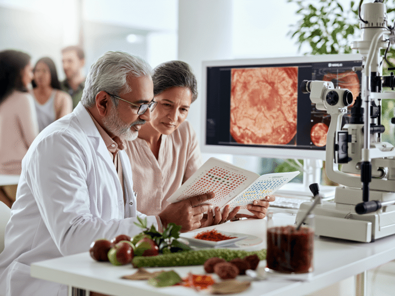
- Color Blindness Is Not One Disease—First Identify the Type
- Which Color-Vision Problem Describes You?
- Can Your Type of Color Blindness Actually Improve?
- Sudden Color-Vision Change May Be an Eye or Neurological Emergency
- Where Does the Color-Vision Problem Actually Begin?
- How Ayurveda Understands Color Perception
- Ayurvedic Understanding of Inherited Color Blindness
- Ayurvedic Understanding of Acquired Color-Vision Loss
- Modern Tests Used Before Selecting Ayurvedic Treatment
- Ayurvedic Rogi–Roga Assessment Before the Avaleha Is Selected
- Root-Cause Ayurvedic Treatment Pathway for Color-Vision Problems
- The Central Ayurvedic Medicine—Chakshushya Drishti Rasayana Avaleha
- What May Go Into a Personalized Chakshushya Drishti Rasayana Avaleha?
- How the Chakshushya Drishti Rasayana Avaleha Changes According to Your Condition
- Why Diabetic Patients Should Not Automatically Receive the Same Sweet Avaleha
- Chakshushya Drishti Rasayana Avaleha-Medicine
- Patient Friendly Preparation Method
- Color-Vision Recovery Passport
- Safety of Ayurvedic Eye Treatment
- What Published Evidence Exists for Ayurveda in Color Vision Disorders?
- Genetics, Family History and Future Children
- Failed Ishihara Test for Pilot, Navy, Railway or Employment: What Should You Do Next?
- Three Patient Scenarios: Why the Treatment Must Be Different
- Frequently Asked Questions
- Reference
Color blindness treatment in Ayurveda should not begin with a ready-made eye tonic. It should begin by understanding why you are unable to identify certain colors correctly.
You may have confused red and green since childhood. Your color vision may have started changing only recently. You may also have a mild inherited problem that has become worse because of diabetes, retinal disease, optic-nerve weakness, nutritional deficiency, cataract or a medicine you are taking.
These conditions may produce similar color-test results, but they are not the same disease. Therefore, they should not receive the same Ayurvedic treatment.
Inherited color-vision deficiency is usually present from childhood and remains relatively stable. At present, no established treatment can reliably replace a missing or genetically altered cone pigment. However, color vision that becomes weaker later in life may have a treatable retinal, optic-nerve, metabolic, nutritional, medicinal or systemic cause [1,2,7].
Your Treatment Possibility Depends on the Type of Color-Vision Problem
If you have always confused certain colors, the first aim is to confirm the type and severity of the inherited deficiency. Your retina, optic nerve and general eye health should also be examined to determine whether another treatable problem is present.
If you previously recognised colors normally but they now appear faded, dull, yellowish or different in one eye, you require a detailed investigation. Treating the underlying cause may help improve color discrimination when the damage is functional or reversible.
A sudden change in one eye, pain during eye movement, dim vision, a new blind spot or rapidly worsening color perception should be examined urgently. Ayurvedic treatment should not delay necessary retinal, neurological or optic-nerve care [7–9].
How Ayurveda Looks Beyond the Color Test
Ayurveda does not assess you only through an Ishihara result. Your condition is studied through Alochaka Pitta, Drishti, Dosha, Dushya, Agni, Ama, Srotas, tissue strength, digestion, metabolism, sleep, diet, medicines and associated diseases.
A lifelong inherited condition may be understood through Adibala-Pravritta Vyadhi and Beeja-related concepts. An acquired retinal problem may show Pitta–Rakta, Vata or mixed involvement. Optic-nerve weakness may require a Vata–Majja approach, while diabetes-associated color deterioration may require treatment of Prameha, Meda, Agni and Ama [10–17].
This is why two people with the same number of incorrect Ishihara plates may need completely different treatment.
The Medicine Is Selected From Your Reports, Not Only From the Disease Name
After the cause and Ayurvedic Samprapti are understood, a personalized oral Drishti Rasayana Avaleha may be prepared. Its herbal base, Rasayana ingredients, metabolic support, neural support, Avaleha medium and Anupana are selected according to your individual findings.
The formulation may focus more strongly on retinal support, optic-nerve nourishment, Pitta–Rakta regulation, Vata–Majja support, diabetes management, nutritional restoration or Agni–Ama correction. It is not the same commercial “eye tonic” for every patient.
The treatment model may use 15 g twice daily for 30 days, requiring approximately 900 g of finished Avaleha. However, the dosage, medium and suitability must be reviewed according to your age, digestion, diabetes status, concurrent medicines and overall health [27,28].
Improvement Must Be Measured, Not Assumed
Feeling that colors look brighter does not automatically mean that your color vision has improved. Progress should be checked through repeat standardized color testing, separate examination of both eyes and other relevant tests such as visual acuity, contrast sensitivity, OCT, visual fields, HbA1c or electrophysiological testing when required [20–25].
The purpose of treatment is therefore not merely to give you an Ayurvedic medicine. It is to identify whether your problem is inherited, acquired or mixed, treat the reversible component, protect the remaining visual function and verify the result objectively.
Color Blindness Is Not One Disease—First Identify the Type

The term “color blindness” covers several different problems. You may have difficulty separating red from green, blue from yellow or several colors from one another. The problem may be inherited, acquired later in life or caused by a combination of both.
The type matters because it decides what should be investigated, what may be treated and what result you can reasonably expect.
Lifelong Color Confusion Since Childhood
You are more likely to have an inherited color-vision deficiency when the problem has been present since childhood, affects both eyes similarly and has remained stable over time.
You may confuse red with green, brown with green, purple with blue or orange with red. You may only discover the problem during a school test, driving assessment or occupational medical examination.
Inherited red–green color-vision deficiency commonly results from genetic changes affecting cone photopigments. It is more common in males because many red–green deficiencies follow an X-linked inheritance pattern [2–4].
In Ayurveda, an inherited condition may be examined through the concepts of Adibala-Pravritta Vyadhi, Beeja, Beejabhaga and Beejabhagavayava. These concepts help explain hereditary or developmental susceptibility, but they should not be presented as exact substitutes for modern genes or chromosomes [13–15].
A lifelong inherited deficiency should not automatically be called Pittaja Timira. Your Alochaka Pitta, Drishti and overall ocular health can still be assessed, but the inherited origin must remain clear.
The treatment aim may include protecting remaining visual function, correcting nutritional or metabolic problems, reducing visual strain and checking whether an additional acquired problem is making your color discrimination worse. It should not include an unsupported promise of replacing a genetically altered cone pigment.
Color Vision That Has Changed Recently
You should be evaluated differently when you previously recognised colors normally but they have recently become faded, dull, yellowish or less distinct.
Acquired color-vision deficiency may develop because of retinal disease, optic-nerve dysfunction, diabetes, cataract, neurological disease, trauma, medicine toxicity or another systemic condition [2,7,8].
You may notice that red looks less bright, colors appear washed out or one eye sees colors differently from the other. You may also develop reduced contrast, dim vision, visual-field changes or difficulty recognising colors in low light.
In these cases, Ayurveda should focus on the underlying disease rather than only the abnormal color test.
A retinal or vascular problem may require assessment of Pitta, Rakta, Vata, Meda, Agni and Ama. An optic-nerve problem may require greater attention to Vata, Majja Dhatu, Dhatukshaya and associated neurological factors. A diabetes-related problem may require treatment of Prameha, Meda, Kleda, Agni and the retinal microvascular environment [10–12,16,17].
When the cause is identified early and remains reversible, treating the underlying pathology may improve color discrimination or prevent further deterioration.
Mixed Color-Vision Deficiency
You may have a mild inherited deficiency and later develop an acquired retinal, optic-nerve, metabolic or lens-related problem.
For example, you may have confused red and green since childhood but begin noticing that all colors are becoming dull after developing diabetes. You may also have a stable inherited deficiency that becomes more noticeable after cataract, retinal disease or medicine exposure.
In such a situation, the inherited component and the acquired component should be separated.
The inherited part may remain unchanged, while the newer retinal, neural or metabolic component may have treatment potential. Without this distinction, all deterioration may be wrongly dismissed as congenital color blindness.
Your assessment should therefore ask whether your color difficulty has always been present, whether it is worsening, whether both eyes are equally affected and whether you have diabetes, retinal disease, cataract, neurological symptoms or recent medication changes.
Why Different Types Need Different Ayurvedic Treatment
Two people may make the same number of errors on an Ishihara test but have completely different causes.
One person may have a stable inherited red–green deficiency. Another may have diabetic retinal dysfunction. A third may have optic-nerve inflammation, medicine toxicity or cataract.
The same Avaleha, herb, mineral preparation or eye procedure should not be given to all three.
Your treatment should be selected only after identifying the dominant pathology and constructing the Ayurvedic Samprapti. The formulation may then focus on Chakshushya Rasayana, Pitta–Rakta regulation, Vata–Majja support, Prameha management, Agni–Ama correction or Dhatu restoration according to your findings.
The first therapeutic decision is therefore not which medicine to give. It is deciding which type of color-vision problem you actually have.
Which Color-Vision Problem Describes You?

Your symptoms can provide an early clue about whether the problem is inherited, acquired or mixed. They cannot confirm the diagnosis by themselves, but they help decide which tests you need and whether urgent examination is required.
You Have Confused Red and Green Since Childhood
You may have difficulty separating red from green, brown from green, orange from red or purple from blue. The problem may have been noticed by a parent, teacher or during a school or employment examination.
When the difficulty has been present since childhood, remains stable and affects both eyes similarly, an inherited red–green color-vision deficiency is more likely [1–4].
You should still have a proper eye examination at least once. This helps confirm that your retina, optic nerve and visual acuity are healthy and that no additional acquired condition is present.
Colors Have Recently Become Dull or Washed Out
You need a more detailed assessment when you previously saw colors normally but now feel that they are becoming faded, pale or less distinct.
This may occur with retinal disease, optic-nerve dysfunction, diabetes, cataract, neurological illness, nutritional deficiency, trauma or medicine exposure [2,7,8].
Acquired color change should not be dismissed as normal ageing or assumed to be inherited. The underlying cause may still be active and may require treatment.
One Eye Sees Colors Differently
You can compare each eye separately by covering one eye and then the other. If red, green or another bright color appears noticeably duller through one eye, the difference may indicate retinal or optic-nerve dysfunction.
A marked difference between the two eyes is not typical of common inherited red–green deficiency, which usually affects both eyes in a similar way.
You should arrange a prompt ophthalmic assessment, especially when the difference is new or worsening [7,9].
You Have Eye Pain and Colors Look Faded
Pain during eye movement, reduced brightness, dim vision and faded colors may occur with optic-nerve inflammation.
You may notice that red appears less vivid through the affected eye. Central vision or part of your visual field may also become blurred or dark.
This situation requires urgent ophthalmic or neurological evaluation. Ayurvedic medicine should not be started as a substitute for investigating possible optic neuritis or another acute optic-nerve disorder [7,9].
Your Color Vision Has Worsened With Diabetes
Diabetes may affect the retinal blood vessels, retinal nerve cells and visual processing. You may notice weaker contrast, difficulty separating similar shades or gradually faded colors.
In this situation, treatment should not focus only on the eye. Your HbA1c, glucose control, blood pressure, lipid levels, kidney health and retinal findings may also need review.
From an Ayurvedic perspective, the assessment may include Prameha, Meda, Kleda, Agni, Ama, Rakta and retinal involvement. Treating the systemic metabolic disturbance is an important part of protecting visual function [7,17,29–31,50].
Your Color Perception Changed After Starting a Medicine
Some medicines can affect the retina, optic nerve or visual pathways. The color change may begin gradually and may be accompanied by reduced contrast, blurred vision or difficulty seeing particular shades.
The timing is important. Your doctor should know when the medicine was started, when the visual symptoms began and whether the symptoms are affecting one or both eyes.
Do not stop an essential medicine suddenly. Contact the prescribing doctor and arrange an eye examination so that the risks and alternatives can be reviewed safely [8].
You Have Cataract and Colors Look Yellow or Dull
Cataract can change the quality of light reaching your retina. White objects may look yellowish, blue shades may become difficult to recognise and colors may lose brightness.
Ayurvedic treatment may help address associated metabolic, digestive or systemic factors, but it cannot make an advanced opaque lens transparent.
When cataract is significantly affecting daily vision, an ophthalmologist should decide whether surgery is required [2].
Your Child Has Color Difficulty With Poor Vision or Severe Light Sensitivity
Common inherited red–green deficiency usually does not cause severe loss of visual acuity, marked photophobia or nystagmus.
If your child has involuntary eye movements, strong light sensitivity, poor central vision or difficulty recognising most colors, a more serious inherited cone disorder may be present.
Your child may require a paediatric retinal examination, electroretinography and genetic assessment. These symptoms should not be treated as ordinary color confusion [5].
You Failed a Pilot, Navy, Railway or Employment Color Test
Failing an Ishihara test does not necessarily mean that you see no colors. It means that you made errors on a test designed mainly to detect red–green deficiency.
Your result should be interpreted according to the number and type of errors, the lighting conditions and the occupational rules that apply to your country and profession.
Additional tests such as HRR, Farnsworth D-15, Farnsworth–Munsell 100 Hue or an anomaloscope may help define the type and severity of the deficiency [20–25].
Treatment may be considered when an acquired and reversible condition is contributing to the result. However, no treatment should guarantee that you will pass a pilot, military, railway, maritime or other occupational medical examination. The final decision belongs to the relevant authority [55–60].
Your Lifelong Color Problem Has Recently Become Worse
You may have had a mild inherited deficiency for many years and then notice a new decline after diabetes, cataract, retinal disease, optic-nerve damage or medication exposure.
This is a mixed presentation.
The inherited component may remain unchanged, but the newer acquired component may be treatable. Your assessment should therefore compare your earlier ability with your present symptoms and examine each eye separately.
The main aim is to identify what has always been present and what has changed recently. This distinction often decides whether the treatment goal should be protection, stabilization or improvement.
Can Your Type of Color Blindness Actually Improve?
Your chance of improvement depends mainly on whether the problem is inherited, acquired or mixed. A color-vision test can show that you are making color errors, but it cannot by itself explain why they are happening. The underlying cause must be identified before any treatment outcome can be predicted.
Acquired Color-Vision Problems May Have a Reversible Cause
If you previously recognised colors normally and the difficulty developed later, the cause may involve the retina, optic nerve, lens, metabolism, nutrition, medicine exposure or another systemic condition. Improvement may be possible when the responsible disease is detected early and remains reversible [2,7].
For example, color discrimination may improve after better diabetes control, correction of a nutritional deficiency, treatment of retinal or optic-nerve disease, review of a causative medicine or appropriate management of cataract. Ayurveda may support this process by treating the systemic Dosha–Dushya disturbance, Agni, Ama, Prameha, Rakta, Meda or Majja involvement according to your individual findings.
Mixed Cases May Have a Treatable Component
You may have had mild color confusion since childhood but now notice that colors are becoming duller or more difficult to recognise. In this situation, the inherited deficiency may be combined with a new retinal, metabolic, neural or lens-related problem.
The inherited component may not change, but the newer acquired component may respond to treatment. Your treatment should therefore focus on separating what has always been present from what has recently worsened.
Inherited Color Blindness Requires Realistic Expectations
Inherited red–green color-vision deficiency is usually caused by genetically altered or absent cone photopigments. At present, there is no established treatment that can reliably replace these pigments or permanently correct the genetic cause [1,4,6].
Ayurvedic treatment should therefore not promise that every inherited case will become completely normal. Its role may include protecting ocular health, treating associated nutritional or metabolic abnormalities, reducing visual strain and checking whether another treatable condition is worsening your color perception.
Advanced Retinal or Optic-Nerve Damage May Limit Recovery
When retinal cells or optic-nerve fibres have been permanently damaged, complete restoration may not be possible. In such cases, the treatment goal may shift from reversal to stabilization, protection of remaining function and prevention of further deterioration.
OCT, visual fields, visual acuity, contrast sensitivity and other investigations help show how much structural or functional damage is present. These findings are important before deciding whether the realistic goal is improvement, stabilization or supportive care.
Improvement Must Be Confirmed Objectively
You may initially feel that colors look brighter or that visual strain has reduced. These changes can be encouraging, but they do not by themselves prove that color discrimination has improved.
A meaningful response should be confirmed through repeat standardized color testing, comparison of both eyes and relevant retinal, optic-nerve or metabolic investigations [20–25]. The same test conditions should be used whenever possible so that lighting, memorisation or test familiarity does not create a false impression of recovery.
Sudden Color-Vision Change May Be an Eye or Neurological Emergency

A sudden change in color perception is different from lifelong color blindness. If colors become faded, dull or unequal between your two eyes over hours or days, you should not wait for Ayurvedic treatment to begin. The retina, optic nerve or visual pathway may be under active stress and requires prompt examination [7–9].
Sudden Color Fading in One Eye
Common inherited color-vision deficiency usually affects both eyes in a similar and stable manner. A new difference between your eyes may suggest retinal disease, optic-nerve dysfunction, inflammation, reduced blood supply or another acquired problem.
You may notice that red appears darker through one eye, white looks less bright or all colors seem washed out. A prompt ophthalmic examination is required even when your visual acuity appears normal.
Eye Pain With Faded or Dim Colors
Pain behind the eye or pain during eye movement, together with reduced brightness or faded colors, may occur in optic neuritis. You may also experience blurred central vision, a blind spot or reduced contrast [7,9].
This requires urgent ophthalmic or neurological assessment. Ayurveda may be considered later as supportive care, but it should not delay imaging, blood tests or treatment advised for an acute optic-nerve condition.
Color Change With Flashes, Floaters or a Visual Shadow
New flashes, a sudden increase in floaters or a curtain-like shadow may indicate a retinal tear or detachment. Color perception may also become abnormal when part of the retina is no longer functioning correctly.
This is an eye emergency. Immediate retinal examination is more important than starting any oral Avaleha or local eye procedure.
Color Deterioration After Trauma
Color vision may change after injury to the eye, head or optic nerve. Even a seemingly minor injury can affect retinal or neural function without obvious external damage.
You should be examined promptly if color change follows trauma, especially when it is accompanied by blurred vision, double vision, headache, field loss or reduced brightness.
Color Change After Starting a Medicine
Some medicines can affect the retina, optic nerve or visual processing pathways. The change may involve one color range or make all colors appear less vivid. Reduced contrast, blurred vision or visual-field changes may also occur [8].
Do not stop an essential medicine suddenly. Contact the prescribing doctor, record when the symptoms began and arrange an ophthalmic review. The medicine can then be adjusted safely if it is suspected to be the cause.
When Ayurveda Can Be Added
Ayurvedic treatment may be considered after urgent pathology has been identified and medically stabilized. The later plan may address Pitta–Rakta involvement, Vata–Majja weakness, metabolic disturbance, Dhatu Kshaya or recovery from medicine-related stress according to your findings.
The priority is always clear: sudden, painful, one-sided or rapidly worsening color change must be investigated first.
Where Does the Color-Vision Problem Actually Begin?
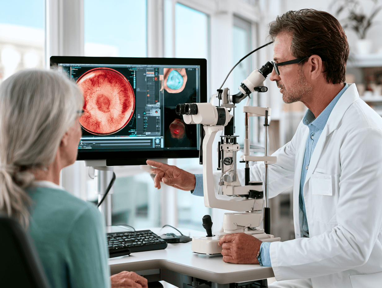
Color vision depends on several parts of your visual system working together. Light must pass clearly through the lens, reach the cone cells in the retina, travel through the optic nerve and finally be interpreted by the brain. A problem at any of these levels can change how you recognise colors.
Cone Photoreceptors and Inherited Color Deficiency
Cone cells are concentrated in the central retina and help you see fine details and colors. Different cone pathways respond mainly to long, medium and short wavelengths of light.
Inherited red–green color-vision deficiency usually develops when the photopigment in one cone pathway is absent or functions differently. This problem is generally present from childhood, affects both eyes similarly and remains relatively stable [3,4,6].
Because the basic alteration is genetic, treatment should not be described as simply correcting Pitta or strengthening the eye. The inherited component must be recognised honestly before discussing supportive Ayurvedic care.
Retina and Macula-Related Color Loss
Your macula is responsible for detailed central vision and contains a high concentration of cone cells. Retinal or macular disease can reduce the ability of these cells to separate similar shades, even when you could recognise colors normally in the past.
You may notice faded colors, reduced contrast, distorted central vision or difficulty identifying blue–yellow shades. Diabetes, retinal degeneration, inflammation and vascular problems may contribute to this type of acquired color-vision loss [2,7].
In such cases, treatment must address both the retinal condition and the systemic factors affecting it.
Optic-Nerve Dysfunction and Faded Colors
The optic nerve carries visual information from your retina to your brain. When it becomes inflamed, compressed, injured or damaged by reduced blood supply or toxic exposure, colors may appear less bright or washed out.
Red may look particularly dull through the affected eye. You may also develop reduced brightness, central blurring, a blind spot or visual-field loss [7,9].
A new difference between your two eyes requires prompt investigation. It should not be treated as ordinary inherited color blindness.
Cataract and Lens-Related Color Changes
The natural lens inside your eye can become yellow or cloudy with age. This changes the quality of light reaching the retina and can make colors look dull, brownish or yellow.
Blue shades may become more difficult to recognise, and white objects may no longer appear completely white. When cataract is the main cause, removing the cloudy lens may provide the most direct correction [2].
Ayurveda may support your general metabolic and ocular health, but it should not be presented as a substitute for surgery when the lens has become significantly opaque.
Brain and Visual-Pathway Causes
Color information is also processed in the brain. Stroke, head injury, neurological disease or damage to the visual pathways can disturb color recognition even when the eyes themselves appear healthy [2,7].
You may have additional symptoms such as visual-field loss, weakness, numbness, headache, confusion or difficulty recognising objects. These symptoms require neurological evaluation before Ayurvedic treatment is planned.
Metabolic, Nutritional and Medicine-Related Causes
Diabetes, nutritional deficiency, chronic systemic illness and selected medicines can affect the retina, optic nerve or visual pathways. Color changes may develop gradually and may be accompanied by blurred vision, poor contrast or visual fatigue [7,8].
Your assessment may therefore require more than an eye test. HbA1c, glucose, nutritional markers, liver and kidney function, medicine history and other systemic investigations may be relevant.
From an Ayurvedic perspective, these cases may involve Prameha, Meda, Agni, Ama, Rakta, Majja or Dhatu Kshaya. The treatment should focus on the systemic cause together with the affected visual structure.
Why Locating the Problem Matters
The same color-test error can arise from a cone-pigment difference, retinal disease, optic-nerve dysfunction, cataract, metabolic disturbance or neurological disease.
Modern investigations help identify where the problem is occurring. Ayurvedic assessment then helps explain the individual Dosha–Dushya pattern and guides the treatment strategy.
Without locating the actual source, giving a general eye medicine or Avaleha may miss the real cause of your color-vision difficulty.
How Ayurveda Understands Color Perception
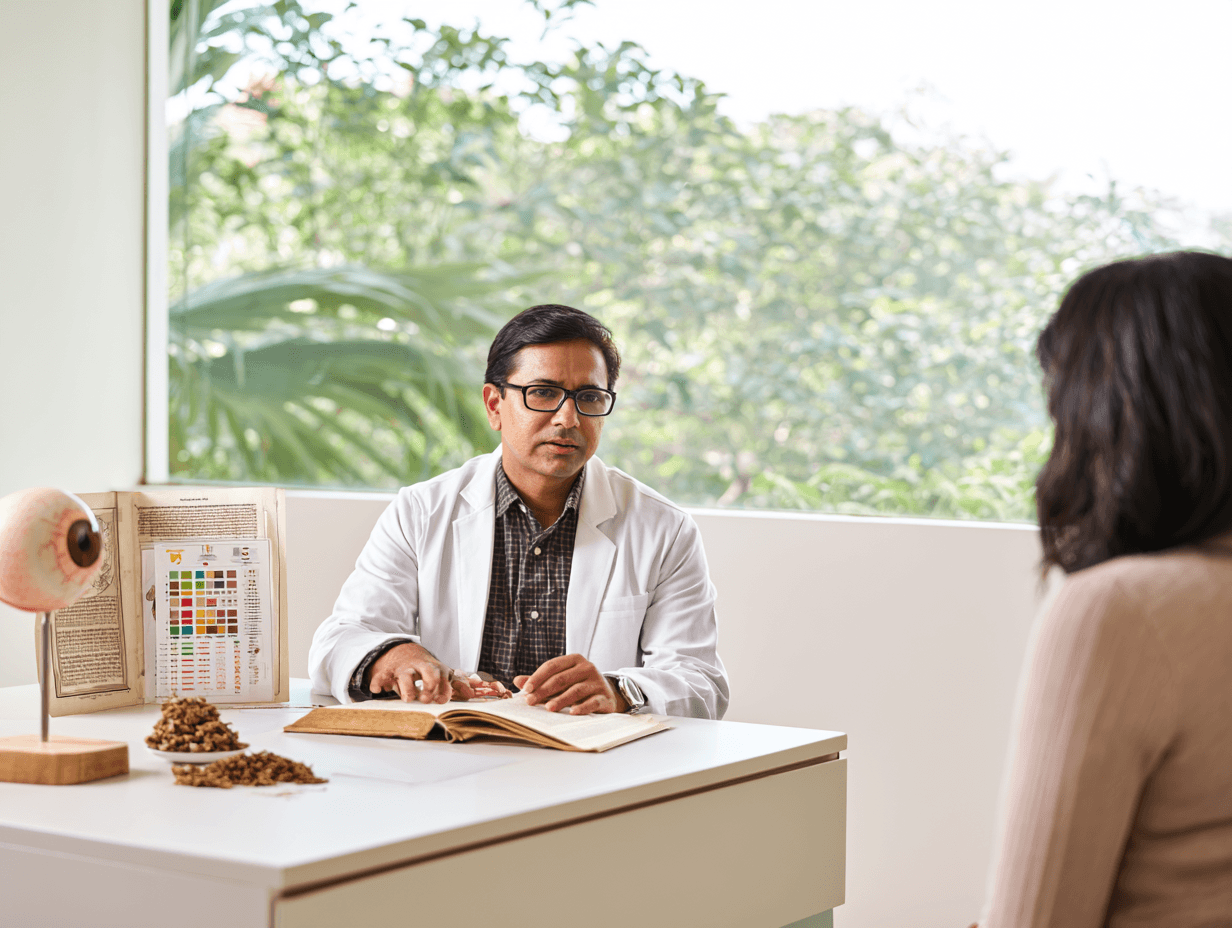
Ayurveda does not view color perception as the function of the eyes alone. Your ability to recognise colors depends on the condition of Drishti, the balance of Doshas, the nourishment of the tissues, the clarity of the visual channels and the health of the entire body.
Modern investigations help identify whether the disturbance is located in the cones, retina, optic nerve, lens or brain. Ayurvedic assessment then examines why that structure has become weak, obstructed, inflamed, depleted or functionally disturbed.
Alochaka Pitta and Visual Perception
Alochaka Pitta is the principal Ayurvedic functional concept associated with vision. It is described in Ashtanga Hridaya, Sutrasthana, Chapter 12, Doshabhediya Adhyaya as the form of Pitta situated in the eyes and involved in visual perception [10].
Alochaka Pitta helps you receive and understand visual information. When its function is disturbed, you may experience changes in clarity, brightness, contrast or the appearance of objects.
However, an abnormal color-vision test does not automatically mean that you have excessive Pitta. Alochaka Pitta may be affected by Vata depletion, Rakta disturbance, Kapha obstruction, metabolic disease, tissue weakness or an inherited developmental variation.
Your treatment therefore cannot be selected only by giving a cooling or Pitta-reducing medicine.
Drishti Is More Than the Visible Eye
In Ayurveda, Drishti refers to the functional system through which you perceive what is in front of you. It should not be understood only as one visible part of the eye.
The classical description of Drishtigata Roga in Sushruta Samhita, Uttara Tantra, Chapter 7, Drishtigata Roga Vijnaniya Adhyaya provides the main Ayurvedic framework for disorders that affect visual perception [11].
When your color vision changes, the assessment may consider whether the disturbance is affecting the retinal level, the transmission of visual information or the clarity with which that information is interpreted.
This broader understanding allows the treatment to address the eye together with your metabolism, nervous system, digestion, circulation and nutritional condition.
When Timira May Be Relevant
Timira is a classical Drishtigata disorder associated with progressive disturbance of vision. It may provide a useful Ayurvedic framework when your color difficulty is part of a broader acquired visual problem.
For example, Timira-related reasoning may be considered when you have gradually worsening vision, reduced contrast, retinal disease, metabolic dysfunction or changes in the appearance of objects.
The treatment principles for Drishtigata disorders are discussed in Sushruta Samhita, Uttara Tantra, Chapter 17, Drishtigata Roga Pratishedha Adhyaya [12].
However, every person who fails an Ishihara test should not be diagnosed with Timira. A stable inherited red–green deficiency present since childhood has a different origin from an acquired progressive visual disorder.
How Doshas Can Affect Color Perception
Pitta and Rakta may become important when your condition includes retinal inflammation, vascular disturbance, burning, redness, light sensitivity or progressive alteration in color perception.
Vata and Majja may be more important when the optic nerve, sensory transmission, degeneration, trauma or tissue depletion is involved. You may then experience faded colors, reduced brightness, fluctuating vision or associated neurological symptoms.
Kapha, Meda and Ama may have a stronger role when color deterioration occurs with diabetes, obesity, sluggish metabolism, cataract, heaviness or microvascular obstruction.
Many chronic cases do not fit into one Dosha. Your condition may involve Pitta–Rakta at the retinal level, Vata–Majja at the neural level and Meda–Ama at the systemic level at the same time.
Why Color Blindness Cannot Be Given One Ayurvedic Diagnosis
Modern color blindness includes inherited cone-pigment variations, retinal disorders, optic neuropathies, cataract, medicine toxicity and neurological disease. These conditions do not share one cause, even when they produce similar color-test errors.
Ayurveda therefore should not force all cases into one diagnosis such as Pittaja Timira or Alochaka Pitta Dushti.
Your Ayurvedic diagnosis should be based on the origin of the problem, affected structure, Dosha, Dushya, Agni, Ama, Srotas, chronicity and associated systemic disease.
This is the reason your treatment must be individualized. The aim is not simply to “balance Pitta,” but to understand the complete Samprapti that is disturbing your color perception and treat the part that may still be reversible.
Ayurvedic Understanding of Inherited Color Blindness

If you have confused the same colors since childhood and both eyes are affected similarly, your condition is more likely to be inherited. Ayurveda does not describe modern color blindness as one exact disease, but it provides a clear framework for understanding disorders that arise through parental reproductive factors and abnormalities present from birth.
Modern research shows that common inherited red–green color-vision deficiency is usually related to changes affecting cone photopigments. It generally remains stable throughout life and is commonly inherited through the X chromosome [2–4]. (National Eye Institute)
Adibala-Pravritta Vyadhi: The Classical Hereditary Framework
Sushruta classifies diseases according to their origin and places disorders connected with parental reproductive factors under Adibala-Pravritta Vyadhi.
Classical source: Sushruta Samhita, Sutrasthana, Chapter 24, Vyadhi Samuddeshiya Adhyaya, verse 5 [13].
Sanskrit
तत्र, आदिबलप्रवृत्ता ये शुक्रशोणितदोषान्वयाः कुष्ठार्शःप्रभृतयः; तेऽपि द्विविधाः—मातृजाः, पितृजाश्च ॥५॥
Transliteration
Tatra, ādibalapravṛttā ye śukraśoṇitadoṣānvayāḥ kuṣṭhārśaḥprabhṛtayaḥ; te’pi dvividhāḥ—mātṛjāḥ, pitṛjāś ca.
Translation
“Adibala-Pravritta diseases are associated with abnormalities of the paternal and maternal reproductive elements. They may therefore be of maternal or paternal origin.”
This passage does not mention color blindness specifically. It provides the classical principle that some health conditions may arise from factors carried through the mother or father rather than from your present diet, lifestyle or Dosha imbalance alone.
Beeja and Beejabhaga: Why a Particular Function May Be Affected
Charaka explains that a disturbance affecting the reproductive source responsible for a particular body part may produce an abnormality in that corresponding part.
Classical source: Charaka Samhita, Sharirasthana, Chapter 4, Mahati Garbhavakranti Sharira, verse 30 [14].
Sanskrit
यस्य यस्य ह्यवयवस्य बीजे बीजभागे वा दोषाः प्रकोपमापद्यन्ते, तं तमवयवं विकृतिराविशति ॥३०॥
Transliteration
Yasya yasya hy avayavasya bīje bījabhāge vā doṣāḥ prakopam āpadyante, taṃ tam avayavaṃ vikṛtir āviśati.
Translation
“When disturbance affects the Beeja or Beejabhaga responsible for a particular body part, abnormality develops in that corresponding part.”
In this framework, Beeja refers to the reproductive source, while Beejabhaga indicates a more specific component responsible for the development of a particular structure or function. Charaka also uses the term Beejabhagavayava for a still finer subdivision of that reproductive component [14,15]. (siva.sh)
These terms may be compared conceptually with hereditary organization, but they should not be declared identical to genes, chromosomes or DNA. Ayurveda developed these concepts within its own classical language, while modern genetics identifies specific cone-opsin genes such as OPN1LW, OPN1MW and OPN1SW in different inherited color-vision disorders [4]. (MedlinePlus)
Why Inherited Color Blindness Is Not Simply Pittaja Timira
Inherited color blindness should not automatically be diagnosed as Pittaja Timira or excessive Alochaka Pitta. In many inherited cases, the basic difference lies in how one or more cone photopigments have developed or function.
Alochaka Pitta remains relevant because it represents the Ayurvedic functional principle of visual perception. However, it does not remove the inherited origin of the condition. Giving only cooling herbs or Pitta-reducing medicines would therefore be an incomplete approach.
Your Ayurvedic assessment should consider the inherited Adibala-Pravritta and Beeja-related background first. Alochaka Pitta, Dosha, Agni, Ama, Rakta, Majja and your general ocular health may then be assessed to identify additional disturbances that could still be treated.
What Ayurvedic Treatment May Realistically Address
When your color deficiency has remained unchanged since childhood, treatment should not promise to recreate a missing cone pigment or alter an inherited opsin variation. Current medical evidence does not establish a reliable treatment that permanently corrects the genetic cause of common inherited color-vision deficiency [1–4]. (National Eye Institute)
Ayurvedic treatment may still be considered to support your overall ocular health, correct nutritional or metabolic problems, reduce associated visual fatigue and identify whether an acquired condition is worsening your inherited difficulty.
This distinction becomes especially important if your lifelong color confusion has recently become worse. Diabetes, cataract, retinal disease, optic-nerve dysfunction or medicine exposure may add a potentially treatable acquired component to an existing inherited deficiency.
The correct goal is therefore to separate the stable hereditary component from any newer reversible disturbance. Your treatment can then focus precisely on the part of your color-vision problem that may still improve.
Ayurvedic Understanding of Acquired Color-Vision Loss

If you recognised colors normally in the past but they later became faded, dull or difficult to separate, Ayurveda treats this as an acquired disturbance rather than a fixed hereditary condition. The assessment looks at the affected visual structure together with Dosha, Dushya, Agni, Ama, Srotas, metabolism, nutrition and the disease causing the change.
A retinal problem, optic-nerve disorder, diabetes, nutritional deficiency or medicine-related injury may produce a different Samprapti. Therefore, the treatment cannot be selected only from your Ishihara score.
Acquired Disease Can Disturb the Clarity of Drishti
Sushruta describes how the perception of forms becomes unclear when aggravated Doshas affect deeper levels of Drishti.
Classical source: Sushruta Samhita, Uttara Tantra, Chapter 7, Drishtigata Roga Vijnaniya Adhyaya, verse 7 [11].
Sanskrit
अव्यक्तानि स रूपाणि सर्वाण्येव प्रपश्यति ।
दृष्टिर्भृशं विह्वलति द्वितीयं पटलं गते ॥७॥
Transliteration
Avyaktāni sa rūpāṇi sarvāṇy eva prapaśyati ।
Dṛṣṭir bhṛśaṃ vihvalati dvitīyaṃ paṭalaṃ gate ॥7॥
Translation
“When the Dosha reaches the second Patala, the person sees forms indistinctly and visual perception becomes greatly disturbed.”
This verse describes disturbed visual perception, not modern color blindness specifically. It nevertheless provides a classical basis for examining acquired changes in clarity, contrast, form and color as part of a wider Drishtigata disorder.
Pitta–Rakta Dominant Retinal Pattern
Pitta and Rakta may become important when your color disturbance is associated with retinal inflammation, vascular changes, burning, redness, light sensitivity or a yellow, red or unusually bright appearance of objects.
Sushruta directly describes altered visual appearance under the influence of Pitta and Rakta.
Classical source: Sushruta Samhita, Uttara Tantra, Chapter 7, Drishtigata Roga Vijnaniya Adhyaya, verse 25 [11].
Sanskrit
पित्तं कुर्यात् परिम्लायि मूर्च्छितं रक्ततेजसा ।
पीता दिशस्तथोद्यन्तमादित्यमिव पश्यति ॥२५॥
Transliteration
Pittaṃ kuryāt parimlāyi mūrcchitaṃ raktatejasā ।
Pītā diśas tathodyantam ādityam iva paśyati ॥25॥
Translation
“Pitta, influenced by the heat of Rakta, produces disturbed vision in which the surroundings may appear yellow, like the rising sun.”
This verse should not be presented as a description of inherited red–green deficiency. It shows that classical Ayurveda recognised that Dosha and Rakta disturbances could alter the perceived color and appearance of the visual field.
In a modern retinal case, the Ayurvedic plan may therefore assess Alochaka Pitta, Rakta, retinal circulation, inflammation, Agni and associated metabolic disease. Treatment should still be guided by the actual retinal diagnosis.
Vata–Majja Dominant Optic-Nerve Pattern
Vata becomes more relevant when the visual problem is associated with optic-nerve dysfunction, trauma, degeneration, fluctuating vision, reduced brightness or neurological symptoms.
Classical source: Sushruta Samhita, Uttara Tantra, Chapter 7, Drishtigata Roga Vijnaniya Adhyaya, verse 18 [11].
Sanskrit
स एव लिङ्गनाशस्तु नीलिकाकाचसञ्ज्ञितः ।
तत्र वातेन रूपाणि भ्रमन्तीव स पश्यति ॥१८॥
Transliteration
Sa eva liṅganāśas tu nīlikākācasañjñitaḥ ।
Tatra vātena rūpāṇi bhramantīva sa paśyati ॥18॥
Translation
“In the visual disorder influenced by Vata, the person perceives forms as though they are moving or revolving.”
The verse supports the role of Vata in unstable or distorted visual perception. It does not state that Vata is identical to the optic nerve. The connection with Majja Dhatu and neural function is an Ayurvedic clinical interpretation made after modern examination confirms optic-nerve or neurological involvement.
A sudden reduction in color saturation, especially in one eye or with pain during eye movement, requires urgent ophthalmic or neurological investigation before Ayurvedic treatment [7,9].
Kapha–Meda–Ama Dominant Metabolic Pattern
When your color vision worsens with diabetes, obesity, insulin resistance or sluggish metabolism, the eye should not be treated separately from the systemic disease.
Ayurveda may examine Prameha, Kapha, Meda, Kleda, Agni, Ama and Srotorodha. Your retinal circulation and nerve function may be affected while the deeper disease process remains metabolic.
Charaka also makes it clear that patients with the same metabolic disease should not automatically receive the same treatment.
Classical source: Charaka Samhita, Chikitsa Sthana, Chapter 6, Prameha Chikitsa, verse 15 [17].
Sanskrit
स्थूलः प्रमेही बलवानिहैकः कृशस्तथैकः परिदुर्बलश्च ।
सम्बृंहणं तत्र कृशस्य कार्यं संशोधनं दोषबलाधिकस्य ॥१५॥
Transliteration
Sthūlaḥ pramehī balavān ihaikaḥ kṛśas tathaikaḥ paridurbalaś ca ।
Sambṛṃhaṇaṃ tatra kṛśasya kāryaṃ saṃśodhanaṃ doṣabalādhikasya ॥15॥
Translation
“One patient with Prameha may be strong and obese, while another may be lean and very weak. Nourishing treatment is required for the weak patient, while Dosha-reducing treatment is considered for the strong patient with greater Dosha involvement.”
This principle is important for color-vision problems associated with diabetes. A strong patient with Kapha–Meda excess should not receive the same sweet, nourishing Avaleha as a weak patient with Dhatu Kshaya. The formulation, medium and treatment intensity must follow your actual condition. (Caraka Samhita Online)
Dhatu Kshaya and Nutritional Depletion
Color perception may deteriorate when chronic illness, poor nutrition or malabsorption affects the retina or optic nerve [7]. Ayurveda may then assess Mandagni, Grahani dysfunction, Rasa–Rakta depletion, Majja weakness and reduced Ojas.
In such a case, strong Langhana or excessive Dosha-reducing treatment may further weaken you. The priority may instead be to improve digestion and absorption before introducing carefully selected Brimhana, Chakshushya and Rasayana medicines.
Medicine- or Toxin-Associated Color Disturbance
Some medicines and toxic exposures can affect the retina or optic nerve and change how you perceive colors [8]. Ayurveda may examine the case through Nidana, Agantuja factors, Rakta–Pitta disturbance, Ama and, where clinically appropriate, Dushi Visha concepts.
The first step is not detoxification medicine. The suspected cause must be reviewed with the prescribing doctor, and the retina and optic nerve must be examined. You should not stop an essential medicine suddenly.
Ayurvedic treatment may be added after the damaging exposure has been controlled and the extent of reversible injury has been assessed.
Mixed Dosha Involvement Is Common
Long-standing acquired color-vision problems may involve more than one Dosha. Sushruta describes varied visual appearances when several Doshas are involved together.
Classical source: Sushruta Samhita, Uttara Tantra, Chapter 7, Drishtigata Roga Vijnaniya Adhyaya, verse 23 [11].
Sanskrit
हरितश्यावकृष्णानि धूमधूम्राणि चेक्षते ।
सन्निपातेन चित्राणि विप्लुतानि च पश्यति ॥२३॥
Transliteration
Haritaśyāvakṛṣṇāni dhūmadhūmrāṇi cekṣate ।
Sannipātena citrāṇi viplutāni ca paśyati ॥23॥
Translation
“With combined Dosha involvement, the person may perceive greenish, dark, black, smoky or hazy forms and varied disturbed visual appearances.”
This verse should not be treated as a modern classification of color-vision deficiency. It supports the broader Ayurvedic principle that mixed pathology can produce complex changes in visual perception. (siva.sh)
Your condition may therefore involve Pitta–Rakta at the retinal level, Vata–Majja at the neural level and Kapha–Meda–Ama at the metabolic level. The treatment should address the dominant cause first rather than attempting to reduce all three Doshas without a clear sequence.
Modern Tests Used Before Selecting Ayurvedic Treatment
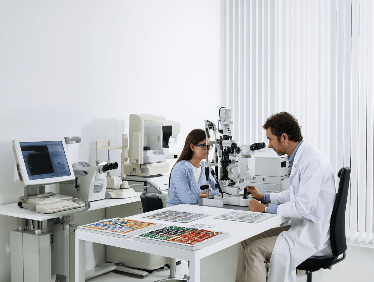
Your Ayurvedic treatment should not be selected from symptoms alone. Color-vision testing helps identify the type and severity of the problem, while eye and blood investigations help locate the underlying cause.
You may not need every test mentioned below. The required tests depend on whether your problem has been present since childhood, developed recently, affects one eye more than the other or occurs with diabetes, retinal disease, optic-nerve symptoms or medicine exposure.
Ishihara Test
The Ishihara test uses plates made of colored dots. You are asked to identify numbers, lines or patterns within each plate.
It is mainly used to screen for inherited red–green color-vision deficiency. It can show that you are making red–green errors, but it does not fully measure every type of color-vision problem [20,21].
An Ishihara result should not be interpreted only as “pass” or “fail.” The number of errors, type of errors, lighting conditions, viewing distance and whether each eye was tested separately should also be recorded.
A normal Ishihara result does not exclude every acquired color-vision disorder. Blue–yellow changes and some retinal or optic-nerve problems may require other tests [21,23].
Hardy–Rand–Rittler Test
The Hardy–Rand–Rittler test, commonly called the HRR test, can help identify both red–green and blue–yellow color-vision problems.
It may also help estimate whether the deficiency is mild, moderate or severe. This can be useful when your symptoms developed later in life or when an acquired retinal or optic-nerve disorder is suspected.
The HRR test may provide information that is not available from Ishihara plates alone. However, the result must still be interpreted together with your complete eye examination.
Farnsworth D-15 Test
The Farnsworth D-15 test asks you to arrange colored caps in the correct sequence according to similarity of shade.
It helps identify the direction and severity of your color confusion. Your pattern of arrangement may show whether the errors are mainly red–green, blue–yellow or more widespread.
The D-15 test is useful for classifying functional color discrimination, but it has limitations. Mild deficiencies may not always be detected, and one test result should not be used alone to declare recovery or occupational fitness [22].
Farnsworth–Munsell 100 Hue Test
The Farnsworth–Munsell 100 Hue test provides a more detailed assessment of your ability to separate similar shades.
You arrange many colored caps in a gradual sequence. The result produces an error score and may show the main axis of your color confusion.
This test can be useful for measuring smaller changes over time. It may therefore help compare your baseline color discrimination with your response after treatment.
The same test conditions should be maintained during follow-up. Differences in lighting, concentration, fatigue or test familiarity can influence the result.
Anomaloscope Testing
An anomaloscope is considered one of the most precise methods for classifying inherited red–green color-vision deficiency.
You are asked to match a mixture of red and green light with a yellow reference light. The test can help distinguish different red–green deficiency patterns and measure their severity [21,25].
Anomaloscope testing is not available in every clinic. It may be especially useful when your diagnosis is uncertain, when occupational standards require detailed classification or when a treatment claim needs stronger objective documentation.
Testing Each Eye Separately
Inherited color-vision deficiency usually affects both eyes in a similar manner. A clear difference between your right and left eye raises greater concern for an acquired retinal or optic-nerve problem.
Each eye should therefore be tested separately when you report recent color fading, red desaturation, dim vision or unequal brightness.
A simple red comparison may provide an early clue, but red-desaturation testing alone is not reliable enough to diagnose optic-nerve disease. Some healthy people also report small differences between their eyes [24].
Visual Acuity and Refraction
Visual acuity testing measures how clearly you can see letters or symbols at a set distance. Refraction checks whether glasses or contact lenses are needed.
Common inherited red–green deficiency usually does not cause major loss of visual acuity. Reduced acuity may therefore suggest an additional retinal, macular, lens or neurological disorder.
Your color testing should be performed with the correct refractive correction. Uncorrected blurred vision can make some test plates more difficult to recognise.
Contrast-Sensitivity Testing
Contrast sensitivity measures how well you can distinguish an object from its background when the difference in brightness is small.
You may have normal letter-chart vision but still experience difficulty with faded colors, low-light vision or similar shades. Contrast testing can help identify these functional problems.
Improved visual comfort without improved color discrimination may still show as better contrast sensitivity. These outcomes should be recorded separately.
Dilated Retinal Examination
A dilated examination allows the ophthalmologist to inspect your retina, macula, optic disc and retinal blood vessels.
This is especially important when your color vision has changed recently, you have diabetes, one eye is worse, visual acuity has reduced or you have flashes, floaters or a visual-field defect.
The examination may identify diabetic retinopathy, macular disease, retinal degeneration, inflammation, vascular changes or optic-nerve abnormalities.
Optical Coherence Tomography of the Macula
Optical coherence tomography, commonly called OCT, produces detailed cross-sectional images of your retina.
Macular OCT can show swelling, thinning, degeneration, fluid or changes affecting the cone-rich central retina. These findings may help explain why your color discrimination or contrast has deteriorated.
OCT does not directly measure color vision. It shows whether structural retinal disease may be contributing to the problem.
OCT of the Retinal Nerve-Fibre Layer
OCT-RNFL measures the thickness of nerve fibres around your optic disc.
It may help identify or monitor optic-nerve damage associated with glaucoma, inflammation, compression, trauma or other neuropathies. Reduced nerve-fibre thickness can support the diagnosis of an optic-nerve-related color disturbance.
A normal OCT does not exclude every early optic-nerve disorder. Your symptoms, visual fields, color testing and neurological findings must also be considered.
Visual-Field Testing
Visual-field testing checks whether any part of your peripheral or central vision is missing or reduced.
An optic-nerve or neurological disorder may cause a central blind spot, reduced sensitivity or a characteristic field defect together with faded colors.
Changes in your field should be monitored objectively. A worsening field requires specialist review even when you feel that color perception has improved.
Electroretinography
Electroretinography, or ERG, measures the electrical response of your retinal cells to light.
It may be needed when you have poor color vision with severe light sensitivity, nystagmus, reduced visual acuity or suspected cone or cone–rod disease.
ERG can help distinguish common inherited red–green deficiency from more serious retinal disorders. It may also help determine whether the cone system is functioning abnormally.
Visual Evoked Potential
Visual evoked potential, or VEP, measures how visual signals travel from your eyes to the visual areas of your brain.
It may be considered when optic-nerve dysfunction, delayed neural transmission or a neurological cause is suspected.
VEP does not replace an eye examination, OCT or visual-field testing. It provides additional information about the visual pathway.
Genetic Testing
Genetic testing may be useful when you have a severe inherited cone disorder, strong family history, nystagmus, marked photophobia or reduced visual acuity from childhood.
It may help identify changes involving cone-opsin or retinal-disease genes. Genetic counselling can then explain the inheritance pattern and possible implications for other family members [4,5].
Genetic testing is not necessary for every person with stable red–green deficiency. It is selected when the result is likely to change diagnosis, counselling or future management.
Metabolic and Nutritional Investigations
Blood investigations may be required when your color vision changed after previously being normal.
Depending on your history, these may include fasting glucose, HbA1c, lipid profile, complete blood count, vitamin B12, folate, thyroid function, liver function and kidney function.
These tests do not diagnose color blindness directly. They help identify diabetes, nutritional depletion, organ dysfunction or another systemic factor that may be affecting your retina or optic nerve.
Why Online Color Tests Are Not Enough
Online color tests may provide a useful initial indication, but their accuracy depends on your screen, brightness, display settings, room lighting and device quality.
They should not be used as the only basis for diagnosis, Ayurvedic treatment, occupational certification or a claim of recovery.
Your baseline and follow-up results should ideally come from validated tests performed under similar conditions by a trained examiner.
How Test Results Guide Your Ayurvedic Treatment
Your reports help determine whether the dominant problem is inherited, retinal, optic-nerve-related, metabolic, nutritional or mixed.
A stable inherited result may lead to a protective and supportive Rasayana approach. Retinal disease may require stronger attention to Pitta–Rakta, Vata and metabolic factors. Optic-nerve dysfunction may require a Vata–Majja-focused plan, while diabetes-associated changes require treatment of Prameha, Meda, Agni and Ama.
The purpose of testing is not merely to give your condition a name. It is to make sure that your Ayurvedic treatment addresses the correct cause and that any future improvement can be measured honestly.
Ayurvedic Rogi–Roga Assessment Before the Avaleha Is Selected
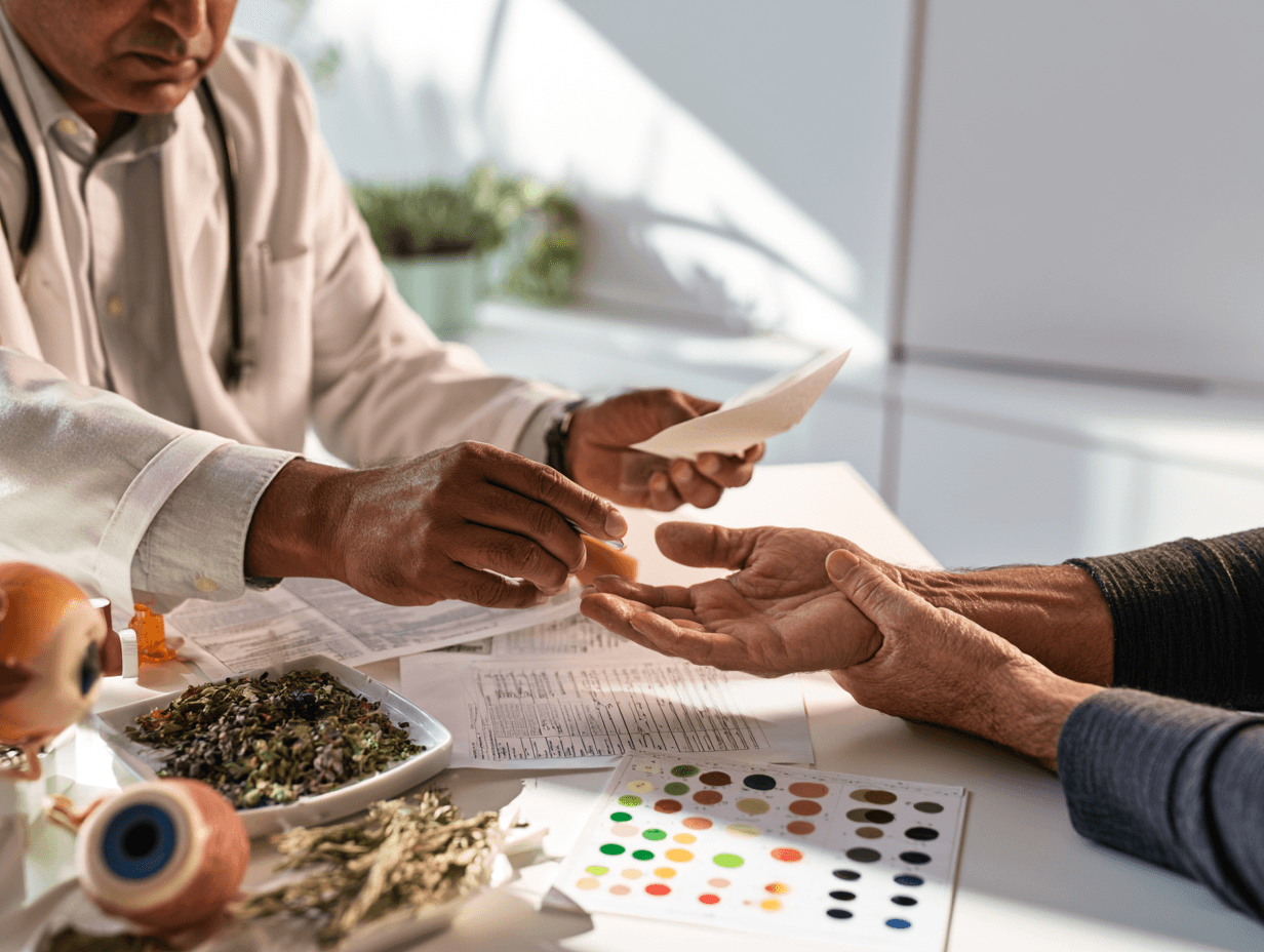
Modern tests help identify whether your color-vision problem involves the cones, retina, optic nerve, lens or visual pathway. Ayurvedic assessment then examines why the disturbance developed in you, which tissues are affected, how strong the disease is and how much treatment your body can tolerate.
This assessment is essential because a powerful formulation is not suitable merely because it contains many herbs or minerals. The medicine, dose, medium and duration must match both the disease and your individual strength.
The Disease Must Be Understood Before the Medicine Is Chosen
Charaka clearly places diagnosis before medicine.
Classical source: Charaka Samhita, Sutra Sthana, Chapter 20, Maharoga Adhyaya, verse 20 [61].
Sanskrit
रोगमादौ परीक्षेत ततोऽनन्तरमौषधम् ।
ततः कर्म भिषक् पश्चाज्ज्ञानपूर्वं समाचरेत् ॥२०॥
Transliteration
Rogam ādau parīkṣeta tato’nantaram auṣadham ।
Tataḥ karma bhiṣak paścāj jñānapūrvaṃ samācaret ॥20॥
Translation
“The physician should first examine the disease and then select the medicine. Treatment should be administered only after gaining proper knowledge of the condition.”
This principle is especially important in color-vision disorders. An inherited cone-pigment difference, diabetic retinal change and optic-nerve disorder may produce similar color errors, but they should not receive the same Avaleha.
Your Complete Strength Is Examined Before Strong Treatment
Charaka describes a detailed examination of the patient before selecting the strength and quantity of treatment.
Classical source: Charaka Samhita, Vimana Sthana, Chapter 8, Rogabhishagjitiya Vimana, verse 94 [62].
Sanskrit
तस्मादातुरं परीक्षेत प्रकृतितश्च विकृतितश्च सारतश्च संहननतश्च प्रमाणतश्च सात्म्यतश्च सत्त्वतश्च आहारशक्तितश्च व्यायामशक्तितश्च वयस्तश्चेति ॥९४॥
Transliteration
Tasmād āturaṃ parīkṣeta prakṛtitaś ca, vikṛtitaś ca, sārataś ca, saṃhananataś ca, pramāṇataś ca, sātmyataś ca, sattvataś ca, āhāraśaktitaś ca, vyāyāmaśaktitaś ca, vayastaś ceti ॥94॥
Translation
“Therefore, the patient should be examined according to constitution, present morbidity, tissue excellence, compactness, body measurements, suitability, mental strength, capacity to consume and digest food, exercise capacity and age.”
The same verse explains that treatment that is too strong may harm a weak patient, while treatment that is too mild may be ineffective in a strong patient with severe disease. This is why your Avaleha cannot be designed only by choosing the maximum number of potent ingredients.
Prakriti and Vikriti Are Examined Separately
Your Prakriti is your underlying constitution. It helps explain your natural digestive capacity, tissue characteristics, tolerance to heat or cold and tendency toward particular Dosha patterns.
Your Vikriti is the imbalance present now. You may have a Vata-dominant Prakriti but currently show Pitta–Rakta involvement in the retina. You may also have Kapha–Meda excess from diabetes together with Vata-related optic-nerve weakness.
Your medicine should primarily address the present disease while remaining suitable for your constitution. Prakriti alone should not decide the formulation.
Dosha and Dushya Show What Is Disturbed
Dosha assessment identifies whether Vata, Pitta, Kapha or a mixed pattern is driving the present condition. Dushya assessment identifies the tissues and functional systems being affected.
Pitta and Rakta may be important when your retinal condition includes burning, light sensitivity, inflammation or vascular changes. Vata and Majja may become more relevant when color fading is linked with optic-nerve dysfunction, degeneration, trauma or reduced neural transmission.
Kapha, Meda and Ama may dominate when your visual changes occur with diabetes, obesity, sluggish metabolism or cataract. Chronic cases may involve several Doshas and tissues at the same time.
The dominant pathology must be treated first rather than adding medicines for every Dosha without a clear sequence.
Agni and Ama Decide Whether You Can Use a Nourishing Avaleha
Agni reflects your ability to digest food, absorb medicine and transform nutrition into healthy tissues. Ama indicates incomplete digestion or disturbed metabolic processing.
If you have poor appetite, heaviness, coated tongue, bloating, irregular bowel movements or intolerance to rich foods, directly starting a dense Rasayana Avaleha may not be appropriate. Your treatment may first require gentle Agni correction and Ama management.
When digestion is stable, the body may be better prepared to receive Chakshushya and Rasayana medicines. This is also why the same dose and Avaleha base cannot be assumed suitable for every patient.
Rasa and Rakta Are Considered in Retinal Nourishment
Rasa supports the primary nourishment of the tissues, while Rakta is closely related to circulation, heat and Pitta-associated functions.
When retinal disease, vascular changes, inflammation or nutritional depletion is present, your assessment may examine Rasa–Rakta nourishment, Rakta Dushti, Pitta aggravation and the quality of systemic circulation.
This does not mean that every retinal disorder is caused only by impure blood. Your OCT, fundus findings, diabetes status and modern diagnosis must be considered together with the Ayurvedic assessment.
Meda and Prameha Are Important When Diabetes Is Present
If your color vision has deteriorated with diabetes, your treatment should not focus only on the eyes. Meda, Kleda, Agni, Ama, glucose control and microvascular health may all contribute to retinal dysfunction.
Your body type also matters. A strong patient with obesity and Kapha–Meda excess may need a different approach from a lean diabetic patient with weakness and Dhatu Kshaya.
For this reason, a conventional sugar- or jaggery-rich Avaleha should not automatically be given to every diabetic patient. The medium, quantity and dosage form must be selected according to your metabolic condition [17].
Majja and Vata Are Assessed in Optic-Nerve Cases
When you experience faded colors, reduced brightness, one-eye difference or associated neurological symptoms, the optic nerve must first be investigated medically.
After the cause is identified, Ayurvedic assessment may consider Vata, Majja Dhatu, Dhatukshaya, sleep, stress, nutritional status and any history of trauma or chronic illness.
A Vata–Majja-dominant case may require a more nourishing and neural-supportive strategy. However, strong nourishment should not be given without considering Agni, Ama, diabetes and the activity of the underlying disease.
Srotas Assessment Identifies Obstruction or Poor Nourishment
Srotas are the channels through which nourishment, metabolism and functional communication occur. A disturbance may involve obstruction, inadequate flow, excessive flow or damage to the channel.
In a metabolic case, Kapha, Meda and Ama may contribute to Srotorodha. In a depleted patient, the main problem may be insufficient tissue nourishment. In a retinal or optic-nerve disorder, both obstruction and degeneration may be present.
The treatment should therefore decide whether your first need is clearing, regulation, nourishment or a carefully balanced combination.
Bala, Satmya and Age Influence the Medicine Strength
Bala reflects your physical and functional strength. Satmya describes what your body has become accustomed to and can tolerate safely.
Your age, body weight, digestion, sleep, kidney and liver status, current medicines and previous reactions all affect the selection of herbs, minerals, dosage and Anupana.
A young, strong patient with good digestion may tolerate a different formulation from an older patient with weak digestion, several medicines and reduced organ function. Stronger treatment does not always mean better treatment.
Your Diet, Lifestyle and Medicines Are Part of the Diagnosis
Your assessment should examine when the color difficulty began, whether it is progressing and what changed before the symptoms appeared.
Diabetes control, long-term medicine exposure, poor nutrition, occupational chemicals, sleep deprivation, smoking, alcohol, digestive disturbance and untreated eye disease may continue to damage visual function.
If the cause remains active, adding Rasayana alone may provide limited benefit. Nidana Parivarjana—removing or controlling the continuing cause—must be part of the treatment.
How This Assessment Changes Your Avaleha
Your assessment determines whether the formulation should focus mainly on Chakshushya Rasayana, Pitta–Rakta regulation, Vata–Majja support, Prameha management, Agni–Ama correction or Dhatu restoration.
It also determines whether a traditional Avaleha base is suitable, whether the medium requires modification, whether any mineral medicine is justified and whether local Netra Kriyakalpa should be added.
The final aim is not to make the medicine appear stronger. It is to make the formulation precise enough for your cause, your Samprapti and your capacity to tolerate treatment.
Root-Cause Ayurvedic Treatment Pathway for Color-Vision Problems

Your treatment should follow a clear sequence. It should not begin by giving the same eye tonic, Avaleha or procedure to every person who fails a color-vision test.
The first aim is to identify which part of your condition may be reversible. The next aim is to stop any continuing damage, correct the systemic disturbance and then introduce suitable Chakshushya and Rasayana treatment. Your progress must finally be checked through repeat objective testing.
Phase One—Confirm Whether Your Condition Is Inherited, Acquired or Mixed
If you have confused the same colors since childhood and the problem has remained stable, an inherited color-vision deficiency is more likely. In this situation, treatment should focus on ocular protection, associated symptoms and any additional treatable problem rather than promising genetic reversal.
If you previously recognised colors normally but they later became faded or difficult to separate, an acquired retinal, optic-nerve, metabolic, nutritional, lens-related or medicine-related cause must be investigated.
You may also have a mixed condition. This means that a mild inherited deficiency has been present since childhood, but diabetes, cataract, retinal disease, optic-nerve dysfunction or another acquired problem has recently made it worse [1–9].
Phase Two—Locate the Main Site of the Problem
Your physician should determine whether the main disturbance involves the cone cells, retina, macula, optic nerve, lens or visual pathway.
This cannot be decided from an Ishihara result alone. Your visual acuity, contrast sensitivity, retinal examination, OCT, visual fields and other investigations may be required according to your symptoms [20–25].
Locating the affected structure helps prevent incorrect treatment. A retinal problem should not be treated as though it were only optic-nerve weakness, while a cataract-related color change should not be treated as though it were an inherited cone disorder.
Phase Three—Construct Your Individual Ayurvedic Samprapti
After the modern diagnosis is understood, your Ayurvedic Samprapti is constructed through the sequence:
Nidana → Dosha → Dushya → Agni and Ama → Srotas → Adhisthana → Lakshana.
Nidana identifies the causative or aggravating factors. Dosha shows whether Vata, Pitta, Kapha or a mixed disturbance is dominant. Dushya identifies the affected tissues, while Agni and Ama show whether digestion and metabolic transformation are functioning properly.
Srotas assessment helps determine whether the main problem involves obstruction, excessive flow, poor nourishment or degeneration. Adhisthana identifies the principal site of disease, such as the retina, optic nerve or systemic metabolic pathway.
The formulation should be designed only after this complete sequence is understood.
Phase Four—Remove or Control the Continuing Cause
Ayurveda gives central importance to Nidana Parivarjana. Continuing the same disease-causing factor while taking Rasayana may reduce the possibility of improvement.
Classical source: Sushruta Samhita, Uttara Tantra, Chapter 1, Aupadravika Adhyaya, verse 25.
Sanskrit
संक्षेपतः क्रियायोगो निदानपरिवर्जनम् ॥२५॥
Transliteration
Saṅkṣepataḥ kriyāyogo nidānaparivarjanam.
Translation
“In brief, treatment begins with avoiding or removing the causative factors.”
For you, Nidana Parivarjana may mean improving uncontrolled diabetes, correcting nutritional deficiency, reviewing a potentially harmful medicine, reducing chemical exposure, treating an active retinal condition or addressing poor sleep and digestive disturbance.
You should not stop a prescribed medicine independently. When medicine-related toxicity is suspected, the prescribing doctor and ophthalmologist should review it safely.
Phase Five—Correct Agni and Ama Before Intensive Rasayana
A dense, nourishing Avaleha may not be suitable as the first step when you have poor appetite, coated tongue, heaviness, bloating, irregular bowel movements or poor tolerance to rich foods.
In such cases, treatment may first support Agni and reduce Ama. This helps prepare your body to digest and utilise the later Chakshushya and Rasayana medicines.
This phase should not be prolonged unnecessarily. Once your digestion and tolerance improve, the treatment can move toward the retinal, neural or tissue-supportive stage.
A weak patient with Dhatu Kshaya may require a gentler approach. Excessive Langhana or strong reducing treatment could further weaken such a patient.
Phase Six—Treat the Disease Driving the Color-Vision Change
If diabetes is contributing to your retinal dysfunction, the main treatment must include Prameha, Meda, Kleda, Agni and Ama management. Giving only an eye-supportive herb while glucose and metabolic factors remain uncontrolled would be incomplete [17].
If the optic nerve is involved, the cause must first be identified. After urgent disease has been treated or stabilised, the Ayurvedic strategy may consider Vata, Majja, Dhatukshaya, nutrition and neural support.
If the retinal pattern is inflammatory, vascular or heat-dominant, Pitta–Rakta regulation may receive greater importance. If poor nutrition or malabsorption is present, Agni, Grahani, Rasa, Rakta and Dhatu nourishment may become the priority.
The eye symptom and the systemic disease should therefore be treated together.
Phase Seven—Introduce Chakshushya and Rasayana Treatment
Once the cause, Samprapti and digestive capacity are clear, a personalized Drishti Rasayana Avaleha may be introduced.
Its purpose is not simply to add many “eye herbs.” The formulation should combine the therapeutic groups required for your specific condition. These may include Chakshushya Rasayana, retinal support, Pitta–Rakta regulation, Vata–Majja nourishment, metabolic correction, Agni support or Dhatu restoration.
The same formulation should not be copied from another patient. A person with stable inherited deficiency, a diabetic patient with retinal changes and a patient recovering from optic-nerve injury require different therapeutic emphasis.
The exact Avaleha ingredients, quantities, medium and Anupana should therefore follow your reports and Samprapti rather than the disease name alone [16,27,28].
Phase Eight—Add Netra Kriyakalpa Only When Indicated
Netra Tarpana, Putapaka, Seka, Aschyotana, Anjana, Vidalaka or Nasya may be considered in selected cases. These procedures are described within the classical Shalakya Tantra framework [18,19].
They are not compulsory for every color-vision problem. A person with a genetic cone-pigment difference may not benefit from the same procedure as someone with dryness, retinal dysfunction or a stable degenerative condition.
Local procedures should not be performed during an acute eye infection, corneal injury, sudden visual deterioration or an untreated retinal or optic-nerve emergency. Homemade ghee, honey, herbal water or powders should never be placed directly into your eyes.
In many acquired cases, systemic treatment may be more important than a local eye procedure because the disease is being driven by diabetes, nutrition, medicine toxicity or another internal disorder.
Phase Nine—Review the Response After the First 30 Days
The first 30 days should be treated as a structured treatment and assessment phase, not as an automatic cure period.
Your review should compare the same standardized color-vision test wherever possible. Each eye should be assessed separately, and the number and pattern of errors should be recorded. Visual acuity, contrast sensitivity, OCT, visual fields, HbA1c or other relevant findings may also be compared according to your diagnosis [20–25].
Your medicine tolerance, digestion, bowel function, sleep and daily color-recognition difficulties should also be reviewed. Subjective changes such as brighter-looking colors or reduced eye strain are useful, but they do not replace objective testing.
If there is measurable improvement, the next treatment phase can build on it. If the response is partial, the therapeutic emphasis may be modified. If there is no improvement or your vision becomes worse, the diagnosis and treatment plan must be reconsidered rather than repeating the same formulation indefinitely.
Why This Treatment Sequence Matters
A strong Ayurvedic treatment is not the one that contains the greatest number of herbs, bhasmas or procedures. It is the one that correctly matches your diagnosis, Samprapti, strength, digestion and stage of disease.
The sequence is therefore clear: identify the type, locate the pathology, remove the continuing cause, correct systemic imbalance, introduce personalized Drishti Rasayana treatment and verify the result objectively.
The Central Ayurvedic Medicine—Chakshushya Drishti Rasayana Avaleha
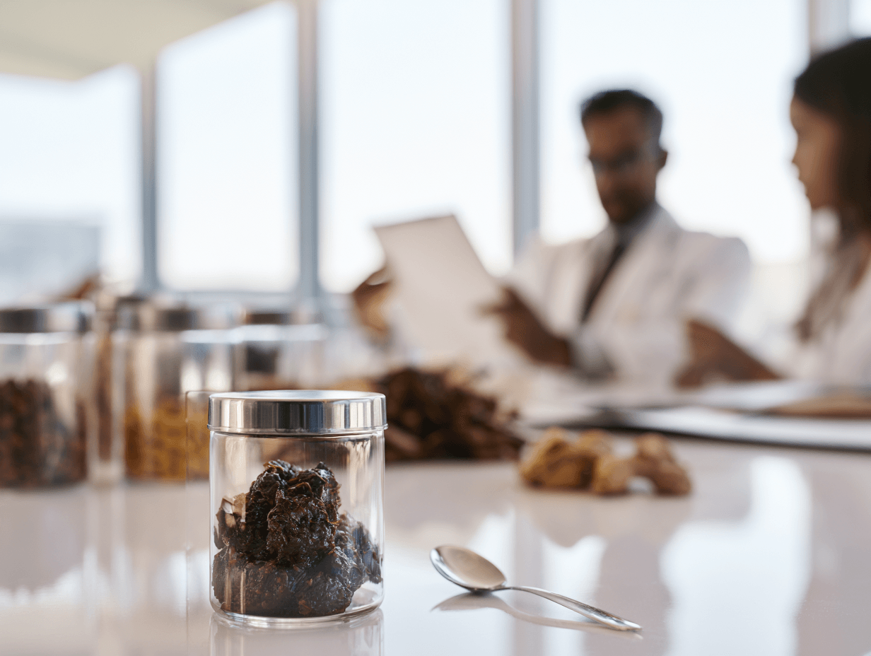
After your color-vision problem is classified and the underlying cause is identified, a personalized oral Avaleha may be considered. The proposed name for this formulation is Chakshushya Drishti Rasayana Avaleha.
This name reflects its intended therapeutic direction. “Chakshushya” refers to substances traditionally considered supportive for the eyes. “Drishti” relates to visual function, while “Rasayana” refers to the nourishment, protection and maintenance of body tissues and sensory functions.
This formulation is not described as a classical medicine quoted exactly from one book. It is a physician-designed Avaleha inspired by the Rasayana principles of Charaka Samhita, the Drishtigata Roga approach of Sushruta Samhita, the concept of Alochaka Pitta in Ashtanga Hridaya and the pharmaceutical method of Avaleha preparation in Sharangadhara Samhita [10–12,16,27,28].
The Classical Rasayana Principle Behind the Formulation
Charaka explains that Rasayana is a method for obtaining and maintaining the best functional quality of Rasa and the tissues nourished through it.
Classical source: Charaka Samhita, Chikitsa Sthana, Chapter 1, Rasayana Adhyaya, Abhaya-Amalakiya Pada, verses 7–8 [16].
Sanskrit
दीर्घमायुः स्मृतिं मेधामारोग्यं तरुणं वयः ।
प्रभावर्णस्वरौदार्यं देहेन्द्रियबलं परम् ॥७॥
वाक्सिद्धिं प्रणतिं कान्तिं लभते ना रसायनात् ।
लाभोपायो हि शस्तानां रसादीनां रसायनम् ॥८॥
Transliteration
Dīrgham āyuḥ smṛtiṃ medhām ārogyaṃ taruṇaṃ vayaḥ ।
Prabhā-varṇa-svaraudāryaṃ dehendriya-balaṃ param ॥7॥
Vāk-siddhiṃ praṇatiṃ kāntiṃ labhate nā rasāyanāt ।
Lābhopāyo hi śastānāṃ rasādīnāṃ rasāyanam ॥8॥
Translation
“Through Rasayana, a person may obtain longevity, memory, intellect, health, youthful strength, healthy appearance, strong sensory functions and vitality. Rasayana is the means of obtaining the best quality of Rasa and the succeeding tissues.”
This verse does not claim that Rasayana can replace an inherited cone pigment. It provides the classical basis for supporting tissue nourishment, sensory strength, recovery capacity and long-term protection.
Why an Avaleha Is Chosen
Avaleha is a semisolid oral dosage form prepared by concentrating a herbal liquid and combining it with suitable supporting substances and fine herbal powders. Its classical preparation is described in Sharangadhara Samhita, Madhyama Khanda, Chapter 8, Avaleha Kalpana [27,28].
This form allows several therapeutic layers to be combined in one medicine. A concentrated Kwatha may form the base, while fine Chakshushya, Rasayana, metabolic, neural or Pitta–Rakta-supportive herbs may be added at the appropriate stage.
The Avaleha can also be modified according to your digestion, diabetes status, strength and treatment response. This makes it more suitable for individualized treatment than giving the same commercial tablet or eye tonic to every patient.
Why This Is Not a Standard Color-Blindness Medicine
The Avaleha is not selected merely because you failed an Ishihara test. Your formulation depends on whether your condition is inherited, retinal, optic-nerve-related, metabolic, nutritional, medicine-associated or mixed.
If diabetes is affecting your retinal function, the formulation may require stronger Prameha, Meda, Agni and Ama management. If optic-nerve dysfunction is dominant, greater attention may be given to Vata, Majja, Dhatukshaya and neural-supportive Rasayana.
A retinal inflammatory or vascular pattern may require greater Pitta–Rakta regulation. A weak patient with nutritional depletion may require Agni correction followed by Rasa, Rakta and Dhatu nourishment.
This is why two people with the same color-test score may receive different herbs, different proportions, a different Avaleha medium and a different Anupana.
The Formulation Treats the Systemic Cause Along With the Visual Problem
Your eyes do not function separately from the rest of your body. Diabetes, poor nutrition, medicine toxicity, chronic inflammation, weak digestion and neurological disease may influence the retina or optic nerve.
The Avaleha should therefore be designed to address both the affected visual structure and the systemic condition contributing to it.
For example, adding only a Chakshushya herb would be incomplete if your HbA1c remains uncontrolled. Similarly, adding only nourishing Rasayana ingredients may be unsuitable when you have Mandagni, Ama, obesity or Kapha–Meda excess.
The correct formulation balances ocular support with the treatment of the underlying disease.
The Avaleha Is an Oral Medicine, Not an Eye Application
Chakshushya Drishti Rasayana Avaleha is intended to be taken orally. It must not be placed directly into your eyes.
Avaleha should not be confused with Aschyotana, Anjana, Seka, Tarpana or Putapaka. These are separate Netra Kriyakalpa procedures and may be considered only when they are suitable for your diagnosis.
You should never place homemade herbal liquids, honey, powders, oils or non-sterile ghee into your eyes. Such practices may cause irritation, corneal injury or infection.
The Proposed 30-Day Treatment Quantity
The physician-directed dosage proposed for this treatment model is 15 g orally twice daily.
You receive 15 g in the morning and 15 g in the evening, giving a total of 30 g per day. For 30 days, the required finished quantity is:
30 g per day × 30 days = 900 g of finished Avaleha.
The medicine may therefore be prepared as a complete 900 g patient-specific batch for the first 30-day treatment phase.
This dosage should not be copied by every reader. Your age, digestive capacity, diabetes status, body strength, concurrent medicines and tolerance must be assessed before the regimen is confirmed.
The Avaleha Medium Must Match Your Metabolic Condition
Classical Avaleha preparations commonly use substances such as Guda or Sharkara to obtain the required semisolid consistency. However, a conventional sweet base may not be appropriate when you have uncontrolled diabetes, severe insulin resistance, obesity or significant Kapha–Meda involvement.
In such cases, the quantity, medium or dosage form may need modification. Another suitable pharmaceutical form may be selected when a 15 g sweet Avaleha would conflict with your metabolic treatment.
A diabetic patient should not receive a sugar-heavy preparation simply because the medicine is called an Avaleha. Your metabolic safety is more important than rigidly following one dosage form.
Why the First Batch Is Prepared for 30 Days
The first 30-day batch allows your response to be reviewed before the same formulation is repeated.
Your digestion, bowel function, appetite, sleep, medicine tolerance and associated visual symptoms should be reassessed. Your color-vision test, contrast sensitivity, HbA1c, OCT or other relevant findings may also be compared according to your diagnosis.
If the response is satisfactory, the next batch may continue the same therapeutic direction. If your response is partial, the proportions or treatment emphasis may be changed. If no objective benefit is found, the diagnosis and formulation should be reconsidered.
The Avaleha should not be repeated indefinitely merely because it is Ayurvedic.
Why the Most Crowded Formula Is Not the Strongest Formula
A formulation does not become stronger merely by adding every available Chakshushya herb, Rasayana or bhasma.
A patient with Pitta–Rakta aggravation may not tolerate the same combination as a patient with Vata–Majja depletion. A diabetic patient may require a different medium from a weak, undernourished patient. A person with reduced kidney or liver function may require additional caution before any mineral medicine is considered.
The true strength of the Avaleha comes from Yukti—the correct selection of ingredients, quantities, medium, Anupana, timing and patient.
Your reports should therefore decide the formulation. The formulation should never be made powerful only by increasing the number of ingredients.
What May Go Into a Personalized Chakshushya Drishti Rasayana Avaleha?
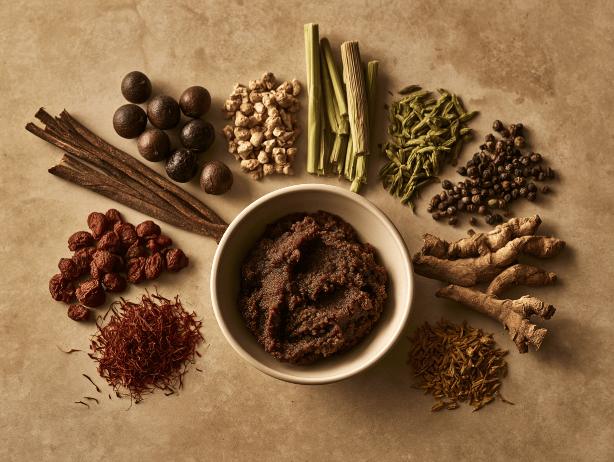
A strong Avaleha is not created by adding every available eye herb or mineral. It is built in therapeutic layers according to your diagnosis, Ayurvedic Samprapti, digestive capacity, metabolic condition and treatment goal.
Your formulation may require a Chakshushya–Rasayana foundation, retinal support, Vata–Majja nourishment, Pitta–Rakta regulation, Prameha management, Agni correction or Dhatu restoration. You should receive only the ingredients that are suitable for your condition.
Triphala as the Chakshushya–Rasayana Foundation
Triphala contains Amalaki, Haritaki and Bibhitaki. It is traditionally used to support Rasayana, digestion, metabolism and ocular health.
Its classical properties are discussed in Bhavaprakasha Nighantu, Haritakyadi Varga, while its broader Rasayana use follows the principles described in Charaka Samhita, Chikitsa Sthana, Chapter 1, Rasayana Adhyaya.
Amalaki, or Phyllanthus emblica, may support antioxidant protection and Pitta-sensitive patients. Experimental research has examined its effects on retinal cells, mitochondrial function and oxidative stress relevant to age-related macular degeneration [30].
Haritaki, or Terminalia chebula, may be selected when Rasayana support must be combined with better bowel regulation, Agni and Srotas function. Bibhitaki, or Terminalia bellirica, may be more relevant when Kapha, Meda or sluggish metabolism is present.
An experimental diabetic-retinopathy study reported that Triphala improved selected biochemical, structural and electrophysiological findings in diabetic rats [31]. This provides an indirect retinal rationale, but it does not prove that Triphala can correct inherited color blindness.
Guduchi for Retinal and Metabolic Support
Guduchi, or Tinospora cordifolia, may be included when your case involves diabetes, metabolic disturbance, Pitta aggravation, chronic inflammation or a need for Rasayana support.
Its classical properties are described in Bhavaprakasha Nighantu, Guduchyadi Varga. It is also relevant to the Rasayana principles of Charaka Samhita, Chikitsa Sthana, Chapter 1, and the metabolic treatment principles of Chapter 6, Prameha Chikitsa.
In an experimental diabetic-retinopathy model, Guduchi reduced retinal oxidative stress and improved antioxidant defence mechanisms [29]. This supports its consideration in selected metabolic retinal cases, but it is not direct evidence of color-vision recovery in humans.
Your liver history, concurrent medicines and previous reactions should still be reviewed before prolonged use.
Yashtimadhu and Shatavari for Pitta-Sensitive and Depleted Patients
Yashtimadhu, or Glycyrrhiza glabra, may be considered when your formulation requires Rasayana, tissue nourishment and Pitta-supportive action.
Experimental research involving glycyrrhizin has reported protection against inflammatory, vascular and neuronal damage in diabetic retinal models [33]. This evidence is indirect because an isolated constituent is not identical to the complete herb or finished Avaleha.
Yashtimadhu requires caution when you have uncontrolled hypertension, fluid retention, low potassium, kidney disease or heart disease.
Shatavari, or Asparagus racemosus, may be considered when a Pitta-sensitive or depleted patient requires gentler Brimhana and Rasayana support. It should not be added automatically to a patient with severe Kapha–Meda excess or poor digestion.
Manjistha, Sariva and Usheera for Pitta–Rakta Patterns
Manjistha, or Rubia cordifolia, may be selected when your retinal condition is associated with Pitta–Rakta disturbance, metabolic inflammation or microvascular stress.
Experimental research has reported antioxidant and anti-apoptotic effects of Manjistha in diabetic neural injury [40]. This is not direct retinal or color-vision evidence, but it supports further study in metabolically driven neural conditions.
Sariva, or Hemidesmus indicus, may be used in selected Pitta–Rakta and heat-dominant patterns. Laboratory research has reported antioxidant and antithrombotic activity, although direct clinical evidence for retinal or color-vision improvement is unavailable [41].
Usheera, or Chrysopogon zizanioides, may be considered when cooling Pitta support is required. Its modern evidence is mainly limited to laboratory antioxidant findings [42].
These herbs should not be added merely because every visual disorder is assumed to be a Pitta disease.
Daruharidra for Retinal-Metabolic Involvement
Daruharidra, or Berberis aristata, may be considered when retinal dysfunction occurs with diabetes, Kapha–Pitta involvement or metabolic inflammation.
Berberine related experimental research has reported effects on retinal inflammatory, vascular and oxidative pathways in diabetic animals [39]. However, berberine research should not be presented as proof that the complete Daruharidra herb improves color vision.
Daruharidra may also interact with medicines and may not be appropriate during pregnancy or in every weak patient. Its inclusion should follow your complete medical history.
Ashwagandha, Brahmi and Bala for Vata–Majja Support
Ashwagandha, or Withania somnifera, may be considered when your condition involves Vata aggravation, tissue depletion, chronic stress, poor sleep or a stable degenerative pattern.
Modern research describes antioxidant, anti-inflammatory and neuroprotective mechanisms of Ashwagandha [34]. These findings provide a general neural rationale, but they do not establish that Ashwagandha repairs the optic nerve or restores color discrimination.
Brahmi, or Bacopa monnieri, may be added when Medhya Rasayana and neural support are required. A systematic review described possible effects on oxidative stress, inflammation, mitochondrial function and neuronal protection [35]. Direct evidence in color blindness remains unavailable.
Bala, or Sida cordifolia, may be considered in selected weak, Vata-dominant patients requiring Brimhana and tissue support. It is not suitable for automatic inclusion, especially when you have hypertension, cardiac concerns or significant Kapha–Meda excess.
Jambu, Meshashringi and Vijayasara for Diabetes-Associated Cases
If diabetes is contributing to retinal or color-vision deterioration, your formulation may require a separate Prameha-focused module.
Jambu Bija, or Syzygium cumini, has been studied clinically for glucose-related outcomes, although results have been limited and it has not been shown to improve color vision directly [38].
Meshashringi, or Gymnema sylvestre, has been investigated for insulin sensitivity, glucose metabolism and metabolic syndrome [36]. Vijayasara, or Pterocarpus marsupium, has also been studied in patients with newly diagnosed type 2 diabetes [37].
These herbs may support the systemic metabolic treatment plan, but they are not “color-blindness herbs.” Their role is to address a disease process that may be affecting your retina or optic nerve.
Because these ingredients may influence blood glucose, your glucose readings and conventional diabetes medicines should be reviewed to reduce the risk of hypoglycaemia.
Agni and Ama-Correcting Ingredients
A nourishing Avaleha may not work properly when your Agni is weak and Ama is prominent. Small quantities of ingredients such as Musta, Shunthi, Pippali or another patient-appropriate Deepana–Pachana medicine may therefore be considered.
The selection depends on your Dosha. Strong heating herbs may be unsuitable when you have marked Pitta aggravation, burning, acidity, active retinal inflammation or poor tolerance.
Agni-supportive herbs are included to improve suitability and digestion of the formulation. They should not dominate the medicine unless Mandagni and Ama are central to your Samprapti.
Dhatu-Restoring Ingredients for Weak or Depleted Patients
When your condition occurs with weight loss, chronic illness, poor nutrition, malabsorption or general weakness, the formulation may require greater Rasa, Rakta, Majja and Ojas support.
Ashwagandha, Bala, Shatavari, Yashtimadhu, Amalaki and suitable Ghrita-based support may be considered according to your digestion and metabolic status.
A depleted patient should not receive the same Lekhana or Kapha-reducing formulation as an obese diabetic patient. The treatment must reflect whether your main problem is obstruction, inflammation, degeneration or inadequate nourishment.
Mineral and Pishti Components Are Not Required in Every Patient
Selected formulations may consider physician-supervised preparations such as Abhraka Bhasma, Yashada Bhasma, Mukta Pishti, Pravala Pishti or Swarna Bhasma when a clear Ayurvedic indication exists.
These substances should never be combined simply to make the Avaleha appear stronger. Your age, body weight, liver function, kidney function, pregnancy status, concurrent medicines and treatment duration must first be assessed.
Only licensed and traceable products with appropriate quality documentation should be used. The exact mineral preparation and quantity should remain patient-specific and should not be published as a universal online prescription [43–46,48].
Modern evidence does not establish that any bhasma recreates a genetically absent cone photopigment or cures inherited color blindness.
Why the Exact Ingredient Weight Cannot Be the Same for Everyone
A 900 g finished Avaleha does not mean that every patient should receive the same raw-herb weights.
A diabetic patient may need a stronger metabolic module and a modified Avaleha medium. An optic-nerve case may require greater Vata–Majja support. A Pitta–Rakta retinal pattern may need a different balance, while a weak patient may require more Dhatu-restorative ingredients.
The decoction herbs, fine Prakshepa powders, Rasayana ingredients, medium and Anupana must therefore be calculated after your reports and Ayurvedic assessment are complete.
The public article can explain the full therapeutic architecture. Your individual formulation sheet should contain the exact ingredients, weights, batch number, dose, preparation date and monitoring plan.
What the Research Actually Supports
Modern research provides preliminary support for antioxidant, metabolic, anti-inflammatory, retinal and neuroprotective actions of several ingredients considered in this formulation [29–42].
Most of this evidence comes from laboratory studies, animal models, small clinical studies or research on related diseases. It does not prove that each herb independently improves color vision or that combining all of them produces a cure.
The scientific purpose of these findings is to support careful formulation and future investigation. Your actual response must still be confirmed through standardized color-vision testing and relevant retinal, optic-nerve or metabolic measurements.
How the Chakshushya Drishti Rasayana Avaleha Changes According to Your Condition

The finished Avaleha may remain approximately 900 g for a 30-day course, but its internal composition should not be identical for every patient. The decoction herbs, fine powders, Rasayana ingredients, metabolic support, Avaleha medium and Anupana must change according to the cause of your color-vision problem.
The purpose of customization is not to add more ingredients. It is to give greater importance to the therapeutic group that matches your dominant pathology.
When Your Color Deficiency Has Been Present Since Childhood
If you have had stable color confusion since childhood, the formulation should not be designed as though you have an active retinal disease. The main emphasis may remain on Chakshushya Rasayana, general ocular nourishment, Agni support and protection of remaining visual function.
Amalaki, Triphala, Guduchi and other suitable Rasayana ingredients may be considered according to your constitution and digestion. Nutritional, metabolic or ocular abnormalities should be corrected when present.
The formulation should not claim to recreate an absent or altered cone photopigment. Its purpose is to support ocular health and identify whether an additional acquired disturbance is worsening your inherited deficiency [1–6,13–16].
When the Retina or Macula Is Mainly Affected
If OCT, retinal examination or other findings show macular or retinal involvement, the Avaleha may require stronger retinal, Pitta–Rakta and metabolic support.
Guduchi, Amalaki, Triphala, Manjistha, Sariva, Daruharidra or other patient-appropriate ingredients may be considered. The exact selection depends on whether your retinal condition is inflammatory, vascular, degenerative, diabetic or associated with nutritional depletion.
A cooling formulation should not be given automatically. If Vata degeneration or tissue depletion is also present, excessive Pitta-reducing treatment may weaken the nourishing aspect required for retinal support.
Experimental studies involving Guduchi, Amalaki and Triphala have reported antioxidant or retinal-protective effects in related models, but these findings remain indirect and do not establish correction of inherited color blindness [29–32].
When the Optic Nerve Is Mainly Involved
If colors appear faded in one eye, red looks less vivid or visual-field and optic-nerve findings are abnormal, the cause must first be medically investigated.
After urgent or active disease has been treated, the Avaleha may give greater importance to Vata–Majja support, Dhatu nourishment and selected Medhya Rasayana ingredients.
Ashwagandha, Brahmi, Bala, Yashtimadhu, Amalaki or suitable Ghrita-based support may be considered according to your strength, digestion and metabolic condition.
These ingredients are selected for their broader neural and Rasayana rationale. They should not be described as medicines that directly regenerate the optic nerve or guarantee restoration of color vision [33–35].
When Diabetes Is Contributing to Color-Vision Deterioration
If your color vision has worsened with diabetes, the formulation must address Prameha, Meda, Kleda, Agni and Ama along with retinal support.
Guduchi, Jambu Bija, Meshashringi, Vijayasara, Daruharidra or other suitable metabolic ingredients may receive greater importance. Your fasting glucose, post-meal glucose, HbA1c, kidney function, current diabetes medicines and risk of hypoglycaemia must be reviewed [17,29,36–39].
A conventional sugar- or jaggery-heavy Avaleha should not be used automatically. The medium, concentration, dose or even the dosage form may need modification.
The main purpose is not to hide the retinal symptom with an eye tonic. It is to improve the metabolic environment that may be continuing to affect your retinal and neural function.
When Poor Digestion and Ama Are Dominant
If you have poor appetite, heaviness, bloating, coated tongue or irregular bowel movements, a dense nourishing Avaleha may be difficult to digest.
The first batch may therefore contain a carefully balanced Agni and Ama-supportive component. Musta, Shunthi, Pippali or another appropriate Deepana–Pachana ingredient may be considered in small quantities.
The choice depends on your Dosha. Strong heating ingredients may be unsuitable when you have acidity, burning, active Pitta aggravation or retinal inflammation.
Once digestion and medicine tolerance improve, the next batch may shift toward stronger Chakshushya and Rasayana support.
When Nutritional Depletion or Dhatu Kshaya Is Present
If your condition occurs with weakness, weight loss, anaemia, malabsorption, chronic illness or poor nourishment, the formulation may require greater Brimhana and Dhatu-restorative support.
Amalaki, Yashtimadhu, Shatavari, Ashwagandha, Bala or other appropriate Rasayana ingredients may be considered. However, nourishment should follow improvement in Agni and absorption.
A depleted patient should not receive the same Kapha-reducing formulation used for an obese patient with uncontrolled diabetes. Your main need may be tissue restoration rather than Lekhana or strong Dosha reduction.
When a Medicine or Toxic Exposure May Be Responsible
If your color perception changed after starting a medicine or after occupational chemical exposure, the first step is to identify and control the suspected cause.
You should not stop a necessary medicine without consulting the prescribing doctor. Your retina, optic nerve, liver and kidney function may require assessment according to the suspected exposure [7,8].
The Avaleha may later focus on recovery support, Pitta–Rakta regulation, Agni–Ama correction or Vata–Majja nourishment. However, Rasayana should not be used to conceal continuing toxic injury.
When Cataract Is Changing Your Color Perception
If your lens has become yellow or opaque, colors may appear dull, brownish or less vivid. In this situation, an Avaleha cannot physically replace the cloudy lens with a clear one.
The formulation may support your general ocular, digestive and metabolic health, but cataract assessment remains essential. When surgery is clinically indicated, delaying it solely for oral Ayurvedic treatment may reduce your quality of life.
The treatment objective should therefore be supportive and systemic rather than making an unrealistic promise of clearing an advanced cataract.
When You Have a Mixed Inherited and Acquired Problem
You may have had mild red–green confusion since childhood and later develop diabetes, cataract, retinal disease or optic-nerve dysfunction.
In such cases, the formulation should target the acquired component while supporting your general visual health. The stable inherited component and the newer deterioration must be recorded separately.
For example, your lifelong Ishihara error pattern may remain, while contrast, brightness difference, retinal function or additional color errors may improve after the acquired disease is treated.
This is why your outcome should not be judged only by whether every Ishihara plate becomes normal.
Why the Second Batch May Differ From the First
The first 30-day batch provides information about your digestion, tolerance and early response. The next formulation should be reviewed against color testing, visual symptoms, retinal or optic-nerve findings and relevant metabolic reports.
If Agni improves but color discrimination remains unchanged, the next batch may increase the disease-specific Rasayana component. If glucose control remains poor, the metabolic strategy may need strengthening. If the formulation causes heaviness or reduced appetite, its medium and nourishing ingredients may need adjustment.
The same prescription should not be repeated automatically. Your next batch should reflect what actually changed during the first month.
Why Customization Is More Important Than the Number of Ingredients
A formula containing every potent herb and mineral may become difficult to digest, unsuitable for your Dosha or unnecessarily complex.
The strongest formulation is the one that correctly matches your inherited or acquired diagnosis, dominant Dosha–Dushya pattern, Agni, metabolic status, tissue strength and treatment stage.
Your 900 g Avaleha should therefore be understood as a patient-specific therapeutic structure , not as one fixed recipe for every person with color-vision difficulty.
Why Diabetic Patients Should Not Automatically Receive the Same Sweet Avaleha
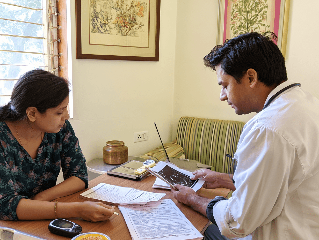
If your color vision has worsened with diabetes, the medicine must support both your eyes and your metabolic condition. A standard Avaleha prepared with a large quantity of sugar or jaggery may not be suitable when your glucose remains uncontrolled.
The dosage form should never work against the disease being treated. Your fasting glucose, post meal glucose, HbA1c, body weight, kidney function, current diabetes medicines and risk of low blood sugar should therefore be reviewed before the Avaleha medium is selected.
Classical Avaleha Commonly Uses a Sweet Medium
Classical Avaleha preparation may use Guda, Sharkara or another suitable sweet medium to create the required consistency, stability and palatability. The preparation principles are described in Sharangadhara Samhita, Madhyama Khanda, Chapter 8, Avaleha Kalpana [27, 28].
This does not mean that every patient must receive the same amount of sugar or jaggery. Classical Ayurveda also requires the medicine to be adjusted according to the disease, patient strength, Agni and suitability.
A dosage form should therefore follow Yukti rather than being used rigidly.
Your Metabolic Pattern Must Be Identified
You may have diabetes with obesity, heaviness, excessive appetite, Kapha, Meda and Ama. Another patient may have diabetes with weight loss, weakness, poor appetite and Dhatu Kshaya.
These two patients should not receive the same Avaleha base or the same nourishing ingredients.
Charaka explains this distinction in Charaka Samhita, Chikitsa Sthana, Chapter 6, Prameha Chikitsa, verse 15 [17].
Sanskrit
स्थूलः प्रमेही बलवानिहैकः कृशस्तथैकः परिदुर्बलश्च ।
सम्बृंहणं तत्र कृशस्य कार्यं संशोधनं दोषबलाधिकस्य ॥१५॥
Transliteration
Sthūlaḥ pramehī balavān ihaikaḥ kṛśas tathaikaḥ paridurbalaś ca ।
Sambṛṃhaṇaṃ tatra kṛśasya kāryaṃ saṃśodhanaṃ doṣabalādhikasya ॥15॥
Translation
“One patient with Prameha may be strong and obese, while another may be lean and weak. The weak patient requires nourishment, while the strong patient with greater Dosha involvement requires Dosha reducing treatment.”
This principle shows why diabetes treatment must be individualized. A sweet and heavily nourishing Avaleha may worsen heaviness and poor glucose control in one patient, while excessive reducing treatment may weaken another.
Honey Is Not Automatically a Safe Replacement
Honey should not be considered freely suitable simply because it is natural. It still affects glucose intake and must be judged according to your total diet, glucose control and medical condition.
Honey should also not be heated during Avaleha preparation. If it is considered appropriate, it should be added only after the formulation has cooled sufficiently and only in a patient specific quantity.
You should not replace sugar with honey independently and assume that the formulation has become suitable for diabetes.
The Avaleha Medium May Need Modification
When your glucose remains controlled and the physician considers Avaleha suitable, the quantity and type of sweet medium may be adjusted carefully.
When your diabetes is poorly controlled, another approach may be safer. The medicine may be prepared in a smaller concentrated dose, divided into separate powders or tablets, or provided in another suitable dosage form.
The aim is to preserve the therapeutic herbs without adding an unnecessary metabolic burden.
Metabolic Herbs Do Not Cancel a High Sugar Base
Adding Guduchi, Jambu Bija, Meshashringi, Vijayasara or Daruharidra does not automatically make a sugar rich Avaleha suitable for diabetes.
These herbs may support the treatment of Prameha, Meda, Agni and metabolic dysfunction, but they do not neutralize an inappropriate quantity of sugar or jaggery.
Research on Guduchi, Meshashringi, Vijayasara, Jambu and berberine related compounds provides indirect support for metabolic and retinal pathways. It does not justify ignoring the carbohydrate content of the final medicine [29, 36, 37, 38, 39].
Blood Glucose Must Be Monitored During Treatment
Some metabolic herbs may influence blood glucose. When they are combined with conventional diabetes medicines, your glucose may fall more than expected.
You should therefore monitor fasting glucose, post meal glucose and any symptoms of low blood sugar. Your conventional medicine should not be reduced or stopped without guidance from the doctor managing your diabetes.
HbA1c should be reviewed at the appropriate clinical interval rather than being repeated after only a few days.
Diabetic Retinal Disease Requires More Than an Eye Formula
If diabetes is affecting your color perception, the deeper treatment target may include retinal circulation, oxidative stress, inflammation, glucose control, blood pressure and lipid levels.
Ayurvedic assessment may additionally consider Prameha, Meda, Kleda, Agni, Ama, Rakta and Srotas.
A related Ayurvedic clinical study has examined an add on treatment protocol in diabetic retinopathy, but it does not prove that one Avaleha can restore inherited color vision [50].
The correct goal is to improve the metabolic environment that supports retinal and neural function.
When a Traditional Avaleha May Not Be the Best Choice
A traditional Avaleha may be unsuitable when your HbA1c is very high, glucose fluctuates widely, obesity is severe, digestion is poor or you already consume several sweet medicines.
In such cases, choosing another dosage form is not a weakness in Ayurvedic treatment. It is a safer application of Yukti.
Your treatment should remain Ayurvedic in principle while the pharmaceutical form is adapted to your actual medical needs.
The Final Decision Should Follow Your Reports
Before confirming a 900 g Avaleha for 30 days, your metabolic reports should be reviewed. The final decision should consider your HbA1c, glucose readings, body weight, appetite, Agni, kidney function, current medicines and risk of hypoglycaemia.
A diabetic patient should not receive the same sweet Avaleha merely because the disease also affects the eyes. The formulation must protect your metabolism while treating the retinal or neural cause of your color vision difficulty.
Chakshushya Drishti Rasayana Avaleha-Medicine
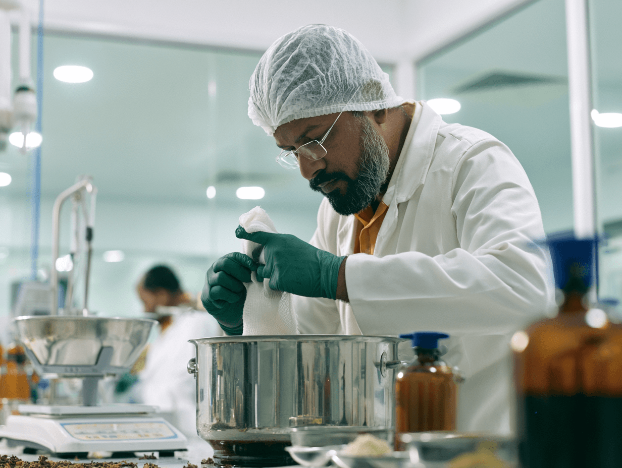
Chakshushya Drishti Rasayana Avaleha is a physician designed oral formulation for selected adults with acquired or mixed color vision difficulty. It is intended to support the retina, optic nerve, metabolism, digestion and tissue nourishment according to the diagnosed cause.
This exact Avaleha is not quoted word for word from one classical book. It is a new formulation inspired by classical Rasayana, Chakshushya, Drishtigata Roga, Saptamrita Lauha and Avaleha preparation principles.
Treatment has greater relevance when an acquired retinal, optic nerve, metabolic, nutritional, medicinal or lens related cause is present [1, 2].
The published Ayurvedic color blindness evidence is limited to a single case report involving several internal medicines and procedures. That report did not study this Avaleha and does not prove that a universal formulation can reverse inherited cone pigment changes [49].
Classical Books That Inspire This Formulation
The Rasayana foundation is inspired by Charaka Samhita, Chikitsa Sthana, Chapter 1, Rasayana Adhyaya. This chapter supports the use of Rasayana for the maintenance of Dhatu quality, strength and sensory function.
The visual assessment is inspired by Sushruta Samhita, Uttara Tantra, Chapter 7, Drishtigata Roga Vijnaniya Adhyaya, and Chapter 17, Drishtigata Roga Pratishedha Adhyaya.
The functional understanding of visual perception is inspired by Ashtanga Hridaya, Sutrasthana, Chapter 12, Doshabhediya Adhyaya, which describes Alochaka Pitta.
The ocular treatment framework is supported by Sushruta Samhita, Uttara Tantra, Chapter 18, Kriyakalpa Adhyaya, and Ashtanga Hridaya, Sutrasthana, Chapter 24, Tarpana Putapaka Vidhi.
The Triphala, Yashtimadhu, Lauha, Ghrita and Madhu logic is inspired by Saptamrita Lauha, traditionally described under Bhaishajya Ratnavali, Netraroga Chikitsa, Chapter 64. Published reviews describe Saptamrita Lauha as a combination of Triphala, Yashtimadhu and Lauha Bhasma, usually administered with Ghrita and Madhu.
The pharmaceutical preparation follows Sharangadhara Samhita, Madhyama Khanda, Chapter 8, Avaleha Kalpana, which explains the use of a concentrated herbal liquid, sweetening medium, Prakshepa Dravya, Sneha, Madhu and Avaleha Siddhi Lakshana [27, 28].
Who This Exact 900 Gram Formula Is Designed For
This formulation model is intended for a non diabetic adult with normal liver and kidney function, satisfactory digestion and an ophthalmologist confirmed acquired or mixed color vision problem.
It is not suitable as a standard formula for children, pregnancy, breastfeeding, uncontrolled diabetes, severe obesity, marked Ama, significant liver or kidney disease, iron overload, acute optic neuritis, sudden one eye color loss, retinal detachment symptoms or rapidly worsening vision.
A diabetic patient requires a different dosage form because this classical Avaleha model contains jaggery and honey. Reducing the sweetening agent without pharmaceutical reassessment can alter consistency, water activity, stability and microbial safety.
Exact Dosage and 30 Day Quantity
| Administration | Quantity |
| Morning dose | 15 g |
| Evening dose | 15 g |
| Total daily dose | 30 g |
| Duration | 30 days |
| Total finished Avaleha required | 900 g |
The usual physician directed method is 15 g orally twice daily after food. The medicine may be followed by approximately 50 to 100 ml of lukewarm water unless another Anupana is selected for you.
The dosage should be reduced or discontinued when you develop persistent nausea, loose stools, abdominal heaviness, reduced appetite, skin reaction, swelling, unusual weakness or any new visual deterioration.
Exact Kwatha Dravya for the 30 Day Batch
All the ingredients below should be taken as authenticated dry raw drugs and made into a coarse powder. The total Kwatha Dravya weight is 500 g.
| Ayurvedic ingredient | Botanical identity | Plant part | Weight |
| Haritaki | Terminalia chebula Retz. | Fruit pericarp | 35 g |
| Bibhitaki | Terminalia bellirica Roxb. | Fruit pericarp | 35 g |
| Amalaki | Phyllanthus emblica L. | Dried fruit | 60 g |
| Guduchi | Tinospora cordifolia | Stem | 50 g |
| Yashtimadhu | Glycyrrhiza glabra L. | Root | 40 g |
| Jivanti | Leptadenia reticulata | Whole plant | 30 g |
| Shatavari | Asparagus racemosus Willd. | Tuberous root | 30 g |
| Bhringaraja | Eclipta prostrata L. | Whole plant | 25 g |
| Brahmi | Bacopa monnieri | Whole plant | 25 g |
| Ashwagandha | Withania somnifera | Root | 30 g |
| Mandukaparni | Centella asiatica L. | Whole plant | 20 g |
| Manjistha | Rubia cordifolia L. | Root | 25 g |
| Sariva | Hemidesmus indicus | Root | 25 g |
| Daruharidra | Berberis aristata DC. | Stem | 20 g |
| Punarnava | Boerhavia diffusa L. | Root | 20 g |
| Musta | Cyperus rotundus L. | Rhizome | 15 g |
| Haridra | Curcuma longa L. | Rhizome | 10 g |
| Usheera | Chrysopogon zizanioides | Root | 5 g |
| Total | 500 g |
This Kwatha group combines Triphala based Chakshushya support, Guduchi and Daruharidra for retinal metabolic stress, Yashtimadhu and Sariva for Pitta sensitive patterns, and Brahmi, Ashwagandha, Mandukaparni and Jivanti for broader neural and Rasayana support.
Exact Prakshepa Dravya
The following ingredients should be made into a very fine powder and kept separately. They should not be boiled with the Kwatha.
| Prakshepa ingredient | Botanical or pharmaceutical identity | Weight |
| Amalaki fine powder | Phyllanthus emblica | 20 g |
| Yashtimadhu fine powder | Glycyrrhiza glabra | 15 g |
| Guduchi Satva | Purified starch extract of Tinospora cordifolia | 10 g |
| Jivanti fine powder | Leptadenia reticulata | 10 g |
| Shatavari fine powder | Asparagus racemosus | 10 g |
| Ashwagandha fine powder | Withania somnifera | 10 g |
| Brahmi fine powder | Bacopa monnieri | 10 g |
| Shankhapushpi fine powder | Convolvulus pluricaulis | 10 g |
| Mandukaparni fine powder | Centella asiatica | 10 g |
| Bhringaraja fine powder | Eclipta prostrata | 10 g |
| Padmaka fine powder | Prunus cerasoides | 10 g |
| Haridra fine powder | Curcuma longa | 6 g |
| Pippali fine powder | Piper longum L. | 3 g |
| Ela fine powder | Elettaria cardamomum | 3 g |
| Tvak fine powder | Cinnamomum verum | 2 g |
| Kumkuma or Kesar | Crocus sativus L. stigma | 1 g |
| Total | 140 g |
The repeated use of selected herbs in the Kwatha and Prakshepa stages is intentional. The decoction extracts water soluble components, while the fine powder stage retains a wider range of plant constituents that may be damaged or lost through prolonged boiling.
Avaleha Base and Final Batch Materials
| Material | Exact quantity |
| Water for Kwatha | 8 litres |
| Prepared Kwatha after reduction | Approximately 1 litre |
| Clean jaggery | 400 g |
| Cow Ghrita | 60 g |
| Honey | 60 g |
| Fine Prakshepa powders | 140 g |
| Final finished batch | 900 g |
The final weight is achieved through controlled evaporation. You should not add untreated water after the Avaleha has reached the correct stage because this can disturb stability and increase microbial risk.
Physician Only Mineral Options
The fixed herbal formulation above should not automatically contain every available bhasma. Combining several minerals merely to call the medicine stronger has no direct evidence in color vision deficiency and creates unnecessary quality, dosing and monitoring concerns.
When a mineral component is clinically justified, it is better to select the minimum required option. The following are 30 day clinician planning amounts, not a universal prescription and not doses established through color blindness trials.
| Mineral option | Maximum proposed 30 day quantity | Approximate daily exposure | When it may be considered |
| Lauha Bhasma | 3 g | 100 mg daily | Saptamrita Lauha inspired approach when iron status permits |
| Abhraka Bhasma, Sahasraputi | 3 g | 100 mg daily | Selected Vata, Majja or Dhatu Kshaya presentation |
| Yashada Bhasma | 1.5 g | 50 mg daily | Selected metabolic or documented zinc related presentation |
| Mukta Pishti | 1.5 g | 50 mg daily | Selected Pitta dominant presentation |
| Pravala Pishti | 1.5 g | 50 mg daily | Alternative to Mukta Pishti in a suitable Pitta pattern |
| Swarna Bhasma | 0.3 g | 10 mg daily | Highly selected Rasayana use, not routine use |
One should not combine all six options in one patient. A practical rule is to use no mineral, one primary bhasma, or one primary bhasma with one specifically indicated Pishti. Mukta and Pravala should not be combined automatically.
Heeraka Bhasma, Tamra Bhasma, Naga Bhasma, Vanga Bhasma and mercury containing Rasa preparations are intentionally excluded from this fixed formulation. There is no direct clinical evidence that they improve color discrimination, while the analytical and safety burden is substantially greater.
When one mineral option is selected, prepare the herbal Avaleha to a weight that allows the selected mineral to be added while retaining the final batch weight of 900 g. For example, when 3 g Lauha Bhasma is selected, prepare 897 g of the completed herbal Avaleha and add 3 g through geometric dilution.
All bhasmas and pishtis must come from a licensed source with batch traceability and appropriate analytical documentation. Official AYUSH guidance advises physician supervision and warns against self medication with mineral and herbo mineral products.
Patient Friendly Preparation Method
Step One: Prepare the Coarse Herbs
Take the 500 g Kwatha ingredients and verify their identity, plant part, appearance, odour and absence of visible mould, insects or foreign material.
Make them into a coarse powder. Do not use a very fine powder for the decoction because it becomes difficult to filter and may produce a gritty Avaleha.
Step Two: Soak the Herbs
Place the 500 g coarse herbal mixture in a clean food grade stainless steel vessel.
Add 8 litres of potable water and allow the mixture to soak for approximately 8 to 12 hours. Overnight soaking helps the harder roots, stems and fruits absorb water before boiling.
Step Three: Prepare the Kwatha
Heat the soaked mixture slowly. Once it begins to boil, maintain gentle heat and stir occasionally.
Continue reducing the liquid until approximately 1 litre remains. Avoid aggressive boiling because the mixture may burn at the bottom before proper extraction occurs.
The traditional proportion used here is sixteen parts water reduced to approximately one eighth of its original volume.
Step Four: Filter Carefully
Filter the hot decoction through a clean double layered muslin cloth.
Press the coarse material gently, but do not force fine sediment through the cloth. The filtered Kwatha should be smooth and free from coarse particles.
Step Five: Add the Jaggery
Add 400 g of clean jaggery to the filtered Kwatha.
Heat gently until the jaggery dissolves completely. Filter the liquid again to remove any sand, fibre or other impurities that may be present in the jaggery.
Return the clean liquid to the vessel.
Step Six: Cook Until the Syrup Reaches the Correct Stage
Continue heating on a low flame while stirring continuously.
As the water evaporates, the liquid becomes thick. Add 60 g cow Ghrita gradually and continue stirring so that the Ghrita becomes properly incorporated.
The mixture should not stick to the bottom or develop a burnt smell.
Step Seven: Check Avaleha Siddhi Lakshana
Take a small amount, allow it to cool and test it between your fingers.
The Avaleha is approaching the correct stage when it produces a soft thread between the fingers, forms a soft bolus that retains a fingerprint and does not disperse immediately when a cooled drop is placed in water.
You should stop heating before the mixture becomes hard or excessively sticky. Overcooking can make the final medicine difficult to measure and consume.
Step Eight: Add the Fine Prakshepa Powders
Remove the vessel from direct heat.
Allow the Avaleha to cool until it remains hot but is no longer boiling. Add the 140 g Prakshepa powder slowly in small portions while stirring continuously.
This gradual addition prevents lumps and helps distribute saffron, Brahmi, Guduchi Satva, Pippali and the other fine ingredients evenly.
Step Nine: Add the Selected Mineral Only When Indicated
When a mineral option has been prescribed, do not pour it directly into the full vessel.
First triturate the measured mineral with a small quantity of the prepared Avaleha. Gradually add larger quantities through geometric dilution until the mixture is uniform. Then incorporate it into the main batch.
This step is essential because a poorly distributed bhasma can produce unequal dosing from one spoonful to another.
Step Ten: Add Honey Only After Cooling
Allow the preparation to cool below approximately 40°C.
Add 60 g honey and mix thoroughly. Honey should not be boiled or added while the preparation remains very hot.
Step Eleven: Confirm the Final Weight
The final net weight must be 900 g, including the selected mineral option when one is used.
If the weight is too high, controlled evaporation should be completed before adding honey. Do not correct an overcooked batch by adding untreated water.
Step Twelve: Pack the 30 Day Course
Pack the Avaleha in two clean, dry, wide mouth amber containers of 450 g each.
Each container should carry the patient identifier, complete ingredient list, batch number, preparation date, dose, storage advice, Anupana, mineral disclosure and review date.
A dry spoon must be used every time. Because this is a fresh patient specific batch with a moderate sweetening concentration, refrigeration after opening is prudent unless finished product stability and microbial testing support room temperature storage.
How to Take the Avaleha
Take 15 g after breakfast and 15 g after the evening meal.
Consume it slowly, followed by lukewarm water unless a different Anupana has been selected for you.
Do not place this Avaleha inside your eyes. It is an oral medicine and is completely different from Tarpana, Aschyotana, Anjana, Seka or Putapaka.
Do not increase the dose because you want a faster response. The first 30 days should be used to assess digestion, tolerance, associated symptoms and objective color vision findings.
Color-Vision Recovery Passport
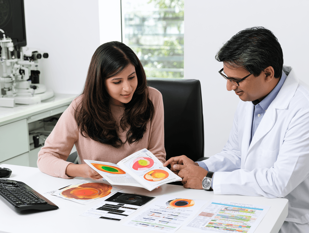
A Color-Vision Recovery Passport is a structured clinical record used to compare your condition before, during and after treatment. It prevents improvement from being judged only by statements such as “colors look brighter” or “my eyes feel better.”
The passport is not a separate diagnostic test and is not a validated medical certificate. It brings your color-vision results, ophthalmic findings, systemic reports, medicine details and daily functional changes into one record. Standardized testing remains necessary [20–25].
Your Baseline Color-Vision Record
Before treatment begins, the exact test name, test date, illumination, viewing distance and refractive correction should be recorded. Your right and left eyes should be tested separately whenever possible.
The passport should record the number of correct and incorrect responses, the specific plates missed and the pattern of color confusion. Writing only “pass” or “fail” does not provide enough information to measure a small but meaningful change.
Ishihara testing is mainly useful for red–green deficiency. HRR, Farnsworth D-15, Farnsworth–Munsell 100 Hue or anomaloscope testing may be added when more detailed classification is required [20–25].
Right-Eye and Left-Eye Comparison
Common inherited color-vision deficiency usually affects both eyes in a similar way. A significant difference between your eyes may suggest an acquired retinal or optic-nerve problem.
Your passport should therefore record color-test performance, brightness perception and contrast separately for each eye. A new or increasing difference requires ophthalmic reassessment rather than automatic continuation of the same treatment.
Ophthalmic Findings Before Treatment
Color-test results should be interpreted with your wider eye findings. The passport may include visual acuity, refraction, contrast sensitivity, fundus examination, OCT of the macula, OCT of the retinal nerve-fibre layer and visual-field findings.
ERG or VEP may be recorded when retinal cone dysfunction or optic-nerve transmission requires further evaluation. These investigations do not directly measure the same function as a color test, but they help show whether an underlying retinal or neural disorder is stable, improving or deteriorating.
Systemic Factors That May Affect Your Vision
When diabetes, nutritional deficiency or another systemic disease may be contributing to your color-vision change, the relevant reports should be included.
The passport may record HbA1c, fasting glucose, post-meal glucose, lipid profile, complete blood count, vitamin B12, folate, thyroid function, kidney function and liver function according to your clinical history.
This makes it possible to see whether visual changes are occurring alongside better metabolic or nutritional control.
Medicine and Avaleha Batch Record
Each 30-day treatment phase should be documented with the formulation name, batch number, preparation date, finished quantity, dose, Anupana and any mineral medicine included.
The record should also note whether you consumed the medicine regularly, missed doses, developed digestive difficulty or experienced any suspected adverse reaction.
This is important because a follow-up result cannot be interpreted correctly when the treatment actually taken is unclear.
Your Daily Functional Difficulties
Your ability to use color in daily life should be recorded before treatment. This may include difficulty selecting clothes, reading color-coded charts, identifying digital indicators, recognising food colors or performing a work-related task.
At follow-up, the same activities should be reviewed. Functional improvement strengthens the importance of a test result, while an improved test score without any practical change may require cautious interpretation.
Your 30-Day Review
At the end of the first 30 days, the same validated color-vision test should be repeated under similar conditions. The same eye should be tested separately, and the error pattern should be compared with the baseline.
The review should also compare medicine tolerance, visual acuity, contrast, retinal or optic-nerve findings and relevant systemic markers. HbA1c should be repeated only at an appropriate clinical interval rather than being expected to change meaningfully within a few days.
How Your Response Is Classified
| Recovery category | Meaning |
| Objective improvement | Reproducible reduction in color-test errors with meaningful functional or related visual improvement |
| Partial response | Improvement in selected tests or daily activities without complete normalization |
| Stabilization | No further deterioration in a previously progressive retinal, optic-nerve or metabolic condition |
| Subjective improvement only | Colors feel brighter or eye strain is reduced, but standardized testing remains unchanged |
| No response | No meaningful objective or functional change after an appropriate treatment period |
| Deterioration | Worsening color errors, acuity, contrast, visual fields, OCT findings or neurological symptoms |
Why One Improved Ishihara Result Is Not Enough
Color-test performance may change because of lighting, concentration, memorisation or familiarity with the plates. A single better result should therefore not be called a cure.
A credible improvement should be reproducible, preferably confirmed with another appropriate color test and supported by better daily function. In acquired disease, the identified retinal, optic-nerve, metabolic or nutritional cause should also show improvement or stabilization [20–25].
What Related Ayurvedic Studies Teach About Monitoring
Ayurvedic studies involving diabetic retinopathy, glaucomatous optic neuropathy and traumatic optic neuropathy have used objective clinical measurements such as visual acuity, retinal findings, visual fields or electrophysiological assessment [50–52]. A published cone–rod dystrophy case also demonstrates the importance of documenting the underlying retinal diagnosis rather than relying only on subjective visual changes [53].
These studies do not validate the Color-Vision Recovery Passport and do not prove correction of inherited color blindness. They support the wider principle that Ayurvedic outcomes should be recorded through measurable ophthalmic findings rather than testimonials alone.
Suggested Passport Review Schedule
| Treatment stage | Main documentation |
| Day 0 | Diagnosis, baseline color tests, separate-eye findings, ophthalmic reports, systemic reports and functional difficulties |
| Day 30 | Medicine tolerance, adherence, repeat standardized color test and early functional response |
| Day 60 | Confirmation of whether early improvement is reproducible and review of the second formulation phase |
| Day 90 | Sustained color-test change, functional outcome and relevant ophthalmic or metabolic reassessment |
| Later follow-up | Stability of the result and monitoring of the underlying retinal, optic-nerve or systemic disease |
Why the Passport Matters
The purpose of the passport is to show exactly what changed, what remained unchanged and whether continuing the same treatment is justified.
Your treatment should not be repeated indefinitely because an Ayurvedic medicine has been prepared. It should continue, change or stop according to your objective results, safety, tolerance and realistic treatment goal [20–25,50–53].
Safety of Ayurvedic Eye Treatment
Ayurvedic treatment for color vision problems should be safe, traceable and matched to your diagnosis. Oral Avaleha, mineral medicines and local eye procedures require different safety standards. A treatment should not be considered safe merely because it is described as natural.
Your retina, optic nerve, cornea, diabetes status, liver function, kidney function, current medicines and age should be reviewed before treatment begins. Sudden visual deterioration should always receive urgent ophthalmic evaluation.
Chakshushya Drishti Rasayana Avaleha Is an Oral Medicine
Chakshushya Drishti Rasayana Avaleha is prepared for oral consumption. It must not be placed inside your eyes.
You should not confuse an Avaleha with Aschyotana, Anjana, Seka, Tarpana or Putapaka. These are separate Shalakya Tantra procedures that require specific indications, suitable medicines and trained supervision.
Putting an oral Avaleha into the eye may cause severe irritation, contamination or corneal injury.
Do Not Use Homemade Eye Drops
You should not place homemade herbal water, decoction, juice, honey, oil, powder or ordinary ghee directly into your eyes.
A preparation may appear clean but can still contain bacteria, fungi, fine particles or irritating plant substances. The eye has limited protection against contaminated products, and an infection can progress quickly.
Medicines intended for direct ophthalmic use require suitable pharmaceutical preparation, appropriate sterility and safe packaging. Ordinary kitchen preparation cannot meet these requirements [47].
Why Sterility Is Essential
A contaminated eye preparation may cause redness, pain, discharge, corneal infection, blurred vision or permanent visual damage.
Any product intended to enter the eye should be manufactured and handled according to ophthalmic quality requirements. The bottle, applicator, storage conditions and period of use after opening also affect safety.
You should stop a local product and obtain an eye examination if you develop increasing pain, marked redness, swelling, discharge, light sensitivity or reduced vision.
Netra Kriyakalpa Requires Clinical Supervision
Netra Tarpana, Putapaka, Seka, Aschyotana and Anjana should be performed only after your eye condition has been properly diagnosed.
The treatment room, instruments, hands, medicines and surrounding area must be hygienically maintained. The material used should be suitable for ocular application and prepared through an appropriate pharmaceutical process.
These procedures should not be performed during active eye infection, corneal abrasion, ulceration, unexplained eye pain, sudden color loss or rapidly worsening vision.
Raw Herbs Must Be Correctly Identified
The safety of an Avaleha begins with correct botanical identification. The Ayurvedic name alone is not enough because one regional name may be used for different plants.
The botanical species, plant part, source, appearance, odour and condition of every raw material should be verified. Mouldy, insect damaged, discoloured or adulterated herbs should not be used.
Official Ayurvedic pharmacopoeias provide standards for identity, purity and quality of medicinal materials [43].
Contaminants Must Be Controlled
Herbal materials may be exposed to soil, pesticides, fungal contamination, storage moisture or environmental pollutants.
Quality assessment may therefore include microbial limits, yeast and mould count, pesticide residues, aflatoxins and relevant toxic elements. The required tests depend on the ingredients, source and intended duration of use.
World Health Organization guidance explains the importance of assessing contaminants and residues in herbal medicines rather than assuming that all natural materials are automatically pure [45].
Manufacturing Conditions Matter
A properly prepared Avaleha requires clean equipment, controlled heating, correct moisture reduction, suitable containers and protection from cross contamination.
Schedule T under the Drugs Rules provides Good Manufacturing Practice requirements for Ayurvedic, Siddha and Unani medicines. These requirements address premises, raw materials, manufacturing control, sanitation, quality control, records and storage [44].
The medicine label should identify the formulation, batch number, preparation date, net quantity, dose, storage advice and manufacturer or dispensing unit.
Fresh Avaleha Must Be Protected From Moisture and Contamination
You should always use a clean and completely dry spoon. Wet fingers or a wet spoon can introduce moisture and microorganisms into the container.
The lid should be closed immediately after each use. The medicine should be stored away from heat, direct sunlight and humid areas.
If the Avaleha develops mould, fermentation, gas, an unusual smell, discoloration or a major change in consistency, you should stop using it. It should not be mixed or reheated to hide signs of deterioration.
Mineral Medicines Require Additional Safety Checks
Lauha Bhasma, Abhraka Bhasma, Yashada Bhasma, Swarna Bhasma, Mukta Pishti and other mineral preparations should not be added merely to make the formulation appear stronger.
Their use requires a clear clinical indication, correct dose, limited duration, licensed sourcing and batch traceability. The quality of the starting material and manufacturing process can significantly affect the finished product.
Safety concerns have been reported with some Ayurvedic products containing lead, mercury, arsenic or other metals. This makes analytical documentation and professional supervision essential [46].
Do Not Use Unlabelled or Untraceable Bhasma
You should not take a mineral medicine supplied in an unlabelled packet or without clear manufacturer and batch information.
A suitable product should come from a licensed source and should have supporting quality documentation. Depending on the preparation, testing may include identity, purity, particle characteristics, elemental composition and relevant contaminant limits.
Terms such as “purified,” “detoxified,” “heavy metal free” or “laboratory tested” should not be accepted without actual batch specific evidence.
Laboratory Monitoring May Be Required
The investigations required depend on the mineral selected, your health status and the treatment duration.
Before prolonged or combined herbomineral treatment, your physician may review complete blood count, liver function, kidney function and relevant mineral studies. Iron studies may be required before Lauha Bhasma, while zinc and copper balance may be considered with longer Yashada use.
Any unexplained nausea, abdominal pain, constipation, weakness, tremor, numbness, jaundice, reduced urination or new neurological symptom requires prompt medical review.
Herbs Can Interact With Conventional Medicines
Ayurvedic herbs may influence blood glucose, blood pressure, thyroid function, sedation, fluid balance or the metabolism of other medicines.
For example, metabolic herbs used with diabetes medicines may increase the risk of low blood glucose. Yashtimadhu may be unsuitable with uncontrolled hypertension, fluid retention or low potassium. Ashwagandha may require caution with thyroid medicines, sedatives or immunosuppressive treatment.
You should provide a complete list of your medicines and supplements before starting the Avaleha. Conventional treatment should not be reduced or stopped without guidance from the doctor managing that condition [46].
Pregnancy, Childhood and Organ Disease Require Separate Assessment
A formula prepared for a healthy adult should not be copied for a child, a pregnant woman or a person with liver or kidney disease.
Children require age appropriate dosing and confirmation of the exact retinal or inherited disorder. Pregnancy and breastfeeding require additional caution because several herbs and all mineral options may be unsuitable.
Reduced liver or kidney function can change how certain ingredients are processed and may increase the risk of adverse effects.
Diabetic Patients Need a Suitable Dosage Form
A jaggery or sugar based Avaleha may be unsuitable when your diabetes is uncontrolled. Adding glucose lowering herbs does not neutralize an excessive sweet base.
Your HbA1c, daily glucose readings, body weight, current diabetes medicines and risk of hypoglycaemia should be reviewed before the dosage form is confirmed.
When a conventional Avaleha is not appropriate, a powder, tablet, capsule or another suitably prepared form may be selected.
Report Every Suspected Adverse Reaction
You should record when a suspected reaction started, which medicine and batch you were using, the dose taken and whether other medicines were being used at the same time.
The suspected medicine should be reviewed rather than automatically continued. Serious reactions require immediate medical care.
Suspected adverse reactions associated with Ayurvedic medicines can also be reported through the national Ayush Suraksha pharmacovigilance system [48].
Ayurvedic Treatment Must Not Delay Emergency Eye Care
Seek urgent ophthalmic or neurological assessment if you develop sudden color loss, rapid reduction in vision, one eye becoming darker, pain during eye movement, new flashes, many floaters, a curtain like shadow, a new blind spot or neurological symptoms.
An oral Avaleha, Tarpana or another Ayurvedic procedure should not be used to postpone emergency examination.
Ayurveda may be integrated after the urgent cause has been identified and appropriately treated.
What Safe Treatment Should Include
A safe treatment plan should document your diagnosis, complete formulation, ingredient sources, dose, duration, batch information, relevant laboratory reports and follow up schedule.
The treatment should also define when the medicine must be stopped, when testing should be repeated and which symptoms require urgent referral.
Safety is not separate from treatment. Correct identification, controlled manufacturing, suitable patient selection, objective monitoring and adverse event reporting are essential parts of responsible Ayurvedic eye care [43–48].
What Published Evidence Exists for Ayurveda in Color Vision Disorders?

Published evidence for Ayurveda in color vision disorders is still limited. The most directly relevant publication is a single case report, while the remaining studies concern related retinal or optic nerve conditions.
These studies can help explain why Ayurveda may be investigated in selected acquired or mixed color vision problems. They do not prove that Ayurveda can universally correct an inherited cone pigment abnormality.
Direct Ayurvedic Case Report on Color Blindness
A published case report described Ayurvedic management of partial color blindness in a 20 year old man. The treatment included internal medicines together with procedures such as Snehapana, Virechana, Nasya, Anjana, Vidalaka, Seka, Tarpana, Shirodhara and Matra Vasti. The authors reported improvement on repeat Ishihara testing after treatment [49].
This report is important because it directly discusses an Ayurvedic approach to a color vision problem. However, it involved only one patient and had no untreated comparison group.
The report did not clearly establish whether the condition was inherited, acquired or mixed. Detailed classification through anomaloscope testing, genetic testing or broader color vision assessment was not reported. Long term stability of the result was also not adequately established.
Familiarity with Ishihara plates, lighting conditions and test repetition may influence performance. Therefore, this case should be presented as an encouraging clinical observation rather than proof that Ayurveda cures all inherited color blindness.
Ayurvedic Clinical Study in Diabetic Retinopathy
A randomized clinical study evaluated an Ayurvedic treatment protocol as an additional treatment in patients with diabetic retinopathy [50].
The study reported improvement in selected clinical and retinal outcomes. It is relevant because diabetes can affect retinal circulation, neural function, contrast and color discrimination.
This study supports the principle that treating the metabolic and retinal disease together may help preserve or improve visual function. It also supports the Ayurvedic approach of addressing Prameha, Meda, Agni, Ama and retinal involvement rather than giving only an eye tonic.
However, the study did not specifically test color blindness and did not establish correction of an inherited cone pigment disorder. It should be used as related retinal evidence, not direct proof of color vision recovery.
Chakshushya Rasayana in Glaucomatous Optic Neuropathy
An open label randomized controlled trial evaluated Chakshushya Rasayana as an additional treatment with conventional beta blocker eye drops in progressive glaucomatous optic neuropathy [51].
The study examined measurable ophthalmic outcomes rather than relying only on subjective patient statements. This is relevant because optic nerve damage can reduce brightness, contrast and color saturation.
The findings support further investigation of Ayurvedic Rasayana treatment in stable optic nerve disorders under ophthalmic supervision.
The study was not conducted in patients with inherited color blindness. It also used Ayurveda as an additional treatment rather than as a replacement for glaucoma management. Therefore, it cannot prove that Chakshushya Rasayana restores normal color vision.
Ayurvedic Management of Traumatic Optic Neuropathy
A published case report described Ayurvedic treatment in a patient with traumatic optic neuropathy [52].
The report is useful because it included objective monitoring of optic nerve and visual pathway function, including visual electrophysiological assessment. This demonstrates how an Ayurvedic case should be documented through measurable findings rather than only through statements such as clearer or brighter vision.
Traumatic optic neuropathy is different from inherited color vision deficiency. The report involved one patient and cannot establish that the same treatment will work in other optic nerve conditions.
Its main value is to show that selected acquired neural visual disorders may be studied through structured Ayurvedic treatment and objective follow up.
Ayurvedic Case Report in Cone Rod Dystrophy
A case report discussed the Ayurvedic understanding and treatment of cone rod dystrophy [53].
Cone rod dystrophy is an inherited retinal disorder that can affect color vision, visual acuity, light tolerance and later peripheral vision. It is much more serious than common stable red green color deficiency.
The report provides a possible Ayurvedic framework using Drishti, Dosha and retinal degeneration concepts. It also shows why poor color vision with photophobia, nystagmus or reduced visual acuity requires detailed retinal assessment.
However, one case report cannot prove restoration of damaged cone cells or correction of the responsible genetic abnormality. The condition, outcome measures and duration of follow up must be examined carefully before drawing wider conclusions.
What This Evidence Means for You
The current evidence is strongest as a reason to investigate Ayurveda in selected acquired retinal, optic nerve and metabolic conditions. It is much weaker for claiming complete correction of inherited color vision deficiency.
The direct color blindness evidence consists mainly of one uncontrolled case report [49]. Studies in diabetic retinopathy, glaucoma, traumatic optic neuropathy and cone rod dystrophy provide related evidence, but they involve different diseases and cannot be treated as direct proof [50–53].
Your treatment possibility should therefore be decided from your exact diagnosis. If your color vision changed because of diabetes, retinal disease, optic nerve dysfunction, nutritional depletion or another acquired cause, there may be a more reasonable therapeutic target.
If your deficiency has been stable since childhood and is caused by an inherited cone pigment variation.
How Future Ayurvedic Evidence Should Be Improved
Future studies should first classify patients as inherited, acquired or mixed. They should test each eye separately and use more than one validated color vision test.
Research should record the exact Ayurvedic diagnosis, complete formulation, dose, treatment duration and any Netra Kriyakalpa procedures used. Follow up should include standardized Ishihara testing, HRR or Farnsworth testing, visual acuity, contrast sensitivity, OCT, visual fields and relevant metabolic findings.
A sustained result should be confirmed after treatment has ended. This would help separate genuine functional improvement from temporary variation, memorisation or familiarity with the test.
Until stronger studies are available, the responsible conclusion is that Ayurveda has preliminary and indirect evidence in selected visual disorders, while universal reversal of inherited color blindness remains unproven [49–53].
Genetics, Family History and Future Children

Inherited color vision deficiency may pass through families, but the pattern depends on the affected cone system and gene. Your family history can help explain the diagnosis, although the condition may still appear when no relative is known to have it.
Why Red Green Color Deficiency Is More Common in Males
Most inherited red green color vision deficiencies involve changes in the OPN1LW or OPN1MW cone opsin genes located on the X chromosome [2–4].
Males have one X chromosome. Therefore, one altered gene may be enough for the deficiency to appear. Females have two X chromosomes and may carry the altered gene without having a significant color vision problem.
In the usual X linked inheritance pattern, a carrier mother may pass the altered gene to her sons or daughters. An affected father does not pass his X chromosome to his sons, but he passes it to all his daughters [4].
Other Inherited Color Vision Disorders Follow Different Patterns
Rare blue yellow color vision deficiency may involve the OPN1SW gene and may follow an autosomal dominant pattern. Severe disorders such as achromatopsia may follow an autosomal recessive pattern and can involve genes such as CNGA3 and CNGB3 [4,5].
Achromatopsia is different from common red green deficiency. It may cause poor visual acuity, marked light sensitivity, nystagmus and greatly reduced color perception from early childhood [5].
Why Family History May Not Always Be Obvious
You may have inherited color vision deficiency even when no close relative reports the condition. Female carriers may have few or no noticeable symptoms, and older family members may never have undergone formal color vision testing.
A detailed history should include color vision problems in your parents, siblings, maternal uncles, grandparents and children. Formal testing is more reliable than relying only on what family members remember.
Ayurvedic Understanding of Hereditary Conditions
Ayurveda describes diseases arising through parental reproductive factors under Adibala Pravritta Vyadhi in Sushruta Samhita, Sutrasthana, Chapter 24, Vyadhi Samuddeshiya Adhyaya [13].
Charaka further explains that disturbance affecting the Beeja or the specific part responsible for a body structure may influence the development of that structure.
Classical source: Charaka Samhita, Sharirasthana, Chapter 4, Mahati Garbhavakranti Sharira, verse 30 [14].
यस्य यस्य ह्यवयवस्य बीजे बीजभागे वा दोषाः प्रकोपमापद्यन्ते, तं तमवयवं विकृतिराविशति ॥३०॥
Yasya yasya hy avayavasya bīje bījabhāge vā doṣāḥ prakopam āpadyante, taṃ tam avayavaṃ vikṛtir āviśati.
“When disturbance affects the Beeja or Beejabhaga responsible for a particular body part, abnormality develops in that corresponding part.”
Beeja, Beejabhaga and Beejabhagavayava provide a classical hereditary framework, but they should not be described as exact equivalents of genes, chromosomes or DNA [13–15].
Should Your Children Be Tested?
Early testing may be useful when color vision deficiency is present in your family, especially before schoolwork becomes heavily dependent on color coded maps, charts and diagrams.
A child should receive a detailed retinal assessment when poor color recognition occurs with reduced visual acuity, severe photophobia or nystagmus. These features may indicate a more serious inherited cone disorder rather than common red green deficiency [3–5].
When Genetic Counselling May Help
Genetic counselling may be useful when you have a severe cone disorder, several affected family members, uncertainty about the inheritance pattern or concerns about future children.
Counselling can explain the likely transmission pattern, whether genetic testing is appropriate and which relatives may benefit from screening. It can estimate risk, but it cannot guarantee whether every future child will or will not inherit the condition.

Failing an Ishihara test does not mean that you see no colors. It means that you made errors on a screening test designed mainly to detect red green color vision deficiency.
Your next step should be proper classification rather than immediately starting medicine or repeatedly practising the same plates. The type, severity, stability and cause of your color vision problem must first be confirmed [20–25].
Confirm That the Test Was Performed Correctly
Ishihara plates should be examined under suitable illumination, at the correct viewing distance and within the recommended viewing time. Your refractive correction should be used when required.
A result obtained through a mobile phone, computer screen, photocopy or poorly lit room should not be treated as a final occupational diagnosis. Screen brightness, display settings and image quality can change the appearance of the plates.
Your report should record the test edition, number of plates used, specific errors and whether each eye was assessed separately.
Determine the Type and Severity of the Deficiency
Ishihara mainly identifies red green deficiencies. It does not fully classify every color vision disorder or measure all occupationally relevant abilities.
Depending on the authority and purpose, further assessment may include HRR testing, Farnsworth D 15, Farnsworth Munsell 100 Hue, anomaloscope testing, lantern testing or another approved occupational assessment [20–25].
These tests may help determine whether your deficiency is mild, moderate or severe and whether it follows a red green, blue yellow or generalized pattern.
Check Whether the Problem Is Inherited or Acquired
When your color difficulty has been present since childhood and remains equal in both eyes, an inherited deficiency is more likely.
When you previously recognised colors normally, one eye performs worse or the condition is progressing, retinal disease, optic nerve dysfunction, cataract, diabetes, medicine toxicity or another acquired cause should be investigated.
Treating an acquired and reversible component may improve your functional color discrimination. A stable inherited cone pigment difference requires more cautious expectations.
Can Ayurvedic Treatment Help You Pass the Test?
Ayurvedic treatment may be considered when a treatable retinal, optic nerve, metabolic, nutritional or mixed component is identified. The treatment should address the diagnosed cause and should be followed by standardized color vision testing.
However, no Ayurvedic medicine, Avaleha, Tarpana or other procedure should guarantee that you will pass an aviation, military, railway, maritime or employment examination.
Even when your test performance improves, the final fitness decision remains with the relevant occupational authority. Each country, licence category, service and employer may apply different standards and approved testing pathways [55–60].
Do Not Memorise Ishihara Plates
Memorising plate numbers does not improve your cone function or real life color discrimination. It may produce an artificial result without correcting your ability to identify signals, warning lights, wiring or safety related colors.
This can create serious risks in professions where accurate color recognition is essential. Genuine improvement should be confirmed through alternate plates, additional validated tests and practical color dependent tasks.
Understand What Treatment Can Realistically Achieve
If an acquired disease is worsening your result, treatment may improve the reversible component. If your condition is inherited and stable, the realistic goals may include protecting ocular health, addressing associated visual problems and helping you understand suitable career options.
Improvement should be reproducible under standardized conditions and should correspond with better functional color recognition.
The Final Decision Belongs to the Relevant Authority
Aviation authorities in the United States, United Kingdom, Australia, Canada and Singapore use their own medical standards and approved assessment pathways [55–60]. Navy, military, railway and other employers may use separate recruitment rules.
You should therefore submit your results to the exact authority responsible for your application. A certificate from a treating physician cannot replace the authority’s official medical decision.
The correct approach is to classify your deficiency accurately, investigate any reversible cause, treat what can genuinely be treated and then undergo assessment through the approved occupational pathway.
Three Patient Scenarios: Why the Treatment Must Be Different
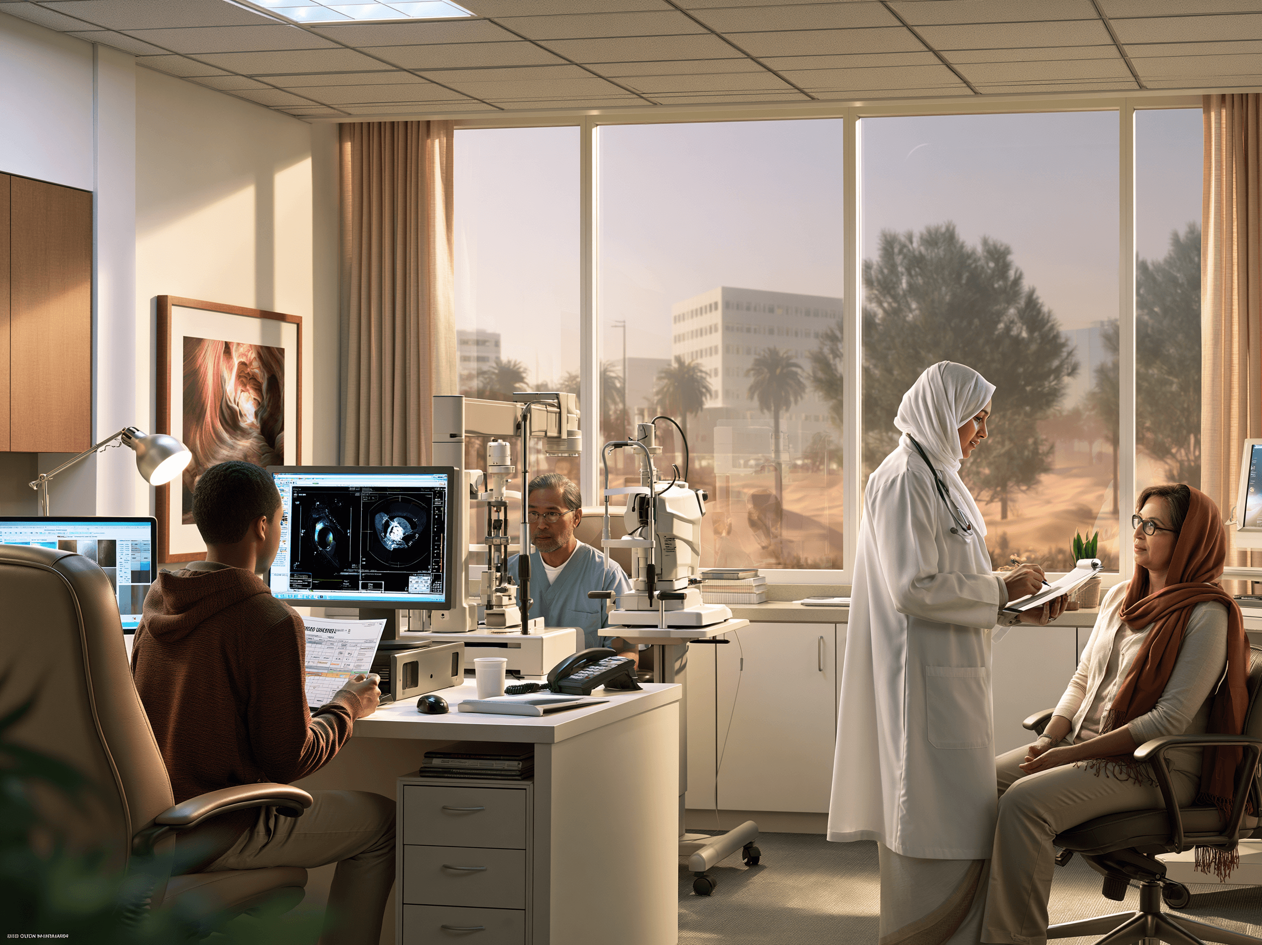
The same abnormal color vision test can arise from completely different causes. Your treatment should therefore be based on when the problem began, whether it is progressing, which visual structure is affected and whether an underlying disease can be treated.
You Discovered Lifelong Color Difficulty During a Navy or Employment Test
You may be a young adult who has always confused red and green but never considered it a medical problem. Your vision may otherwise be clear, both eyes may perform similarly and the difficulty may have remained stable since childhood.
This pattern usually suggests inherited color vision deficiency. Your evaluation should confirm the type and severity, examine the retina and optic nerve and identify whether any additional acquired problem is present [1–6,20–25].
From an Ayurvedic perspective, the hereditary background may be understood through Adibala Pravritta Vyadhi and Beeja related concepts. Alochaka Pitta, Drishti, Agni, nutrition and general ocular health may also be assessed [10,13–16].
The treatment goal should be ocular protection, correction of associated deficiencies and support for visual function. A normal occupational test result or genetic reversal should not be guaranteed.
Your Colors Became Faded After Diabetes Developed
You may have recognised colors normally for most of your life but later notice that shades are becoming dull, contrast is weaker or blue and yellow are more difficult to separate.
In this case, your retina, optic nerve and metabolic condition require evaluation. Fundus examination, OCT, color vision testing, visual fields, HbA1c, glucose, blood pressure and lipid levels may be relevant.
The Ayurvedic assessment may identify Prameha, Meda, Kleda, Agni, Ama, Rakta and retinal involvement. Your treatment should address the metabolic disease together with the visual dysfunction rather than giving only a general eye tonic [17,29–31,50].
A personalized Avaleha may give greater importance to Guduchi, Triphala, Amalaki and suitable Prameha supporting ingredients. The sweetening medium must be modified or another dosage form selected when your glucose control is poor.
The realistic goal may be better metabolic control, stabilization of retinal function and measurable improvement in color discrimination where the damage remains reversible.
Your Color Vision Changed After Starting a Medicine
You may have previously recognised colors normally and notice fading, reduced contrast or a difference between your two eyes after beginning a new medicine.
The first step is to review the medicine with the prescribing doctor and arrange an ophthalmic examination. You should not stop an essential treatment independently [7,8].
The retina, optic nerve, visual fields and organ function may require assessment according to the suspected medicine. The possibility of continuing toxic exposure must be addressed before Rasayana treatment is started.
After the cause has been controlled, the Ayurvedic plan may focus on Pitta Rakta regulation, Agni and Ama correction, Vata Majja support or tissue recovery according to the affected structure.
The objective is to support recovery from a potentially reversible acquired injury. An Avaleha should not be used to hide continuing medicine related damage.
Why These Three Patients Cannot Receive the Same Avaleha
The first patient mainly has a stable inherited cone pigment difference. The second has a metabolic disease affecting retinal or neural function. The third may have medicine related retinal or optic nerve injury.
Their color test errors may appear similar, but their Samprapti, treatment priorities and expected outcomes are different.
Your formulation should therefore be selected from your diagnosis, reports, Dosha, Dushya, Agni, Ama, metabolic condition and tissue strength. The same 900 g Avaleha recipe should not be repeated for every patient merely because the symptom is called color blindness.
Frequently Asked Questions
Can color blindness be cured?
Inherited color blindness currently has no established cure. Acquired color vision problems may improve when the underlying retinal, optic nerve, cataract, medicine related or systemic cause is treated.
Can Ayurveda cure color blindness?
Yes, selected acquired or mixed color-vision problems may improve or resolve when their reversible retinal, optic-nerve, metabolic, nutritional, medicine-related or lens cause is identified and treated. Ayurveda may support this root-cause treatment through an individualized approach.
Which type of color blindness may improve?
Color vision that deteriorated later because of retinal disease, optic nerve dysfunction, cataract, diabetes, medicine exposure or another treatable condition may have greater scope for improvement.
What is the best Ayurvedic medicine for color blindness?
There is no single best medicine for every patient. The formulation should depend on whether your problem is inherited, retinal, optic nerve related, metabolic, nutritional or mixed.
What is Chakshushya Drishti Rasayana Avaleha?
It is a physician designed oral Ayurvedic formulation inspired by classical Chakshushya, Rasayana and Drishtigata Roga principles. It is not an eye drop.
Can color blindness develop later in life?
Yes. Color vision may change later because of eye disease, optic nerve damage, brain injury, cataract, certain medicines or other health conditions.
Is color blindness hereditary?
Most red green color vision deficiency is inherited and commonly involves cone opsin genes on the X chromosome. This is why it is more common in males.
Can diabetes affect color vision?
Yes. Diabetes can damage the retina and may contribute to reduced contrast and altered color discrimination, especially when retinal disease develops.
Why do colors look faded in one eye?
A new difference between your eyes may indicate retinal or optic nerve disease. Faded colors with eye pain or dim vision require prompt ophthalmic assessment.
Which test detects color blindness?
The Ishihara test mainly screens for red green deficiency. Additional color arrangement or matching tests may be needed to identify the exact type and severity.
Can color blind glasses cure color blindness?
No. Special filters may increase contrast between some colors, but they do not replace an absent or altered cone pigment.
Does inherited color blindness worsen with age?
Inherited color vision deficiency is usually stable. New deterioration may indicate cataract, retinal disease, optic nerve dysfunction or another acquired problem.
Can children inherit color blindness?
Yes. Children with a family history or difficulty learning colors should receive proper color vision testing, especially before schoolwork becomes heavily color coded.
Can Netra Tarpana improve color vision?
Netra Tarpana may be considered in selected diagnosed eye conditions, but it is not proven to correct inherited color blindness and should not be performed without supervision.
Can treatment help you pass an Ishihara job test?
Treatment may help only when a reversible acquired problem is affecting your result.
Reference
Modern Color-Vision Foundations
[1] National Eye Institute. (2025, November 5). Color blindness. National Institutes of Health.
https://www.nei.nih.gov/eye-health-information/eye-conditions-and-diseases/color-blindness
Used for: Principal patient-level reference for inherited and acquired color-vision deficiency, symptoms, causes, lack of an established cure for inherited deficiency, treatment of secondary causes, classroom considerations, visual aids, and color-identification applications.
[2] National Eye Institute. (2025, January 30). Causes of color vision deficiency. National Institutes of Health.
https://www.nei.nih.gov/eye-health-information/eye-conditions-and-diseases/color-blindness/causes-color-vision-deficiency
Used for: Genetic inheritance, acquired color-vision changes, glaucoma, macular disease, neurological disease, medicines, retinal injury, brain injury, ageing, and cataract-related color alteration.
[3] National Eye Institute. (2023, August 7). Types of color vision deficiency. National Institutes of Health.
https://www.nei.nih.gov/eye-health-information/eye-conditions-and-diseases/color-blindness/types-color-vision-deficiency
Used for: Red–green deficiency, blue–yellow deficiency, complete color-vision deficiency, and patient-friendly classification of the major phenotypes.
[4] National Library of Medicine. (2015, January 1). Color vision deficiency. MedlinePlus Genetics.
https://medlineplus.gov/genetics/condition/color-vision-deficiency/
Used for: OPN1LW, OPN1MW, and OPN1SW cone-opsin genes, X-linked inheritance, cone function, red–green deficiency, blue–yellow deficiency, blue-cone monochromacy, and acquired causes.
[5] Kohl, S., Jägle, H., Wissinger, B., & Zobor, D. (2018). Achromatopsia. In GeneReviews®. University of Washington, Seattle.
https://www.ncbi.nlm.nih.gov/books/NBK1418/
Used for: Severe inherited cone disorders, poor visual acuity, nystagmus, marked photophobia, absent or reduced color discrimination, electroretinography, genetic testing, and family counselling.
[6] Simunovic, M. P. (2010). Colour vision deficiency. Eye, 24(5), 747–755.
https://pubmed.ncbi.nlm.nih.gov/19927164/
Used for: Clinical overview of congenital and acquired deficiency, diagnosis, daily adaptations, counselling, and limitations of filters or optical aids.
[7] Simunovic, M. P. (2016). Acquired color vision deficiency. Survey of Ophthalmology, 61(2), 132–155.
https://pubmed.ncbi.nlm.nih.gov/26656928/
Used for: Principal clinical review for acquired dyschromatopsia involving retinal disease, optic-nerve disease, systemic disorders, neurological disease, drugs, and toxic exposure.
[8] Ageed, A., Aslam, M. D., & El Haouari, S. (2024). Acquired dyschromatopsia and its link to drug toxicity. Cureus, 16(12), e76190.
https://pmc.ncbi.nlm.nih.gov/articles/PMC11749246/
Used for: Medicine-induced alterations in color vision, mechanisms of drug toxicity, and the need to review new symptoms in relation to medication exposure.
[9] American Academy of Ophthalmology. (2026, February 2). What is optic neuritis?
https://www.aao.org/eye-health/diseases/what-is-optic-neuritis
Used for: Eye pain, pain with movement, dim or faded colors, visual loss, and emergency-referral discussion for possible optic neuritis.
Classical Ayurvedic Foundations
[10] Vāgbhaṭa. (n.d.). Aṣṭāṅga Hṛdaya, Sūtrasthāna, Chapter 12: Doṣabhedīya Adhyāya.
https://www.easyayurveda.com/dosha-types-increase-and-decrease/
Used for: Alochaka Pitta and its association with the eyes and visual perception. Used as a functional Ayurvedic basis, not as proof that every color-vision disorder is caused by Pitta.
[11] Suśruta. (n.d.). Suśruta Saṃhitā, Uttaratantra, Chapter 7: Dṛṣṭigata Roga Vijñānīya Adhyāya.
https://www.siva.sh/sushruta-samhita/uttara-tantra/7
Used for: Drishti, Drishtigata disorders, Timira, and classical descriptions of disturbances affecting visual perception. The chapter does not establish a direct equivalence with modern inherited color blindness.
[12] Suśruta. (n.d.). Suśruta Saṃhitā, Uttaratantra, Chapter 17: Dṛṣṭigata Roga Pratiṣedha Adhyāya.
https://www.siva.sh/sushruta-samhita/uttara-tantra/17
Used for: Classical treatment principles for Drishtigata disorders and the broader Ayurvedic therapeutic framework for selected acquired visual disturbances.
[13] Suśruta. (n.d.). Suśruta Saṃhitā, Sūtrasthāna, Chapter 24: Vyādhi Samuddeśīya Adhyāya (verses 4–5).
https://www.siva.sh/sushruta-samhita/sutra-sthana/24/4
Used for: Adibala-Pravritta Vyadhi, inherited or parental-origin disease categories, and distinctions between hereditary, congenital, Dosha-derived, and other disease origins.
[14] Agniveśa, Caraka, & Dṛḍhabala. (n.d.). Caraka Saṃhitā, Śārīrasthāna, Chapter 4: Mahatīgarbhāvakrānti Śārīra (verses 30–31).
https://www.carakasamhitaonline.com/index.php/Mahatigarbhavakranti_Sharira
Used for: Beeja, Beejabhaga, and Beejabhagavayava concepts used to discuss hereditary susceptibility and developmental abnormalities within the Ayurvedic framework.
[15] Sharma, K., Chaudhary, N., & Sharma, R. (2025). A review on genetics from an Ayurvedic perspective with special focus on Beeja, Beejabhaga and Beejabhagavayava. Journal of Ayurveda and Integrated Medical Sciences, 10(10), 121–125.
https://jaims.in/jaims/article/view/4767
Used for: Secondary scholarly interpretation of classical Beeja concepts in relation to modern hereditary thinking. Classical terminology should not be presented as biologically identical to genes, chromosomes, or DNA.
[16] Agniveśa, Caraka, & Dṛḍhabala. (n.d.). Caraka Saṃhitā, Cikitsāsthāna, Chapter 1: Rasāyana Adhyāya.
https://www.carakasamhitaonline.com/index.php/Rasayana_Adhyaya
Used for: Rasayana, maintenance of tissue function, Bala, longevity, and the general classical rationale behind a Drishti Rasayana formulation.
[17] Agniveśa, Caraka, & Dṛḍhabala. (n.d.). Caraka Saṃhitā, Cikitsāsthāna, Chapter 6: Prameha Cikitsā.
https://www.carakasamhitaonline.com/index.php/Prameha_Chikitsa
Used for: Prameha, Meda, Kleda, Agni, and systemic metabolic treatment principles relevant to diabetes-associated retinal or color-vision dysfunction.
[18] Suśruta. (n.d.). Suśruta Saṃhitā, Uttaratantra, Chapter 18: Kriyākalpa Adhyāya.
https://www.siva.sh/sushruta-samhita/uttara-tantra/18
Used for: Classical ocular procedures including Seka, Aschyotana, Anjana, Tarpana, and Putapaka and the principle of selecting procedures according to the clinical condition.
[19] Vāgbhaṭa. (n.d.). Aṣṭāṅga Hṛdaya, Sūtrasthāna, Chapter 24: Tarpana–Puṭapāka Vidhi.
https://www.easyayurveda.com/tarpana-putapaka-ayurveda-eye-care-ashtanga-hrudaya-sutrasthana-24/
Used for: Classical indications, procedural principles, and patient selection for Tarpana and Putapaka. It does not provide clinical proof of color-blindness correction.
Color-Vision Testing and Objective Monitoring
[20] National Eye Institute. (2024, December 6). Testing for color vision deficiency. National Institutes of Health.
https://www.nei.nih.gov/eye-health-information/eye-conditions-and-diseases/color-blindness/testing-color-vision-deficiency
Used for: Pseudoisochromatic plate testing and hue-arrangement testing, including the importance of formal testing when accurate color discrimination is required.
[21] Fanlo Zarazaga, A., Gutiérrez Vásquez, J., & Pueyo Royo, V. (2019). Review of the main colour vision clinical assessment tests. Archivos de la Sociedad Española de Oftalmología (English Edition), 94(1), 25–32.
https://pubmed.ncbi.nlm.nih.gov/30361001/
Used for: Comparative overview of color-vision tests, their purposes, strengths, and limitations.
[22] Dain, S. J., Atchison, D. A., & Hovis, J. K. (2019). Limitations and precautions in the use of the Farnsworth-Munsell Dichotomous D-15 Test. Optometry and Vision Science, 96(9), 695–705.
https://pubmed.ncbi.nlm.nih.gov/31479025/
Used for: Limitations of D-15 testing, interpretation precautions, and the need to avoid overstating a single test result.
[23] Garip Kuebler, A., Halfter, K., Reznicek, L., Klingenstein, A., Priglinger, S., Rudolph, G., & Hintschich, C. (2021). A pathological indicator for dysthyroid optic neuropathy: Tritan color vision deficiency. Graefe’s Archive for Clinical and Experimental Ophthalmology, 259, 3421–3426.
https://pubmed.ncbi.nlm.nih.gov/34159407/
Used for: Importance of blue–yellow or tritan-sensitive testing in selected acquired optic-nerve disorders and why Ishihara alone may miss relevant acquired changes.
[24] Mikolajczyk, B., Ritter, A., Larson, C., Connett, J., Olson, J., McClelland, C., & Lee, M. S. (2022). Red desaturation prevalence and severity in healthy patients. Neurology: Clinical Practice, 12(1), 1–5.
https://pmc.ncbi.nlm.nih.gov/articles/PMC9491505/
Used for: Limitations of red-desaturation testing. Healthy individuals may report inter-eye differences, so this test should not be used alone to diagnose optic neuropathy or demonstrate treatment success.
[25] Cole, B. L. (2007). Assessment of inherited colour vision defects in clinical practice. Clinical and Experimental Optometry, 90(3), 157–175.
https://pubmed.ncbi.nlm.nih.gov/17425762/
Used for: Clinical classification of inherited defects, test selection, severity assessment, and occupational implications.
[26] Ramachandran, N., Wilson, G. A., & Wilson, N. (2014). Is screening for congenital colour vision deficiency in school students worthwhile? A review. Clinical and Experimental Optometry, 97(6), 499–506.
https://pubmed.ncbi.nlm.nih.gov/25039829/
Used for: Childhood screening, educational consequences, school accommodations, and the value and limitations of early identification.
Avaleha Inspiration and Preparation
[27] Śārṅgadhara. (2013). Śārṅgadhara Saṃhitā, Madhyama Khaṇḍa, Chapter 8: Avaleha Kalpanā (B. Tripathi, Ed.). Chaukhambha Surbharati Prakashan.
https://archive.org/details/dLhu_sharangadhara-samhita-of-sharangadhara-acharya-containing-anjananidana-of-agnive
Used for: Classical Avaleha preparation, use of liquid extracts and sweetening media, concentration, Prakshepa Dravya, Paka Lakshana, storage, and administration principles.
[28] Phunde, R. D., Chikurte, S. N., Patil, P. A., & Chokhar, S. (2019). Avaleha Kalpana—A review. Journal of Ayurveda and Integrated Medical Sciences, 4(3), 89–91.
https://jaims.in/jaims/article/view/627
Used for: Patient-friendly explanation of Avaleha as a semisolid dosage form, pharmaceutical stages, classical quality indicators, and formulation advantages.
Research Supporting the Herbal Modules
[29] Agrawal, S. S., Naqvi, S., Gupta, S. K., & Srivastava, S. (2012). Prevention and management of diabetic retinopathy in STZ diabetic rats by Tinospora cordifolia and its molecular mechanisms. Food and Chemical Toxicology, 50(9), 3126–3132.
https://pubmed.ncbi.nlm.nih.gov/22687550/
Used for: Guduchi. The animal study reported effects on retinal oxidative stress and antioxidant activity in experimental diabetic retinopathy. This is preclinical evidence and does not prove human color-vision recovery.
[30] Nashine, S., Kanodia, R., Nesburn, A. B., Soman, G., Kuppermann, B. D., & Kenney, M. C. (2019). Nutraceutical effects of Emblica officinalis in age-related macular degeneration. Aging, 11(4), 1177–1188.
https://pubmed.ncbi.nlm.nih.gov/30792375/
Used for: Amalaki. Supports cellular and mitochondrial research relevant to retinal ageing and oxidative stress. It is not a human color-blindness treatment trial.
[31] Suryavanshi, S. V., Barve, K., Utpat, S. V., & Kulkarni, Y. A. (2022). Triphala churna ameliorates retinopathy in diabetic rats. Biomedicine & Pharmacotherapy, 148, 112711.
https://pubmed.ncbi.nlm.nih.gov/35168075/
Used for: Triphala. The experimental study assessed retinal structure, biochemical markers, and electrophysiological outcomes in diabetic animals. Evidence is indirect and preclinical.
[32] Peterson, C. T., Denniston, K., & Chopra, D. (2017). Therapeutic uses of Triphala in Ayurvedic medicine. Journal of Alternative and Complementary Medicine, 23(8), 607–614.
https://pmc.ncbi.nlm.nih.gov/articles/PMC5567597/
Used for: General Triphala pharmacology, antioxidant activity, digestive, and metabolic rationale. It does not establish direct efficacy in color-vision deficiency.
[33] Liu, L., Jiang, Y., & Steinle, J. J. (2019). Glycyrrhizin protects the diabetic retina against permeability, neuronal, and vascular damage through anti-inflammatory mechanisms. Journal of Clinical Medicine, 8(7), 957.
https://pmc.ncbi.nlm.nih.gov/articles/PMC6678129/
Used for: Yashtimadhu-related rationale. The study concerns glycyrrhizin in an experimental diabetic-retinal model; it does not establish that whole Yashtimadhu or an Avaleha improves color vision in patients.
[34] Mikulska, P., Malinowska, M., Ignacyk, M., Szustowski, P., Nowak, J., Pesta, K., Szeląg, M., Szklanny, D., Judasz, E., Kaczmarek, G., Ejiohuo, O. P., Paczkowska-Walendowska, M., Gościniak, A., & Cielecka-Piontek, J. (2023). Ashwagandha (Withania somnifera)—Current research on the health-promoting activities: A narrative review. Pharmaceutics, 15(4), 1057.
https://pmc.ncbi.nlm.nih.gov/articles/PMC10147008/
Used for: Ashwagandha. Supports broader neuroprotective, anti-inflammatory, antioxidant, and stress-related rationale. Direct evidence for optic-nerve or color-vision recovery remains insufficient.
[35] Valotto Neto, L. J., de Araujo, M. R., Moretti Junior, R. C., Mendes Machado, N., Joshi, R. K., dos Santos Buglio, D., Barbalho Lamas, C., Direito, R., Fornari Laurindo, L., Tanaka, M., & Barbalho, S. M. (2024). Investigating the neuroprotective and cognitive-enhancing effects of Bacopa monnieri: A systematic review focused on inflammation, oxidative stress, mitochondrial dysfunction, and apoptosis. Antioxidants, 13(4), 393.
https://pubmed.ncbi.nlm.nih.gov/38671841/
Used for: Brahmi. Supports general neuroprotective mechanisms, but not direct retinal, optic-nerve, or color-vision efficacy.
[36] Zuñiga, L. Y., González-Ortiz, M., & Martínez-Abundis, E. (2017). Effect of Gymnema sylvestre administration on metabolic syndrome, insulin sensitivity, and insulin secretion. Journal of Medicinal Food, 20(8), 750–754.
https://pubmed.ncbi.nlm.nih.gov/28459647/
Used for: Meshashringi. Provides limited human metabolic evidence. The study did not establish an ocular or color-vision outcome.
[37] Indian Council of Medical Research Collaborating Centres. (1998). Flexible dose open trial of Vijayasar in cases of newly diagnosed non-insulin-dependent diabetes mellitus. Indian Journal of Medical Research, 108, 24–29.
https://pubmed.ncbi.nlm.nih.gov/9745215/
Used for: Vijayasara. Supports historical clinical investigation in newly diagnosed type 2 diabetes. It was an open trial and did not evaluate retinal or color-vision outcomes.
[38] Teixeira, C. C., Weinert, L. S., Barbosa, D. C., Ricken, C., Esteves, J. F., & Fuchs, F. D. (2004). Syzygium cumini (L.) Skeels in the treatment of type 2 diabetes: Results of a randomized, double-blind, double-dummy, controlled trial. Diabetes Care, 27(12), 3019–3020.
https://pubmed.ncbi.nlm.nih.gov/15562231/
Used for: Jambu. Provides controlled human metabolic evidence and its limitations. It does not establish improvement in diabetic retinopathy or color vision.
[39] Yin, Z., Tan, R., Yuan, T., Chen, S., Quan, Y., Hao, Q., Zeng, J., Zhao, J., & Li, L. (2021). Berberine prevents diabetic retinopathy through inhibiting the HIF-1α/VEGF/NF-κB pathway in db/db mice. Die Pharmazie, 76(4), 165–171.
https://pubmed.ncbi.nlm.nih.gov/33849702/
Used for: Daruharidra-related berberine rationale. The mouse study reported metabolic and retinal-pathway effects but does not prove that Daruharidra, berberine, or an Avaleha restores color vision in humans.
[40] Bana, S., Kumar, N., Sartaj, A., Alhalmi, A., Qurtam, A. A., Nasr, F. A., Al-Zharani, M., Singh, N., Gaur, P., Mishra, R., Bhardwaj, S., Ali, H., & Goel, R. (2023). Rubia cordifolia L. attenuates diabetic neuropathy by inhibiting apoptosis and oxidative stress in rats. Pharmaceuticals, 16(11), 1586.
https://pmc.ncbi.nlm.nih.gov/articles/PMC10674165/
Used for: Manjistha. Provides indirect experimental evidence related to diabetic neural injury, apoptosis, and oxidative stress. It is not a retinal or color-vision trial.
[41] Mary, N. K., Achuthan, C. R., Babu, B. H., & Padikkala, J. (2003). In vitro antioxidant and antithrombotic activity of Hemidesmus indicus (L.) R. Br. Journal of Ethnopharmacology, 87(2–3), 187–191.
https://pubmed.ncbi.nlm.nih.gov/12860306/
Used for: Sariva. Provides laboratory antioxidant and antithrombotic rationale only. It should not be described as having proven retinal or color-vision efficacy.
[42] Subhadradevi, V., Asokkumar, K., Umamaheswari, M., Sivashanmugam, A. T., & Sankaranand, R. (2010). In vitro antioxidant activity of Vetiveria zizanioides root extract. Tanzania Journal of Health Research, 12(4), 274–279.
https://pubmed.ncbi.nlm.nih.gov/24409635/
Used for: Usheera. Provides in vitro antioxidant evidence only; there is no direct clinical evidence for color-vision improvement.
Medicine Quality, Mineral Safety and Ocular Safety
[43] Pharmacopoeia Commission for Indian Medicine & Homoeopathy. (n.d.). Pharmacopoeias and formularies. Ministry of Ayush, Government of India.
https://pcimh.gov.in/
Used for: Official Ayurvedic Pharmacopoeia and Formulary framework, botanical identity, ingredients, standards, monographs, and pharmaceutical quality specifications.
[44] Central Drugs Standard Control Organization. (2022). Drugs Rules, 1945. Ministry of Health and Family Welfare, Government of India.
https://cdsco.gov.in/opencms/resources/UploadCDSCOWeb/2022/drug_rules/Drugs%20Rules%2C%201945.pdf
Used for: Manufacturing licences, Schedule T, Good Manufacturing Practices, authenticated raw materials, contamination prevention, manufacturing-process control, quality control, and GMP certification.
[45] World Health Organization. (2007). WHO guidelines for assessing quality of herbal medicines with reference to contaminants and residues.
https://www.who.int/publications/i/item/9789241594448
Used for: Microbial contamination, pesticides, aflatoxins, toxic elements, contaminants, residues, sampling, and quality evaluation of herbal materials.
[46] National Center for Complementary and Integrative Health. (n.d.). Ayurvedic medicine: In depth.
https://www.nccih.nih.gov/health/ayurvedic-medicine-in-depth
Used for: Safety concerns, possible metal exposure, herb–drug interactions, and the importance of professional supervision and quality-controlled products.
[47] U.S. Food and Drug Administration. (n.d.). What you should know about eye drops.
https://www.fda.gov/drugs/buying-using-medicine-safely/what-you-should-know-about-eye-drops
Used for: Ocular sterility and contamination risks. Supports warnings against homemade eye drops and non-sterile ghee, honey, herbal water, or powders being applied directly to the eyes.
[48] Ministry of Ayush. (n.d.). Ayush Suraksha: Pharmacovigilance programme for Ayurveda, Siddha, Unani and Homoeopathy.
https://suraksha.ayush.gov.in/
Used for: Monitoring and reporting suspected adverse reactions associated with Ayurvedic, Siddha, Unani, and Homoeopathic medicines.
Direct and Related Ayurvedic Clinical Evidence
[49] Singh, D. S., Sahu, S. K., & Sharma, A. B. (2016). Ayurvedic management of colour blindness: A case study. World Journal of Pharmaceutical Research, 5(10), 1084–1088.
https://wjpr.net/abstract_show/5967
Used for: The principal directly relevant Ayurvedic color-blindness case report. The reported intervention included multiple procedures and internal medicines, with improvement reported on repeat Ishihara testing. Evidence limitation: Single patient, no control group, incomplete classification of congenital versus acquired deficiency, no anomaloscope or genetic confirmation, possible test-learning effect, and inadequate long-term verification.
[50] Kumar, V. K., Singh, B. V. D., & Manjusha, R. (2021). Add-on effect of Ayurvedic treatment protocol for diabetic retinopathy: A randomized controlled clinical study. AYU, 42(3), 118–129.
https://pmc.ncbi.nlm.nih.gov/articles/PMC10251286/
Used for: Related clinical evidence involving Ayurvedic treatment as an adjunct in diabetic retinopathy. Evidence limitation: Supports investigation of retinal and metabolic disease but does not prove treatment of inherited cone-photopigment deficiency or direct correction of color vision.
[51] Dhiman, K. S., Adhoor, V. S., Agarwal, R., & Mehta, A. J. (2016). Adjuvant effect of Chakshushya Rasayana with beta-blocker eye drops in the management of progressive glaucomatous optic neuropathy: An open-label randomized controlled trial. AYU, 37(2), 125–134.
https://pmc.ncbi.nlm.nih.gov/articles/PMC5688835/
Used for: Related evidence concerning Chakshushya Rasayana and optic-nerve disease outcomes. Evidence limitation: Open-label adjunctive research in glaucoma; it is not a trial of color blindness and does not establish inherited color-vision reversal.
[52] Bhat, P. M. (2022). Traumatic optic neuropathy and Ayurveda: A case study. Journal of Ayurveda and Integrative Medicine, 13(1), 100494.
https://pmc.ncbi.nlm.nih.gov/articles/PMC8728071/
Used for: Related optic-nerve evidence and the value of objective monitoring, including visual electrophysiology, in an Ayurvedic case report. Evidence limitation: A single traumatic optic-neuropathy case; findings cannot be generalized to congenital or acquired color-vision deficiency.
[53] Vidyashree, H. T., & Rathi, S. (2018). A critical understanding of cone rod dystrophy and its Ayurvedic interventions: A case report. Journal of Ayurveda and Integrated Medical Sciences, 3(4), 241–246.
https://jaims.in/jaims/article/view/481
Used for: Ayurvedic interpretation of an inherited retinal dystrophy and possible integration of Drishtigata concepts. Evidence limitation: Single case report involving cone–rod dystrophy; it does not establish restoration of inherited cone pigments or general treatment of color blindness.
Color-Filtering Glasses and Practical Assistance
[54] Bastien, K., Mallet, D., & Saint-Amour, D. (2020). Characterizing the effects of EnChroma glasses on color discrimination. Optometry and Vision Science, 97(10), 903–910.
https://pubmed.ncbi.nlm.nih.gov/33055508/
Used for: Scientific assessment of spectral-filtering glasses, variable changes in color perception, and the distinction between increased color contrast and biological correction.
Current Official Aviation References
[55] Federal Aviation Administration. (2025, August 27). Guide for aviation medical examiners: Item 52—Color vision.
https://www.faa.gov/ame_guide/app_process/exam_tech/item52/amd
Used for: Current United States aviation medical evaluation, color-vision screening, and certification considerations. Applicable specifically to FAA-regulated aviation.
[56] Federal Aviation Administration. (n.d.). Acceptable test instruments for color vision screening.
https://www.faa.gov/ame_guide/app_process/exam_tech/item52/et
Used for: FAA-accepted color-vision testing instruments and examination requirements. The list should not be extrapolated to non-U.S. aviation authorities.
[57] UK Civil Aviation Authority. (n.d.). Colour vision guidance material.
https://www.caa.co.uk/aeromedical/medical-standards/pilots/medical-conditions/visual/colour-vision-guidance-material-gm/
Used for: United Kingdom aircrew color-vision requirements, Ishihara screening, advanced assessment, and UK regulatory decision-making.
[58] Civil Aviation Safety Authority. (2024, May 3). Colour vision assessment for medical certificates.
https://www.casa.gov.au/licences-and-certificates/aviation-medicals/colour-vision-assessment-medical-certificates
Used for: Australian tiered color-vision assessment using pseudoisochromatic plates, lantern testing, CAD, and operational assessment.
[59] Transport Canada. (n.d.). Handbook for civil aviation medical examiners—TP 13312.
https://tc.canada.ca/en/aviation/publications/handbook-civil-aviation-medical-examiners-tp-13312
Used for: Canadian aviation color-perception assessment, approved screening approaches, testing illumination, and identification of deterioration associated with acquired eye disease.
[60] Civil Aviation Authority of Singapore. (2026, May 12). Medical requirements.
https://www.caas.gov.sg/personnel-licensing/flight-crew-pilot/medical-requirements/
Used for: Current Singapore aviation medical-certification framework, licence classes, examination pathways, and the authority responsible for final aeromedical decisions.