

Updated August 29, 2026
FDA mammogram quality problems can affect how confidently a previous breast screening result is interpreted. Patients covered by the FDA warning may need their original images reviewed at another certified facility, and some may be advised to undergo repeat mammography or diagnostic breast imaging.
The FDA mammogram warning issued on August 28, 2026 concerns patients who had mammography at Mammography Center of Monterey in California on or after December 22, 2023. The United States Food and Drug Administration says some examinations may have had quality problems and advises affected patients to obtain their mammogram images and reports for independent review at another certified facility [1].
The warning does not mean that every mammogram performed at the center was inaccurate. It also does not mean that every affected patient has breast cancer. However, it does mean that a previous normal result may not provide enough reassurance until the images have been reviewed by another qualified radiologist.
The facility is located at 798 Cass Street, Suite 205, Monterey, California. It is not currently accredited, does not have an active Mammography Quality Standards Act certificate, and may not legally perform mammography at this time [1].
Patients should not automatically arrange another screening mammogram without first contacting a different certified mammography facility. The FDA recommends having the existing images reviewed so that the new radiologist can determine whether the original examination is adequate, whether new images are required, or whether more detailed diagnostic evaluation is appropriate [1].
Who Is Covered by the FDA Mammogram Warning?
The FDA notice specifically covers mammograms performed at Mammography Center of Monterey on or after December 22, 2023. It is not a nationwide mammography recall and does not apply to every imaging center in Monterey or California [1].
Patients who subsequently received a mammogram at a different Mammography Quality Standards Act certified facility on or after December 22, 2023 should follow the recommendations provided by that newer facility.
Patients who have not had a more recent mammogram elsewhere should request their original mammogram images and medical reports from Mammography Center of Monterey. Those records should then be reviewed by another certified mammography facility.
| Your situation | What it means | Recommended action |
|---|---|---|
| Mammogram performed at Mammography Center of Monterey on or after December 22, 2023, with no newer examination elsewhere | Your examination is covered by the FDA warning | Obtain the images and reports and arrange review at another certified facility |
| Newer mammogram completed at a different certified facility on or after December 22, 2023 | The newer examination may provide the most relevant assessment | Follow the recommendations from the newer facility and your doctor |
| New breast lump, nipple discharge, skin change or other symptom is present | This is no longer only a routine screening matter | Seek prompt clinical examination and diagnostic breast imaging |
| Mammogram at the center was completed before December 22, 2023 | The examination is not included in the current FDA notification period | Continue screening according to your age, risk and clinician’s advice |
| Records cannot be obtained from the center | The new radiologist may lack important comparison images | Inform the new facility and contact the FDA Mammography Quality Standards Act Hotline |
What Problems Did the FDA Find?
The warning followed a prolonged sequence of complaints, inspections, accreditation action and additional image review.
The American College of Radiology and the FDA received multiple consumer complaints alleging that Mammography Center of Monterey had not provided written mammography reports to some health care professionals or understandable result summaries to some patients within the required 30 day period. Complaints also alleged delays in transferring mammography records after patients requested them [1].
On October 31, 2025, the American College of Radiology instructed the facility to submit corrective action. After the facility did not respond adequately to repeated notifications, the accrediting organization announced its intention to revoke the accreditation.
The center did not appeal, and its accreditation was revoked on December 17, 2025. An FDA inspection conducted one day earlier confirmed that results from some examinations had not been communicated within the required period [1].
On December 22, 2025, the FDA placed the facility’s Mammography Quality Standards Act certificate into a status indicating that it was no longer in effect. From that date, the facility could no longer legally provide mammography.
The FDA subsequently required an Additional Mammography Review to evaluate whether the overall quality of examinations had been compromised. The American College of Radiology issued its review report on June 11, 2026. The center failed the review, and the FDA reported that some clinical image deficiencies were severe [1].
On June 22, 2026, the FDA ordered the center to notify affected patients and their referring health care professionals. As of the August 28 safety notification, the center had not complied with that notification order [1].
This final point is important. An affected patient should not assume that the absence of a letter means her examination was unaffected. The FDA published the warning partly because the required patient and provider notification had not been completed.
Why Mammogram Quality Problems Matter
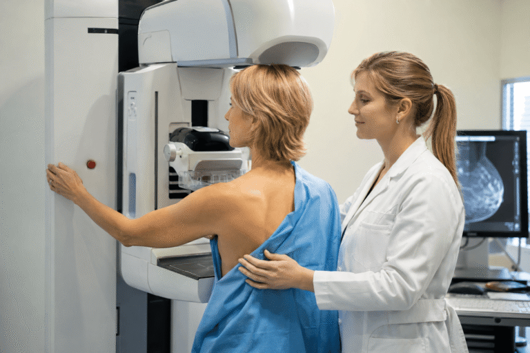
Mammography uses low dose X rays to identify breast masses, calcifications, areas of distortion and other abnormalities. Screening mammography is intended for people without symptoms, while diagnostic mammography is used to investigate a lump, nipple discharge, skin change, unexplained focal pain or an abnormal screening result [2].
For mammography to provide reliable reassurance, the images must capture enough breast tissue with adequate clarity. They must also be interpreted by qualified professionals, compared with previous examinations when available and communicated to the patient and referring clinician within the required time.
A technically inadequate image can conceal or incompletely display an area that needs further assessment. Delayed reporting can also postpone diagnostic imaging, ultrasound, biopsy or specialist referral.
The FDA safety notice does not report how many affected examinations were inaccurate, how many patients may require repeat imaging or whether a specific number of breast cancers were missed. Therefore, patients should take the warning seriously without interpreting it as proof that their previous mammogram was incorrect [1].
Independent review is the safest way to distinguish an acceptable examination from one that needs to be repeated.
Does Every Affected Patient Need a Repeat Mammogram?
No. The FDA has not directed every patient covered by the warning to undergo immediate repeat mammography.
The first step is to obtain the existing images and reports and ask another certified facility to review them. The reviewing radiologist may conclude that the images are adequate and that no immediate repeat examination is necessary.
If the images are incomplete or technically inadequate, the patient may be asked to undergo another screening mammogram. If there is an unexplained area, a prior abnormal finding or a current symptom, the radiologist may recommend diagnostic mammography with additional views, breast ultrasound or another appropriate investigation.
A biopsy is not ordered merely because a facility received a quality warning. It is considered when clinical examination and properly obtained imaging identify a suspicious abnormality.
The correct decision therefore depends on image quality, the original findings, the patient’s age, breast density, symptoms, family history, previous biopsies and overall breast cancer risk.
Screening Mammogram Versus Diagnostic Mammogram
A screening mammogram is generally performed when a person has no breast symptoms. Standard images are obtained and interpreted later by a radiologist.
A diagnostic mammogram is used when there is a symptom or when a screening result needs clarification. It usually involves additional images from different angles and may be reviewed while the patient remains at the facility. Because more images may be needed, the radiation exposure is slightly higher than during routine screening, although the overall exposure remains low [2].
Patients with a new lump, nipple discharge, nipple inversion, persistent focal pain or visible skin change should tell the new facility when booking. They may need a diagnostic appointment rather than another routine screening appointment.
What Affected Patients Should Do Now
Obtain the complete mammography record
Ask Mammography Center of Monterey for the mammogram images, the radiologist’s complete report and the patient summary. Previous comparison images should also be requested when they are available.
The FDA confirms that patients or their authorized representatives have the right to request their mammograms and medical reports [1].
It is often helpful to ask the center to transfer the records directly to the new imaging facility while also requesting a personal copy. Keep written documentation of the request, including the date, the person contacted and the response received.
Arrange review at a certified facility
The new center should have an active Mammography Quality Standards Act certificate. Certification means the facility has undergone an approved accreditation process and is capable of providing mammography under federal quality standards [2].
Patients can search the FDA database of certified facilities or call the National Cancer Institute information service at 1 800 422 6237 for assistance locating a certified facility [1].
Provide relevant breast history
The reviewing radiologist should know about any new lump, nipple discharge, previous breast surgery, breast implants, prior biopsy, atypical hyperplasia, family history of breast or ovarian cancer, genetic risk, hormone use and previous chest radiation.
This information can influence whether simple image review is sufficient or whether diagnostic mammography, ultrasound, magnetic resonance imaging or specialist assessment is needed.
Do not wait for routine screening when symptoms are present
Routine screening schedules apply to people without suspicious breast symptoms. A new symptom requires clinical evaluation even when a recent mammogram was reported as normal.
Should Patients Be Worried About Radiation From a Repeat Mammogram?
A repeat mammogram does involve another small exposure to radiation. However, the FDA states that the radiation risk from mammography is very small compared with the potential benefit of finding breast cancer early [2].
The greater concern in this situation is leaving a potentially unreliable examination unresolved. A delayed diagnosis can carry more serious consequences than a medically justified repeat mammogram.
Independent image review before repetition remains valuable because it may prevent an unnecessary examination when the original images are adequate. When repeat imaging is recommended, the patient should ask why it is required, whether the examination will be screening or diagnostic, and whether previous images will be available for comparison.
What if the Previous Mammogram Was Reported as Normal?
A normal report is reassuring only when the underlying examination is technically adequate and has been properly interpreted.
Patients covered by the FDA warning should therefore not rely solely on the word “normal” in the original report. They should follow the FDA recommendation and obtain an independent review if they have not received a newer mammogram at another certified center [1].
This does not mean the original normal result was necessarily wrong. It means that another qualified facility should confirm whether the images were sufficient to support that conclusion.
How Breast Density Affects the Decision
Breast density and mammogram quality are separate issues.
Dense breast tissue can make an otherwise well performed mammogram more difficult to interpret because dense tissue can obscure some abnormalities. Current federal mammography rules require patients to receive information about breast density in their result summary [2].
The presence of dense breasts does not automatically mean that every patient requires ultrasound or magnetic resonance imaging. The United States Preventive Services Task Force states that evidence remains insufficient to recommend routine supplemental ultrasound or magnetic resonance imaging solely because a person has dense breasts and an otherwise negative mammogram [3].
The decision should instead consider breast density together with age, family history, genetic factors, previous biopsy findings and calculated breast cancer risk.
For a patient covered by the Monterey warning, density does not remove the need to address possible image quality problems. A technically adequate examination is required before the limitations created by breast density can be assessed properly.
Breast Symptoms That Should Not Wait
Patients should seek prompt medical assessment for a new breast or underarm lump, thickening or swelling, nipple retraction, unexplained nipple discharge, bloody discharge, skin dimpling, persistent redness, flaky nipple skin, a change in breast shape or size, or persistent pain in one area [4].
Many of these symptoms are caused by benign conditions. Nevertheless, symptoms should not be managed by waiting for the next routine screening invitation.
A symptomatic patient may require clinical examination, diagnostic mammography and targeted ultrasound even if a previous screening mammogram was described as normal.
Breast Screening Guidance in the United States, United Kingdom, Singapore, Canada and Australia
The Monterey FDA notification applies only to the identified California facility. However, patients in other countries can use the incident as a reminder to choose an accredited service, obtain written results and keep previous images available for future comparison.
The following table summarizes general population screening guidance. Individual recommendations may differ because of symptoms, family history, genetic risk, previous breast cancer, atypical biopsy findings or other clinical factors.
| Country | General routine screening approach | Important qualification |
|---|---|---|
| United States | The United States Preventive Services Task Force recommends mammography every two years from age 40 through 74 [3] | Other professional organizations may recommend different intervals, particularly for higher risk patients |
| United Kingdom | The NHS invites women for their first screening between ages 50 and 53, followed by screening every three years until age 71 [5] | Women aged 71 or older may request continued screening, while symptoms require assessment without waiting for an invitation |
| Singapore | Population guidance recommends mammography every two years for women aged 50 to 69 [6] | Women aged 40 to 49 may be offered annual mammography when recommended by their doctor |
| Canada | Federal public guidance recommends screening every two to three years for women aged 50 to 74 [7] | Eligibility and starting age vary between provincial and territorial screening programs |
| Australia | Women over 40 can access a free mammogram every two years, with active invitations generally directed to women aged 50 to 74 [8] | Symptoms require diagnostic assessment rather than routine BreastScreen participation |
These intervals describe routine screening. They should not be used to postpone evaluation after a facility quality warning, an abnormal result or a new breast symptom.
Can Ayurveda Help After a Mammogram Quality Warning?
Ayurveda cannot determine whether a mammogram image is technically adequate. No herbal medicine, diet, breast massage, detoxification program or Panchakarma procedure can replace independent image review, repeat mammography, diagnostic ultrasound, magnetic resonance imaging or biopsy when these are medically indicated.
The immediate response to this FDA warning must therefore remain conventional and imaging led.
Ayurveda becomes clinically relevant after the structural diagnosis has been clarified. When the repeat assessment confirms a benign breast lump, cyclical breast pain, fibrocystic change, postoperative discomfort or biopsy proven atypical hyperplasia, an individualized Ayurvedic program may address the patient’s wider health pattern alongside continuing breast surveillance.
A whole person Ayurvedic approach may focus on digestive function, sleep, stress regulation, physical activity, body weight, glucose control, menstrual patterns, inflammatory symptoms and recovery after a medical procedure. Improving these areas can reduce symptom burden, strengthen general health and help the patient remain engaged with long term monitoring.
This is more clinically responsible than attempting to treat an unidentified breast abnormality without first establishing what it is.
Classical Ayurvedic Understanding of Breast Lumps
Modern diagnostic terms such as atypical ductal hyperplasia, lobular neoplasia and mammographic distortion do not have exact equivalents in classical Ayurvedic texts. They should not be forced into a one to one comparison.
A relevant classical framework is Granthi, described in Sushruta Samhita, Nidana Sthana, Chapter 11, Verse 3 [11].
वातादयो मांसमसृक् च दुष्टाः सन्दूष्य मेदश्च कफानुविद्धम् ।
वृत्तोन्नतं विग्रथितं तु शोफं कुर्वन्त्यतो ग्रन्थिरिति प्रदिष्टः ॥३॥
Transliteration:
Vatadayo mamsamasrk cha dushtah sandushya medash cha kaphanuviddham,
vrttonnatam vigrathitam tu shopham kurvanty ato granthir iti pradishtah.
Meaning:
When disturbed Doshas affect Mamsa, Rakta and Meda, particularly with Kapha involvement, they may produce a rounded, elevated and knot like swelling described as Granthi.
This verse provides a traditional framework for evaluating nodular swelling through tissue involvement and systemic imbalance. It does not establish that Granthi is identical to breast cancer or atypical hyperplasia. Modern imaging and histopathology remain necessary to determine the biological nature of any breast lesion [11,12].
How Ayurveda May Support Recovery and Long Term Breast Health
The most convincing role for Ayurveda is not as a replacement for breast imaging but as a structured health program after the diagnostic pathway has been completed.
An individualized approach can help address persistent breast tenderness, cyclical swelling, anxiety surrounding repeated scans, disrupted sleep, digestive difficulty, weight related metabolic stress and reduced physical resilience. When surgery or biopsy has been performed, treatment may also be directed toward appetite, bowel regularity, sleep, energy and gradual restoration of function.
Modern evidence remains preliminary. A 2020 single arm pilot study enrolled 32 breast cancer survivors in a four month individualized Ayurvedic program involving nutrition, lifestyle counselling, yoga and Marma based care. Retention was 84 percent, adherence was high, and the investigators observed improvements in several measures of emotional function, cognitive function, sleep, fatigue, stress and quality of life [10].
Because the study did not include a control group and was not designed to assess tumour regression, it cannot prove that Ayurveda eliminates breast cancer, reverses atypical hyperplasia or prevents recurrence. It does, however, support the practical value of a carefully supervised whole systems approach for symptom control and recovery.
The National Center for Complementary and Integrative Health similarly advises that complementary approaches should not replace or delay cancer diagnosis or treatment. Herbal medicines may also interact with hormonal therapy, anticoagulants, chemotherapy and other medicines, making professional supervision important [9].
For patients whose repeat evaluation confirms a benign breast lump or atypical hyperplasia, the Panaceayur clinical guide explains how imaging, biopsy findings, risk assessment and individualized Ayurveda may be integrated before or after surgery without abandoning medical surveillance [12].
Frequently Asked Questions
Does the FDA warning mean I have breast cancer?
No. The warning concerns possible mammogram quality problems. It is not a cancer diagnosis. Independent review is needed to determine whether your previous images were adequate and whether any further investigation is required.
Should I repeat my mammogram immediately?
Not necessarily. Obtain the original images and reports and ask another certified facility to review them. The reviewing radiologist should decide whether you need no immediate action, repeat screening or a diagnostic examination.
Can breast ultrasound replace the affected mammogram?
Ultrasound can provide useful information about a specific lump or area, but it is not automatically a complete substitute for mammography. The appropriate test depends on the patient’s age, symptoms, breast density, original images and clinical risk.
What should I do if I never received my mammogram report?
Contact the facility and your referring clinician to request the report and patient summary. Mammography facilities are generally required to provide patients with a written result summary within 30 calendar days. Suspicious results require faster communication.
What if Mammography Center of Monterey does not provide my records?
Document your requests and inform the new certified facility. Questions about the notification can be directed to the FDA Mammography Quality Standards Act Hotline at 1 800 838 7715 or by email at MQSAhotline@versatechinc.com
Can Ayurveda be started while waiting for the repeat assessment?
General measures involving balanced nutrition, sleep, gentle activity and stress control may be reasonable, but no Ayurvedic treatment should be used to postpone image review or diagnostic testing. Herbal or mineral preparations should be considered only after medication interactions, pregnancy status, liver and kidney health, and the final breast diagnosis have been reviewed.
The Most Important Message for Patients
Patients who had mammography at Mammography Center of Monterey on or after December 22, 2023 should act without assuming the worst.
Obtain the images and medical reports. Arrange review at another Mammography Quality Standards Act certified facility. Inform the new center about any current breast symptoms and relevant family or biopsy history. Follow the new radiologist’s recommendation regarding repeat mammography, diagnostic imaging or further assessment.
The absence of a notification letter should not be treated as reassurance because the FDA reported that the facility had not complied with its patient notification order when the safety notice was published [1].
Ayurveda may offer meaningful help with benign breast symptoms, metabolic health, emotional strain and recovery after the diagnosis is clarified. It cannot correct an inadequate mammogram or replace the investigations needed to exclude breast cancer.
References
[1] U.S. Food and Drug Administration. (2026, August 28). Mammography problems at Mammography Center of Monterey in Monterey, California: FDA safety notification.
https://www.fda.gov/radiation-emitting-products/mqsa-reports-and-safety-notifications/mammography-problems-mammography-center-monterey-monterey-california-fda-safety-notification
Brief: The primary FDA notification identifying the affected facility, examination dates, accreditation history, image quality concerns, record retrieval advice and patient contact information. (U.S. Food and Drug Administration)
[2] U.S. Food and Drug Administration. (n.d.). Frequently asked questions about the Mammography Quality Standards Act.
https://www.fda.gov/radiation-emitting-products/mammography-information-patients/frequently-asked-questions-about-mqsa
Brief: Explains certification, result communication, diagnostic mammography, patient record rights, radiation safety and the FDA complaint process. (U.S. Food and Drug Administration)
[3] U.S. Preventive Services Task Force. (2024, April 30). Breast cancer: Screening.
https://www.uspreventiveservicestaskforce.org/uspstf/recommendation/breast-cancer-screening
Brief: Recommends mammography every two years for women aged 40 to 74 and explains current uncertainty regarding routine supplemental imaging for dense breasts. (USPSTF)
[4] Centers for Disease Control and Prevention. (n.d.). Symptoms of breast cancer.
https://www.cdc.gov/breast-cancer/symptoms/index.html
Brief: Lists breast and nipple changes that require medical assessment rather than waiting for routine screening. (CDC)
[5] UK Health Security Agency. (2026, February 23). Your guide to NHS breast screening. GOV.UK.
https://www.gov.uk/government/publications/breast-screening-helping-women-decide/nhs-breast-screening-helping-you-decide
Brief: Explains the current NHS invitation ages, three year screening interval and the purpose and limitations of routine mammography. (GOV.UK)
[6] HealthHub Singapore. (n.d.). Evidence based recommendations on health screening tests.
https://www.healthhub.sg/well-being-and-lifestyle/personal-care/type-2-screening-tests
Brief: Provides Singapore population guidance recommending mammography every two years for women aged 50 to 69, with individual risk assessment where appropriate. (HealthHub)
[7] Health Canada. (2019, September 3). Mammography: Medical radiation.
https://www.canada.ca/en/health-canada/services/health-risks-safety/radiation/medical/mammography.html
Brief: Explains mammography benefits, radiation considerations and federal screening information while advising patients to consult their provincial or territorial program. (Canada)
[8] Australian Government Department of Health, Disability and Ageing. (2025, August 18). BreastScreen Australia Program.
https://www.health.gov.au/our-work/breastscreen-australia-program
Brief: Describes free mammography access for women over 40 and active screening invitations for women aged 50 to 74. (Health, Disability and Ageing)
[9] National Center for Complementary and Integrative Health. (2021, October). Cancer and complementary health approaches: What you need to know.
https://www.nccih.nih.gov/health/cancer-and-complementary-health-approaches-what-you-need-to-know
Brief: States that complementary approaches should not replace cancer diagnosis or treatment and warns that herbal products may interact with conventional medicines. (NCCIH)
[10] Dhruva, A., Wu, C., Miaskowski, C., Hartogensis, W., Rugo, H. S., Adler, S. R., Kaptchuk, T. J., Kelkar, R., Agarawal, S., Vadodaria, A., Garris, E., & Hecht, F. M. (2020). A 4 month whole systems Ayurvedic medicine nutrition and lifestyle intervention is feasible and acceptable for breast cancer survivors: Results of a single arm pilot clinical trial. Global Advances in Health and Medicine, 9, Article 2164956120964712.
https://pmc.ncbi.nlm.nih.gov/articles/PMC7716077/
Brief: A small pilot study reporting high adherence and encouraging changes in symptoms and quality of life, while not establishing tumour regression, recurrence prevention or cancer cure. (PubMed)
[11] Sushruta. (1907 to 1916). An English translation of the Sushruta Samhita, based on the original Sanskrit text (K. L. Bhishagratna, Trans.). Calcutta. Nidana Sthana, Chapter 11, Verse 3.
https://archive.org/details/englishtranslati00susruoft
Brief: Classical source describing Granthi as a rounded, elevated, knot like swelling involving disturbed Doshas and Mamsa, Rakta and Meda, especially with Kapha association. (Internet Archive)
[12] Panaceayur. (2026, May 14). Benign breast lump cancer risk: Biopsy results and prevention.
https://panaceayur.com/benign-atypia-breast-symptoms-diagnosis-ayurveda/
Brief: Provides a detailed integrative discussion of benign breast lumps, atypical hyperplasia, imaging, biopsy, surveillance and the appropriate role of individualized Ayurveda before or after medical procedures. (panaceayur.com)