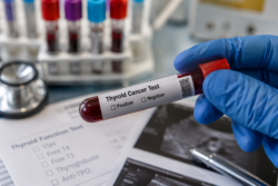



Parathyroid Cancer and High Calcium can initially look like ordinary fatigue, constipation, dehydration, muscle weakness or mental stress. This is one reason the underlying disorder may remain undiagnosed until the calcium level becomes dangerously high or complications develop in the kidneys, bones, nervous system or heart.
Parathyroid cancer, also called parathyroid carcinoma, is extremely rare and accounts for fewer than 1% of primary hyperparathyroidism cases. However, most parathyroid cancers produce excessive parathyroid hormone, or PTH. This can raise the calcium concentration in the blood to a much more dangerous level than is usually seen with a benign parathyroid adenoma [1–4].
The National Cancer Institute warns that the hypercalcemia caused by an overactive parathyroid tumour can be more immediately life-threatening than the cancer itself. Controlling the calcium level is therefore a central part of treatment, not a secondary concern [1].
A major clinical review published in July 2026 also emphasised that marked hypercalcemia, substantially elevated PTH, a palpable neck mass or recurrent laryngeal nerve dysfunction should increase suspicion of parathyroid carcinoma [3].
What Are the Parathyroid Glands?
Most people have four parathyroid glands located behind or close to the thyroid gland in the neck. Although each gland is usually only about the size of a small pea, together they control one of the body’s most important mineral systems.
The parathyroid glands release PTH when the calcium level in the blood falls. PTH helps raise calcium by affecting the bones, kidneys and intestinal absorption of calcium. Once sufficient calcium is available, PTH production should decrease [1,6].
When a parathyroid gland becomes overactive, it may continue releasing PTH even though the blood calcium level is already high. This is called primary hyperparathyroidism. The most common cause is a benign parathyroid adenoma. Cancer is a much rarer cause.
The important point is that a PTH result should not be judged only by whether it falls inside the laboratory’s normal range. When calcium is elevated, PTH should normally be suppressed. A PTH result that appears “normal” may therefore be inappropriately normal and can still indicate a PTH-dependent calcium disorder [6].
Parathyroid Cancer and High Calcium: Why the Link Matters
Most parathyroid carcinomas are functioning tumours, meaning that they release PTH. Persistently excessive PTH can cause calcium to move out of the bones, increase calcium retention by the kidneys and indirectly increase calcium absorption from the digestive tract.
This can produce two problems at the same time. Calcium becomes dangerously concentrated in the blood while the skeleton gradually loses mineral strength.
The kidneys attempt to remove some of the extra calcium through urine. This causes frequent urination, excessive thirst and dehydration. High urinary calcium can also contribute to kidney stones, calcium deposits within the kidneys and reduced kidney function.
At the same time, the digestive system may slow down, muscles may weaken and the nervous system may begin functioning abnormally. This is why the symptoms of high calcium can seem unrelated until they are considered together.
Parathyroid Cancer and High Calcium Symptoms
The following symptoms do not automatically mean that a person has parathyroid cancer. Most people with these symptoms will have another explanation. However, persistent or progressive symptoms should be investigated, especially when a blood test has already shown elevated calcium or PTH.
| Area affected | Symptoms a patient may notice | Why the symptom matters |
|---|---|---|
| Kidneys and hydration | Excessive thirst, frequent urination, waking repeatedly to urinate, dehydration, kidney stones, side or back pain | The kidneys are attempting to remove excess calcium and may gradually become damaged |
| Bones and muscles | Muscle weakness, deep bone pain, osteoporosis, reduced height or fractures after minor injury | Excessive PTH can cause mineral loss from the skeleton |
| Digestive system | Constipation, nausea, vomiting, abdominal discomfort, poor appetite or unexplained weight loss | High calcium can slow intestinal activity and worsen dehydration |
| Brain and nervous system | Poor concentration, irritability, low mood, memory problems, confusion, drowsiness or personality changes | Moderate or severe hypercalcemia can disrupt neurological function |
| Heart and circulation | Palpitations, irregular heartbeat, dizziness, profound weakness or collapse | Significant calcium disturbance can interfere with the electrical activity of the heart |
| Neck and voice | A firm neck lump, pressure in the neck, hoarseness, voice changes or difficulty swallowing | These symptoms can occur when a tumour affects nearby tissues or the recurrent laryngeal nerve |
| General wellbeing | Persistent fatigue, reduced physical strength and declining ability to perform normal activities | Multisystem effects of high calcium can produce significant deterioration before the cause is recognised |
These symptom patterns are recognised by the National Cancer Institute, Canadian Cancer Society, Cancer Research UK and healthdirect Australia [1,7–9].
Excessive Thirst and Frequent Urination

Being much thirstier than usual while passing large quantities of urine is one of the most important high calcium warning patterns.
High calcium reduces the kidneys’ ability to concentrate urine. The patient may urinate repeatedly throughout the day and wake several times at night. Even after drinking water, the mouth may remain dry and the feeling of thirst may continue.
This fluid loss can lead to dehydration. Dehydration then reduces kidney filtration, making it harder for the body to remove excess calcium. The calcium level may rise further, producing a potentially dangerous cycle.
These symptoms should not automatically be attributed to hot weather, diabetes, anxiety or increased water intake. A patient may need blood tests for calcium, albumin, PTH, glucose, electrolytes and kidney function.
People with heart failure, advanced kidney disease or another condition requiring fluid restriction should not attempt aggressive self-hydration. Fluid replacement must be medically supervised.
Kidney Stones and Declining Kidney Function
Persistently elevated calcium can increase the amount of calcium entering the urine. Over time, this may contribute to kidney stones, nephrocalcinosis, recurrent urinary symptoms or progressive kidney impairment.
A person may experience severe pain in the side or back, pain travelling towards the lower abdomen or groin, nausea, vomiting or blood in the urine. However, kidney damage can also develop without obvious stone pain.
The combination of recurrent kidney stones, elevated calcium and elevated or inappropriately normal PTH strongly supports the need for a complete parathyroid evaluation.
Kidney stones alone are common and usually do not indicate cancer. Concern increases when renal disease occurs together with severe hypercalcemia, very high PTH, significant bone disease or a suspicious neck lesion [2–4].
Constipation, Nausea and Loss of Appetite
High calcium can slow the movement of food and waste through the digestive tract. Constipation may become persistent and may not respond properly to ordinary dietary changes or laxatives.
As the calcium level rises, nausea, vomiting, abdominal pain and loss of appetite may appear. Some people begin losing weight because they are unable to eat normally.
Repeated vomiting is particularly concerning because it worsens dehydration and can accelerate the rise in calcium. Digestive symptoms accompanied by excessive thirst, frequent urination, muscle weakness or confusion should be assessed promptly.
Fatigue and Muscle Weakness
The fatigue associated with hypercalcemia can be more severe than ordinary tiredness. A patient may struggle to climb stairs, rise from a chair, carry normal household items or complete a regular working day.
Muscle weakness can gradually increase because high calcium affects normal nerve and muscle function. Weakness may also be aggravated by dehydration, reduced food intake, kidney impairment and prolonged inactivity.
When fatigue is persistent and associated with constipation, thirst, kidney stones, bone pain or abnormal blood results, it should not be dismissed as ageing or stress.
Brain Fog, Mood Changes and Confusion
Mental and behavioural changes can begin subtly. The person may notice poor concentration, forgetfulness, irritability, low motivation, reduced interest in normal activities or a feeling of mental fog.
Family members may notice personality changes before the patient recognises them. As hypercalcemia becomes more severe, mild confusion can progress to agitation, marked drowsiness, difficulty communicating, loss of coordination, reduced consciousness or coma [7,8].
Sudden confusion in someone with known high calcium requires urgent medical assessment.
Bone Pain, Osteoporosis and Unexpected Fractures
Excessive PTH can cause calcium to be released from the skeleton. This can weaken the bones even while the blood calcium level remains high.
Possible signs include deep bone pain, reduced bone density, fractures after minor injury, spinal discomfort or a gradual reduction in height. Severe, prolonged hyperparathyroidism can produce extensive skeletal changes.
The combination of significant kidney disease and obvious skeletal involvement is particularly concerning when calcium and PTH are markedly elevated [2,4].
A bone density scan can help identify skeletal damage, but a normal result cannot independently exclude parathyroid cancer.
A Neck Lump, Hoarseness or Difficulty Swallowing
A firm or enlarging neck lump is an important warning sign, particularly when it occurs with high calcium and high PTH.
Persistent hoarseness or a change in voice may develop if a tumour affects the recurrent laryngeal nerve, which controls vocal-cord movement. A person may also experience pressure in the neck, discomfort while swallowing or a sensation that food is becoming stuck.
These symptoms do not prove that a lesion is cancerous. Thyroid nodules, benign parathyroid adenomas and other neck conditions may produce similar complaints. Nevertheless, a palpable neck mass or vocal-cord dysfunction substantially increases clinical concern in a patient with severe hyperparathyroidism [3,4].
Can High Calcium Affect the Heart?
Calcium is essential for normal cardiac contraction and electrical conduction. However, a marked calcium disturbance can interfere with normal heart rhythm.
Some people experience palpitations, dizziness, faintness or unexplained weakness. Severe hypercalcemia may contribute to clinically important rhythm changes, particularly when dehydration, kidney dysfunction or other electrolyte abnormalities are also present [4,7].
Chest discomfort, collapse, severe dizziness or a sustained irregular heartbeat requires emergency evaluation.
High Calcium Does Not Automatically Mean Cancer
An elevated calcium result should be investigated, but it does not automatically mean that a patient has parathyroid cancer.
Most cases of primary hyperparathyroidism are caused by a non-cancerous adenoma. High calcium can also be related to dehydration, certain medications, excessive calcium or vitamin D intake, familial hypocalciuric hypercalcemia, prolonged immobility, granulomatous disorders and cancers arising in other organs.
The PTH result helps clinicians narrow the possibilities. When calcium is elevated because of most non-parathyroid cancers, the natural PTH level is usually suppressed. When calcium is high and PTH is also high or inappropriately normal, a parathyroid-dependent cause becomes more likely [6].
No single calcium, PTH or imaging result can confirm parathyroid carcinoma. Results must be interpreted together with the symptoms, physical examination, kidney function, phosphate, vitamin D, urinary calcium and imaging findings.
Findings That Increase Suspicion of Parathyroid Cancer
Parathyroid cancer remains rare even among people with hyperparathyroidism. Suspicion increases when several concerning findings occur together.
| Finding | Why it is important |
|---|---|
| Calcium above 12 mg/dL or 3.0 mmol/L | Represents marked hypercalcemia and requires prompt evaluation, especially when symptoms are present |
| Calcium above 14 mg/dL or 3.5 mmol/L | Generally classified as severe hypercalcemia and may require emergency hospital treatment |
| PTH three to ten times the upper reference limit | A very high result can occur in parathyroid carcinoma, although it is not diagnostic by itself |
| Firm or enlarging neck mass | May suggest a large or locally invasive parathyroid lesion |
| Persistent hoarseness or vocal-cord weakness | Can indicate involvement of the recurrent laryngeal nerve |
| Kidney stones together with severe bone disease | Simultaneous renal and skeletal complications are more concerning than either problem alone |
| Large lesion with irregular borders or invasion on imaging | May increase suspicion, although benign adenomas can also become large |
| Persistent or recurrent high calcium after parathyroid surgery | Requires investigation for residual, recurrent or previously unrecognised malignant disease |
Specialist literature commonly reports calcium above 14 mg/dL, ionised calcium above approximately 1.77 mmol/L and PTH three to ten times the upper reference limit as findings that should increase suspicion. These values are warning signs rather than independent diagnostic criteria [3,4,7].
When High Calcium Becomes an Emergency
A calcium result above 14 mg/dL or 3.5 mmol/L is generally considered severe. The Endocrine Society recommends urgent calcium-lowering treatment for severe hypercalcemia of malignancy, including hypercalcemia caused by parathyroid carcinoma [5].
Emergency assessment is necessary when high calcium is accompanied by severe or worsening confusion, extreme drowsiness, repeated vomiting, inability to drink, severe dehydration, very little urine, sudden muscle weakness, an irregular heartbeat, seizure, loss of consciousness or collapse.
A rapid increase in calcium can produce serious symptoms even before the result reaches 14 mg/dL. The speed of the rise, kidney function, hydration status and the patient’s overall condition are therefore as important as the number itself.
Patients experiencing these symptoms should attend an emergency department or A&E. They should not wait for a routine appointment or attempt to lower the calcium through diet, supplements, detoxification or alternative treatment at home.
Tests Used to Investigate Parathyroid Cancer and High Calcium

Investigation usually begins by repeating the calcium test. Total calcium must be considered alongside albumin because a proportion of calcium is attached to blood proteins. If albumin is abnormal or the result remains uncertain, ionised calcium may provide a clearer measurement [6].
PTH should be measured at the same time as calcium. Other useful tests commonly include kidney function, phosphate, magnesium, alkaline phosphatase and 25-hydroxyvitamin D.
Urinary calcium measurement may help distinguish primary hyperparathyroidism from familial hypocalciuric hypercalcemia, an inherited condition that can cause lifelong mild hypercalcemia but usually requires a different management approach.
A bone density scan can identify skeletal damage. Kidney ultrasound or CT may be required to look for stones or nephrocalcinosis.
An electrocardiogram may be performed when the calcium level is markedly elevated or the patient reports cardiac symptoms.
Imaging Tests Used for a Suspected Parathyroid Tumour
Once blood tests have established PTH-dependent hypercalcemia, imaging can help identify the location of the abnormal gland and assist surgical planning.
Neck ultrasound may show an enlarged parathyroid gland, its relationship with the thyroid and features such as irregular borders, calcification or possible tissue invasion.
Technetium-99m sestamibi scanning, often combined with SPECT/CT, can help localise an overactive parathyroid gland. Four-dimensional CT, MRI or fluorocholine PET/CT may be considered in complex cases, recurrent disease or when ordinary imaging does not clearly identify the lesion [1,3,4].
Imaging does not independently determine whether a tumour is benign or malignant. A large adenoma can resemble carcinoma, while some cancers may not show obvious invasion on the first scan.
Why a Needle Biopsy Is Usually Avoided
Fine-needle aspiration is widely used to assess thyroid nodules, but a suspicious parathyroid lesion requires a different approach.
A needle biopsy cannot reliably distinguish a benign parathyroid adenoma from parathyroid carcinoma. It may also cause bleeding, rupture of the tumour capsule or seeding of tumour cells along the needle path [3,4].
For this reason, preoperative needle biopsy is generally not recommended when parathyroid cancer is suspected. The case should first be reviewed by an endocrinologist, endocrine surgeon and radiologist experienced in parathyroid disease.
How Parathyroid Cancer Is Confirmed
Parathyroid cancer can be difficult to confirm before surgery because benign and malignant parathyroid cells may appear similar.
Blood tests, clinical findings and imaging establish the level of suspicion, but a definite diagnosis commonly depends on evidence that the tumour has invaded nearby tissues, blood vessels or nerves, or has spread to lymph nodes or distant organs [3,4,10].
The removed tissue should be examined by an experienced pathologist. Features such as capsular invasion, vascular invasion, perineural invasion, mitotic activity, Ki-67 index and parafibromin expression may contribute to the final assessment.
Because the first operation can have a major influence on long-term outcome, a suspicious case should ideally be referred to a specialist endocrine surgery centre before an unplanned limited excision is performed.
Treatment of Severe High Calcium
The first objective is to stabilise the patient and reduce the calcium level safely.
Hospital treatment may involve carefully monitored intravenous fluids, calcitonin for rapid short-term calcium reduction, an intravenous bisphosphonate or denosumab. A calcimimetic medicine such as cinacalcet may be used in selected patients with PTH-driven hypercalcemia [5].
The treatment selected depends on the severity of the calcium elevation, symptoms, kidney function, hydration status, previous treatment and whether surgery can be performed.
Calcitonin is generally used only briefly because its effect can diminish after approximately 48 to 72 hours. Calcium-lowering medication may stabilise the patient, but it does not remove the tumour producing excessive PTH.
The Endocrine Society specifically recommends considering surgery when feasible after severe hypercalcemia has been controlled [5].
Surgery and the Possibility of Long-Term Recovery
Complete removal during the first properly planned operation offers the strongest possibility of durable disease control and potential cure.
When parathyroid carcinoma is suspected, specialist reviews recommend en bloc removal of the tumour without rupturing its capsule. This may include the affected parathyroid gland, the thyroid lobe on the same side and any directly involved surrounding tissue [2–4,10].
An experienced surgeon should determine the extent of the operation. Routine removal of uninvolved structures is not automatically necessary, but visible invasion must be addressed appropriately.
Incomplete excision or rupture of the tumour can increase the risk of local recurrence. The 2026 clinical review reported recurrence rates ranging from approximately 23% to 65% across published series, reinforcing the need for lifelong surveillance [3].
After treatment, calcium and PTH should be monitored even when the patient feels completely well. A gradual increase may be an early indication of recurrent disease before a new lump or other symptoms appear.
Genetic Testing and Family History
A proportion of parathyroid carcinomas are associated with inherited changes in the CDC73 gene. This gene is linked with hyperparathyroidism-jaw tumour syndrome and some familial forms of parathyroid disease [3,4].
Genetic counselling or testing may be considered when parathyroid carcinoma is confirmed, particularly in younger patients or those with a personal or family history of parathyroid tumours, jaw tumours, kidney lesions, uterine tumours or recurrent hyperparathyroidism.
A genetic result can influence the patient’s follow-up and may help determine whether close relatives should also be assessed.
How Ayurveda May Support a Patient With Parathyroid Disease
Ayurveda can have a meaningful complementary role when it is integrated with endocrinology, oncology and endocrine surgery rather than used as a substitute for urgent calcium reduction or tumour-directed treatment.
A personalised Ayurvedic plan may focus on the broader health problems that often accompany prolonged parathyroid disease, including reduced appetite, disturbed digestion, constipation, poor sleep, emotional stress, loss of strength and difficulty rebuilding normal daily function.
From an Ayurvedic perspective, long-standing parathyroid dysfunction may be assessed in relation to Agni, the metabolic processing of nutrients, Asthi Dhatu, which represents the structural bone system, Majja Dhatu, which is associated with deeper tissue and neurological support, and disturbances affecting Mutravaha Srotas, the urinary system.
This whole-person assessment can be useful because parathyroid disease rarely affects only one laboratory value. It can influence nutrition, muscle strength, bowel function, kidney health, bone stability, sleep and psychological wellbeing at the same time.
An individualised Ayurvedic program may therefore aim to support digestive stability, improve the patient’s nutritional tolerance, maintain regular bowel function, strengthen recovery after medical or surgical treatment and restore a more sustainable daily routine. Such support may help a patient remain physically and mentally better prepared for necessary investigations, surgery and long-term follow-up.
Patients can read Panaceayur’s detailed approach to parathyroid disorders, symptoms, diagnosis and root-level Ayurvedic management here: https://panaceayur.com/hyperparathyroidism-treatment-without-surgery/
In a suspected or confirmed parathyroid cancer case, responsible Ayurvedic care must remain coordinated with the treating endocrinologist and surgeon. Ayurveda has not been established through clinical trials as a standalone cure for parathyroid carcinoma or as an emergency treatment capable of rapidly lowering dangerously high calcium.
The most medically responsible integrative model is one in which emergency treatment controls the calcium, specialist surgery addresses the tumour when feasible, and carefully selected Ayurvedic care supports the patient’s digestion, strength, sleep, emotional resilience and long-term recovery.
Patients should disclose every herb, mineral preparation and supplement to their medical team. The National Center for Complementary and Integrative Health advises against postponing conventional medical assessment and warns that some poorly manufactured Ayurvedic products may contain unsafe concentrations of lead, mercury or arsenic [11].
Supplement and Diet Precautions
A person with high blood calcium should not automatically eliminate every naturally calcium-containing food. In PTH-driven hypercalcemia, the main problem is abnormal hormonal regulation, not simply the amount of calcium consumed.
Extreme dietary restriction can worsen malnutrition, muscle loss and bone weakness without correcting the overactive parathyroid gland. Dietary decisions should be based on the calcium level, kidney function, nutritional status and the treating clinician’s advice [6].
Calcium tablets, vitamin D, multivitamins, antacids and mineral-containing Ayurvedic preparations should not be started without reviewing recent blood tests. Even when vitamin D is low, replacement should be medically supervised because the appropriate dose and monitoring requirements vary.
The patient should also provide the healthcare team with a complete list of prescription medicines, over-the-counter products, herbs and supplements. Certain medicines can influence calcium levels or complicate their interpretation.
Frequently Asked Questions
Is high calcium always a sign of parathyroid cancer?
No. Parathyroid cancer is extremely rare. Most PTH-dependent cases of high calcium are caused by a benign parathyroid adenoma or gland enlargement. Other medicines and medical conditions can also raise calcium.
What calcium level should be considered dangerous?
The laboratory’s reference range should always be considered. Calcium above 12 mg/dL, or 3.0 mmol/L, represents marked hypercalcemia and needs prompt assessment. Calcium above 14 mg/dL, or 3.5 mmol/L, is generally considered severe and may require emergency hospital treatment.
Can dangerous high calcium occur without severe symptoms?
Yes. Symptom severity does not always correspond to the calcium result. Some people with a modest increase feel extremely unwell, while others with severe hypercalcemia initially report only fatigue, constipation or thirst.
Does very high PTH confirm parathyroid cancer?
No. PTH three to ten times the upper reference limit increases concern, particularly when calcium is also severely elevated, but it does not prove malignancy. Some large benign adenomas can also produce very high PTH.
Can PTH be normal when calcium is high?
Yes. When calcium is elevated, PTH should normally become low. A result within the laboratory reference range can therefore be “inappropriately normal” and may still indicate primary hyperparathyroidism.
Is a neck lump always present in parathyroid cancer?
No. Many patients do not have a noticeable neck lump. However, a firm mass, hoarseness, difficulty swallowing or vocal-cord weakness becomes important when it occurs with marked hypercalcemia and high PTH.
Should a suspicious parathyroid lump be biopsied?
Preoperative needle biopsy is generally avoided when parathyroid carcinoma is suspected. It cannot reliably differentiate carcinoma from adenoma and may cause bleeding, tumour rupture or seeding.
Can parathyroid cancer be cured?
Complete removal during the first properly planned operation offers the highest chance of cure and durable disease control. The result depends on whether the tumour can be completely removed, whether it has invaded surrounding structures and whether it has spread.
Can Ayurveda lower dangerously high calcium?
Ayurveda should not be relied upon for rapid treatment of severe hypercalcemia. Severe or symptomatic high calcium requires urgent medical care. Ayurvedic treatment may be used as complementary support for digestion, nutrition, bowel function, sleep, strength and recovery after the patient has been properly evaluated.
Is lifelong follow-up necessary after surgery?
Yes. Parathyroid carcinoma can recur years after apparently successful treatment. Calcium and PTH should be monitored long term, even when the patient has no symptoms.
The Most Important Message for Patients
Parathyroid Cancer and High Calcium should be considered when a person has marked hypercalcemia together with very high or inappropriately normal PTH, kidney stones, severe bone disease, a firm neck mass, hoarseness or difficulty swallowing.
Most people with high calcium do not have parathyroid cancer. Nevertheless, persistent thirst, excessive urination, constipation, vomiting, unexplained weakness, confusion, bone pain or a neck lump should never be ignored.
Severe high calcium must be stabilised promptly. A suspicious parathyroid tumour should then be evaluated by an endocrinologist and an experienced endocrine surgeon. Carefully coordinated Ayurvedic care may support the patient’s overall recovery, but it should not delay emergency treatment, essential investigations or potentially curative surgery.
References
[1] National Cancer Institute. Parathyroid Cancer Treatment, Patient Version. Explains the relationship between parathyroid cancer, excessive PTH, high calcium, symptoms, and diagnostic testing.
https://www.cancer.gov/types/parathyroid/patient/parathyroid-treatment-pdq
[2] National Cancer Institute. Parathyroid Cancer Treatment, Health Professional Version. Provides detailed clinical information on warning signs, severe hypercalcemia, renal and skeletal involvement, surgery, and recurrence.
https://www.cancer.gov/types/parathyroid/hp/parathyroid-treatment-pdq
[3] Song J, et al. Parathyroid carcinoma: From molecular pathogenesis to multidisciplinary management. The Journal of Clinical Endocrinology & Metabolism. Published July 4, 2026. Reviews diagnosis, imaging, surgery, calcium control, recurrence, and lifelong follow-up.
https://doi.org/10.1210/clinem/dgag266
[4] Cetani F, et al. Approach to the Patient With Parathyroid Carcinoma. The Journal of Clinical Endocrinology & Metabolism. 2024;109(1):256–268. Reviews clinical warning signs, imaging, biopsy precautions, genetics, and surgical management.
https://doi.org/10.1210/clinem/dgad455
[5] Endocrine Society. Treatment of Hypercalcemia of Malignancy in Adults: Clinical Practice Guideline. Includes recommendations specifically addressing high calcium caused by parathyroid carcinoma.
https://www.endocrine.org/clinical-practice-guidelines/hypercalcemia
[6] Bilezikian JP, et al. Evaluation and Management of Primary Hyperparathyroidism: Guidelines from the Fifth International Workshop. Journal of Bone and Mineral Research. 2022;37(11):2293–2314. Covers calcium interpretation, PTH testing, kidney evaluation, bone health, and long-term management.
https://doi.org/10.1002/jbmr.4677
[7] Canadian Cancer Society. Signs and Symptoms of Parathyroid Cancer. Medically reviewed October 2025. Describes hypercalcemia symptoms and findings that may raise suspicion of parathyroid cancer.
https://cancer.ca/en/cancer-information/cancer-types/parathyroid/signs-and-symptoms
[8] Cancer Research UK. High Calcium Levels in People With Cancer. Explains the early and advanced symptoms of hypercalcemia and the need for prompt treatment.
https://www.cancerresearchuk.org/about-cancer/coping/physically/blood-calcium/high-calcium-people-cancer
[9] Healthdirect Australia. Parathyroid Glands. Reviews PTH function, hyperparathyroidism symptoms, complications, diagnosis, and treatment.
https://www.healthdirect.gov.au/parathyroid-glands
[10] Hadoux J, et al. ESMO Clinical Practice Guideline for Rare Endocrine Tumours, Including Parathyroid Carcinoma. ESMO Open. 2024;9(10):103664. Provides recommendations for diagnosis, treatment, and follow-up.
https://doi.org/10.1016/j.esmoop.2024.103664
[11] National Center for Complementary and Integrative Health. Ayurvedic Medicine: In Depth. Reviews the available evidence, safety considerations, product quality, and the importance of coordinated medical care.
https://www.nccih.nih.gov/health/ayurvedic-medicine-in-depth