




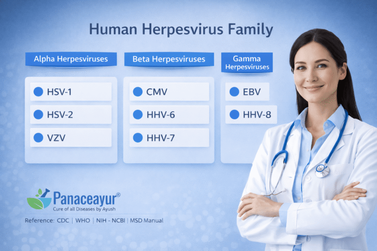
- Types of Herpes Viruses in Humans
- How Many Types of Herpes Viruses Infect Humans?
- The Eight Human Herpesviruses at a Glance
- Alpha, Beta and Gamma Herpesviruses
- Alpha Herpesviruses: HSV-1, HSV-2 and VZV
- Beta Herpesviruses: CMV, HHV-6 and HHV-7
- Gamma Herpesviruses: EBV and HHV-8
- What All Human Herpesviruses Have in Common
- Herpes Simplex Versus Other Human Herpesviruses
- Are All Herpes Viruses Sexually Transmitted?
- Can One Person Carry More Than One Herpesvirus?
- Which Herpesviruses Commonly Coexist?
- How the Different Herpesviruses Are Tested
- How Treatment Differs Across the Herpesvirus Family
- Can HSV-1 and HSV-2 Be Cured Permanently?
- Ayurvedic Clinical Perspective on Herpesvirus
- When Herpesvirus Symptoms Require Urgent Medical Evaluation
- Frequently Asked Questions
- References
Types of Herpes Viruses in Humans
The main types of herpes viruses in humans are traditionally described as eight numbered groups: herpes simplex virus type 1 (HSV-1), herpes simplex virus type 2 (HSV-2), varicella-zoster virus (VZV), Epstein–Barr virus (EBV), cytomegalovirus (CMV), human herpesvirus 6 (HHV-6), human herpesvirus 7 (HHV-7), and human herpesvirus 8 (HHV-8). These are different members of the herpesvirus family—not eight strains of the virus responsible for genital herpes. Their names matter because each has different clinical associations, transmission patterns, and testing requirements. [1]
Why Some Sources Describe Eight and Others Nine
The difference comes from HHV-6. Scientists initially described HHV-6A and HHV-6B as variants of one virus, but they were formally classified as separate species in 2012 because of important biological, genetic, and clinical differences. Consequently, the familiar HHV-1–HHV-8 numbering contains nine distinct viruses when HHV-6A and HHV-6B are counted separately. The numbers identify viruses; they do not indicate stages of infection or increasing disease severity. [1,3]
What the Different Names Mean for Patients
In everyday conversation, “herpes” usually refers to HSV-1 or HSV-2. HSV-1 commonly causes cold sores and can also cause genital herpes, while HSV-2 predominantly causes genital infection. A genital outbreak therefore does not automatically mean HSV-2: identifying the type is useful because genital HSV-1 generally causes fewer recurrences and less frequent viral shedding than genital HSV-2. [4,5]
Other members of the family cause different illnesses. VZV causes chickenpox during primary infection and can later reactivate as shingles. EBV is the most common cause of infectious mononucleosis, often called “mono” or glandular fever. Having shingles or a previous EBV infection does not mean that a person has genital herpes. [6,7]
Herpesvirus infection also does not automatically indicate sexual transmission. HSV-1, EBV, and CMV are frequently acquired during childhood, sometimes without recognizable symptoms. Their presence should be interpreted according to the specific virus and clinical history rather than used to make assumptions about a person’s sexual relationships. [4,7,8]
When reviewing a laboratory report, distinguish the virus being tested from the type of test performed. For example, an HSV test taken directly from a genital lesion can identify whether HSV-1 or HSV-2 is causing that lesion. An HSV-1 antibody blood test, however, cannot determine whether the infection is oral or genital. This distinction helps prevent a positive result from being assigned the wrong diagnosis. [5]
How Many Types of Herpes Viruses Infect Humans?
The types of herpes viruses in humans are conventionally counted as eight numbered groups, encompassing nine distinct viruses when HHV-6A and HHV-6B are counted separately. This count refers to the viruses themselves, rather than the number of illnesses, affected body sites, or outbreaks a person experiences. [1,3]
The abbreviations on medical records help distinguish these identities. HHV-1 and HSV-1 refer to the same virus, as do HHV-2 and HSV-2. However, the remaining HHV numbers identify other members of the family: HHV-3 is VZV, HHV-4 is EBV, and HHV-5 is CMV. Recognizing these alternative names prevents the same infection from being mistaken for two separate diagnoses when comparing reports from different laboratories or clinicians. [1,7]
Several conditions commonly described as “types of herpes” are actually different presentations of HSV infection. Herpes labialis refers to cold sores around the lips, herpetic whitlow describes infection of a finger, and herpes keratitis involves the cornea—the clear front surface of the eye. These names identify the affected tissue or clinical presentation; they do not add new viruses to the family. The location remains important because an eye infection requires a different assessment and treatment approach from a localized finger or lip infection. [1]
The reverse distinction also matters: different herpesviruses can produce similar illnesses. EBV and CMV can both cause a mononucleosis-like illness, with symptoms such as fever and fatigue. Identifying the responsible virus therefore involves the clinical history and appropriately selected laboratory tests, rather than relying on symptoms alone. This is particularly important when pregnancy or reduced immune function changes the implications of the diagnosis. [7,8]

A laboratory test labeled “HSV-1 and HSV-2” evaluates herpes simplex, rather than screening for every human herpesvirus. Reviewing the exact organisms named on the laboratory request clarifies what has been investigated. Additional testing should answer a specific clinical question based on symptoms, exposure history, pregnancy, or immune status, rather than treating the entire herpesvirus family as a single diagnosis. [5,8,10]
The Eight Human Herpesviruses at a Glance
HSV-1 (HHV-1) commonly causes episodes of tingling, burning, and clustered blisters around the lips. A first symptomatic oral infection can also involve fever, sore throat, and swollen lymph nodes. Oral-to-genital contact can transmit HSV-1 to the genital area, so prevention depends on the infected site and contact involved. [4,5]
HSV-2 (HHV-2) predominantly affects the genital and anal regions, where it can produce painful blisters or ulcers. Some infections cause mild or unrecognized symptoms, while viral shedding can occur between visible outbreaks. Care therefore addresses both the patient’s symptoms and practical measures to reduce transmission to partners. [4,5]
Varicella-zoster virus (HHV-3) causes chickenpox and, following reactivation, shingles. A characteristic shingles presentation is pain followed by a band of blisters on one side of the torso or face. Persistent nerve pain is an important complication, particularly in older adults, while facial involvement requires attention to possible eye or ear complications. [6]
Epstein–Barr virus (HHV-4) is particularly recognizable when adolescents or adults develop infectious mononucleosis with an inflamed throat, enlarged neck lymph nodes, fever, and substantial fatigue. The spleen or liver may also enlarge. Recovery from the initial illness commonly takes several weeks, and fatigue can continue after other symptoms improve. [7]
Cytomegalovirus (HHV-5) usually produces few symptoms in otherwise healthy people, but its clinical importance increases during pregnancy and in patients with substantially reduced immune function. Infection acquired before birth can affect hearing and development. For a newborn with suspected congenital CMV, testing within the first three weeks helps establish whether infection occurred before birth. [8]
HHV-6, comprising HHV-6A and HHV-6B, has its clearest childhood disease association with HHV-6B, the principal cause of roseola. HHV-6B also has an established association with encephalitis after stem-cell transplantation. Distinguishing the two viruses is particularly useful in specialized investigations because their biological behavior and clinical associations differ. [3]
HHV-7 can persist in CD4 T cells, which help coordinate immune responses, and can be shed in saliva without obvious symptoms. Its presence and its clinical significance are therefore separate questions. Interpretation requires attention to the patient’s symptoms, immune status, and the specimen tested, especially when investigating possible reactivation. [2]
HHV-8, also called Kaposi sarcoma-associated herpesvirus, is associated with Kaposi sarcoma, primary effusion lymphoma, and certain forms of multicentric Castleman disease. Most infected people remain free of these conditions. The risk of associated disease rises with significant immune suppression, making immune-health assessment and evaluation of suspicious skin lesions or lymph-node changes important. [9]
Alpha, Beta and Gamma Herpesviruses
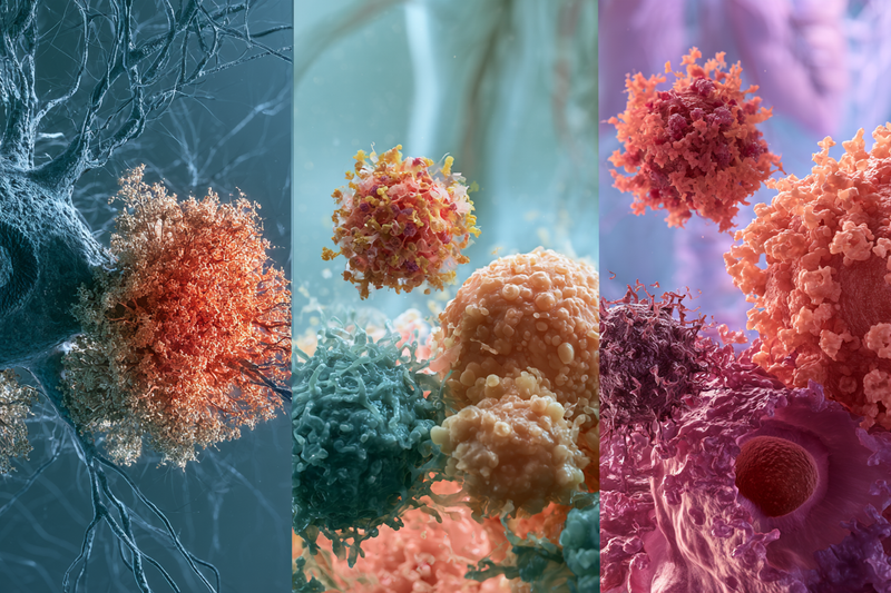
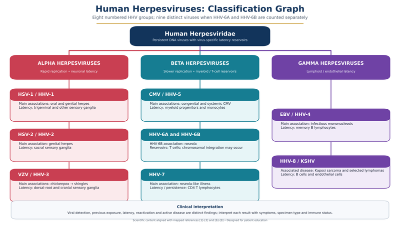
The types of herpes viruses in humans are organized into alpha, beta and gamma subfamilies according to their biological properties, genetic relationships and preferred sites of latency. These categories help explain which cells a virus affects and how it can reactivate. They describe viral biology rather than stages of illness or a ranking of severity. [1,2]
Alpha Herpesviruses
HSV-1, HSV-2 and VZV belong to the alpha subfamily, formally called Alphaherpesvirinae. They multiply relatively rapidly in susceptible cells and establish latency primarily in sensory ganglia—clusters of nerve-cell bodies outside the brain and spinal cord. These nerves connect with particular areas of skin and mucosal tissue, such as the lips or genital region. [1,2]
During reactivation, virus can travel along nerve pathways back to the surface tissues. This relationship helps explain why HSV outbreaks often return within the same general area and why shingles commonly follows the distribution of an affected sensory nerve. A recurrent eruption can therefore arise from an existing infection, without a new exposure each time. [1,2]
Beta Herpesviruses
CMV, HHV-6A, HHV-6B and HHV-7 belong to the beta subfamily, or Betaherpesvirinae. These viruses generally have a longer replication cycle and slower spread in laboratory cell cultures than alpha herpesviruses. That laboratory characteristic describes how they reproduce; the patient’s symptoms, immune function and affected organs determine the urgency of clinical care. [1,3,8]
Their cellular reservoirs differ from the predominantly neuronal reservoirs of HSV and VZV. CMV establishes latency chiefly in certain blood-forming precursor cells and monocytes, a type of white blood cell. HHV-6 and HHV-7 also have important relationships with immune cells, although their reservoirs are not identical. These differences help explain why evaluating suspected reactivation may involve blood or organ-specific investigations rather than examining a skin eruption alone. [2]
Gamma Herpesviruses
EBV and HHV-8 belong to the gamma subfamily, known as Gammaherpesvirinae. Both can establish latency in B lymphocytes, immune cells involved in antibody responses. EBV primarily persists in memory B cells in otherwise healthy carriers. HHV-8 can also remain latent in endothelial cells—the cells lining blood vessels—in Kaposi sarcoma. [2]
A distinctive feature of these viruses is their ability to influence the growth and survival of infected cells. This provides a biological explanation for their association with particular cancers and lymphoproliferative disorders, conditions involving abnormal growth of lymphocytes. Most infected people remain free of these complications. Clinical assessment therefore considers the specific virus, immune status and any abnormal tissue findings, rather than interpreting the gamma classification itself as a cancer diagnosis. [2,9]
| Feature | Alpha herpesviruses | Beta herpesviruses | Gamma herpesviruses |
|---|---|---|---|
| Scientific subfamily | Alphaherpesvirinae. [1] | Betaherpesvirinae. [1] | Gammaherpesvirinae. [1] |
| Human viruses included | HSV-1, HSV-2 and varicella-zoster virus (VZV). [1] | Cytomegalovirus (CMV), HHV-6A, HHV-6B and HHV-7. [1,3] | Epstein–Barr virus (EBV) and HHV-8, also called Kaposi sarcoma-associated herpesvirus (KSHV). [1,2] |
| Characteristic biological behavior | Relatively rapid replication in susceptible cells, followed by the ability to establish latency in nerve cells. [1] | Generally slower replication in laboratory cell cultures, with virus-specific relationships to blood-forming and immune cells. [1,2] | Establish latency in lymphocytes and can influence infected-cell growth and survival. [2] |
| Principal latency reservoirs | Sensory neurons within nerve ganglia, including trigeminal and sacral ganglia for HSV. [1,2] | CMV primarily persists in myeloid precursor cells and monocytes. HHV-6 has several blood and immune-cell reservoirs; HHV-7 establishes latency in CD4 T cells. [2] | EBV primarily persists in memory B cells. HHV-8 persists in B cells and is also latent in endothelial cells within Kaposi sarcoma lesions. [2] |
| Examples of associated illness | HSV-1 and HSV-2 cause oral or genital herpes and can affect other sites. VZV causes chickenpox and shingles. [1] | CMV can cause a mononucleosis-like illness, congenital infection or organ disease. HHV-6B causes roseola and can cause encephalitis following stem-cell transplantation. [3,8] | EBV causes infectious mononucleosis and is associated with selected cancers. HHV-8 causes Kaposi sarcoma and certain lymphoproliferative disorders. [9] |
| Why the distinction matters clinically | Nerve-associated latency helps explain recurring HSV lesions in a similar area and the nerve-distribution pattern of shingles. [1,2] | Pregnancy, congenital infection and reduced immune function can substantially change the significance of infection and the investigations required. [3,8] | The specific virus, immune status and tissue findings guide assessment. Most people infected with EBV or HHV-8 do not develop the associated cancers. [2,9] |
Alpha Herpesviruses: HSV-1, HSV-2 and VZV
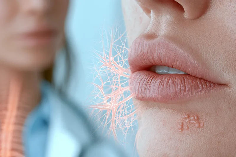
HSV-1: Oral, Genital and Eye Infections
A first symptomatic oral HSV-1 infection can be more extensive than a familiar cold sore. It may involve inflamed gums, multiple ulcers inside the mouth, fever and a sore throat. Recurrent oral episodes are generally more localized, often appearing along the lip border. When mouth pain interferes with drinking, prompt assessment helps address pain relief and hydration, particularly in young children. [1,4]
For patients with confirmed genital HSV-1, viral shedding from the genital area decreases substantially during the first year after infection. This helps guide follow-up: daily suppressive antiviral treatment is generally considered for frequent recurrences through shared decision-making, taking account of symptoms and their effect on daily life. The initial genital episode still requires appropriate treatment, even when the expected recurrence pattern is relatively infrequent. [5]
The affected tissue determines the urgency of care. HSV-1 infection involving the cornea requires ophthalmic assessment because deeper inflammation can impair vision. A painful red eye, light sensitivity or a change in vision warrants same-day evaluation rather than treatment as an ordinary cold sore. [1]
HSV-2: Genital Symptoms and Recurrent Episodes
A first symptomatic genital episode can affect the vulva, vagina, cervix, penis or surrounding skin. Painful urination, fever and tender groin lymph nodes may accompany the lesions. Some affected surfaces are internal, making a clinical examination valuable when genital discomfort or urinary symptoms persist despite few visible external sores. [1,10]
Recognizing an individual’s early warning symptoms can make recurrent-episode treatment more effective. For example, a patient who consistently develops localized tingling or burning before genital lesions appear can agree on a treatment plan in advance. Episodic antiviral treatment works best when started during this warning phase or within the first day of lesion onset. [5]
For frequent or distressing recurrences, daily suppressive therapy can reduce outbreaks and improve quality of life. Partner protection combines an appropriate treatment strategy with consistent condom use and avoiding sexual contact during warning symptoms or active lesions. Condoms reduce exposure, while uncovered skin can still transmit infection. [5]
During pregnancy, a history of genital herpes or any new genital symptoms should be discussed promptly with the maternity team. Newly acquired infection near delivery carries the greatest neonatal transmission risk. Early identification allows pregnancy-specific treatment and delivery planning to protect the baby; this consideration applies to genital infection caused by either HSV-1 or HSV-2. [4,5]
VZV: Chickenpox, Shingles and Nerve Pain
Shingles can begin with localized pain, itching or tingling before its rash becomes visible. For example, an older adult may first notice burning along one side of the chest, followed by blisters within that area. The rash generally heals within two to four weeks, although nerve pain can persist afterward as postherpetic neuralgia. [6]
Exposure to someone with active shingles can transmit VZV to a susceptible person, producing chickenpox rather than directly causing shingles. Covering the lesions and avoiding contact with susceptible people until every lesion has dried and crusted helps reduce transmission. Particular care is important around people with weakened immunity. [6]
Suspected shingles benefits from prompt assessment because antiviral treatment is most effective early and can reduce new lesions and acute pain. Facial involvement, especially around the eye, requires urgent attention to protect vision. Atypical eruptions can be investigated with lesion PCR to confirm VZV. [6]
Prevention also differs from HSV care. In the United States, a two-dose recombinant shingles vaccine is recommended for adults aged 50 years and older and for adults aged 19 years and older who are immunocompromised because of disease or treatment. Previous shingles does not remove the possibility of another episode. [6]
Beta Herpesviruses: CMV, HHV-6 and HHV-7
CMV: Pregnancy, Childhood Exposure and Organ Disease
Young children can shed CMV in saliva and urine while appearing well. During pregnancy, practical precautions include washing hands after diaper changes and avoiding shared cups, utensils or food that has contacted a child’s saliva. These measures help reduce exposure. Pregnancy-related assessment remains relevant after previous CMV infection because congenital infection can follow a first maternal infection, reinfection with another strain or reactivation. [8]
In otherwise healthy adults, symptomatic CMV may cause prolonged fever and liver inflammation rather than a recognizable blistering eruption. Care generally focuses on recovery and assessment of persistent symptoms. Immune status changes the treatment approach: patients with substantial immune suppression may require antiviral treatment for sight-threatening or other serious CMV disease, with investigations directed toward the affected organs. [8]
For infants diagnosed with congenital CMV, follow-up extends beyond the initial examination. Hearing loss may develop after a normal newborn hearing screen or progress over time. Regular hearing and vision assessments help identify changes early, allowing timely support for communication and development. Most infants with congenital CMV grow up healthy. [8]
HHV-6: Childhood Illness and Specialized Testing
Primary HHV-6B infection commonly occurs during infancy, particularly during the second half of the first year in studied Western populations. Its clinical spectrum includes fever-associated illness, roseola and, in some children, seizures. HHV-6A has a different and less clearly defined clinical profile. Recording the identified virus accurately helps distinguish established disease associations from findings that require further investigation. [3]
After stem-cell or organ transplantation, HHV-6B accounts for most documented HHV-6 reactivation in studied populations. Neurological involvement is particularly important because encephalitis—inflammation of the brain—requires prompt specialist assessment. New neurological symptoms following transplantation should therefore be evaluated within the transplant team’s diagnostic pathway rather than interpreted as an ordinary recurrence of a childhood infection. [3]
Inherited chromosomally integrated HHV-6 is an important laboratory distinction. In some people, HHV-6 DNA is incorporated into their chromosomes and inherited from a parent. A molecular test can consequently detect viral genetic material that is part of the person’s inherited DNA. Interpreting a positive result requires consideration of this possibility alongside active infection, particularly before changing treatment on the basis of viral DNA detection alone. [2]
Routine HHV-6 antibody testing also has limits when identifying the specific virus: serological methods generally do not reliably distinguish HHV-6A from HHV-6B. When that distinction matters clinically, an appropriately selected molecular assay can provide more specific information. [3]
HHV-7: Interpreting Detection in Context
HHV-7 can persist in immune cells and infect salivary-gland tissue, with viral shedding occurring in people who have no symptoms. The specimen and the clinical question therefore matter. Detection in saliva has a different significance from evidence being investigated in relation to suspected organ disease. A positive salivary result should be interpreted as evidence of viral presence, with further assessment establishing whether there is an associated illness. [2]
For a patient with substantial immune suppression, the assessment focuses on new symptoms, the affected organ and laboratory findings relevant to that presentation. This approach helps distinguish asymptomatic persistence or shedding from clinically significant reactivation, so that treatment decisions address the illness being demonstrated rather than the viral name alone. [2]
Gamma Herpesviruses: EBV and HHV-8

EBV: Recovery, Transmission and Clinical Assessment
For uncomplicated EBV-associated mononucleosis, care focuses on maintaining hydration, allowing adequate rest and using appropriate relief for throat pain and fever. Recovery should be assessed through changes in symptoms and daily functioning. A patient whose fever and throat discomfort improve but whose energy remains reduced may need continued recovery time and reassessment when symptoms persist or worsen. [7]
EBV transmission can begin before symptoms appear and continue for weeks during a first infection. Sharing a drinking bottle, toothbrush or eating utensil can transfer saliva even when the other person appears well. Keeping these items separate and avoiding kissing during symptomatic illness are practical precautions. Because transmission can precede recognizable illness, the onset of symptoms does not reliably identify when or from whom infection was acquired. [7]
EBV has established associations with particular subsets of Hodgkin lymphoma, Burkitt lymphoma, nasopharyngeal carcinoma and gastric carcinoma. These associations involve specific disease processes in infected cells; they are different from the usual course of mononucleosis. A history of EBV infection or an antibody-positive blood test should therefore be interpreted within the patient’s clinical assessment, rather than treated as a cancer diagnosis. [2,7,9]
After transplantation, EBV can also contribute to post-transplant lymphoproliferative disease, in which infected lymphocytes multiply abnormally. This is a distinct clinical situation from uncomplicated mononucleosis and requires transplant-specialist evaluation. The patient’s immune suppression and evidence of abnormal cell growth determine the investigation and treatment approach. [2]
HHV-8: Transmission and Disease-Specific Care
HHV-8 transmission patterns vary between populations. Saliva is an important route, while sexual transmission contributes to its spread in settings such as the United States and Northern Europe. Infection can also be transmitted through organ or bone marrow transplantation. The exposure history is therefore considered alongside geographic background and immune status, rather than assuming a single route of acquisition. [9]
The disorders associated with HHV-8 involve different tissues. In Kaposi sarcoma, the virus is present in tumor cells of endothelial origin, related to the cells lining blood vessels. In primary effusion lymphoma and HHV-8-associated multicentric Castleman disease, infected B cells are involved. Identifying the associated disorder is essential: these conditions require different specialist assessments and treatment plans, rather than a standard treatment for oral or genital herpes. [2,9]
For someone living with HIV, effective antiretroviral therapy is an important protective measure because it lowers the risk of HHV-8-related complications. For other patients receiving immune-suppressing treatment, the prescribing specialist should be involved when HHV-8-associated disease is suspected. Coordinating care around the underlying immune condition helps ensure that treatment addresses both the associated disease and the circumstances allowing it to develop. [9]
What All Human Herpesviruses Have in Common
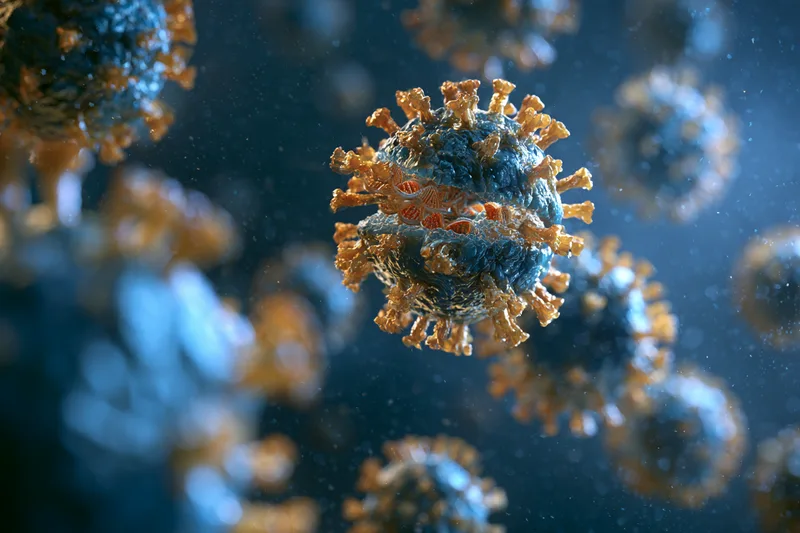
All human herpesviruses contain double-stranded DNA enclosed within a protein shell and an outer lipid envelope. Their genetic material directs replication inside infected cells. This shared biology helps explain why laboratory investigations often look for viral DNA, while the specific virus and suspected site of disease determine which specimen should be tested. [1,5]
During latency, herpesviruses regulate their gene activity and limit the production of proteins that would otherwise draw the immune system’s attention. Their persistence therefore involves active biological mechanisms rather than simply remaining unchanged inside a cell. This helps explain how an infection can remain clinically quiet for long periods while its genetic material is retained in particular cellular reservoirs. [2]
Immune control involves both antibodies and cellular defenses. T cells help recognize and control infected cells, while herpesviruses have mechanisms that reduce their visibility to these defenses. Reactivation can occur despite functioning immunity, so an occasional recurrent episode should be assessed according to its frequency, severity and clinical context rather than automatically interpreted as an immune deficiency. [2,5]
Visible symptoms and viral activity also follow different timelines. Several herpesviruses can reactivate and be shed without causing an obvious illness, although the frequency and routes differ substantially between viruses. For patients, this means that prevention should follow the identified infection and relevant exposure route, rather than relying entirely on the presence of a rash or how well someone feels. [2,5]
Changes in immune function can change the significance of an established infection. Cancer treatment, transplantation and certain immune-suppressing medicines can increase the risk of clinically important reactivation. Sharing a history of previous herpesvirus-related illness with the treating team helps place new symptoms in context. The assessment should consider the current treatment and affected organ, rather than assuming that a familiar viral diagnosis will always behave as it did previously. [6,8]
Herpes Simplex Versus Other Human Herpesviruses
HSV Versus Shingles
Similar-looking blisters can require different laboratory confirmation. When the cause of an eruption is uncertain, testing should target the suspected infection: an HSV lesion test identifies herpes simplex, while a VZV test investigates chickenpox or shingles. Requesting the appropriate viral targets helps establish which infection is responsible before planning treatment for repeated episodes. [5,6]
Some medicines overlap: acyclovir, valacyclovir and famciclovir are used for both genital HSV and shingles. However, the prescribed regimen depends on the infection, its severity and the patient’s circumstances. A familiar medicine name therefore does not make a previous cold-sore or genital-herpes prescription interchangeable with a shingles prescription. [5,6]
HSV Versus EBV and CMV
A patient with established HSV who develops prolonged fever, substantial fatigue or liver inflammation benefits from assessment of the new illness on its own merits. EBV and CMV can produce systemic illness with a different clinical course from a localized HSV recurrence. The examination and appropriately selected investigations help determine whether these viruses, or another condition, explain the symptoms. [7,8]
Treatment decisions also differ after the diagnosis is established. A first clinical episode of genital herpes warrants antiviral treatment. Uncomplicated EBV mononucleosis is generally approached through hydration, rest and symptom relief, while CMV infection in an otherwise healthy person usually requires no virus-specific treatment. These differences reflect the illness and the patient’s risk factors, rather than a single treatment rule for the herpesvirus family. [5,7,8]
HSV Versus HHV-6 and HHV-7
Molecular results have different meanings depending on the virus and specimen. For an active genital lesion, an HSV swab directly investigates the suspected cause at the affected site. Blood HSV PCR is reserved for particular clinical concerns, such as disseminated infection, rather than routine diagnosis of genital herpes. [5]
HHV-6 and HHV-7 can persist in blood or immune cells, and HHV-6 DNA can also be inherited within human chromosomes. Consequently, their detection requires interpretation alongside the specimen, symptoms and evidence of disease. A positive molecular result should answer a specific clinical question, rather than being interpreted identically across HSV, HHV-6 and HHV-7. [2,5]
HSV Versus HHV-8-Associated Disease
Cancer associations within the herpesvirus family are virus-specific. The abnormal cell growth involved in HHV-8-associated Kaposi sarcoma or certain lymphoproliferative disorders represents a different disease process from an ordinary HSV blistering eruption. Likewise, EBV-associated cancers involve particular infected tissues and cellular changes. These associations should be applied to the relevant virus and diagnosed condition, rather than to everyone with a herpes simplex diagnosis. [2,9]
Are All Herpes Viruses Sexually Transmitted?

No. Sexual contact is one route of transmission, not a defining feature of the herpesvirus family. Several herpesviruses are commonly acquired through nonsexual contact during childhood. The specific virus, infected body site and type of exposure determine how transmission occurs. [4,7,8]
HSV-1 commonly spreads through oral contact, including nonsexual kissing, and can reach the genital area through oral sex. HSV-2 is predominantly transmitted through sexual contact with infected genital or anal skin and mucosal surfaces. Both can spread when the infected area looks normal, making symptom-free transmission relevant when discussing protection with a partner. [4,5]
Saliva exposure does not necessarily involve sexual activity. EBV commonly spreads through kissing or sharing saliva-contaminated drinks and utensils. HHV-6 and HHV-7 can also be shed in saliva, including during periods without symptoms. Their detection should therefore be interpreted according to the clinical circumstances rather than assumed to indicate sexual exposure. [2,7]
CMV has both sexual and nonsexual transmission routes. Exposure can occur through saliva, urine, semen, breast milk, transplanted organs or blood transfusions. It can also pass from a pregnant patient to the developing baby. For someone regularly caring for young children, contact with saliva and urine is a relevant exposure to discuss during pregnancy planning. [8]
VZV spreads through contact with infectious blister fluid or inhalation of virus-containing particles. Exposure to active shingles can cause chickenpox in a susceptible person; neither the exposure nor a later shingles episode requires sexual contact. HHV-8 has a different pattern: saliva is an important transmission route, while transmission associated with sexual contact varies between populations. [6,9]
A newly identified HSV infection also needs careful interpretation within a relationship. Many infections remain unrecognized, and antibodies can persist long after acquisition. Consequently, a single positive antibody result does not establish when infection began or identify the person who transmitted it. A new diagnosis alone should not be treated as evidence of recent exposure or infidelity. [5,10]
Can One Person Carry More Than One Herpesvirus?
Yes. A person can carry several herpesviruses acquired at different times. Someone with recurrent HSV-1 cold sores may also carry EBV or CMV from earlier infections. Each virus has its own clinical history and pattern of activity, so one may cause symptoms while others remain clinically quiet. [2,7,8]
It is also possible to have both HSV-1 and HSV-2. Previous HSV-1 infection leaves a person susceptible to acquiring HSV-2. When blood tests suggest both infections, each result should be assessed separately. A low-positive HSV-2 antibody result may require confirmation using a second testing method before a dual diagnosis is established. [5]
The important distinction is between carrying several viruses and having several active illnesses simultaneously. For example, a patient with a confirmed genital HSV outbreak may also have EBV antibodies reflecting an earlier infection. Those antibodies alone do not establish that EBV is contributing to the current outbreak. Interpreting the findings together with the symptom timeline helps identify which infection requires attention now. [5,7,10]
Multiple infections become especially relevant when immune function changes, such as during transplantation or immune-suppressing treatment. Several previously acquired herpesviruses may then warrant consideration when investigating new symptoms. The treating team selects investigations according to the affected organ, immune status and suspected virus, distinguishing clinically significant reactivation from evidence of an infection that remains quiet. [2,8]
A personalized assessment focuses on the significance of each finding, rather than the number of positive results. Recurrent genital lesions, prolonged fever and new symptoms during immune suppression raise different diagnostic questions. Additional testing should answer those questions and guide a specific treatment decision. A broad panel for every herpesvirus is therefore not an automatic requirement for someone with an otherwise typical HSV presentation. [5,8,10]
Which Herpesviruses Commonly Coexist?

HSV With EBV or CMV
EBV and CMV are relevant additional findings in adults with HSV because previous exposure is widespread. Approximately nine in ten adults have EBV antibodies, while more than half of adults in the United States have acquired CMV by age 40. These high exposure rates help explain why someone with herpes simplex may also have evidence of EBV or CMV. They describe population exposure, rather than the frequency of simultaneous active infections. [7,8]
When these viruses appear together on a patient’s reports, each result should be assigned its own clinical meaning. A positive HSV lesion swab identifies the cause of that lesion, whereas an EBV antibody result may document a much earlier infection. This distinction helps keep treatment directed toward the illness currently affecting the patient. [5,7]
EBV With CMV
EBV and CMV can coexist following infections acquired at different times. They also overlap clinically because either can produce a mononucleosis-like illness. During an investigation for prolonged fever, fatigue or liver inflammation, the diagnostic question is whether EBV, CMV or another condition explains the current symptoms—not simply whether antibodies to both viruses are present. [2,7,8]
For example, a person with previous EBV infection may subsequently develop symptomatic CMV infection. The earlier EBV finding and the new CMV illness represent different events. Establishing the timing and significance of each result is particularly valuable during pregnancy or immune suppression, when a new CMV infection can change the care plan. [7,8]
CMV With HHV-6 or HHV-7
CMV, HHV-6 and HHV-7 can persist in the same person, with different cellular reservoirs and patterns of activity. After transplantation, CMV and HHV-6B are particularly relevant diagnostic considerations because both can reactivate in that setting. Their shared association with reduced immune control warrants virus-specific assessment rather than an assumption that one caused the other. [2,3,8]
HHV-6 and HHV-7 can also be shed from salivary-gland tissue. When either is detected alongside another herpesvirus, the specimen matters as much as the virus name: salivary detection, circulating viral DNA and evidence of organ disease answer different clinical questions. Treatment decisions should follow evidence of clinically significant infection for each virus. [2,8]
How the Different Herpesviruses Are Tested

HSV-1 and HSV-2
A fresh blister or ulcer is the best opportunity for direct HSV testing. A clinician collects a swab for PCR, a type of nucleic acid amplification test that detects viral genetic material and distinguishes HSV-1 from HSV-2. Viral culture is an alternative, although its sensitivity decreases as lesions heal. Arrange sampling promptly rather than waiting until the eruption has almost disappeared. [5,10]
Type-specific IgG blood testing is useful in selected circumstances, including recurrent suspicious symptoms with negative lesion testing or a partner with genital herpes. After suspected recent HSV-2 acquisition, repeat antibody testing at approximately 12 weeks after exposure allows time for antibodies to develop. HSV IgM is unsuitable for this purpose because it cannot reliably distinguish the viral types or separate a new infection from a recurrence. [5]
Low-positive HSV-2 enzyme immunoassay results—particularly index values between 1.1 and 3.0—require careful confirmation with a different method, such as Biokit or Western blot. These index values are assay-specific antibody measurements, not a measurement of viral load. A negative swab from an older lesion may also require reassessment because shedding is intermittent. [5]
Varicella-Zoster Virus
A characteristic shingles rash can often be diagnosed clinically. VZV PCR provides confirmation when the presentation is unusual or the diagnosis is uncertain. The laboratory request should identify VZV specifically rather than simply requesting an HSV test. Confirmation is particularly useful when a mild or atypical eruption does not clearly follow the expected shingles pattern. [6]
Epstein–Barr Virus
EBV testing commonly uses blood samples to detect virus-specific antibodies. Interpretation should connect the laboratory findings with the onset and course of the illness, since antibodies are widespread among adults and may reflect previous exposure. Bring the complete report, including the antibodies measured and collection date, rather than relying on a summary stating only “EBV positive.” [7]
The clinical question also matters: investigating a new mononucleosis-like illness differs from assessing persistent fatigue after recovery. Recording when fever, throat symptoms and fatigue began helps the clinician interpret the blood results alongside the current examination and decide whether further investigation is appropriate. [7]
Cytomegalovirus
CMV investigations depend strongly on age and clinical circumstances. During pregnancy, identifying the timing and significance of infection requires specialist interpretation; maternal laboratory results alone cannot predict whether a developing baby will be affected. Testing should therefore address a defined pregnancy-related question rather than treating every positive result identically. [8]
For suspected congenital CMV, saliva PCR followed by urine confirmation is the preferred diagnostic approach. Samples should be obtained within the first three weeks after birth to distinguish congenital infection from infection acquired afterward. Newborn IgG or IgM antibody testing does not establish congenital CMV. [8]
HHV-6 and HHV-7
When distinguishing HHV-6A from HHV-6B is clinically important, an appropriately designed molecular assay is needed because routine antibody tests do not reliably separate them. The requesting clinician should check whether the laboratory reports the two viruses individually. [3]
HHV-6 DNA results also require consideration of inherited chromosomal integration. For HHV-7, persistence in immune cells and shedding in saliva affect interpretation. The specimen, symptoms and immune status must be considered together before attributing an illness to either virus. [2]
HHV-8
Evaluation focuses on the suspected associated disorder rather than viral exposure alone. When Kaposi sarcoma or an HHV-8-associated lymphoproliferative disorder is suspected, specialist assessment should determine whether abnormal tissue or fluid requires examination. This distinction matters because most people carrying HHV-8 do not develop these diseases. [2,9]
How Treatment Differs Across the Herpesvirus Family

HSV-1 and HSV-2
A first symptomatic genital herpes episode is generally treated with oral acyclovir, valacyclovir or famciclovir for seven to ten days, with treatment extended when healing remains incomplete. Subsequent episodes may be managed with shorter courses or daily suppression, depending on recurrence frequency, symptom burden and the patient’s preferences. [5]
For patients with frequent genital HSV-2 recurrences, daily suppressive treatment reduces outbreak frequency by approximately 70–80%. Its continued use is reviewed periodically as the recurrence pattern changes. Severe infection affecting the brain or other organs requires hospital-based treatment, often with intravenous acyclovir, rather than an ordinary outpatient outbreak regimen. [5]
VZV: Early Treatment and Pain Relief
Shingles treatment prioritizes prompt antiviral therapy, particularly within the first 72 hours of symptom onset. Acyclovir, valacyclovir or famciclovir can accelerate lesion healing, reduce new blister formation and lessen acute pain. Treatment planning also considers eye involvement, immune suppression and persistent nerve pain, rather than assessing recovery solely by whether the rash has disappeared. [6]
Vaccination serves a separate preventive purpose. An eligible patient recovering from shingles can discuss vaccination with their clinician to reduce future risk; vaccination is distinct from the antiviral treatment used for an active episode. [6]
CMV: Treatment According to Clinical Risk
Uncomplicated CMV infection in an otherwise healthy adult generally requires observation and symptom-directed care. The treatment threshold changes substantially when CMV threatens vision or causes serious illness in someone with reduced immune function. In those circumstances, specialist-directed antiviral treatment addresses the affected organ and the severity of disease. [8]
For selected newborns with symptomatic congenital CMV, ganciclovir or valganciclovir may improve hearing and developmental outcomes. Treatment requires specialist supervision because these medicines can cause significant adverse effects. Regular hearing and vision assessments remain important throughout follow-up, including when the infant initially appears to be developing normally. [8]
EBV: Recovery Support or Disease-Specific Treatment
Uncomplicated infectious mononucleosis is managed primarily through hydration, rest and appropriate relief of pain and fever. The treatment goal is recovery from the acute illness and restoration of daily functioning, with reassessment when symptoms persist or new complications develop. An HSV prescription should therefore not be assumed to provide the appropriate treatment for an EBV-related illness. [7]
An EBV-associated lymphoma or post-transplant lymphoproliferative disorder requires a different approach directed at the abnormal infected cells. These conditions belong under specialist hematology, oncology or transplant care, rather than the recovery plan used for uncomplicated mononucleosis. [2,9]
HHV-6 and HHV-7: Treating Demonstrated Disease
For HHV-6 and HHV-7, treatment decisions depend on the clinical syndrome and immune status, rather than viral detection alone. HHV-6B-associated encephalitis after transplantation is a particularly important example requiring urgent specialist management. This is a different clinical situation from a childhood febrile illness or asymptomatic viral persistence. [2,3]
Before committing to treatment, the specialist interprets the molecular findings in context. Inherited chromosomally integrated HHV-6 and symptom-free HHV-7 shedding can produce positive results with meanings different from active organ disease. Establishing that distinction helps direct treatment toward the condition actually responsible for the patient’s symptoms. [2]
HHV-8: Managing the Associated Disorder
HHV-8 care focuses on the diagnosed condition, such as Kaposi sarcoma, primary effusion lymphoma or HHV-8-associated multicentric Castleman disease. Each requires its own specialist treatment plan. For patients also living with HIV, effective antiretroviral therapy is an important component of care because it lowers the risk of HHV-8-related complications. The associated disease and underlying immune condition determine the treatment strategy, rather than the HHV-8 result in isolation. [2,9]
Can HSV-1 and HSV-2 Be Cured Permanently?

Within the personalised Ayurvedic model described here, the answer is yes: HSV-1 and HSV-2 are treated with the objective of permanent cure rather than temporary suppression of outbreaks alone. The intended clinical outcomes include complete healing of active lesions, disappearance of tingling or burning before outbreaks, cessation of recurrences, restoration of normal intimate life and sustained stability after completing treatment.
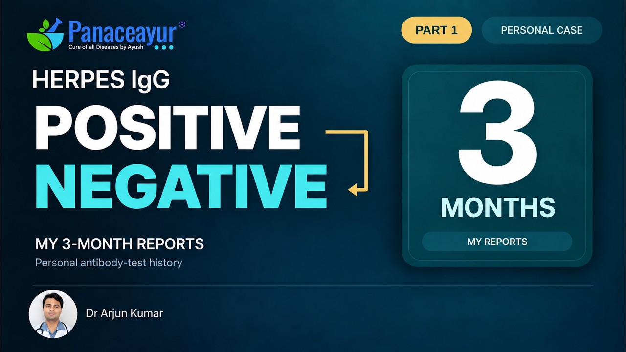
The protocol also sets a laboratory target in which type-specific HSV-1 and HSV-2 IgG values move below the reporting laboratory’s reference range and are reported as negative. This should be documented as a protocol-defined treatment endpoint after objective retesting rather than promised solely from the disappearance of symptoms. Type-specific IgG measures the immune response to HSV, not the latent virus within sensory nerve cells; international guidance states that these antibodies generally develop within the first weeks after infection and commonly persist indefinitely. Any conversion from a confirmed positive result to negative is therefore an important serological observation that requires repeat confirmation using a reliable laboratory method. [5,10] CDC
Personalised Ayurvedic Curative-Intent Treatment
Personalisation is essential because two patients with the same HSV type may have different infection sites, recurrence patterns, disease duration, prodromal symptoms, digestive capacity, metabolic health, associated infections and responses to previous medicines. Treatment planning considers Prakriti, the patient’s constitutional pattern; Vikriti, the present imbalance; Agni, digestive and metabolic function; and Ojas, physiological resilience and recovery capacity.
The prescription is further adjusted according to whether the patient has oral HSV-1, genital HSV-1, genital HSV-2, both HSV-1 and HSV-2, ocular involvement, frequent asymptomatic shedding concerns, recurrent urinary discomfort, menstrual triggers, chronic stress, sleep disruption or another clinically relevant condition. Liver and kidney function, gastrointestinal disorders, diabetes, pregnancy, immune suppression and concurrent medicines must also be reviewed before selecting ingredients, dose and duration.
A generic market Avaleha or fixed herbal combination cannot provide this level of individualisation. The medicine must be selected, prepared and monitored under the supervision of a qualified Ayurvedic physician. Patients should not copy another person’s prescription, independently combine herbs with bhasma preparations or stop prescribed antiviral medicines without coordinated clinical guidance.
Retesting and Confirmation of IgG Conversion
Baseline testing should preferably be completed before treatment begins at an accredited laboratory. The report should state whether the assay is type-specific, the test method, the HSV-1 and HSV-2 numerical index values and the laboratory’s positive, equivocal and negative reference intervals. Follow-up testing is most comparable when performed through the same laboratory using the same assay.
An HSV-2 result with a low-positive index value requires special attention because some commonly used assays produce false-positive findings within the lower positive range. Values between approximately 1.1 and 3.0 should be confirmed through a second method, such as Biokit or Western blot, before being accepted as the patient’s definitive baseline diagnosis. HSV IgM should not be used to establish cure or recurrence because it is not reliably type-specific. [5] CDC
When a previously confirmed HSV-1 or HSV-2 IgG report becomes negative, the result should be repeated on a fresh blood sample. Ideally, the repeat report should reproduce the negative result using the same assay, while an independent confirmatory method provides additional strength where available. The final outcome record should include the pre-treatment values, treatment duration, post-treatment values, duration without recurrence, antiviral use and any lesion PCR result obtained if a suspicious lesion subsequently appears.
The strongest evidence of recovery combines sustained freedom from outbreaks, disappearance of prodromal symptoms, normal daily and intimate functioning, reduced dependence on symptomatic medicines and carefully documented laboratory follow-up. Because conventional guidance currently uses HSV IgG primarily as evidence of present or previous exposure rather than a validated cure test, IgG conversion within this Ayurvedic protocol should be reported transparently as a clinic-defined serological endpoint supported by repeat testing and long-term clinical observation. [2,5,10] CDC
The complete treatment framework, including HSV latency, personalised formulation planning, expected treatment duration and long-term outcome assessment, is presented in Can Herpes Be Cured Permanently? Real Cure Approach.
Ayurvedic Clinical Perspective on Herpesvirus
Modern laboratory testing identifies the specific virus, while Ayurvedic assessment evaluates how that infection is expressing itself in the individual patient. Two people with the same HSV type may differ markedly in lesion location, recurrence frequency, prodromal nerve sensations, digestion, sleep, stress response, menstrual association, metabolic health and recovery time. The strongest clinical approach therefore combines virus-specific diagnosis with assessment of Prakriti, Vikriti, Agni, Ojas, affected tissues, disease stage and associated disorders. [1,2,5]
This distinction is especially important across the wider herpesvirus family. HSV-1, HSV-2 and VZV commonly involve skin, mucosal tissue and sensory nerves, whereas EBV, CMV, HHV-6, HHV-7 and HHV-8 may present through systemic, immune, neurological, congenital or disease-specific patterns. Ayurvedic treatment planning should follow the confirmed virus and the patient’s actual presentation rather than treating every herpesvirus as one identical disorder. [1,6,7,8,9]
Classical Ayurvedic Framework
Classical Ayurvedic texts predate modern virology and do not name HSV-1, HSV-2, VZV, EBV or CMV as laboratory-defined viruses. They instead classify illness through observable patterns such as burning, redness, pain, itching, blistering, ulceration, discharge, fever, fatigue, tissue involvement and the speed or direction of progression.
Charaka Saṃhitā, Cikitsāsthāna, Chapter 21, Visarpa Cikitsā, provides a useful framework for assessing rapidly spreading inflammatory skin presentations associated with burning, pain, discoloration and systemic disturbance. Visarpa should be applied as a classical pattern of assessment rather than treated as an automatic one-to-one synonym for herpes simplex or shingles.
Charaka Saṃhitā, Cikitsāsthāna, Chapter 1, Rasāyana Cikitsā, provides the broader foundation for restoring physiological resilience, supporting tissue recovery and strengthening long-term stability. In recurrent viral illness, Rasāyana planning becomes relevant after evaluating digestion, nutrition, sleep, bowel function, stress load, age, chronic disease and the patient’s capacity to metabolize the prescribed medicine.
Burning, marked redness, heat and rapid inflammation may indicate stronger Pitta and Rakta involvement. Sharp, radiating, electric or persistent nerve pain may indicate prominent Vata involvement. Oozing lesions, swelling, heaviness, sluggish healing or thick discharge may suggest Kapha participation. Mixed presentations are common, and the dominant pattern may change between the first episode, recurrent outbreaks and the recovery phase.
How the Viral Presentation Changes Ayurvedic Assessment
In recurrent oral HSV-1, the assessment considers the lips, oral mucosa, gums, throat, appetite, bowel pattern, acidity, mouth dryness, fever history and triggers such as sunlight, dental procedures, travel or sleep loss. A patient with a localized recurrent lip lesion requires a different plan from someone whose first oral episode includes widespread mouth ulcers, fever and difficulty eating or drinking. [1,5]
In genital HSV-1 or HSV-2, evaluation extends beyond the visible lesion. Burning during urination, pelvic or sacral discomfort, constipation, vaginal discharge, menstrual timing, sexual pain, sleep disruption and prodromal tingling can influence treatment planning. The anatomical site, HSV type, recurrence interval and response to previous antivirals should all be documented because genital HSV-1 and HSV-2 may follow different recurrence patterns. [4,5]
For VZV, the Ayurvedic assessment gives particular attention to unilateral burning pain, hypersensitivity of the skin, dermatomal distribution, age, diabetes, immune status and the persistence of pain after the rash heals. Eye, ear or facial involvement requires coordinated specialist care because these locations carry different clinical risks from an uncomplicated truncal eruption. [6]
EBV and CMV presentations require a systemic assessment rather than a skin-lesion model. Persistent fatigue, fever, swollen lymph nodes, throat symptoms, reduced appetite, liver or spleen involvement, sleep quality and exercise intolerance become more important than blister morphology. During pregnancy, infancy, transplantation or significant immune suppression, modern medical monitoring remains central because the implications of CMV or EBV differ substantially from an ordinary HSV recurrence. [7,8]
HHV-6 and HHV-7 findings require careful interpretation of the specimen, immune status and symptoms before they are incorporated into an Ayurvedic plan. HHV-8-associated disease requires diagnosis-specific specialist management, with Ayurvedic support individualized around strength, digestion, treatment tolerance and recovery needs rather than approached as routine oral or genital herpes. [2,3,9]
Personalised Formulation and Follow-Up
A personalised Ayurvedic prescription is selected after reviewing the confirmed viral diagnosis, infection site, recurrence pattern, symptom severity, disease duration, previous treatment, co-infections, liver and kidney function, blood glucose, gastrointestinal health, pregnancy status, immune-suppressing medicines and other concurrent therapies. The dosage form, ingredients, dose, timing and duration should reflect the patient’s present condition rather than a standard formula applied to every herpesvirus case.
Clinical monitoring should document outbreak dates, prodromal symptoms, lesion number and location, healing time, pain, urinary or bowel symptoms, sleep, energy, appetite, stress exposure, menstrual association and use of conventional antivirals. Laboratory reports should be reviewed with their collection dates, numerical values, testing method and reference ranges so that changes are interpreted consistently. [5,8,10]
Patients should not purchase a generic Avaleha or attempt to prepare an antiviral Ayurvedic formulation without qualified supervision. A physician-prepared medicine allows adjustment for Prakriti, Agni, age, body weight, disease stage, chronic disorders, co-infections, current medicines and treatment tolerance. Raw-material identity, purification, processing temperature, ingredient compatibility, dose accuracy and storage conditions also influence safety and clinical performance.
When a herbo-mineral preparation is considered, ingredient authenticity, classical processing, precise dosing and appropriate safety monitoring are essential. The treatment plan should remain transparent, with the patient informed about the ingredients, intended role of each component, expected duration and follow-up schedule. This personalised method supports a curative-intent approach while keeping clinical progress measurable and coordinated with virus-specific medical care.
When Herpesvirus Symptoms Require Urgent Medical Evaluation
Eye, Brain and Nervous-System Symptoms
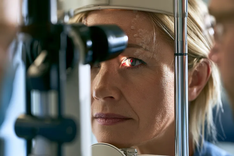
Eye pain, marked redness, light sensitivity, blurred vision or a blistering eruption near the eye requires same-day medical assessment. HSV can involve the cornea, while shingles affecting the forehead, eyelid or tip of the nose may threaten deeper eye structures. Early ophthalmic examination and virus-specific treatment help protect vision. [1,6]
A severe headache accompanied by fever, confusion, unusual behaviour, speech difficulty, weakness, neck stiffness, loss of consciousness or seizures requires emergency evaluation. HSV can cause encephalitis or meningitis, and HHV-6B can cause encephalitis in selected transplant recipients. Hospital assessment may include neurological examination, brain imaging, cerebrospinal-fluid testing and intravenous antiviral treatment when indicated. [1,3,5]
New facial weakness, severe ear pain, hearing changes, dizziness or blisters within or around the ear also warrant prompt assessment. These findings can occur when VZV affects facial or auditory nerves, and early treatment supports better neurological recovery. [6]
Pregnancy, Newborns and Young Infants
A new genital outbreak, genital pain, unexplained ulcers or suspected first HSV infection during pregnancy should be reported promptly to the maternity team. The timing of infection, HSV type, lesion status and stage of pregnancy influence antiviral treatment and delivery planning. This coordinated approach is especially important when infection is acquired close to delivery because neonatal protection becomes a central treatment objective. [4,5]
A newborn exposed to genital HSV during delivery, or an infant who develops poor feeding, unusual sleepiness, irritability, fever, low temperature, breathing difficulty, skin blisters or seizures, requires immediate pediatric evaluation. Neonatal HSV may initially resemble other common newborn illnesses, so early testing and intravenous treatment are important when infection is suspected. [1,5]
Possible congenital CMV also benefits from time-sensitive assessment. Infants with failed hearing screening, jaundice, petechiae, reduced head growth, enlarged liver or spleen, seizures or other compatible findings should be evaluated promptly. Saliva PCR with urine confirmation should be collected within the first three weeks after birth when congenital infection is being investigated. [8]
Widespread, Severe or Rapidly Progressive Illness
A rapidly spreading eruption, extensive blistering, severe pain, high fever, inability to drink, marked weakness or signs of dehydration requires urgent medical review. Extensive HSV can occur across damaged skin, including in people with eczema, while disseminated VZV may involve areas beyond a typical localized dermatome. Early recognition allows pain control, hydration support and appropriately intensive antiviral treatment. [1,5,6]
Genital herpes also requires prompt assessment when swelling or pain prevents urination, lesions are unusually extensive, symptoms continue to intensify, or a bacterial infection is suspected. Internal vaginal or cervical lesions may be present even when few external sores are visible, making examination valuable when urinary or pelvic symptoms are significant. [1,5]
Persistent high fever, profound fatigue, worsening throat swelling, breathing difficulty, severe abdominal pain or jaundice during a suspected EBV or CMV illness should be assessed without delay. In EBV-associated mononucleosis, sudden pain in the upper-left abdomen or left shoulder may indicate splenic involvement and requires emergency evaluation. [7,8]
Reduced Immunity and Cancer-Associated Presentations
Patients receiving chemotherapy, transplant medicines, high-dose corticosteroids or other immune-suppressing treatments should contact their medical team promptly when new fever, rash, visual symptoms, breathing difficulty, confusion or unexplained organ symptoms develop. HSV, VZV, CMV, EBV and HHV-6 can cause more extensive disease in this setting, and the relevant virus may need to be identified through blood, lesion, cerebrospinal-fluid or organ-specific testing. [1,3,6,8]
Persistent purple, red or brown skin lesions, unexplained lymph-node enlargement, abdominal swelling, night sweats or unexplained weight loss in a person with significant immune suppression requires specialist evaluation for HHV-8-associated or other lymphoproliferative disease. Tissue examination and disease-specific testing guide management more reliably than herpesvirus antibody results alone. [2,9]
Urgent medical care and personalised Ayurveda can be coordinated rather than treated as competing approaches. Emergency antiviral therapy, ophthalmic care, neurological investigation, maternity management or hospital treatment should begin according to clinical need, while Ayurvedic support is adjusted around hydration, digestion, strength, recovery, concurrent medicines and organ function. This integrated approach protects the patient during the acute stage and preserves continuity of the longer-term personalised treatment plan.
Frequently Asked Questions
How many types of herpes viruses infect humans?
Eight numbered human herpesvirus groups are commonly recognized: HSV-1, HSV-2, VZV, EBV, CMV, HHV-6, HHV-7 and HHV-8. When HHV-6A and HHV-6B are counted as separate species, these groups contain nine distinct viruses.
What are the eight human herpesviruses?
They are HSV-1 or HHV-1, HSV-2 or HHV-2, varicella-zoster virus or HHV-3, Epstein–Barr virus or HHV-4, cytomegalovirus or HHV-5, HHV-6, HHV-7 and HHV-8. Each virus has different transmission routes, latency sites and clinical effects.
Why do some sources say there are nine herpesviruses?
The difference arises because HHV-6A and HHV-6B are now classified as separate viral species. Patient information commonly retains the traditional eight-numbered-group system, while modern virological classification may count HHV-6A and HHV-6B individually.
What is the difference between HSV-1 and HSV-2?
HSV-1 commonly causes oral herpes but can also cause genital infection. HSV-2 predominantly causes genital herpes and usually produces more frequent genital recurrence and shedding than genital HSV-1. The infection site alone cannot reliably identify the viral type.
Can HSV-1 cause genital herpes?
Yes. HSV-1 can reach the genital area through oral-genital contact and is an important cause of first-episode genital herpes. Genital HSV-1 usually recurs less frequently than genital HSV-2, although the initial episode can still cause painful lesions and systemic symptoms.
Is chickenpox a type of herpes infection?
Yes. Chickenpox is the primary infection caused by varicella-zoster virus, also called HHV-3. After recovery, VZV remains latent in sensory nerve ganglia and may later reactivate as shingles.
Is shingles the same as genital herpes?
No. Shingles results from reactivation of varicella-zoster virus, while genital herpes is usually caused by HSV-1 or HSV-2. Shingles often produces a painful unilateral rash following a nerve distribution, whereas genital herpes affects genital or anal skin and mucosal tissues.
Is Epstein–Barr virus a type of herpesvirus?
Yes. Epstein–Barr virus is HHV-4 and belongs to the gamma herpesvirus subfamily. It commonly causes infectious mononucleosis and establishes latency in B lymphocytes, but it does not usually produce the recurrent oral or genital blisters associated with HSV.
Is cytomegalovirus sexually transmitted?
CMV can spread through sexual contact, but it also spreads through saliva, urine, breast milk, blood products, organ transplantation and pregnancy. Young children can shed CMV without appearing ill, making nonsexual household exposure clinically relevant, especially during pregnancy.
Can one person have more than one herpesvirus?
Yes. A person may carry HSV-1, HSV-2, EBV, CMV or other herpesviruses acquired at different times. Positive results should be interpreted individually because previous exposure, latent infection, asymptomatic shedding, reactivation and active disease have different clinical meanings.
Which herpesviruses are associated with cancer?
EBV is associated with selected lymphomas, nasopharyngeal carcinoma and some gastric cancers. HHV-8 is associated with Kaposi sarcoma, primary effusion lymphoma and certain forms of multicentric Castleman disease. Infection alone does not establish cancer; diagnosis depends on clinical findings and tissue-specific assessment.
Can HSV-1 and HSV-2 be cured permanently through personalised Ayurveda?
The personalised Ayurvedic model described in this article treats HSV-1 and HSV-2 with curative intent by individualizing medicine according to viral type, infection site, recurrence pattern, Prakriti, Vikriti, Agni, Ojas, disease duration, co-infections and associated disorders. Progress should be documented through sustained clinical recovery and carefully interpreted repeat laboratory testing.
References
[1] Whitley, R. J. (1996). Herpesviruses. In S. Baron (Ed.), Medical microbiology (4th ed., Chapter 68). University of Texas Medical Branch at Galveston. NCBI Bookshelf
Brief. Supports the overview of herpesvirus structure, the traditional eight numbered groups, the alpha, beta, and gamma subfamilies, and sensory-ganglion latency in HSV and VZV. Use [3] for the distinction between HHV-6A and HHV-6B.
[2] Cohen, J. I. (2020). Herpesvirus latency. The Journal of Clinical Investigation, 130(7), 3361–3369. Full text
Brief. Reviews how herpesviruses persist in specific cells, regulate gene expression during latency, and reactivate. Supports the sections distinguishing latent infection, reactivation, viral shedding, and symptomatic disease.
[3] Ablashi, D., Agut, H., Alvarez-Lafuente, R., Clark, D. A., Dewhurst, S., DiLuca, D., Flamand, L., Frenkel, N., Gallo, R., Gompels, U. A., Höllsberg, P., Jacobson, S., Luppi, M., Lusso, P., Malnati, M., Medveczky, P., Mori, Y., Pellett, P. E., Pritchett, J. C., … Yoshikawa, T. (2014). Classification of HHV-6A and HHV-6B as distinct viruses. Archives of Virology, 159(5), 863–870. PubMed
Brief. Explains why HHV-6A and HHV-6B are classified as separate species and why the eight traditionally numbered groups comprise nine distinct viruses when both are counted.
[4] World Health Organization. (2025, May 30). Herpes simplex virus. WHO fact sheet
Brief. Supports the HSV-1 and HSV-2 overview, including oral and genital symptoms, transmission without visible sores, recurrence patterns, global prevalence, and neonatal herpes.
[5] Workowski, K. A., Bachmann, L. H., Chan, P. A., Johnston, C. M., Muzny, C. A., Park, I., Reno, H., Zenilman, J. M., & Bolan, G. A. (2021). Sexually transmitted infections treatment guidelines, 2021. MMWR Recommendations and Reports, 70(4), 1–187. CDC full guideline
Brief. Principal reference for genital HSV lesion PCR or NAAT, culture limitations, type-specific serology, confirmation of low-positive HSV-2 results, antiviral treatment, pregnancy management, and partner counselling.
[6] Centers for Disease Control and Prevention. (2024, June 27). Clinical overview of shingles (herpes zoster). CDC clinical overview
Brief. Supports the distinction between chickenpox and shingles, VZV reactivation, dermatomal rash, postherpetic neuralgia, lesion PCR, antiviral treatment, and shingles vaccination.
[7] Centers for Disease Control and Prevention. (2024, May 9). About Epstein-Barr virus (EBV). CDC overview
Brief. Supports the EBV section on salivary transmission, infectious mononucleosis, common symptoms, latency and reactivation, antibody testing, and supportive care.
[8] Centers for Disease Control and Prevention. (2024, November 6). Clinical overview of CMV and congenital CMV. CDC clinical overview
Brief. Supports the sections on CMV transmission, infection during pregnancy, congenital CMV, newborn saliva PCR with urine confirmation, and hearing and developmental follow-up.
[9] National Cancer Institute. (2019, March 4). Infectious agents. National Cancer Institute
Brief. Supports the discussion of EBV associations with selected lymphomas and nasopharyngeal cancer, and HHV-8 associations with Kaposi sarcoma, primary effusion lymphoma, and multicentric Castleman disease.
[10] National Library of Medicine. (2025, August 19). Herpes (HSV) test. MedlinePlus. MedlinePlus test guide
Brief. Provides a patient-facing explanation of lesion swabs, PCR, viral culture, and blood antibody testing. Use [5] for decisions about repeat testing and confirmation of low-positive results.




One Response
Great write-up, I’m regular visitor of one’s web site, maintain up the excellent operate, and It’s going to be a regular visitor for a lengthy time.