



- Why HCC Is Both a Cancer and a Chronic Liver Disease
- Causes and Risk Factors for Hepatocellular Carcinoma
- Symptoms and Warning Signs of Hepatocellular Carcinoma
- When You Need Urgent Medical Assessment
- How Hepatocellular Carcinoma Is Diagnosed
- What Your Reports Reveal About the Cancer and Remaining Liver Function
- Understanding HCC Staging Without Creating Unnecessary Fear
- Factors That Determine Your Treatment Plan
- Modern Treatment Options for Hepatocellular Carcinoma
- When Surgery Is Not Recommended for HCC
- What “Inoperable HCC” Actually Means
- Master Clinical Framework: Treating the Tumour, Liver Disease and the Whole Patient
- The Evidence-Informed Role of Ayurveda in HCC Care
- Ayurveda Explained Through Modern Clinical Concepts
- Ayurveda for Recovery When You Are Not Undergoing Surgery
- Ayurveda After Liver Surgery
- Ayurveda After Surgery for Long-Term Recurrence-Risk Management
- Ayurveda After Liver Transplantation
- Personalised Yakrit–Ojas Rasayana Avaleha (Medicine)
- Modern Pharmaceutical Explanation of the Avaleha Dosage Form
- Sarcopenia and Muscle Loss in Liver Cancer
- Frequently Asked Questions About Hepatocellular Carcinoma and Ayurveda
- References
Hepatocellular carcinoma treatment depends on the size, number and location of the liver tumours, whether major blood vessels are involved, whether the cancer has spread, and how well your remaining liver is functioning. Because HCC often develops alongside cirrhosis, hepatitis B, hepatitis C, alcohol-related liver disease or metabolic fatty liver disease, your treatment must address both the tumour and the underlying liver condition.
This complete guide explains hepatocellular carcinoma treatment through liver resection, transplantation, ablation, TACE, TARE, radiotherapy, immunotherapy and targeted therapy. You will also understand how physician-supervised Ayurveda may support appetite, digestion, nutrition, muscle strength, postoperative recovery and long-term liver health without replacing necessary oncology care.
Your hepatocellular carcinoma treatment plan should be based on your imaging, tumour stage, liver reserve, overall strength and treatment goals. No single treatment is suitable for every patient, which is why multidisciplinary evaluation and personalised recovery planning remain essential.
What Hepatocellular Carcinoma Means
Hepatocellular carcinoma, commonly abbreviated as HCC, is a primary cancer that develops from hepatocytes, the principal functional cells of your liver. A primary liver cancer begins within the liver itself. It is therefore different from a cancer that begins in another organ, such as the colon, pancreas, stomach, breast, or lung, and subsequently spreads to the liver.
HCC must also be distinguished from intrahepatic cholangiocarcinoma, which originates from the bile ducts located inside the liver. These cancers differ in their biological behaviour, diagnostic criteria, staging, and treatment. HCC accounts for approximately 90% of primary liver cancers, making it the predominant form of malignant tumour originating in the liver [2,5].
When your scan or biopsy report identifies HCC, the diagnosis does not by itself describe the complete severity of your condition. Your doctors must also determine the number of tumours, their dimensions, their location within the liver, whether blood vessels are involved, whether the disease has spread outside the liver, and how effectively the remaining liver is functioning [2–5].
Why HCC Is a Major Global Health Concern
According to the GLOBOCAN 2024 estimates, liver cancer accounted for approximately 843,000 new diagnoses and 732,000 deaths worldwide in 2024. It ranked seventh among cancers by the number of new cases but third as a cause of cancer-related death [1]. These figures refer to liver cancer as an overall category rather than HCC alone; however, because HCC represents approximately 90% of primary liver cancers, it accounts for most of this global burden [1,2].
The comparatively small difference between the number of new cases and deaths demonstrates the high mortality burden associated with liver cancer at the population level. This does not mean that every person with HCC has the same prognosis. Your likely outcome can differ considerably depending on whether the cancer is detected at an early stage, whether potentially curative treatment is possible, how much functional liver remains, and how your disease responds to treatment [2–5].
Why HCC Is Different From Many Other Solid Cancers
In many cancers, prognosis is determined mainly by the tumour’s size, lymph-node involvement, and spread to distant organs. In HCC, those tumour-related findings remain important, but they are only one part of your clinical assessment.
Your prognosis is influenced simultaneously by the cancer and by the condition of the liver in which it has developed. Cirrhosis is involved in as many as 80% of HCC cases. Consequently, your liver may already have reduced capacity to synthesise proteins, regulate coagulation, process medicines, control fluid balance, and recover after treatment [3,5].
This dual problem explains why two people with apparently similar tumours may receive different treatment recommendations. You may have a small tumour but severely impaired liver function, portal hypertension, ascites, or previous hepatic decompensation. Another person may have a larger tumour but relatively well-preserved liver function. The balance between tumour control and preservation of adequate functioning liver tissue will differ in these two situations.
For this reason, your treatment cannot be selected from tumour size alone. Your assessment usually considers the size and number of lesions, vascular invasion, spread outside the liver, bilirubin, albumin, coagulation status, ascites, encephalopathy, portal hypertension, general physical condition, and ability to tolerate treatment [2–5].
Understanding Tumour Burden and Liver Reserve
Tumour burden describes the anatomical extent of your cancer. It includes the number and size of tumours, their distribution within the liver, invasion of the portal or hepatic veins, regional lymph-node involvement, and spread to distant organs.
Liver reserve describes how effectively your remaining non-cancerous liver tissue can continue performing essential functions. It is assessed through your clinical history, examination, blood tests, and evidence of complications such as ascites or hepatic encephalopathy. Bilirubin, albumin, INR, platelet count, portal hypertension, Child-Pugh classification, and ALBI grade may contribute to this assessment.
These two domains are interdependent. A tumour-directed treatment may control or remove the visible cancer but may not be safe if your liver cannot tolerate the loss or injury of additional liver tissue. Conversely, severe liver dysfunction may restrict treatment even when the tumour appears technically removable.
The Barcelona Clinic Liver Cancer system reflects this complexity by considering tumour extent, liver function, physical performance, and cancer-related symptoms together. The updated BCLC framework also recognises that your treatment pathway may change as your tumour response, liver reserve, and overall clinical condition change [3–5].
What This Means for You
You should understand HCC as a disease that requires simultaneous evaluation of the tumour, the remaining liver, and your overall functional condition. A complete assessment should clarify not only where the cancer is located, but also why it developed, how much liver function remains, which treatment objectives are realistic, and what can be done to preserve your strength and treatment tolerance.
Statistics can help you understand the seriousness of HCC, but they cannot determine your individual outcome. Your prognosis depends on the specific characteristics of your tumour, the condition of your liver, associated medical illnesses, your nutritional and physical reserve, the treatments available to you, and your response over time [2–5].
Why HCC Is Both a Cancer and a Chronic Liver Disease

The Tumour and the Liver Disease Usually Exist Together
Hepatocellular carcinoma is not simply a tumour growing inside an otherwise healthy organ. In most cases, the cancer develops after your liver has been exposed to years of inflammation, repeated cellular injury, fibrosis and regenerative stress. By the time HCC is diagnosed, your doctors may therefore be managing two interconnected diseases: the malignant tumour and the chronic liver disorder in which it developed [2,3,5].
This distinction is clinically important because your prognosis is determined not only by the number, size and spread of the tumours but also by the functional capacity of the liver that remains. A treatment may be capable of removing or destroying the visible cancer, yet it may be unsafe when the surrounding liver is severely cirrhotic or unable to tolerate further loss of functioning tissue. In HCC, tumour control and preservation of liver function must therefore be planned together [2,3,5].
Cirrhosis Creates the Most Common Background for HCC
Cirrhosis is the advanced stage of chronic liver scarring. It develops when repeated injury causes normal liver tissue to be replaced by fibrosis and structurally abnormal regenerative nodules. This process alters blood flow through the liver, reduces the organ’s functional reserve and creates a biological environment in which malignant transformation becomes more likely.
The National Cancer Institute reports that cirrhosis is present in approximately 70% to 90% of people with HCC at the time of diagnosis. The estimated five-year cumulative risk of developing HCC among people with cirrhosis ranges from approximately 5% to 30%, although the actual risk varies according to the cause and severity of the liver disease [5,6].
If you have cirrhosis, the non-cancerous portion of your liver may already be struggling to produce albumin and coagulation proteins, process bilirubin and medicines, regulate fluid balance and maintain normal blood flow. This is why bilirubin, albumin, INR, platelet count, ascites, encephalopathy and portal hypertension can be as important as tumour measurements when your treatment is selected [2,3,5].
Cirrhosis is not merely a historical risk factor that becomes irrelevant after the tumour appears. It remains an active disease that may influence whether you can undergo resection, ablation, embolisation, radiotherapy or systemic treatment. It also remains clinically relevant after apparently successful cancer treatment because the chronically injured liver can continue to produce new malignant lesions [2,3].
Chronic Hepatitis B Can Drive Continuing Liver Injury
Chronic hepatitis B is one of the major causes of HCC worldwide. The virus can remain in your liver for many years, producing persistent inflammation, progressive fibrosis and eventual cirrhosis. Hepatitis B is also unusual because HCC can sometimes develop before established cirrhosis is present, particularly when viral replication, duration of infection and other clinical risk factors are substantial [2,3,6,7].
The World Health Organization estimated that approximately 240 million people were living with chronic hepatitis B in 2024. During the same year, hepatitis B was associated with an estimated 1.1 million deaths, most of which resulted from cirrhosis and hepatocellular carcinoma [7].
If you have HCC and active hepatitis B, treating the tumour alone does not stop the viral cause of continuing liver injury. Your HBV DNA level, hepatitis B surface antigen status, liver fibrosis and antiviral-treatment history must also be reviewed. Oral antiviral medicines such as tenofovir or entecavir can suppress viral replication, slow the progression of cirrhosis and reduce the risk of liver-related complications and future HCC. They do not directly remove an existing tumour, and they do not reduce the risk to zero [2,3,7].
This distinction matters after surgery, ablation or another apparently successful treatment. Continued viral suppression may protect the remaining liver, but your imaging and tumour surveillance must continue because the previous HCC and the underlying liver disease remain important risk factors.
Chronic Hepatitis C Often Acts Through Fibrosis and Cirrhosis
Hepatitis C is another major cause of chronic liver inflammation, fibrosis, cirrhosis and HCC. WHO estimated that approximately 47 million people were living with chronic hepatitis C in 2024 and that around 239,000 deaths resulted from the infection, mainly because of cirrhosis and HCC [8].
Approximately 15% to 30% of people with chronic hepatitis C may develop cirrhosis over a period of about 20 years, although progression varies according to age, alcohol exposure, metabolic health, coinfection and other factors [8]. HCC is uncommon when hepatitis C has caused little or no fibrosis, but the risk rises substantially once cirrhosis develops. The NCI reports an annual HCC incidence of approximately 1% to 8% among people with HCV-related cirrhosis [6].
Modern direct-acting antiviral medicines cure more than 95% of treated hepatitis C infections. Achieving viral cure reduces continuing injury and future liver-cancer risk, but it does not immediately reverse all advanced fibrosis or remove the cancer risk created by established cirrhosis. If you already have cirrhosis or have previously been treated for HCC, surveillance generally remains necessary even after the virus is no longer detectable [2,3,8].
Alcohol Can Damage the Liver Through Several Connected Pathways
Long-term heavy alcohol consumption can cause fatty change, inflammation, fibrosis and cirrhosis. Alcohol may also worsen oxidative stress, nutritional deficiency and metabolic disturbance. Most alcohol-associated HCC develops after cirrhosis has formed, although HCC can occasionally occur in heavily exposed individuals without documented cirrhosis [5,6].
The NCI concludes that heavy alcohol consumption increases HCC risk by at least approximately twofold, with some studies reporting larger associations. The magnitude of risk varies because alcohol exposure often coexists with hepatitis, smoking, fatty liver disease, obesity or diabetes. Alcohol and chronic hepatitis C may act together more strongly than either exposure alone [6].
If alcohol contributed to your liver disease, stopping alcohol remains important even after the tumour has been treated. Continued exposure can accelerate injury in the remaining liver, worsen portal hypertension, impair nutrition and reduce your ability to tolerate surgery or systemic treatment. Alcohol abstinence should therefore be understood as part of ongoing liver-disease management rather than as a stand-alone cancer treatment.
MASLD and MASH Are Increasingly Important Causes of HCC
Metabolic dysfunction-associated steatotic liver disease, or MASLD, describes the accumulation of excess liver fat in association with cardiometabolic risk factors. When fat accumulation is accompanied by inflammation and cellular injury, the condition may progress to metabolic dysfunction-associated steatohepatitis, or MASH. Over time, MASH may lead to fibrosis, cirrhosis and HCC [3,6,9].
Your risk is influenced by the combined metabolic environment rather than by liver fat alone. Type 2 diabetes, central obesity, insulin resistance, abnormal lipid levels and hypertension frequently occur together. The 2024 EASL–EASD–EASO guideline recommends active assessment for advanced fibrosis in people with cardiometabolic risk factors, particularly when type 2 diabetes or obesity is present [9].
Unlike many other causes of HCC, MASLD-associated HCC can occasionally develop before cirrhosis is formally diagnosed. Nevertheless, the greatest risk remains concentrated among people with advanced fibrosis or cirrhosis. This means that apparently mild liver-enzyme abnormalities should not be used to assume that your liver disease is harmless. Fibrosis assessment, metabolic evaluation and appropriate imaging may provide more clinically useful information [2,3,9].
Diabetes and Obesity Add to the Metabolic Risk
Type 2 diabetes and obesity are associated with increased HCC risk, although they frequently coexist with MASLD, insulin resistance and other metabolic factors. This makes it difficult to determine how much risk is attributable to each condition independently.
The NCI review identified a pooled relative risk of approximately 2.18 for HCC among people with type 2 diabetes in the studies analysed. A separate meta-analysis summarised by the NCI found obesity to be associated with an approximately 1.83-fold higher risk of primary liver cancer. These figures describe epidemiological associations and should not be interpreted as proof that diabetes or obesity alone directly caused an individual person’s tumour [6].
If you have HCC together with diabetes, obesity or MASLD, your treatment plan should not focus only on rapid weight loss. Severe calorie restriction can worsen muscle loss and treatment tolerance. The clinical objective is controlled metabolic improvement while protecting nutrition, muscle strength and liver reserve. Glucose control, appropriate food intake, physical activity, body composition and treatment-related nutritional needs must be considered together [3,9].
The Original Liver Injury May Continue After the Cancer Is Diagnosed
The diagnosis of HCC does not automatically stop the disease process that caused it. Hepatitis viruses may remain active, alcohol exposure may continue, metabolic disease may remain uncontrolled, and cirrhosis may continue to progress. These factors can reduce your tolerance of treatment, contribute to hepatic decompensation and increase the risk of additional tumours within the remaining liver [2,3,5,6].
This is why a complete HCC treatment plan has two parallel objectives. The first is to control the existing tumour through surgery, transplantation, ablation, embolisation, radiotherapy, immunotherapy or targeted therapy when clinically appropriate. The second is to identify and manage the chronic liver disease that continues to affect the rest of the organ.
Treating the underlying liver disease does not replace treatment of the established tumour. Similarly, removing or controlling the visible tumour does not mean that the underlying hepatitis, cirrhosis, alcohol-related injury or metabolic liver disease has been resolved.
What This Means for Your Treatment Decisions
Your treatment should be based on more than the anatomical stage of the cancer. Your doctors need to understand the tumour burden, the cause of the liver disease, the degree of fibrosis or cirrhosis, the remaining functional reserve and the presence of metabolic or viral factors that may continue causing injury.
If the tumour is successfully treated but the underlying liver disease remains active, your liver may continue to deteriorate or develop another tumour. If the liver disease is treated but the established cancer is not controlled, the tumour may continue to progress. The safest and most complete strategy therefore addresses both conditions at the same time [2,3,5].
Understanding HCC in this way helps you interpret why your treatment may involve several specialists and several different objectives. Your oncologist may focus on tumour control, your hepatologist on cirrhosis or viral hepatitis, your surgeon or interventional radiologist on local treatment, and your nutrition and supportive-care team on preserving strength and functional reserve. These are not separate or competing plans. They are different parts of managing the same combined cancer-and-liver-disease process.
Causes and Risk Factors for Hepatocellular Carcinoma

Hepatocellular carcinoma usually develops after prolonged exposure to one or more causes of liver injury. These causes do not produce cancer immediately. They create repeated inflammation, hepatocyte damage, fibrosis, abnormal regeneration and, in many cases, cirrhosis over several years or decades [2,3,6].
Your individual risk depends on the underlying cause of liver disease, duration of exposure, severity of fibrosis, viral activity, alcohol intake, metabolic health, age, sex, family history and the presence of more than one risk factor. For example, chronic viral hepatitis combined with alcohol exposure, diabetes or obesity may create a greater risk than any single factor acting alone [2,3,6].
A risk factor increases the probability of HCC but does not prove the cause of an individual tumour. Some people with several risk factors never develop HCC, while others develop HCC without an obvious history of cirrhosis or chronic liver disease. Your assessment must therefore identify both recognised risk factors and the actual condition of your liver.
Chronic Hepatitis B
Chronic hepatitis B is one of the leading causes of HCC worldwide. The hepatitis B virus can persist inside your liver cells and cause repeated inflammation, fibrosis and cirrhosis. Unlike most other liver diseases, hepatitis B can occasionally lead to HCC even before cirrhosis is established [2,3,6,7].
Your risk is influenced by the duration of infection, HBV DNA level, hepatitis B e antigen status, degree of fibrosis, family history of HCC, age, sex and exposure to additional factors such as alcohol, smoking, diabetes or hepatitis D coinfection [2,3,7].
If you have chronic hepatitis B, antiviral treatment can suppress viral replication and reduce the risk of liver failure and HCC. However, antiviral therapy does not eliminate an established tumour and does not reduce future HCC risk to zero. Continued surveillance may remain necessary, particularly when you have cirrhosis, advanced fibrosis, a strong family history or previous HCC [2,3,7].
Vaccination is the most effective method of preventing new hepatitis B infection. Population-level vaccination programmes have also reduced the incidence of childhood and early-adult HCC in regions where hepatitis B was previously common [6,7].
Chronic Hepatitis C
Chronic hepatitis C can cause progressive inflammation, fibrosis and cirrhosis over many years. HCC usually develops after advanced fibrosis or cirrhosis has formed, although the pace of progression varies between individuals [2,3,6,8].
Your risk may increase when hepatitis C is accompanied by alcohol use, obesity, diabetes, older age at infection, male sex, HIV coinfection or other causes of chronic liver injury. Once cirrhosis has developed, the annual risk of HCC remains clinically significant [3,6,8].
Direct-acting antiviral medicines can cure hepatitis C in more than 95% of treated patients [8]. Viral cure substantially reduces continuing liver injury and lowers HCC risk, but it does not completely remove the risk when advanced fibrosis or cirrhosis is already present. If you have cirrhosis or a previous history of HCC, you generally require continued imaging surveillance even after hepatitis C has been cured [2,3,8].
Cirrhosis From Any Cause
Cirrhosis is the most important common background condition for HCC. It represents the final stage of repeated liver injury from many different causes, including hepatitis B, hepatitis C, alcohol-related liver disease, MASH, autoimmune liver disease, haemochromatosis and other metabolic or genetic conditions [2,3,6].
Cirrhosis alters the structure of your liver and promotes continuous cycles of cell death and regeneration. These cycles increase the probability that genetic and epigenetic abnormalities will accumulate within hepatocytes, eventually contributing to malignant transformation.
Your HCC risk is not the same in every form of cirrhosis. It varies according to the underlying cause, age, sex, severity of liver disease, ongoing exposure and whether the damaging factor has been controlled. However, the presence of cirrhosis usually places you in a group that requires regular HCC surveillance [2,3].
Alcohol-Related Liver Disease
Long-term heavy alcohol intake can produce fatty liver, alcoholic hepatitis, fibrosis and cirrhosis. Most alcohol-related HCC develops after cirrhosis has formed, although occasional cases occur without previously recognised cirrhosis [5,6].
Alcohol can damage your liver through oxidative stress, inflammation, altered fat metabolism, nutritional deficiency and direct toxic effects on hepatocytes. It can also worsen the effects of hepatitis B, hepatitis C, obesity and diabetes.
The risk depends on the amount consumed, duration of exposure and interaction with other factors. There is no established safe level of alcohol intake after HCC or cirrhosis has been diagnosed. Continued alcohol consumption may accelerate loss of liver function, worsen portal hypertension, reduce treatment tolerance and increase the risk of decompensation.
Stopping alcohol cannot remove an existing tumour, but it may reduce continuing liver injury and improve your ability to tolerate treatment. Alcohol abstinence should therefore be considered part of liver-disease management rather than an alternative cancer treatment [2,3,6].
Metabolic Dysfunction-Associated Steatotic Liver Disease
Metabolic dysfunction-associated steatotic liver disease, or MASLD, is increasingly recognised as a major cause of chronic liver disease and HCC. MASLD develops when excess liver fat occurs in association with metabolic risk factors such as obesity, type 2 diabetes, insulin resistance, abnormal blood lipids or hypertension [3,6,9].
When fatty accumulation is accompanied by inflammation and hepatocyte injury, the condition is described as metabolic dysfunction-associated steatohepatitis, or MASH. MASH may progress to fibrosis, cirrhosis and HCC.
Most MASLD-related HCC occurs in people with advanced fibrosis or cirrhosis. However, HCC can also develop in some patients without established cirrhosis, particularly when several metabolic risk factors are present [2,3,9].
Your risk is not determined by body weight alone. A person with central obesity, diabetes and advanced fibrosis may have substantial risk even when overall body weight does not appear extremely high. Conversely, rapid weight loss or severe dietary restriction after HCC diagnosis may worsen muscle loss and treatment tolerance. Metabolic management must therefore protect both liver health and nutritional reserve.
Type 2 Diabetes
Type 2 diabetes is associated with a higher risk of HCC. This association is partly explained by insulin resistance, chronic inflammation, fatty liver disease, obesity and progression to fibrosis or cirrhosis [6,9].
Population studies have reported approximately a twofold increase in HCC risk among people with type 2 diabetes, although the exact level varies between studies [6]. This association does not mean that diabetes alone directly caused your cancer. Diabetes frequently acts as part of a broader metabolic pattern involving MASLD, obesity, abnormal lipids and hypertension.
If you have HCC and diabetes, glucose control remains important because uncontrolled diabetes can worsen infection risk, wound healing, nutritional instability and treatment tolerance. However, treatment should avoid severe calorie restriction when you are already losing weight or muscle.
Obesity and Central Adiposity
Obesity is associated with increased HCC risk, particularly when it is accompanied by insulin resistance, diabetes and MASLD. Central adiposity may be especially important because visceral fat is metabolically active and contributes to inflammatory signalling and insulin resistance [6,9].
Epidemiological studies have reported an increased risk of primary liver cancer among people with obesity, but obesity should not be viewed as an isolated cause. Its effect is usually mediated through metabolic liver disease and interaction with other risk factors.
After diagnosis, the clinical objective is not simply to reduce weight rapidly. Your treatment should aim to improve metabolic health while preserving muscle mass, food intake and physical strength.
Aflatoxin Exposure
Aflatoxin B1 is a toxin produced by certain Aspergillus moulds that can contaminate improperly stored grains, maize, peanuts and other foods in warm and humid environments. Long-term exposure is associated with an increased risk of HCC, particularly when chronic hepatitis B is also present [6].
Aflatoxin can damage DNA and is strongly associated with mutations in the TP53 tumour-suppressor gene. The combined effect of aflatoxin and hepatitis B may be greater than the effect of either factor alone.
Food-storage standards, moisture control and removal of visibly mouldy products are important public-health measures. For an individual patient, aflatoxin exposure may be difficult to confirm unless there is a clear environmental or dietary history.
Tobacco Smoking
Smoking has been associated with an increased risk of HCC. Tobacco smoke contains carcinogens that can contribute to oxidative stress and DNA damage, while smoking may also interact with alcohol, viral hepatitis and metabolic disease [6].
The increase in risk is generally smaller than that associated with chronic viral hepatitis or cirrhosis, but smoking remains a modifiable factor. Stopping tobacco may improve cardiovascular, respiratory and surgical risk even when the direct reduction in HCC recurrence risk cannot be predicted for an individual patient.
Hereditary Haemochromatosis
Hereditary haemochromatosis causes excessive iron absorption and progressive iron deposition in the liver and other organs. Untreated iron overload can lead to fibrosis, cirrhosis and a markedly increased risk of HCC [6].
Your doctors may consider haemochromatosis when you have elevated transferrin saturation, high ferritin, a family history of iron overload, diabetes, skin pigmentation, heart disease or unexplained cirrhosis. The HCC risk is greatest after cirrhosis has developed.
Other Chronic Liver Diseases
Less common causes of HCC include autoimmune hepatitis, primary biliary cholangitis, alpha-1 antitrypsin deficiency, Wilson disease, glycogen-storage disorders and other inherited or metabolic liver conditions. Their contribution to HCC risk usually becomes clinically important after advanced fibrosis or cirrhosis develops [2,3,6].
The absence of hepatitis, alcohol exposure or obesity does not exclude chronic liver disease. A complete evaluation may therefore be necessary when the cause of your liver damage is not immediately apparent.
Age, Sex and Family History
HCC risk generally rises with age because chronic liver injury and genetic damage accumulate over time. Men develop HCC more often than women in most populations, although the size of this difference varies according to the underlying cause and regional exposure patterns [1,2,6].
A family history of HCC is particularly important when hepatitis B is present. Shared viral exposure, inherited susceptibility and common environmental factors may all contribute.
Age, sex and family history cannot be modified, but they help determine whether you require surveillance and how intensively your overall risk should be assessed.
When Several Risk Factors Occur Together
Your HCC risk may be greater when several causes of liver injury are present simultaneously. Examples include hepatitis B with aflatoxin exposure, hepatitis C with alcohol use, or MASLD with diabetes and obesity [2,3,6,9].
These combinations may accelerate fibrosis, increase oxidative stress and reduce your liver’s ability to recover. They also complicate treatment because one factor may continue causing injury even after another has been controlled.
A complete risk assessment should therefore examine more than the most obvious diagnosis. If hepatitis C has been cured but alcohol use continues, or if hepatitis B is suppressed but diabetes and MASH remain uncontrolled, the liver may still be exposed to continuing injury.
Why Risk-Factor Management Remains Important After HCC Is Diagnosed
Once HCC has developed, identifying its likely causes remains clinically relevant. Treatment of the visible tumour does not automatically resolve the chronic disease affecting the remaining liver.
Controlling hepatitis B, curing hepatitis C, avoiding alcohol, managing MASLD and diabetes, stopping smoking and correcting iron overload may help preserve liver function and reduce ongoing damage. These measures can also influence whether you remain suitable for surgery, transplantation, locoregional treatment or systemic therapy [2,3,6–9].
Risk-factor management should not be presented as a substitute for cancer treatment. Its purpose is to protect the liver that remains, reduce preventable injury and support long-term surveillance after tumour-directed treatment.
Symptoms and Warning Signs of Hepatocellular Carcinoma

Why HCC May Not Cause Symptoms in Its Early Stages
Hepatocellular carcinoma may remain clinically silent while the tumour is small. Your liver has considerable functional reserve, and an early tumour may not immediately interfere with bile flow, blood circulation or the overall performance of the organ. This is one reason why surveillance with ultrasound and AFP is recommended for people at increased risk, particularly those with cirrhosis or selected forms of chronic hepatitis B [5,6].
You should not assume that the absence of pain, jaundice or weakness means that HCC is absent. Some tumours are detected during routine surveillance before you notice any physical change. Early detection is important because potentially curative treatments, including resection, transplantation and ablation, are generally more feasible when the tumour remains localised and your liver function is preserved [5].
Symptoms are more likely to appear when the tumour enlarges, affects blood vessels or bile ducts, spreads beyond the liver, or develops in a liver that is already losing functional reserve.
Pain or Discomfort in the Right Upper Abdomen
You may experience a dull ache, pressure or persistent discomfort beneath the ribs on the right side of your abdomen. This can occur when the tumour stretches the capsule surrounding the liver, enlarges the organ or affects nearby tissues [5].
The pain may be mild and intermittent at first. It may later become persistent or extend towards your right shoulder or back. Abdominal pain is not specific to HCC and may also arise from gallbladder disease, gastritis, muscle strain or other liver conditions. Persistent or unexplained right-sided abdominal pain should therefore be assessed through examination and appropriate imaging rather than interpreted from symptoms alone.
A sudden onset of severe abdominal pain in a person with a known liver tumour may indicate bleeding into the tumour or rupture. This is uncommon but requires urgent medical assessment.
A Feeling of Fullness, Pressure or Abdominal Heaviness
An enlarging liver or tumour can create a feeling of pressure or heaviness in your upper abdomen. You may notice that your clothes feel tighter around the abdomen or that you become uncomfortable after eating a small meal.
When the liver becomes enlarged, your doctor may be able to feel a firm or irregular mass beneath the right rib margin. A palpable liver does not confirm cancer, because enlargement can also occur with fatty liver, hepatitis, congestion or other conditions. Imaging is necessary to determine the cause [5].
Abdominal fullness can also result from ascites, which is the accumulation of fluid within the abdominal cavity. Ascites may be caused by advanced cirrhosis, portal hypertension, tumour progression or a combination of these factors.
Loss of Appetite and Early Satiety
You may notice that your desire to eat has reduced or that you feel full after consuming only a small amount of food. Early satiety can occur when an enlarged liver, tumour or ascites places pressure on your stomach. Nausea, altered taste, treatment effects, inflammation and emotional distress may also reduce food intake [5].
Loss of appetite is clinically important because it can lead to inadequate energy and protein intake. When reduced intake continues, you may lose muscle and functional strength even before a substantial change in body weight becomes visible.
If you have ascites or oedema, fluid accumulation may conceal tissue and muscle loss. Your body weight may remain stable or increase even while your nutritional reserve is declining. Appetite and functional ability should therefore be assessed together rather than relying only on the weighing scale.
Unintentional Weight Loss
Unexplained weight loss may occur when the cancer alters metabolism, reduces appetite or increases inflammatory activity. Chronic liver disease can also interfere with nutrient storage, protein metabolism and normal energy balance [5].
You should pay attention to weight loss that occurs without a deliberate change in diet or activity. However, weight loss should be interpreted carefully when you have ascites, leg swelling or are taking diuretics. A rapid reduction after fluid treatment may reflect loss of retained water rather than loss of tumour or body fat.
Changes in your muscle strength, clothing fit, walking ability, food intake and appearance may provide additional information about nutritional decline.
Weight loss alone cannot establish HCC. It can occur in many malignant and non-malignant diseases. In a person with cirrhosis, chronic viral hepatitis or another HCC risk factor, unexplained weight loss requires timely evaluation.
Fatigue, Weakness and Reduced Physical Capacity
You may experience persistent tiredness that does not improve adequately with rest. Activities such as walking, climbing stairs, bathing or preparing food may become more difficult.
Fatigue in HCC can have several causes. The tumour may contribute through inflammatory and metabolic effects, while chronic liver disease may reduce energy production and protein synthesis. Anaemia, infection, poor nutrition, sleep disturbance, pain, anxiety and cancer treatment can add to the problem [5].
Because fatigue is common in many conditions, it should not be attributed automatically to tumour progression. Your blood count, liver function, kidney function, thyroid function, glucose, nutrition, sleep and medicines may need to be reviewed.
A sudden or marked decline in physical ability is more concerning than stable mild tiredness. It may indicate hepatic decompensation, infection, bleeding, dehydration, treatment toxicity or disease progression.
Jaundice
Jaundice causes yellow discolouration of your eyes and skin. It develops when bilirubin accumulates in the blood.
In HCC, jaundice may occur because the underlying cirrhosis has worsened, the tumour is obstructing bile flow, a major portion of the liver has been replaced or damaged, or treatment has temporarily impaired liver function [5].
You may also notice dark urine, pale stool, itching, reduced appetite or increasing tiredness. Jaundice generally suggests clinically significant liver or biliary dysfunction and requires medical assessment.
The severity of jaundice cannot be judged reliably from skin colour alone. Bilirubin levels and the cause of the obstruction or liver failure must be established. A rise in bilirubin can affect whether surgery, embolisation, systemic treatment or certain supportive medicines remain safe.
Abdominal Swelling From Ascites
Ascites is the accumulation of fluid inside your abdomen. You may notice increasing abdominal size, tightness, reduced appetite, breathlessness or difficulty bending forward.
In a person with HCC, ascites often reflects portal hypertension or worsening cirrhosis, although tumour progression, portal-vein involvement, infection or low albumin can also contribute [5].
New or rapidly worsening ascites may indicate that your liver has moved from compensated to decompensated disease. This change is important because it can alter your treatment options and your ability to tolerate procedures or systemic therapy.
Ascites should not be managed only by reducing water intake or taking unprescribed diuretics. Sodium intake, kidney function, electrolytes, infection risk and the cause of fluid accumulation must be assessed together.
Swelling of the Legs and Feet
You may develop swelling around your ankles, feet or lower legs. This can occur when your liver produces insufficient albumin, when portal hypertension is present, when kidney function deteriorates or when fluid balance becomes abnormal [5].
Leg swelling may also be caused by heart disease, venous thrombosis, infection or medicines. New one-sided swelling, pain or redness requires prompt assessment because it may indicate a blood clot.
Bilateral swelling that develops with abdominal distension, jaundice or reduced urine output may suggest worsening liver or kidney function.
Nausea and Vomiting
Nausea may develop because of liver dysfunction, abdominal pressure, treatment effects, pain medicines, infection, electrolyte disturbance or gastrointestinal disease. Persistent vomiting can quickly cause dehydration and kidney dysfunction, particularly when your liver reserve is already limited.
You should report repeated vomiting, inability to retain food or fluids, blood in vomit or a sudden increase in abdominal pain. These symptoms require investigation rather than being assumed to be a normal part of cancer or treatment.
Itching and Changes in Urine or Stool
Itching may occur when bile acids accumulate because of impaired bile flow or liver dysfunction. It may be generalised and can interfere significantly with sleep.
Dark urine may reflect increased bilirubin, while pale or clay-coloured stool may indicate reduced bile reaching the intestine. These changes do not identify the exact cause, but they may indicate cholestasis or biliary obstruction and should be evaluated with blood tests and imaging [5].
Blood in stool, black tar-like stool or vomiting blood may indicate gastrointestinal bleeding associated with portal hypertension or another cause. These symptoms require urgent medical attention.
Fever and Unexplained Temperature Changes
Some people with HCC may experience low-grade fever, although infection is usually a more important and treatable cause that must be excluded [5].
If you have cirrhosis, ascites, recent surgery, embolisation or systemic treatment, fever may indicate bacterial infection, post-procedure inflammation or treatment-related complications. Fever should not be interpreted automatically as a sign that the tumour is “breaking down” or that a treatment is working.
Persistent or high fever, chills, confusion, abdominal tenderness or low blood pressure requires prompt medical review.
Signs of Worsening Liver Function
HCC may become apparent not because of a new tumour-specific symptom but because your previously stable chronic liver disease begins to deteriorate.
You may develop increasing ascites, jaundice, confusion, sleep reversal, bleeding, reduced appetite, muscle loss or declining physical function. In a person with known cirrhosis, an unexplained episode of decompensation should prompt investigation for HCC as well as infection, bleeding, medication effects and other causes [5,6].
Mental confusion, unusual sleepiness or personality change may indicate hepatic encephalopathy. This occurs when the damaged liver cannot adequately process substances that affect brain function. Encephalopathy is a medical complication and should not be described as a psychological reaction or a normal part of ageing.
Symptoms Caused by Tumour Spread
When HCC spreads outside the liver, your symptoms may depend on the organs involved. Spread to the lungs may cause cough, chest discomfort or breathlessness. Bone involvement may cause persistent localised pain or increase fracture risk. Lymph-node involvement may create pressure or pain depending on the location [5].
These symptoms are not specific to metastatic HCC. Imaging is required to establish whether they are caused by cancer, treatment, infection or another condition.
A Sudden Change in a Previously Stable Condition
A new symptom or rapid deterioration is often more important than the presence of a long-standing mild complaint.
If your liver disease has been stable and you suddenly develop pain, weight loss, jaundice, ascites, weakness or reduced appetite, your doctors should assess whether HCC has developed or progressed. They should also consider infection, bleeding, portal-vein thrombosis, biliary obstruction and drug-induced liver injury.
Similarly, if you already have HCC, a sudden increase in symptoms does not automatically prove tumour progression. Treatment complications, liver decompensation and unrelated medical problems may produce similar changes.
Symptoms Cannot Determine the Stage
Your symptoms do not reliably reveal whether HCC is early or advanced. A small tumour in a severely cirrhotic liver can cause significant clinical deterioration, while a larger tumour in a well-compensated liver may produce few symptoms.
Pain intensity does not measure tumour size. The absence of jaundice does not confirm that liver function is normal. A reduction in pain or improvement in appetite does not prove that the tumour has responded to treatment.
Cancer stage and response must be determined through imaging, liver-function assessment, physical examination and relevant laboratory tests [2,3,5]. Symptoms remain important because they show how the disease affects your daily life and may identify complications requiring treatment.
Why Surveillance Matters Even When You Feel Well
HCC surveillance is designed to detect cancer before symptoms develop. If you have cirrhosis, chronic hepatitis B or another recognised high-risk condition, waiting for pain, weight loss or jaundice may allow the tumour to reach a more advanced stage [5,6].
Feeling well should not be used as a reason to miss scheduled ultrasound, AFP testing or specialist follow-up. Surveillance cannot identify every tumour, but it improves the possibility of detecting HCC when more treatment options may still be available.
What These Warning Signs Mean for You
You should not diagnose or exclude HCC from symptoms alone. Most of the symptoms associated with HCC can also be caused by cirrhosis, hepatitis, gallbladder disease, infection, treatment effects or other medical conditions.
Their clinical importance depends on your risk factors, previous liver disease, speed of onset, laboratory results and imaging findings. Persistent abdominal discomfort, unexplained weight loss, reduced appetite, new jaundice, abdominal swelling or sudden functional decline deserves medical evaluation, particularly when you have chronic liver disease [5,6].
Early HCC may cause no symptoms at all. Regular surveillance and report-based assessment therefore remain more reliable than waiting for your body to provide a clear warning.
When You Need Urgent Medical Assessment
Hepatocellular carcinoma and advanced chronic liver disease can produce complications that worsen rapidly. Some symptoms require same-day medical review, while others require immediate emergency care. You should not wait for a routine appointment when a new symptom suggests internal bleeding, infection, liver failure, kidney dysfunction, tumour rupture or a serious treatment-related adverse effect [5,22].
Your threshold for seeking help should be lower when you have cirrhosis, portal hypertension, ascites, previous hepatic encephalopathy, low platelets, impaired kidney function, recent surgery, embolisation, ablation, radiotherapy, immunotherapy or targeted therapy. A symptom that appears minor in a person with healthy liver function may become clinically important when your liver reserve is limited.
Vomiting Blood or Passing Black Stool
Vomiting fresh blood, dark material resembling coffee grounds or passing black, tar-like stool may indicate bleeding from the upper gastrointestinal tract. In a person with cirrhosis, this may result from oesophageal or gastric varices caused by portal hypertension. It may also arise from an ulcer, tumour-related bleeding, medication use or another gastrointestinal condition [5].
You should treat these symptoms as a medical emergency. Even a small visible amount of blood can be followed by substantial internal bleeding. You may also develop dizziness, sweating, weakness, a rapid heartbeat, fainting or reduced blood pressure.
You should not attempt to manage possible gastrointestinal bleeding with food, herbal preparations, antacids or home remedies. Emergency assessment is required to stabilise circulation, check haemoglobin and coagulation, identify the bleeding source and provide endoscopic, radiological or medical treatment.
Sudden Severe Abdominal Pain
A sudden onset of intense pain in the right upper abdomen, particularly when accompanied by weakness, sweating, dizziness, abdominal rigidity or fainting, may indicate bleeding into the tumour or rupture of an HCC lesion. Tumour rupture is uncommon, but it can cause life-threatening internal bleeding [5].
Severe abdominal pain may also result from biliary obstruction, infection, portal-vein thrombosis, bowel disease or a complication of a recent procedure. The cause cannot be established safely from the character of the pain alone.
You should seek emergency care rather than waiting to see whether the pain settles. Imaging, blood counts, liver tests, coagulation studies and haemodynamic assessment may be required.
New Confusion or Excessive Sleepiness
Confusion, unusual behaviour, disorientation, slurred speech, difficulty recognising people, reversal of the sleep–wake cycle or excessive drowsiness may indicate hepatic encephalopathy. This complication can occur when your liver is unable to process substances that affect brain function [5].
Encephalopathy may be triggered by infection, constipation, gastrointestinal bleeding, dehydration, kidney dysfunction, electrolyte imbalance, sedative medicines or progression of liver disease.
You or your caregiver should not assume that confusion is caused only by emotional stress, ageing or lack of sleep. A sudden change in mental state requires urgent medical assessment. You should not drive yourself to hospital when you are confused or unusually drowsy.
Rapidly Increasing Jaundice
A noticeable increase in yellow discolouration of your eyes or skin, particularly when accompanied by dark urine, pale stool, itching, nausea, abdominal pain or increasing weakness, may indicate worsening liver failure or obstruction of bile flow [5].
Jaundice can worsen because of tumour progression, blockage of a bile duct, infection, drug-induced liver injury, recent embolisation, surgery or systemic treatment. A rapidly rising bilirubin level can affect your eligibility for further procedures and can alter the safe dosing of medicines.
You should seek prompt assessment rather than starting a “liver detox” or increasing herbal medicines. The first priority is to determine whether the problem is obstruction, inflammation, treatment toxicity, infection or loss of liver function.
Rapid Abdominal Swelling or Breathlessness
A rapid increase in abdominal size may indicate worsening ascites, internal bleeding or another acute complication. Ascites can press against your stomach and diaphragm, causing early satiety, nausea, discomfort or difficulty breathing [5].
You require urgent review when abdominal swelling develops quickly, becomes painful, is associated with fever, reduces your ability to eat or causes breathlessness. New tenderness in a person with ascites may indicate spontaneous bacterial peritonitis, a serious infection that requires prompt treatment.
You should not independently increase diuretics, severely restrict water or begin strong purgation. These measures can worsen dehydration, sodium imbalance and kidney function.
Fever, Chills or Suspected Infection
Fever may occur after certain procedures, but infection must always be considered, especially when you have cirrhosis, ascites, reduced immunity, a biliary stent, recent surgery or systemic cancer treatment [5,22].
You should seek urgent assessment when fever is persistent, high or accompanied by chills, abdominal pain, confusion, cough, breathlessness, painful urination, low blood pressure or severe weakness.
Infection can cause rapid deterioration in liver and kidney function. It may also trigger encephalopathy or decompensation. You should not assume that fever represents tumour destruction, detoxification or a normal healing response.
Persistent Vomiting or Inability to Take Fluids
Repeated vomiting can lead to dehydration, electrolyte disturbance and acute kidney injury. These complications are especially dangerous when you have cirrhosis, ascites or are taking diuretics [5].
You should seek medical review when you cannot retain fluids, vomit repeatedly, produce very little urine or develop dizziness, weakness or confusion.
Persistent vomiting may be caused by treatment toxicity, infection, bowel obstruction, metabolic disturbance, severe pain, medication effects or worsening liver disease. The cause should be identified rather than managed only with dietary restriction or herbal antiemetics.
Markedly Reduced Urine Output
A significant reduction in urine volume may indicate dehydration, acute kidney injury, infection, medication toxicity or hepatorenal syndrome. Hepatorenal syndrome is a serious form of kidney dysfunction that can occur in advanced liver disease [5].
You should seek urgent medical assessment if urine output becomes markedly reduced, particularly when this occurs with abdominal swelling, leg oedema, vomiting, low blood pressure, confusion or increasing jaundice.
You should not respond by drinking excessive amounts of water without medical advice, because some patients with advanced cirrhosis also have sodium imbalance or fluid restrictions.
Sudden Breathlessness or Chest Pain
New breathlessness may result from large-volume ascites, fluid around the lungs, infection, anaemia, pulmonary embolism, heart disease or spread of cancer to the lungs [5].
Sudden breathlessness, chest pain, coughing blood, fainting or a rapid heartbeat requires emergency assessment. These symptoms can indicate a blood clot in the lungs or another acute cardiopulmonary problem.
Breathlessness should not be attributed automatically to weakness or anxiety, particularly when it appears suddenly or is worsening.
New One-Sided Leg Swelling
Swelling, pain, warmth or redness affecting one leg may indicate deep-vein thrombosis. Cancer, reduced mobility, surgery and certain systemic treatments can increase the risk of blood clots.
A clot in the leg can travel to the lungs and cause pulmonary embolism. You should seek urgent assessment when one leg becomes suddenly more swollen than the other, especially if breathlessness or chest pain is also present.
Uncontrolled Pain
Pain that becomes severe, rapidly worsens or is not controlled by the prescribed plan requires medical review. New severe pain may indicate tumour progression, bleeding, fracture, biliary obstruction, infection or treatment complications [5,22].
Your pain treatment can be adjusted, but pain medicines must be selected carefully when liver function is impaired. Some medicines may accumulate or increase bleeding, kidney or encephalopathy risk.
You should not repeatedly increase the dose of pain medicines or combine them with sedative herbs without medical supervision.
Bleeding or Unusual Bruising
Your liver produces several proteins required for blood clotting. Advanced liver dysfunction, low platelets, portal hypertension and cancer treatment can all increase bleeding risk [5].
You should obtain urgent medical advice if you develop persistent nosebleeds, bleeding gums, blood in urine, large unexplained bruises, prolonged bleeding from a wound or any sign of internal bleeding.
Bleeding risk may also be affected by anticoagulants, antiplatelet medicines, certain herbs and supplements. Your complete medicine list should be reviewed.
Severe Diarrhoea, Rash or New Symptoms During Immunotherapy
Immunotherapy can cause inflammation in healthy organs, including the liver, bowel, lungs, skin, thyroid, adrenal glands and other tissues. New diarrhoea, abdominal pain, blood in stool, persistent cough, breathlessness, severe rash, profound weakness or rapidly worsening liver tests may represent an immune-related adverse effect.
You should report these symptoms promptly to your oncology team. Early treatment can reduce the risk of severe organ damage.
You should not attempt to suppress these symptoms only with Ayurvedic medicines or assume that they are signs of immune activation or tumour response.
High Blood Pressure, Bleeding or Severe Weakness During Targeted Treatment
Some targeted medicines and anti-VEGF treatments can increase blood pressure, bleeding risk, protein loss through urine, fatigue and other complications.
You should seek urgent review for severe headache, visual disturbance, chest pain, breathlessness, heavy bleeding, black stool, vomiting blood or sudden neurological symptoms.
Your blood pressure, kidney function, urine protein and bleeding risk may require regular monitoring during treatment.
Symptoms After TACE, TARE, Ablation or Surgery
Pain, fever, nausea and fatigue can occur after liver-directed treatment, but severe or worsening symptoms should not be dismissed as expected recovery.
You require urgent assessment when post-procedure symptoms include persistent high fever, severe abdominal pain, progressive jaundice, repeated vomiting, confusion, breathlessness, uncontrolled bleeding, rapidly increasing ascites or a marked reduction in urine output.
These symptoms may indicate infection, liver decompensation, bile-duct injury, bleeding, kidney dysfunction or another procedural complication [5].
Rapid Decline in Daily Function
A sudden inability to walk, bathe, eat, communicate or remain awake is an important warning sign, even when no single dramatic symptom is present.
Rapid functional decline may result from infection, dehydration, bleeding, encephalopathy, electrolyte imbalance, medication toxicity, severe malnutrition or cancer progression. You should not wait for the next scheduled consultation when your condition changes substantially over a short period.
Your caregiver’s observations may be particularly valuable because you may not recognise gradual confusion, weakness or reduced food intake.
When Your Caregiver Should Seek Help on Your Behalf
Your caregiver should contact the medical team or emergency services when you become confused, unconscious, severely breathless, unable to retain fluids, visibly bleeding or too weak to communicate your needs.
A caregiver should also seek help when your behaviour changes suddenly, you stop taking food or fluids, your urine output falls markedly or your abdomen expands rapidly.
In advanced disease, it is useful for you and your caregiver to know in advance whom to contact during the day, at night and on weekends. Palliative-care services can provide symptom support and emergency planning alongside active cancer treatment [22].
Why You Should Not Wait for Every Test Result
When you develop a major warning sign, treatment may need to begin before the exact cause has been fully confirmed. Stabilising breathing, circulation, bleeding, infection or altered consciousness takes priority over completing a routine outpatient investigation.
You should carry an updated summary of your diagnosis, liver condition, cancer treatment, allergies and current medicines whenever possible. This information can help emergency teams make safer decisions.
What Urgent Assessment Means for You
Seeking urgent care does not necessarily mean that the cancer has suddenly progressed. Many serious symptoms are caused by treatable complications such as infection, dehydration, bleeding, medication toxicity or electrolyte imbalance.
Early assessment may prevent a reversible problem from progressing to liver failure, kidney failure or prolonged hospitalisation. Your emergency plan should therefore be treated as part of HCC management rather than as a sign that routine treatment has failed [5,22].
How Hepatocellular Carcinoma Is Diagnosed

The diagnosis of hepatocellular carcinoma is based on a combination of your clinical history, liver-disease risk, blood tests, imaging findings and, in selected cases, biopsy. Unlike many other cancers, HCC can sometimes be diagnosed without a tissue biopsy when you have cirrhosis or another recognised high-risk liver condition and your CT or MRI shows a characteristic vascular pattern [2,3,5,10].
Your diagnostic pathway should answer several questions at the same time. Your doctors need to determine whether the liver lesion is truly HCC, how many tumours are present, whether blood vessels are involved, whether the disease has spread outside the liver and how much functioning liver remains.
A diagnosis should not be based on one abnormal blood test, one ultrasound image or one tumour marker alone. Each finding must be interpreted within the context of your underlying liver disease and complete imaging pattern.
Your Clinical History and Liver-Disease Background
Your doctors usually begin by reviewing the conditions that may have increased your risk of HCC. These may include cirrhosis, chronic hepatitis B, chronic hepatitis C, alcohol-related liver disease, MASLD, MASH, diabetes, obesity, haemochromatosis or another chronic liver disorder [2,3,5].
They may also ask whether you have experienced abdominal discomfort, reduced appetite, unexplained weight loss, jaundice, abdominal swelling, weakness or a recent decline in physical function.
Your previous imaging and laboratory history are important. A lesion that has appeared or enlarged over time may be more concerning than a stable benign lesion. Similarly, a new rise in AFP may be more informative than a single isolated value.
Your complete medicine and supplement list should also be reviewed because some products can alter liver tests or cause liver injury that may complicate interpretation.
Blood Tests Used During Assessment
Blood tests cannot diagnose HCC on their own, but they help your doctors evaluate the tumour, liver reserve and treatment safety.
Your assessment may include AFP, complete blood count, bilirubin, albumin, AST, ALT, ALP, GGT, INR, creatinine, electrolytes and hepatitis-related tests. These results provide different types of information.
AFP may support suspicion of HCC or help monitor a known tumour, but a normal AFP level does not exclude HCC. Some HCC tumours do not produce AFP, while AFP can also rise in active hepatitis, pregnancy and certain other tumours [2,3,5].
Bilirubin, albumin and INR help estimate how well your liver is functioning. Platelet count may provide indirect information about portal hypertension. Creatinine and electrolytes are important because kidney function influences treatment tolerance and prognosis.
Your blood tests should therefore be interpreted as part of a broader clinical picture rather than as a stand-alone cancer diagnosis.
The Role of Liver Ultrasound
Ultrasound is commonly used for surveillance in people at increased risk of HCC. It is non-invasive, widely available and does not expose you to radiation [2,3].
During ultrasound, your liver is examined for new nodules, changes in existing lesions, altered liver texture, portal-vein abnormalities, ascites and other signs of chronic liver disease.
Ultrasound is useful for detection but usually cannot provide enough information to confirm HCC. If a suspicious lesion is found, your doctors generally require multiphasic CT or contrast-enhanced MRI for further characterisation.
Ultrasound quality may be reduced when you have obesity, severe fatty liver, advanced nodular cirrhosis or a lesion located in a difficult area. In these situations, your surveillance strategy may need to be adapted.
Triphasic CT Imaging
A triphasic or multiphasic CT scan examines how a liver lesion behaves before and after contrast enters the bloodstream. The scan usually includes an arterial phase, portal venous phase and delayed phase [2,3,5,10].
HCC often receives a substantial blood supply from the hepatic artery. This may cause the tumour to enhance more strongly than the surrounding liver during the arterial phase.
During the portal venous or delayed phase, the lesion may become relatively darker than the surrounding liver. This feature is commonly described as washout.
The combination of arterial-phase hyperenhancement and later washout is highly characteristic of HCC in an at-risk liver. Additional findings may include a capsule appearance, threshold growth, internal necrosis, fat or tumour invasion into a vein.
CT also helps assess tumour number, size, distribution, vascular involvement, lymph nodes and possible spread beyond the liver.
The contrast used in CT requires attention to your kidney function and previous contrast reactions. Radiation exposure is another consideration when repeated imaging is required.
Contrast-Enhanced MRI
MRI provides detailed assessment of liver tissue and tumour vascularity without ionising radiation. It may be particularly useful when CT findings are uncertain, when lesions are small or when your doctors need more detailed characterisation [2,3,5,10].
MRI can evaluate arterial enhancement, washout, capsule appearance, diffusion restriction, fat content, internal haemorrhage and other tissue characteristics.
Some MRI contrast agents also provide a hepatobiliary phase, which can help distinguish lesions that contain functioning liver cells from lesions that do not. This may improve detection of small or atypical nodules.
MRI quality can be affected by movement, difficulty holding your breath, severe claustrophobia or certain implanted devices. Your kidney function and the type of contrast agent must also be considered.
CT and MRI are complementary rather than competing tests. The choice depends on your lesion, liver condition, kidney function, previous imaging and local expertise.
How LI-RADS Helps Standardise Imaging
The Liver Imaging Reporting and Data System, known as LI-RADS, provides a standardised method for describing liver observations in people at risk of HCC [10].
LI-RADS categories estimate how likely a lesion is to represent HCC based on imaging features. A lesion may be classified from definitely benign to definitely HCC, with additional categories for tumour in a vein or non-HCC malignancy.
The system improves communication between radiologists, hepatologists, surgeons and oncologists. It also reduces ambiguity in reports by using consistent terms for enhancement, washout, capsule, growth and vascular invasion.
A LI-RADS category does not replace clinical judgement. Your doctors still need to consider tumour size, prior scans, liver function, AFP, treatment history and whether the imaging was technically adequate.
When HCC Can Be Diagnosed Without Biopsy
If you have cirrhosis or another recognised high-risk condition and your CT or MRI shows the classic vascular pattern of HCC, a biopsy may not be required [2,3,5].
This approach is possible because the imaging pattern can be highly specific in the appropriate clinical setting. Avoiding biopsy may reduce the risk of bleeding, sampling error and tumour seeding.
However, imaging-based diagnosis is not appropriate in every situation. The reliability of the diagnosis depends on your risk background, lesion size, imaging quality and whether the appearance is typical.
Your doctors should be cautious when a lesion is found in a liver without cirrhosis or another accepted high-risk condition. In that situation, the same imaging pattern may not provide the same diagnostic certainty.
When a Liver Biopsy May Be Needed
A biopsy may be recommended when imaging is indeterminate, when your liver is not considered high risk, when another cancer is possible or when the pathology may influence systemic treatment [2,3,5].
During biopsy, a small tissue sample is obtained and examined by a pathologist. The sample may help distinguish HCC from intrahepatic cholangiocarcinoma, combined hepatocellular-cholangiocarcinoma, metastatic cancer or a benign lesion.
Biopsy can also provide information about tumour differentiation and molecular characteristics, although not every molecular test has an established role in routine HCC treatment.
A biopsy has limitations. A small sample may miss the most representative part of the tumour, particularly when the lesion contains necrosis or mixed tissue. Bleeding risk may be higher when you have low platelets, abnormal coagulation or significant portal hypertension.
A negative or inconclusive biopsy does not always exclude cancer. Your doctors may recommend repeat imaging, repeat biopsy or multidisciplinary review when suspicion remains high.
Assessing Whether the Cancer Has Spread
Once HCC is suspected or confirmed, staging imaging is used to determine whether the disease remains confined to the liver.
Your doctors may use CT or MRI of the liver and CT of the chest. Additional imaging may be considered when symptoms or initial scans suggest bone, lymph-node or other distant involvement [2,3,5].
PET-CT is not routinely required for every HCC case because some HCC tumours show limited uptake. It may be useful in selected situations but should not replace high-quality liver CT or MRI.
The presence of spread outside the liver can change the treatment objective and may shift management from local treatment towards systemic therapy or combined approaches.
Evaluating Vascular Invasion
HCC can invade the portal vein, hepatic veins or other blood vessels. Vascular invasion is an important staging and treatment factor because it may affect surgery, transplantation, embolisation, radiotherapy and systemic treatment decisions [2,3,5,10].
Imaging may show tumour tissue expanding a vein, enhancing during the arterial phase or extending along the vessel.
Tumour thrombus must be distinguished from a non-malignant blood clot because the treatment and prognosis differ. Your radiology report should describe the vessel involved and whether the appearance is consistent with tumour invasion.
Assessing the Condition of the Remaining Liver
Diagnosing the tumour is only one part of your assessment. Your doctors must also determine whether your remaining liver can tolerate treatment.
This assessment may include bilirubin, albumin, INR, platelets, ascites, encephalopathy, portal hypertension, spleen size and kidney function [2,3,5].
A person with a small resectable tumour may still be unsuitable for surgery if the remaining liver reserve is poor. Conversely, a person with preserved liver function may be able to tolerate a more intensive treatment plan.
Your diagnosis should therefore include both the cancer stage and the severity of the underlying liver disease.
Why a Multidisciplinary Review Is Important
HCC imaging and treatment decisions can be complex. A multidisciplinary team may include a hepatologist, radiologist, hepatobiliary surgeon, transplant surgeon, interventional radiologist, medical oncologist, radiation oncologist and pathologist [2–5].
This review is especially important when your imaging is indeterminate, the tumour is close to major vessels, transplantation is being considered, downstaging may be possible or several treatment options appear reasonable.
A multidisciplinary discussion can also determine whether another scan, biopsy, angiographic procedure or second radiology review is necessary before treatment begins.
What Your Diagnostic Report Should Clarify
Your final assessment should clarify whether the diagnosis is definite or probable HCC, the number and size of lesions, their liver segments, vascular invasion, extrahepatic spread and the condition of the remaining liver.
It should also state whether the diagnosis was established through imaging or biopsy and whether further testing is required.
You should understand that a diagnosis such as “liver mass” or “suspicious lesion” is not always equivalent to confirmed HCC. Similarly, a high AFP level alone does not prove the diagnosis.
What This Means for You
Your HCC diagnosis should be based on a structured evaluation rather than one isolated finding. Imaging confirms the tumour pattern, blood tests assess liver reserve and treatment safety, and biopsy is used when the diagnosis remains uncertain or tissue information is required [2,3,5,10].
The quality of this diagnostic process directly influences your treatment options. An accurate diagnosis can help avoid unnecessary biopsy, prevent inappropriate surgery and identify whether resection, transplantation, ablation, embolisation, radiotherapy or systemic treatment is most suitable.
You should keep copies of your imaging reports, scan images, pathology findings and laboratory results. These documents allow your case to be reviewed accurately and make a multidisciplinary second opinion more meaningful.
What Your Reports Reveal About the Cancer and Remaining Liver Function

Your laboratory tests and imaging reports do much more than confirm the presence of hepatocellular carcinoma. They explain how extensive the tumour is, how well your liver continues to function, whether treatment can be performed safely and which treatment pathway is likely to provide the greatest benefit [2,3,5].
Many people focus only on the words “liver cancer” in the report. In reality, the details contained in the remaining pages often determine whether surgery, transplantation, ablation, embolisation, radiotherapy or systemic therapy is appropriate. Two patients with the same diagnosis may receive completely different recommendations because their reports describe different tumour characteristics and different levels of liver reserve.
For this reason, your reports should always be interpreted as a complete clinical package rather than as isolated test results.
Your Imaging Report Describes More Than the Presence of a Tumour
The first objective of imaging is to determine whether the lesion is consistent with hepatocellular carcinoma. Once HCC has been identified, the report should describe the tumour in sufficient detail to guide treatment planning.
Your CT or MRI report generally records the number of tumours, their maximum diameter, their location within individual liver segments, their relationship to major blood vessels and bile ducts, and whether additional suspicious lesions are present elsewhere in the liver [2,3,5,10].
The report should also describe whether arterial-phase hyperenhancement, portal venous washout, capsule appearance or tumour growth supports the diagnosis of HCC. These imaging features form the basis of non-invasive diagnosis in appropriately selected patients with chronic liver disease [2,3].
The report therefore provides considerably more information than simply confirming that a tumour exists. It describes how the tumour behaves and how extensive the disease appears at the time of diagnosis.
The Number of Tumours Changes Your Treatment Options
A solitary liver tumour and multiple liver tumours are managed differently because they represent different tumour burdens.
If imaging identifies a single small lesion with preserved liver function, curative treatments such as resection, transplantation or thermal ablation may be considered in appropriate candidates. As tumour number increases, treatment planning often shifts towards embolisation, radiotherapy, systemic therapy or combined approaches [2–5].
Multiple tumours do not automatically mean that treatment is no longer possible. Your doctors will also consider whether the lesions are confined to one lobe, involve both lobes, can be downstaged or remain suitable for transplantation under accepted criteria.
For this reason, you should avoid comparing your situation with another patient based only on tumour size. Tumour number and distribution are equally important.
Tumour Size Alone Does Not Determine Prognosis
Many people ask whether a tumour measuring two centimetres is “better” than one measuring five centimetres. Although tumour size is clinically important, it is only one part of the overall assessment.
A small tumour located next to a major blood vessel or accompanied by severe cirrhosis may present greater therapeutic challenges than a larger tumour occurring in a well-compensated liver.
Your doctors therefore assess tumour size together with tumour number, vascular invasion, liver function, physical condition and evidence of disease outside the liver [2–5].
Tumour size should never be interpreted independently from the remainder of the report.
Vascular Invasion Is One of the Most Important Findings
Your imaging report should specifically comment on whether the tumour involves the portal vein, hepatic veins or other major blood vessels.
Vascular invasion indicates that the tumour has extended into the vascular system rather than remaining confined within liver tissue. This finding significantly influences staging and treatment selection because it may limit surgical options and increase the likelihood that systemic treatment, radiotherapy or combined approaches will be required [2–5].
Radiologists also distinguish tumour thrombus from ordinary blood clot because the biological behaviour and management differ.
The absence of vascular invasion is generally associated with a broader range of potentially curative treatment options than extensive vascular involvement.
Extrahepatic Spread Must Be Evaluated
Your staging investigations also determine whether HCC remains confined to the liver or has spread beyond it.
The lungs, regional lymph nodes, bones and adrenal glands are among the recognised sites of extrahepatic spread. When metastases are identified, treatment objectives often change from local tumour eradication towards systemic disease control, symptom management or combined treatment strategies [2–5].
This assessment usually requires CT of the chest in addition to liver imaging. Additional investigations may be performed when symptoms suggest disease outside the liver.
The absence of extrahepatic spread expands the range of potential liver-directed treatments that may be considered.
Your Liver Function Is as Important as Your Cancer Stage
One of the unique features of HCC is that your treatment depends not only on the tumour but also on the remaining liver.
Your blood tests provide information about whether the liver can continue performing its essential physiological functions. This assessment often influences treatment more strongly than tumour size alone [2,3,5].
The principal laboratory measurements include bilirubin, albumin, INR, AST, ALT, alkaline phosphatase, gamma-glutamyl transferase and platelet count.
These values should always be interpreted together rather than individually.
What Bilirubin Tells Your Doctors
Bilirubin reflects your liver’s ability to process and excrete bile pigments.
An elevated bilirubin level may indicate worsening liver function, bile duct obstruction, advanced cirrhosis, tumour-related biliary compression or another hepatobiliary disorder.
Your bilirubin level is important because significant elevation may reduce the safety of surgery, embolisation and some systemic treatments [2,3,5].
However, bilirubin should never be interpreted in isolation. Temporary increases may occur because of infection, dehydration, medication, haemolysis or recent procedures.
Why Albumin Matters
Albumin is a protein produced by the liver. It helps maintain fluid balance and transports many substances within the bloodstream.
A reduced albumin level often reflects impaired liver synthetic function, although malnutrition, kidney disease, inflammation and protein loss from other causes may also contribute.
Low albumin is associated with reduced physiological reserve and forms part of established liver-function scoring systems such as Child-Pugh classification and ALBI grading [2,3].
Albumin therefore reflects your overall recovery capacity rather than the tumour itself.
The Importance of INR
The liver produces many proteins involved in blood clotting. When liver function deteriorates, the international normalised ratio (INR) may increase because clotting-factor production decreases.
An elevated INR suggests impaired hepatic synthetic function and influences surgical planning, biopsy decisions and bleeding-risk assessment.
It is important to remember that INR may also be affected by anticoagulant medicines. Your doctors therefore interpret INR together with your medication history and the remainder of your liver-function assessment.
Why Platelet Count Is Relevant
A reduced platelet count often develops because portal hypertension causes enlargement of the spleen, leading to increased platelet sequestration.
Although low platelets do not diagnose portal hypertension by themselves, they frequently provide indirect evidence of chronic liver disease.
Platelet count is particularly important before surgery, biopsy or invasive procedures because thrombocytopenia may increase bleeding risk.
AST and ALT Do Not Measure Liver Reserve
Many people become concerned when AST or ALT are elevated and reassured when they are normal.
In reality, these enzymes primarily indicate hepatocyte injury rather than overall liver function.
A patient with advanced cirrhosis may have relatively modest AST and ALT levels while liver synthetic capacity is severely impaired. Conversely, acute hepatitis may produce markedly elevated enzymes while long-term liver reserve remains preserved.
AST and ALT should therefore never be used alone to determine prognosis or treatment suitability.
Child-Pugh Classification
Child-Pugh classification estimates liver functional reserve by combining bilirubin, albumin, INR, ascites and hepatic encephalopathy.
The score classifies liver function into Child-Pugh A, B or C.
Patients with Child-Pugh A generally have better preserved liver function than those with Child-Pugh B or C. This influences treatment selection because more intensive procedures may be tolerated when liver reserve remains adequate [2,3].
Child-Pugh classification evaluates liver function rather than tumour biology.
ALBI Grade
The Albumin-Bilirubin (ALBI) grade provides another objective assessment of liver function using only serum albumin and bilirubin.
Unlike Child-Pugh classification, ALBI does not include subjective variables such as ascites or encephalopathy. It therefore provides a reproducible method for comparing liver reserve between patients [2,3,11].
Many multidisciplinary teams now incorporate ALBI alongside Child-Pugh classification when planning treatment.
MELD Score
The Model for End-stage Liver Disease (MELD) score estimates the severity of advanced liver disease and plays an important role in liver-transplant assessment.
The score incorporates bilirubin, INR, creatinine and, in many clinical settings, serum sodium.
Although MELD was originally developed to predict mortality in chronic liver disease rather than HCC specifically, it remains valuable when transplantation or advanced cirrhosis is being considered [12].
AFP Should Never Be Read Alone
Alpha-fetoprotein is widely recognised as the principal serum tumour marker used in HCC.
However, AFP should never be interpreted independently of imaging.
Some HCC tumours produce large amounts of AFP, whereas others produce very little or none. AFP may also increase because of active hepatitis, pregnancy and certain germ-cell tumours [2,3,5].
Serial changes often provide more useful information than a single measurement. Rising AFP during follow-up may prompt additional imaging, while falling AFP after successful treatment may support evidence of tumour response when interpreted together with CT or MRI.
Kidney Function Influences Treatment Safety
Creatinine and estimated glomerular filtration rate are important because kidney dysfunction frequently accompanies advanced liver disease.
Kidney impairment may alter contrast-agent selection, medicine dosing, eligibility for certain treatments and overall prognosis.
Patients with cirrhosis are particularly vulnerable to hepatorenal syndrome, dehydration, infection-related kidney injury and medication-associated nephrotoxicity.
Your kidney function should therefore be reviewed whenever treatment decisions are made.
Your Reports Should Always Be Reviewed Together
No individual laboratory value determines your treatment.
Similarly, one abnormal scan finding should not be interpreted without understanding the remainder of the report.
Your doctors combine tumour burden, vascular invasion, liver reserve, kidney function, nutritional status, physical performance and underlying liver disease before recommending treatment [2–5].
This comprehensive assessment explains why two patients with apparently similar diagnoses may receive very different treatment recommendations.
What Your Reports Mean for You
Your reports are not simply documents confirming the diagnosis of HCC. They explain the biological behaviour of the tumour, the condition of your remaining liver and the safety of potential treatment options.
Understanding these reports allows you to participate more confidently in treatment discussions. Rather than asking only whether cancer is present, you can also understand how extensive it is, how well your liver is functioning, whether complications have developed and why one treatment is recommended over another.
For this reason, every important treatment decision should begin with careful review of your complete imaging studies, pathology when available and the full set of laboratory investigations rather than relying on a single result or tumour marker [2,3,5,10–12].
Understanding HCC Staging Without Creating Unnecessary Fear

Receiving a staging report is often one of the most emotionally difficult moments after being diagnosed with hepatocellular carcinoma. Many people immediately search the internet for survival statistics or assume that a higher stage means there is no meaningful treatment available. In reality, HCC staging is a clinical tool designed to guide treatment decisions rather than to predict the exact outcome for an individual person [2–5].
Unlike many other cancers, HCC staging considers both the tumour and the condition of your liver. Two patients with similar tumour sizes may receive different stages because one has preserved liver function while the other has advanced cirrhosis or portal hypertension. Similarly, two patients with the same stage may experience different outcomes depending on their overall health, nutritional status, treatment response and underlying liver disease.
Understanding what your stage actually means can help you make informed decisions without unnecessary anxiety.
Why HCC Staging Is Different From Other Cancers
In many solid cancers, staging is determined primarily by the size of the tumour, involvement of nearby lymph nodes and spread to distant organs.
HCC requires a broader assessment because the tumour develops in an organ that is frequently damaged before the cancer appears. Your liver reserve influences not only your prognosis but also which treatments can be performed safely [2,3].
For this reason, HCC staging systems combine tumour characteristics with liver function and, in some systems, your physical performance and cancer-related symptoms.
This integrated approach explains why surgery may not be recommended for a relatively small tumour if your liver reserve is severely impaired, while another patient with a larger tumour and preserved liver function may still receive potentially curative treatment.
What Staging Is Designed to Achieve
The primary purpose of staging is to help your medical team answer several practical questions.
Can the tumour be removed safely?
Is liver transplantation an option?
Would thermal ablation provide adequate local control?
Should embolisation, radiotherapy or systemic therapy be considered?
Can more than one treatment be combined?
Would treatment benefit outweigh the potential risks?
These decisions depend on the complete clinical picture rather than on tumour measurements alone [2–5].
Staging therefore helps match the most appropriate treatment to your current condition. It should not be interpreted as a fixed prediction of your future.
Barcelona Clinic Liver Cancer (BCLC) Staging
The Barcelona Clinic Liver Cancer classification is currently one of the most widely used staging systems for HCC because it integrates tumour burden, liver function, physical performance and treatment recommendations within a single framework [2–4].
Unlike anatomical staging systems, BCLC does not simply describe where the tumour is located. It attempts to identify which treatment is most appropriate for your individual situation.
Your BCLC stage is determined by considering tumour size and number, vascular invasion, spread outside the liver, Child-Pugh classification, performance status and cancer-related symptoms.
Because these variables may change after treatment, your BCLC stage should not always be regarded as permanently fixed. The updated BCLC recommendations recognise that patients may move between treatment pathways when tumour response, liver reserve or overall health changes [4].
Very Early and Early HCC
When HCC is diagnosed at a very early or early stage, the tumour is generally confined to the liver without extensive vascular invasion or distant spread. Liver function is often well preserved, although underlying cirrhosis may still be present.
Depending on your individual assessment, potentially curative treatments such as liver resection, liver transplantation or thermal ablation may be considered [2–5].
Being classified within an early stage does not guarantee cure because recurrence remains possible. However, early detection generally provides the greatest number of treatment options.
This explains why surveillance programmes are recommended for people with cirrhosis or other recognised high-risk liver diseases.
Intermediate-Stage HCC
Intermediate-stage HCC usually describes liver-confined disease that is more extensive than early-stage cancer but has not yet developed major vascular invasion or widespread metastases.
You may have multiple tumours that cannot be removed safely through surgery while your liver function remains adequate enough to tolerate locoregional treatment.
In these situations, transarterial chemoembolisation, radioembolisation, radiotherapy or combinations of liver-directed therapies may be considered [2–5].
Intermediate stage should not be interpreted as the point at which active treatment ends. Many patients continue to receive effective tumour-directed therapy at this stage.
Advanced HCC
Advanced HCC generally refers to disease involving major vascular invasion, spread outside the liver or significant cancer-related symptoms while liver function remains sufficient to permit systemic treatment.
Modern management has changed substantially during the past decade because immunotherapy and targeted therapies have expanded treatment options beyond traditional oral kinase inhibitors [2–5].
Although advanced-stage HCC is associated with a less favourable prognosis than localised disease, treatment continues to evolve. Some patients experience meaningful disease control and improved quality of life with contemporary systemic treatment.
Your treatment objectives at this stage should be discussed individually rather than assumed from internet survival statistics.
Terminal-Stage Disease
Terminal-stage HCC generally refers to patients with very poor functional status, severe liver failure or advanced illness in whom the burden of aggressive treatment is likely to exceed its expected benefit.
This stage does not mean that medical care has ended. Symptom control, nutritional support, emotional care, caregiver support and palliative care remain essential components of treatment [5].
Many symptoms, including pain, nausea, breathlessness, constipation, anxiety and sleep disturbance, can still be managed actively.
The goal shifts from attempting tumour control towards maintaining comfort, dignity and quality of life.
Tumour Stage and Liver Function Do Not Always Match
One of the most important concepts in HCC is that tumour stage and liver function often progress independently.
You may have an early tumour but severely impaired liver function because of advanced cirrhosis.
Alternatively, you may have a larger tumour while the remaining liver continues to function relatively well.
This difference explains why treatment decisions cannot rely only on tumour measurements.
Your doctors must determine whether your liver can tolerate surgery, embolisation, systemic treatment or another intervention before recommending therapy [2–5].
TNM Staging
The TNM staging system describes the anatomical extent of cancer using three principal components.
The “T” category describes the primary tumour, including its size, number and vascular invasion.
The “N” category indicates whether nearby lymph nodes contain tumour.
The “M” category records whether the cancer has spread to distant organs.
TNM staging is particularly useful for pathological classification, surgical reporting and communication between specialists [5].
However, TNM does not assess liver function. For this reason, it is usually interpreted together with systems such as BCLC, Child-Pugh or ALBI rather than used in isolation.
Child-Pugh Classification
Child-Pugh classification estimates the functional reserve of your liver.
The score incorporates bilirubin, albumin, INR, ascites and hepatic encephalopathy.
Patients are classified into Child-Pugh A, B or C according to the severity of liver dysfunction.
A person with Child-Pugh A generally has better preserved liver reserve than someone with Child-Pugh C, although treatment decisions still depend on tumour burden and other clinical factors [2,3].
Child-Pugh classification does not describe the cancer itself. It evaluates the condition of the remaining liver.
ALBI Grade
The Albumin-Bilirubin grade provides another method of evaluating liver function using only serum albumin and bilirubin.
Because ALBI relies entirely on objective laboratory values, it avoids some of the subjective features included within Child-Pugh classification.
Many multidisciplinary liver-cancer teams now use ALBI alongside Child-Pugh to improve assessment of treatment tolerance and prognosis [2,3,11].
ALBI should not replace tumour staging. It complements it by describing liver reserve.
MELD Score
The Model for End-stage Liver Disease was originally developed to estimate short-term mortality in advanced liver disease and later became an important component of liver-transplant allocation.
The score uses bilirubin, INR, creatinine and, in many settings, serum sodium.
Although MELD is not an HCC staging system, it provides valuable information regarding the severity of liver dysfunction and transplantation planning [12].
Patients with advanced cirrhosis may have a high MELD score despite relatively limited tumour burden.
Performance Status Matters
Your physical condition is an important part of HCC staging because it influences whether you can tolerate treatment.
Performance status reflects how well you can carry out daily activities such as walking, dressing, eating, bathing and working.
Reduced performance status may result from tumour burden, cirrhosis, malnutrition, sarcopenia, anaemia, infection or other medical illnesses.
Improving nutrition, physical activity and supportive care may sometimes improve performance status sufficiently to expand treatment options.
This is one reason why supportive care remains important throughout the cancer journey.
Stage Does Not Predict Your Individual Future
Many people ask, “How long do I have?” immediately after learning their stage.
Unfortunately, no staging system can answer this question accurately for an individual patient.
Survival statistics describe outcomes observed across large groups of patients treated in different countries, healthcare systems and time periods. They cannot account for your specific tumour biology, liver reserve, age, nutrition, treatment response, medical care or future therapeutic advances [2–5].
Two patients with identical stages may experience very different clinical courses.
Stage should therefore guide treatment rather than define your expectations.
Your Stage May Change During Treatment
Staging is not necessarily permanent.
Your tumour may shrink after embolisation, immunotherapy or targeted treatment. New lesions may appear. Liver function may improve after successful hepatitis treatment or deteriorate because of progressive cirrhosis.
Modern HCC management therefore involves repeated reassessment rather than a single lifelong classification.
Treatment decisions are reviewed whenever imaging, laboratory results or your overall condition changes [2–4].
Why You Should Not Compare Your Stage With Another Patient
Two patients may both describe themselves as having “Stage B HCC” while having completely different tumour numbers, liver function, age, nutritional status and treatment possibilities.
Similarly, survival stories shared on social media or internet forums often omit important clinical details.
Meaningful comparison requires understanding the tumour characteristics, liver reserve, treatment received, response to therapy and underlying liver disease.
For this reason, your treatment should always be individualised rather than based on another person’s experience.
What Your Stage Means for You
Your HCC stage should help you understand the current extent of the disease and the treatments that may be appropriate.
It should not be viewed as a fixed prediction of survival or as a reason to lose hope.
Your prognosis depends on many interacting factors, including tumour burden, liver reserve, treatment response, nutritional status, physical performance and management of the underlying liver disease [2–5].
Understanding your stage allows you to participate more confidently in treatment discussions and to appreciate why different specialists may recommend different but complementary treatment approaches.
Factors That Determine Your Treatment Plan
Your treatment plan for hepatocellular carcinoma is not decided by tumour size alone. It is based on the combined assessment of the cancer, the remaining liver function, your general health, your treatment goals and the expertise available within the treating centre [2–5].
This is why two people with apparently similar HCC may receive different recommendations. One person may be suitable for surgery, while another may be advised ablation, embolisation, radiotherapy or systemic therapy because the surrounding liver cannot safely tolerate resection.
Your treatment should therefore be individualised rather than selected from a fixed protocol.
Table: What Determines Your HCC Treatment Plan?
| Clinical factor | What your doctors assess | Why it changes your treatment |
|---|---|---|
| Tumour number and size | Single or multiple tumours and maximum diameter | Smaller, limited tumours may be suitable for resection, transplantation or ablation |
| Tumour location | Relationship to major vessels, bile ducts and remaining liver | A small tumour in a difficult location may be less suitable for surgery or ablation |
| Vascular invasion | Portal-vein, hepatic-vein or inferior vena cava involvement | Major vascular invasion may shift treatment towards radiotherapy or systemic therapy |
| Spread outside the liver | Lymph nodes, lungs, bones, adrenal glands or other organs | Extrahepatic disease usually increases the importance of systemic treatment |
| Liver reserve | Bilirubin, albumin, INR, ascites, encephalopathy and ALBI or Child-Pugh status | Poor liver reserve may make surgery, embolisation or intensive treatment unsafe |
| Portal hypertension | Varices, low platelets, enlarged spleen and ascites | Clinically significant portal hypertension may increase surgical complications |
| Physical and nutritional reserve | Performance status, frailty, weight loss and muscle strength | Reduced strength can limit treatment tolerance and recovery |
| Previous treatment | Surgery, ablation, TACE, TARE, radiotherapy or systemic therapy | Previous response and toxicity influence the next treatment option |
| Personal treatment goals | Curative intent, disease control, symptom relief or quality of life | Your treatment should reflect both clinical evidence and your informed priorities |
The Number and Size of Tumours
The number and dimensions of your tumours are among the first factors considered.
A single small tumour may be suitable for resection, transplantation or thermal ablation when liver function is preserved. Several tumours may shift treatment towards transplantation, embolisation, radioembolisation, radiotherapy or systemic therapy depending on their distribution and your liver reserve [2–5].
Tumour size also affects the likelihood of complete local treatment. Smaller lesions are generally easier to ablate completely, while larger tumours may require combined or repeated treatment.
Size alone, however, does not determine operability. A small lesion close to a major vessel or bile duct may be more difficult to treat than a larger lesion in a safer anatomical location.
The Location of the Tumour
Your tumour’s position within the liver can influence which treatments are technically possible.
A lesion located near the main portal vein, hepatic veins, major bile ducts, diaphragm, gallbladder or bowel may be difficult to remove or ablate safely.
Tumours situated deep within the liver may require a larger resection than their size initially suggests. Your surgeon must consider how much healthy liver would remain after removing the tumour-bearing segment.
The anatomical location is therefore reviewed in detail on multiphasic CT or MRI before treatment is selected [2,3,5,10].
Whether Major Blood Vessels Are Involved
HCC may invade the portal vein, hepatic veins or other vessels.
Vascular invasion is an important sign of tumour aggressiveness and can alter your treatment options significantly. It may make transplantation or resection unsuitable and increase the likelihood that radiotherapy, systemic therapy or combined treatment will be recommended [2–5].
The report should distinguish tumour thrombus from a non-cancerous blood clot because their treatment differs.
The extent of vascular involvement also matters. Limited branch involvement may be treated differently from invasion of the main portal vein.
Whether the Cancer Has Spread Outside the Liver
The presence or absence of extrahepatic spread is a major treatment determinant.
If your cancer remains confined to the liver, liver-directed treatments may still be possible. If it has spread to lymph nodes, lungs, bones, adrenal glands or other organs, systemic therapy generally becomes more important [2–5].
This does not mean that local treatment is always abandoned. In selected cases, radiotherapy, embolisation or ablation may still be used to control a dominant liver lesion or relieve symptoms.
Your treatment objective may change from curative intent to long-term disease control when extrahepatic spread is present.
The Condition of Your Remaining Liver
Your liver reserve is one of the most important factors in HCC treatment.
Even when a tumour is technically removable, surgery may be unsafe if the remaining liver is severely cirrhotic or unable to perform essential functions.
Your doctors assess bilirubin, albumin, INR, ascites, encephalopathy, platelet count, portal hypertension and previous episodes of decompensation [2,3,5].
Child-Pugh classification and ALBI grade may help describe liver reserve, while MELD or MELD-Na may be relevant when transplantation or advanced liver failure is being considered [11,12].
A treatment that is appropriate for a person with Child-Pugh A liver function may be too risky for someone with Child-Pugh C disease.
Portal Hypertension
Portal hypertension develops when blood flow through the liver becomes obstructed, usually because of cirrhosis.
Evidence of portal hypertension may include enlarged spleen, low platelets, oesophageal varices, ascites or increased portal pressure.
Clinically significant portal hypertension can increase the risk of bleeding, liver failure and postoperative complications. It may therefore reduce the suitability of liver resection even when the tumour appears technically removable [2,3].
In some situations, transplantation may provide a more complete solution because it treats both the tumour and the cirrhotic liver.
Your Physical Performance
Your ability to carry out normal daily activities influences whether you can tolerate treatment.
Performance status considers whether you can walk, work, dress, bathe, eat and perform other routine activities independently.
A reduced performance status may result from the tumour, cirrhosis, anaemia, infection, malnutrition, muscle loss, pain or another illness.
Poor performance status can increase treatment risk, but it should not automatically be assumed to be permanent. Correcting dehydration, infection, anaemia, poor nutrition or uncontrolled symptoms may improve your condition and make additional treatment possible [2–5].
Your Nutritional and Muscle Reserve
Weight loss, poor appetite and sarcopenia can reduce your ability to tolerate surgery, embolisation, radiotherapy and systemic treatment.
Body weight alone may be misleading when ascites or oedema is present. You may appear weight stable while losing substantial muscle.
Your treatment team may therefore assess recent food intake, grip strength, walking ability, CT-based muscle measurements and functional decline.
Preserving muscle and nutritional reserve can affect recovery, treatment adherence and quality of life even though nutrition alone cannot control the tumour.
Your Kidney and Cardiac Function
Kidney function influences the use of contrast agents, diuretics, certain systemic medicines and the safety of invasive treatment.
Advanced liver disease can also cause hepatorenal dysfunction. A rise in creatinine may therefore change the balance between benefit and risk.
Cardiac disease, uncontrolled blood pressure, lung disease and previous vascular events may influence whether surgery, anti-VEGF therapy or another treatment is suitable.
Your treatment plan should therefore include more than liver-related information.
The Underlying Cause of Liver Disease
Your doctors should determine whether your HCC developed in the setting of hepatitis B, hepatitis C, alcohol-related liver disease, MASLD, MASH, haemochromatosis or another condition.
The underlying cause may continue damaging your liver even after the tumour has been treated.
HBV suppression, HCV treatment, alcohol abstinence and metabolic-risk management may help preserve your remaining liver and improve long-term treatment tolerance [2,3].
Treating the underlying cause does not replace tumour-directed treatment, but it remains an essential part of the overall plan.
Your Previous Treatments
Previous surgery, ablation, TACE, TARE, radiotherapy, immunotherapy or targeted therapy can influence what can be offered next.
Your doctors review how the tumour responded, how long the response lasted and whether the treatment caused liver or kidney toxicity.
A previously treated area may be unsuitable for the same procedure again, while another treatment may become more appropriate after progression.
Treatment sequencing is increasingly important in HCC because your pathway may involve several different modalities over time [2–4].
Whether the Tumour Can Be Downstaged
Some tumours are initially outside accepted criteria for surgery or transplantation but may become eligible after successful treatment.
Downstaging may involve TACE, TARE, ablation, radiotherapy or systemic therapy.
The purpose is not merely to reduce tumour size. Your doctors also assess tumour number, vascular behaviour, AFP response and stability over time.
A favourable response may indicate less aggressive tumour biology and allow reconsideration of transplantation or another curative-intent treatment [2–4].
Your Bleeding Risk
Bleeding risk is especially important when biopsy, surgery, embolisation or anti-VEGF therapy is being considered.
Low platelets, elevated INR, oesophageal varices, portal hypertension and anticoagulant use can increase this risk.
Before certain systemic treatments, your doctors may recommend endoscopy to assess and treat varices because serious bleeding can occur in people with portal hypertension.
Your complete medicine and supplement list should be reviewed because some products may affect platelet function or coagulation.
Your Age and Other Medical Conditions
Chronological age alone does not determine whether you can receive treatment.
A physically independent older person with preserved liver function may tolerate treatment better than a younger person with severe frailty or multiple uncontrolled illnesses.
Your heart, lungs, kidneys, diabetes, neurological condition and previous surgeries may all influence the safest treatment choice.
The decision should therefore be based on physiological reserve rather than age alone.
The Expected Benefit of Treatment
Every proposed treatment should have a clear clinical objective.
The aim may be cure, transplantation, downstaging, local tumour control, delayed progression, symptom relief or preservation of liver function.
You should understand what benefit is realistically expected, how long that benefit may last, what adverse effects may occur and what alternatives are available.
A treatment with limited expected benefit may still be reasonable when it can reduce symptoms or maintain quality of life. Conversely, an aggressive treatment may not be appropriate when the risk of liver failure is greater than the chance of meaningful tumour control.
Your Own Preferences
Your values and priorities should be included in treatment planning.
You may place greater importance on the possibility of cure, maintaining independence, avoiding hospitalisation, preserving quality of life or remaining close to family.
These preferences should be discussed openly after you understand the expected benefits, risks and uncertainties.
Shared decision-making does not mean that every requested treatment is medically suitable. It means that the final recommendation should consider both clinical evidence and your informed priorities [2–5].
The Experience of the Treatment Centre
HCC treatment often requires several specialties working together.
The availability of transplant surgery, hepatobiliary surgery, interventional radiology, radiation oncology, medical oncology, advanced imaging and hepatology can affect which options are considered.
A tumour may be labelled inoperable in one setting but considered suitable for downstaging, transplantation or advanced liver-directed treatment in another specialised centre.
Complex cases therefore benefit from multidisciplinary review before treatment options are considered exhausted.
Why Your Treatment Plan May Change
Your treatment plan is not always fixed at diagnosis.
Imaging may show tumour response or progression. Liver function may improve after infection treatment or worsen after decompensation. Your physical condition may become stronger or weaker.
Modern HCC management therefore involves repeated reassessment. A treatment that was unsuitable at diagnosis may become possible after downstaging, while another may need to be stopped if liver reserve declines [2–4].
This adaptive approach is more appropriate than following a rigid sequence regardless of your response.
What This Means for You
Your treatment plan should reflect the balance between controlling the tumour and protecting the liver that remains.
No single test, tumour size or stage can determine the best option by itself.
Your doctors must integrate tumour number, location, vascular invasion, extrahepatic spread, liver reserve, portal hypertension, physical performance, nutrition, kidney function, previous treatments, underlying liver disease and your own priorities [2–5,10–12].
Understanding these factors can help you see why treatment recommendations may differ between patients and why multidisciplinary reassessment is essential throughout your care.
Modern Treatment Options for Hepatocellular Carcinoma

The treatment of hepatocellular carcinoma has changed significantly during the past two decades. Improvements in surgery, liver transplantation, interventional radiology, radiotherapy, immunotherapy and targeted therapy have expanded the number of options available for many patients. Today, HCC management is based on selecting the treatment that offers the greatest potential benefit while preserving as much functioning liver as possible [2–5].
Table : Modern Treatment Options for Hepatocellular Carcinoma
| Treatment | When it may be considered | Main objective | Important limitation |
|---|---|---|---|
| Liver resection | Localised, removable tumour with adequate liver reserve | Remove the tumour with curative intent | Recurrence can occur in the remaining diseased liver |
| Liver transplantation | Selected early HCC with cirrhosis or successful downstaging | Remove both the tumour and the diseased liver | Donor availability, selection criteria and lifelong immunosuppression |
| Radiofrequency or microwave ablation | Selected small tumours, especially when surgery is unsuitable | Destroy the tumour locally | Effectiveness may fall with larger tumours or difficult locations |
| TACE | Multifocal, liver-confined HCC with adequate liver function | Control tumour growth or downstage disease | Repeated treatment may worsen liver function |
| TARE or Y-90 | Selected liver-confined disease, vascular involvement or downstaging | Deliver targeted internal radiation | Requires specialised planning and adequate liver reserve |
| Stereotactic radiotherapy | Tumours unsuitable for surgery or ablation | Achieve precise local tumour control | Radiation exposure must be limited in cirrhotic liver |
| Immunotherapy | Unresectable or advanced HCC in eligible patients | Produce systemic disease control | Immune-related inflammation can affect the liver, bowel, lungs and other organs |
| Targeted therapy | Advanced HCC or when immunotherapy is unsuitable | Slow tumour growth and angiogenesis | May cause hypertension, diarrhoea, fatigue and liver-test abnormalities |
| Supportive and palliative care | At any stage alongside active treatment | Relieve symptoms and preserve quality of life | Does not replace tumour-directed treatment when treatment remains appropriate |
Unlike many cancers, there is rarely a single treatment that is suitable for every person with HCC. Your treatment depends on tumour burden, vascular invasion, liver reserve, physical performance, associated liver disease and your overall medical condition. The same tumour may be managed differently in two patients because the surrounding liver is not the same.
In many situations, treatment is delivered in stages rather than through one procedure alone. You may undergo surgery followed by surveillance, embolisation before transplantation, immunotherapy before surgery or several different treatments over time. Modern HCC care therefore focuses on individualised treatment sequencing rather than a fixed pathway [2–5].
Liver Resection
Liver resection involves surgical removal of the tumour together with the surrounding portion of liver tissue.
It remains one of the principal treatments with curative intent for patients whose tumours are technically removable and whose remaining liver has sufficient functional reserve. The objective is complete removal of all visible cancer while preserving enough healthy liver to maintain normal physiological function [2–5].
Your surgeon considers much more than tumour size before recommending resection. The operation depends on tumour location, relationship to blood vessels and bile ducts, expected future liver remnant, portal hypertension, bilirubin, albumin, INR, platelet count and the overall condition of the liver.
Many patients with advanced cirrhosis cannot safely undergo major liver surgery because the remaining liver may not recover adequately after resection.
Even after apparently complete tumour removal, continued surveillance remains essential because recurrence can occur either from microscopic residual disease or from the development of a new tumour within the chronically diseased liver [2–5].
Liver Transplantation
Liver transplantation removes both the tumour-bearing liver and the chronically diseased liver in which the cancer developed.
Unlike resection, transplantation treats two conditions simultaneously. It removes the visible HCC while also eliminating cirrhosis, portal hypertension and much of the underlying environment that contributed to tumour formation [2–5,13].
Transplantation is generally considered for carefully selected patients whose tumours fall within accepted transplant criteria or who have been successfully downstaged into those criteria.
Selection is influenced by tumour number, tumour size, vascular invasion, extrahepatic spread, response to previous treatment and the availability of donor organs.
Because donor organs are limited, many patients undergo bridging treatment such as TACE, radioembolisation or ablation while awaiting transplantation.
After transplantation, lifelong immunosuppressive treatment is necessary to prevent rejection of the new liver. This changes the long-term management strategy and requires careful monitoring for infection, medicine interactions and recurrence.
Radiofrequency and Microwave Ablation
Thermal ablation destroys tumour tissue by generating heat within the lesion.
Radiofrequency ablation uses alternating electrical current, whereas microwave ablation generates electromagnetic energy to produce tissue destruction.
These techniques are generally considered for patients with relatively small tumours who are not suitable for surgery or who require local treatment while awaiting transplantation [2–5].
Ablation is usually performed through a needle inserted under imaging guidance, making it less invasive than open surgery.
Complete tumour destruction is more likely when lesions are small and located away from large blood vessels. Tumours situated close to major vessels may be more difficult to eradicate completely because circulating blood removes heat from the treatment area.
Following ablation, CT or MRI is required to determine whether complete tumour destruction has been achieved. Improvement in symptoms alone cannot confirm treatment success.
Transarterial Chemoembolisation (TACE)
TACE is one of the most frequently used liver-directed treatments for intermediate-stage HCC.
During the procedure, a catheter is advanced into the hepatic artery supplying the tumour. Chemotherapy is delivered directly into the arterial supply, followed by embolic material that reduces blood flow to the tumour [2–5,14].
Because HCC receives much of its blood supply from the hepatic artery, whereas the normal liver receives most of its blood from the portal vein, this approach preferentially affects tumour tissue.
TACE is not intended to cure every patient. Depending on your clinical situation, it may be used to control tumour growth, relieve symptoms, bridge you to transplantation or downstage disease before reconsidering surgery.
After treatment, you may experience abdominal pain, fever, nausea, reduced appetite and fatigue. These symptoms are commonly referred to as post-embolisation syndrome and usually improve over several days, although liver function should be monitored carefully.
Repeated TACE procedures may be performed when clinically appropriate, but continued embolisation is not beneficial if liver function deteriorates or the tumour no longer responds.
Transarterial Radioembolisation (TARE or Y-90)
Radioembolisation delivers radioactive microspheres containing yttrium-90 directly into the arteries supplying the tumour.
Unlike TACE, tumour destruction results primarily from localised radiation rather than arterial occlusion.
Radioembolisation may be considered when portal-vein invasion limits the suitability of embolisation or when selective radiation can provide local tumour control [2–5,15].
The procedure requires careful planning with angiography and nuclear medicine imaging before treatment.
Depending on tumour location and liver anatomy, radioembolisation may be used for local tumour control, downstaging before transplantation or treatment of selected patients with liver-confined disease.
Although the treatment is delivered internally, follow-up CT or MRI remains necessary to determine radiological response.
Stereotactic Body Radiotherapy
Advances in radiation technology now allow high doses of precisely targeted radiation to be delivered while minimising exposure to surrounding liver tissue.
Stereotactic body radiotherapy may be considered when surgery or ablation is not feasible, when vascular invasion is present or when other liver-directed treatments are unsuitable [2–5,16].
Treatment is usually delivered over several sessions rather than a prolonged course of conventional radiotherapy.
Careful planning is required because the liver has limited tolerance for radiation, particularly when cirrhosis is already present.
Radiotherapy may also be used for symptom relief, treatment of vascular invasion or palliation of bone metastases in selected situations.
Tumour response is evaluated using follow-up imaging rather than symptom improvement alone.
Immunotherapy
Immunotherapy has become one of the most important developments in modern HCC treatment.
Instead of directly attacking cancer cells, immune checkpoint inhibitors modify your immune response so that immune cells are better able to recognise and attack tumour tissue [2–5,17,18].
Several immunotherapy-based combinations are now recommended as first-line treatment for many patients with advanced unresectable HCC.
Your suitability depends on liver function, bleeding risk, autoimmune disease, transplantation status, previous treatment and your overall physical condition.
Unlike chemotherapy, immunotherapy may produce immune-related adverse effects because activated immune cells can also attack healthy organs.
The liver, bowel, lungs, thyroid, skin, adrenal glands and other tissues may become inflamed. Early recognition and treatment of these complications is essential.
A temporary increase in symptoms should not automatically be interpreted as disease progression or treatment success. Imaging and laboratory assessment remain necessary.
Targeted Therapy
Targeted therapies interfere with specific molecular pathways that contribute to tumour growth, blood-vessel formation and cancer-cell survival.
Several oral tyrosine kinase inhibitors remain important options for advanced HCC, particularly when immunotherapy is unsuitable or after disease progression [2–5].
These medicines are generally taken continuously rather than in treatment cycles.
Common adverse effects may include hypertension, fatigue, diarrhoea, reduced appetite, weight loss, hand-foot skin reaction and changes in thyroid or liver function.
Regular monitoring of blood pressure, liver function, kidney function and nutritional status is therefore important throughout treatment.
Treatment response is evaluated by imaging rather than by symptom improvement alone.
Combination Treatment Approaches
Many patients receive more than one type of treatment during the course of their disease.
For example, embolisation may be followed by transplantation, immunotherapy may precede surgery, or radiotherapy may be combined with systemic treatment.
Combination therapy aims to improve tumour control while preserving future treatment options.
The sequence depends on tumour response, liver reserve and treatment tolerance rather than following the same order for every patient [2–5].
Your treatment plan should therefore be reviewed regularly rather than remaining unchanged throughout the disease.
Downstaging Before Curative Treatment
Some patients initially have tumours that exceed accepted criteria for surgery or transplantation.
Downstaging attempts to reduce tumour burden using embolisation, radioembolisation, ablation, radiotherapy or systemic therapy so that potentially curative treatment becomes possible later [2–5].
Successful downstaging is assessed through repeat imaging, AFP trends and stability over time.
Not every tumour responds sufficiently, but patients who achieve durable downstaging may subsequently become candidates for transplantation or resection.
Supportive and Palliative Care
Supportive care should begin early in the treatment pathway rather than only during advanced disease.
Its purpose is to maintain nutrition, control symptoms, preserve physical function, manage psychological distress and improve quality of life.
Pain, nausea, constipation, diarrhoea, fatigue, sleep disturbance, ascites and emotional stress should all be addressed proactively.
Supportive care does not indicate that active treatment has failed. It complements surgery, embolisation, radiotherapy and systemic treatment throughout the cancer journey [5].
Clinical Trials
Clinical trials remain an important treatment option for selected patients.
They evaluate new medicines, treatment combinations, biomarkers, immunotherapy strategies, radiation techniques and liver-directed therapies.
Participation in a clinical trial may provide access to promising treatments while also contributing to future improvements in HCC management.
However, clinical trials are research studies rather than guaranteed therapeutic options. Eligibility depends on tumour stage, liver function, previous treatment and specific study criteria.
Your treating team should discuss whether a suitable trial is available when standard treatment options are limited.
Multidisciplinary Decision-Making
Modern HCC treatment is rarely planned by one specialist alone.
Your care may involve hepatologists, hepatobiliary surgeons, transplant surgeons, interventional radiologists, medical oncologists, radiation oncologists, pathologists, diagnostic radiologists, nutrition specialists and palliative-care physicians.
Each specialist contributes different expertise to determine the safest and most effective treatment strategy.
Multidisciplinary discussion is particularly valuable when more than one treatment appears appropriate or when downstaging, transplantation or combined therapy is being considered [2–5].
No Single Treatment Is Best for Every Patient
It is common to ask which treatment is “the best” for HCC.
The more appropriate question is which treatment offers the greatest benefit for your tumour, your liver reserve and your overall health.
A treatment considered optimal for one patient may expose another patient to unacceptable risk because of severe cirrhosis, poor physical condition or extensive vascular invasion.
For this reason, HCC management should remain individualised throughout the disease course.
What This Means for You
Modern treatment options for HCC continue to expand, giving many patients more opportunities than were available only a decade ago.
Your treatment should be selected after integrating tumour burden, vascular invasion, liver function, physical performance, nutritional status, previous treatments and your personal treatment goals [2–5,13–18].
The objective is not only to control the tumour but also to preserve liver function, maintain quality of life and retain future treatment options whenever possible.
When Surgery Is Not Recommended for HCC
Surgery can provide curative-intent treatment for selected patients with hepatocellular carcinoma, but it is not appropriate for every tumour or every liver. A recommendation against surgery does not mean that no treatment is available. It usually means that the expected surgical risk is greater than the likely benefit or that another treatment is more suitable for your tumour and liver condition [2–5].
Your doctors must consider whether the tumour can be removed completely, how much functioning liver would remain after surgery, whether your liver can regenerate safely and whether the cancer has already extended beyond a surgically treatable area.
The decision is therefore based on both technical resectability and your physiological ability to tolerate the operation.
Your Tumour May Be Technically Difficult to Remove
A tumour may be considered technically unresectable when it involves both sides of the liver, surrounds major blood vessels, extends into the main portal vein or hepatic veins, or lies close to structures that cannot be removed safely.
The important question is not only whether the surgeon can reach the tumour. The operation must also leave enough functioning liver to support you after surgery [2–5].
A large tumour may still be removable when it is confined to one liver segment and the remaining liver is healthy. By contrast, a smaller tumour may be unsuitable for surgery when it is positioned near the main bile duct, portal vein or several major vascular branches.
This is why tumour size alone cannot determine whether resection is possible.
Multiple Tumours May Limit Resection
When several tumours are distributed throughout both lobes of your liver, removing all visible disease may require an unsafe amount of liver tissue to be sacrificed.
Multifocal HCC may also indicate that the remaining liver has a broad field of chronic injury capable of producing additional tumours.
In selected cases, limited multifocal disease may still be considered for resection or transplantation. In other cases, TACE, TARE, ablation, radiotherapy or systemic therapy may provide a safer and more appropriate treatment pathway [2–5,14–18].
The number of tumours must therefore be interpreted together with their distribution, size, vascular behaviour and your remaining liver reserve.
Major Vascular Invasion Can Change the Treatment Strategy
HCC may grow into the portal vein, hepatic veins or inferior vena cava.
Major vascular invasion increases the risk of incomplete tumour removal, postoperative liver failure and early recurrence. It may therefore make standard resection or transplantation unsuitable [2–5].
This does not mean that active treatment must stop. Depending on the extent of vascular invasion and your liver function, radiotherapy, TARE, systemic therapy or carefully selected combined approaches may still be considered.
Your imaging report should clearly distinguish limited branch involvement from invasion of the main portal vein because the treatment implications can differ substantially.
Spread Outside the Liver Usually Reduces the Role of Surgery
If HCC has spread to distant lymph nodes, lungs, bones, adrenal glands or other organs, removing the liver tumour alone may not control the whole disease.
In this situation, systemic therapy usually becomes more important because treatment must reach cancer cells outside the liver [2–5,17,18].
Surgery may occasionally be considered in highly selected circumstances, but it is not the standard approach for widespread metastatic HCC.
Local treatments may still be used to control a dominant liver lesion, reduce symptoms or manage a specific metastatic site. These decisions require multidisciplinary assessment.
Your Remaining Liver May Not Tolerate Resection
The liver can regenerate after surgery, but this capacity is reduced when cirrhosis, severe fibrosis, portal hypertension or previous decompensation is present.
Even when a tumour can be removed anatomically, resection may be unsafe if the remaining liver is unlikely to maintain essential functions.
Your doctors assess bilirubin, albumin, INR, ascites, encephalopathy, platelet count, portal hypertension and the expected future liver remnant before recommending surgery [2,3,5].
Post-hepatectomy liver failure is one of the most serious complications of liver surgery. Avoiding surgery when the remaining liver reserve is inadequate is therefore a safety decision rather than therapeutic neglect.
Clinically Significant Portal Hypertension May Increase Surgical Risk
Portal hypertension develops when cirrhosis obstructs blood flow through your liver.
Signs may include oesophageal varices, enlarged spleen, low platelets, ascites or elevated portal pressure.
Clinically significant portal hypertension can increase the risk of bleeding, ascites, hepatic decompensation and postoperative liver failure [2,3].
For some patients with a small tumour and portal hypertension, liver transplantation may be more suitable than resection because transplantation removes both the tumour and the cirrhotic liver.
The final decision depends on tumour criteria, donor availability, transplant eligibility and your overall health.
Decompensated Cirrhosis Can Make Major Surgery Unsafe
Decompensated cirrhosis means that your liver disease has already produced complications such as ascites, variceal bleeding, hepatic encephalopathy or marked jaundice.
These features indicate limited liver reserve and a high risk of postoperative complications.
Major resection is generally unsuitable when decompensation is present because removing additional liver tissue may precipitate liver failure [2,3,5].
Transplantation may be considered when the tumour remains within accepted criteria and your overall condition permits it. If transplantation is not possible, treatment may focus on carefully selected locoregional therapy, systemic therapy or supportive care according to your liver function.
An Inadequate Future Liver Remnant May Prevent Surgery
Before resection, your surgeon estimates how much liver will remain after the operation.
The required volume depends on the quality of the liver. A healthy liver may tolerate a smaller remnant than a cirrhotic, fatty or previously treated liver.
If the expected future liver remnant is too small, your risk of postoperative liver failure rises substantially.
In selected cases, portal-vein embolisation or staged surgical techniques may be used to encourage growth of the future remnant before resection. These approaches are suitable only for carefully selected patients and require specialised expertise [2–5].
Severe Frailty or Poor Performance Status May Increase Operative Risk
Your ability to recover from surgery depends partly on your physical reserve.
Severe weakness, inability to perform daily activities, advanced muscle loss, uncontrolled infection, poor nutritional intake or major comorbid illness may increase the risk of complications and prolonged recovery.
A poor performance status does not always mean that surgery will never be possible. Some reversible problems, including malnutrition, infection, dehydration, anaemia or uncontrolled symptoms, may improve with treatment.
However, when frailty is severe and unlikely to improve, the burden of major surgery may exceed its expected benefit [2–5].
Serious Heart, Lung or Kidney Disease May Affect Surgical Suitability
Liver surgery places significant demands on your cardiovascular, respiratory and renal systems.
Severe heart disease, uncontrolled arrhythmia, advanced lung disease or significant kidney dysfunction may increase anaesthetic and postoperative risk.
Your doctors may request cardiology, respiratory or renal assessment before making the final decision.
Chronological age alone should not exclude surgery, but age-related frailty and associated illnesses must be considered.
Tumour Biology May Suggest a High Risk of Early Recurrence
Surgical decisions are not based only on whether the tumour can be physically removed.
Very high AFP, rapidly increasing tumour size, poor differentiation, multifocal growth or vascular invasion may suggest aggressive tumour biology.
When the likelihood of rapid recurrence is high, your team may consider systemic or locoregional treatment before surgery, or they may decide that surgery is unlikely to provide meaningful long-term benefit [2–5].
Response to downstaging treatment can sometimes provide additional information about tumour behaviour. A sustained response may support reconsideration of surgery or transplantation.
Previous Treatment May Restrict Further Surgery
Previous liver resection, ablation, embolisation or radiotherapy can alter liver anatomy and reduce the amount of safely usable liver tissue.
Scar formation, vascular changes and reduced liver reserve may make another operation more difficult.
Repeat surgery is possible in selected patients, but the decision requires detailed imaging and assessment of cumulative treatment effects.
Your previous response and recovery also help your doctors estimate whether another operation is likely to be beneficial.
Transplantation May Be More Appropriate Than Resection
When you have early HCC together with advanced cirrhosis or significant portal hypertension, transplantation may offer an advantage over resection because it removes both the tumour and the diseased liver [2–5,13].
A recommendation against resection does not therefore necessarily mean that curative-intent treatment is unavailable.
Your tumour may still need to fall within accepted transplant criteria or be successfully downstaged. Donor availability, waiting time, age, medical fitness and social factors also influence transplant eligibility.
Bridging treatment may be used to control the tumour while you wait for transplantation.
You May Need Downstaging Before Surgery Becomes Possible
Some HCC tumours are not suitable for immediate surgery but may become operable after treatment reduces tumour burden.
TACE, TARE, ablation, radiotherapy or systemic therapy may be used for downstaging [2–5,14–18].
Successful downstaging is judged through repeat imaging, AFP trends, absence of new lesions and stability over time.
Downstaging does not guarantee that surgery will become possible, but it can create another opportunity for curative-intent treatment in selected cases.
Your case should therefore be reassessed after treatment rather than permanently labelled inoperable without review.
Surgery May Not Match Your Treatment Goal
In advanced disease, severe liver failure or major frailty, surgery may not provide enough benefit to justify its risks.
Your treatment objective may be disease control, symptom relief, preservation of liver function or maintenance of quality of life rather than complete tumour removal.
A less invasive treatment may be more appropriate when it can offer meaningful control with a lower risk of prolonged hospitalisation or loss of independence.
The decision should reflect both clinical evidence and your informed priorities.
Declining Surgery Is Different From Being Medically Ineligible
You may decide not to undergo surgery even when it is medically possible.
This decision should follow a clear discussion of the expected benefits, risks, alternatives and consequences of delay.
Fear of surgery, cost, travel, family responsibilities, previous experiences and concern about recovery may influence your choice.
Your doctors should address these concerns without coercion. However, you should understand that delaying a potentially curative operation may allow the tumour to progress beyond the point at which surgery remains possible.
A second hepatobiliary or transplant opinion may be valuable before you decline a curative-intent procedure.
A Recommendation Against Surgery Should Be Specific
The phrase “surgery is not possible” is incomplete unless the reason is explained.
You should understand whether the limitation is caused by tumour anatomy, vascular invasion, spread outside the liver, inadequate liver reserve, portal hypertension, medical frailty, transplant strategy or the need for downstaging.
These reasons have different implications.
Some are permanent. Others may change after treatment, improvement in liver function or review at a specialised centre.
Clear explanation helps you avoid assuming that no active treatment remains.
Why Multidisciplinary Review Matters
Surgical suitability can be interpreted differently depending on available expertise.
A multidisciplinary team can determine whether resection, transplantation, ablation, TACE, TARE, radiotherapy or systemic treatment offers the most appropriate balance of benefit and risk [2–5].
Your case may benefit from review at a centre with hepatobiliary surgery, transplantation, interventional radiology, radiation oncology, hepatology and medical oncology.
Complex anatomy or advanced disease should not be considered untreatable until all relevant options have been evaluated.
What This Means for You
When surgery is not recommended, you should understand the precise clinical reason and whether that reason is temporary or permanent.
You may still have options involving transplantation, ablation, embolisation, radioembolisation, radiotherapy, immunotherapy, targeted therapy or clinical trials [2–5,14–18].
The central objective remains the same: to control the tumour as effectively as possible while preserving your remaining liver function and overall quality of life.
A non-surgical plan should therefore be understood as an active treatment strategy, not as an absence of treatment.
What “Inoperable HCC” Actually Means

The term “inoperable” can be frightening because it is often interpreted as meaning that no treatment remains. In hepatocellular carcinoma, this interpretation is frequently incorrect.
When your doctors describe HCC as inoperable, they may mean that the tumour cannot be removed safely through liver resection at the present time. They may also mean that your liver function is too limited for surgery, that the cancer has spread beyond a surgically treatable area, or that another treatment is expected to provide a better balance of benefit and risk [2–5].
Table: What “Inoperable HCC” May Actually Mean
| Type of inoperability | What it means | Treatment pathways that may remain |
|---|---|---|
| Technically unresectable | The tumour cannot be removed safely because of its location, size or relationship to vessels and bile ducts | TACE, TARE, radiotherapy, systemic therapy or downstaging |
| Functionally unresectable | Too little functioning liver would remain after surgery | Transplantation assessment, locoregional treatment or systemic therapy |
| Medically inoperable | Heart, lung, kidney disease, frailty or poor liver reserve makes surgery unsafe | Less invasive liver-directed treatment or systemic therapy |
| Oncologically inoperable | Disease has spread beyond a surgically controllable area | Immunotherapy, targeted therapy, radiotherapy or symptom-directed care |
| Currently inoperable | Surgery is not possible now but may become possible after treatment response | Downstaging followed by reassessment for resection or transplantation |
| Permanently inoperable | Tumour extent, liver failure or overall condition makes surgery unlikely to become safe | Disease control, symptom management and quality-of-life-focused care |
The term should therefore never be accepted without a clear explanation of the reason behind it.
Inoperable Does Not Mean Untreatable
A tumour may be unsuitable for surgery while still being treatable through ablation, TACE, TARE, stereotactic radiotherapy, immunotherapy, targeted therapy or a carefully selected combination of these treatments [2–5].
Your treatment may aim to control the tumour, reduce its size, delay progression, relieve symptoms, preserve liver function or make another treatment possible later.
In some patients, nonsurgical treatment can downstage the disease sufficiently for transplantation or resection to be reconsidered. In others, the goal remains long-term disease control rather than surgery.
The word inoperable therefore describes one treatment limitation. It does not describe the full range of available care.
Technically Unresectable HCC
A tumour is technically unresectable when its anatomical position or extent prevents complete removal while leaving enough functioning liver behind.
This may occur when the tumour involves both lobes of your liver, surrounds major blood vessels, extends into the main portal vein or hepatic veins, or lies too close to critical bile ducts and vascular structures [2–5].
A technically difficult tumour may still be treated with radioembolisation, radiotherapy, systemic therapy or another liver-directed approach. In selected cases, treatment may shrink or isolate the tumour sufficiently to permit later surgery.
Technical unresectability is therefore different from complete absence of treatment options.
Medically Inoperable HCC
Your tumour may be anatomically removable, but surgery may still be unsafe because of your liver function or general medical condition.
Severe cirrhosis, portal hypertension, ascites, encephalopathy, elevated bilirubin, impaired coagulation, kidney dysfunction, advanced frailty or serious heart and lung disease can make major liver surgery too risky [2,3,5].
In this situation, the limiting factor is not the tumour alone. It is your ability to survive and recover from the operation without developing liver failure or another serious complication.
A medically inoperable patient may still be eligible for less invasive treatment, depending on liver reserve and overall condition.
Oncologically Inoperable HCC
Surgery may not be advised when removing the visible liver tumour is unlikely to control the disease adequately.
This may occur when HCC has spread to distant organs, involves multiple lymph nodes or shows extensive vascular invasion. In such situations, cancer cells are no longer confined to a surgically removable area [2–5].
Systemic treatment becomes more important because it can reach tumour cells inside and outside the liver. Local treatment may still be used for symptom relief or control of a dominant lesion, but surgery alone is unlikely to address the entire disease.
Functionally Inoperable HCC
Your HCC may also be considered inoperable because too little functioning liver would remain after resection.
The liver can regenerate, but cirrhosis, severe fatty liver, previous embolisation, radiotherapy or earlier surgery may reduce that regenerative capacity.
Your surgeon estimates the future liver remnant before recommending an operation. If the expected remnant is inadequate, the risk of postoperative liver failure may be unacceptably high [2–5].
In selected cases, portal-vein embolisation or another preoperative strategy may encourage the future remnant to grow. This may create an opportunity for later surgery, but it is not suitable for every patient.
Currently Inoperable Is Different From Permanently Inoperable
Some patients are initially considered inoperable but become eligible for surgery or transplantation after successful treatment.
This may happen after TACE, TARE, ablation, radiotherapy or systemic therapy reduces tumour burden or demonstrates favourable tumour biology [2–5].
Your doctors may reassess tumour size, number, vascular involvement, AFP trend and liver function after treatment. A sustained response without new lesions may support reconsideration of curative-intent therapy.
This is why the phrase “currently not suitable for surgery” is often more accurate than “never operable.”
Inoperable Because of Poor Liver Reserve
A small tumour does not automatically make surgery safe.
If you have decompensated cirrhosis, significant portal hypertension or poor synthetic liver function, removing even a modest amount of liver tissue may lead to liver failure.
Your bilirubin, albumin, INR, platelet count, ascites, encephalopathy, Child-Pugh class and ALBI grade help your doctors estimate this risk [2,3].
When the tumour remains within accepted criteria, liver transplantation may sometimes be more appropriate because it replaces the entire diseased liver rather than removing only the tumour-bearing portion.
Inoperable Because of Vascular Invasion
HCC frequently spreads into the portal vein or hepatic veins.
When major vessels are involved, standard surgery may be unlikely to achieve complete removal or may leave too little functioning liver. Vascular invasion is also associated with a greater risk of recurrence and progression [2–5].
Depending on the extent of involvement, your treatment may include radiotherapy, TARE, immunotherapy, targeted therapy or a combined approach.
Limited branch invasion and main portal-vein invasion should not be treated as identical conditions. The exact vessel and extent of involvement matter.
Inoperable Because of Multifocal Disease
Several tumours distributed throughout the liver may prevent safe resection.
Removing every lesion could require too much liver tissue, particularly when cirrhosis is already present. Multifocal disease may also indicate that the liver has developed a widespread tendency to form tumours.
You may still be considered for TACE, TARE, systemic treatment or transplantation in selected circumstances [2–5].
The decision depends on tumour number, distribution, size, liver reserve and whether the disease can be downstaged.
Inoperable Because of Extrahepatic Spread
When HCC has spread beyond the liver, surgery is usually not the main treatment because removing the liver lesion would not eliminate disease elsewhere.
Systemic therapy is generally prioritised in this situation [2–5].
Local treatment may still be used when a liver lesion is causing pain, bleeding, pressure or risk of rupture. It may also be used to treat a limited metastatic site in carefully selected patients.
The goal becomes whole-disease control rather than removal of one visible tumour.
Inoperable Because of Frailty or Poor General Health
Major liver surgery requires substantial physical reserve.
Severe muscle loss, malnutrition, inability to perform daily activities, uncontrolled infection or serious cardiopulmonary disease may make postoperative recovery unlikely.
Some of these problems are reversible. Nutrition, rehabilitation, treatment of infection, correction of anaemia and better symptom control may improve your operative fitness.
Frailty should therefore be assessed carefully rather than assumed from age alone. However, when physical reserve remains very poor, less invasive treatment may be safer and more appropriate.
Why a Second Specialist Review May Matter
The definition of operability can vary according to the experience and resources of the treating centre.
A hospital without transplantation, advanced interventional radiology or specialised liver surgery may have fewer options than a dedicated liver-cancer centre.
A second opinion can be particularly valuable when your case involves major vessels, bilobar disease, transplant eligibility, downstaging or uncertainty about the future liver remnant [2–5].
A second opinion does not guarantee that surgery will become possible. It helps ensure that the decision reflects the full range of available expertise.
What Your Doctors Should Explain Clearly
You should be told whether your HCC is technically, medically, functionally or oncologically inoperable.
You should also understand whether this status is considered temporary or permanent, whether downstaging is possible and what the treatment objective will be without surgery.
A clear explanation should address why surgery is unsafe, what alternatives exist, how response will be measured and when your case will be reassessed.
Without this information, the word inoperable can create unnecessary fear and confusion.
What This Means for You
If you are told that your HCC is inoperable, the next question should not be whether all hope is lost. The next question should be why surgery is not suitable and what active treatment pathway remains.
Your options may still include ablation, embolisation, radioembolisation, radiotherapy, immunotherapy, targeted therapy, transplantation assessment or clinical trials [2–5].
The most accurate interpretation is that surgery is not presently the safest or most effective treatment. Your care should still aim to control the tumour, preserve liver function, maintain physical strength and reassess future options as your condition changes.
Master Clinical Framework: Treating the Tumour, Liver Disease and the Whole Patient

Hepatocellular carcinoma should not be managed as if the tumour exists separately from the liver in which it developed. Your treatment must address the visible cancer, the chronic liver disease, the remaining liver reserve, your nutritional condition, your physical strength and your ability to tolerate ongoing treatment [2,3,6–9,19–22].
This broader approach does not replace tumour-directed oncology. It improves the clinical context in which oncology is delivered. Your tumour may require surgery, transplantation, ablation, embolisation, radiotherapy, immunotherapy or targeted therapy, while your underlying liver disease may simultaneously require antiviral treatment, metabolic management, alcohol abstinence, nutritional rehabilitation and complication prevention.
The purpose of this framework is to prevent your care from becoming fragmented. A scan may show the tumour response, but it does not reveal whether you are losing muscle, developing frailty, eating inadequately or approaching liver decompensation. Similarly, improvement in appetite or energy does not establish that the tumour has responded. Each domain must be assessed separately and then interpreted together.
Controlling the Established Tumour
The first responsibility of HCC treatment is to control the malignant tumour through the most appropriate evidence-based treatment available for your stage and liver reserve.
Your options may include resection, liver transplantation, thermal ablation, TACE, TARE, stereotactic radiotherapy, immunotherapy, targeted therapy or a sequence of several treatments [2,3].
The treatment objective should be stated clearly. It may be complete tumour removal, local tumour destruction, downstaging, delayed progression, symptom relief or preservation of liver function.
You should understand that improvement in appetite, pain, sleep or energy does not prove that the cancer has reduced. Tumour response must be assessed through contrast-enhanced CT or MRI, vascular behaviour, lesion measurements and relevant tumour markers.
This distinction is essential in integrative care. Supportive improvement is clinically valuable, but it should not be confused with radiological tumour control.
Treating the Underlying Cause of Liver Damage
Your HCC may have developed because of chronic hepatitis B, hepatitis C, alcohol-related liver disease, MASLD, MASH, diabetes, obesity or another chronic liver disorder [6–9].
If the underlying cause remains active, it may continue damaging the non-cancerous portion of your liver even while the tumour is being treated.
When hepatitis B is present, antiviral suppression may be required. When hepatitis C is present, direct-acting antiviral treatment may be appropriate according to your clinical situation. When alcohol has contributed to the disease, complete abstinence remains important. When MASLD or MASH is present, glucose control, metabolic-risk management and appropriate physical activity must be considered [6–9].
Treating the underlying cause does not remove an established tumour. Its purpose is to reduce continuing injury, preserve liver function and improve the condition of the organ that must support you through future treatment.
This is one of the most medically accurate meanings of root-cause care in HCC.
Preserving Remaining Liver Function
Your remaining liver function may determine whether you can continue cancer treatment.
Bilirubin, albumin, INR, ascites, encephalopathy, portal hypertension, kidney function and previous episodes of decompensation help your doctors estimate liver reserve [2,3].
A treatment may control the tumour but still leave you clinically worse if it causes severe hepatic decompensation. This is why the potential benefit of treatment must always be balanced against the amount of functioning liver that may be lost or injured.
Preserving liver reserve includes managing ascites, preventing infection, avoiding unnecessary hepatotoxic medicines, treating constipation that may worsen encephalopathy, monitoring kidney function and maintaining adequate nutrition.
Your care should also identify early signs of deterioration. Increasing jaundice, confusion, abdominal swelling, reduced urine output or a rapid decline in activity may indicate that the remaining liver is struggling.
Preventing Malnutrition and Muscle Loss
Malnutrition and sarcopenia are common in chronic liver disease and HCC. They may develop because of poor appetite, early satiety, nausea, inflammation, restrictive diets, altered metabolism and reduced physical activity [19–21].
Sarcopenia means loss of skeletal muscle mass and function. It can reduce your ability to tolerate surgery, embolisation, systemic therapy and prolonged hospitalisation.
Your body weight may not reveal the full problem. Ascites and oedema can increase weight while muscle mass continues to fall. For this reason, your assessment should include food intake, strength, walking ability, recent functional decline and, where available, muscle measurements from CT imaging [19,20].
Nutrition should aim to preserve energy intake and protein adequacy according to your liver and kidney condition. Severe fasting, juice-only plans and unnecessary food restriction may worsen weakness rather than improve recovery.
The objective is not simply to increase calories. It is to maintain muscle, support treatment tolerance and prevent avoidable functional decline.
Supporting Treatment Tolerance
Cancer treatment may produce fatigue, nausea, diarrhoea, appetite loss, pain, skin reactions, hypertension or liver-test abnormalities. These adverse effects can lead to dose reduction, treatment delay or discontinuation if they are not recognised and managed early.
Supportive care should help you continue the treatment that remains medically indicated. This may include symptom management, nutritional support, sleep correction, graded activity and careful review of all medicines and supplements [21,22].
Ayurvedic interventions, when used, should have defined supportive objectives such as improving appetite, bowel regularity, sleep or daily function. They should not be introduced without reviewing interaction risk and liver tolerance.
The clinical value of supportive treatment should be measured through observable outcomes. These may include improved meal intake, reduced symptom burden, preserved walking ability, fewer treatment interruptions or stable laboratory values.
Maintaining Physical Function
Your functional status influences prognosis and treatment eligibility.
You may become less active because of fatigue, pain, fear, hospitalisation or loss of muscle. Prolonged inactivity can accelerate weakness and reduce independence.
When medically safe, your recovery plan should include gradual movement, walking and resistance activity appropriate to your condition [19].
Exercise should not be prescribed as a fixed programme for every patient. Your plan must account for ascites, anaemia, bone metastases, recent procedures, fall risk, severe fatigue and cardiovascular condition.
The aim is to preserve what you can still do and to prevent avoidable loss of function. Even small improvements in walking, standing or self-care may have meaningful clinical value.
Managing Symptoms Without Masking Complications
Pain, nausea, constipation, fatigue and sleep disturbance deserve active treatment, but symptoms should not be suppressed without understanding their cause.
For example, constipation may worsen hepatic encephalopathy. New diarrhoea during immunotherapy may indicate immune-related colitis. Increasing abdominal pain after TACE may represent post-embolisation syndrome, infection or another complication.
Your symptom plan should therefore combine relief with diagnostic vigilance.
You and your caregiver should know which symptoms can be managed at home, which require same-day review and which require emergency care [22].
Protecting Kidney Function
Kidney dysfunction is common in advanced liver disease and can rapidly limit treatment options.
Dehydration, infection, diuretics, contrast agents, vomiting, diarrhoea and certain medicines may contribute to kidney injury. In severe cirrhosis, hepatorenal syndrome may also develop.
Your creatinine, urine output, electrolytes and fluid status should be reviewed during treatment. You should not independently increase water intake, diuretics or herbal diuretics without medical guidance.
Preserving kidney function supports contrast imaging, medicine dosing and overall treatment tolerance.
Addressing Psychological and Caregiver Burden
HCC affects your emotional health as well as your physical condition.
Fear of progression, uncertainty about surgery, financial pressure, treatment fatigue and concern about family can contribute to anxiety, insomnia and depression.
Your caregiver may also experience exhaustion, uncertainty and difficulty recognising which symptoms require urgent attention.
Supportive and palliative care can be introduced alongside active treatment to address emotional distress, communication, symptom planning and caregiver needs [22].
Psychological support should not be interpreted as a sign that medical treatment is ending. It is part of comprehensive cancer care.
Avoiding Treatment Fragmentation
Your hepatologist, surgeon, oncologist, interventional radiologist, nutrition team and Ayurvedic physician should not work from separate and conflicting plans.
Every practitioner involved in your care should know your diagnosis, treatment schedule, liver function, kidney function and complete medicine list.
This is particularly important when herbs, mineral preparations, anticoagulants, immunotherapy or transplant medicines are being used.
A fragmented plan increases the risk of interactions, duplicated treatment, delayed recognition of toxicity and contradictory dietary advice.
Monitoring What Matters
Your care should be monitored through both cancer-specific and whole-patient outcomes.
Tumour status is assessed through imaging and relevant tumour markers. Liver reserve is assessed through bilirubin, albumin, INR, ascites and encephalopathy. Nutritional recovery is assessed through food intake, muscle strength and functional ability. Treatment tolerance is assessed through adverse effects, dose interruptions and hospital admissions.
No single measure is sufficient.
A falling AFP does not replace imaging. Stable weight does not exclude muscle loss. Improved appetite does not confirm tumour shrinkage. Normal AST and ALT do not prove adequate liver reserve.
Each result must be interpreted in the context of the complete clinical picture.
Maintaining Long-Term Surveillance
Even after apparently successful treatment, your risk does not become zero.
HCC may recur because of microscopic residual disease or because a new tumour develops in the remaining chronically injured liver.
Long-term surveillance therefore remains necessary after resection, ablation, embolisation, radiotherapy or transplantation according to your treatment pathway [2,3].
Your follow-up may include contrast CT or MRI, AFP, liver-function testing and assessment of the underlying liver disease.
Surveillance should not be discontinued because you feel well. Early recurrence may produce no symptoms.
What Root-Cause Care Means in HCC
Root-cause care should be defined carefully.
It means identifying and treating the factors that continue to damage your liver, weaken your recovery and reduce treatment tolerance. These may include viral hepatitis, alcohol exposure, metabolic liver disease, poor nutrition, muscle loss, disturbed sleep, inactivity and unsafe self-medication.
It does not mean that correcting these factors will automatically remove an established malignant tumour.
A responsible treatment model therefore combines tumour-directed oncology with management of chronic liver disease and measurable recovery barriers [2,3,6–9,19–22].
What This Means for You
Your treatment should not be divided into separate plans for cancer, liver disease and recovery. These problems influence one another and must be managed together.
The most complete clinical strategy controls the tumour, treats continuing liver injury, preserves organ function, protects muscle and nutrition, supports treatment tolerance and maintains long-term surveillance.
This approach does not promise a uniform outcome. It gives you a structured and medically accountable plan based on the full complexity of HCC rather than the tumour alone.
The Evidence-Informed Role of Ayurveda in HCC Care

Ayurveda may have a supportive role in hepatocellular carcinoma, but that role must be defined carefully. You should not be told that one herb, one Avaleha, one Panchakarma procedure or one dietary programme has been proved to remove HCC, replace surgery, prevent recurrence or prolong survival. At present, reliable clinical evidence does not support those claims [23–25].
Table : Evidence-Informed Role of Ayurveda Across the HCC Treatment Journey
| Treatment phase | Possible supportive role of Ayurveda | What must remain under modern medical care |
|---|---|---|
| Before surgery | Appetite, bowel regularity, sleep, nutrition and physical preparation | Surgical assessment, staging, anaesthesia clearance and treatment timing |
| After liver surgery | Gradual restoration of digestion, food intake, strength and sleep | Wound care, liver-function monitoring, complication management and imaging |
| During TACE or TARE | Support for food tolerance, fatigue, bowel function and recovery between procedures | Post-procedure liver tests, pain, fever, infection and tumour-response imaging |
| During radiotherapy or ablation | Support for appetite, fatigue, sleep and daily routine | Radiation or ablation response, liver toxicity and procedural complications |
| During immunotherapy | Carefully selected supportive care after interaction review | Recognition and treatment of immune-related hepatitis, colitis, pneumonitis and other toxicities |
| During targeted therapy | Symptom-specific support for appetite, digestion and fatigue | Blood pressure, diarrhoea, bleeding, thyroid, kidney and liver monitoring |
| After transplantation | Very limited supportive role only with transplant-team approval | Immunosuppressant levels, graft function, infection prevention and rejection monitoring |
| Advanced or palliative HCC | Comfort, sleep, bowel support, gentle nutrition and caregiver guidance | Pain management, ascites, bleeding, infection, encephalopathy and palliative medicine |
The medically appropriate role of Ayurveda is to complement your oncology and hepatology care by addressing selected problems such as poor appetite, disturbed digestion, bowel irregularity, fatigue, sleep difficulty, reduced daily function, nutritional decline and emotional stress. Any intervention should be chosen according to your liver reserve, kidney function, current treatment, glucose status and possible herb–drug interactions [23–27].
Your treatment should therefore distinguish between tumour control and recovery support. Tumour response must be evaluated through contrast-enhanced imaging, clinical staging and appropriate laboratory tests. Improvement in appetite, energy or sleep can be valuable, but it does not prove that the tumour has reduced.
Ayurveda Should Complement, Not Replace, Tumour-Directed Treatment
Complementary care is used together with standard medical treatment. Alternative care is used instead of standard treatment. This distinction is especially important in HCC because delays in surgery, transplantation, ablation, embolisation, radiotherapy or systemic therapy may allow the cancer to progress beyond a treatable stage [23,24].
If your multidisciplinary team recommends a potentially curative treatment, Ayurveda should not be used as a reason to postpone it. The same principle applies when TACE, TARE, radiotherapy, immunotherapy or targeted therapy is required for disease control.
Your Ayurvedic plan should be organised around your oncology schedule rather than in competition with it. This means that medicines may need to be paused, modified or avoided before surgery, invasive procedures, embolisation or the introduction of a new systemic treatment.
The Evidence for Ayurveda in HCC Remains Limited
Ayurvedic practice includes medicines, diet, sleep regulation, physical routine, behavioural guidance and traditional restorative approaches. These components do not all have the same evidence base or safety profile.
Some laboratory studies report that isolated plant compounds can affect cancer-related pathways or cultured liver-cancer cells. Such findings may help generate research questions, but they do not establish that the same substance cures HCC in a human patient. Laboratory concentrations, purified extracts and experimental conditions may differ substantially from the formulations used in clinical practice.
Small observational studies and individual case reports may suggest possible benefits for symptoms or quality of life, but they cannot establish tumour response, survival advantage or recurrence prevention. Reliable conclusions require standardised formulations, appropriate comparison groups, adequate sample sizes and clinically meaningful outcomes [24,25].
You should therefore ask whether a proposed claim is supported by laboratory research, an uncontrolled observation, a randomised trial or a recognised guideline. These forms of evidence should not be treated as equivalent.
Supportive Goals Should Be Defined Before Treatment Begins
An Ayurvedic intervention should have a specific and measurable clinical purpose.
Your objective may be to improve food intake, reduce constipation, support sleep, maintain daily activity or reduce a defined symptom burden. The goal should be documented before treatment begins so that benefit can be reviewed objectively.
For example, appetite support can be measured through the proportion of meals consumed, changes in early satiety and dietary intake. Functional recovery can be followed through walking ability, chair-stand performance or independence in daily activities. Bowel support can be assessed through frequency, consistency and associated discomfort.
Vague statements such as “detoxifying the liver,” “boosting immunity” or “removing the root cause” are not sufficient clinical endpoints. They should be translated into measurable outcomes and monitored alongside laboratory and imaging findings.
Symptom Improvement Is Not the Same as Tumour Response
You may feel stronger, eat better or sleep more comfortably after supportive treatment. These improvements are important because they may enhance quality of life and treatment tolerance.
However, they do not demonstrate that HCC has reduced, become inactive or stopped spreading.
Tumour response must be established through multiphasic CT or MRI, recognised radiological criteria, AFP trends when informative and multidisciplinary interpretation [2,3].
This distinction protects you from continuing an ineffective anticancer claim simply because one general symptom has improved.
Your Liver Function Determines What May Be Safe
A medicine that may be tolerated by a person with preserved liver function may be unsuitable when you have jaundice, ascites, encephalopathy, elevated INR or reduced albumin.
Your liver processes many medicines and plant constituents. Cirrhosis may alter absorption, metabolism, protein binding and excretion, increasing the risk that an apparently ordinary dose causes toxicity [23,26].
Before an Ayurvedic formulation is introduced, your clinician should review bilirubin, albumin, INR, AST, ALT, ALP, GGT, platelet count, creatinine, electrolytes and any evidence of decompensation.
A stable liver panel does not guarantee that every product is safe. The complete formulation, dose, duration and interaction profile must still be assessed.
Herb–Drug Interactions Must Be Considered
Herbal products can affect the same enzymes, transport systems, receptors and physiological pathways involved in cancer treatment.
A herb may alter CYP-mediated metabolism, intestinal absorption, blood pressure, glucose, platelet function or drug transport. These effects may increase toxicity or reduce the effectiveness of systemic therapy [23].
This concern is particularly important when you receive immunotherapy, anti-VEGF treatment, tyrosine-kinase inhibitors, anticoagulants, antiplatelet medicines or immunosuppressants.
Interaction risk cannot be assessed from the word “natural.” Your oncology team needs the complete ingredient list, dose, manufacturer and timing of every formulation you use.
Ayurveda During Immunotherapy Requires Caution
Immune-checkpoint inhibitors can cause inflammation of the liver, bowel, lungs, skin, thyroid and other organs. New symptoms may represent immune-related toxicity rather than tumour progression or a temporary healing response.
If you develop diarrhoea, abdominal pain, rash, cough, breathlessness, marked weakness or worsening liver tests, you require oncology assessment.
You should not use Ayurvedic medicines to suppress these symptoms before the cause has been clarified. Delayed recognition of immune-related hepatitis or colitis can lead to serious complications.
Products promoted as strong immune stimulants also require caution because immunotherapy already modifies immune activity in complex ways. There is insufficient evidence to assume that combining them will improve treatment response.
Ayurveda During Targeted Therapy Requires Monitoring
Targeted treatments may cause hypertension, diarrhoea, appetite loss, fatigue, hand-foot skin reactions, thyroid disturbance and liver-test abnormalities.
Ayurvedic support may be considered for selected symptoms, but the cause of each symptom must first be established.
For example, diarrhoea may result from the anticancer medicine, infection, diet or a herbal product. Adding several medicines without identifying the cause can make treatment more difficult and may delay necessary dose adjustment.
Your blood pressure, liver function, kidney function, weight and food intake should be monitored throughout combined treatment.
The Risk of Herb-Induced Liver Injury Must Be Taken Seriously
Herbal and dietary supplements can cause liver injury. The clinical pattern may resemble viral hepatitis, biliary obstruction, tumour progression or treatment toxicity [26].
A suspected reaction should be considered when liver tests worsen after a new formulation is started, particularly if no other cause is immediately evident.
The product should be reviewed for every ingredient, dose, manufacturing source and batch. Your doctors may need to stop the suspected product and monitor whether laboratory values improve.
You should not interpret worsening jaundice or liver enzymes as evidence that the medicine is “drawing out toxins.” This explanation can delay recognition of drug-induced liver injury.
Product Quality Can Affect Safety
The name of an Ayurvedic formulation does not guarantee consistent quality.
Different products may contain different plant species, concentrations, contaminants, undeclared pharmaceuticals or incorrectly processed mineral ingredients. Microbial contamination, aflatoxins, pesticides and toxic elements are additional concerns [25,27,34].
These risks are especially important in HCC because your liver and kidneys may already have reduced reserve.
A clinically used formulation should have traceable raw materials, batch documentation, appropriate manufacturing standards and relevant laboratory testing. The exact ingredient list should be disclosed rather than hidden behind proprietary language.
Herbo-Mineral Medicines Require Additional Safeguards
Some classical Ayurvedic formulations contain mineral or metal-derived ingredients. Their use requires greater scrutiny in a person with HCC or cirrhosis.
You should not receive such a product solely because it is described as traditional or powerful. Your clinician should verify the indication, source, processing method, batch testing, toxic-element results and dose [25,27,34].
Kidney function, liver function and concurrent medicines must be considered before and during use.
Evidence that some commercially available Ayurvedic products have contained lead, mercury or arsenic supports the need for testing and traceability. These findings should not be generalised to every carefully manufactured preparation, but they cannot be ignored [34].
Panchakarma and Aggressive Cleansing Are Not Universally Appropriate
Strong purgation, induced vomiting, prolonged fasting and dehydrating procedures may be unsafe when you have cirrhosis, ascites, low blood pressure, kidney dysfunction, poor nutrition or recent cancer treatment.
Such procedures can worsen dehydration, electrolyte imbalance, weakness, encephalopathy or renal function.
Your treatment should be proportionate to your physiological reserve. A weakened patient should not be subjected to an aggressive intervention simply because it is described as detoxification.
Any traditional procedure should have a clear indication, defined safety criteria and a monitoring plan.
Dietary Support Should Avoid Unnecessary Restriction
Ayurvedic dietary planning may help establish regular meals, improve food tolerance and reduce unsuitable or heavily processed foods.
However, rigid food prohibitions can become harmful when you already have poor appetite, weight loss or sarcopenia.
Your diet should account for liver function, ascites, kidney function, diabetes, treatment effects and cultural food habits. The objective is to support adequate nutrition and digestive tolerance rather than create a restrictive list that you cannot maintain.
A food plan should be changed when it causes declining intake, progressive weakness or loss of muscle.
Ayurvedic Assessment Can Add Individual Context
Ayurvedic assessment traditionally considers Prakriti, Agni, bowel pattern, sleep, appetite, strength and present symptom patterns.
These observations may help structure supportive care, particularly when they are translated into clinically observable problems.
For example, reduced Agni may be discussed alongside poor appetite, nausea, early satiety or food intolerance. Reduced Bala may be considered alongside frailty, walking ability and muscle loss.
These traditional concepts should not replace HCC staging, imaging, pathology or liver-function scoring. They provide an additional framework for individualisation, not an alternative cancer diagnosis.
Monitoring Should Be Planned Before the First Dose
A responsible integrative plan defines what will be measured, how often it will be reviewed and what findings require treatment modification.
Monitoring may include symptoms, meal intake, bowel pattern, sleep, weight, muscle strength, bilirubin, albumin, INR, creatinine and treatment-related adverse effects.
Cancer imaging and AFP follow your oncology schedule. They should not be replaced by pulse assessment, tongue examination or subjective feelings of improvement.
Your Ayurvedic medicines should be reviewed when oncology treatment changes because a formulation considered acceptable during one phase may become unsuitable during another.
Clear Stop Criteria Are Essential
An Ayurvedic medicine should be stopped or urgently reviewed when you develop increasing jaundice, worsening liver enzymes, reduced urine output, persistent vomiting, new confusion, bleeding, severe diarrhoea, rash, swelling or suspected interaction.
Treatment should also be reconsidered when it produces no measurable benefit, adds excessive pill burden or interferes with food intake and prescribed cancer therapy.
Continuing a medicine simply because it is classical or expensive is not clinically justified.
Pharmacovigilance Is Part of Responsible Ayurveda
Suspected adverse reactions should be documented and reported. This includes the complete product name, manufacturer, batch number, dose, start date, stop date, symptoms and relevant laboratory findings [27].
Formal pharmacovigilance helps distinguish isolated reactions from recurring product or formulation problems.
Reporting an adverse event does not weaken Ayurveda. It strengthens patient safety and creates better evidence for future practice.
What This Means for You
The evidence-informed role of Ayurveda in HCC is supportive, individualised and closely monitored.
It may help address defined problems involving appetite, digestion, bowel function, sleep, fatigue, activity and recovery. It should not delay tumour-directed treatment, replace imaging or be promoted as a guaranteed cure or recurrence-prevention strategy [23–25].
Your safest plan is one in which your Ayurvedic physician understands your cancer stage, liver reserve, oncology treatment, laboratory results and complete medicine list.
The value of integrative care should be judged by transparent goals, measurable benefit, verified product quality and absence of preventable harm.
Ayurveda Explained Through Modern Clinical Concepts

Ayurvedic concepts can help describe your appetite, digestion, strength, sleep, bowel pattern, food tolerance and recovery capacity. However, they should not be presented as exact substitutes for modern biomedical measurements.
Agni is not the same as one digestive enzyme. Ojas is not the same as a white-cell count. Bala is not identical to ECOG performance status. Ama is not a laboratory-confirmed toxin. Rasayana is not equivalent to immunotherapy or a proven anticancer treatment.
The clinically responsible approach is to identify where traditional observations overlap with measurable modern problems while preserving the differences between the two systems [19–21,23–25,28–30].
Agni and Nutritional Tolerance
In Ayurveda, Agni describes your ability to digest, process and assimilate food. In modern clinical practice, the closest observable concerns include appetite, nausea, early satiety, bloating, food tolerance, bowel regularity, meal intake and nutritional absorption.
When you have HCC, your digestive capacity may decline because of ascites, enlarged liver, systemic inflammation, treatment-related nausea, constipation, medication effects, anxiety or reduced physical activity. You may begin eating smaller quantities, avoiding multiple foods or feeling full after only a few bites.
From an Ayurvedic perspective, this may be described as reduced or disturbed Agni. From a modern perspective, your clinicians should assess how much you are actually eating, whether you are losing weight or muscle, whether nausea or constipation is present and whether liver or kidney dysfunction is affecting nutritional planning [19,21].
The treatment objective is not simply to “increase Agni.” It is to improve food tolerance without causing irritation, dehydration, diarrhoea, reflux or further loss of nutritional intake.
Why Meal Quantity Must Be Individualised
Ashtanga Hridaya, Sutra Sthana, Chapter 8, Matrashitiya Adhyaya, emphasises that food quantity should correspond to digestive capacity [29].
This principle has practical value when you are unable to tolerate standard meal sizes. Smaller, more frequent meals may be more appropriate when you have early satiety, ascites or nausea. A softer consistency may help when chewing, fatigue or upper abdominal pressure limits intake.
However, reducing meal size should not reduce total daily nutrition. If you eat less at each sitting, your plan may need greater meal frequency or more nutrient-dense food.
Ayurvedic individualisation should therefore be combined with modern assessment of calorie intake, protein requirement, glucose status, kidney function and muscle loss [19–21].
Ama and the Language of Impaired Processing
Ama is traditionally associated with incomplete digestion, heaviness, coating of the tongue, disturbed appetite and altered elimination.
In modern clinical language, these symptoms may overlap with poor food tolerance, constipation, delayed gastric emptying, medication effects, reduced mobility, inflammation or metabolic disturbance.
Ama should not be described as a visible cancer toxin, circulating tumour material or a substance that can be washed out through detoxification. No validated laboratory test measures Ama as a discrete biomedical entity.
The useful clinical question is not whether your body contains “toxins,” but whether you have treatable problems such as poor intake, constipation, nausea, dehydration, metabolic instability or adverse medicine effects.
Bala and Functional Performance
Bala refers to your strength and ability to tolerate physical and mental stress.
In modern HCC care, related measurable domains include walking ability, grip strength, chair-stand performance, independence in daily activities, performance status, frailty and muscle reserve [19,20].
If you become unable to walk the same distance, rise from a chair without support or complete routine activities, your Bala has declined in an Ayurvedic sense and your functional reserve has declined in modern clinical terms.
This change matters because reduced physical function may affect treatment eligibility, surgical recovery and tolerance of systemic therapy.
Your treatment should therefore aim to preserve movement and muscle rather than focus only on the tumour and liver tests.
Bala Is More Than Body Weight
You may maintain or even gain body weight while losing muscle because ascites and oedema can conceal tissue loss.
For this reason, Bala should not be judged by weight alone.
Your assessment should consider food intake, strength, mobility, recent functional decline and, when available, muscle measurements on CT imaging [19,20].
A person with a higher body weight can still have severe sarcopenia. Similarly, a thinner person may retain good functional strength.
The objective is to preserve useful muscle and independence rather than pursue weight change without understanding body composition.
Ojas and Physiological Resilience
Ojas is traditionally associated with vitality, stability and resistance to physical and mental stress.
In modern language, the closest practical interpretation is your overall recovery reserve. This includes your ability to sleep, eat, remain active, recover after a procedure, tolerate treatment and maintain emotional stability.
Ojas should not be equated with immune-cell counts, antibodies, cytokines or tumour immunity. It is a broad traditional concept rather than a measurable biomedical parameter.
When your appetite, sleep, strength and mental resilience deteriorate together, Ayurveda may describe reduced Ojas. Modern clinicians may describe declining physiological reserve, frailty, malnutrition or reduced quality of life.
Both descriptions point towards the need for restorative care, but neither proves tumour progression or treatment response.
Rasayana and Structured Recovery Care
Charaka Samhita, Chikitsa Sthana, Chapter 1, Rasayana Adhyaya, describes Rasayana as a means of supporting health, strength and the quality of body tissues [28].
In a modern HCC context, Rasayana should be understood as a structured recovery programme rather than as one medicine.
Your programme may include nutritional support, bowel regulation, sleep correction, graded physical activity, emotional support, treatment adherence and carefully selected formulations.
The value of Rasayana should be measured through outcomes such as better meal intake, improved bowel function, maintained strength, reduced fatigue or improved sleep.
It should not be presented as equivalent to tumour eradication, immunotherapy or recurrence prevention.
Pathya-Apathya and Personalised Nutrition
Pathya refers to food and behaviour that support your condition, while Apathya refers to those that may worsen it.
In modern clinical practice, this becomes individualised nutrition and lifestyle planning.
Your Pathya depends on liver function, ascites, diabetes, kidney function, appetite, food tolerance, treatment effects and cultural habits.
For one patient, a particular food may be easy to digest and nutritionally useful. For another, it may worsen nausea, bloating, diarrhoea or glucose control.
Pathya-Apathya should therefore not become a rigid universal list. It should remain responsive to your reports, symptoms and nutritional needs [19,21,29].
Rasayana Is Not the Same as Immune Stimulation
The phrase “immune boosting” is commonly used but can be misleading.
Your immune system is complex, and stronger activity is not always beneficial. During immunotherapy, excessive or misdirected immune activation can contribute to inflammation of the liver, bowel, lungs or other organs.
After liver transplantation, immune suppression is necessary to prevent rejection.
For these reasons, Rasayana should not be marketed as indiscriminate immune stimulation. It is more appropriately described as restorative care aimed at supporting your resilience, nutrition, sleep and functional recovery [23–25].
Avaleha and Modern Dosage-Form Logic
Sharangadhara Samhita, Madhyama Khanda, Chapter 8, Avaleha Kalpana, describes the preparation of semi-solid oral formulations [30].
In modern pharmaceutical terms, an Avaleha can provide a measured dosage form that combines selected ingredients in a stable, palatable medium.
Its potential practical advantages include easier administration, structured dosing and improved adherence for some patients.
However, the dosage form does not prove anticancer activity. The clinical effect depends on the ingredients, dose, quality, safety, interaction profile and the condition being treated.
Your Avaleha must therefore be assessed as a formulation, not assumed to be effective simply because it is classical.
Why Sugar and Metabolic Health Matter
Many Avaleha preparations use sugar, jaggery, honey or another sweet base.
This may be unsuitable when you have diabetes, poor glucose control, obesity, MASLD or reduced tolerance for concentrated sweetness.
A modern interpretation of Avaleha Kalpana requires evaluation of the carrier, total sugar load, dose frequency, microbial stability and overall nutritional effect.
The traditional form can be adapted, but adaptation should preserve safety, stability and dose consistency.
Ayurvedic Assessment and Modern Monitoring
Ayurvedic assessment may provide valuable information about appetite, sleep, bowel pattern, food tolerance, daily routine and subjective strength.
Modern monitoring provides objective information about tumour size, vascular involvement, liver reserve, kidney function, nutritional status and treatment toxicity.
Both can contribute to your care when their roles remain clear.
Pulse examination or tongue appearance cannot replace CT, MRI, AFP or liver-function tests. Imaging cannot fully describe your appetite, sleep or personal food tolerance.
A coordinated model uses traditional assessment for individualisation and modern assessment for diagnosis, staging and safety.
Why Clinical Parallels Must Not Become False Equivalences
It may be useful to compare Agni with digestive tolerance, Bala with functional strength and Ojas with recovery reserve.
These comparisons help explain Ayurvedic reasoning to patients and caregivers.
However, they remain parallels rather than identical scientific concepts.
Agni has no single laboratory value. Ojas has no accepted biomarker. Bala cannot be reduced to one performance score. Rasayana is not a standardised modern treatment category.
Clear language protects you from exaggerated claims and allows traditional care to be discussed honestly.
What This Means for You
Ayurveda can help organise your supportive care around digestion, nutrition, strength, bowel function, sleep and recovery.
Modern medicine determines your tumour stage, liver reserve, treatment eligibility and radiological response.
The most useful integration occurs when traditional concepts are translated into observable clinical goals without pretending that they are exact biomedical equivalents [19–21,23–25,28–30].
This approach allows Ayurveda to remain individualised and clinically meaningful while preserving the accuracy of modern HCC diagnosis and treatment.
Ayurveda for Recovery When You Are Not Undergoing Surgery
When surgery is not part of your current treatment plan, your care should still remain active, structured and medically supervised. A non-surgical pathway does not mean that your disease is untreated. You may still receive ablation, TACE, TARE, radiotherapy, immunotherapy, targeted therapy, transplantation assessment, clinical-trial treatment or symptom-directed care according to your tumour stage and liver reserve [2–5,14–18].
Ayurveda may be used during this period to support defined recovery needs such as poor appetite, digestive intolerance, bowel disturbance, fatigue, sleep difficulty, muscle loss, anxiety and reduced daily function. It should not be used to postpone an oncological treatment that is medically indicated or to imply that symptomatic improvement proves tumour control [23–27].
Your Ayurvedic plan should therefore be adapted to the treatment you are receiving, the condition of your liver, your kidney function, your food intake, your physical strength and your current medicines.
First Understand Why Surgery Is Not Being Performed
Before any supportive plan is designed, you should understand why surgery is not recommended.
Your tumour may be technically difficult to remove, distributed across both liver lobes, involving major blood vessels or accompanied by disease outside the liver. Your liver reserve may be too limited because of cirrhosis, portal hypertension, ascites, jaundice or previous decompensation. You may also require downstaging before resection or transplantation can be reconsidered [2–5].
In some cases, surgery is not permanently excluded. TACE, TARE, radiotherapy, ablation or systemic therapy may reduce tumour burden and create another opportunity for curative-intent treatment. Your condition should therefore be reassessed through imaging and laboratory testing rather than being labelled permanently inoperable without review [2–4].
Ayurvedic treatment during this period should support your ability to undergo and recover from the recommended non-surgical therapy. It should not replace the treatment pathway selected by your liver-cancer team.
Recovery Support During TACE
TACE delivers treatment directly into the arterial blood supply of the tumour and then reduces that blood flow through embolisation. It is commonly used for selected liver-confined HCC that is not suitable for immediate resection or ablation [2–5,14].
After TACE, you may experience abdominal pain, fever, nausea, vomiting, reduced appetite, tiredness and temporary changes in liver tests. This cluster of symptoms is often described as post-embolisation syndrome.
Your Ayurvedic plan during this phase should be conservative. The immediate priorities are maintaining hydration within your medical restrictions, controlling nausea and pain, preserving food intake, monitoring fever and observing liver and kidney function.
Strong purgation, prolonged fasting, dehydrating therapies and multiple new formulations are inappropriate during the acute recovery period. These interventions may worsen fluid imbalance, weakness, electrolyte disturbance or kidney function.
Once the acute post-procedure period has passed and your treating team has confirmed clinical stability, selected supportive measures may be considered for appetite, bowel regularity, sleep and fatigue. Any formulation should be reviewed against your liver tests and prescribed post-procedure medicines.
Recovery Support During TARE
TARE, also called Y-90 radioembolisation, delivers radioactive microspheres into the arterial supply of the tumour. It may be used for selected liver-confined tumours, vascular involvement, radiation segmentectomy or downstaging [2–5,15].
You may experience fatigue, nausea, abdominal discomfort, appetite reduction or temporary deterioration in liver function after treatment.
Your Ayurvedic care should focus on maintaining nutritional intake, reducing unnecessary dietary restriction, supporting bowel function and helping you preserve daily activity without overexertion.
You should not assume that fatigue after TARE represents successful tumour destruction or that worsening jaundice is a normal cleansing response. Increasing bilirubin, abdominal swelling, reduced urine output or confusion requires prompt medical assessment.
The effectiveness of TARE must be determined through scheduled imaging. Improvement in appetite or strength remains a supportive outcome and should be documented separately from radiological tumour response.
Ayurveda During Thermal Ablation
Radiofrequency or microwave ablation may be used to destroy selected small tumours. Although the procedure is less invasive than major liver surgery, it still produces local tissue injury and may require sedation or anaesthesia [2–5].
Your early recovery may involve pain, fatigue, nausea, temporary food intolerance or reduced mobility.
Ayurvedic support should remain gentle during this period. Your food should be easy to tolerate but sufficiently nourishing. Bowel regularity and hydration should be maintained, especially when pain medicines or reduced movement cause constipation.
You should not apply unapproved oils, pastes or herbal substances over the puncture site. The wound should be managed according to the instructions of the interventional team.
Follow-up imaging is essential because symptom relief does not confirm complete ablation.
Ayurveda During Radiotherapy
Stereotactic radiotherapy can be used when surgery or ablation is unsuitable, when vascular invasion is present or when precise local control is required [2–5,16].
You may develop fatigue, nausea, appetite reduction or discomfort during treatment. The degree of toxicity depends on the irradiated liver volume, your baseline liver function and previous treatment.
Your Ayurvedic programme may support sleep, meal tolerance, bowel function and graded physical activity. However, every new symptom should still be evaluated because fatigue and nausea can also indicate liver dysfunction, infection, dehydration or treatment toxicity.
No Ayurvedic medicine should be described as protecting the liver from radiation unless that effect has been established clinically. The realistic objective is to support your recovery and reduce avoidable functional decline while your radiotherapy team monitors treatment safety.
Ayurveda During Immunotherapy
Immune-checkpoint inhibitors may improve disease control in selected patients with unresectable or advanced HCC [17,18]. These treatments alter immune regulation and can cause inflammation in healthy organs.
Your liver, bowel, lungs, thyroid, skin, adrenal glands and other organs may be affected. New diarrhoea, abdominal pain, persistent cough, breathlessness, severe rash, unusual weakness or worsening liver tests requires prompt oncology assessment.
An Ayurvedic formulation should not be started simply because it is described as immune enhancing. Strong immune stimulation is not automatically beneficial during checkpoint-inhibitor treatment.
Your Ayurvedic physician should know the exact immunotherapy regimen, dosing schedule, baseline liver tests and history of autoimmune disease. The formula should be reviewed for possible effects on immune activity, liver function, bleeding and drug metabolism [23–26].
If immune-related toxicity is suspected, Ayurvedic medicines should not delay corticosteroids, hospital assessment or other treatment recommended by your oncology team.
Ayurveda During Targeted Therapy
Targeted medicines may be used when immunotherapy is unsuitable or after progression. These treatments can cause hypertension, diarrhoea, appetite loss, hand-foot skin reactions, fatigue, thyroid disturbance and liver-test abnormalities [2–5].
Ayurvedic support should be symptom-specific and monitored. If diarrhoea develops, your clinician should first determine whether it is caused by targeted therapy, infection, diet or a newly introduced herbal medicine.
If your appetite declines, the objective should be to protect total nutritional intake rather than prescribe a rigid cleansing diet. If hand-foot symptoms develop, treatment should be coordinated with your oncology team rather than managed only through local oils or herbal applications.
Blood pressure, liver function, kidney function, glucose, thyroid function and food intake may require repeated monitoring.
Supporting Appetite and Food Tolerance
Poor appetite and early satiety are common in HCC, particularly when ascites, hepatomegaly, nausea or systemic treatment is present.
From an Ayurvedic perspective, this may be interpreted as disturbed Agni. From a modern perspective, your team should measure what you can eat, whether you are losing muscle, whether nausea or constipation is present and whether food restriction is worsening your condition [19–21].
Your diet may need smaller, more frequent meals, softer textures and energy-dense foods that are easier to tolerate. The total nutritional value of the day matters more than the size of one meal.
Ayurvedic digestive support should not irritate your stomach, worsen diarrhoea or interact with treatment. Strong pungent, heating or concentrated preparations may be unsuitable when you have mucosal irritation, reflux, diarrhoea or significant Pitta-dominant symptoms.
Supporting Bowel Regularity
Constipation can worsen discomfort, reduce appetite and contribute to hepatic encephalopathy in susceptible patients. It may occur because of reduced mobility, dehydration, pain medicines, poor intake or dietary restriction.
Your bowel plan should be gentle and predictable. Severe purgation can cause dehydration, electrolyte imbalance, kidney injury and worsening weakness.
If you have diarrhoea, the cause must be identified before treatment. Diarrhoea during immunotherapy or targeted therapy may represent a serious adverse effect rather than a simple digestive imbalance.
Your Ayurvedic clinician should therefore distinguish between constipation, diarrhoea, incomplete evacuation and treatment-related bowel inflammation rather than prescribing one bowel medicine for all patients.
Preserving Body Weight and Muscle Strength
Weight loss and sarcopenia can reduce treatment tolerance and physical independence. In HCC, body weight may be misleading because ascites and oedema can conceal muscle loss [19,20].
Your recovery plan should therefore monitor food intake, walking ability, grip strength, chair-rise ability and visible muscle loss.
When medically safe, gentle resistance activity and regular walking can help preserve muscle. The programme should be adjusted when you have severe fatigue, anaemia, ascites, bone metastases, recent procedures or fall risk.
Ayurveda may support this process through appropriate food, sleep, routine and selected restorative medicines. It cannot replace adequate protein, energy intake and physical rehabilitation.
Managing Fatigue
Fatigue may result from cancer, cirrhosis, anaemia, treatment toxicity, poor sleep, malnutrition, infection or reduced physical activity.
You should not assume that fatigue is unavoidable or that it always means tumour progression. Your blood count, liver function, kidney function, thyroid status, food intake and sleep should be reviewed.
Ayurvedic support may include regular meal timing, sleep correction, gentle activity and carefully selected restorative treatment. The objective should be measurable, such as improved walking, better wakefulness or increased participation in daily activities.
If fatigue worsens suddenly, it requires medical assessment rather than simply increasing a tonic.
Supporting Sleep and Emotional Stability
HCC can produce persistent anxiety, fear of progression and uncertainty about treatment. Sleep may be disturbed by pain, itching, hospital visits, medication effects or emotional stress.
Ayurvedic care may support a structured sleep routine, calming practices and reduction of behaviours that worsen sleep. These measures should be integrated with psychological and palliative support when anxiety or depression is significant [22].
Sedative herbs require caution because they may worsen drowsiness or confusion in hepatic encephalopathy. They may also interact with pain medicines, anti-anxiety medicines or other central nervous system depressants.
New confusion, sleep reversal or unusual daytime drowsiness should be treated as a possible medical complication rather than only as insomnia.
Supporting Daily Function
Your functional ability is an important treatment outcome.
If you can maintain eating, walking, bathing, communication and basic independence, you may tolerate treatment more effectively and preserve a better quality of life.
Your Ayurvedic programme should therefore not be judged only by changes in pain or laboratory values. It should also be judged by whether you remain able to perform meaningful daily activities.
Supportive care should be modified when the medicine schedule itself becomes burdensome or interferes with food, sleep and prescribed cancer treatment.
Ayurveda During Advanced HCC
When HCC is advanced, the objective of care may shift from cure to disease control, symptom relief and preservation of quality of life.
Ayurveda may be used for selected supportive goals, provided that it does not increase treatment burden or create interaction risk.
Your priorities may include pain relief, nausea control, easier food intake, bowel comfort, sleep and emotional support. Your caregiver may also require guidance regarding medication timing, food, warning signs and emergency planning [22].
A long list of medicines is not necessarily better care. In advanced disease, the most useful programme may be the simplest plan that provides measurable comfort without causing harm.
Ayurveda During Palliative Care
Palliative care can be introduced alongside active anticancer treatment. It focuses on symptom relief, communication, caregiver support and quality of life [22].
Ayurveda may contribute through gentle diet planning, bowel support, sleep support, comfort measures and selected oral medicines when swallowing and liver function permit.
However, oral formulations may become inappropriate when you have repeated vomiting, severe dysphagia, aspiration risk, advanced encephalopathy or very limited consciousness.
The plan should be reassessed frequently so that treatment does not become more burdensome than beneficial.
Why Aggressive Detoxification Is Usually Inappropriate
The idea of removing cancer toxins through fasting, purgation, induced vomiting or dehydration is not supported by modern evidence and may be dangerous in HCC.
If you have cirrhosis, ascites, low blood pressure, poor nutrition or kidney impairment, aggressive cleansing can precipitate electrolyte disturbance, encephalopathy or renal failure.
Your treatment should support physiological stability rather than force the body through an intense procedure.
Ayurvedic care in HCC should be proportional to your Bala and liver reserve. A weak patient requires gentler treatment than a healthy person.
How Benefit Should Be Measured
The benefit of Ayurvedic supportive care should be assessed through defined outcomes.
You may be monitored for improved meal intake, more regular bowel movements, better sleep, reduced fatigue, preserved walking ability, fewer treatment interruptions and stable liver or kidney tests.
Tumour response must remain separate and should be measured through scheduled imaging and oncology assessment.
If no supportive benefit is observed, if your laboratory values worsen or if an interaction is suspected, the formulation should be changed or stopped.
What This Means for You
When surgery is not part of your current HCC treatment, Ayurveda may support recovery during TACE, TARE, ablation, radiotherapy, immunotherapy, targeted therapy or palliative care.
Its role is to help you maintain appetite, digestion, bowel function, sleep, strength and daily activity while your oncology team controls and monitors the tumour [2–5,14–27].
The safest plan is one in which every Ayurvedic intervention has a clear objective, a defined monitoring method and compatibility with your liver function and modern treatment.
Ayurveda After Liver Surgery

Liver surgery is one of the most important treatments with curative intent for selected patients with hepatocellular carcinoma. Although the visible tumour may be removed successfully, your recovery continues long after you leave the operating room. Your remaining liver must regenerate, your digestive capacity must gradually recover and your body must regain the strength lost before and during surgery [2–5].
Ayurveda should not be introduced immediately after surgery without careful clinical assessment. During the early postoperative period, your surgeon’s instructions always take priority. The first objective is safe recovery from the operation rather than beginning multiple supportive medicines.
Once your surgical team confirms that recovery is progressing appropriately, Ayurveda may be considered as part of a structured rehabilitation programme aimed at restoring appetite, digestion, nutritional status, muscle strength and overall physiological reserve. The emphasis should remain on recovery support rather than claims of preventing recurrence or replacing postoperative surveillance.
The First Few Weeks After Liver Surgery Are Critical
During the first days after liver resection, your body is responding to surgical stress, anaesthesia, blood loss, inflammation and tissue healing.
Your remaining liver begins the process of regeneration while simultaneously maintaining essential functions such as protein synthesis, bile production, glucose regulation, detoxification and coagulation.
At this stage, your doctors closely monitor bilirubin, albumin, INR, liver enzymes, haemoglobin, kidney function, urine output and fluid balance [2,3,5].
The priorities during this period include preventing bleeding, infection, bile leakage, liver failure, pneumonia and blood clots. These clinical objectives are far more important than introducing multiple supportive formulations.
Any new medicine should therefore be discussed with your surgical team before it is started.
Your Digestive Capacity Does Not Recover Immediately
After major abdominal surgery, your digestive system often functions more slowly than usual.
You may experience reduced appetite, early satiety, abdominal discomfort, constipation, nausea or temporary intolerance to certain foods. Pain medicines, reduced movement and changes in normal eating patterns may contribute further.
From an Ayurvedic perspective, this period can be understood as temporary disturbance of Agni. However, treatment should remain gentle because your digestive system is still adapting after surgery.
The objective is not to stimulate digestion aggressively. It is to help your digestive capacity recover gradually while ensuring that your nutritional intake remains adequate.
Heavy meals, prolonged fasting and highly irritating foods should generally be avoided during the early recovery period.
Gradual Return of Appetite
Many patients become concerned when their appetite does not return immediately after surgery.
Reduced appetite during the first postoperative days is common and may result from anaesthesia, pain, inflammation, altered bowel function and emotional stress.
Your nutritional plan should therefore focus on tolerance rather than quantity during the earliest phase.
Small meals eaten more frequently may be easier to manage than three large meals. As your appetite improves, meal size and dietary variety can gradually increase according to your tolerance and nutritional requirements [19,21].
From an Ayurvedic perspective, the gradual restoration of Agni should occur alongside adequate nourishment rather than severe dietary restriction.
Supporting Wound Recovery
The surgical wound requires appropriate healing before additional therapies are considered.
Sushruta Samhita emphasises cleanliness, protection of the wound and selection of appropriate treatment according to the stage of healing [31].
These classical principles remain relevant as supportive concepts but should always be applied within the framework of modern surgical practice.
Your wound should be managed according to your surgeon’s instructions regarding dressing changes, bathing, activity restrictions and infection prevention.
You should not apply herbal pastes, oils or powders directly over the surgical wound unless specifically approved by your treating surgical team.
Any increase in redness, swelling, discharge, fever or wound separation requires immediate medical review.
Rebuilding Your Strength After Surgery
Loss of muscle and physical strength commonly occurs after major liver surgery.
Reduced food intake before surgery, the metabolic effects of cancer, hospitalisation and postoperative inactivity can all contribute to declining muscle mass.
From an Ayurvedic perspective, this may be interpreted as reduction in Bala.
From a modern perspective, your recovery should include nutritional rehabilitation, gradual mobilisation and progressive restoration of physical function [19,20].
Walking should usually begin as early as your surgical team considers safe because prolonged bed rest increases the risk of muscle loss, blood clots and respiratory complications.
Recovery should progress gradually according to your energy level, wound healing and liver function.
Preventing Muscle Loss During Recovery
Body weight alone is not an adequate measure of recovery.
Fluid shifts, postoperative swelling and changes in hydration can conceal ongoing muscle loss.
Your recovery should therefore include assessment of food intake, walking ability, stair climbing, independence in daily activities and progressive return of physical endurance [19,20].
Your nutritional plan should provide sufficient energy and protein according to your liver function, kidney function and overall clinical condition.
Ayurveda may support this process through appropriate dietary planning and carefully selected restorative formulations, but adequate nutrition remains the primary requirement for muscle recovery.
Restoring Healthy Bowel Function
Constipation is common after liver surgery because of pain medicines, reduced mobility, altered food intake and dehydration.
Your bowel pattern should return gradually without aggressive purgation.
Strong laxatives or traditional cleansing therapies may increase dehydration, electrolyte disturbance and abdominal discomfort during the early postoperative period.
Gentle bowel regulation, adequate fluid intake according to your medical advice, appropriate dietary fibre and gradual physical activity usually provide safer support.
Persistent constipation, severe abdominal pain, inability to pass gas or repeated vomiting requires medical assessment rather than self-treatment.
Supporting Healthy Sleep
Sleep is frequently disturbed after major surgery.
Pain, hospital routines, anxiety, medicines and reduced physical activity may all contribute.
Poor sleep slows physical recovery, increases fatigue and may reduce appetite and motivation for rehabilitation.
Your recovery plan should include regular sleep timing, daytime mobilisation, appropriate pain control and reduction of unnecessary stimulation before bedtime.
Sedative herbal medicines require caution because they may interact with pain medicines or contribute to excessive drowsiness, particularly in patients with impaired liver function.
Managing Postoperative Fatigue
Fatigue often persists for several weeks after liver resection.
This does not necessarily indicate recurrence or treatment failure.
Recovery requires energy for wound healing, liver regeneration, immune activity and restoration of normal metabolism.
Your fatigue should gradually improve as nutritional intake, mobility and liver function recover.
However, worsening fatigue accompanied by jaundice, fever, confusion, abdominal swelling or reduced urine output requires immediate medical assessment because these symptoms may indicate postoperative complications.
When Ayurveda Should Be Introduced
There is no universal postoperative day on which Ayurveda should begin.
The timing depends on your clinical stability, wound healing, liver function, bowel function, oral intake and current medicines.
Your surgeon should first confirm that there is no active bleeding, infection, bile leak, uncontrolled jaundice or progressive liver failure.
Only then should your Ayurvedic physician determine whether supportive treatment is appropriate.
Beginning multiple formulations before recovery has stabilised may complicate interpretation if liver tests worsen or new symptoms appear.
Individualising Ayurvedic Treatment After Surgery
No two patients recover at the same pace.
Your age, liver reserve, nutritional status, diabetes, kidney function, body composition, previous treatment and postoperative complications all influence recovery.
An Ayurvedic programme should therefore be individualised rather than copied from another patient’s prescription.
Your physician should reassess appetite, digestion, bowel function, sleep, physical strength, laboratory findings and current medicines before selecting any formulation.
The same Avaleha, Rasayana or herbal combination should not automatically be prescribed to every postoperative HCC patient.
Your Current Medicines Must Always Be Reviewed
Following liver surgery, you may receive antibiotics, pain medicines, anticoagulants, proton pump inhibitors, nutritional supplements or medicines for diabetes, blood pressure or other conditions.
Every Ayurvedic formulation should therefore be evaluated for possible interaction.
Herbal products can alter drug metabolism, bleeding tendency, blood pressure or liver function [23,26].
Your complete medicine list should be reviewed before introducing any new Ayurvedic preparation.
Monitoring Recovery Objectively
Recovery should be measured through observable clinical outcomes rather than subjective impressions alone.
Your doctors may monitor liver function tests, kidney function, blood count, food intake, body composition, walking ability, wound healing and imaging according to your surgical follow-up schedule [2–5].
From an Ayurvedic perspective, improvements in Agni, Bala and Ojas may correspond clinically to improved appetite, increased strength, better sleep and greater independence in daily activities.
These supportive improvements are important, but they should never replace postoperative imaging or laboratory surveillance.
What Ayurveda Cannot Replace After Surgery
Ayurveda cannot replace your surgical follow-up appointments.
It cannot determine whether microscopic tumour recurrence has occurred.
It cannot replace contrast-enhanced CT or MRI.
It cannot replace AFP monitoring when clinically appropriate.
It cannot replace pathology review or liver-function assessment.
Your recovery programme should therefore complement postoperative oncology care rather than substitute for it.
What This Means for You
Recovery after liver surgery involves far more than healing the incision.
Your remaining liver must adapt to reduced volume, your digestive capacity must recover, your nutritional status must improve and your physical strength must gradually return.
Ayurveda may contribute to this rehabilitation through individualised support for appetite, digestion, bowel function, sleep, strength and overall recovery, provided that your surgical team confirms clinical stability and your treatment remains coordinated with modern postoperative care [2–5,19–21,23,26,31].
The safest recovery programme is one that protects your liver, supports your body and continues careful surveillance while avoiding unnecessary interventions during the vulnerable postoperative period.
Ayurveda After Surgery for Long-Term Recurrence-Risk Management
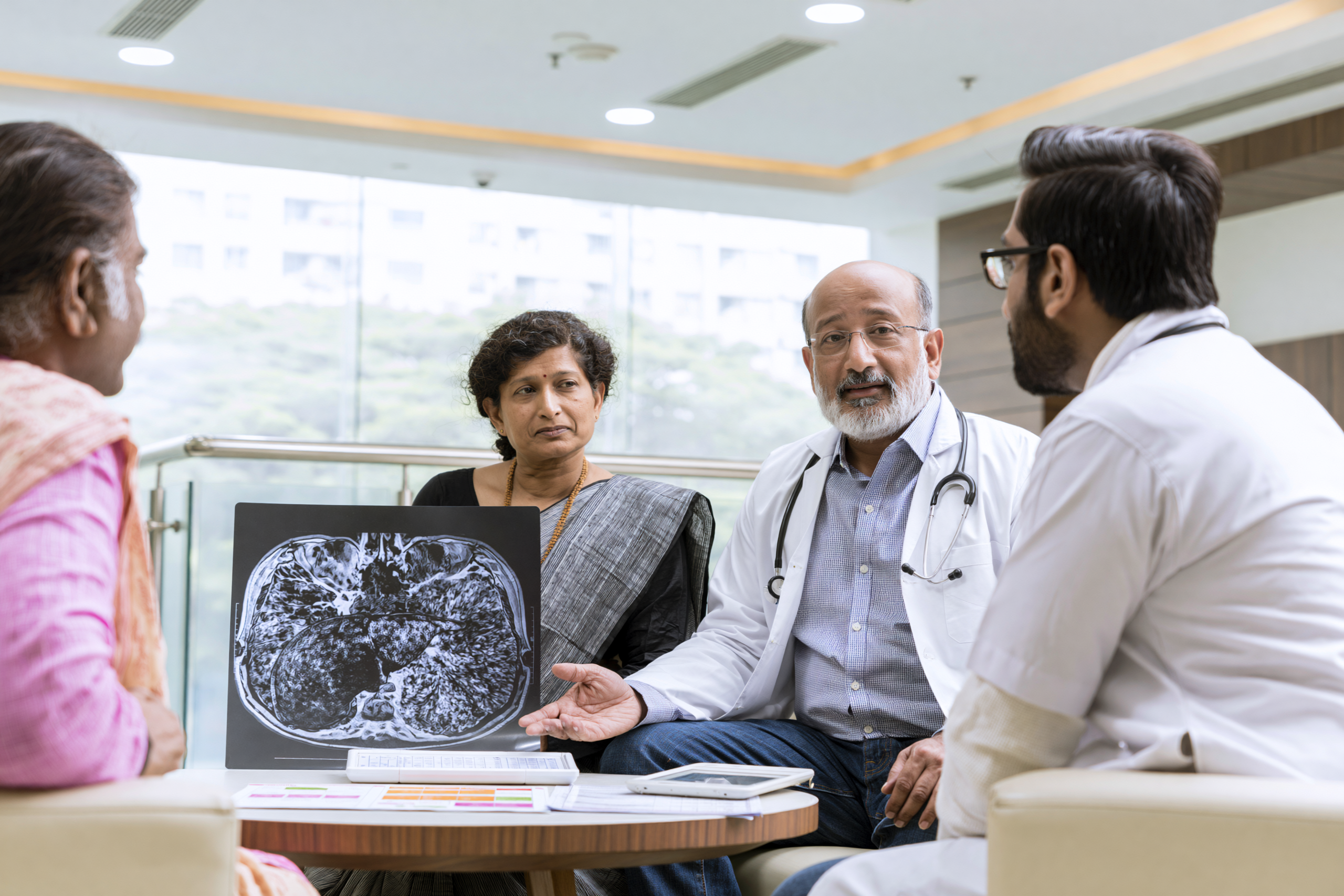
Successful liver surgery removes the visible tumour, but it does not necessarily remove every factor that contributed to the development of hepatocellular carcinoma. Your remaining liver may still contain cirrhosis, chronic hepatitis, fibrosis, metabolic dysfunction or other conditions that continue to influence long-term health [2–4].
Table: HCC Recurrence-Risk Management After Liver Surgery
| Continuing risk factor | Why it matters after surgery | Long-term management |
|---|---|---|
| Cirrhosis in the remaining liver | The liver environment that contributed to HCC may still be present | Hepatology follow-up, liver-function monitoring and regular imaging |
| Chronic hepatitis B | Viral replication may continue damaging the remaining liver | Continued antiviral suppression and HBV monitoring |
| Chronic hepatitis C | Advanced fibrosis or cirrhosis may continue to carry HCC risk after viral cure | Direct-acting antiviral treatment where indicated and continued surveillance |
| Alcohol-related liver disease | Continued alcohol exposure accelerates further liver injury | Complete alcohol abstinence |
| MASLD or MASH | Diabetes, insulin resistance and obesity may continue metabolic liver injury | Glucose control, nutrition, physical activity and body-composition management |
| Sarcopenia | Muscle loss reduces recovery reserve and treatment tolerance | Adequate nutrition, protein, walking and resistance activity |
| Smoking | Smoking adds cardiovascular, respiratory and cancer-related risk | Tobacco cessation |
| Missed surveillance | Early recurrence may remain asymptomatic | CT or MRI, AFP when informative and specialist follow-up |
| Unverified supplements | Some products can cause liver injury or interact with medicines | Complete ingredient review, quality testing and adverse-event monitoring |
For this reason, postoperative care should not end when the surgical wound heals. Long-term management focuses on preserving liver function, reducing ongoing liver injury, maintaining nutritional and physical reserve and detecting recurrence at the earliest possible stage.
Ayurveda may contribute to this recovery programme by supporting digestion, nutrition, strength, bowel function, sleep and healthy daily routines. However, it should not be presented as a proven method of preventing HCC recurrence. Current scientific evidence does not support the claim that any Ayurvedic medicine, Rasayana or Avaleha can eliminate the risk of recurrence after curative liver surgery [23–25].
Your postoperative strategy should therefore combine continued oncology surveillance with management of the underlying liver disease and carefully supervised supportive care.
Why HCC Can Recur Even After Successful Surgery
One of the most important facts you should understand is that recurrence after liver resection is relatively common.
Recurrence can occur because microscopic tumour cells remained undetected at the time of surgery or because a completely new tumour develops within the chronically diseased liver. These two mechanisms are biologically different, but both are recognised in long-term follow-up [2–5].
Your recurrence risk depends on many factors, including tumour size, tumour number, vascular invasion, tumour differentiation, surgical margin, AFP level, underlying cirrhosis and the continuing activity of liver disease.
This is why complete tumour removal does not eliminate the need for continued medical follow-up.
The Remaining Liver Still Requires Treatment
If your HCC developed in a cirrhotic liver, the liver remaining after surgery may still contain fibrosis, regenerative nodules and chronic inflammation.
Similarly, hepatitis B, hepatitis C, MASLD, MASH, diabetes or alcohol-related liver disease may continue unless they are actively treated [2,3,6–9].
From a modern perspective, these conditions require continued management because they contribute to ongoing liver injury.
From an Ayurvedic perspective, the focus shifts from managing the tumour itself towards supporting Agni, Bala, Ojas and healthy tissue nourishment while reducing factors that continue disturbing normal physiological function.
The objective is to preserve the quality of the remaining liver rather than assume that surgery has restored complete hepatic health.
Managing Chronic Hepatitis B After Surgery
If chronic hepatitis B contributed to your HCC, antiviral treatment often continues after surgery.
Suppressing HBV replication reduces ongoing liver injury and may lower the likelihood of further deterioration of liver function [2,3,7].
Your antiviral medicine should not be discontinued because the tumour has been removed.
Ayurvedic supportive care should be planned around your antiviral treatment rather than replacing it.
Your liver function, viral status and oncology surveillance remain essential parts of long-term management.
Managing Chronic Hepatitis C After Surgery
If hepatitis C is still active, treatment with direct-acting antiviral medicines may be recommended according to your clinical situation.
Successful viral eradication reduces ongoing liver inflammation and fibrosis progression. However, patients who already have cirrhosis remain at increased risk for future HCC even after viral cure [2,3,8].
You should therefore continue scheduled imaging and liver follow-up.
Ayurvedic supportive treatment should complement rather than replace antiviral management.
Managing MASLD and MASH
Many patients develop HCC in association with metabolic dysfunction-associated steatotic liver disease.
After surgery, controlling obesity, insulin resistance, diabetes and abnormal lipid metabolism becomes an important part of protecting your remaining liver [6,9].
From an Ayurvedic perspective, long-term Pathya-Apathya becomes particularly important during this phase.
Your dietary programme should support metabolic health without causing progressive muscle loss or severe nutritional restriction.
Rapid weight-loss programmes, prolonged fasting and extreme diets should generally be avoided unless medically supervised.
Complete Alcohol Abstinence
If alcohol contributed to your liver disease, complete abstinence remains one of the most important long-term interventions.
Continued alcohol exposure may accelerate fibrosis, worsen portal hypertension and reduce liver reserve even when the original tumour has been removed [6].
Ayurveda also recognises the importance of avoiding substances that repeatedly disturb digestive and metabolic balance.
The objective is not only to prevent another tumour but also to preserve the function of the remaining liver.
Stopping Tobacco
Smoking remains a modifiable health risk after HCC treatment.
Although tobacco cessation alone cannot eliminate recurrence risk, it contributes to better cardiovascular health, respiratory function, wound healing and overall long-term recovery.
Your postoperative recovery should therefore include smoking cessation support whenever applicable.
Preserving Muscle and Functional Capacity
Muscle loss often continues after surgery unless nutrition and physical activity are actively restored.
Your recovery should include adequate protein intake, gradual resistance activity, walking and repeated assessment of functional ability [19,20].
From an Ayurvedic perspective, restoration of Bala becomes a major objective during this period.
The purpose is not to produce rapid weight gain but to restore useful physical strength and independence.
A structured programme combining nutrition, graded exercise, sleep regulation and supportive Ayurvedic care may contribute to improved recovery.
Supporting Agni During Long-Term Recovery
Digestive capacity often improves gradually over several weeks or months after surgery.
Your Agni should not be challenged with excessive fasting, overeating or highly restrictive food plans.
Instead, your meals should become progressively more varied as tolerance improves while continuing to respect your liver function, diabetes status, bowel pattern and nutritional requirements.
The classical principle of matching food quantity to digestive capacity remains relevant during this phase [29].
As your appetite recovers, your nutritional intake should also increase appropriately to support muscle rebuilding and liver regeneration.
The Role of Rasayana After Surgery
Charaka Samhita describes Rasayana as supporting longevity, strength, tissue nourishment and physiological resilience [28].
In the postoperative HCC setting, Rasayana should be interpreted as a structured recovery strategy rather than as a recurrence-prevention treatment.
Your Rasayana programme may include individualised diet, appropriate daily routine, adequate sleep, gradual physical activity, bowel regulation and carefully selected formulations.
Its intended outcomes should include improved appetite, better nutritional intake, preserved muscle strength, improved sleep, enhanced daily activity and better overall quality of life.
These objectives are clinically measurable and should be reviewed regularly.
Maintaining Ojas During Recovery
Ojas may be viewed as your recovery reserve rather than a specific immune marker.
After surgery, maintaining Ojas means preserving physical resilience, nutritional stability, emotional wellbeing and your capacity to tolerate ongoing surveillance and any future treatment.
Good sleep, adequate nutrition, regular activity, emotional support and avoidance of unnecessary physiological stress all contribute to this broader recovery process.
This concept should not be presented as evidence that Ojas prevents cancer recurrence.
Long-Term Pathya-Apathya
Your dietary and behavioural guidance should evolve as recovery progresses.
Initially, your meals may be softer and smaller.
As your digestive tolerance improves, your diet should gradually become nutritionally complete while avoiding excessive alcohol, highly processed foods and unnecessary dietary extremes.
Pathya should remain individualised according to your liver function, diabetes, bowel pattern, kidney function and metabolic health rather than following one universal food list.
Apathya should include behaviours that repeatedly damage liver health, such as alcohol consumption, smoking, prolonged fasting, contaminated food and unverified supplements.
Continuing Imaging Surveillance
Even when you feel completely well, postoperative imaging remains essential.
Contrast-enhanced CT or MRI is used to detect recurrence before symptoms appear. AFP may also be monitored when clinically appropriate [2–5].
Your follow-up schedule depends on tumour characteristics, liver reserve and your treating team’s recommendations.
Feeling healthy does not mean surveillance is no longer necessary.
Many recurrent tumours are detected before causing symptoms, allowing earlier treatment.
Monitoring Liver Function
Your long-term follow-up should also include bilirubin, albumin, INR, liver enzymes, kidney function and assessment for complications of chronic liver disease.
These investigations help determine whether your liver remains stable and whether additional treatment can be performed safely if required.
Ayurvedic supportive care should always be adjusted according to these objective laboratory findings.
Monitoring Your Daily Function
Your recovery should not be judged only by scans.
You should also ask whether you are eating normally, maintaining muscle strength, walking independently, sleeping well, returning to work or family activities and maintaining emotional wellbeing.
These outcomes reflect the practical success of your rehabilitation.
Ayurveda may contribute meaningfully to these domains when treatment is individualised and appropriately monitored.
What Ayurveda Cannot Promise After Surgery
No Ayurvedic formulation has demonstrated in high-quality clinical trials that it can prevent recurrence after liver resection.
Similarly, no Avaleha, Rasayana or classical medicine has been shown to replace postoperative surveillance.
Claims that surgery is unnecessary because Ayurveda alone prevents recurrence are not supported by current scientific evidence [23–25].
Your recovery programme should therefore remain transparent regarding both its strengths and its limitations.
What This Means for You
Long-term recovery after liver surgery involves much more than removing the original tumour.
Your remaining liver continues to require protection, your nutritional reserve must be restored, your physical strength should improve and the causes of chronic liver injury should be actively managed.
Ayurveda may support this process by helping maintain digestion, nutrition, sleep, bowel function, daily activity and overall physiological resilience while your oncology and hepatology teams continue surveillance through imaging and laboratory monitoring [2–9,19–25,28,29].
The most effective long-term strategy is one that treats the remaining liver with the same care that was given to removing the tumour.
Ayurveda After Liver Transplantation

Liver transplantation changes the clinical context completely. After transplantation, your diseased liver has been replaced, but your recovery now depends on protecting the graft, preventing rejection, avoiding infection and maintaining stable immunosuppressant levels [2,3,13,32,33].
Ayurveda may still have a limited supportive role, but it must be approached with greater caution than after liver resection. Your transplant medicines have narrow therapeutic ranges, and even small changes in absorption or metabolism can increase toxicity or reduce protection against rejection [32,33].
For this reason, no Ayurvedic medicine, Avaleha, herbal supplement, mineral preparation or “immune-boosting” product should be started after liver transplantation without the explicit approval of your transplant team.
Why Transplantation Is Different From Liver Resection
After liver resection, part of your own liver remains. After transplantation, the entire diseased liver is replaced with a donor liver.
This may remove the tumour-bearing organ and the cirrhotic environment in which HCC developed. However, transplantation introduces new long-term responsibilities, including lifelong immunosuppression, infection prevention, graft monitoring, kidney protection and recurrence surveillance [2,3,13].
Your recovery plan must therefore prioritise graft survival and medicine stability above all other supportive goals.
Immunosuppressant Medicines Are Essential
Following transplantation, you may receive tacrolimus, cyclosporine, mycophenolate, corticosteroids, everolimus, sirolimus or a combination of these medicines.
These drugs reduce immune activity so that your body does not reject the transplanted liver.
You should not reduce, stop or replace them because you feel well or because an alternative practitioner describes them as harmful. Missing doses or altering the schedule can lead to graft rejection, while excessive exposure can contribute to kidney injury, infection, neurological symptoms, diabetes or other complications [32,33].
Every supportive treatment must be planned around these medicines.
Why Herb–Drug Interactions Are Particularly Important
Tacrolimus and several other transplant medicines are affected by intestinal transport proteins and CYP3A-related metabolism. Certain foods, herbs and supplements can therefore alter blood concentrations [33].
If a product increases tacrolimus exposure, you may develop kidney injury, tremor, headache, high blood pressure, confusion or other toxicity. If it lowers exposure, your graft may become vulnerable to rejection.
The risk cannot be assessed from the phrase “natural” or “traditional.” Your transplant team requires the exact ingredient list, dose, manufacturer, batch and proposed duration before deciding whether a product is acceptable.
Why “Immune Boosting” Is Not an Appropriate Goal
After transplantation, your immune system is deliberately controlled to protect the donor liver.
A product advertised as strongly stimulating immunity may conflict with the purpose of immunosuppressive treatment. It may also produce unpredictable effects when combined with checkpoint pathways, corticosteroids or calcineurin inhibitors.
This does not mean that your recovery should ignore resilience, nutrition or infection prevention. It means that immune support should focus on adequate nutrition, vaccination according to transplant guidance, sleep, hygiene, safe activity and adherence rather than indiscriminate immune stimulation [32].
When Ayurveda May Be Considered
Ayurveda may be considered only for clearly defined supportive goals after your transplant team confirms that graft function and medicine levels are stable.
Possible goals may include support for appetite, bowel regularity, sleep, gentle rehabilitation or food tolerance.
Even then, the simplest effective intervention is usually preferable. A long list of herbs increases the difficulty of identifying an interaction or adverse effect.
Your supportive plan should be reviewed whenever your immunosuppressant dose changes, an infection occurs or liver and kidney tests become abnormal.
Avaleha Use After Transplantation
A personalised Avaleha should not be introduced routinely after transplantation.
The formulation may contain multiple herbs, sweeteners, oils, ghee, honey, minerals or spices that affect glucose control, absorption, metabolism or microbial safety.
If an Avaleha is being considered, your transplant team should receive the full composition and manufacturing documentation before you take the first dose.
You should not use a market-bought Avaleha with an incomplete label or proprietary mixture because interaction assessment becomes impossible.
Sugar, Diabetes and Metabolic Complications
Post-transplant medicines can increase your risk of diabetes, weight gain, high blood pressure and abnormal lipids.
Many traditional Avaleha preparations contain concentrated sugar, jaggery or honey. These may be unsuitable when your glucose is elevated or when metabolic complications are developing.
A formulation described as sugar-free still requires review because alternative sweeteners, preservatives and other ingredients may also affect tolerance or stability.
Your nutritional plan should support recovery without worsening post-transplant metabolic disease.
Infection Risk and Product Contamination
Immunosuppression increases your susceptibility to bacterial, viral and fungal infections.
This makes microbial quality particularly important. An inadequately manufactured herbal product may expose you to contamination that would be less serious in a healthy person but dangerous after transplantation [25,27,32].
Any product considered for use should have reliable manufacturing records, microbial testing, batch identification and appropriate storage instructions.
Home-prepared formulations and unpackaged powders are especially difficult to verify and should generally be avoided.
Herbo-Mineral Preparations Require Extreme Caution
Metal- or mineral-containing preparations should not be used automatically after transplantation.
Your kidneys may already be vulnerable because of tacrolimus, cyclosporine, dehydration or other medicines. Exposure to lead, mercury, arsenic or another toxic element can create additional organ stress [25,34].
If any herbo-mineral medicine is proposed, documented toxic-element testing and transplant-team approval are essential.
A traditional name or long history of use does not remove the need for modern quality control.
Ayurveda and Kidney Protection
Kidney dysfunction is common after liver transplantation, particularly with calcineurin-inhibitor therapy.
Your creatinine, electrolytes, blood pressure and urine output require regular monitoring.
You should not begin herbal diuretics, “kidney cleanses” or aggressive detoxification because these may alter fluid balance, interact with medicines or worsen renal function.
A rise in creatinine should be assessed medically rather than attributed to normal adjustment or detoxification.
Bowel Function After Transplantation
Constipation may occur because of reduced activity, pain medicines, dehydration or dietary changes. Diarrhoea may result from infection, mycophenolate, antibiotics or other medicines.
Ayurvedic bowel support should not be prescribed until the cause is understood.
Severe purgation can alter absorption of immunosuppressants and increase dehydration. Persistent diarrhoea may also change tacrolimus levels and requires prompt review [32,33].
The safest bowel plan is usually gentle, predictable and coordinated with the transplant team.
Supporting Appetite and Nutrition
Your appetite may fluctuate after transplantation because of surgery, medicines, infection, altered taste or emotional stress.
Nutritional support should help you restore strength while considering glucose, kidney function, blood pressure and infection risk.
Food hygiene is especially important because immunosuppression increases vulnerability to food-borne illness.
Ayurvedic dietary guidance may help structure meal timing and digestive tolerance, but it should not conflict with transplant nutrition recommendations.
Physical Recovery After Transplantation
Muscle loss and frailty may persist after transplantation, particularly when you were weak before surgery.
Your rehabilitation should include gradual walking, strength-building and restoration of daily independence according to your surgical and transplant guidance.
Ayurveda may contribute through routine, sleep regulation and appropriate supportive nutrition, but physical recovery still depends on progressive mobilisation and adequate protein and energy intake.
Your activity plan should be modified when you have wound problems, infection, anaemia or unstable graft function.
Monitoring Graft Function
Your graft is monitored through liver enzymes, bilirubin, INR, albumin, imaging and clinical assessment.
Abnormal liver tests after transplantation may result from rejection, infection, biliary complications, vascular problems, recurrent disease, medicine toxicity or another cause.
You should not begin or intensify herbal treatment when liver tests worsen without identifying the cause.
Any new product introduced before abnormal results appeared should be disclosed immediately.
Recurrence Surveillance After Transplantation
Transplantation reduces recurrence risk in appropriately selected HCC patients but does not make the risk zero.
Your surveillance schedule depends on the characteristics of the original tumour, pathology, vascular invasion, AFP and transplant-centre protocol [2,3,13].
Ayurveda cannot replace this surveillance or prove that recurrence has been prevented.
Your follow-up should continue even when graft function and general wellbeing are excellent.
When Ayurvedic Medicines Should Be Stopped
Any Ayurvedic product should be stopped and reviewed if you develop rising creatinine, abnormal tacrolimus levels, jaundice, rash, diarrhoea, vomiting, tremor, confusion, reduced urine output or unexpected liver-test changes.
You should also stop a formulation if the complete ingredients cannot be verified or if the manufacturer cannot provide batch and quality documentation.
A medicine that interferes with food intake, prescribed treatment or transplant monitoring has no place in your recovery plan.
What This Means for You
After liver transplantation, your safest treatment strategy is based on strict immunosuppressant adherence, graft monitoring, infection prevention, nutrition, rehabilitation and continued HCC surveillance [2,3,13,32,33].
Ayurveda may be considered only for narrow supportive goals after your transplant team has reviewed the formulation and confirmed that it does not create an unacceptable interaction or contamination risk.
The central principle is simple: your transplanted liver must be protected first. Any complementary treatment is acceptable only when it supports that objective without disturbing medicine levels, kidney function, graft stability or oncological follow-up.
Personalised Yakrit–Ojas Rasayana Avaleha (Medicine)

Yakrit–Ojas Rasayana Avaleha is a proposed clinical name for a personalised Ayurvedic recovery formulation. It is not a classical formulation reproduced word-for-word from one Ayurvedic text, and it should not be presented as a standard medicine for every person with hepatocellular carcinoma.
The formulation is inspired by the Rasayana principles described in Charaka Samhita, Chikitsa Sthana, Chapter 1, Rasayana Adhyaya; the principles of Matra, Agni and food tolerance described in Ashtanga Hridaya, Sutra Sthana, Chapter 8, Matrashitiya Adhyaya; and the pharmaceutical principles of Avaleha preparation described in Sharangadhara Samhita, Madhyama Khanda, Chapter 8, Avaleha Kalpana [28–30].
The word Yakrit reflects the liver-centred clinical context. Ojas represents your physiological reserve and ability to recover. Rasayana refers to restorative care, while Avaleha describes the semi-solid dosage form.
This formulation may be considered for defined recovery goals such as poor appetite, disturbed digestion, constipation, sleep difficulty, nutritional decline, fatigue and loss of physical strength. It has not been proved to eradicate HCC, replace tumour-directed treatment or guarantee that recurrence will not occur [23–25].
Essential Warning Before This Avaleha Is Prescribed
You should never purchase a ready-made “liver cancer Avaleha” merely because its label contains the words Yakrit, Rasayana, Arbuda, detoxification or immunity.
You should never prepare this formulation at home or ask a pharmacy to copy a formula obtained from a website, social-media post or another patient.
The medicine must always be formulated, prescribed and monitored under the direct supervision of a qualified Ayurvedic physician who has reviewed your complete oncology and hepatology records. Where you are receiving surgery, embolisation, radiotherapy, immunotherapy, targeted therapy or post-transplant immunosuppression, coordination with your treating specialist is essential.
The formulation of herbs, minerals, dosage-form base, dose and Anupana will vary from person to person. The same medicine may be supportive for one patient, ineffective for another and unsafe for a third.
Your physician must review your tumour stage, tumour distribution, vascular involvement, bilirubin, albumin, INR, platelet count, liver enzymes, kidney function, glucose, ascites, encephalopathy, appetite, bowel pattern, body strength and current medicines before selecting any ingredient [23–27].
Why a Market-Bought Avaleha May Not Work for You
A commercial product may contain many impressive ingredients and still fail because it has not been designed for your individual disease stage, liver reserve and current treatment.
| Factor | Why the market product may not work for you | Possible consequence |
| No review of your HCC stage | The manufacturer does not know the number, size or location of your tumours | The product cannot be matched to your actual treatment pathway |
| No assessment of vascular invasion | Portal-vein or hepatic-vein involvement is ignored | You may wrongly depend on supportive medicine while the cancer requires urgent tumour-directed treatment |
| No evaluation of liver reserve | The same product is sold to people with normal bilirubin and those with jaundice or decompensated cirrhosis | A standard dose may worsen digestive intolerance or liver dysfunction |
| No evaluation of kidney function | Creatinine, electrolytes and urine output are not reviewed | Diuretic, purgative or mineral ingredients may increase kidney risk |
| No assessment of glucose | Traditional Avaleha bases may contain substantial sugar or jaggery | Diabetes, MASLD and metabolic instability may worsen |
| No assessment of ascites | Fluid retention, sodium restriction and prescribed diuretics are ignored | Additional Mutrala or Rechana ingredients may disturb fluid balance |
| No assessment of encephalopathy | Confusion, constipation and sedative exposure are not considered | Sedating or dehydrating medicines may worsen mental status |
| No assessment of Agni | Appetite, nausea, early satiety, reflux and diarrhoea are ignored | Bitter, heating or heavy ingredients may further reduce food intake |
| No assessment of Bala | Severe weakness, sarcopenia and poor performance status are not considered | Strong cleansing or Lekhana medicines may increase depletion |
| No review of oncology medicines | Immunotherapy, targeted therapy, anticoagulants and supportive medicines are ignored | Herb–drug interactions or overlapping toxicity may occur [23] |
| No review of recent procedures | The product may be started immediately after surgery, TACE, TARE or ablation | New symptoms and abnormal liver tests become difficult to interpret |
| One formula for every patient | Your Prakriti, Dosha, Dhatu, bowel pattern and treatment objective are ignored | The formulation may not address your actual recovery barriers |
| Incorrect botanical species | Similar vernacular names may refer to different plants | You may receive an ineffective substitute or a plant with a different safety profile |
| Incorrect plant part | The root, bark, leaf, fruit and seed are not interchangeable | The expected traditional and pharmacological action may not be obtained |
| Poor-quality raw materials | Old, mouldy or improperly stored herbs may be used | Potency may decline and contamination risk may increase |
| Aflatoxin contamination | Herbs and food-derived ingredients may not be tested adequately | Additional liver exposure may occur in an already vulnerable patient |
| Pesticide contamination | Supplier quality may not be verified | Unnecessary chemical exposure may occur |
| Batch-to-batch variation | Ingredient concentration may change between batches | The effect and tolerability may be inconsistent |
| Unstandardised extracts | Concentrated extracts may replace ordinary powders without dose correction | Your actual exposure may be several times higher than expected |
| Undeclared bioavailability enhancers | Piperine or concentrated Trikatu may be added | Absorption of your prescription medicines may also change |
| Incomplete label disclosure | Proprietary blends may conceal exact quantities | Reliable interaction assessment becomes impossible |
| Undeclared adulterants | The product may contain substances not listed on the label | Unexpected toxicity or misleading symptom relief may occur |
| Inadequately processed Bhasma | Shodhana, Marana and final testing may not be documented | Your exposure to an improperly processed mineral cannot be excluded |
| No batch-specific elemental analysis | A general certificate may be shown instead of the certificate for your actual batch | Lead, mercury, arsenic or cadmium exposure may remain unknown [25,34] |
| Wrong Avaleha base | Sugar, jaggery, honey or ghee may be chosen for manufacturing convenience | The product may worsen glucose, nausea, diarrhoea or fat intolerance |
| Wrong dose | The label cannot account for your liver reserve, kidney function, age or strength | The prescribed amount may be ineffective or excessive |
| Wrong timing | The medicine may be taken too close to anticancer medicines | Absorption or gastrointestinal tolerance may be affected |
| Wrong Anupana | The same vehicle is recommended for every patient | The medicine may not suit your digestion, diabetes or fluid restrictions |
| Excessive ingredient count | Too many herbs and minerals are combined | The cause of benefit or toxicity becomes impossible to identify |
| No baseline testing | Liver, kidney and glucose values are not documented before use | Later deterioration cannot be attributed accurately |
| No monitoring plan | The seller does not review your reports after treatment begins | A preventable adverse reaction may continue unnoticed |
| No stop criteria | You may be advised to continue despite jaundice, diarrhoea or abnormal tests | Toxicity may be misrepresented as detoxification or a healing reaction |
| Improper storage | Moisture, heat or use of a wet spoon may contaminate the Avaleha | Fermentation, microbial growth or instability may develop |
| False interpretation of improvement | Better appetite or sleep may be described as tumour regression | Necessary imaging or oncology treatment may be delayed |
Market failure is therefore not limited to poor-quality manufacturing. A perfectly manufactured product may still fail if it is given to the wrong patient, at the wrong dose, during the wrong phase of treatment or for the wrong clinical objective.
How Potent Herbs Should Be Understood
In Ayurvedic oncology support, potent does not mean that every available herb should be added. A potent ingredient is one that has a clear indication in you, an appropriate dose, acceptable manufacturing quality and reasonable compatibility with your current treatment.
Laboratory findings involving cancer cells, isolated compounds or animal models do not establish that a whole herb can cure human HCC. The evidence should be presented as preclinical unless reliable human trials have demonstrated a clinical effect.
The herbs below may be considered during individualised formulation planning. They should not all be combined in one Avaleha.
Potential Potent Herbs and Their Functions
| Ayurvedic herb | Botanical name | Traditional function | Potential recovery relevance | Important caution |
| Amalaki | Phyllanthus emblica L. | Rasayana, Pitta-shamana, Dhatu-poshana and Balya | May support nutrition, digestive tolerance and restorative care | Human HCC regression has not been demonstrated |
| Haritaki | Terminalia chebula Retz. | Anulomana, Rasayana and Vata regulation | May support constipation and regular bowel movement | Excess may cause diarrhoea, dehydration or weakness |
| Bibhitaki | Terminalia bellirica Roxb. | Kapha-Meda regulation and Rasayana | May be considered in heaviness, metabolic disturbance or selected bowel patterns | May be unsuitable when you are dry, underweight or constipated |
| Triphala | Combination of Amalaki, Haritaki and Bibhitaki | Rasayana and bowel regulation | May provide gentle bowel support in an individualised proportion | Standard equal proportions are not suitable for everyone |
| Guduchi | Tinospora cordifolia | Rasayana and traditional support of resilience | Has been investigated for immunomodulatory and metabolic effects | Reported liver injury means that it should not be included routinely in HCC [26] |
| Bhumyamalaki | Phyllanthus amarus and authenticated related species | Traditional Pitta, Kamala and Yakrit support | May be considered in selected liver-related symptom patterns | Several species are sold under similar names; authentication is essential |
| Kalmegha | Andrographis paniculata | Tikta, Deepana and Pitta-Kapha regulation | Andrographolide has shown activity in experimental HCC models | May worsen nausea, loose stool and poor appetite |
| Katuki | Picrorhiza kurroa | Tikta, Bhedana and traditional Yakrit support | Experimental hepatoprotective effects have been studied | It may be purgative and is often substituted or adulterated |
| Punarnava | Boerhavia diffusa | Shothahara and Mutrala | May be considered when oedema is present and renal function is stable | It does not replace medical treatment of ascites |
| Bhringaraja | Eclipta prostrata | Traditional Yakrit and Rakta support | Experimental hepatoprotective activity has been reported | Human HCC benefit remains unproved |
| Daruharidra | Berberis aristata | Tikta, Pitta-Kapha regulation and Rakta support | Berberine-related compounds have shown preclinical anticancer activity | May affect glucose, bowel function and drug transport |
| Haridra | Curcuma longa | Shothahara and metabolic support | Curcumin has been studied extensively in experimental cancer pathways | Concentrated products have been associated with liver injury in some patients [26] |
| Yashtimadhu | Glycyrrhiza glabra | Rasayana, Balya and Pitta-shamana | May support mucosal comfort and selected inflammatory symptoms | May worsen hypertension, oedema and low potassium |
| Ashwagandha | Withania somnifera | Balya, Brimhana and Rasayana | May be considered for sleep, weakness and recovery in selected patients | Liver injury has been reported; it is not an automatic HCC ingredient [26] |
| Shatavari | Asparagus racemosus | Brimhana, Rasayana and nourishing support | May be considered when depletion, dryness and poor intake dominate | Glucose and gastrointestinal tolerance require review |
| Pippali | Piper longum | Deepana, Pachana, Rasayana and Yogavahi | May support digestion in carefully selected low doses | Piperine may alter medicine metabolism and transport [23] |
| Maricha | Piper nigrum | Deepana and Pachana | May support selected digestive symptoms | Concentrated piperine may alter drug exposure |
| Shunthi | Zingiber officinale | Deepana, Pachana and Vata-Kapha regulation | May support nausea and meal tolerance | Reflux, anticoagulants and bleeding risk require consideration |
| Trikatu | Pippali, Maricha and Shunthi | Strong Deepana-Pachana | May be considered in very small quantities when sluggish digestion is clear | It may worsen burning, diarrhoea and drug-interaction risk |
| Musta | Cyperus rotundus | Deepana, Pachana and Grahi | May support selected nausea or bowel disturbances | The indication differs between diarrhoea, constipation and colitis |
| Chitraka | Plumbago zeylanica | Potent Deepana and Pachana | May occasionally be considered in highly selected Mandagni patterns | It is irritant and generally unsuitable for weak or inflamed patients |
| Guggulu | Commiphora wightii | Lekhana, Medohara and Shothahara | May be considered when metabolic disturbance is prominent | It may irritate the gut and interact with medicines |
| Kanchanara | Bauhinia variegata | Traditionally used in Granthi and Kapha-Meda patterns | May be considered only when supported by the complete Ayurvedic assessment | Granthi references do not prove efficacy in modern HCC |
| Nimba | Azadirachta indica | Tikta, Pitta-Kapha regulation and Kleda support | Experimental anti-inflammatory pathways have been investigated | Concentrated preparations may be poorly tolerated or toxic |
| Arjuna | Terminalia arjuna | Hridya and Rakta-supportive | May be considered when cardiovascular support is relevant | It may interact with cardiac, blood-pressure and anticoagulant medicines |
| Gokshura | Tribulus terrestris | Mutrala and Balya | May be considered for a defined urinary indication | It should not be used as routine treatment for ascites |
| Bhallataka | Semecarpus anacardium | Potent Lekhana and specialised Rasayana use after Shodhana | Experimental antineoplastic activity has been explored | It is highly irritant and should generally be excluded from routine HCC recovery formulations |
The experimental effects of curcumin, andrographolide, withanolides, berberine and related phytochemicals are scientifically interesting but remain insufficient to claim that adding their source herbs to an Avaleha will shrink your tumour or prolong survival.
Potential Minerals and Herbo-Mineral Ingredients
Minerals should never be added merely because the disease is serious or because the formulation is expected to be “strong.” A mineral ingredient requires a separate indication, authenticated manufacture, batch-specific testing and a patient-specific dose.
No Bhasma, Pishti or Rasaushadhi has been proved through high-quality human trials to cure HCC or prevent its recurrence.
Minerals, Their Traditional Functions and Cautions
| Ingredient | Traditional function | Possible physician-defined role | Critical caution |
| Swarna Bhasma | Rasayana, Balya and Ojas support | May be considered only for a specific restorative indication | No established human HCC benefit; purity and dose verification are essential |
| Rajata Bhasma | Cooling and restorative use in selected conditions | May be considered only for a separate classical indication | Unnecessary silver exposure should be avoided |
| Abhraka Bhasma | Rasayana, Balya and Deepana | May be considered in selected depletion patterns | It should not be claimed to regenerate the liver or destroy cancer |
| Lauha Bhasma | Pandu and Rakta support | May be considered only when iron deficiency is confirmed | Anaemia in cancer is not always iron deficiency |
| Mandura Bhasma | Pandu, Shotha and liver-spleen-related traditional use | May be considered in documented iron deficiency with an appropriate Ayurvedic indication | Ferritin, transferrin saturation and inflammation must be assessed |
| Kasis Bhasma | Iron-related support | Rarely considered for a confirmed iron-related indication | Gastrointestinal irritation and iron excess are concerns |
| Tamra Bhasma | Lekhana and selected Yakrit-Pliha use | Should be reserved for exceptional indications | Copper exposure can be hazardous in liver disease |
| Swarna Makshika Bhasma | Traditional Rasayana, Pandu and Yakrit support | May be considered only after detailed assessment | Copper, iron and elemental testing require review |
| Yashada Bhasma | Traditional Prameha and Vrana support | May be considered when a defined zinc-related indication exists | Excess zinc may cause copper deficiency and gastrointestinal effects |
| Mukta Pishti | Pitta-shamana and Daha support | May be considered for a defined burning or Pitta-related presentation | Calcium load, kidney function and stone history require consideration |
| Pravala Pishti | Pitta-shamana and Amlapitta support | May be considered for a specific symptom indication | It has no established HCC anticancer action |
| Shankha Bhasma | Deepana and Amlapitta-related support | May be considered for a defined digestive indication | Calcium content and concurrent antacid use require review |
| Shukti Bhasma | Pitta and acid-related support | May be considered for selected digestive symptoms | It should not be combined automatically with multiple calcium preparations |
| Godanti Bhasma | Traditional Jvara, Daha and pain-related use | May be considered for a separate symptom indication | It has no proven role in tumour control |
| Heeraka Bhasma | Specialised Rasayana and traditionally promoted Arbuda-related use | Should not be routine in HCC care | Human efficacy, authenticity and safety remain uncertain |
| Kajjali | Traditional Rasaushadhi base and Yogavahi | Only within a licensed, precisely manufactured medicine for a specific indication | It contains mercury and sulfur and requires exceptional scrutiny |
| Rasasindura | Specialised Yogavahi and Rasayana use | Not a routine HCC recovery ingredient | Mercury-related composition, quality and toxicity require caution |
| Makaradhwaja | Potent Balya and Rasayana | May be considered only for an independent, highly specific indication | Mercury-, sulfur- and gold-related constituents require stringent testing |
| Shilajit | Rasayana, Yogavahi and metabolic support | May be considered in selected depletion or metabolic patterns | Purity, iron status, gout, kidney function and contamination require review |
| Hartala preparations | Narrow traditional Rasaushadhi indications | Should not be used in routine HCC recovery | These are arsenic-containing preparations |
| Manahshila preparations | Narrow specialised classical indications | Should not be included in a general Avaleha | Arsenic exposure creates substantial safety concerns |
| Naga Bhasma | Selected traditional genitourinary and Prameha use | No routine role in HCC recovery | Lead-related composition requires extreme caution |
| Vanga Bhasma | Selected traditional Prameha and genitourinary use | No routine role in HCC recovery | It should not be added without an independent indication |
| Trivanga Bhasma | Combined traditional use in selected metabolic and genitourinary conditions | No routine role in HCC care | Unnecessary multi-element exposure should be avoided |
Properly processed Bhasmas may differ chemically from unprocessed environmental metals, but this does not remove the requirement for modern characterization, toxic-element testing, pharmacovigilance and patient monitoring [25,27,34].
Why Herbs and Minerals Must Vary From Person to Person
If your principal problem is poor appetite and early satiety, your formula may prioritise mild Deepana, nutritional support and an easily tolerated base.
If constipation is prominent, gentle Anulomana may be considered. The same approach would be inappropriate if you have diarrhoea, dehydration or suspected immune-related colitis.
If your bilirubin is rising, every non-essential herb and mineral should be reconsidered. A new medicine should not be added simply because it is traditionally associated with Yakrit or Kamala.
If you have ascites or reduced urine output, Punarnava, Gokshura and other Mutrala ingredients require careful coordination with your medical diuretics, electrolytes and kidney function.
If you have diabetes or MASLD, the Avaleha base must be modified. A high-sugar or jaggery preparation may conflict with your metabolic treatment.
If you are receiving immunotherapy, immune-active and potentially hepatotoxic herbs require greater caution. If you are receiving targeted therapy, ingredients affecting CYP enzymes, P-glycoprotein, blood pressure, platelet function or diarrhoea risk require review [23].
If you have undergone liver transplantation, no Avaleha, herb, Bhasma or supplement should be started without explicit transplant-team approval.
Required Quantity for Thirty Days
At a prescribed dose of 15 g twice daily, your total daily dose is 30 g.
| Treatment period | Daily dose | Finished Avaleha required |
| 30 days | 30 g | 900 g |
The 900 g refers to the final finished weight after cooking and cooling. It does not represent the total weight of the raw herbs, decoction water, sugar, jaggery, ghee or other materials used during manufacturing.
The exact weight of every herb and mineral cannot be standardised publicly because the composition must change according to your reports. The prescribing physician should provide a patient-specific batch sheet to the licensed manufacturing unit.
Patient-Friendly Preparation Method
Clinical Review Before Preparation
Your physician first reviews your latest CT or MRI, tumour stage, bilirubin, albumin, INR, liver enzymes, platelet count, kidney function, electrolytes, glucose, ascites, encephalopathy, bowel pattern, appetite, current cancer treatment and complete medicine list.
The purpose of the Avaleha is documented before manufacturing begins. The purpose may be improving appetite, supporting bowel regularity, preserving strength or improving medicine tolerance. It should not be described vaguely as “destroying cancer” or “detoxifying the tumour.”
Preparation of the Prescription Sheet
The prescription sheet records the Sanskrit name, botanical name, plant part, raw-material form and exact quantity of every selected herb.
If a Bhasma or Pishti is clinically justified, its name, manufacturer, batch number, prescribed dose and laboratory documentation are entered separately.
The manufacturer should not add any extra herb or mineral because it is considered powerful or commercially attractive.
Authentication of Raw Materials
Every herb is checked for the correct species and plant part. The material is examined for mould, insects, moisture, substitution and deterioration.
Testing should include microbial limits, aflatoxins, pesticide residues and toxic elements according to the raw material and manufacturing requirements [25,27,34].
Preparation of the Decoction
Only herbs selected for Kwatha are converted into coarse powder. They are processed with purified water according to the pharmacy’s validated decoction method.
The liquid is reduced to the prescribed volume and filtered carefully. Coarse plant particles should not remain in the finished Avaleha.
Preparation of the Avaleha Base
The base is selected according to your diabetes status, MASLD, appetite, nutritional needs and digestive tolerance.
Sugar, jaggery, honey and ghee should not be used automatically. Any modification of the traditional base must also consider preservation, stability and dose consistency.
Addition of Fine Powders
Fine herbal powders are sieved and added at the correct stage of preparation. Prakshepa Dravya are incorporated according to the principles described in Sharangadhara Samhita, Madhyama Khanda, Chapter 8 [30].
Heat-sensitive ingredients should not be subjected to unnecessary prolonged heating. Honey should not be boiled routinely.
Addition of Approved Minerals
Minerals should never be measured by visual estimation.
Where a Bhasma or Pishti has been prescribed, its exact amount is weighed using calibrated equipment and mixed uniformly throughout the batch. This helps prevent one dose from containing substantially more mineral material than another.
Mercurial, arsenical, copper-containing or lead-containing ingredients should not form part of a routine universal HCC recovery Avaleha.
Final Yield and Packaging
The Avaleha is weighed after cooling. The target finished quantity for the proposed course is 900 g.
The final product should have uniform texture, no burnt smell, no water separation and no visible coarse particles.
The container should display your name, complete ingredient list, batch number, manufacturing date, expiry date, dose, storage instructions and prescribing physician.
Dosage
Where your physician approves this regimen, the dose is 15 g twice daily after food for 30 days.
The dose may need to be reduced, paused or discontinued according to your bilirubin, glucose, bowel pattern, kidney function, recent procedure or oncology treatment.
The medicine should be measured with a clean, dry spoon. A wet spoon should never be placed inside the container.
The Anupana must be individualised. Warm water, an appropriate herbal liquid or another physician-selected vehicle may be used according to your digestive tolerance and clinical condition.
Monitoring and Stop Criteria
Before you begin, your physician should document your appetite, food intake, bowel pattern, sleep, physical function and baseline laboratory values.
You should stop the Avaleha and obtain medical review if you develop increasing jaundice, dark urine, severe itching, persistent nausea, repeated vomiting, diarrhoea, rash, facial swelling, confusion, rapidly increasing abdominal swelling, reduced urine output, unusual bleeding or unexplained deterioration in liver or kidney tests.
Suspected reactions should be documented with the full ingredient list, dose, batch number, starting date, stopping date, symptoms and laboratory findings. Pharmacovigilance is an essential part of responsible Ayurvedic practice [27].
How the Benefit Should Be Measured
| Recovery area | What should be assessed |
| Appetite | Proportion of meals consumed, missed meals and early satiety |
| Digestion | Nausea, bloating, reflux and post-meal discomfort |
| Bowel function | Frequency, stool consistency, straining and diarrhoea |
| Strength | Walking, chair-rise ability and independence in daily activities |
| Sleep | Duration, awakenings and daytime alertness |
| Nutrition | Food intake, functional muscle recovery and weight interpreted with ascites |
| Treatment compatibility | Whether your prescribed cancer treatment continues without avoidable interaction |
| Safety | Liver, kidney, coagulation and glucose results |
Better appetite, sleep, digestion or strength represents supportive benefit. It does not prove that the tumour has reduced.
Your HCC response must continue to be assessed through contrast-enhanced CT or MRI, AFP when informative and multidisciplinary oncology review.
Clinical Position of Yakrit–Ojas Rasayana Avaleha
Yakrit–Ojas Rasayana Avaleha should be understood as a patient-specific, physician-supervised restorative formulation inspired by classical Rasayana and Avaleha Kalpana principles [28–30].
It should never be sold as one standard medicine for all patients with HCC. It should never be copied from another patient’s prescription, purchased without report review or prepared without the supervision of a qualified Ayurvedic physician.
The strongest formulation is not the one containing the greatest number of potent herbs and minerals. It is the formulation in which every selected ingredient has a clear purpose, every excluded ingredient reduces a specific risk, the manufacturing quality is verified and the clinical benefit is measured without delaying or interfering with your cancer treatment.
Modern Pharmaceutical Explanation of the Avaleha Dosage Form

Avaleha is a classical semi-solid oral dosage form in which selected herbal decoctions, fine powders, aromatic ingredients and an appropriate base are processed into a uniform preparation. Sharangadhara Samhita, Madhyama Khanda, Chapter 8, Avaleha Kalpana, describes the traditional principles governing its preparation, consistency, administration and preservation [30].
When Avaleha is used during hepatocellular carcinoma recovery, the classical method must be combined with modern pharmaceutical controls. Your formulation should not be judged only by its taste, colour, thickness or traditional name. Its safety depends on correct ingredient identification, accurate measurement, controlled manufacturing, contaminant testing, stability assessment, complete traceability and continued monitoring after you begin using it [25,27,34].
A properly manufactured Avaleha may provide a practical way to deliver a measured oral dose. However, the dosage form itself does not prove that the formulation can treat HCC, reverse cirrhosis or prevent recurrence. Its clinical value depends on the individual ingredients, their quantities, their quality, your liver and kidney function, and compatibility with your ongoing medicines [23,25,26].
Why Avaleha Is More Than a Herbal Paste
Avaleha should not be understood as herbs mixed casually with sugar, honey or ghee. It is a processed pharmaceutical preparation in which the physical characteristics of the final product influence dose uniformity, stability and patient tolerance.
The decoction provides the water-soluble components of selected herbs. Fine powders contribute ingredients that may not be completely extracted through decoction. Prakshepa Dravya are generally incorporated during the finishing stage to preserve their aromatic or heat-sensitive qualities. The base provides texture, palatability and, in traditional preparations, a degree of preservation [30].
Each stage must be controlled. If the decoction is reduced too little, excessive moisture may remain and microbial growth may occur. If it is heated excessively, the preparation may burn, become difficult to digest or lose heat-sensitive constituents. If powders are not mixed uniformly, one spoon may contain a different concentration from another.
A clinically prepared Avaleha must therefore be treated as a measured dosage form rather than as a household food preparation.
Measured Dosage and Dose Uniformity
The principal pharmaceutical advantage of an Avaleha is that it can deliver several selected ingredients through a defined quantity of the finished preparation.
When your prescribed dose is 15 g, that amount should contain a predictable proportion of every ingredient included in the batch. This requires accurate weighing of the raw materials, controlled final yield and thorough mixing.
If the final batch is not homogeneous, the first portion of the container may contain more fine powder or mineral material than the last portion. This becomes particularly concerning when the formulation includes highly concentrated extracts, Bhasmas, Pishtis or ingredients with narrow safety margins.
The manufacturing unit should therefore use calibrated weighing equipment and a validated mixing process. Visual estimation, household spoons and unrecorded additions are not acceptable for a clinical formulation.
You should also receive a standard measuring device or clear weight-based instructions. The words “one spoon” are unreliable because different spoons can hold substantially different quantities.
The Final Weight Must Be Controlled
For a dose of 15 g twice daily over 30 days, the total required finished quantity is 900 g.
This refers to the final cooled Avaleha, not the total quantity of raw herbs, water, sweetening base and other ingredients used during preparation.
Water is lost during decoction and heating. The manufacturer must therefore document the initial materials, the volume of decoction prepared, the degree of reduction and the final yield.
If the batch produces less than the required quantity, additional sugar, water or base should not be added casually to increase the weight. Such correction would dilute the ingredients and change the dose delivered in each 15 g serving.
The final yield must correspond to the written prescription and manufacturing record.
Botanical Identification of Every Herb
Correct botanical identification is fundamental because one vernacular name may refer to several plant species in different regions.
For example, different Phyllanthus species may be marketed under similar names. Bhringaraja may be supplied under more than one botanical designation. Guduchi may be substituted with another Tinospora species. Such variation can affect both efficacy and safety.
Your batch record should state the Sanskrit name, accepted botanical name, plant family, plant part and raw-material form of every herb.
The root, stem, bark, leaf, seed and fruit of the same plant are not automatically interchangeable. They may contain different concentrations of active constituents and may have different traditional indications.
Macroscopic and microscopic identification may be required, and chemical fingerprinting can provide additional confirmation when substitution or adulteration is common.
A supplier label alone should not be considered sufficient evidence of identity.
Why Extracts and Whole-Herb Powders Cannot Be Interchanged Casually
A concentrated extract may provide several times the constituent exposure of an ordinary powder.
If a prescription specifies 10 g of coarse raw material for decoction, it cannot be replaced automatically with 10 g of a concentrated extract. The same weight may represent a much higher pharmacological exposure.
The extract ratio, solvent, marker-compound concentration and residual solvent profile should be known. A water extract, hydroalcoholic extract and supercritical extract may behave differently even when they come from the same plant.
This distinction becomes important during cancer treatment because concentrated extracts may have stronger effects on liver enzymes, drug metabolism, blood pressure, glucose, platelet function or bowel activity [23,26].
Your formulation sheet should therefore specify whether each ingredient is a raw herb, coarse powder, fine powder, aqueous extract or another standardised form.
Microbial Testing
Avaleha contains moisture and organic material, both of which can support microbial growth if the manufacturing process or storage is inadequate.
Testing should assess the total bacterial and fungal load and screen for clinically important pathogens according to applicable pharmacopoeial or regulatory standards.
Microbial safety is particularly important when you are receiving cancer treatment, recovering from surgery or taking immunosuppressive medicines after transplantation. Your ability to tolerate a contaminated product may be lower than that of a healthy person.
A preparation can appear normal in colour and smell while still exceeding acceptable microbial limits.
The manufacturer should therefore test the actual finished batch rather than relying only on the raw-material supplier’s certificate.
Aflatoxin Screening
Aflatoxins are toxic metabolites produced by certain moulds, particularly when plant materials are stored in warm, humid or poorly ventilated conditions.
Aflatoxin exposure is especially relevant in a liver-cancer formulation because these compounds are hepatotoxic and some are recognised carcinogens. A person with HCC should not be exposed to an avoidable contaminant through a medicine intended to support recovery.
Raw herbs, seeds, grains, sweetening substances and other plant-derived ingredients should be assessed according to their contamination risk.
Visual examination alone cannot exclude aflatoxins. A raw material may no longer show obvious mould while still containing fungal toxins.
Aflatoxin testing should therefore be performed through a validated laboratory method, and the report should be linked to the actual batch used in your preparation.
Pesticide and Environmental Contaminant Testing
Medicinal plants may accumulate pesticide residues, soil contaminants and environmental pollutants.
The risk depends on the plant species, cultivation method, geographic source, soil, water and storage conditions.
A product described as natural or organic cannot be assumed to be free from contaminants without documentation.
Where relevant, raw materials should undergo pesticide-residue screening and testing for contaminants likely to occur in the source region.
This is particularly important when the formulation is taken daily for several weeks or months, because repeated low-level exposure may become clinically relevant.
Toxic-Element Testing
Toxic-element testing is essential when a formulation contains mineral ingredients or when the herbs may have accumulated metals from soil, water, processing equipment or contamination.
Lead, mercury, arsenic and cadmium are among the elements most frequently discussed in relation to traditional medicines [25,34].
Published testing has identified potentially harmful levels of these elements in some commercially available Ayurvedic products. These findings should not be generalised to every properly manufactured formulation, but they demonstrate why assumptions of safety are inadequate [34].
The laboratory report should correspond to the final batch you receive. A generic certificate from an earlier batch or a certificate for one ingredient does not confirm the safety of the finished Avaleha.
Testing should distinguish intentional mineral ingredients from unintended contamination. When a Bhasma or Rasaushadhi is prescribed, its identity, processing and elemental profile should be documented separately.
Why Classical Processing Does Not Eliminate the Need for Modern Testing
Shodhana, Marana and other classical pharmaceutical processes are intended to transform and prepare mineral substances for therapeutic use.
However, the existence of a classical process does not remove the need for modern quality assessment.
The manufacturer should document the source material, each processing stage, heating cycles, intermediate tests, final particle characteristics and batch-specific elemental composition.
Traditional Siddhi Lakshana can support process assessment, but they cannot detect low-level contamination, microbial growth, aflatoxins or an incorrect elemental profile.
Modern testing therefore complements rather than rejects classical pharmaceutical principles.
Selection of the Avaleha Base
The base influences palatability, stability, texture, calorie content and metabolic tolerance.
Traditional preparations may use sugar, jaggery, honey, ghee or combinations of these substances. The choice should not be automatic in HCC care.
If you have diabetes, insulin resistance, MASLD, obesity or unstable glucose, a high-sugar base may be unsuitable. If you have severe nausea or fat intolerance, a high-ghee preparation may reduce your willingness to take the medicine. If you have swallowing difficulty, the viscosity may need adjustment.
Replacing sugar or jaggery with another substance also changes preservation and stability. A sugar-free formulation may require a different manufacturing method, validated preservative strategy and shorter shelf life.
The base should therefore be selected through both Ayurvedic and pharmaceutical reasoning.
Water Activity and Moisture Control
Moisture is one of the most important determinants of Avaleha stability.
A formulation containing excessive free water may ferment, develop mould or separate during storage. A preparation that is too dry may become difficult to measure and consume.
Modern manufacturing may assess moisture content or water activity to estimate the likelihood of microbial growth and physical instability.
The final preparation should have uniform consistency without visible water separation, bubbling, gas formation or unexpected fermentation.
You should stop using the product if the smell, colour, texture or taste changes unexpectedly, even when the printed expiry date has not been reached.
Stability Testing
Stability testing determines whether the formulation remains within acceptable quality limits throughout its proposed shelf life.
The manufacturer should evaluate physical appearance, odour, texture, moisture, microbial quality and, where feasible, relevant chemical markers.
The expiry date should be based on product-specific evidence rather than copied automatically from another Avaleha.
A patient-specific formulation prepared in a small batch may have a shorter shelf life than a commercially validated product. This is acceptable when the limitation is stated honestly and the preparation is stored correctly.
A 30-day clinical batch does not require an unnecessarily long expiry period, but it must remain stable throughout the intended treatment course.
Packaging Requirements
The packaging should protect the Avaleha from moisture, light, heat, air and contamination.
The container should be made from a material compatible with the formulation. It should close securely and should not react with the ingredients.
The label should state your name, complete ingredient list, final weight, batch number, manufacturing date, expiry date, prescribed dose, Anupana, storage instructions and prescribing physician.
If the medicine contains sugar, honey, ghee, known allergens, mineral ingredients or concentrated extracts, these should be declared clearly.
You should not receive an unlabelled container or a product identified only by a handwritten disease name.
Traceability From Raw Material to Final Patient
Traceability means that every ingredient in your Avaleha can be followed from its source to the finished container.
The manufacturing record should identify the supplier, raw-material batch, internal testing, date of processing, quantity used, manufacturing personnel, final yield and packaging details.
If an adverse reaction occurs, traceability allows the physician and manufacturer to determine whether another patient received the same batch, whether one ingredient was substituted or whether a contaminant was detected later.
Without traceability, a suspected reaction cannot be investigated properly.
Traceability also prevents undocumented additions by the manufacturer after the physician has approved the prescription.
Why Complete Ingredient Disclosure Matters
A proprietary blend is not suitable for medically coordinated HCC care.
Your oncologist, hepatologist and Ayurvedic physician need to know every ingredient and its approximate dose to assess interaction risk.
Cancer medicines may interact with herbs through CYP enzymes, P-glycoprotein, platelet function, blood pressure, glucose regulation, immune pathways and gastrointestinal absorption [23].
When the exact quantities are hidden, it becomes impossible to determine whether exposure is clinically significant.
Complete disclosure protects you and allows your medical teams to make informed decisions.
Interaction Assessment Before Manufacturing
Interaction review should occur before the Avaleha is prepared, not after you develop a reaction.
Your physician should compare the proposed herbs and minerals with your current oncology medicines, anticoagulants, antiplatelet medicines, antivirals, immunosuppressants, diabetes medicines, antihypertensives, pain medicines and other supplements.
Particular caution is required during immunotherapy, tyrosine-kinase inhibitor therapy, anti-VEGF treatment and post-transplant immunosuppression.
If an interaction cannot be reasonably excluded, the non-essential Ayurvedic ingredient should usually be removed.
The pharmaceutical strength of the formulation lies in rational selection, not in the number of ingredients.
Batch Release Before Dispensing
The finished Avaleha should not be dispensed merely because the cooking process is complete.
The batch should be reviewed against the written manufacturing record. The final weight, appearance, consistency, packaging and labelling should be confirmed.
Where laboratory testing is required, the batch should not be released until the results meet the applicable acceptance criteria.
Any deviation, substitution or unexpected yield should be documented and reviewed by the prescribing physician.
A product that does not match the prescription should not be given to you.
Storage After Dispensing
You should store the Avaleha according to the instructions on the label, usually in a cool, dry place away from direct sunlight and excessive heat.
The container should remain tightly closed.
You should use a clean, dry spoon or measuring device for every dose. A wet spoon can introduce moisture and microorganisms into the container.
The medicine should not be shared with another patient, even when that person has the same diagnosis.
If you notice mould, fermentation, gas formation, unusual separation, foul odour or a significant change in taste, you should stop using the product and contact the physician or pharmacy.
Adverse-Event Monitoring
Avaleha is a medicinal dosage form and should be monitored like any other treatment.
Before you begin, your physician should document your baseline symptoms, appetite, bowel pattern, current medicines, liver tests, kidney function and glucose status.
If you develop increasing jaundice, dark urine, itching, repeated vomiting, diarrhoea, rash, facial swelling, confusion, reduced urine output, abdominal swelling or unusual bleeding, the formulation should be stopped and reviewed.
A suspected reaction should not be dismissed as detoxification, a healing crisis or tumour breakdown.
Herbal and dietary supplements can cause clinically significant liver injury, and distinguishing this from tumour progression, viral hepatitis or oncology-treatment toxicity may require prompt investigation [26].
Pharmacovigilance
Pharmacovigilance is the formal process of detecting, documenting, evaluating and reporting suspected adverse effects.
Responsible Ayurvedic practice requires the product name, complete ingredient list, manufacturer, batch number, dose, date started, date stopped, symptoms and relevant laboratory findings to be recorded [27].
The AYUSH pharmacovigilance system provides a mechanism for reporting suspected adverse reactions to Ayurvedic medicines.
Reporting does not imply that the entire system of Ayurveda is unsafe. It helps identify problematic ingredients, manufacturing failures, batch contamination and patient-specific reactions.
A hospital that uses personalised Avaleha should maintain its own adverse-event register and periodically review recurring patterns.
Why Multi-Ingredient Formulations Require Greater Caution
The more ingredients an Avaleha contains, the more difficult it becomes to identify which substance produced benefit or harm.
A reaction may result from one herb, an interaction between several herbs, a mineral ingredient, the base, a contaminant or a prescription medicine.
A formulation containing numerous unnecessary ingredients may appear powerful but can be pharmacologically less rational.
Your Avaleha should therefore contain only ingredients with a clear purpose in your treatment plan.
If one ingredient is not necessary, excluding it improves the ability to monitor safety and response.
How Pharmaceutical Quality Supports Clinical Personalisation
Personalisation does not mean that manufacturing standards can vary casually from one patient to another.
The selected ingredients and proportions may change according to your condition, but botanical identity, weighing accuracy, contaminant limits, mixing quality, documentation and labelling should remain consistent.
Clinical individualisation and pharmaceutical standardisation must operate together.
Without personalisation, the formulation may not suit your condition. Without standardisation, the physician cannot know what dose you actually received.
What This Means for You
A personalised Avaleha should provide a known dose of identified ingredients in a stable and traceable preparation.
Its quality depends on botanical authentication, accurate weighing, controlled processing, microbial testing, aflatoxin screening, toxic-element testing, appropriate packaging and continued adverse-event monitoring [23,25–27,30,34].
You should not judge an Avaleha by the number of herbs, its price, taste, colour or claims printed on the label.
The most reliable formulation is the one that can answer five questions clearly: what it contains, how much it contains, how it was tested, why it was selected for you and how its safety and benefit will be monitored.
Sarcopenia and Muscle Loss in Liver Cancer
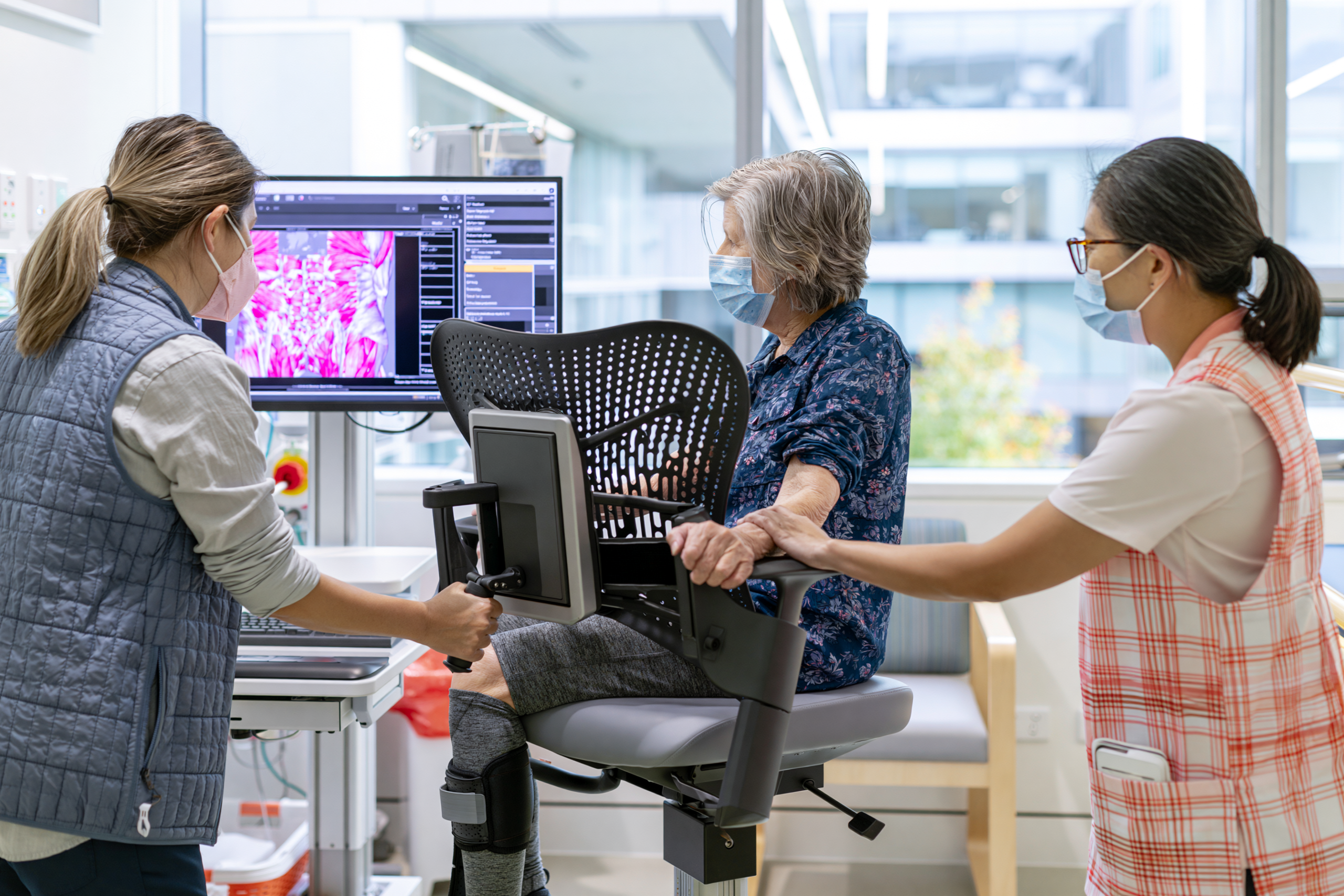
Sarcopenia is the progressive loss of skeletal muscle mass, strength and physical performance. In hepatocellular carcinoma, it is not a minor nutritional problem. It can affect your treatment tolerance, recovery after surgery, risk of complications, independence and overall prognosis [19,20].
A systematic review and meta-analysis estimated that approximately 42% of people with HCC had sarcopenia. The reported prevalence varied between studies because researchers used different populations, muscle measurements and diagnostic thresholds [20]. This means that the exact percentage cannot be applied to every clinic or patient, but it confirms that muscle loss is common enough to require routine assessment.
You may develop sarcopenia even when your body weight appears normal or elevated. Ascites, leg oedema and excess body fat can conceal substantial loss of muscle. For this reason, your weight alone cannot show whether your nutritional and physical reserve is stable.
Why Muscle Loss Is Common in HCC
HCC often develops in the setting of cirrhosis, where protein metabolism, energy storage and nutrient processing are already disturbed. Your liver normally stores glycogen and supports the regulation of amino acids, glucose and fats. When liver function declines, your body may enter a fasting state more quickly between meals and begin using muscle protein for energy [19].
Cancer-related inflammation can further accelerate muscle breakdown. Poor appetite, nausea, early satiety, abdominal pressure from ascites, altered taste, repeated hospitalisation and restrictive diets may reduce your food intake.
Pain, fatigue and fear of exertion can also make you less active. Reduced activity leads to further loss of muscle strength, creating a cycle in which weakness causes inactivity and inactivity causes greater weakness.
Systemic treatment, embolisation, radiotherapy and surgery may add temporary periods of reduced intake and physical inactivity. If these periods are not followed by structured nutritional and physical recovery, your muscle reserve may continue to decline.
Why Sarcopenia Matters Clinically
Sarcopenia is associated with poorer outcomes in cirrhosis and HCC. It may increase your risk of frailty, falls, infection, treatment intolerance, postoperative complications and prolonged hospitalisation [19,20].
When muscle mass and strength decline, you may find it more difficult to recover after liver resection, transplantation, TACE, TARE or systemic therapy. You may require longer rehabilitation and may be more likely to experience interruptions or dose reductions in treatment.
Sarcopenia may also affect your ability to remain eligible for certain procedures. A technically treatable tumour may become more difficult to manage if your functional reserve deteriorates substantially.
These associations do not mean that sarcopenia alone determines your prognosis. Tumour stage, liver function, kidney function, age, treatment response and other illnesses remain important. Muscle status adds another clinically meaningful dimension to your assessment.
Sarcopenia Is Not the Same as Being Thin
You can have sarcopenia at a low, normal or high body weight.
When low muscle mass occurs together with obesity, the condition is sometimes described as sarcopenic obesity. You may have excess abdominal fat while your arms, legs and core muscles are progressively weakening.
This pattern is particularly relevant when HCC develops in the background of MASLD, diabetes or obesity. A recommendation to lose weight without protecting muscle may worsen your functional condition.
Your treatment should therefore focus on body composition and strength rather than only on reducing the number shown on the weighing scale.
Why Body Weight Can Be Misleading
Ascites and oedema can add several kilograms of fluid. Diuretics or fluid drainage may then cause rapid weight reduction without any change in tumour burden or body fat.
Your weight may therefore remain stable while muscle is lost, or fall suddenly because fluid has been removed.
A proper assessment considers weight together with fluid status, food intake, physical function and available imaging measurements [19].
Changes in the fit of your clothes, thinning of your arms or thighs, reduced grip and slower walking may reveal muscle loss before the scale does.
How Muscle Mass Can Be Assessed
CT scans obtained for cancer staging can also provide information about skeletal muscle. The cross-sectional muscle area at the third lumbar vertebral level is frequently used in research and specialised clinical assessment [20].
This approach can identify low muscle mass without requiring an additional scan because the relevant images may already exist.
However, measurement methods and cut-off values vary between studies. This variation is one reason why the reported prevalence of sarcopenia in HCC is not identical across populations [20].
Other methods may include bioelectrical impedance, dual-energy X-ray absorptiometry or anthropometric measurements, although fluid retention can affect some of these results.
Muscle quantity should not be assessed without considering muscle function. A person may have relatively preserved muscle size but poor strength, or low muscle mass with better-than-expected function.
How Muscle Strength Can Be Assessed
Hand-grip strength is a simple and useful measure of overall muscle function. It can be repeated over time to identify improvement or decline.
The chair-stand test assesses how easily you can rise repeatedly from a seated position. Walking speed and short physical performance tests may also provide practical information about frailty and mobility [19].
Your daily experience matters as well. Difficulty climbing stairs, carrying groceries, bathing, dressing or walking to the bathroom may indicate clinically important functional decline.
These measures are often more meaningful than asking only whether you feel weak.
How Sarcopenia Affects Surgical Recovery
Before liver surgery, low muscle mass and frailty may increase the risk of postoperative complications and delayed recovery.
After surgery, pain, bed rest and reduced food intake can accelerate muscle breakdown. Even a successful operation may leave you considerably weaker if rehabilitation and nutrition are not addressed early.
Your recovery plan should therefore begin before surgery whenever possible. Prehabilitation may include nutrition optimisation, walking, resistance activity and treatment of reversible problems such as anaemia, dehydration or infection.
After surgery, mobilisation and nutritional support should resume as soon as your surgical team considers them safe.
How Sarcopenia Affects Nonsurgical Treatment
During TACE or TARE, poor muscle reserve may make post-procedure fatigue and reduced appetite more difficult to tolerate.
During immunotherapy or targeted therapy, sarcopenia may increase the clinical impact of diarrhoea, nausea, appetite loss and prolonged inactivity.
When you are already close to the threshold of functional dependence, even a short treatment-related decline can lead to inability to manage meals, medicines or personal care.
For this reason, muscle preservation should be treated as part of your cancer-management plan rather than as an optional fitness goal.
Nutrition for Muscle Preservation
AASLD guidance emphasises the importance of adequate energy and protein intake in cirrhosis and advises against unnecessary protein restriction [19].
Older recommendations sometimes restricted protein because of concern about hepatic encephalopathy. Routine long-term protein restriction is now discouraged because it can worsen muscle loss. Your protein source and quantity should be individualised according to liver function, kidney function, tolerance and nutritional status [19].
Your meals may need to be smaller and more frequent when you have early satiety or ascites. The aim is to reduce long fasting periods while maintaining sufficient total intake.
A late-evening snack may help reduce prolonged overnight fasting in cirrhosis by providing an additional source of energy before sleep [19].
The exact protein and calorie targets should be decided by your clinical team. They cannot be safely prescribed from body weight alone when ascites, obesity, kidney disease or severe malnutrition is present.
Why Prolonged Fasting Can Worsen Muscle Loss
When your liver has reduced glycogen storage, your body begins using fat and muscle protein for energy sooner during fasting.
Long periods without food may therefore accelerate muscle breakdown in cirrhosis.
Juice-only plans, severe detoxification diets and repeated fasting may reduce protein intake and worsen sarcopenia, even when they temporarily reduce body weight.
A lower number on the scale after fasting does not mean that the liver or tumour has improved. The change may represent loss of fluid, glycogen and muscle.
Your food plan should support adequate nourishment and treatment tolerance rather than create avoidable depletion.
The Role of Protein
Protein provides amino acids needed for muscle repair, immune function, enzyme production and wound healing.
Your protein should be distributed across the day rather than concentrated entirely in one meal. This may improve tolerance and provide repeated opportunities for muscle-protein synthesis.
Plant proteins, dairy products, eggs, fish or other sources may be used according to your preferences, liver condition, kidney function and cultural diet.
If one food source causes bloating, nausea or poor tolerance, the solution is not necessarily to remove all protein. Another source or preparation method may be more suitable.
Exercise and Muscle Preservation
Nutrition alone cannot fully restore muscle if physical activity remains very low.
AASLD guidance supports individualised aerobic and resistance exercise for people with cirrhosis when medically safe [19].
Walking helps maintain endurance, while resistance activity is more directly related to preserving or rebuilding muscle strength. Simple exercises may include repeated sit-to-stand movements, supported squats, resistance bands or light weights under professional guidance.
Your programme should begin at a level you can tolerate and progress gradually. Excessive exercise may worsen fatigue or injury risk, while complete rest accelerates deconditioning.
When Exercise Must Be Modified
Your activity plan requires modification when you have severe anaemia, uncontrolled ascites, active bleeding, significant breathlessness, unstable heart disease, infection, recent surgery or severe treatment toxicity.
Bone metastases may require avoidance of loading or twisting movements that could increase fracture risk.
If you have poor balance, encephalopathy or a history of falls, supervised exercise may be safer.
New chest pain, dizziness, severe breathlessness, confusion or sudden weakness during activity requires medical assessment.
Exercise should challenge your muscles appropriately without ignoring your liver disease and overall medical condition.
Why Bed Rest Should Be Minimized
Hospitalisation and bed rest can lead to rapid loss of muscle, particularly when you are older, frail or already malnourished.
When medically permitted, sitting out of bed, standing, short walks and simple resistance exercises should begin early.
The amount may appear small, but maintaining daily movement can reduce functional decline.
Your caregiver can help encourage safe movement without forcing activity during periods of severe fatigue or instability.
The Ayurvedic Interpretation of Muscle Loss
From an Ayurvedic perspective, progressive loss of muscle and strength may be understood through Mamsa Dhatu Kshaya, reduced Bala, disturbed Agni and declining Ojas.
This language can help structure supportive care, but it should not replace objective assessment of food intake, strength, body composition and liver function.
Brimhana and Rasayana principles may be considered when depletion is present. However, a heavy or excessively sweet formulation is not automatically appropriate. Your appetite, glucose, ascites and digestive tolerance still determine what can be used safely.
Ayurvedic support should work alongside adequate nutrition and physical rehabilitation rather than being treated as a substitute for them.
How Progress Should Be Monitored
Your muscle-recovery plan should be reviewed through repeated functional and nutritional measures.
Useful indicators include meal intake, grip strength, chair-rise ability, walking distance, independence in daily activities and changes in CT-based muscle area when available [19,20].
Weight can be recorded, but it should always be interpreted with ascites and oedema status.
A meaningful improvement may be your ability to walk farther, rise from a chair more easily or resume personal care without assistance, even when body weight changes very little.
Why Early Intervention Is Better
Severe sarcopenia is more difficult to reverse than early functional decline.
You should not wait until you are unable to walk independently or have lost a large amount of weight before nutritional and physical rehabilitation begins.
Early screening allows your team to identify reduced intake, muscle weakness and inactivity before they become major barriers to treatment.
Muscle preservation should begin at diagnosis and continue before, during and after tumour-directed treatment.
What This Means for You
Sarcopenia is common in HCC, affecting approximately 42% of patients in the available pooled research, although the estimate varies according to how muscle loss is defined and measured [20].
Your muscle condition can influence treatment tolerance, recovery, independence and prognosis. It cannot be assessed reliably from body weight alone.
A complete plan should combine adequate nutrition, avoidance of unnecessary fasting, individualised protein intake, regular meal timing, resistance activity, walking and repeated functional assessment [19,20].
Preserving your muscle is not separate from treating your cancer. It is part of maintaining the physical reserve required to undergo treatment, recover from it and remain independent for as long as possible.
Frequently Asked Questions About Hepatocellular Carcinoma and Ayurveda
What is hepatocellular carcinoma?
Hepatocellular carcinoma is the most common primary cancer arising from liver cells. Your treatment depends on the tumour’s size, number, location and spread, together with the condition of your remaining liver.
Can hepatocellular carcinoma be cured?
HCC may be treated with curative intent when it is detected early and you are suitable for liver resection, transplantation or thermal ablation. Your outcome depends on tumour stage, liver reserve, overall health and treatment response.
What does inoperable hepatocellular carcinoma mean?
Inoperable HCC means that surgery is not currently considered safe or likely to provide sufficient benefit. You may still be eligible for ablation, embolisation, radiotherapy, immunotherapy, targeted therapy, transplantation assessment or downstaging treatment.
Can Ayurveda replace surgery or cancer treatment for HCC?
Ayurveda should not replace surgery, transplantation, ablation, embolisation, radiotherapy, immunotherapy or targeted therapy when these treatments are medically indicated. It may support your appetite, digestion, bowel function, strength, sleep and recovery alongside oncology care.
How can Ayurveda help when surgery is not possible?
A personalised Ayurvedic programme may support food intake, digestion, bowel regularity, sleep, muscle strength, fatigue and daily function during nonsurgical treatment. Your tumour must still be monitored through imaging and oncology review.
Can Ayurveda prevent HCC recurrence after surgery?
Ayurveda cannot guarantee that HCC will not return. It may support postoperative recovery, liver health, nutrition, physical strength, metabolic control and management of factors that continue damaging your remaining liver.
What is Yakrit–Ojas Rasayana Avaleha?
Yakrit–Ojas Rasayana Avaleha is a personalised supportive formulation inspired by classical Rasayana and Avaleha principles. It is designed according to your reports, liver function, digestion, strength and ongoing cancer treatment.
Can you buy HCC Avaleha from the market?
You should not purchase a ready-made HCC Avaleha without medical assessment. A commercial product cannot account for your bilirubin, kidney function, glucose, ascites, medicines, treatment interactions or individual digestive capacity.
Can you prepare Yakrit–Ojas Rasayana Avaleha at home?
You should never prepare this Avaleha without supervision from a qualified Ayurvedic physician and a licensed manufacturing unit. Incorrect herbs, inaccurate quantities, contamination, untested minerals or improper processing may make it ineffective or unsafe.
Are Bhasmas and minerals necessary for every HCC patient?
No. Bhasmas and mineral preparations should be considered only when you have a specific clinical indication. Their selection and dose must depend on your liver function, kidney function, blood reports, current medicines and individual Ayurvedic assessment.
Can you take Ayurvedic medicines during immunotherapy or targeted therapy?
You should take Ayurvedic medicines only after your complete formulation and oncology medicines have been reviewed for interactions. Some herbs may affect liver function, drug metabolism, bleeding risk, blood pressure, glucose or bowel function.
Is muscle loss common in liver cancer?
Muscle loss is common in HCC and cirrhosis. It may occur even when your body weight appears stable because ascites, swelling or excess body fat can conceal declining muscle mass.
How can you preserve muscle during HCC treatment?
Your recovery plan should include adequate food and protein, avoidance of unnecessary fasting, regular meal timing, walking and resistance activity when medically safe. Your progress should be assessed through strength and daily function, not weight alone.
Which HCC symptoms require urgent medical attention?
You need urgent medical assessment for vomiting blood, black stool, new confusion, worsening jaundice, severe abdominal pain, fever, repeated vomiting, reduced urine output, sudden breathlessness or rapidly increasing abdominal swelling.
How do you know whether Ayurvedic treatment is helping?
Supportive benefit should be assessed through appetite, food intake, bowel function, sleep, strength, walking ability and treatment tolerance. Tumour response must be evaluated separately through appropriate scans and oncology follow-up.
References
[1] Sung, H., Filho, A. M., Laversanne, M., Ferlay, J., Siegel, R. L., Soerjomataram, I., Jemal, A., & Bray, F. (2026). Global cancer statistics 2024: GLOBOCAN estimates of incidence and mortality worldwide for 34 cancers in 186 countries. CA: A Cancer Journal for Clinicians, 76(4), e70090. https://pubmed.ncbi.nlm.nih.gov/42417444/
Used for: Global cancer incidence, mortality, liver-cancer burden, and international epidemiological comparisons.
[2] European Association for the Study of the Liver. (2025). EASL clinical practice guidelines on the management of hepatocellular carcinoma. Journal of Hepatology, 82(2), 315–374. https://www.journal-of-hepatology.eu/article/S0168-8278%2824%2902508-X/fulltext
Used for: HCC surveillance, diagnosis, staging, surgery, transplantation, ablation, embolisation, radiotherapy, systemic treatment, and follow-up.
[3] Singal, A. G., Llovet, J. M., Yarchoan, M., Mehta, N., Heimbach, J. K., Dawson, L. A., Jou, J. H., Kulik, L. M., Agopian, V. G., Marrero, J. A., Mendiratta-Lala, M., Brown, D. B., Rilling, W. S., Goyal, L., Wei, A. C., & Taddei, T. H. (2023). AASLD practice guidance on prevention, diagnosis, and treatment of hepatocellular carcinoma. Hepatology, 78(6), 1922–1965. https://pmc.ncbi.nlm.nih.gov/articles/PMC10663390/
Used for: HCC-risk populations, surveillance, imaging, biopsy, resection, transplantation, locoregional treatment, systemic therapy, and recurrence monitoring.
[4] Reig, M., Sanduzzi-Zamparelli, M., Forner, A., Rimola, J., Ferrer-Fàbrega, J., Burrel, M., Garcia-Criado, Á., Díaz, A., Llarch, N., Iserte, G., Mollà, M., Kelley, R. K., Galle, P. R., Mazzaferro, V., Salem, R., Sangro, B., Singal, A. G., Vogel, A., Yanagihara, T. K., . . . Bruix, J. (2026). BCLC strategy for prognosis prediction and treatment recommendations: The 2026 update. Journal of Hepatology, 84(3), 631–654. https://www.journal-of-hepatology.eu/article/S0168-8278%2825%2902571-1/fulltext
Used for: BCLC staging, prognosis, treatment allocation, multidisciplinary decision-making, and movement between treatment pathways.
[5] National Cancer Institute. (2025, April 17). Primary liver cancer treatment (PDQ®)—Health professional version. https://www.cancer.gov/types/liver/hp/adult-liver-treatment-pdq
Used for: HCC symptoms, diagnostic assessment, TNM staging, surgery, transplantation, ablation, embolisation, radiotherapy, systemic therapy, and survival data.
[6] National Cancer Institute. (2025, April 30). Liver cancer prevention (PDQ®)—Health professional version. https://www.cancer.gov/types/liver/hp/liver-prevention-pdq
Used for: Hepatitis, cirrhosis, alcohol, tobacco, aflatoxin, obesity, diabetes, metabolic liver disease, and HCC risk reduction.
[7] World Health Organization. (n.d.). Hepatitis B. Retrieved July 12, 2026, from https://www.who.int/news-room/fact-sheets/detail/hepatitis-b
Used for: HBV transmission, vaccination, testing, antiviral treatment, cirrhosis, and HBV-associated HCC risk.
[8] World Health Organization. (n.d.). Hepatitis C. Retrieved July 12, 2026, from https://www.who.int/news-room/fact-sheets/detail/hepatitis-c
Used for: HCV transmission, testing, cirrhosis, direct-acting antiviral treatment, and continuing HCC surveillance.
[9] European Association for the Study of the Liver, European Association for the Study of Diabetes, & European Association for the Study of Obesity. (2024). EASL–EASD–EASO clinical practice guidelines on the management of metabolic dysfunction-associated steatotic liver disease. Journal of Hepatology, 81(3), 492–542. https://pubmed.ncbi.nlm.nih.gov/38851997/
Used for: MASLD, MASH, diabetes, obesity, fibrosis, metabolic-risk control, and HCC risk related to metabolic liver disease.
[10] American College of Radiology. (n.d.). Liver Imaging Reporting and Data System: LI-RADS®. Retrieved July 12, 2026, from https://www.acr.org/Clinical-Resources/Clinical-Tools-and-Reference/Reporting-and-Data-Systems/LI-RADS
Used for: Standardised liver-imaging terminology, arterial enhancement, washout, tumour-in-vein, and CT or MRI interpretation.
[11] Johnson, P. J., Berhane, S., Kagebayashi, C., Satomura, S., Teng, M., Reeves, H. L., O’Beirne, J., Fox, R., Skowronska, A., Palmer, D., Yeo, W., Mo, F., Lai, P., Iñarrairaegui, M., Chan, S. L., Sangro, B., Miksad, R., Tada, T., Kumada, T., & Toyoda, H. (2015). Assessment of liver function in patients with hepatocellular carcinoma: A new evidence-based approach—The ALBI grade. Journal of Clinical Oncology, 33(6), 550–558. https://pubmed.ncbi.nlm.nih.gov/25512453/
Used for: Albumin-bilirubin grading, objective liver-function assessment, prognosis, and treatment stratification.
[12] Kamath, P. S., Wiesner, R. H., Malinchoc, M., Kremers, W., Therneau, T. M., Kosberg, C. L., D’Amico, G., Dickson, E. R., & Kim, W. R. (2001). A model to predict survival in patients with end-stage liver disease. Hepatology, 33(2), 464–470. https://pubmed.ncbi.nlm.nih.gov/11172350/
Used for: MELD score development, liver-disease severity, survival prediction, and transplant-related assessment.
[13] Mazzaferro, V., Regalia, E., Doci, R., Andreola, S., Pulvirenti, A., Bozzetti, F., Montalto, F., Ammatuna, M., Morabito, A., & Gennari, L. (1996). Liver transplantation for the treatment of small hepatocellular carcinomas in patients with cirrhosis. The New England Journal of Medicine, 334(11), 693–699. https://pubmed.ncbi.nlm.nih.gov/8594428/
Used for: Milan criteria, liver-transplant selection, and transplantation outcomes in early HCC.
[14] Llovet, J. M., Real, M. I., Montaña, X., Planas, R., Coll, S., Aponte, J., Ayuso, C., Sala, M., Muchart, J., Solà, R., Rodés, J., & Bruix, J. (2002). Arterial embolisation or chemoembolisation versus symptomatic treatment in patients with unresectable hepatocellular carcinoma: A randomised controlled trial. The Lancet, 359(9319), 1734–1739. https://pubmed.ncbi.nlm.nih.gov/12049862/
Used for: Randomised evidence supporting embolisation and TACE in selected unresectable HCC.
[15] Salem, R., Johnson, G. E., Kim, E., Riaz, A., Bishay, V., Boucher, E., Fowers, K., Lewandowski, R. J., & Padia, S. A. (2021). Yttrium-90 radioembolization for the treatment of solitary, unresectable HCC: The LEGACY study. Hepatology, 74(5), 2342–2352. https://pmc.ncbi.nlm.nih.gov/articles/PMC8596669/
Used for: Y-90 radioembolisation, radiation segmentectomy, tumour response, and local control in selected unresectable HCC.
[16] Dawson, L. A., Winter, K. A., Knox, J. J., Zhu, A. X., Krishnan, S., Guha, C., Kachnic, L. A., Gillin, M. T., Hong, T. S., Craig, T. D., Williams, T. M., Hosni, A., Chen, E., Noonan, A. M., Koay, E. J., Sinha, R., Lock, M. I., Ohri, N., Dorth, J. A., . . . Crane, C. H. (2025). Stereotactic body radiotherapy versus sorafenib alone in hepatocellular carcinoma: The NRG Oncology/RTOG 1112 phase 3 randomized clinical trial. JAMA Oncology, 11(2), 136–144. https://pubmed.ncbi.nlm.nih.gov/39699905/
Used for: Stereotactic radiotherapy in locally advanced HCC and interpretation of survival and progression outcomes.
[17] Finn, R. S., Qin, S., Ikeda, M., Galle, P. R., Ducreux, M., Kim, T.-Y., Kudo, M., Breder, V., Merle, P., Kaseb, A. O., Li, D., Verret, W., Xu, D.-Z., Hernandez, S., Liu, J., Huang, C., Mulla, S., Wang, Y., Lim, H. Y., . . . Cheng, A.-L. (2020). Atezolizumab plus bevacizumab in unresectable hepatocellular carcinoma. The New England Journal of Medicine, 382(20), 1894–1905. https://pubmed.ncbi.nlm.nih.gov/32402160/
Used for: IMbrave150 evidence, atezolizumab-bevacizumab, survival, progression-free survival, and treatment selection.
[18] Abou-Alfa, G. K., Lau, G., Kudo, M., Chan, S. L., Kelley, R. K., Furuse, J., Sukeepaisarnjaroen, W., Kang, Y.-K., Dao, T. V., De Toni, E. N., Rimassa, L., Breder, V., Vasilyev, A., Heurgué, A., Tam, V. C., Mody, K., Thungappa, S. C., Ostapenko, Y., Yau, T., . . . Sangro, B. (2022). Tremelimumab plus durvalumab in unresectable hepatocellular carcinoma. NEJM Evidence, 1(8), EVIDoa2100070. https://pubmed.ncbi.nlm.nih.gov/38319892/
Used for: HIMALAYA trial, STRIDE regimen, durvalumab, survival outcomes, and immunotherapy safety.
[19] Lai, J. C., Tandon, P., Bernal, W., Tapper, E. B., Ekong, U., Dasarathy, S., & Carey, E. J. (2021). Malnutrition, frailty, and sarcopenia in patients with cirrhosis: 2021 practice guidance by the American Association for the Study of Liver Diseases. Hepatology, 74(3), 1611–1644. https://pmc.ncbi.nlm.nih.gov/articles/PMC9134787/
Used for: Nutrition, protein requirements, frailty, sarcopenia, exercise, meal timing, and muscle preservation.
[20] Liu, J., Luo, H., Huang, L., & Wang, J. (2023). Prevalence of sarcopenia among patients with hepatocellular carcinoma: A systematic review and meta-analysis. Oncology Letters, 26(1), 283. https://pmc.ncbi.nlm.nih.gov/articles/PMC10236145/
Used for: Sarcopenia prevalence in HCC and the importance of muscle assessment and rehabilitation.
[21] National Cancer Institute. (2024, September 20). Nutrition in cancer care (PDQ®)—Health professional version. https://www.cancer.gov/about-cancer/treatment/side-effects/appetite-loss/nutrition-hp-pdq
Used for: Appetite loss, malnutrition, cachexia, early satiety, nutritional assessment, and treatment tolerance.
[22] National Cancer Institute. (2021, November 1). Palliative care in cancer. https://www.cancer.gov/about-cancer/advanced-cancer/care-choices/palliative-care-fact-sheet
Used for: Early palliative care, symptom relief, emotional support, caregiver care, and quality of life.
[23] National Cancer Institute. (2024, April 5). Cancer therapy interactions with foods and dietary supplements (PDQ®)—Health professional version. https://www.cancer.gov/about-cancer/treatment/cam/hp/dietary-interactions-pdq
Used for: Herb-drug interactions, CYP enzymes, transport proteins, pharmacokinetic interactions, and treatment toxicity.
[24] National Cancer Institute. (2024, October 31). Complementary and alternative medicine. https://www.cancer.gov/about-cancer/treatment/cam
Used for: Distinguishing complementary care from alternative care and explaining evidence limitations.
[25] National Center for Complementary and Integrative Health. (n.d.). Ayurvedic medicine: In depth. Retrieved July 12, 2026, from https://www.nccih.nih.gov/health/ayurvedic-medicine-in-depth
Used for: Ayurveda research limitations, safety, product contamination, heavy metals, and avoiding delay of conventional cancer treatment.
[26] National Institute of Diabetes and Digestive and Kidney Diseases. (n.d.). LiverTox: Clinical and research information on drug-induced liver injury. National Library of Medicine. Retrieved July 12, 2026, from https://www.ncbi.nlm.nih.gov/books/NBK547852/
Used for: Drug-induced and supplement-associated liver injury, causality assessment, injury patterns, and monitoring.
[27] Ministry of Ayush, Government of India. (n.d.). Ayush Suraksha: Pharmacovigilance database. Retrieved July 12, 2026, from https://suraksha.ayush.gov.in/
Used for: Adverse-reaction reporting, Ayurvedic pharmacovigilance, batch documentation, and medicine-safety monitoring.
[28] Agniveśa. (1981–1994). Caraka saṃhitā (P. V. Sharma, Trans.; Vols. 1–4). Chaukhambha Orientalia. https://www.carakasamhitaonline.com/index.php/Rasayana_Adhyaya
Classical section: Chikitsa Sthana, Chapter 1, Rasayana Adhyaya.
Used for: Rasayana, Bala, Ojas, recovery capacity, diet, conduct, and traditional rejuvenative principles.
[29] Vāgbhaṭa. (1991). Aṣṭāṅga hṛdaya (K. R. Srikantha Murthy, Trans.; Vol. 1). Krishnadas Academy. https://archive.org/details/AstangaHrdayam.Eng
Classical section: Sutra Sthana, Chapter 8, Matrashitiya Adhyaya.
Used for: Food quantity, digestive capacity, meal timing, wholesome food, and individualised Pathya-Apathya.
[30] Śārṅgadhara. (2017). Śārṅgadhara saṃhitā (B. Tripathi, Ed.). Chaukhambha Surbharati Prakashan. https://archive.org/details/dLhu_sharangadhara-samhita-of-sharangadhara-acharya-containing-anjananidana-of-agnive
Classical section: Madhyama Khanda, Chapter 8, Avaleha Kalpana.
Used for: Avaleha definition, preparation, Prakshepa Dravya, Siddhi Lakshana, Anupana, dose, preservation, and storage.
[31] Suśruta. (1907–1916). An English translation of the Sushruta Samhita (K. L. Bhishagratna, Trans.; Vols. 1–3). Author. https://archive.org/details/englishtranslati00susruoft
Classical section: Chikitsa Sthana, Chapter 1, Dvivraniya Chikitsa.
Used for: Classical Vrana assessment, wound cleanliness, wound management, and postoperative recovery principles.
[32] Mathew, J. S., & Philips, C. A. (2023). Drug interactions and safe prescription writing for liver transplant recipients. Journal of Clinical and Experimental Hepatology, 13(5), 869–877. https://pmc.ncbi.nlm.nih.gov/articles/PMC10483006/
Used for: Immunosuppressant interactions, polypharmacy, herbal-product risks, and safe prescribing after liver transplantation.
[33] Miedziaszczyk, M., Bajon, A., Jakielska, E., Primke, M., Sikora, J., Skowrońska, D., & Idasiak-Piechocka, I. (2022). Controversial interactions of tacrolimus with dietary supplements, herbs, and food. Pharmaceutics, 14(10), 2154. https://pmc.ncbi.nlm.nih.gov/articles/PMC9611668/
Used for: Tacrolimus interactions, CYP3A4 metabolism, altered immunosuppressant exposure, and post-transplant safety.
[34] Saper, R. B., Phillips, R. S., Sehgal, A., Khouri, N., Davis, R. B., Paquin, J., Thuppil, V., & Kales, S. N. (2008). Lead, mercury, and arsenic in US- and Indian-manufactured Ayurvedic medicines sold via the Internet. JAMA, 300(8), 915–923. https://pmc.ncbi.nlm.nih.gov/articles/PMC2755247/
Used for: Heavy-metal contamination in selected Ayurvedic products and the need for batch testing, traceability, and quality control.